laser therapy vred - be-opticalbeoptical.eu/public/shareddocs/schools/laser...
TRANSCRIPT

LASER THERAPY
Meritxell VilasecaUniversitat Politècnica de Catalunya, SPAIN
1
CONTENTS
Therapeutic applications of lasersPhotochemical effectsPhotothermal applicationsPhotoablationPhotomechanical effects
There are four major categories of light-tissue interactions that lead to alteration of the tissue structure/composition:
• Photochemical: Absorption of light by molecules present or added to tissue.• Photothermal: Biological effects due to deposition ofthermal energy in tissue.• Photoablative: in UV, photons possess sufficient energy to cause photo-dissociation of bio-polymers and subsequent desorption of fragments (a substance is released from or through a surface).• Photomechanical: occurs at high fluence rateswhere dielectric breakdown of tissue is induced whichcan lead to plasma formation. Rapid plasma expansiongenerates a shock wave which can rupture tissue.
THERAPEUTIC APPLICATIONS OF LASERS
3
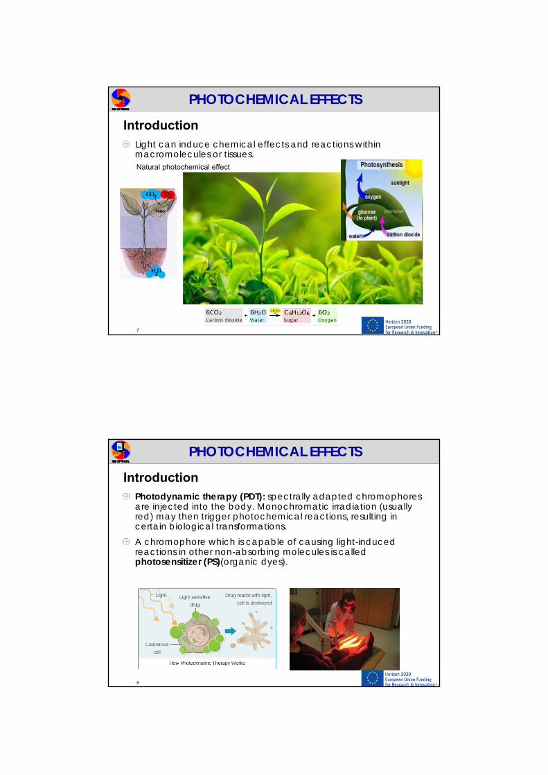
The biological tissue interaction is determined by the laser irradiance or power density [W/cm2] (1).
4
THERAPEUTIC APPLICATIONS OF LASERS
Lasers used for medical
applications must have
fluences ranging from 1 J/cm2
to 1000 J/cm2.

The exposure time [s] (2) is also responsible for thevariety of interaction mechanisms:
5
THERAPEUTIC APPLICATIONS OF LASERS
CW or exposure times > 1s: photochemical interactions1min-1ms: thermal interactions1ms-1ns: photoablation<1ns: plasma-induced ablationand photodisruption. Thedifferences between those twodue to different energy densities(Plasma-induced ablation basedon ionization, photodisruptionbased on mechanical effects)
The biological tissue interaction also depends on optical (reflection/absorption [a]and scattering [s]), thermal –such as heat conduction, heat capacity, and chemical properties of tissue (3).
6
Absorption spectra of main absorbers in biological tissue.
• Proteins absorb in the UVregion (mainly 200 - 300nm).
• Pigments such as hemoglobin in blood and melanin, the basic chromophore of skin, absorb in the visible range.
• Absorption in the IR region (2000-10000nm) originates from water, which is the main constituent of most tissues.
THERAPEUTIC APPLICATIONS OF LASERS
PHOTOCHEMICAL EFFECTS
7
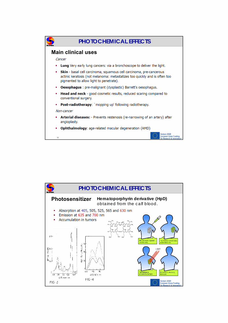
IntroductionLight can induce chemical effects and reactions within macromolecules or tissues. Natural photochemical effect
PHOTOCHEMICAL EFFECTS
8
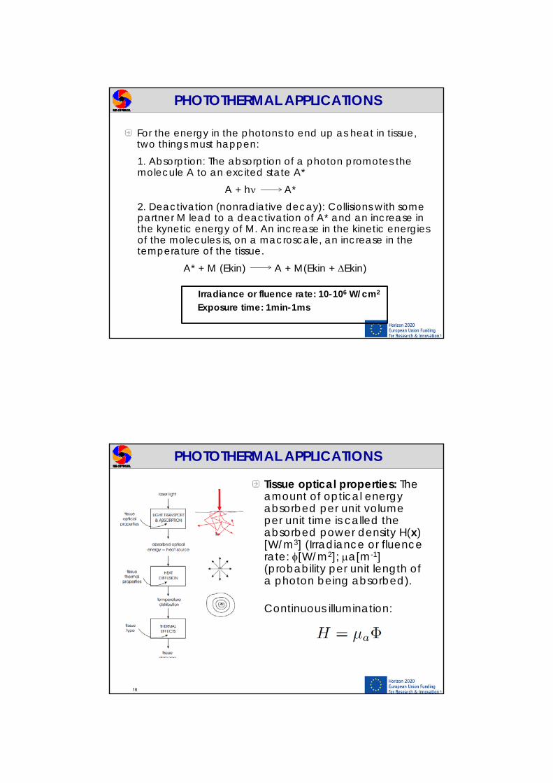
IntroductionPhotodynamic therapy (PDT): spectrally adapted chromophores are injected into the body. Monochromatic irradiation (usually red) may then trigger photochemical reactions, resulting in certain biological transformations.A chromophore which is capable of causing light-induced reactions in other non-absorbing molecules is called photosensitizer (PS)(organic dyes).
90731.jpg

9
After absorption of laser photons, PS is transferred to S1. After this, a radiative decay might occur to an excited triplet state (intercrossing system) (T1) besides other alternatives such as non-radiative decays or radiative singlet decay to the single ground state. Radiative singlet and triplet decays: fluorescence(ns) and phosphorescence (ms-s).
PHOTOCHEMICAL EFFECTS
Singlet statesSpin momentum s=0
Triplet statesSpin momentum s=1
PS are compounds whose energy difference T1 − S0 is close to the energy needed for oxygen molecules to be excited from the triplet ground state to a excited singlet state (energy is transferred to the oxygen molecule).
Irradiance: 1 W/cm2
Exposure time: seconds to CW
10
PHOTOCHEMICAL EFFECTS
The excited singlet oxygen resulting from this process are very reactive and lead to lipids and protein oxygenation and other destructive processes, which could start necrosis of cancer cells (PDT: photodynamic therapy).
The main disadvantage of PDT is the long time of the compound’s decay and removal from the body—patients became photosensitive from several days to weeks after application of such drugs and the related effects (toxicity etc.)
PHOTOCHEMICAL EFFECTS
Main clinical uses
11
PHOTOCHEMICAL EFFECTS
Photosensitizer Hematoporphyrin derivative (HpD) obtained from the calf blood.
12

PHOTOCHEMICAL EFFECTS
Light sources
13
PHOTOCHEMICAL EFFECTS
Light sources
14

15
PDT EXAMPLES
Lung cancer
15
Skin cancer
Introduction
Thermal interaction: large group of interaction types where the increase in local temperature is the significant parameter change.
16
PHOTOTHERMAL APPLICATIONS
The damage done to the tissue depends on the temperature that is reached, and the duration at which it is held at that temperature.
For the energy in the photons to end up as heat in tissue, two things must happen:1. Absorption: The absorption of a photon promotes the molecule A to an excited state A*
A + h A*2. Deactivation (nonradiative decay): Collisions with some partner M lead to a deactivation of A* and an increase in the kynetic energy of M. An increase in the kinetic energies of the molecules is, on a macroscale, an increase in the temperature of the tissue.
A* + M (Ekin) A + M(Ekin + Ekin)
Irradiance or fluence rate: 10-106 W/cm2
Exposure time: 1min-1ms
PHOTOTHERMAL APPLICATIONS
Tissue optical properties: The amount of optical energy absorbed per unit volume per unit time is called the absorbed power density H(x) [W/m3] (Irradiance or fluencerate: [W/m2]; a[m-1] (probability per unit length of a photon being absorbed).
18
Continuous illumination:
PHOTOTHERMAL APPLICATIONS
Absorption coefficient:

19
Tissue thermal properties:
Material Cp J /(Kg ºC)Copper 0.388Air 1.005Fat 1.930Ethanol 2.430Blood 3.220Water 4.183
- Heat capacity (Cp) [J/kg·K] defined as the heat energy required to raise 1kg of substance by 1K at constant pressure. It is related with the ability to keep heat.- Thermal conductivity (k) is related with the speed at whichthe heat flows from a point with high temperature to anotherwith low temperature.
Material Thermal conducitivity W /(m ºC)Copper 418.00Blood 0.62Water 0.58Fat 0.3Ethanol 0.16Air 0.02
PHOTOTHERMAL APPLICATIONS
20
Taking into account all this the heat diffusion equation is formulated, which inform us about the temperature spreads out in space as time goes on:
Thermal interaction (Heat diffusion equation)
PHOTOTHERMAL APPLICATIONS
: mass density [kg/m3]
If the tissue does not change phase then an increase in the heat energy per unit volume (dE [J/m3]) leads to a rise in temperature (dT):

21
Solution for a point source in 3D:
Solutions to this equation by means of numerical methods under general conditions serve as a starting point to predict what will happen in a particular case (Numerical analysis packages).
Thermal interaction (Heat diffusion equation)
E: Joules of optical energy deposited at a point
PHOTOTHERMAL APPLICATIONS
Coagulation (45-60ºC): change from a fluid into a thickened mass. Coagulated tissue appears darker and becomes necrotic.Vaporization (100ºC): water absorbs light, tries to expand in volume as it vaporizes inducing microexplosions and tissue is removed.
Effects of temperature on tissue
PHOTOTHERMAL APPLICATIONS
Carbonization (>100ºC): Blackening in color of the tissue. It should be avoided, since necrosis is already achieved at lower temperatures.Melting (>200-300ºC): melted and afterwards cooled tissue can be though as solidified lava (with gas bubbles).
22

CO2Er:YAG
Ho:YAG
Er:glass
Nd:YAGKTP
Nd:YAGAr
+
Rubi
Alexandrite
DyeExcimer
PHOTOTHERMAL APPLICATIONSLasers used in thermal applications
Absorption coefficientPenetration depth
Dependance on media and λ
Laser λ(nm) Penetration depth (mm)
Skin Liver Muscle Blood
CO2 10600 0.01 0.01 0.01 0.01 Water
Er:YAG 2940 0.001 0.001 0.001 0.001 Water
Nd:YAG 1064 4 5 4 0.8 Haemoglobin
Ar+
514 2 0.5 1 0.03 Haemoglobin
23
Temperature dependance with the absorption
ΔT on the skin surface produced by lasers.
Laser λ (nm) μa (cm-1) H (J cm-2) ΔT (ºC)Alejandrite 755 10 1 2.4
Nd:YAG 1064 2 1 0.5
Er:YAG 2940 10000 1 2400
CO210600 1000 1 240
Higher A Higher T
Low penetration depthHigh A
Laser with low A
Penetration depth
Affected volume
Low temperature
Affected volume
High temperature
PHOTOTHERMAL APPLICATIONS
Absorption coefficientPenetration depth
Dependance on media and λ
Laser with high A
24

25
Lasers used in thermal applications
Typical lasers for coagulation are Nd:YAG, diode lasers or Ar+ (with emission in the VIS).
CO2 and Er:YAG lasers are very suitable forvaporization and precise thermal cutting.
PHOTOTHERMAL APPLICATIONS
26
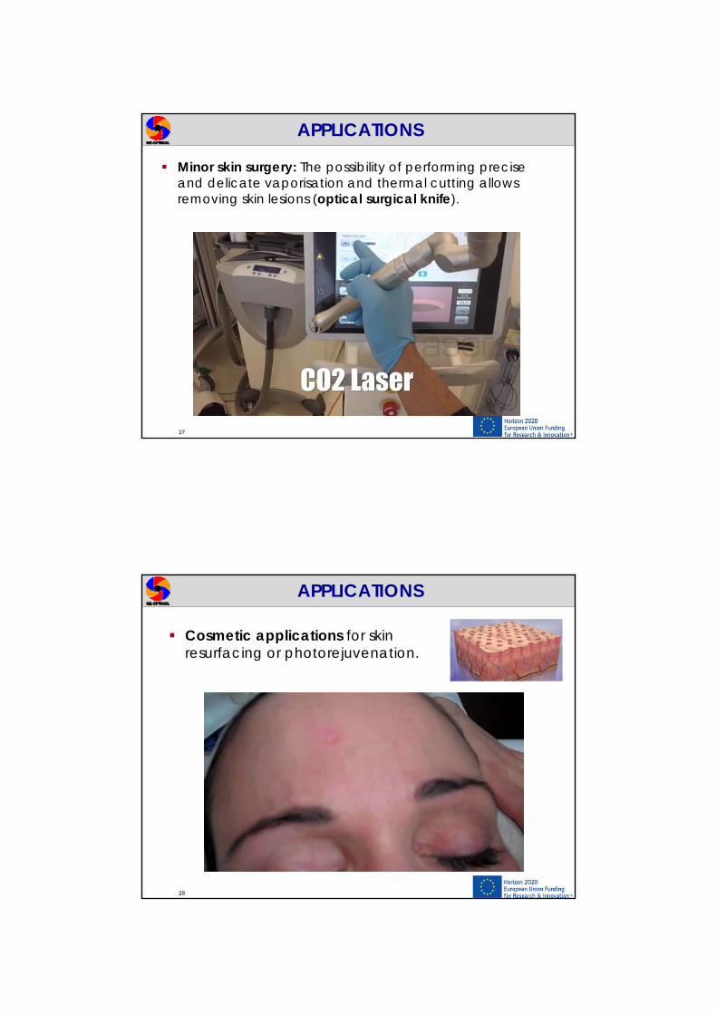
Minor skin surgery: The possibility of performing precise and delicate vaporisation and thermal cutting allows removing skin lesions (optical surgical knife)(No bleeding).
APPLICATIONS
27
Minor skin surgery: The possibility of performing precise and delicate vaporisation and thermal cutting allows removing skin lesions (optical surgical knife).
APPLICATIONS
28
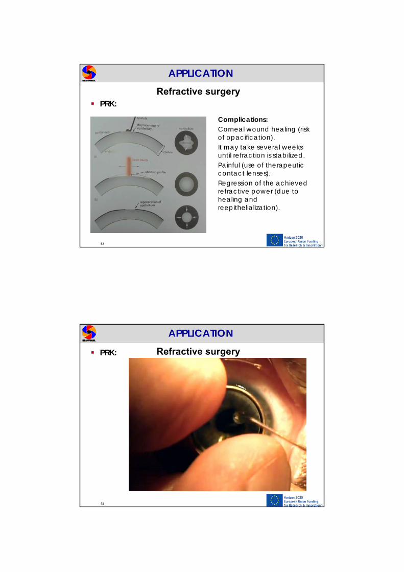
Cosmetic applications for skin resurfacing or photorejuvenation.
APPLICATIONS

29
Hair/Tattoo removal:
http://www.skincancer.org/prevention/are-you-at-risk/fitzpatrick-skin-quiz
Different wavelengths have different melanin absorption coefficients, making it possible to choose the most suitable source based on the patient’s characteristics (phototype, hair colour, etc.) (Ruby, Diode, alexandrite…).
Black tattoo pigment absorbs all laser wavelengths, making it the easiest to treat (Nd:YAG Laser). Other colors can only be treated by selected lasers based upon the pigment color (Yellow and green are the hardest colors to remove; blue and black are the easiest).
APPLICATIONS
30
Photothermal treatments in ophthalmology:The retina contains blood vessels. When too many of them grow, a vein is occluded and there hemorragies, such as in diabetic retinopathy, vision can be seriously impaired. By thermally photocoagulating the vessels, vision can be restored to some extent and the likelihood of recurrence can be minimised. When the whole of the retina - except the macula - is coagulated it is termed panretinal photocoagulation.
APPLICATIONS

31
Photothermal treatments in ophthalmology:
APPLICATIONS
32
Photothermal treatments in ophthalmology:
A detached retina, where the retina comes away from the back of the eye, can treated by “gluing” it back on again by photocoagulating it at a number of spots (away from the macula).Retinal tumours, retinoblastomas, can be thermally necrosed.
APPLICATIONS

33
Glaucoma is caused by a build-up of pressure in the eye. Closed-angle glaucoma can be treated by making a hole surgically in the iris, thus releasing the pressure. This procedure is called laser iridotomy. Typical lasers are argon and Nd:YAG.
APPLICATIONS
Photothermal treatments in ophthalmology:
34
APPLICATIONS
Photothermal treatments in ophthalmology:Iridotomy

35
Main characteristics of photoablation:
PHOTOABLATION
It involves the use of UV light. It is based on the fact that
material is decomposed when exposed to high intense laser irradiation.
It is very predictable and precise in the etching process.
It was discovered in 1982 and firstly used in polymers (PMMA, polyimide, Teflon etc.).
Let us assume that two atoms A and B are bound by a common electron. Absorption of a UV photon may promote them to an excited state (AB)*.1. Excitation: AB + h (AB)*
If the energy gain exceeds the bond energy, the two atoms may dissociate at the very next vibration.
2. Dissociation: (AB)* A + B + Ekin
Photoablation:- 107-108 W/cm2
- Pulse duration: ns
PHOTOABLATION
36

Model of photoablation
37
Lambert’s law of light absorption:
Photoablation will take place if:
Iph: threshold intensity of photoablation (determined by the minimal number of bonds that have to be dissociated to yield decomposition).
PHOTOABLATION
Ablation depth (depth at which ):
d
Ablation curve of rabbit cornea obtained with ArF excimer laser (pulse duration: 14ns)
38
An excimer laser is a gas laser emitting in the UV-wavelength range from 157 to 351nm. It uses a combination of a noble gas (argon, krypton, or xenon) and a reactive gas (fluorine [F2] or chlorine [Cl2]).
Excimer lasers
PHOTOABLATION
ArF (Argon Fluoride) excimer laser (193nm) is the one commonly applied for biomedical purposes, specifically refractive surgery techniques used in ophthalmology, since the cornea has a maximum absorption coefficient at 200nm approx.

Refractive surgery is the term used to describe a variety of eye surgeries to correct refractive errors such as myopia, hyperopia, astigmatism and presbyopia to reduce dependency on eyeglasses or contact lenses. There are various surgical procedures for correcting the refractive error such as reshaping the cornea (laser surgery) or implanting a lens inside the eye (phakicintraocular lens, IOL). This last one is used for very high refractions K as cornea is not thick enough!
APPLICATION
39
Refractive surgery
40
The eye is like a camera. Light comes in through the cornea, it is refracted by the cornea and the lens, and focused on the retina.
The averaged equivalent power of the eye for adults is of 60D.
APPLICATION

41
mmP
nnr
c
aah
c 80542
133741
.
.
The cornea provides most of the refracting power ( 42 D) (70% of the overall refraction) as its first surface is in contact to air.
Refractive surgery
r<< : High powerr>>: Low power
APPLICATION
The power of the lens is 22D approx. (when it is relaxed) and changes when the eye needs to focus at different distances (Accommodation).
Most common refractive errors: Farsightedness and nearsightedness, which are related to the shape of the eyeball and are caused by genetic and environmental factors.- Myopia
- Hyperopia
Refractive surgery
42
APPLICATION

Refractive surgery
43
Myopia: the eye is too powerful (refractive)
Hyperopia: the eye is not powerful enough(refractive)
E.g. A myopic eye of -2D means that its power is 2D more powerful than what it is expected in relation to its length.
APPLICATION
E.g. An hyperopic eye of +2D means that its power is 2D less powerful than what it is expected in relation to its length.
44
Keratomileusis technique (Barraquer, 1964. Trokel, 1983): from Greek “to detach”.
Large corneal ablations are performed in order to remove or add power to the eye.
Refractive surgery
APPLICATION
1st_c
c1st_c P
nr
1
rnn
P
'
Power of a spherical surface:

Keratomileusis technique: How reshaping of the eye has to be done? E.g.: myopic eye
Pre-operative
Post-operativei: initialf: finalR: curvatures of the corneax: optical axisY: arbitrary axis
Refractive surgery
APPLICATION
45
APPLICATION
46
Refractive surgery

Keratomileusis technique:
APPLICATIONRefractive surgery
2ymax
47
APPLICATION
48
Refractive surgery
i
ci R
nP
1
f
cf R
nP
1
D=Pf - Pi
ifc RR
nD11
1
where D is the degree of myopia expressed in diopters and nc the refractiveindex of the cornea
Rf

The depth of ablation at y=0 d(0) can also be easily calculated by using the definitions of sagitta of the anterior corneal surface and the power of a spherical surface:
2max
2 )(yrrsag r
nnP
'
APPLICATIONRefractive surgery
Keratomileusis technique:
d(0)=sagi - sagf=0.32mm=320m49
ymax
E.g. Patient with myopia of K=-7D, initial radius of the anterior corneal surface of 7.8mm, a radius of ablation of 5mm (ymax) and a refractive index of the cornea of 1.3771.
1 pulse of ArF laser typically ablates 0.1-1mm (0.01-0.1D). Energy densities of 1-5 J/cm2 are applied to be less dependent on energy fluctuations.
Correction of 1D 10 pulses.
Plasma generation
d
APPLICATIONRefractive surgery
Keratomileusis technique:
50

Delivery systems:
APPLICATIONRefractive surgery
Keratomileusis technique:
51
Scanning slit (a: myopia, b: hyperopia). Rotating disk (c: myopia)
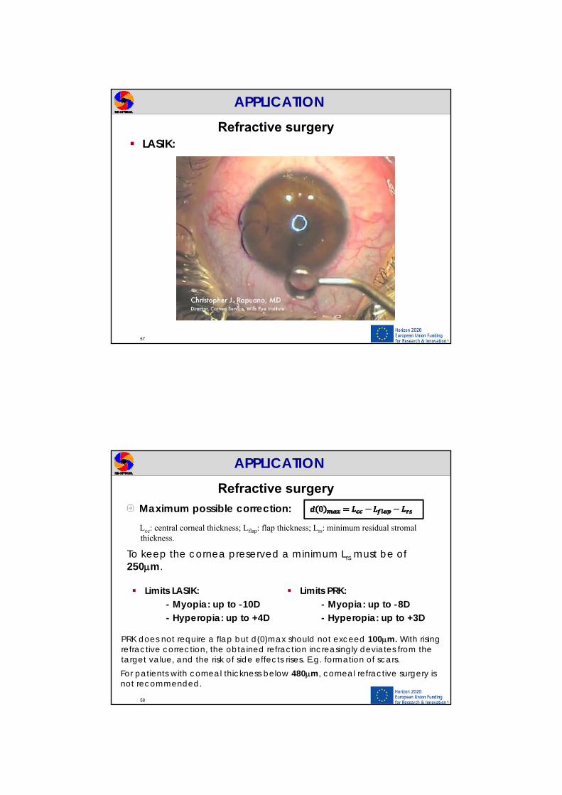
PRK Photorefractive keratectomy (1986): Direct carving of the cornea. The surgeon removes the epithelium, using an alcohol solution. The underlying corneal stroma (which does not regenerate) is then reshaped with an excimer laser. A new epithelial layer grows back within five days.
APPLICATIONRefractive surgery
52
Total thicknesses corneal layers about 560 m
53
APPLICATIONRefractive surgery
PRK:
Complications: Corneal wound healing (risk of opacification). It may take several weeks until refraction is stabilized. Painful (use of therapeutic contact lenses). Regression of the achieved refractive power (due to healing and reepithelialization).
APPLICATIONRefractive surgery PRK:
54

LASIK (Laser-assisted in-situ keratomileusis):
Similar to PRK but the following steps are done:
1- Cutting a flap into the anterior section of the cornea2- Removing intrastromal tissue with the laser3-Pulling the flap down again with surgical tweezers
APPLICATIONRefractive surgery
55
LASIK:
APPLICATIONRefractive surgery
Complications: Flap complicationsEye irritation, temporary visual discomfort.
56
How the flap is created? - Mechanical keratome: Surgical knife (blade)- Femtosecond laser: Intralase* (see next
section)
LASIK:
APPLICATIONRefractive surgery
57
Maximum possible correction:
Limits LASIK:- Myopia: up to -10D- Hyperopia: up to +4D
Lcc: central corneal thickness; Lflap: flap thickness; Lrs: minimum residual stromalthickness.
To keep the cornea preserved a minimum Lrs must be of 250m.
APPLICATIONRefractive surgery
Limits PRK:- Myopia: up to -8D- Hyperopia: up to +3D
PRK does not require a flap but d(0)max should not exceed 100m. With rising refractive correction, the obtained refraction increasingly deviates from the target value, and the risk of side effects rises. E.g. formation of scars.
58
For patients with corneal thickness below 480m, corneal refractive surgery is not recommended.

Photomechanical effects include plasma-induced ablation and photodisruption.
Plasma-induced ablation:- For very high power densities
in solids and fluids optical breakdown occurs. The physical effects of that are plasma formation and shock wave generation.
Photodisruption:- If optical breakdown occurs
inside soft tissues, cavitation and jet formation can also take place (more mechanical).
PHOTOMECHANICAL EFFECTS
59
Plasma is one of the four fundamental states of matter (the others being solid, liquid, and gas). Ionization is the process by which an atom or a molecule acquires a negative or positive charge by gaining or losing electrons to form ions. It can result from the loss of an electron after collisions with sub atomic particles, with other atoms, or through the interaction with light.
Lighting and electric sparks and neon lights are everyday examples of phenomena made from plasma.
PHOTOMECHANICAL EFFECTS
60

61
PHOTOMECHANICAL EFFECTS Ionization with subsequent electron avalanche (plasma formation – accumulation of free e- and ions):A free e- absorbs a photon and accelerates. The accelerated e- collides with another atom and ionizes it, resulting in two free electrons. And so forth.
Plasma-induced ablation
62
PHOTOMECHANICAL EFFECTS
If the applied electric field forces ionization of tissue optical breakdown occurs leading to well-defined removal of tissue.
Plasma-induced ablation:- 1011-1013 W/cm2
- Pulse duration: 500 10-12s (ps) – 10-15 s (fs)
Laser-induced plasma sparking on tooth surface caused by a single pulse from a Nd:YLF laser Typical sparking noise is heard if several pulses are applied.
63
The electric field E is related to the local power density I by the basic electrodynamic equation:
PHOTOMECHANICAL EFFECTS
For ps pulses: I = 1011 W/cm2 equivalent to E = 107 V/cm(comparable to the atomic and intramolecular Coulomb electric field)
Threshold allowing plasma generation (within a few hundred picoseconds 1018/cm3 free electron density is created).
Dielectric breakdown refers to plasma generation by intense electric field. Optical breakdown refers to the fact that this field is generated using light (UV, VIS and IR).
Plasma-induced ablation
Plasma-induced ablation
Optical breakdown renders possible energy deposition not only in pigmented tissue but also in weakly absorbing media due to the increased absorption coefficient of the induced plasma (a(pl)>>a). Moreover, plasma absorption is enhanced in the IR region (Maxwell’s equations).
is the frequency of the electromagnetic fieldN is the density of free electrons
64
PHOTOMECHANICAL EFFECTS
a(pl) N2 a(pl) 1/2
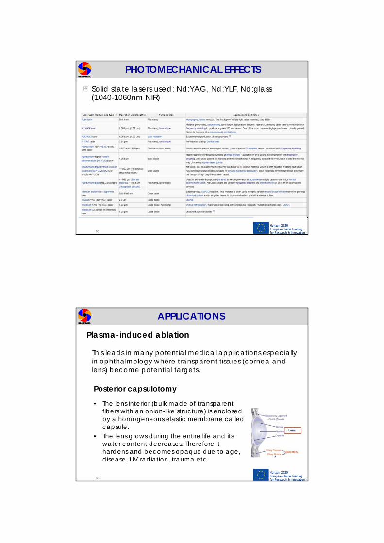
Solid state lasers used: Nd:YAG, Nd:YLF, Nd:glass (1040-1060nm NIR)
65
PHOTOMECHANICAL EFFECTS
This leads in many potential medical applications especially in ophthalmology where transparent tissues (cornea and lens) become potential targets.
Posterior capsulotomy
• The lens interior (bulk made of transparent fibers with an onion-like structure) is enclosed by a homogeneous elastic membrane called capsule.
• The lens grows during the entire life and its water content decreases. Therefore it hardens and becomes opaque due to age, disease, UV radiation, trauma etc.
66
APPLICATIONS
Plasma-induced ablation

Cataract surgery involves extracting the lens interior. Conventional methods rely on phaco-emulsification (ultrasonic handpiece) followed by aspiration of the fragments. Afterwards, an intraocular lens is inserted. The posterior capsule is retained to prevent collapse of the vitreous body and subsequent retinal detachment.
Posterior capsulotomyPlasma-induced ablation
67
APPLICATIONS
However, new lens fibers frequently emerge from this posterior capsule forming a scattering membrane, which must be removed.
Posterior capsulotomy with a Nd:YAG laser (IR) is done during a second surgery. It consists of focusing the laser on the posterior capsule (a He-Ne can be used as an aiming beam) and cutting it using several pulses (pulse duration: 30ns, energy: 5mJ, power density: 1010 W/cm2). After several cuts, the capsule opens as a zipper.
Posterior capsulotomyPlasma-induced ablation
68
APPLICATIONS

Posterior capsulotomyPlasma-induced ablation
69
APPLICATIONS
PhotodisruptionAt higher pulse energies shock waves and other mechanical side effects (CAVITATION BUBBLES and JET FORMATION) become significant and might determine the global effect upon the tissue.
70
PHOTOMECHANICAL EFFECTS
Photodisruption:- 1011-1016 W/cm2
- Pulse duration: 100 10-9s (ns) – 100 10-15 s (fs)
Bubble inside a cornea

Shock wave generation is associated with the expansion of the plasma (damage at subcellular level due to short displacement length 1-4 mm).
Cavitation occurs when focusing the laser beam not on the surface of a tissue but into it. Cavitation bubbles consist of gaseous vapors (water and carbon oxides) which diffuse again into the surrounding tissue.
If cavitations collapse in fluids and near a solid boundary, a jetformation is achieved.
Photodisruption
71
PHOTOMECHANICAL EFFECTS
PhotodisruptionApplication: Flap creation
• Use of a fs laser to create the corneal flap during LASIK replacing the microkeratome. They have been shown to be more precise, with fewer collateral tissue effects.
The technology is based on creating bubbles which separate the cells of the stroma. Thousands of these tiny bubbles are stacked together to create a flap. The whole process only takes 20 seconds (Intralase laser, Nd:glass 1053nm).
72
APPLICATIONS
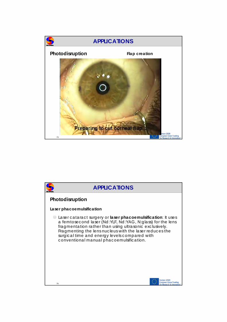
Photodisruption Flap creation
73
APPLICATIONS
Laser cataract surgery or laser phacoemulsification: It uses a femtosecond laser (Nd:YLF, Nd:YAG, N:glass) for the lens fragmentation rather than using ultrasonic exclusively. Fragmenting the lens nucleus with the laser reduces the surgical time and energy levels compared withconventional manual phacoemulsification.
Photodisruption
Laser phacoemulsification
74
APPLICATIONS

Photodisruption Conventional phacoemulsification
75
APPLICATIONS
Photodisruption Laser phacoemulsification
76
APPLICATIONS

QUESTIONS?
77
THANK YOU!