laser spectroscopy combined with mass spectrometry · laser spectroscopy combined with . mass...
TRANSCRIPT

Laser Spectroscopy combined with
mass spectrometry
Jos Oomens
FELIX Laboratory, Radboud University
Nijmegen, The Netherlands [email protected]

FELIX Free Electron Laser for Infrared eXperiments

Free Electron Laser: widely tunable, high intensity
undulator
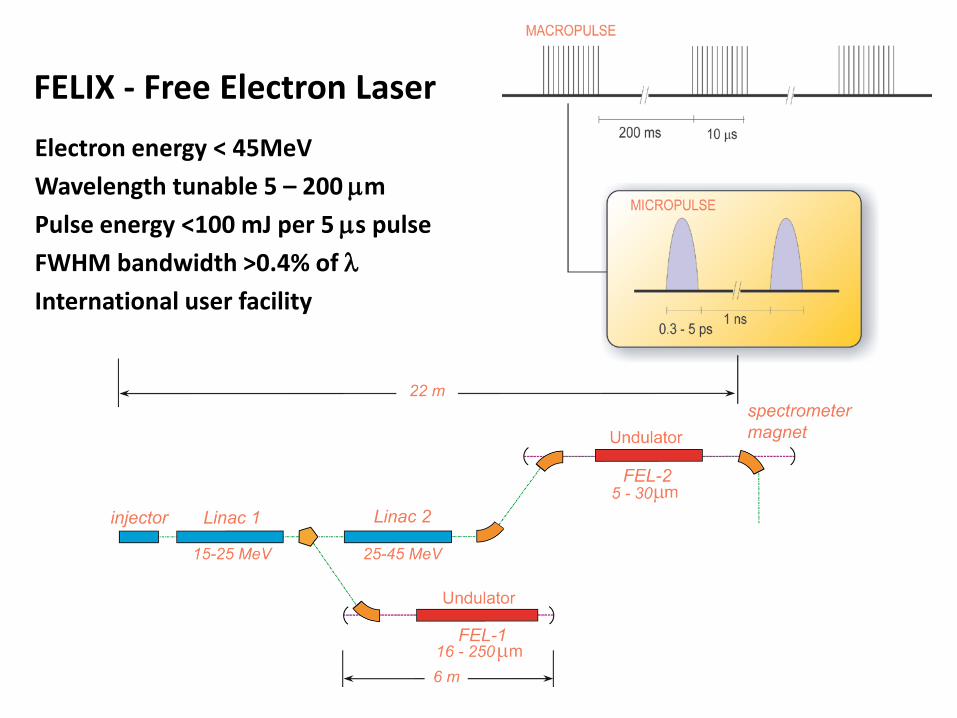
FELIX - Free Electron Laser
gun
accelerator
Electron energy < 45MeV Wavelength tunable 5 – 200 µm Pulse energy <100 mJ per 5 µs pulse FWHM bandwidth >0.4% of λ International user facility

Other common IR laser sources
CO2 laser line tunable 9-11 µm
OPO: hνpump = hνsignal + hνidler
tuning range depends on NL crystal transparency
DFG: hν = hνpump - hνtune
tuning range depends on NL crystal transparency
Quantum cascade / diode laser narrow tuning range
Far IR Time-Domain Spectrometer THz range (<100 cm-1)
1064 nm ~1.5 µm ~3.0 µm
dye laser 1064 nm ~3.0 µm
LiNbO3
ps THz
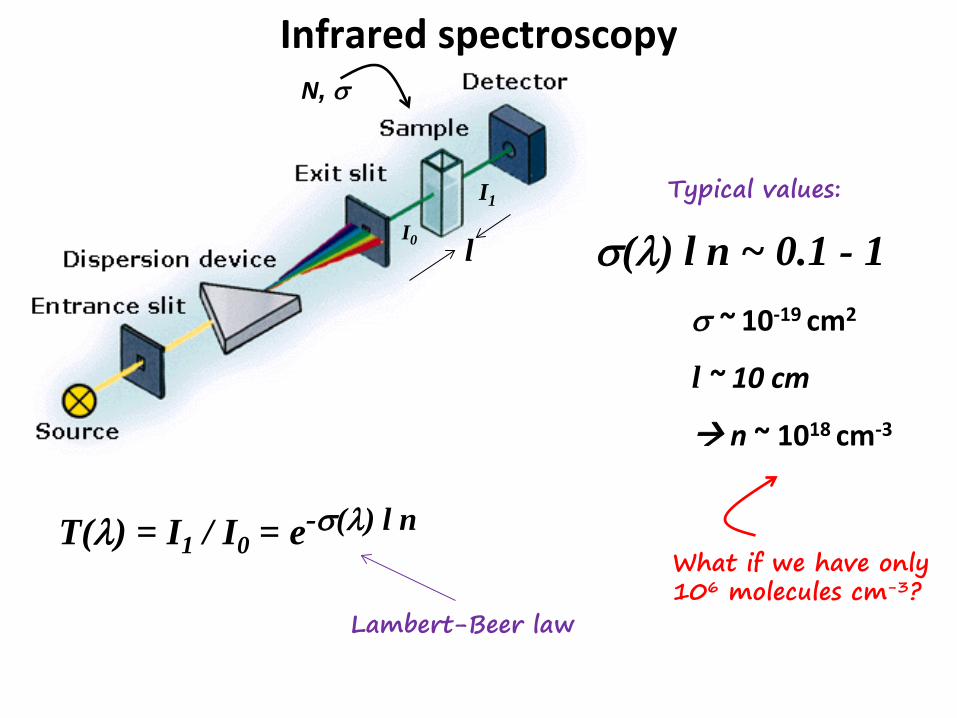
Infrared spectroscopy
T(λ) = I1 / I0 = e-σ(λ) l n
σ(λ) l n ~ 0.1 - 1 σ ~ 10-19 cm2
l ~ 10 cm
n ~ 1018 cm-3
Lambert-Beer law
Typical values:
N, σ
l
What if we have only 106 molecules cm-3?
I0
I1
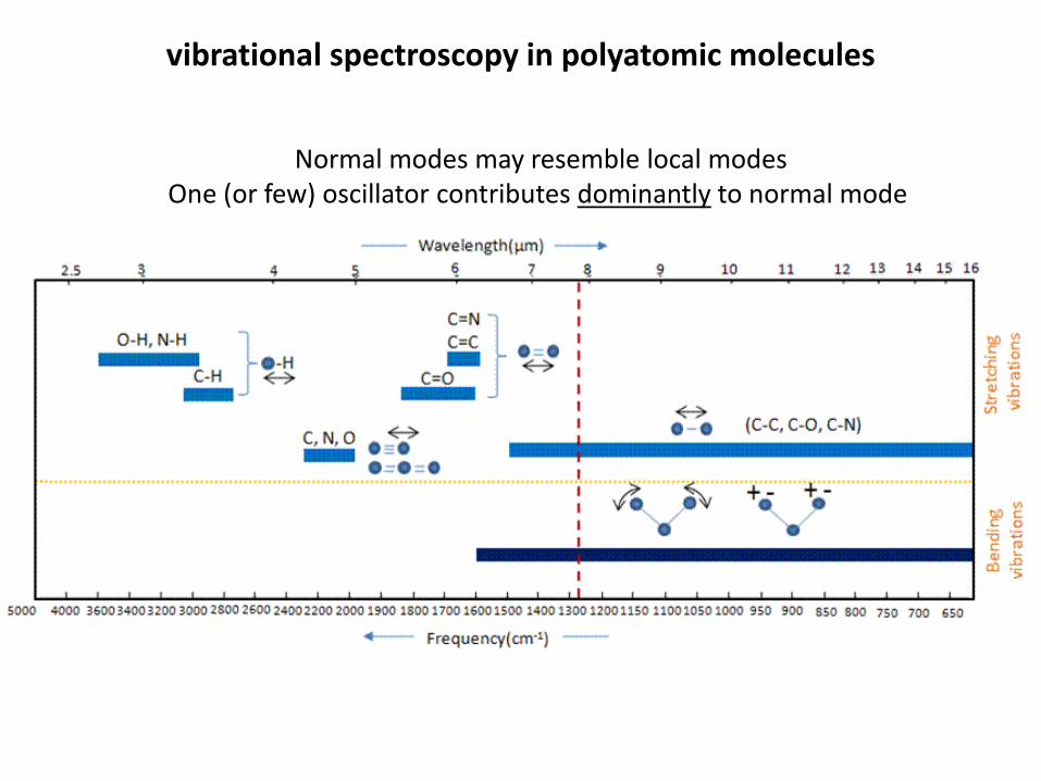
vibrational spectroscopy in polyatomic molecules
Normal modes may resemble local modes One (or few) oscillator contributes dominantly to normal mode
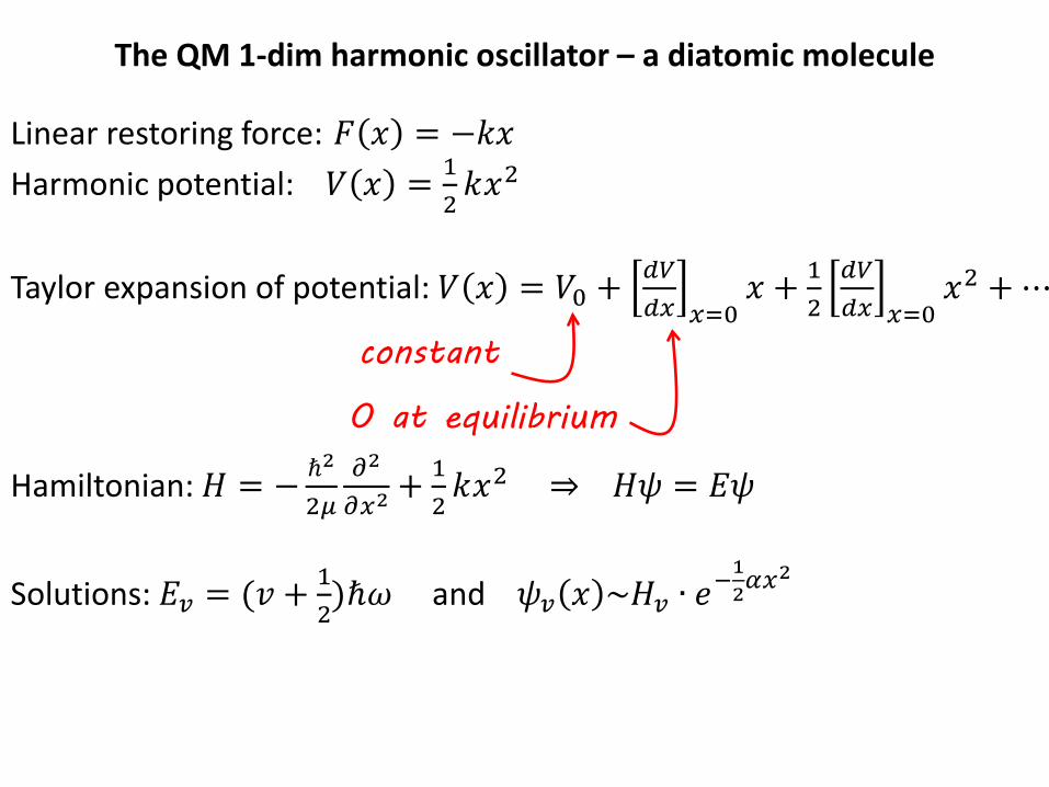
The QM 1-dim harmonic oscillator – a diatomic molecule
Linear restoring force: 𝐹 𝑥 = −𝑘𝑥 Harmonic potential: 𝑉 𝑥 = 1
2𝑘𝑥2
Taylor expansion of potential: 𝑉 𝑥 = 𝑉0 + 𝑑𝑑
𝑑𝑑 𝑑=0𝑥 + 1
2𝑑𝑑𝑑𝑑 𝑑=0
𝑥2 + ⋯
Hamiltonian: 𝐻 = − ℏ2
2𝜇𝜕2
𝜕𝑑2+ 1
2𝑘𝑥2 ⇒ 𝐻𝐻 = 𝐸𝐻
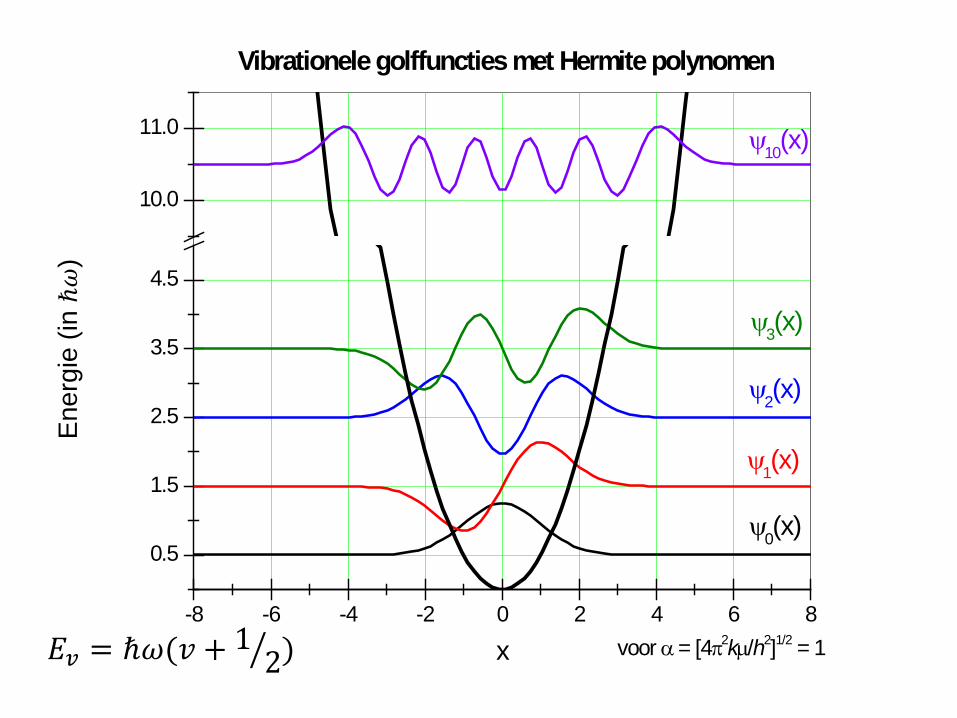
Solutions: 𝐸𝑣 = (𝑣 + 1
2)ℏ𝜔 and 𝐻𝑣 𝑥 ~𝐻𝑣 ∙ 𝑒
−12𝛼𝑑2
constant
0 at equilibrium

Hermite polynomials
𝐻𝑛 𝑥 = (−1)𝑛𝑒𝑑2𝑑𝑛
𝑑𝑥𝑛𝑒−𝑑2
Vibrational wavefunctions: 𝐻𝑣 𝑥 ~𝐻𝑣 ∙ 𝑒−12𝛼𝑑
2
-8 -6 -4 -2 0 2 4 6 8
0.5
1.5
2.5
3.5
4.5
10.0
11.0 ψ10(x)
voor α = [4π2kµ/h2]1/2 = 1
ψ3(x)
ψ2(x)
ψ1(x)
ψ0(x)
Vibrationele golffuncties met Hermite polynomen
x
Ener
gie
(in ℏ𝜔
)
𝐸𝑣 = ℏ𝜔(𝑣 + 12� )

-8 -6 -4 -2 0 2 4 6 8
0.5
1.5
2.5
3.5
4.5
10.0
11.0 |ψ10(x)|2
voor α = [4π2kµ/h2]1/2 = 1
|ψ3(x)|2
|ψ2(x)|2
|ψ1(x)|2
|ψ0(x)|2
Vibrationele waarschijnlijkheidsverdeling
x

Intensities / selection rules: tranition dipole moment
Taylor expansion for dipole moment: 𝜇 𝑥 = 𝜇0 + 𝑑𝜇𝑑𝑑 𝑑=0
𝑥 + ⋯
𝑇𝑇𝑇 = � 𝐻𝑣𝜇 𝑥 𝐻𝑣′𝑑𝑥 =∞
−∞
∫ 𝐻𝑣𝐻𝑣′𝑑𝑥
∞−∞ + 𝑑𝜇
𝑑𝑑 𝑑=0∫ 𝐻𝑣𝑥𝐻𝑣′𝑑𝑥∞−∞
0 due to orthogonality
𝜇0
dipole derivative ∆v = 1

-8 -6 -4 -2 0 2 4 6 8
ψ1(x) ψ2(x)
ψ0(x) ψ3(x)
ψ0(x) ψ1(x)
ψ0(x) ψ2(x)
x-8 -6 -4 -2 0 2 4 6 8
Vibrational wavefunctions are orthogonal
ψ2(x) ψ3(x)
ψ1(x) ψ3(x)
x

-8 -6 -4 -2 0 2 4 6 8
ψ1(x) x ψ2(x)
ψ0(x) x ψ3(x)
ψ0(x) x ψ1(x)
ψ0(x) x ψ2(x)
x-8 -6 -4 -2 0 2 4 6 8
ψ2(x) x ψ3(x)
ψ1(x) x ψ3(x)
x
Overlap vibrational wavefunctions selection rules
0 1
0 2
0 3
1 2
1 3
2 3

CH stretch normal modes
voor C2H4 (D2h)
B1u Ag
B3g B2u
y
z
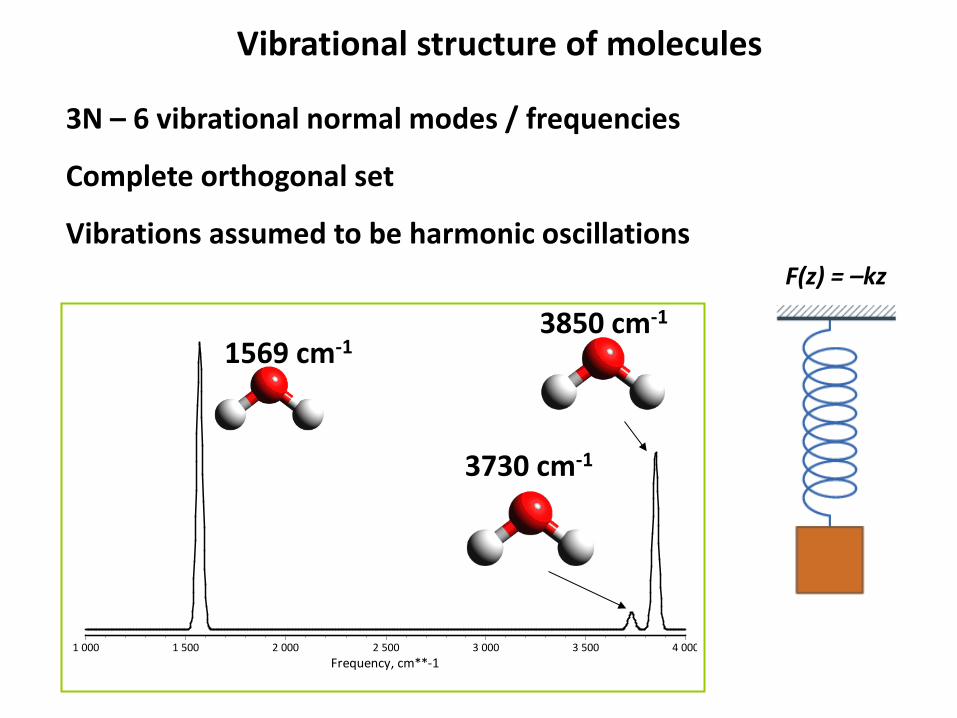
3N – 6 vibrational normal modes / frequencies
Complete orthogonal set
Vibrations assumed to be harmonic oscillations
3806 cm-1
Frequency, cm**-14 0003 5003 0002 5002 0001 5001 000
3850 cm-1
3730 cm-1
1569 cm-1
Vibrational structure of molecules
F(z) = –kz

Frequency, cm**-13 5003 0002 5002 0001 5001 000 500 0
Normal modes – localized vs. delocalized
CO stretch 1729 cm-1 NH bend 1561 cm-1 CH stretch 3094 cm-1
delocalized 661 cm-1
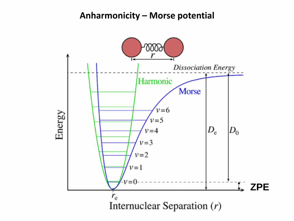
Anharmonicity – Morse potential
ZPE

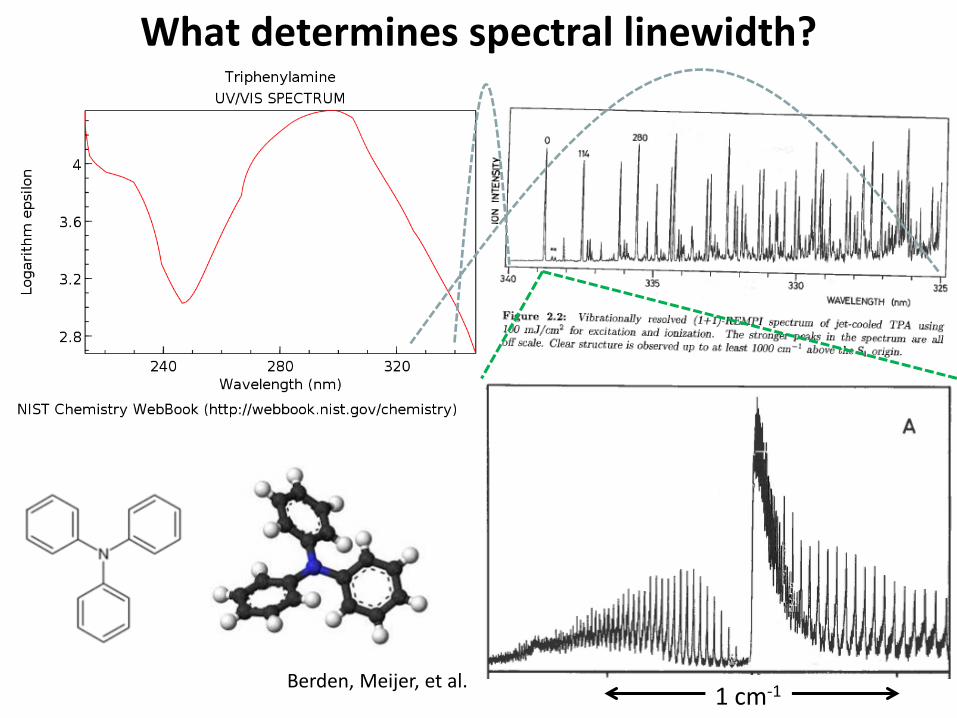
What determines spectral linewidth?
1 cm-1 Berden, Meijer, et al.

What determines spectral linewidth?
Homogeneous broadening - lifetime (excited state) - pressure broadening - transit time Inhomogeneous broadening - solvent (or environment) interactions - Doppler broadening (velocity distribution) - conformeric heterogeneity Unresolved fine structure (rotational, T-dependent) Instrumental resolution
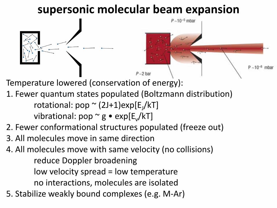
supersonic molecular beam expansion
Temperature lowered (conservation of energy): 1. Fewer quantum states populated (Boltzmann distribution) rotational: pop ~ (2J+1)exp[EJ/kT] vibrational: pop ~ g • exp[Ev/kT] 2. Fewer conformational structures populated (freeze out) 3. All molecules move in same direction 4. All molecules move with same velocity (no collisions) reduce Doppler broadening low velocity spread = low temperature no interactions, molecules are isolated 5. Stabilize weakly bound complexes (e.g. M-Ar)

E
# 11 128 164
164 minima 714 TS’s
Complexity gap

Conformational energy landscape As function of two torsional coordinates
As function of multiple coordinates: hypersurface
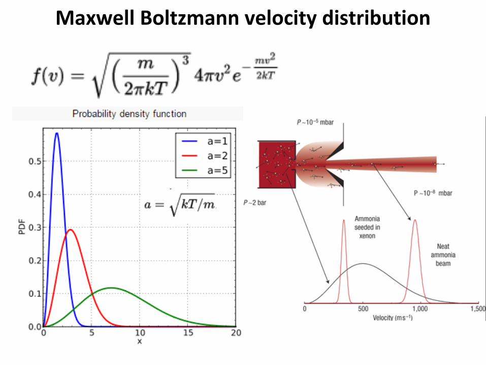
Maxwell Boltzmann velocity distribution
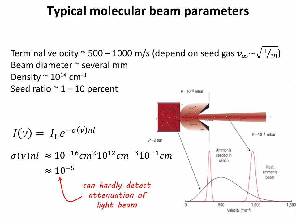
Typical molecular beam parameters
Terminal velocity ~ 500 – 1000 m/s (depend on seed gas 𝑣∞~ 1 𝑚⁄ ) Beam diameter ~ several mm Density ~ 1014 cm-3
Seed ratio ~ 1 – 10 percent
𝐼 𝜈 = 𝐼0𝑒−𝜎 𝜈 𝑛𝑛
𝜎 𝜈 𝑛𝑛 ≈ 10−16𝑐𝑐21012𝑐𝑐−310−1𝑐𝑐 ≈ 10−5
can hardly detect attenuation of
light beam
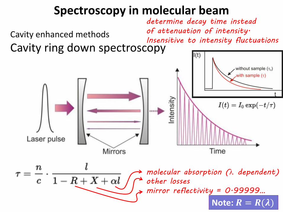
Spectroscopy in molecular beam
Cavity enhanced methods Cavity ring down spectroscopy
molecular absorption (λ dependent) other losses mirror reflectivity = 0.99999…
Note: 𝑹 = 𝑹(𝝀)
determine decay time instead of attenuation of intensity. Insensitive to intensity fluctuations

Action spectroscopy in molecular beam UV/vis (electronic spectroscopy)
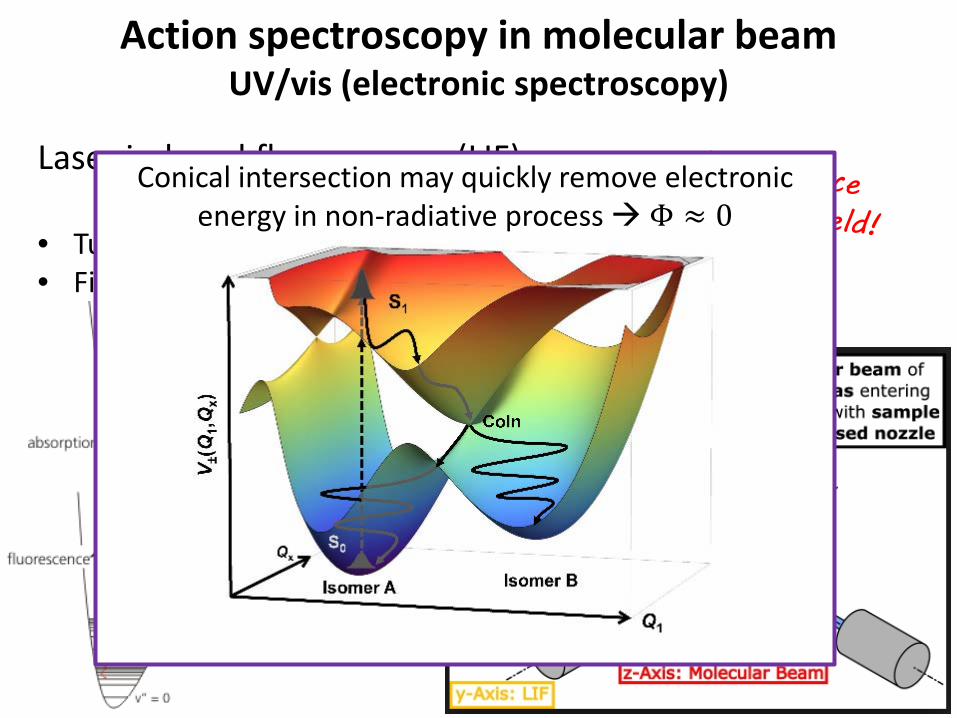
Laser induced fluorescence (LIF) detect fluorescence with photomultiplier • Tune laser λ: excitation spectrum • Fixed λ, disperse fluorescence: ground state spectrum
Action spectroscopy in molecular beam UV/vis (electronic spectroscopy)
Laser induced fluorescence (LIF) detect fluorescence • Tune laser λ: excitation spectrum • Fixed λ, disperse fluorescence: ground state spectrum
Conical intersection may quickly remove electronic energy in non-radiative process Φ ≈ 0

Watson-Crick
PNAS 2004

Action spectroscopy in molecular beam UV/vis (electronic spectroscopy)
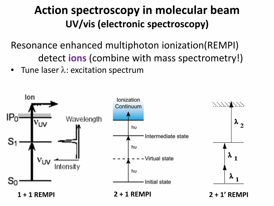
Resonance enhanced multiphoton ionization(REMPI) detect ions (combine with mass spectrometry!) • Tune laser λ: excitation spectrum
mol beam
ions
E
Action spectroscopy in molecular beam UV/vis (electronic spectroscopy)
Resonance enhanced multiphoton ionization(REMPI) detect ions (combine with mass spectrometry!) • Tune laser λ: excitation spectrum
1 + 1 REMPI 2 + 1 REMPI 2 + 1’ REMPI

Action spectroscopy in molecular beam IR (vibrational spectroscopy) ?
Mirror reflectivity typically lower and over limited λ range No fluorescence in IR (Einstein A coefficient ~ ν3, detectors insensitive in IR, thermal background radiation, …)
IR multiple photon excitation leads to dissociation rather than ionization (IP > D0)

Action spectroscopy in molecular beam IR (vibrational spectroscopy) ?
JPCA 2003
Fullerenes have high D0 and low IP Transition metals have low IP
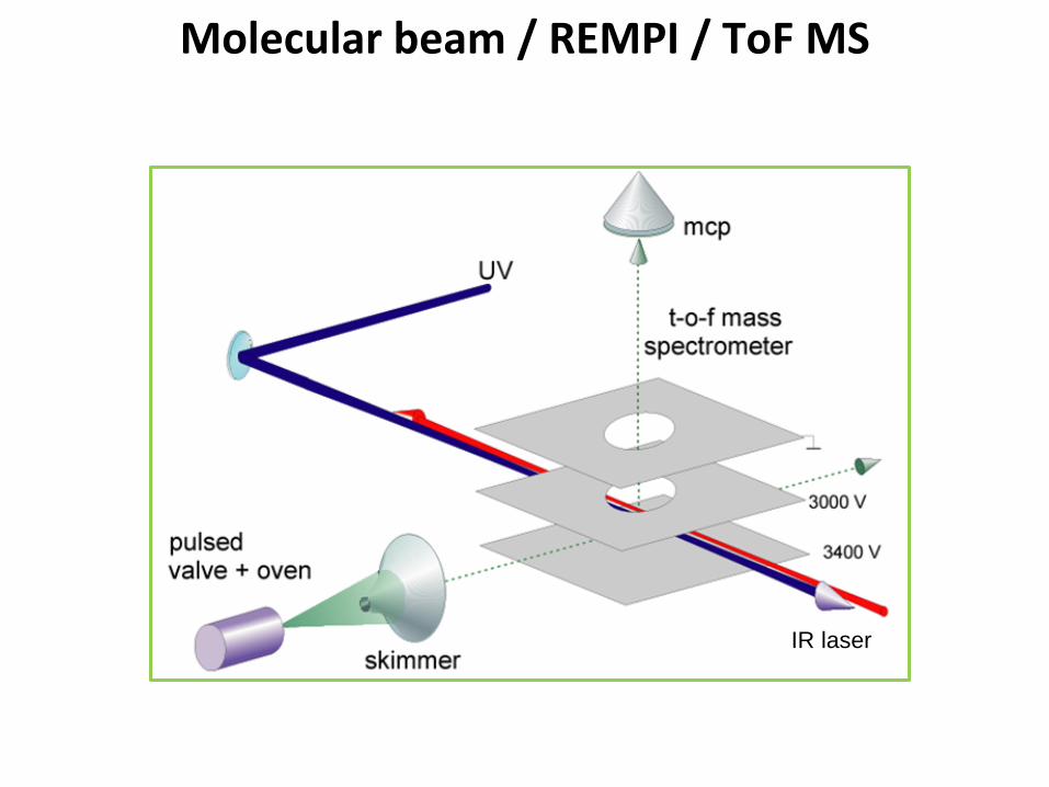
IR laser
Molecular beam / REMPI / ToF MS

REMPI is conformation specific!
S1 S0 transition is slightly different for different conformers of the same molecule Excite one specific conformer with narrow-band laser
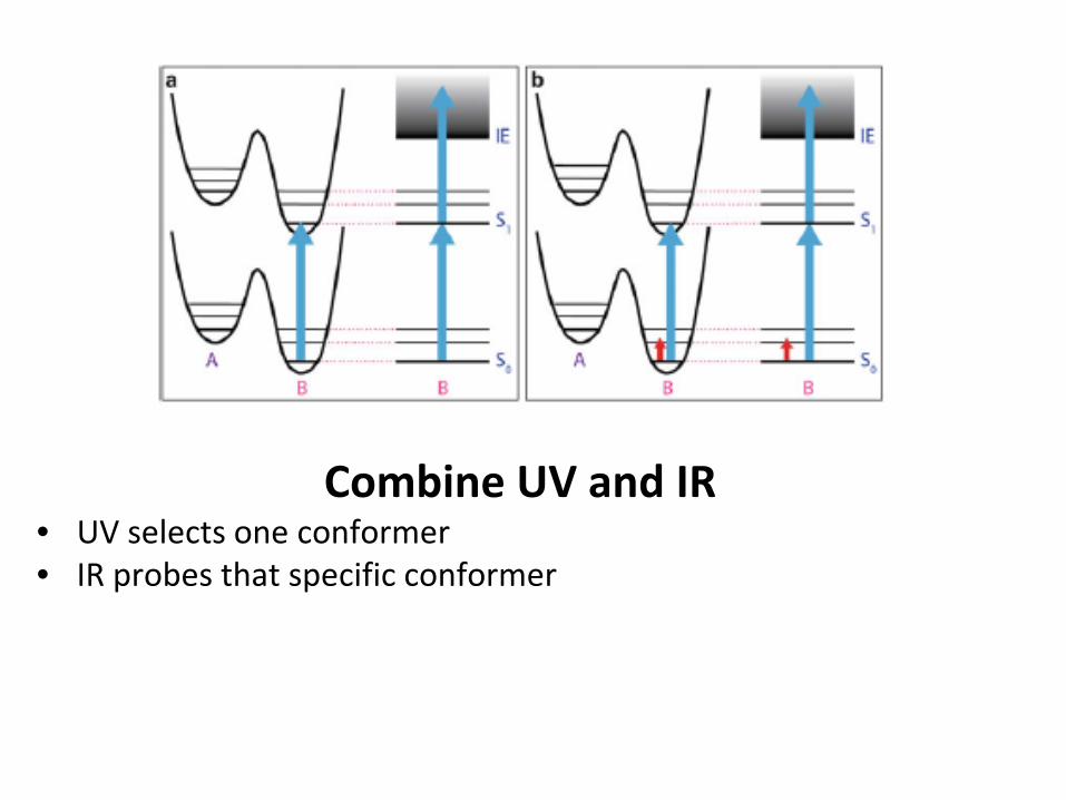
Combine UV and IR • UV selects one conformer • IR probes that specific conformer

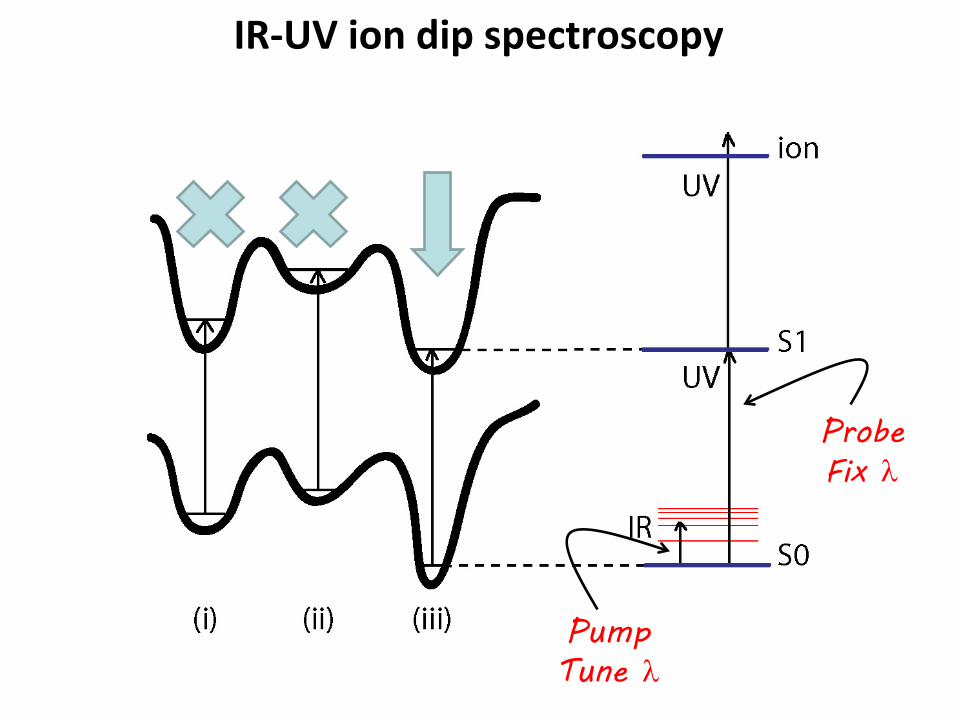
IR-UV ion dip spectroscopy
Pump Tune λ
Probe Fix λ
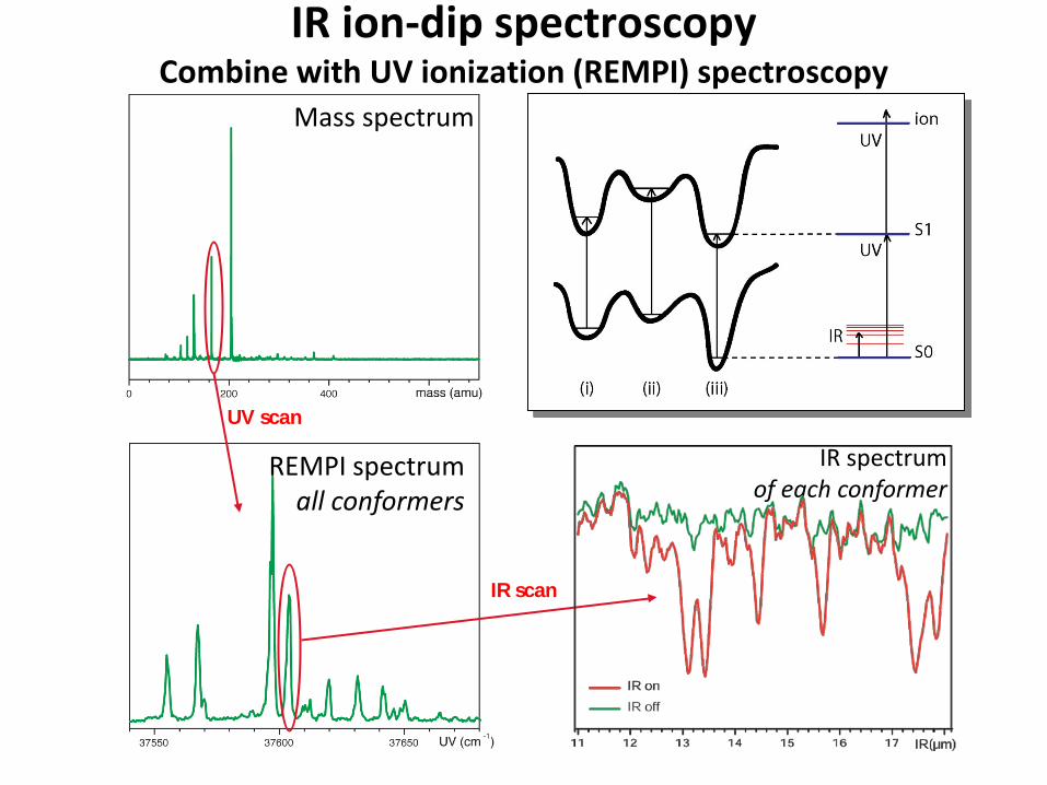
Mass spectrum
REMPI spectrum all conformers
IR spectrum of each conformer
UV scan
IR scan
IR ion-dip spectroscopy Combine with UV ionization (REMPI) spectroscopy

IR-UV hole-burning spectroscopy
Pump Fix λ
Probe Tune λ

Different conformers with different H-bonding network
Stabilization by intramolecular hydrogen bonds
Can we see differences in the IR spectrum ?
Ion dip spectroscopy Neutral molecules in beam
Conformer selective due to UV excitation step !
Application to tryptophan
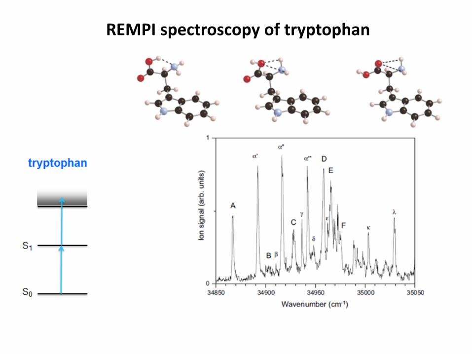
REMPI spectroscopy of tryptophan
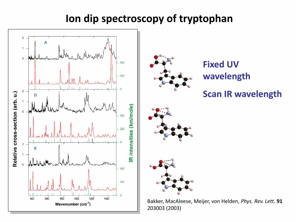
Bakker, MacAleese, Meijer, von Helden, Phys. Rev. Lett. 91 203003 (2003)
Ion dip spectroscopy of tryptophan
Fixed UV wavelength
Scan IR wavelength

IR-VUV double resonance spectroscopy If no chromophore is present: valine
VUV 118 nm
Not conformer selective

IR-VUV double resonance spectroscopy If no chromophore is present: NMA
IR photodissociation VUV ionization of fragments Background free No conformer selectivity
Yatsyna et al. PRL 2016

Spectroscopy in far-IR • Delocalized modes • Shallow potentials • Large amplitude motions

Frequency, cm**-13 5003 0002 5002 0001 5001 000 500 0
Normal modes – localized vs. delocalized
CO stretch 1729 cm-1 NH bend 1561 cm-1 CH stretch 3094 cm-1
delocalized 661 cm-1

Born-Oppenheimer Molecular Dynamics Alternative method to compute IR frequencies
Solve t.i. Schrodinger eq for nuclear geometry q (single point calc of electronic wavefunction)
Determine PES derivatives to calculate new atom positions
Trajectory on ‘on-the-fly’ calculated PES
Take FT of fluctuating dipole moment over trajectory

Probing dynamics Finding the barriers between different conformers

IR pump A
IR pump B
Difference spectra pump on/off
LIF spectrum no pump

Probing dynamics Finding the barriers between different conformers
Mass spectrometers
Determine molecular weight based on trajectories of ionized molecule in electric or magnetic field
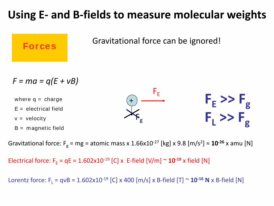
Using E- and B-fields to measure molecular weights
Forces
F = ma = q(E + vB)
where q = charge
E = electrical field
v = velocity
B = magnetic field
Gravitational force: Fg = mg = atomic mass x 1.66x10⁻²⁷ [kg] x 9.8 [m/s2] ≈ 10-26 x amu [N]
+
Fg
FE
Electrical force: FE = qE = 1.602x10⁻¹⁹ [C] x E-field [V/m] ~ 10-19 x field [N]
FE >> Fg FL >> Fg
Lorentz force: FL = qvB = 1.602x10⁻¹⁹ [C] x 400 [m/s] x B-field [T] ~ 10-16 N x B-field [N]
Gravitational force can be ignored!
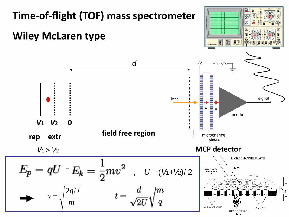
Time-of-flight (TOF) mass spectrometer
Wiley McLaren type
MCP detector
d
V1 V2 0 field free region rep extr
= , U = (V1+V2)/2
V1 > V2
mqUv 2
=
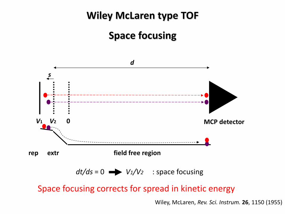
Wiley McLaren type TOF
Space focusing
MCP detector
d
V1 V2 0
field free region rep extr
dt/ds = 0 V1/V2 : space focusing
Wiley, McLaren, Rev. Sci. Instrum. 26, 1150 (1955)
s
Space focusing corrects for spread in kinetic energy

Lorentz force Right-hand rule (for + ions) Thumb: ion velocity v Index: magnetic field B Middle finger: Lorentz force FL
Absolute value only:
)(sinαqvBFL =α = angle between v and B α = 90o → sin(90o) = 1
F
B
I
BqvFL ⊗=

Ions moving in B-field
FL always perpendicular to v ion traverses a circular path
rvm
FqvBF CL
2
===
FC=centripetal force
qBvm
r = Momentum – magnetic sector is a momentum analyzer
UBr
qm
2
22
=Remember from TOF: mqUv 2
=
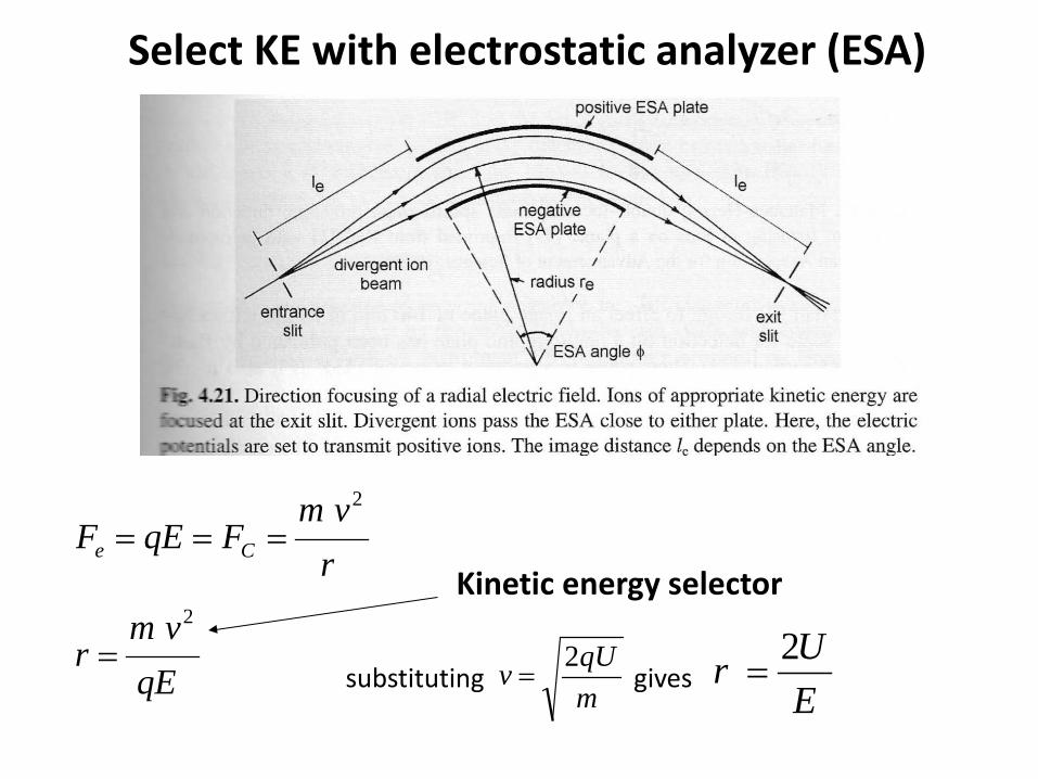
Select KE with electrostatic analyzer (ESA)
qEvm
r
rvm
FqEF Ce
2
2
=
===
EUr 2
=mqUv 2
=substituting gives
Kinetic energy selector
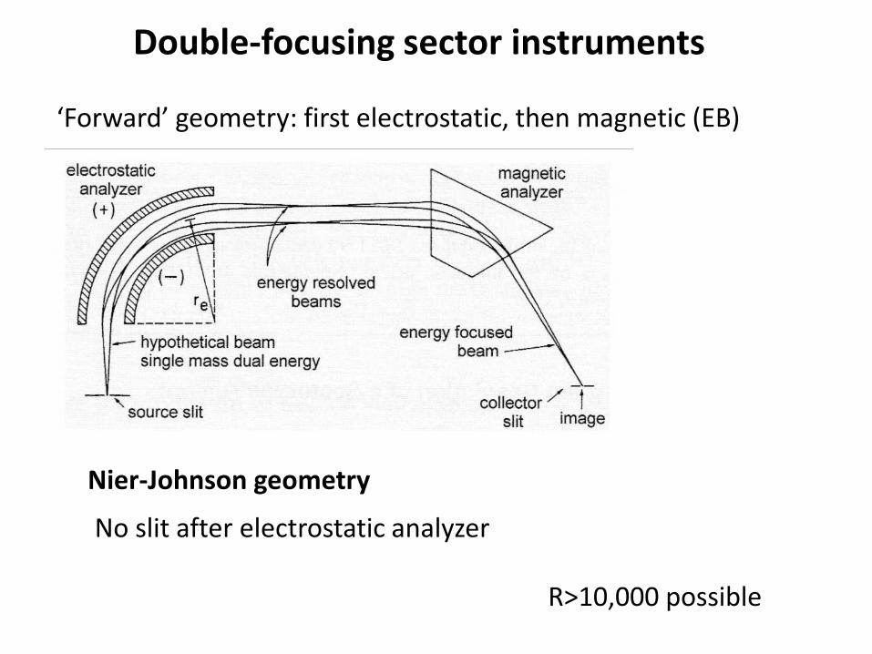
Double-focusing sector instruments
‘Forward’ geometry: first electrostatic, then magnetic (EB)
Nier-Johnson geometry
No slit after electrostatic analyzer
R>10,000 possible
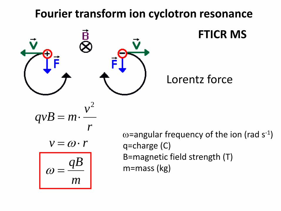
Fourier transform ion cyclotron resonance
mqB
rvrvmqvB
=
⋅=
⋅=
ω
ω
2
ω=angular frequency of the ion (rad s-1) q=charge (C) B=magnetic field strength (T) m=mass (kg)
FTICR MS
Lorentz force

4.7 T actively shielded magnet ESI source (Z-Spray) Ion optics controls
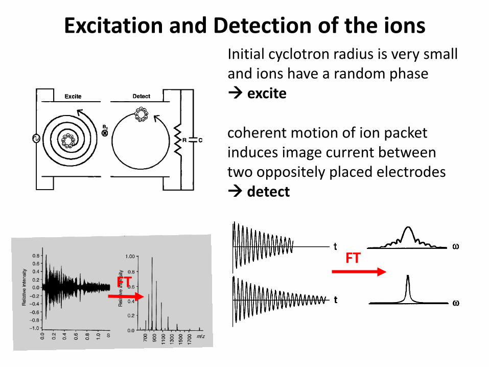
Excitation and Detection of the ions
coherent motion of ion packet induces image current between two oppositely placed electrodes detect
Initial cyclotron radius is very small and ions have a random phase excite
FT FT

Excitation and Detection of the ions
Mass-selective excitation also for ion manipulation:
1. Collisional activation 2. ejection

Resolution and accuracy
Same nominal mass, but different elemental formulae. e.g. to determine S-content in crude oil
Marshall et al. NHMFL, Tallahassee, FL

Quadrupole mass analyzer
VA = VDC + VRFcos(ωt) VB = – VDC – VRFcos(ωt) 0 2 4 6 8 10 12 14 16 18 20
-6
-4
-2
0
2
4
6
V
time
A A B
B

3-D quadrupole ion trap (QIT) a.k.a. Paul trap
1 ring electrode (middle) and two end-cap electrodes.
Wolfgang Paul (1913 – 1993) Nobel Prize Physics 1989

Stability diagram in z-dimension
VRF
VDC
Note: DC voltage between ring and end-cap electrodes
If VDC=0, az=0 Very similar to quadrupole filter (transmit wide range of m/z’s), in QIT trap wide range of m/z’s.
For az=0, βz=1 qz=0.908 (outside of qz<0.4 limit)
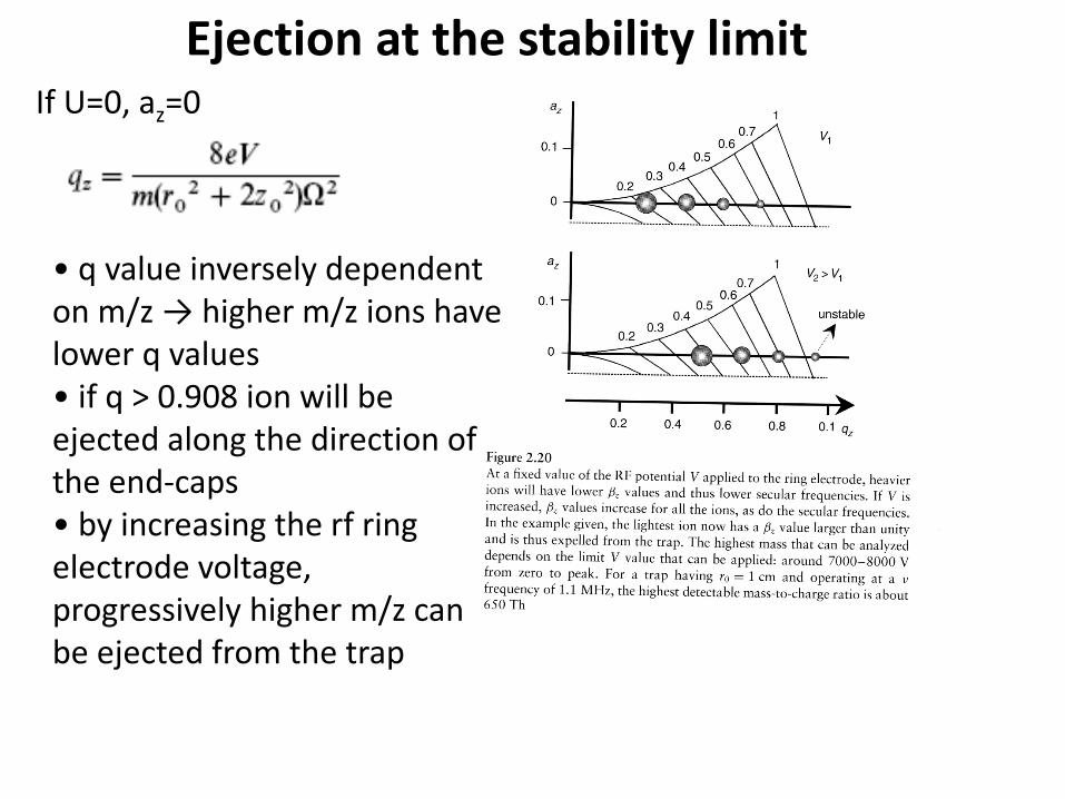
Ejection at the stability limit
• q value inversely dependent on m/z → higher m/z ions have lower q values • if q > 0.908 ion will be ejected along the direction of the end-caps • by increasing the rf ring electrode voltage, progressively higher m/z can be ejected from the trap
If U=0, az=0