large intestine - judoctors · pdf fileanatomy of the large intestine 2. ... • it is a...
TRANSCRIPT

Large intestine
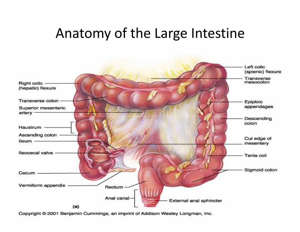
Anatomy of the Large Intestine
2

Large Intestine
• Extends from ileocecal valve to anus
• Length = 1.5- 2.5m = 5 feet
• Regions
– Cecum = 2.5- 3 inch
– Appendix= 3-5 inch
3
– Appendix= 3-5 inch
– Colon
• Ascending= 5 inch
• Transverse= 15 inch
• Descending= 10 inch
• Sigmoid colon = 10 – 15 inch
– Rectum= 5 inch
– Anal canal= 4 cm
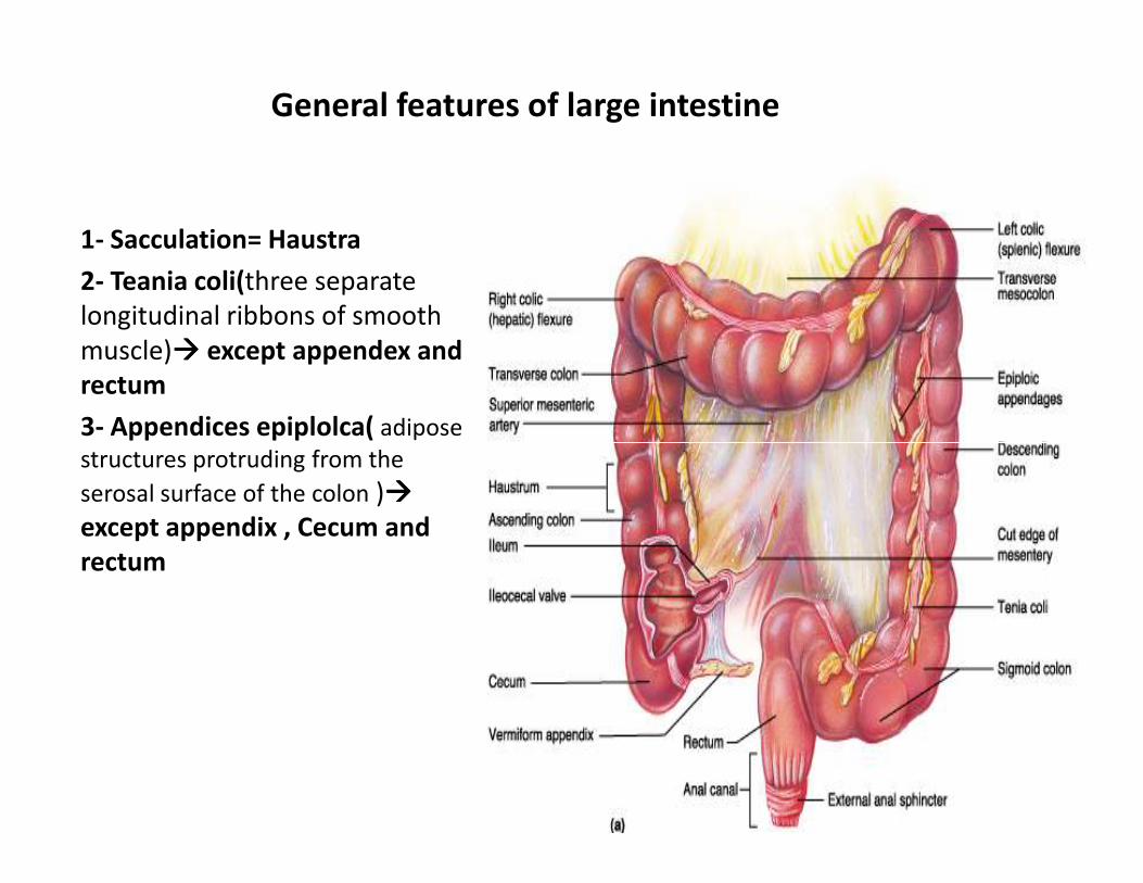
General features of large intestine
1- Sacculation= Haustra
2- Teania coli(three separate
longitudinal ribbons of smooth
muscle)���� except appendex and
rectum
3- Appendices epiplolca( adipose
structures protruding from the structures protruding from the
serosal surface of the colon )����
except appendix , Cecum and
rectum
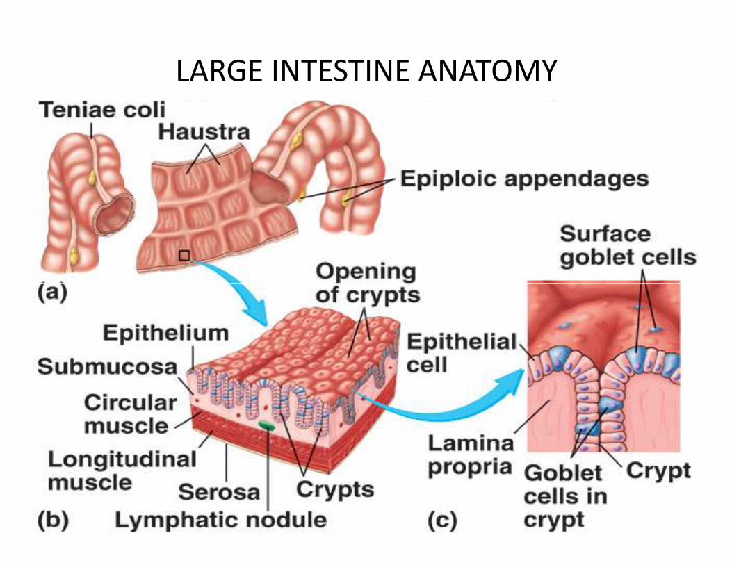
LARGE INTESTINE ANATOMY
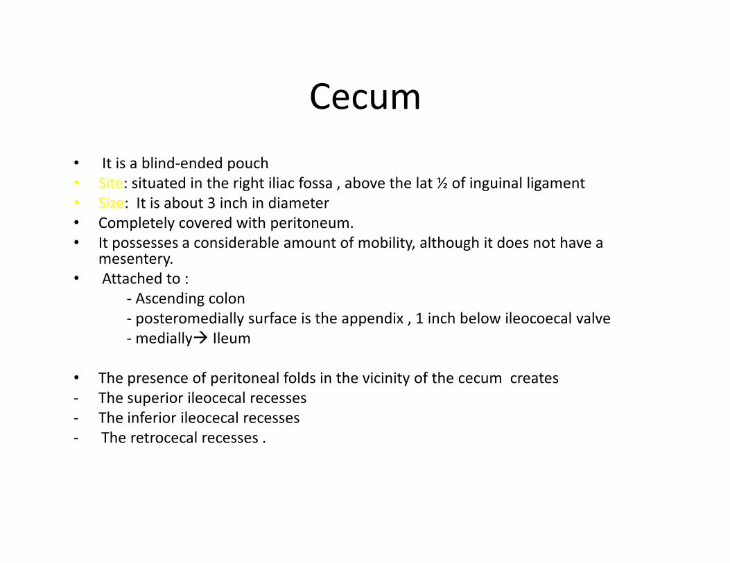
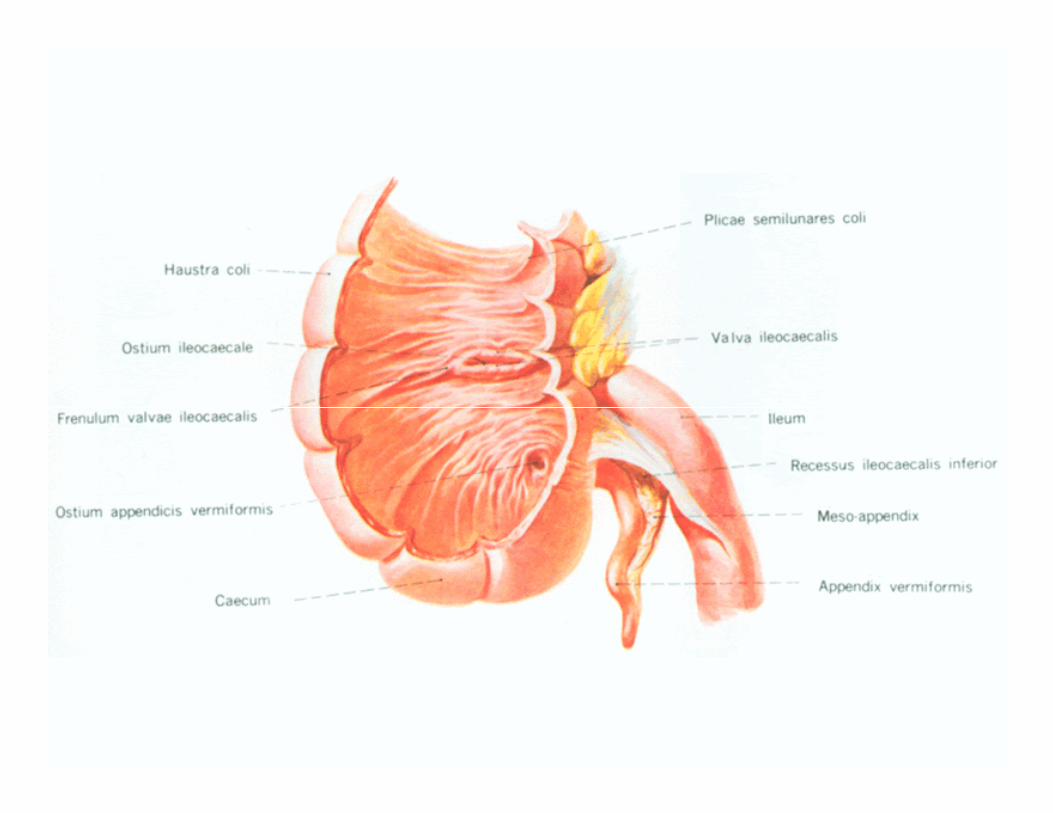
Cecum
• It is a blind-ended pouch
• Site: situated in the right iliac fossa , above the lat ½ of inguinal ligament
• Size: It is about 3 inch in diameter
• Completely covered with peritoneum.
• It possesses a considerable amount of mobility, although it does not have a mesentery.
• Attached to :• Attached to :
- Ascending colon
- posteromedially surface is the appendix , 1 inch below ileocoecal valve
- medially� Ileum
• The presence of peritoneal folds in the vicinity of the cecum creates
- The superior ileocecal recesses
- The inferior ileocecal recesses
- The retrocecal recesses .

Cecum….cont
• The longitudinal muscle is restricted to three
flat bands, the taenia coli, which converge on
the base of the appendix and provide for it a
complete longitudinal muscle coat . complete longitudinal muscle coat .

Relations of cecum
• Anteriorly:
- Coils of small intestine
- the greater omentum
- the anterior abdominal wall in the right iliac region
• Posteriorly: • Posteriorly:
- The psoas and the iliacus muscles
- the femoral nerve
- and the lateral cutaneous nerve of the thigh .
- Postero- medially � The appendix is commonly �retrocecal common.
• Medially:
- Small intestine( ileum)

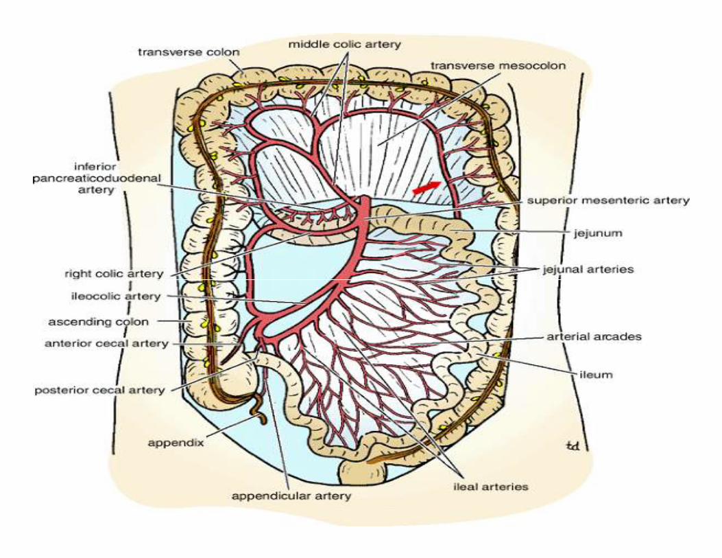
Blood Supply of cecum
Arteries
• Anterior and posterior cecal arteries � a
branch of Superior mesenteric artery
The veins correspond to the arteries and drain
into the superior mesenteric vein.

Blood supply of cecum
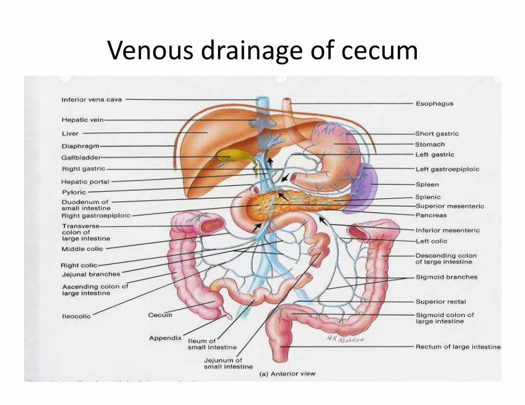
Venous drainage of cecum

Lymphatic Drainage of cecum
• The lymph vessels pass through several
mesenteric nodes � finally reach the superior
mesenteric nodes.
Nerve Supply of cecum
• Branches from the sympathetic and
parasympathetic (vagus) nerves form the
superior mesenteric plexus.
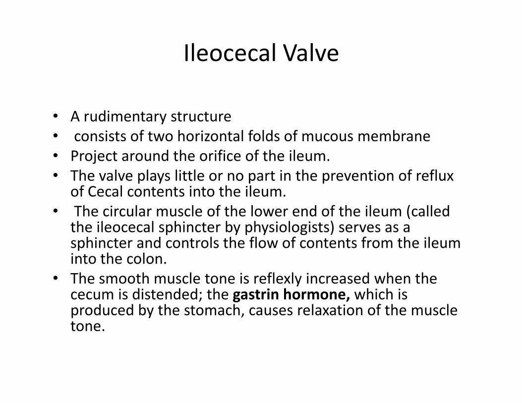
Ileocecal Valve
• A rudimentary structure
• consists of two horizontal folds of mucous membrane
• Project around the orifice of the ileum.
• The valve plays little or no part in the prevention of reflux of Cecal contents into the ileum.
• The circular muscle of the lower end of the ileum (called • The circular muscle of the lower end of the ileum (called the ileocecal sphincter by physiologists) serves as a sphincter and controls the flow of contents from the ileum into the colon.
• The smooth muscle tone is reflexly increased when the cecum is distended; the gastrin hormone, which is produced by the stomach, causes relaxation of the muscle tone.
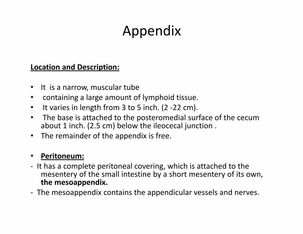
Appendix
Location and Description:
• It is a narrow, muscular tube
• containing a large amount of lymphoid tissue.
• It varies in length from 3 to 5 inch. (2 -22 cm).
• The base is attached to the posteromedial surface of the cecum • The base is attached to the posteromedial surface of the cecum about 1 inch. (2.5 cm) below the ileocecal junction .
• The remainder of the appendix is free.
• Peritoneum:
- It has a complete peritoneal covering, which is attached to the mesentery of the small intestine by a short mesentery of its own, the mesoappendix.
- The mesoappendix contains the appendicular vessels and nerves.

APPENIX

Appendix….cont
• Position
- The appendix lies in the right iliac fossa, and in relation to the anterior abdominal wall
1- Retrocecal in retrocaecal recess behind cecum� in 74% of people
2- pelvic: in pelvis related to Rt. Ovary and uterine tube� in 21% of people
3- Subcaecal: below cecum� in 3.5%
4- Preileal: infront of ileum � 1%4- Preileal: infront of ileum � 1%
5- Postileal: behind the ileum�0.5%
• Surface anatomy of appendix= McBurney's point
- Its base is situated one third of the way up the line joining the right anterior superior iliac spine to the umbilicus
- To reach the appendix during operation follow the taenia coli which converge toward the appendix
Blood Supply of appendix
Arteries
• The appendicular artery is a branch of the
posterior cecal artery(ilio-cecal.a)which
descends behind the ileum.descends behind the ileum.
Veins
• The appendicular vein drains into the
posterior cecal vein.

• Appendicular artery runs in
free margin of the
mesoappendix
Lymphatic Drainage of appendix
• The lymph vessels drain into one or two nodes
lying in the mesoappendix � eventually into
the superior mesenteric nodes.

Nerve Supply of appendix
• The appendix is supplied by the sympathetic and parasympathetic (vagus) nerves from the superior mesenteric plexus.
• Afferent nerve fibers concerned with the conduction of visceral pain from the appendix conduction of visceral pain from the appendix accompany the sympathetic nerves and enter the spinal cord at the level of the 10th thoracic segment.
• The peritoneum over the appendix is innervated by the 10th intercostal nerve= skin of umbilicus

Clinical notes
• Acute appendetitis� uncommon in the two
extremes of life
• Thrombosis of appendicular .a�
gangrene(just one artery for appendix) gangrene(just one artery for appendix)
�perforation �Lt.paracolic gutter while in
Acute cholecystitis� no gangrene( more than
one artery supply the gallbladder)
• Appendiectomy
Ascending Colon
Location and Description:
• The ascending colon is about 5 inch. (13 cm) long
• lies in the right lower quadrant.
• It extends upward from the cecum to the inferior surface of the right lobe of the liver, where it turns to the left, forming the right colic flexureforming the right colic flexure
• Then becomes continuous with the transverse colon.
• Taenia coli, sacculation & appendeces epiplolca are present
The peritoneum
- Covers the front and the sides of the ascending colon
- Binding it to the posterior abdominal wall.

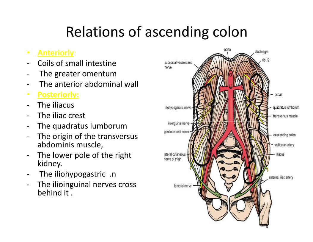
Relations of ascending colon
• Anteriorly:
- Coils of small intestine
- The greater omentum
- The anterior abdominal wall
• Posteriorly:
- The iliacus
- The iliac crest
- The quadratus lumborum- The quadratus lumborum
- The origin of the transversus abdominis muscle,
- The lower pole of the right kidney.
- The iliohypogastric .n
- The ilioinguinal nerves cross behind it .
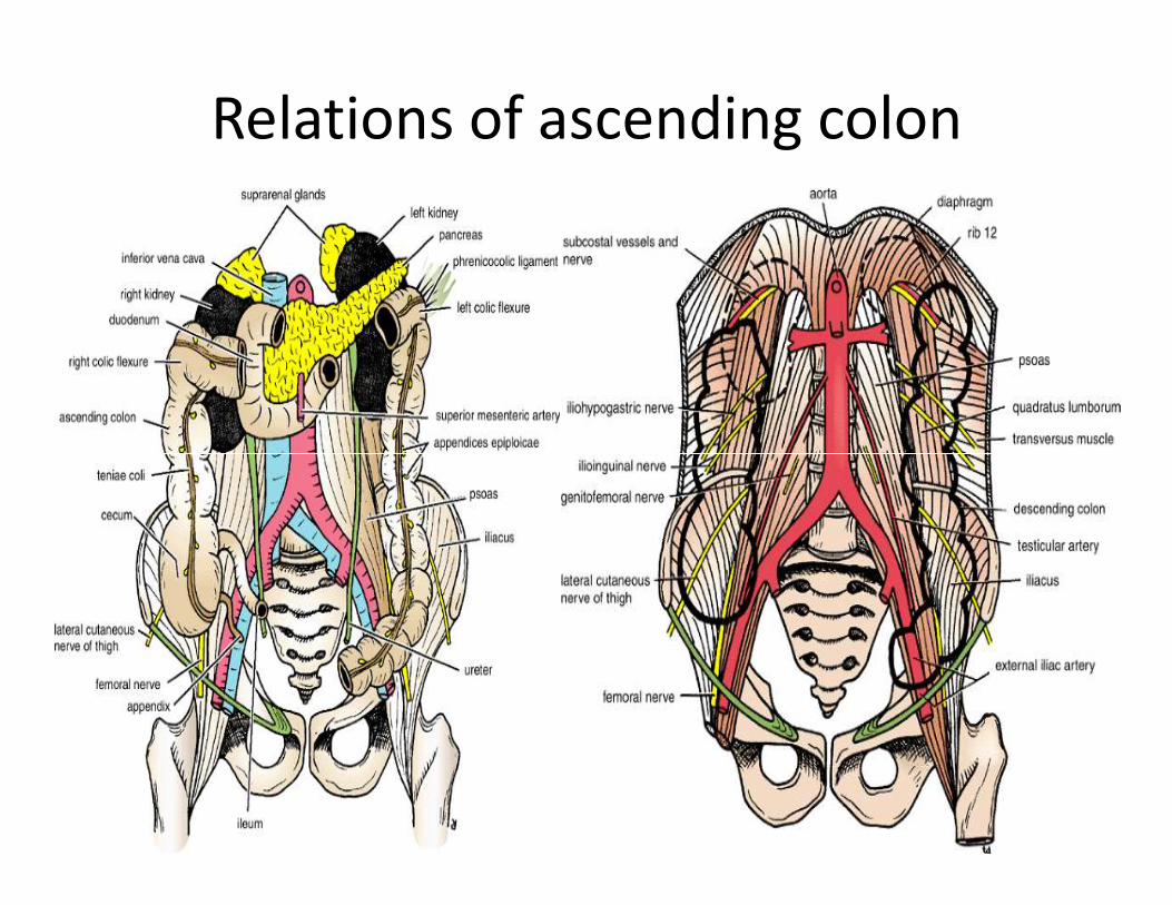
Relations of ascending colon
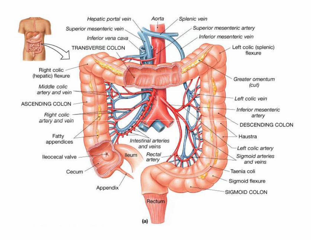
Blood Supply of Ascending colon
Arteries
• The ileocolic & right colic branches of the
superior mesenteric artery supply this area.
VeinsVeins
• The veins correspond to the arteries and drain
into the superior mesenteric vein.

Lymphatic drainage of Ascending colon
• The lymphatic vessels � lymph nodes lying
along the course of the colic blood vessels �
the superior mesenteric nodes.the superior mesenteric nodes.
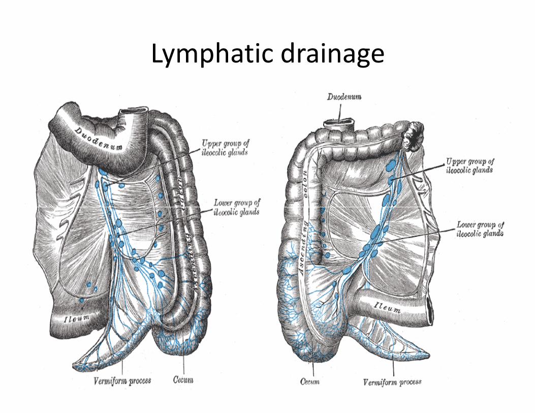
Lymphatic drainage
Nerve Supply of ascending colon
• Sympathetic and parasympathetic (vagus)
nerves from the superior mesenteric plexus .
Transverse colonTransverse colon
Transverse Colon
Location and Description
• The transverse colon is about 15 in. (38 cm) long
• extends across the abdomen
• occupying the umbilical region.
• It begins at the right colic flexure below the right lobe of the liver
• Hangs downward• Hangs downward
• Suspended by the transverse mesocolon from the pancreas
• It then ascends to the left colic flexure below the spleen.
• The left colic flexure is higher than the right colic flexure and is suspended from the diaphragm by the phrenicocolic ligament .
• Taenia coli, sacculation & appendeces epiplolca are present
The transverse mesocolon= mesentery of the transverse colon
• suspends the transverse colon from the anterior border of the pancreas .
• The mesentery is attached to the superior border of the transverse colon
• The posterior layers of the greater omentum • The posterior layers of the greater omentum are attached to the inferior border .
• The position of the transverse colon is extremely variable and may sometimes reach down as far as the pelvis.
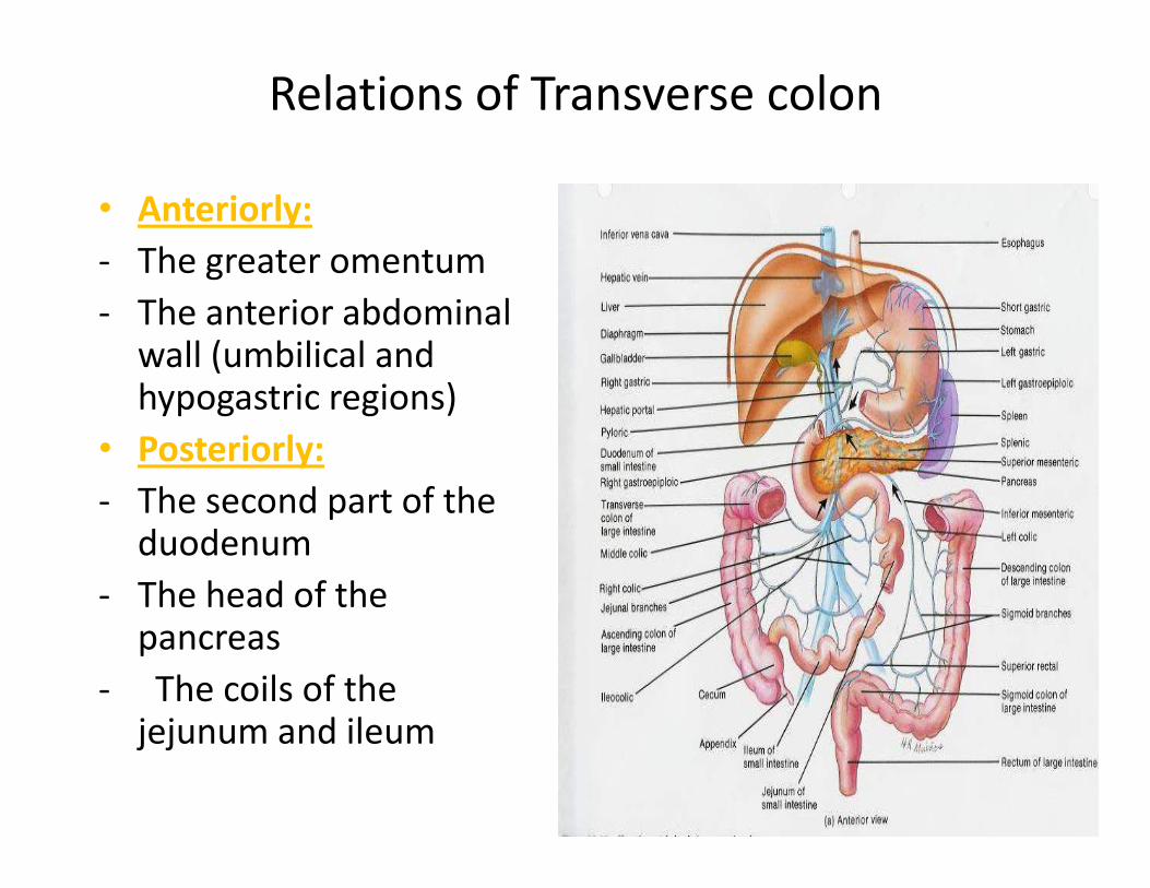
Relations of Transverse colon
• Anteriorly:
- The greater omentum
- The anterior abdominal wall (umbilical and hypogastric regions)
• Posteriorly: • Posteriorly:
- The second part of the duodenum
- The head of the pancreas
- The coils of the jejunum and ileum
Blood Supply of transverse colon
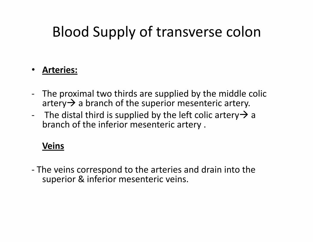
• Arteries:
- The proximal two thirds are supplied by the middle colic artery� a branch of the superior mesenteric artery.
- The distal third is supplied by the left colic artery� a branch of the inferior mesenteric artery .branch of the inferior mesenteric artery .
Veins
- The veins correspond to the arteries and drain into the superior & inferior mesenteric veins.
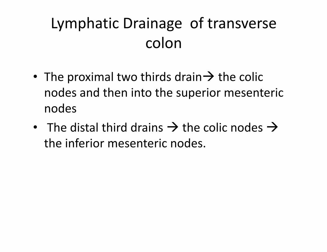
Lymphatic Drainage of transverse
colon
• The proximal two thirds drain� the colic
nodes and then into the superior mesenteric
nodes
• The distal third drains � the colic nodes �• The distal third drains � the colic nodes �
the inferior mesenteric nodes.
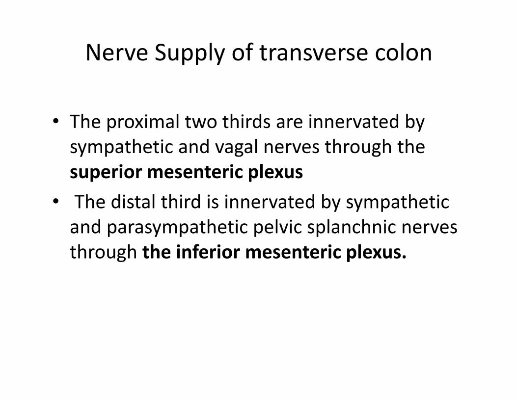
Nerve Supply of transverse colon
• The proximal two thirds are innervated by
sympathetic and vagal nerves through the
superior mesenteric plexus
• The distal third is innervated by sympathetic • The distal third is innervated by sympathetic
and parasympathetic pelvic splanchnic nerves
through the inferior mesenteric plexus.
Descending Colon
Location and Description:
• The descending colon is about 10 in. (25 cm) long
• It extends downward from the left colic flexure, to the pelvic brim, where it becomes continuous with the pelvic brim, where it becomes continuous with the sigmoid colon.
• Taenia coli, sacculation & appendeces epiplolca are present
• The peritoneum
- Covers the front and the sides and binds it to the posterior abdominal wall.
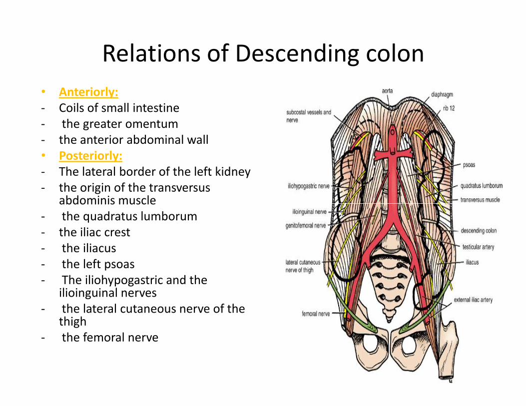
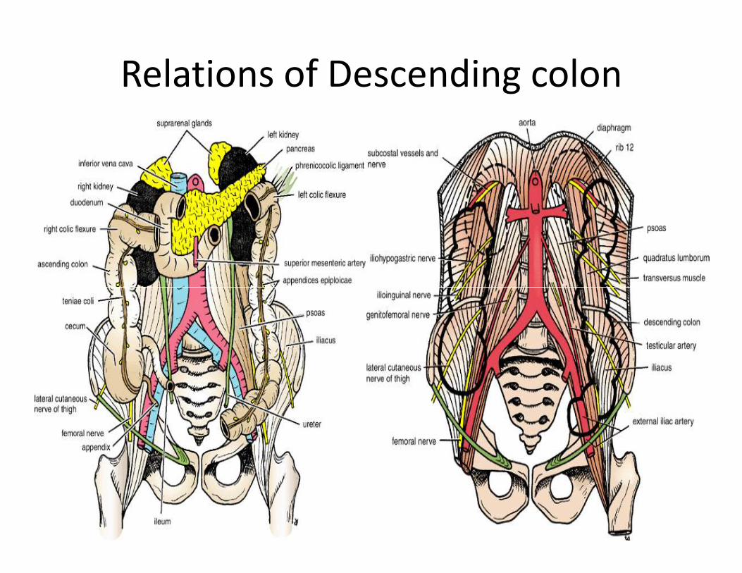
Relations of Descending colon
• Anteriorly:
- Coils of small intestine
- the greater omentum
- the anterior abdominal wall
• Posteriorly:
- The lateral border of the left kidney
- the origin of the transversus abdominis muscleabdominis muscle
- the quadratus lumborum
- the iliac crest
- the iliacus
- the left psoas
- The iliohypogastric and the ilioinguinal nerves
- the lateral cutaneous nerve of the thigh
- the femoral nerve
Relations of Descending colon
Blood Supply of Descending colon
• Arteries
- The left colic and the sigmoid branches of the
inferior mesenteric artery.
• Veins• Veins
- The veins correspond to the arteries �drain
into the inferior mesenteric vein.

Lymphatic Drainage of descending colon
• Lymphatic drains �the colic lymphatic nodes
& the inferior mesenteric nodes around the
origin of the inferior mesenteric artery.
Nerve Supply of descending colon
• The nerve supply is the sympathetic and
parasympathetic pelvic
• Splanchnic nerves through the inferior
mesenteric plexusmesenteric plexus