laforin is required for the functional activation of malin in endoplasmic reticulum stress...
TRANSCRIPT

Laforin is required for the functional activation of malin inendoplasmic reticulum stress resistance in neuronal cellsLi Zeng1,2, Yin Wang1, Otto Baba3, Pan Zheng1,4, Yang Liu1 and Yan Liu1
1 Department of Surgery, University of Michigan Medical Center, Ann Arbor, MI, USA
2 Department of Neurology, Tongji Hospital, Huazhong University of Science and Technology, Wuhan, China
3 Department of Hard Tissue Engineering, Tokyo Medical and Dental University, Tokyo, Japan
4 Department of Pathology, University of Michigan Medical Center, Ann Arbor, MI, USA
Keywords
endoplasmic reticulum stress; laforin; malin;
neuronal cells; polyglucosan
Correspondence
Yan Liu, Section of General Surgery,
Department of Surgery, University of
Michigan Medical Center, Ann Arbor,
MI 48109, USA
Fax: +1 734 615 5307
Tel: +1 734 763 8010
E-mail: [email protected]
Y. Wang, Section of General Surgery,
Department of Surgery, University of
Michigan Medical Center, Ann Arbor, MI
48109, USA
Fax: +1 734 615 5307
Tel: +1 734 763 8010
E-mail: [email protected]
Note
Li Zeng and Yin Wang contributed equally
to this work
(Received 25 March 2012, revised 22 April
2012, accepted 8 May 2012)
doi:10.1111/j.1742-4658.2012.08627.x
Mutations in either EPM2A, the gene encoding a dual-specificity phospha-
tase named laforin, or NHLRC1, the gene encoding an E3 ubiquitin ligase
named malin, cause Lafora disease in humans. Lafora disease is a fatal
neurological disorder characterized by progressive myoclonus epilepsy,
severe neurological deterioration and accumulation of poorly branched gly-
cogen inclusions, called Lafora bodies or polyglucosan bodies, within the
cell cytoplasm. The molecular mechanism underlying the neuropathogenesis
of Lafora disease remains unknown. Here, we present data demonstrating
that in the cells expressing low levels of laforin protein, overexpressed
malin and its Lafora disease-causing missense mutants are stably polyubiq-
uitinated. Malin and malin mutants form ubiquitin-positive aggregates in
or around the nuclei of the cells in which they are expressed. Neither wild-
type malin nor its mutants elicit endoplasmic reticulum stress, although the
mutants exaggerate the response to endoplasmic reticulum stress. Overex-
pressed laforin impairs the polyubiquitination of malin while it recruits
malin to polyglucosan bodies. The recruitment and activities of laforin and
malin are both required for the polyglucosan body disruption. Consistently,
targeted deletion of laforin in brain cells from Epm2a knockout mice
increases polyubiquitinated proteins. Knockdown of Epm2a or Nhlrc1 in
neuronal Neuro2a cells shows that they cooperate to allow cells to resist
ER stress and apoptosis. These results reveal that a functional laforin–
malin complex plays a critical role in disrupting Lafora bodies and reliev-
ing ER stress, implying that a causative pathogenic mechanism underlies
their deficiency in Lafora disease.
Structured digital abstract
Malin physically interacts with Laforin and GS1 by pull down (View interaction)
GS1 and Laforin colocalize by fluorescence microscopy (View interaction)
Laforin physically interacts with Malin by anti tag coimmunoprecipitation (View Interaction: 1,
2, 3, 4, 5, 6, 7, 8, 9, 10, 11)
Abbreviations
ATF6, activating transcriptional factor 6; CHOP, proapoptotic C ⁄ EBP homologous protein; EGFP, enhanced green fluorescent protein;
EPM2A, epilepsy of progressive myoclonus type 2A; ER, endoplasmic reticulum; GS1, glycogen synthase 1; GSK3b, glycogen synthase
kinase 3b; HEK293, human embryonic kidney 293; KO, knockout; LD, Lafora disease; PGB, polyglucosan body; PTG, protein targeting to
glycogen; WT, wild-type.
FEBS Journal (2012) ª 2012 The Authors Journal compilation ª 2012 FEBS 1

Introduction
Laforin, encoded by the epilepsy of progressive myoc-
lonus type 2A gene (EPM2A), is highly expressed in
adult brain [1,2]. Loss-of-function mutations of
EPM2A in humans cause Lafora disease (LD), an
early-onset fatal epileptic neurodegenerative disorder
marked by the accumulation of abnormally branched
glycogen inclusions called Lafora bodies or polygluco-
san bodies (PGBs), in brain neurons [3–6]. Although
Lafora bodies are also distributed in glycogen metabo-
lism-active tissues, such as liver and muscle, LD is not
classified as a glycogen storage disease. Loss-of-function
mutations of NHL repeat-containing 1 (NHLRC1),
which encodes an E3 ubiquitin ligase named malin,
can also cause LD [7]. Interestingly, although mutation
of either gene can cause the development of LD, some
LD patients do not harbor either mutation [8].
Mice with targeted deletion of either Epm2a or Nhlrc1
do not recapitulate the early-onset lethal neurological
features of LD [9–11], which indicates that the
mutations themselves should be considered in the
quest to elucidate the molecular mechanism(s) of LD
development.
Laforin has two functional domains: a carbohy-
drate-binding domain and a dual-specificity phospha-
tase domain [3,12–14]. The carbohydrate-binding
domain is critical for the in vitro binding of laforin to
glycogen [13], the in vivo binding of laforin to poly-
glucosan [12,15] and the in vivo binding of laforin to
itself [16]. Malin also has two functional domains:
a RING finger E3 ubiquitin ligase domain and six
repeats of NHL that are defined by (and named after)
amino acid sequence homologies with NCL-1, HT2A
and LIN41 proteins [17]. Three missense mutations of
NHLRC1 in the RING domain have been reported to
impair malin’s E3 ligase activity, whereas two missense
mutations in the NHL repeats have been reported to
impair the association of malin with laforin [18,19].
Interrelationship studies of laforin and malin have
demonstrated that the combination of these proteins is
responsible for reducing glycogen content in neuronal
cells that ectopically express protein targeting to glyco-
gen (PTG), a glycogenesis activator that induces pro-
tein phosphatase 1 to dephosphorylate glycogen
synthase [20,21]. The combination of laforin and malin
has also been shown to degrade PTG [22,23], misfold-
ed proteins [24] and even laforin itself [18]; it can be
stabilized and activated by AMP-activated protein
kinase in hepatoma cells [25,26]. Laforin has been
shown to be an in vitro phosphatase of glycogen; it
removes phosphates from phosphate-labeled amylopec-
tin, isolated muscle glycogen and muscle glycogen syn-
thesized by muscle glycogen synthase (GS1) [15,27–29].
We and the Minassian laboratory have shown that
laforin dephosphorylates and inactivates glycogen syn-
thase kinase (GSK)3b at serine 9 in serum-starved,
growth factor-stimulated cells [19,30]. However, under
physiological conditions in Epm2a knockout (KO)
mice, increased serine 9 phosphorylation in GSK3bwas not observed in the soluble portion of the tissue
lysate [28,31]. This suggests that laforin dephosphory-
lates GSK3b at serine 9 in a context-dependent
manner.
We have also shown that laforin reduces and its
mutants exaggerate the neuronal cell response to ER
stress stimulation, and that laforin protects cells from
apoptosis induced by energy-deprivation stress [32,33].
Consistent with this, increased ER stress in both
Epm2a KO liver cells and an autopsy sample from an
LD patient has been revealed [34]. Besides preventing
these stresses, laforin with associated malin protect
cells from thermal stress by activation of heat-shock
factor 1, a transcriptional factor that activates heat-
shock genes [35].
Here, we present data demonstrating that laforin
recruits malin to PGBs, where it activates functional
malin for PGB disruption. Both the PGB-binding abil-
ity and the phosphatase activity of laforin are required
for the recruitment and activation of malin and the
disruption of PGBs. Likewise, both the laforin-binding
ability and the E3 ligase activity of malin are required
for the disruption of PGBs by the laforin–malin com-
plex. The functional assembly of the laforin–malin
complex alleviates ER stress and prevents the apop-
tosis of neuronal cells exposed to stress stimuli.
Results
Malin and its mutants aggregate and are
polyubiquitinated but do not contribute
to ER stress
To determine the relationship between malin and lafo-
rin, we generated malin mutations at the same sites
found in LD patients. These included three sites near
the RING finger E3 ubiquitin ligase domain and nine
sites in the first five of the six NHL repeats (Fig. 1A).
Similar to some mutants previously reported in trans-
formed monkey kidney fibroblast COS7 cells [36,37],
we found that overexpressed wild-type (WT) malin
and its mutants in mouse neuroblastoma Neuro2a
(N2A) cells and human embryonic kidney (HEK)293
cells were expressed as monomers as well as polymerized
Laforin–malin complex for ER stress resistance L. Zeng et al.
2 FEBS Journal (2012) ª 2012 The Authors Journal compilation ª 2012 FEBS
aggregates (Fig. 1B). No statistically significant differ-
ences in protein stability were seen between WT malin
and its mutants in these two cell types, although trans-
fection efficiency varied in separate experiments.
Immunocytochemistry revealed that in HEK293 cells,
Flag-tagged aggregates of malin or its mutants mostly
localized in the cytoplasm (Fig. 1C). Immunoprecipita-
tion of the Flag-tagged malin or its mutants demon-
strated that the aggregates were polyubiquitinated
(Fig. 1D). RING domain mutants (C26S, C68Y and
L87P) and NHL mutants (D146N, D245N and
Q308A) were polyubiquitinated to a lesser extent than
the WT protein, as quantified by densitometry
(Fig. 1D). Some aggregates observed in the ligase-inac-
tive RING mutant cells were polyubiquitin-negative
(Fig. 1E), indicating that the polyubiquitination of
aggregates is likely attributable to malin autoubiquiti-
nation. This is consistent with the characteristics of the
RING finger E3 ubiquitin ligase family [38] and with
studies showing that malin protein is autoubiquitinated
in vitro [18]. In support of this, our results showed that
the three E3 ligase-inactive RING finger mutants had
much lower levels of ubiquitination than did WT or
other malin mutants (Fig. 1D).
To test the effect of malin and its mutants on ER
stress, we transfected them into N2A cells and treated
the transfected cells with either thapsigargin or tunica-
mycin, two molecules that induce ER stress by reduc-
ing ER calcium pump activity and dysglycosylating
ER proteins, respectively [39,40]. We then measured
two common ER stress markers, ER chaperone
78-kDa glucose-regulated protein (GRP78) and the
transcription factor proapoptotic C ⁄EBP homologous
protein (CHOP). Our results show that, in the absence
of thapsigargin or tunicamycin, N2A cells expressing
WT malin or its mutants expressed little or no CHOP.
However, in the presence of either stressor, significant
levels of CHOP were induced (Fig. 2A,B). Induced
C26S C68Y D146N I198N D233A P264HRING NHL NHL NHL NHL NHL NHL
L87P
D245N
R253K E280K
Q302P
Q308AMalin
N C
Mal
inC
26S
C68
YL8
7PD
146N
I198
ND
233A
D24
5NR
253K
P26
4HE
280K
Q30
2P
Vect
or
Aggre.
S6K
Q30
8A
Malin-V5
Vec
tor
Mal
in
Mal
in
C26
S
C68
Y
L87P
D14
6N
I198
N
D23
3A
D24
5N
R25
3KP
264H
E28
0K
Q30
2P
Q30
8A
Anti-Flag
Poly-Ub
IgG
Aggre.
Poly-Ub/total Malin
Malin-Flag
Malin-FlagIgG
IP: IgG
1 .4 .3 .4 .6 1.0 .9 .6 .8 .9 .9.8 .6
Malin C26S L87P
D146N D233A D245N
Malin-Flag/Ub
L87P-Flag/Ub D233A-Flag/Ub
Ub
A
B C
ED
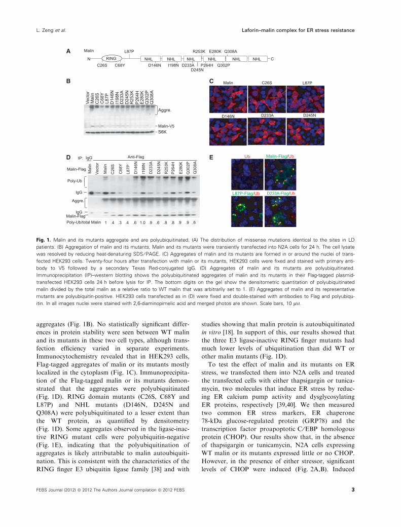
Fig. 1. Malin and its mutants aggregate and are polyubiquitinated. (A) The distribution of missense mutations identical to the sites in LD
patients. (B) Aggregation of malin and its mutants. Malin and its mutants were transiently transfected into N2A cells for 24 h. The cell lysate
was resolved by reducing heat-denaturing SDS ⁄ PAGE. (C) Aggregates of malin and its mutants are formed in or around the nuclei of trans-
fected HEK293 cells. Twenty-four hours after transfection with malin or its mutants, HEK293 cells were fixed and stained with primary anti-
body to V5 followed by a secondary Texas Red-conjugated IgG. (D) Aggregates of malin and its mutants are polyubiquitinated.
Immunoprecipitation (IP)–western blotting shows the polyubiquitinated aggregates of malin and its mutants in their Flag-tagged plasmid-
transfected HEK293 cells 24 h before lysis for IP. The bottom digits on the gel show the densitometric quantitation of polyubiquitinated
malin divided by the total malin as a relative ratio to WT malin that was arbitrarily set to 1. (E) Aggregates of malin and its representative
mutants are polyubiquitin-positive. HEK293 cells transfected as in (D) were fixed and double-stained with antibodies to Flag and polyubiqu-
itin. In all images nuclei were stained with 2,6-diaminopimelic acid and merged photos are shown. Scale bars, 10 lM.
L. Zeng et al. Laforin–malin complex for ER stress resistance
FEBS Journal (2012) ª 2012 The Authors Journal compilation ª 2012 FEBS 3
CHOP levels were higher in most of the cells transfect-
ed with malin mutants than in cells transfected by WT
malin. The inconsistent induction of GRP78 and
CHOP in some mutant transfected cells after exposure
to stressors indicates that the mutants elicited signs of
both early and late ER stress at different stages. Usu-
ally, GRP78 is an early-stage marker of ER stress,
whereas CHOP is a late stage marker. Regardless of
whether the cells were stressed, N2A cells transfected
with malin mutants showed a marked increase in
cleaved activating transcriptional factor 6 (ATF6), an
active form of ATF6 and another ER stress marker.
Neither WT nor mutant malin affected the levels
of phosphor-eIF2a, an early-stage marker of ER
stress, in transfected N2A cells exposed to stress
stimulation (Fig. 2C). It is interesting to note that
although overexpressed WT malin formed polyubiq-
uitinated aggregates in the transfected N2A cells
(Fig. 1C), cells transfected with malin showed a
decreased ER stress response to stimuli in compari-
son with empty vector transfected cells (Fig. 2B),
suggesting that WT malin plays a role in the pre-
vention of ER stress. Taken together, these results
showed that aggregated, polyubiquitinated WT malin
alone does not induce ER stress. By contrast, the
malin mutants themselves induce slight ER stress,
but exacerbate this response upon the addition of
exogenous ER stress inducers.
Malin mutants have impaired binding to laforin
We next characterized the ability of malin mutants to
bind to laforin; although WT malin is known as a
binding partner of laforin, the precise domains
required for binding of malin to laforin remain unde-
fined [18,19]. We constructed plasmids that expressed
different truncated versions of laforin and malin
(Fig. 3A,B). By analysis of immunoprecipitation data
and cotransfection of these plasmids into HEK293
cells, we were able to define the reciprocal binding
regions of laforin and malin. The region of laforin that
binds malin was near the carbohydrate-binding domain
end and included the entirety of exon 2; the region of
malin that binds laforin began near the RING domain
end and spanned the first five NHL repeats
(Fig. 3A,B). The critical region of malin that binds
laforin encompasses the end of the RING domain and
extends to the first NHL repeat. Because the glycogen
binding ability is maintained in exon-deleted mutants
of laforin, cellular glycogen might cause nonspecific
binding between the deleted forms of laforin and malin
(Fig. 3A). Consistent with what is known about the
region of malin that binds laforin, the RING domain
mutants C26S, C68Y and L87P did not differ in their
binding to laforin. By contrast, other NHL repeat
mutants demonstrated an impaired ability to bind to
laforin, with the exception of the two mutants D245N
Vector– + – + – + +–
Malin L87P D233AThapsigargin
Malin-V5
β-actin
p-eIF2d
c-ATF6
GRP78
CHOP
Malin-V5
β-actin
Mal
in
C26
S
C68
Y
L87P
D14
6N
I198
N
D23
3A
D24
5N
R25
3K
P26
4H
E28
0K
Q30
2P
Tunicamycin
Mal
in
Vec
tor
– + – + +– – + – + +– – + +– – –+ + –– + +–– –+ +
Thapsigargin
GRP78
CHOP
Malin-V5
β-actin
D23
3A
D24
5N
R25
3K
P26
4H
E28
0K
Q30
2P
C68
Y
L87P
Mal
in
D14
6N
I198
N
Mal
in
Vec
tor
– + – + – + – + – + – +– + – + – + – +– +– –+ +
A
B
C
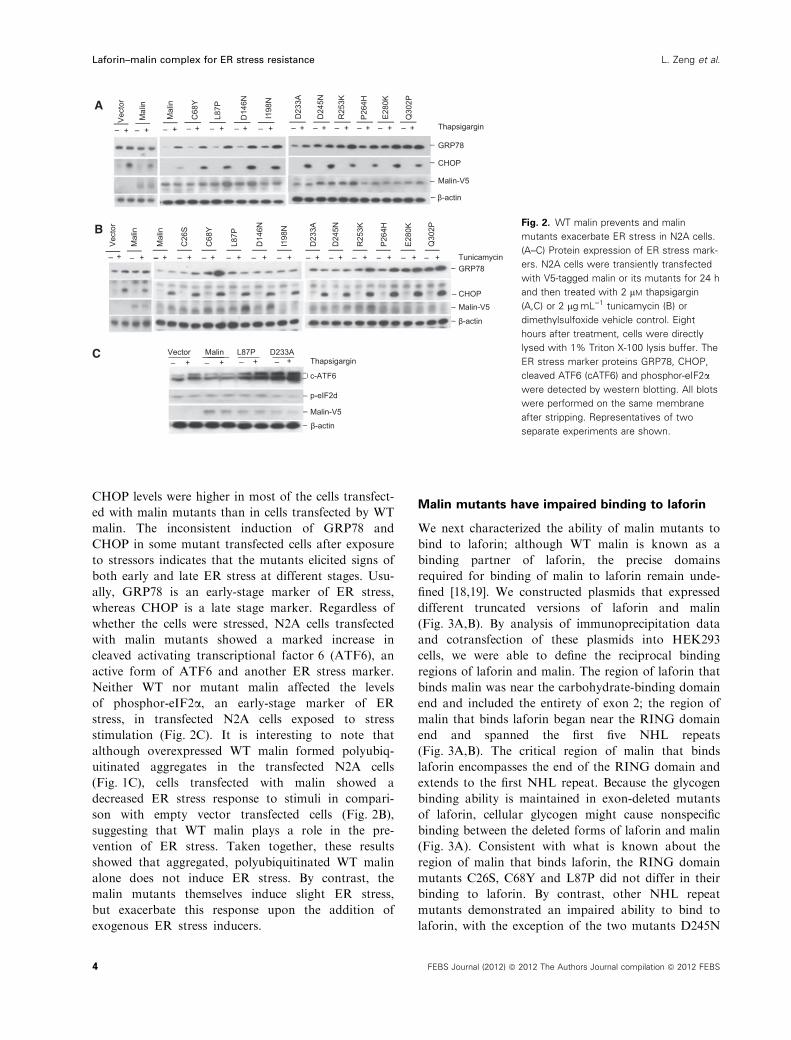
Fig. 2. WT malin prevents and malin
mutants exacerbate ER stress in N2A cells.
(A–C) Protein expression of ER stress mark-
ers. N2A cells were transiently transfected
with V5-tagged malin or its mutants for 24 h
and then treated with 2 lM thapsigargin
(A,C) or 2 lgÆmL)1 tunicamycin (B) or
dimethylsulfoxide vehicle control. Eight
hours after treatment, cells were directly
lysed with 1% Triton X-100 lysis buffer. The
ER stress marker proteins GRP78, CHOP,
cleaved ATF6 (cATF6) and phosphor-eIF2a
were detected by western blotting. All blots
were performed on the same membrane
after stripping. Representatives of two
separate experiments are shown.
Laforin–malin complex for ER stress resistance L. Zeng et al.
4 FEBS Journal (2012) ª 2012 The Authors Journal compilation ª 2012 FEBS

in and R253K next to the third NHL repeat (Fig. 3C).
These results demonstrated that most NHL repeat
mutants of malin were defective in their ability to bind
laforin.
Laforin is required for malin recruitment
to polyglucosan
Subsequent experiments were undertaken to reveal the
binding target that laforin–malin complex might work
on. Based on the finding demonstrating that transgenic
mice expressing GS1 under the control of skeletal mus-
cle-specific promoter display elevated polyglucosan lev-
els in the tissue [41], we constructed and transfected
GS1 into HEK293 cells and found that it synthesized
polyglucosan that was glycogen positive, periodic acid
Schiff positive and resistant to a-amylase hydrolysis,
as previously reported [41]. After cotransfecting or sin-
gly transfecting GS1 with laforin, laforin mutant
C265S, malin or malin mutants L87P or D233A into
HEK293 cells, we found that laforin and C265S, but
not WT malin or its mutants, bound GS1-synthesized
polyglucosan (Fig. 4A). In the triple combination indi-
cated in Fig. 4A, only WT laforin and WT malin
bound to and disrupted large PGBs into relatively
small granules. In cells containing these small granules,
GS1 was still distributed within the cytoplasm and
nucleus. This indicates that if the laforin–malin com-
plex degrades GS1 to limit polyglucosan formation,
this process takes place in PGBs only (Fig. 4A). Nei-
ther phosphatase-dead mutant C265S with WT malin
nor E3 ligase-inactive mutant L87P or laforin-binding-
deficient mutant D233A in combination with WT
laforin was able to disrupt the PGBs, demonstrating
that the disruption of PGBs requires the activities of
both laforin phosphatase and malin E3 ligase, as well
as appropriate recruitment of the laforin–malin com-
plex. To prove the requirement of laforin for malin in
Malin-Flag
Laforin-HA
Laforin-HA
∆E1p
Malin-Flag + + + + + +
Lafo
rin
∆E1
∆E2
∆E3
∆E2p
3p
Laforin-HA
IP: anti-HA
IgG
Malin-Flag
S6K
Malin aggre.
Lysate control
∆E2p3p
Laforin
N
∆E1
CBD DSP
∆E2∆E3
∆E1pC
+ + + + + + +M
alin
L126
RL1
26F
G15
8V
221
S29
8V
367
IP: anti-Myc
+
F88 Malin-Flag
Laforin-Myc
Malin-Flag
IgGLaforin-Myc
R 1 2 3 4 5 6
G158V221 S298
V367F88
L126RL126F
C
MalinNHL
N
Laforin-flag
Malin-V5
Laforin-Flag
Malin-V5
Mal
inC
26S
C68
YL8
7PD
146N
I198
ND
233A
D24
5NR
253K
P26
4HE
280K
Q30
2PQ
308A
Malin aggre.
IgG
Malin aggre.
Lysate control
Malin-V5GSK3
IP: anti-Flag
Total malin/malin input
1 .9 1.3 .9 .6 .5 .5 1.0 .9 .7 .6 .7 .7
Lysate control
Malin-Flag
S6K
A B C
Fig. 3. Most NHL repeat mutants of malin impair their binding to laforin. (A,B) The reciprocal binding regions of malin and laforin are defined.
HEK293 cells were transiently cotransfected with protein-expressing plasmids as indicated. Twenty-four hours after transfection, cells were
directly lysed by 1% Triton X-100 plus 0.02% SDS lysis buffer and supernatants were immunoprecipitated and then blotted with the indi-
cated antibodies. Areas critical for binding between malin and laforin are shown with bold lines. (C) Impaired binding ability of malin mutants
to laforin. HEK293 cells were transiently cotransfected with V5-tagged malin or malin mutants together with Flag-tagged laforin, and then
lysed directly with Triton X-100 plus 0.02% SDS buffer. The resultant supernatants were immunoprecipitated with Flag antibody and the
immunoprecipitates were subjected to western blotting. Densitometric quantitation of total malin divided by total malin input of the lysate is
represented as a relative ratio to WT malin that was arbitrarily set to 1.
L. Zeng et al. Laforin–malin complex for ER stress resistance
FEBS Journal (2012) ª 2012 The Authors Journal compilation ª 2012 FEBS 5

binding to PGBs, we combined purified laforin protein
and malin immunoprecipitate with the isolated GS1
polyglucosan in vitro and found that the malin immu-
noprecipitated the GS1 polyglucosan only in the pres-
ence of laforin (Fig. 4B, lane 2 compared with lane 4).
Also, by determining the glycogen content in HEK293
cells that possessed endogenous GS1 glycogen, we
found that only cells transfected with both malin and
laforin had a reduced glycogen content (Fig. 4C),
which is consistent with results observed in neuronal
cells that ectopically express PTG [20]. These results
demonstrate that GS1 polyglucosan is a target of the
laforin–malin complex, and that polyglucosan disrup-
tion requires the binding and activity of laforin and
malin. This conclusion is supported by previous results
showing that GS1, not PTG, accumulates in Lafora
bodies [42].
Laforin and malin functionally depend on each
other to prevent ER stress
Laforin recruits malin to PGBs, which both proteins
disrupt in combination. Disruption of PGBs either
provides glucose (energy) to cells to counteract ER
stress stimulation (which results in a decrease in cellu-
lar energy), or releases laforin and malin for recycling.
Based on this hypothesis, knockdown of either laforin
or malin in N2A cells could enhance cell sensitivity to
ER stress. As expected, knockdown of either laforin or
malin in N2A cells increased thapsigargin-induced
CHOP levels (Fig. 5A,B). Restoration of laforin did
not diminish the increased sensitivity of malin-silenced
N2A cells to CHOP induction by thapsigargin; like-
wise, restoration of malin did not diminish the
increased sensitivity of laforin-silenced N2A cells to
CHOP induction, even though they increased the resis-
tance of cells transfected with scrambled small hairpin
RNA to ER stress (Fig. 5B). To determine whether
both phosphatase and E3 ligase activities were required
to prevent ER stress-induced apoptosis, we transfected
malin or its mutants into N2A cells expressing either
laforin or C265S, and observed that only a combina-
tion of WT laforin and WT malin significantly
prevented an ER stress-induced increase in annexin
V-positive (apoptotic) cells (12.68% in WT laforin–
WT malin versus 23.23% in WT laforin–mutant
L87P). Other combinations could not prevent apopto-
sis (Fig. 5C). These results show that a functional lafo-
rin–malin complex is necessary for cellular resistance
to ER stress.
Laforin and ER stressor impair malin
autoubiquitination
The question of how autoubiquitinated malin becomes
functionally activated once it has bound laforin
beads + +– –Laforin – +– +
Malin – + – +GS1Gly + + + +
GS1Gly
IgGMalin
Laforin
GS1GlyInput
Gly
coge
n (μ
g/m
g P
r.)
Malin L87P D233AV
Malin L87P D233ALaforin C265S
Malin0
100
200
300
400
500
GS1/glycogen GS1 + Laforin
GS1 + Malin GS1+L87P GS1+ D233A
GS1-Malin-Laforin GS1-L87P-Laforin GS1-D233A-Laforin
GS1+ C265S+Malin
1 2 3 4
A B
C
Fig. 4. Laforin recruits malin and both desrupt polyglucosan. (A) Laforin is required for malin recruitment to GS1-synthesized polyglucosan.
HEK293 cells were transfected with a plasmid of GS1–Flag alone or in combination with the indicated plasmids of laforin–myc and malin–V5
or their mutants for 24 h. The transfected cells were fixed and double-stained with antibodies to glycogen or Flag, V5 or Myc tags. (B) Lafo-
rin is essential for malin binding to polyglucosan in vitro. Isolated GS1 polyglucosan was added to binding buffer containing malin–protein G
beads or empty beads in the presence or absence of purified laforin protein. After 2 h of binding at 4 �C, the washed beads were lysed with
1· SDS loading buffer for western blotting to detect GS1 polyglucosan. (C) Laforin and malin together decrease glycogen content in vivo.
HEK293 cells transfected with the indicated plasmids were directly lysed in NaAc buffer for glycogen determination. Results of glycogen
determination are presented as the mean ± SEM of three separate experiments. Scale bars, 10 lM.
Laforin–malin complex for ER stress resistance L. Zeng et al.
6 FEBS Journal (2012) ª 2012 The Authors Journal compilation ª 2012 FEBS

remained. To determine the mechanism by which this
occurs, we cotransfected laforin along with malin or
one of its mutants into HEK293 cells, and found that
in malin immunoprecipitate containing laforin, malin
became less ubiquitinated, whereas mutants of malin
did not (despite the presence or absence of laforin in
their immunoprecipitates) (Fig. 6A). To ascertain
whether an ER stressor activates malin by impairing
its autoubiquitination, we treated malin-transfected
N2A cells with tunicamycin or thapsigargin. Ubiquiti-
nation of malin was significantly induced by both
stressors (Fig. 6B). Detection of endogenous malin
activation by laforin or ER stress stimulation cannot
be performed because, at present, no convincing, com-
mercially available malin antibodies exist. However,
via immunoprecipitation using anti-polyubiquitin IgG
[43], we were able to detect significantly more poly-
ubiquitinated proteins in the brain cells of Epm2a KO
mice than in age-matched WT mice (Fig. 6C,D). This
indicates that laforin plays a role in preventing the
accumulation of polyubiquitinated proteins, which is
consistent previous work [44] showing increased poly-
ubiquitinated proteins in the lysate of laforin-deficient
human fibroblasts by western blotting. Taken together,
these data demonstrated that laforin is required not only
for the binding of malin to PGBs, but also for the acti-
vation of malin by preventing its autoubiquitination.
Discussion
Through a systematic analysis of malin, laforin and
their missense mutants, and using KO approaches, we
demonstrate for the first time the cooperation of lafo-
rin and malin that confers neuronal protection against
ER stress. Although the exact mechanism underlying
the prevention of ER stress by laforin–malin
complexes remains to be defined, we hypothesize that
polyglucosan disruption governed by laforin–malin
complexes not only provides endogenous energy in the
form of glucose, but also lowers the level of stress
within the cytoplasm, easing the ER burden. We are
the first to demonstrate that polyglucosan disruption
absolutely requires both laforin and malin. However,
these proteins alone are not sufficient. Thus, for dis-
ruption of PGBs, a consecutive process may be
required. First, laforin recruits malin and other
sh-L sh-M Sr sh-M Sr sh-L Sr– + – + – + – + – + – + – +
GRP78
CHOP
Malin-V5
Laforin-Flag
β-actin
Thap
1.27 1.51 1.27 1.70 1.71 1.93
–
V
Malin L87P
Ann
exin
V
18.45 21.46 12.68 23.23 26.27 28.68
Thap
DAPI
Laforin
Malin
C265S
Malin P78LP78L
β-actin
Laforin
Sr sh-L
β-actin
MalinSr sh-M
A B
C
Fig. 5. Laforin and malin cooperate in ER stress relief and apoptosis prevention. (A) Knockdown of laforin or malin in N2A cells. Silencer of
small hairpin RNA of laforin (sh-L) or malin (sh-M) was cotransfected with Flag-tagged laforin or malin into N2A cells for 24 h. Knockdown
efficiency was determined by western blotting using anti-Flag IgG. (B) Codependence of laforin and malin in the prevention of ER stress.
Scrambled sh (Sr), sh-L or sh-M cells of N2A were transiently transfected with vector, malin or laforin for 24 h and then treated with 2 lM
thapsigargin or vehicle for an additional 8 h. After treatment the cells were lysed for western blotting and probed for ER stress proteins. (C)
Activities of both laforin and malin are required for the inhibition of apoptosis induced by ER stressors. N2A cells expressing WT laforin or
its mutant C265S were transiently transfected with malin–EGFP or malin mutant L87P–EGFP. Twenty-four hours after transfection, cells
were divided into two test groups and treated with 1 lM thapsigargin or vehicle for 24 h in 2.5% fetal bovine serum Dulbecco’s modified
Eagle’s medium. Treated cells were stained with annexin V (for apoptotic cells) and 2,6-diaminopimelic acid (for dead cells). The annexin V in
the EGFP-positive population was analyzed by flow cytometry.
L. Zeng et al. Laforin–malin complex for ER stress resistance
FEBS Journal (2012) ª 2012 The Authors Journal compilation ª 2012 FEBS 7

proteins or enzymes to PGBs, where the functional
assembly of a laforin–malin complex disrupts PGBs
into relatively small, ‘normal’ glycogen granules that
can be degraded by conventional glycogen metabolic
enzymes. GS1 is the key enzyme involved in PGB for-
mation, and may be the first enzyme targeted by the
functional complex of laforin and malin. The binding
and recruitment of laforin and malin to GS1 polyg-
lucosan suggests that the laforin–malin complex
degrades GS1 and thus inhibits the ability of GS1 to
synthesize polyglucosan, a process that takes place in
an insoluble glycogen pool. Degradation of GS1 by
the complex has been hinted at by a decrease in GS1
protein levels in the lysate of N2A cells expressing
laforin, malin and PTG [20], and in the accumulation
of GS1 protein in Lafora bodies from Epm2a or
Nhlrc1 KO mice [10,42,45,46]. Disruption of PGBs by
the laforin–malin complex may subsequently prevent
laforin and malin from becoming trapped in PGBs.
The requirement for both laforin phosphatase activity
and malin E3 ligase activity in PGB disruption sug-
gests that the dephosphorylation and ubiquitination of
key components in PGBs may take place simulta-
neously. These processes occurring in the insoluble gly-
cogen pool may create a situation in which detecting
alterations in the target protein becomes difficult [45].
Stresses that result in intracellular energy decline,
such as energy deprivation and ER stress, may induce
PGB formation; activation of laforin–malin complexes
may subsequently disrupts PGBs, thereby supplying
energy for cell recovery from stress. Furthermore,
under homeostatic conditions, the laforin–malin com-
plex plays a critical role in the surveillance and preven-
tion of PGB formation; thus, deletion of either gene
causes PGB accumulation [9,45–47]. The laforin–malin
complex also prevents the formation of and disrupts
PTG-activated GS1-synthesized abnormal, but not
normal, glycogen [20,22,23], because laforin preferen-
tially binds to polyglucosan over normal glycogen [15].
We also predict that polyubiquitination of malin under
physiological conditions may be a form of self-inacti-
vation to control its activity when it is not needed.
Laforin is not a deubiquitinating enzyme, and there-
fore an unknown alternative may be recruited to PGBs
or perhaps earlier in the malin–laforin pathway to
counteract the cellular effects of ER stress. In addition,
increased protein levels of ER stress markers were not
observed in brain extracts from 9-month-old Epm2a
-- Tu ThTu Th
IP: Flag malin-Flag
IgGIgGmalin-Flag
malin-Flagaggre.
malin-FlagPoly-Ub
Mal
inL8
7PD
146N
D23
3AR
253K
Mal
inL8
7PD
146N
D23
3AR
253K
E28
0K
Vector Laforin-myc
Poly-Ub
IgG
aggre.
IgGLaforin-dimer
malin-FlagLaforin-monomer
IP: Flag
WB: Anti-Ub
Anti-Flag/Anti-myc
malin-Flag
Poly-Ub/total malin
E28
0K
1 .4 .6 .8 .8 .8 .4 .3 .5 .6 .6 1.0
IgG
poly-Ub
KO WT KOPoly-Ub
Rabbit anti-Poly-Ub
KO WT KOIP: IgG IgG Poly-Ub
Mouse anti-Ubiquitin
Epm2a
Pol
y-U
biqu
itin
rela
tive
to W
T
WT Epm2a KO
p < 0.05
0
1
2
3
4
5
Poly-Ub/total malin.28 .13 .03
A
C D
B
Fig. 6. Laforin and ER stressors promote malin deubiquitination. (A) Laforin favors the deubiquitination of malin. HEK293 cells cotransfected
with malin or its mutants together with vector or laforin were lysed 24 h after transfection to detect polyubiquitination of malin by IP–wes-
tern blotting using tag antibodies. (B) ER stress stimulation activates malin deubiquitination. N2A cells expressing malin–Flag were treated
with 2 lgÆmL)1 tunicamycin (Tu) or 1 lgÆmL)1 thapsigargin (Th). 8 h after treatment, the cells were lysed for anti-Flag IP and anti-polyubiqu-
itin blotting to detect malin ubiquitination. (C) Polyubiquitinated proteins accumulated in brain cells from Epm2a KO mice. Equal amounts of
Triton X-100-soluble proteins in the brain lysate of 6-month-old Epm2a KO and WT mice were immunoprecipitated with polyubiquitin Lys48-
linkage antibody. Ubiquitinated proteins were detected by mouse anti-ubiquitin IgG and rabbit anti-(Lys48-linkage polyubiquitin) IgG. (D)
Graphical representation of polyubiquitinated protein levels in the two strains.
Laforin–malin complex for ER stress resistance L. Zeng et al.
8 FEBS Journal (2012) ª 2012 The Authors Journal compilation ª 2012 FEBS

KO mice, probably because it is difficult to extract
brain proteins from tissues with massive PGB accumu-
lations [34].
Because PGBs are found in other neuronal disor-
ders, including Alzheimer’s disease [48] and temporal
lobe epilepsy [49], decreased functional laforin and ⁄ormalin might also contribute to the progression of these
diseases. Therefore, our study has clinical implications
across a broad range of neurological disorders.
Experimental procedures
Mice and cells
The Epm2a KO mice (from a 129Sv strain) [9] used in this
study have been backcrossed onto a C57BL ⁄ 6 background
for more than 10 generations. Experiments were performed
using WT and Epm2a KO mice that were littermates born
from homozygous breeding pairs. All mice were kept and
used according to the procedures approved by the Unit for
Laboratory Animal Medicine (ULAM) at the University of
Michigan.
HEK293 and Neuro2a cell lines were from Invitrogen
(Grand Island, NY, USA) and ATCC (Manassas, VA,
USA), respectively. The HEK293 cells were cultured in
Dulbecco’s modified Eagle’s medium supplemented with
4.5 g glucose, 2 mM glutamine, 2% penicillin and 10% fetal
bovine serum. The N2A cells were cultured in minimal
essential medium supplemented with 2 mM glutamine, 2%
penicillin and 10% fetal bovine serum.
Reagents and antibodies
Sources of rabbit polyclonal antibodies to specific proteins
are as follows: Flag (Sigma, St Louis, MO, USA), phos-
phor–Ser52–eIF2a (Cell Signaling (Danvers, MA, USA))
and polyubiquitin Lys48 linkage for immunoprecipitation
(clone Apu2; Millipore Billerica, MA, USA). The sources
of mouse mAbs to specific proteins are as follows: ubiquitin
for western blotting (P4D1; Santa Cruz, Santa Cruz, CA,
USA), polyubiquitin for immunocytochemistry (Ubi-1;
Thermo Scientific, Waltham, MA, USA), ATF6 (IMGE-
NEX, San Diego, CA, USA), V5 and Myc tags (Invitro-
gen), Flag tag (M2, Sigma), Flag-Cy3 (Sigma), GRP78 ⁄Bip(BD Transduction Lab, Franklin, NJ, USA), S6K (H-9,
Santa Cruz), b-actin (Sigma) and CHOP ⁄ GADD153
(MA1–250, Thermo Scientific). The flow cytometry anti-
body to hycocyanin APC–annexin V used in the present
study was from BD Pharmingen (San Diego, CA, USA).
Monoclonal anti-glycogen IgM was from O. Baba (Tokyo
Medical and Dental University, Tokyo, Japan). Thapsigar-
gin and tunicamycin, enzymes and standard for amyloglu-
cosidase (A7420), amylase (A6814) and glycogen (G0885),
and glucose (GO) assay kit were all purchased from Sigma.
Plasmids and RT-PCR
The coding regions of cDNAs of human malin and mouse
GS1 (Gys1) were amplified from reverse transcription
mRNA of human cord blood cells and C57BL ⁄ 6J bone
marrow cells, respectively, and cloned into a pcDNA vector
with Myc, V5 or Flag tag at the N-terminus (malin) or
C-terminus (GS1). After sequencing confirmation of the
WT malin, all point mutants were made by site-directed
mutagenesis using WT malin as a template. Truncated
forms of malin were generated by PCR using full-length
malin as a template. Plasmids of laforin and its mutants
have been described previously [16]. Gene-silenced
sequences were: Epm2a, 5-TTCCAGACTGAATGGGAT
A-3, and Nhlrc1, 5-ATTCTCTTCTTGTGCTGGA-3.
These were cloned into small hairpin RNA lentiviral vec-
tors with enhanced green fluorescent protein (EGFP) as a
reporter. The lenti–sh vector was made by substituting the
CMV prompter of plenti6–TOPO ⁄V5 (Invitrogen) with the
U6–siRNA–PGK–EGFP cassette. Malin and its mutants
were cloned into a lenti–EGFP vector that was made by
substituting the T7-blasticidin cassette of plenti6–TOPO ⁄V5with the PGK–EGFP cassette.
Transfection, western blotting and
immunoprecipitation
In general, HEK293 and N2A cells were transiently trans-
fected with 0.25 lg plasmid and 0.75 lL Lipofecta-
mine 2000 (Invitrogen) per well on a 24-well plate for 24 h
in 0.5 mL Opti-MEM containing 10% fetal bovine serum.
To prevent cells from detaching when the culture medium
was changed to 0.5 mL Opti-MEM, one-third of the origi-
nal culture medium was not removed. Overnight-passaged
cells grown to � 75% confluence were used for transfec-
tion. In double or triple transfections, the total DNA plas-
mids did not exceed 0.375 lg (double) or 0.5 lg (triple) per
well in 24-well plates. Transfected cells were lysed with 1%
Triton X-100 lysis buffer containing 20 mM Tris ⁄HCl,
pH 7.4, 150 mM NaCl, 40 mM NaF, 1 mM dithiothreitol
and a protease and phosphatase inhibitor cocktail (Sigma).
Supernatants of the lysate were used for western blots and
resolved on a reducing and heat-denaturing 10%
SDS ⁄PAGE gel. Immunoprecipitation was carried out with
protein G beads at 4 �C, overnight, with rotation. The
supernatant was preincubated with protein G beads for 2 h,
and cleared supernatant was used for immunoprecipitation.
After washing three times with 1% Triton X-100 lysis buf-
fer, the immunoprecipitates were dissolved in SDS loading
buffer and resolved on a 10% SDS ⁄PAGE gel.
L. Zeng et al. Laforin–malin complex for ER stress resistance
FEBS Journal (2012) ª 2012 The Authors Journal compilation ª 2012 FEBS 9

PGB isolation and malin ubiquitination
To isolate PGBs from transfected cells, 0.55% NP-40 in
Hepes buffer containing 10 mM Hepes, 1 mM EDTA and
1 mM EGTA was used for lysing cells. Nuclei were removed
by centrifugation at 1000 g for 5 min and washed twice
with Hepes buffer. The combined supernatants were centri-
fuged at 8000 g for 15 min to remove debris and were then
centrifuged again at 18 000 g for 45–60 min to obtain pel-
lets containing insoluble PGBs. To detect malin ubiquitina-
tion and the binding of malin to laforin, the pellets were
digested with 1 UÆmL)1 amyloglucosidase and 5 UÆmL)1
amylase in NaCl ⁄Pi, pH 7.4, at 37 �C for 2 h and then
dissolved by 5· 1% Triton X-100 plus 0.02% SDS buffer.
The pellet-digesting solution in combination with cytosolic
supernatant was centrifuged and resultant supernatant was
used for immunoprecipitation in a final solution containing
1% Triton X-100, 0.5% NP-40 and 0.02% SDS.
Immunofluorescence staining
Cells were fixed in cold methanol for 10 min and then per-
meabilized with 0.3% Triton X-100 in 10 mM Tris ⁄HCl
buffer for 30 min. Immunofluorescence staining with the
primary antibody was performed overnight in 10 mM
Tris ⁄HCl buffer containing 2% BSA at 4 �C. Secondary
antibody staining was carried out in 2% BSA Tris ⁄HCl
buffer at room temperature for 2 h.
Acknowledgements
This work is supported by grants from the National
Institutes of Health, 1R21NS062391 and 1R21CA164469.
We are thankful to Dr Judith Connett and Dr Cailin
M. Wilke for their assistance in editing and revising the
grammar within the manuscript.
References
1 Ganesh S, Agarwala KL, Ueda K, Akagi T, Shoda K,
Usui T, Hashikawa T, Osada H, Delgado-Escueta AV
& Yamakawa K (2000) Laforin, defective in the pro-
gressive myoclonus epilepsy of Lafora type, is a dual-
specificity phosphatase associated with polyribosomes.
Hum Mol Genet 9, 2251–2261.
2 Ganesh S, Agarwala KL, Amano K, Suzuki T, Delgad-
o-Escueta AV & Yamakawa K (2001) Regional and
developmental expression of Epm2a gene and its evolu-
tionary conservation. Biochem Biophys Res Commun
283, 1046–1053.
3 Minassian BA, Lee JR, Herbrick JA, Huizenga J, Soder
S, Mungall AJ, Dunham I, Gardner R, Fong CY, Car-
penter S et al. (1998) Mutations in a gene encoding a
novel protein tyrosine phosphatase cause progressive
myoclonus epilepsy. Nat Genet 20, 171–174.
4 Gambetti P, Di Mauro S, Hirt L & Blume RP (1971)
Myoclonic epilepsy with lafora bodies. Some ultrastruc-
tural, histochemical, and biochemical aspects. Arch
Neurol 25, 483–493.
5 Sakai M, Austin J, Witmer F & Trueb L (1970) Studies
in myoclonus epilepsy (Lafora body form). II. Polyg-
lucosans in the systemic deposits of myoclonus epilepsy
and in corpora amylacea. Neurology 20, 160–176.
6 Cavanagh JB (1999) Corpora-amylacea and the family
of polyglucosan diseases. Brain Res Brain Res Rev 29,
265–295.
7 Chan EM, Young EJ, Ianzano L, Munteanu I, Zhao X,
Christopoulos CC, Avanzini G, Elia M, Ackerley CA,
Jovic NJ et al. (2003) Mutations in NHLRC1 cause
progressive myoclonus epilepsy. Nat Genet 35, 125–127.
8 Chan EM, Omer S, Ahmed M, Bridges LR, Bennett C,
Scherer SW & Minassian BA (2004) Progressive myoc-
lonus epilepsy with polyglucosans (Lafora disease):
evidence for a third locus. Neurology 63, 565–567.
9 Ganesh S, Delgado-Escueta AV, Sakamoto T, Avila
MR, Machado-Salas J, Hoshii Y, Akagi T, Gomi H,
Suzuki T, Amano K et al. (2002) Targeted disruption of
the Epm2a gene causes formation of Lafora inclusion
bodies, neurodegeneration, ataxia, myoclonus epilepsy
and impaired behavioral response in mice. Hum Mol
Genet 11, 1251–1262.
10 Valles-Ortega J, Duran J, Garcia-Rocha M, Bosch C,
Saez I, Pujadas L, Serafin A, Canas X, Soriano E,
Delgado-Garcia JM et al. (2011) Neurodegeneration
and functional impairments associated with glycogen
synthase accumulation in a mouse model of Lafora
disease. EMBO Mol Med 3, 667–681.
11 Depaoli-Roach AA, Segvich DM, Meyer CM, Rahimi
Y, Worby CA, Gentry MS & Roach PJ (2012)
Laforin and malin knockout mice have normal
glucose disposal and insulin sensitivity. Hum Mol
Genet 21, 1604–1610.
12 Ganesh S, Tsurutani N, Suzuki T, Hoshii Y, Ishihara
T, Delgado-Escueta AV & Yamakawa K (2004) The
carbohydrate-binding domain of Lafora disease protein
targets Lafora polyglucosan bodies. Biochem Biophys
Res Commun 313, 1101–1109.
13 Wang J, Stuckey JA, Wishart MJ & Dixon JE (2002) A
unique carbohydrate binding domain targets the Lafora
disease phosphatase to glycogen. J Biol Chem 277,
2377–2380.
14 Ganesh S, Puri R, Singh S, Mittal S & Dubey D (2006)
Recent advances in the molecular basis of Lafora’s pro-
gressive myoclonus epilepsy. J Hum Genet 51, 1–8.
15 Chan EM, Ackerley CA, Lohi H, Ianzano L, Cortez
MA, Shannon P, Scherer SW & Minassian BA (2004)
Laforin preferentially binds the neurotoxic starch-like
Laforin–malin complex for ER stress resistance L. Zeng et al.
10 FEBS Journal (2012) ª 2012 The Authors Journal compilation ª 2012 FEBS

polyglucosans, which form in its absence in progressive
myoclonus epilepsy. Hum Mol Genet 13, 1117–1129.
16 Liu Y, Wang Y, Wu C, Liu Y & Zheng P (2006)
Dimerization of Laforin is required for its optimal
phosphatase activity, regulation of GSK3beta phos-
phorylation, and Wnt signaling. J Biol Chem 281,
34768–34774.
17 Slack FJ & Ruvkun G (1998) A novel repeat domain
that is often associated with RING finger and B-box
motifs. Trends Biochem Sci 23, 474–475.
18 Gentry MS, Worby CA & Dixon JE (2005) Insights
into Lafora disease: malin is an E3 ubiquitin ligase that
ubiquitinates and promotes the degradation of laforin.
Proc Natl Acad Sci USA 102, 8501–8506.
19 Lohi H, Ianzano L, Zhao XC, Chan EM, Turnbull J,
Scherer SW, Ackerley CA & Minassian BA (2005)
Novel glycogen synthase kinase 3 and ubiquitination
pathways in progressive myoclonus epilepsy. Hum Mol
Genet 14, 2727–2736.
20 Vilchez D, Ros S, Cifuentes D, Pujadas L, Valles J,
Garcia-Fojeda B, Criado-Garcia O, Fernandez-Sanchez
E, Medrano-Fernandez I, Dominguez J et al. (2007)
Mechanism suppressing glycogen synthesis in neurons
and its demise in progressive myoclonus epilepsy. Nat
Neurosci 10, 1407–1413.
21 Printen JA, Brady MJ & Saltiel AR (1997) PTG, a
protein phosphatase 1-binding protein with a role in
glycogen metabolism. Science 275, 1475–1478.
22 Vernia S, Solaz-Fuster MC, Gimeno-Alcaniz JV, Rubio
T, Garcia-Haro L, Foretz M, de Cordoba SR & Sanz P
(2009) AMP-activated protein kinase phosphorylates
R5 ⁄PTG, the glycogen targeting subunit of the
R5 ⁄PTG-protein phosphatase 1 holoenzyme, and accel-
erates its down-regulation by the laforin-malin complex.
J Biol Chem 284, 8247–8255.
23 Worby CA, Gentry MS & Dixon JE (2008) Malin
decreases glycogen accumulation by promoting the deg-
radation of protein targeting to glycogen (PTG). J Biol
Chem 283, 4069–4076.
24 Garyali P, Siwach P, Singh PK, Puri R, Mittal S, Seng-
upta S, Parihar R & Ganesh S (2009) The malin-laforin
complex suppresses the cellular toxicity of misfolded
proteins by promoting their degradation through the
ubiquitin–proteasome system. Hum Mol Genet 18,
688–700.
25 Moreno D, Towler MC, Hardie DG, Knecht E & Sanz
P (2010) The laforin–malin complex, involved in Lafora
disease, promotes the incorporation of K63-linked
ubiquitin chains into AMP-activated protein kinase beta
subunits. Mol Biol Cell 21, 2578–2588.
26 Solaz-Fuster MC, Gimeno-Alcaniz JV, Ros S, Fernan-
dez-Sanchez ME, Garcia-Fojeda B, Criado Garcia O,
Vilchez D, Dominguez J, Garcia-Rocha M, Sanchez-
Piris M et al. (2008) Regulation of glycogen synthesis
by the laforin–malin complex is modulated by the
AMP-activated protein kinase pathway. Hum Mol
Genet 17, 667–678.
27 Worby CA, Gentry MS & Dixon JE (2006) Laforin,
a dual specificity phosphatase that dephosphorylates
complex carbohydrates. J Biol Chem 281, 30412–
30418.
28 Tagliabracci VS, Turnbull J, Wang W, Girard JM,
Zhao X, Skurat AV, Delgado-Escueta AV, Minassian
BA, Depaoli-Roach AA & Roach PJ (2007) Laforin is
a glycogen phosphatase, deficiency of which leads to
elevated phosphorylation of glycogen in vivo. Proc Natl
Acad Sci USA 104, 19262–19266.
29 Tagliabracci VS, Heiss C, Karthik C, Contreras CJ,
Glushka J, Ishihara M, Azadi P, Hurley TD, DePaoli-
Roach AA & Roach PJ (2011) Phosphate incorporation
during glycogen synthesis and Lafora disease. Cell
Metab 13, 274–282.
30 Wang Y, Liu Y, Wu C, Zhang H, Zheng X, Zheng Z,
Geiger TL, Nuovo GJ, Liu Y & Zheng P (2006) Epm2a
suppresses tumor growth in an immunocompromised
host by inhibiting Wnt signaling. Cancer Cell 10, 179–
190.
31 Puri R, Suzuki T, Yamakawa K & Ganesh S (2009)
Hyperphosphorylation and aggregation of Tau in lafo-
rin-deficient mice, an animal model for Lafora disease.
J Biol Chem 284, 22657–22663.
32 Liu Y, Wang Y, Wu C, Liu Y & Zheng P (2009) Dele-
tions and missense mutations of EPM2A exacerbate
unfolded protein response and apoptosis of neuronal
cells induced by endoplasm reticulum stress. Hum Mol
Genet 18, 2622–2631.
33 Wang Y, Liu Y, Wu C, McNally B, Liu Y & Zheng
P (2008) Laforin confers cancer resistance to energy
deprivation-induced apoptosis. Cancer Res 68, 4039–
4044.
34 Vernia S, Rubio T, Heredia M, Rodriguez de Cordoba
S & Sanz P (2009) Increased endoplasmic reticulum
stress and decreased proteasomal function in lafora dis-
ease models lacking the phosphatase laforin. PLoS
ONE 4, e5907.
35 Sengupta S, Badhwar I, Upadhyay M, Singh S &
Ganesh S (2011) Malin and laforin are essential
components of a protein complex that protects
cells from thermal stress. J Cell Sci 124, 2277–
2286.
36 Mittal S, Dubey D, Yamakawa K & Ganesh S (2007)
Lafora disease proteins malin and laforin are recruited
to aggresomes in response to proteasomal impairment.
Hum Mol Genet 16, 753–762.
37 Rao SN, Maity R, Sharma J, Dey P, Shankar SK, Sat-
ishchandra P & Jana NR (2010) Sequestration of chap-
erones and proteasome into Lafora bodies and
proteasomal dysfunction induced by Lafora disease-
associated mutations of malin. Hum Mol Genet 19,
4726–4734.
L. Zeng et al. Laforin–malin complex for ER stress resistance
FEBS Journal (2012) ª 2012 The Authors Journal compilation ª 2012 FEBS 11

38 Pickart CM (2001) Mechanisms underlying ubiquitina-
tion. Annu Rev Biochem 70, 503–533.
39 Treiman M, Caspersen C & Christensen SB (1998)
A tool coming of age: thapsigargin as an inhibitor of
sarco-endoplasmic reticulum Ca(2+)-ATPases. Trends
Pharmacol Sci 19, 131–135.
40 Helenius A (1994) How N-linked oligosaccharides affect
glycoprotein folding in the endoplasmic reticulum. Mol
Biol Cell 5, 253–265.
41 Pederson BA, Csitkovits AG, Simon R, Schroeder JM,
Wang W, Skurat AV & Roach PJ (2003) Overexpres-
sion of glycogen synthase in mouse muscle results in
less branched glycogen. Biochem Biophys Res Commun
305, 826–830.
42 Tagliabracci VS, Girard JM, Segvich D, Meyer C,
Turnbull J, Zhao X, Minassian BA, Depaoli-Roach AA
& Roach PJ (2008) Abnormal metabolism of glycogen
phosphate as a cause for Lafora disease. J Biol Chem
283, 33816–33825.
43 Newton K, Matsumoto ML, Wertz IE, Kirkpatrick DS,
Lill JR, Tan J, Dugger D, Gordon N, Sidhu SS, Fel-
louse FA et al. (2008) Ubiquitin chain editing revealed
by polyubiquitin linkage-specific antibodies. Cell 134,
668–678.
44 Aguado C, Sarkar S, Korolchuk VI, Criado O, Vernia
S, Boya P, Sanz P, de Cordoba SR, Knecht E & Ru-
binsztein DC (2010) Laforin, the most common protein
mutated in Lafora disease, regulates autophagy. Hum
Mol Genet 19, 2867–2876.
45 DePaoli-Roach AA, Tagliabracci VS, Segvich
DM, Meyer CM, Irimia JM & Roach PJ (2010) Genetic
depletion of the malin E3 ubiquitin ligase in mice leads
to Lafora bodies and the accumulation of insoluble
laforin. J Biol Chem 285, 25372–25381.
46 Turnbull J, Wang P, Girard JM, Ruggieri A, Wang TJ,
Draginov AG, Kameka AP, Pencea N, Zhao X,
Ackerley CA et al. (2010) Glycogen hyperphosphoryla-
tion underlies lafora body formation. Ann Neurol 68,
925–933.
47 Valles-Ortega J, Duran J, Garcia-Rocha M, Bosch C,
Saez I, Pujadas L, Serafin A, Canas X, Soriano E,
Delgado-Garcia JM et al. (2011) Neurodegeneration
and functional impairments associated with glycogen
synthase accumulation in a mouse model of Lafora
disease. EMBO Mol Med.
48 Inoue M, Yagishita S, Itoh Y, Amano N & Matsushita
M (1996) Coexistence of paired helical filaments and
polyglucosan bodies in the same neuron in an autopsy
case of Alzheimer’s disease. Acta Neuropathol 92,
511–514.
49 Abubakr A, Wambacq I, Donahue JE & Zappulla R
(2005) The presence of polyglucosan bodies in temporal
lobe epilepsy: its role and significance. J Clin Neurosci
12, 911–914.
Laforin–malin complex for ER stress resistance L. Zeng et al.
12 FEBS Journal (2012) ª 2012 The Authors Journal compilation ª 2012 FEBS