lactate in the brain: from metabolic end-product to …the ‘bad reputation’ of lactate as a...
TRANSCRIPT
When studied at the organ level, brain energy metabolism can be considered as being almost fully oxidative: elegant studies pioneered in the 1940s and 1950s by Schmitt and Kety1 and later by Sokoloff 2 showed that glucose is the obligatory physiological energy substrate of the brain and is metabolized to CO2 and water to yield 32–36 ATP molecules per molecule of oxidized glucose3. Studies over the past 20 years have added a cellular resolution to these organ studies, demonstrating a cellspecific metabolism of glucose. Given the cellular heterogeneity of the brain, it is not surprising that different cell types have distinct metabolic profiles. In particular, the emerging view is that neurons are mostly oxidative, whereas glial cells — notably, astrocytes and oligodendrocytes — predominantly process glucose glycolytically, meaning that they produce lactate and pyruvate from glucose4,5 (FIG. 1). Estimates of glucose uptake from the circulation indicate that, at the most, neurons take up an amount approximately equal to that taken up by astrocytes under basal conditions6,7.
During functional activation, most of the increase in glucose uptake occurs in astrocytes7–9. As neurons use 80–90% of the total energy consumed by the brain10, and as oxidative activity is the most efficient ATPproducing pathway (FIG. 1a), energy substrates such as pyruvate and lactate must be transferred from astrocytes to neurons, particularly during functional activation. This consideration is fully compatible with the original organlevel studies, because the sequential twostep glucose processing (that is, the transient glycolysis in astrocytes followed by oxidation in neurons) results in brain glucose oxidation.
Notably, under physiological conditions, the lactate:pyruvate concentration ratio is at least 10:1 (REF. 11). Thus, when considering the intercellular transfer of a glycolytic substrate, lactate is the predominant substrate in the brain. This ratio can also massively increase under hypoxia, as decreases in the partial pressure of oxygen (pO2) impair the oxidative capacity of the brain. Indeed, owing to this association with hypoxia, lactate has long been considered a metabolic endproduct, at best a marker of pathology, if not a toxic molecule (BOX 1). However, many recent studies in various tissues and cell types under physiological and pathological conditions12–14 now call for lactate to also be considered as an important signalling molecule. By acting through different molecular effectors, lactate contributes to several homeostatic processes (FIG. 2). This Review focuses on the emerging roles of lactate in brain function, with a particular focus on the modulation of neuronal excitability, neuronal plasticity and neuroprotection.
Lactate production and metabolismLactate is formed through glycolysis, one of the various metabolic pathways through which glucose can be processed to produce energy (FIG. 1a). With normal oxygen tension, ATP is mostly produced through the mitochondrial electron transport chain. Glucose is processed to pyruvate, which under the action of the enzyme pyruvate dehydrogenase (PDH) provides carbon atoms for the tricarboxylic acid (TCA) cycle. TCAcycle products, such as NADH and FADH2, feed the electron transport chain, resulting in the production of 32–36 ATP molecules. By contrast, under hypoxic or
1King Abdullah University of Science and Technology (KAUST), Thuwal, Kingdom of Saudi Arabia.2Brain Mind Institute, École Polytechnique Fédérale de Lausanne (EPFL), Lausanne, Switzerland.3Département de Psychiatrie, UNIL-CHUV, Site de Cery, Prilly Lausanne, Switzerland.
*e-mail: [email protected]
doi:10.1038/nrn.2018.19Published online 8 Mar 2018
Pyruvate dehydrogenase(PDH). The first component enzyme of the pyruvate dehydrogenase complex; it converts pyruvate into acetyl-CoA, which enters the tricarboxylic acid (TCA) cycle for cellular respiration.
Lactate in the brain: from metabolic end-product to signalling moleculePierre J. Magistretti1,2,3* and Igor Allaman2
Abstract | Lactate in the brain has long been associated with ischaemia; however, more recent evidence shows that it can be found there under physiological conditions. In the brain, lactate is formed predominantly in astrocytes from glucose or glycogen in response to neuronal activity signals. Thus, neurons and astrocytes show tight metabolic coupling. Lactate is transferred from astrocytes to neurons to match the neuronal energetic needs, and to provide signals that modulate neuronal functions, including excitability, plasticity and memory consolidation. In addition, lactate affects several homeostatic functions. Overall, lactate ensures adequate energy supply, modulates neuronal excitability levels and regulates adaptive functions in order to set the ‘homeostatic tone’ of the nervous system.
R E V I E W S
NATURE REVIEWS | NEUROSCIENCE VOLUME 19 | APRIL 2018 | 235
© 2018
Macmillan
Publishers
Limited,
part
of
Springer
Nature.
All
rights
reserved.
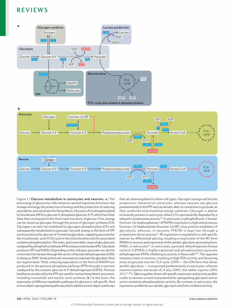
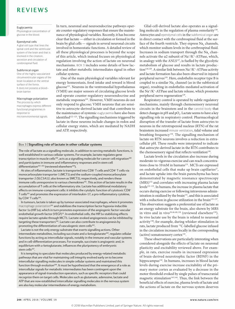
Figure 1 | Glucose metabolism in astrocytes and neurons. a | The processing of glucose by cells subserves several important functions: the storage of energy, the production of energy and the production of reducing equivalents and substrates for biosynthesis. Glucose is first phosphorylated by hexokinase (HEX) to glucose‑6‑phosphate (glucose‑6‑P), which has three fates that correspond to the three main functions of glucose. First, energy can be stored as glycogen through the action of glycogen synthase (GS). Glycogen can later be mobilized by glycogen phosphorylase (GP) and subsequently metabolized to pyruvate. Second, energy in the form of ATP can be produced by glucose‑6‑P entering glycolysis, supplying pyruvate for the tricarboxylic acid (TCA) cycle in the mitochondria and the associated oxidative phosphorylation. The main, and irreversible, steps of glycolysis are catalysed by phosphofructokinase (PFK) and pyruvate kinase (PK). Glycolysis produces ATP and NADH. Depending on the cell type, pyruvate can also be converted into lactate through the action of lactate dehydrogenase (LDH); in doing so, NAD+ levels (which are necessary to maintain the glycolytic flux) are regenerated. Third, reducing equivalents in the form of NADPH are produced in the pentose phosphate pathway (PPP) through a reaction catalysed by the enzyme glucose‑6‑P dehydrogenase (G6PD). Pentose backbones produced by the PPP are used for various biosynthetic processes, including nucleotide and nucleic‑acid synthesis. b | In the brain, the expression of different metabolic pathways for glucose is cell specific. Red arrows depict upregulated pathways; black dashed arrows depict pathways
that are downregulated in these cell types. Glycogen storage and lactate production characterize astrocytes, whereas neurons use glucose predominantly in the PPP and use lactate, after its conversion to pyruvate, as their preferred mitochondrial energy substrate. Glycogen is almost exclusively present in astrocytes, where GS is permanently degraded by a ubiquitin–proteasome process195. In astrocytes, 6‑phosphofructo‑2‑kinase/fructose‑2,6‑bisphosphatase 3 (PFKFB3) expression is high and produces fructose‑2,6‑bisphosphate (fructose‑2,6‑BP; a key positive modulator of glycolysis), whereas, in neurons, PFKFB3 is kept low through a proteasome‑driven process32. PK expression is regulated in a cell‑specific manner by differential splicing, resulting in expression of the M1 form (PKM1) in neurons and expression of the aerobic glycolysis‑promoting form, PKM2, in astrocytes34. In astrocytes, pyruvate dehydrogenase kinase isoform 4 (PDK4) is highly expressed and phosphorylates pyruvate dehydrogenase (PDH), inhibiting its activity in these cells20,34. The opposite situation exists in neurons, resulting in high PDH activity and favouring entry of pyruvate into the TCA cycle. LDH5 — the LDH form that drives aerobic glycolysis — is expressed predominantly in astrocytes, whereas neurons express low levels of, if any, LDH5, but rather express LDH1 (REFS 25,208). Taken together, these cell‑specific expression and activity profiles confer to neurons a restricted potential for upregulating glycolysis and an active oxidative phosphorylation activity. By contrast, in astrocytes, the expression profiles favour aerobic glycolysis and limit oxidative activity.
Nature Reviews | Neuroscience
Glycogen
Glycogen synthesis
Mitochondria
Lactate production
GS GP
Lactate
NAD+
NADH
NAD+ NADH
G6PD
ATP
PKPFKHEX
ADP
Glucose Glucose-6-P Pyruvate
H+
Glycolysis
LDH
NADPH
MoleculesPPP
TCA cycle and oxidative phosphorylation
TCA cycle
CO2
ATPADP
a
b
Astrocyte
Neuron
Glucose Glucose-6-P Acetyl-CoAFructose-6-P
Fructose-2,6-BP
Phosphoenolpyruvate
Glycogen
Glycogen
Pyruvate TCA cycle
Glucose Glucose-6-P Acetyl-CoAFructose-6-P Phosphoenolpyruvate Pyruvate TCA cycle
ATPADP
Lactate
PFKFB3
PKM2
PKM1
LDH5 LDH1
ATPADP
Lactate
LDH1
PDH
PDK4
ATP ADP
P PDH
R E V I E W S
236 | APRIL 2018 | VOLUME 19 www.nature.com/nrn
© 2018
Macmillan
Publishers
Limited,
part
of
Springer
Nature.
All
rights
reserved. ©
2018
Macmillan
Publishers
Limited,
part
of
Springer
Nature.
All
rights
reserved.

Glycolytic fluxThe rate at which glucose and its metabolites proceed through the glycolytic pathway.
Flux analysisA technique used to examine production and consumption rates of metabolites. It determines the transfer of moieties containing isotopic tracers from one metabolite into another using stoichiometric models of metabolism and mass spectrometry methods.
anoxic conditions, only 2 ATP molecules are produced per glucose molecule. Under such conditions, pyruvate is converted to lactate (and NADH to NAD+) by lactate dehydrogenase (LDH) in a process known as anaerobic glycolysis (FIG. 1a). The regeneration of NAD+ is necessary to maintain a glycolytic flux, as NAD+ enables reduction during the initial steps of glycolysis.
A third way to process glucose is aerobic glycolysis, which was initially described in cancer cells by Otto Warburg. In aerobic glycolysis, lactate is formed despite the presence of normal oxygen tension15. This metabolic processing of glucose is typical of astrocytes and is due to a cellspecific gene expression profile that favours the conversion of pyruvate to lactate rather than the use of pyruvate in the TCA cycle (FIG. 1).
Cell-specific metabolism of lactateCellspecific metabolic profiles exist in different organs and tissues. For example, in skeletal muscle, fast ‘white’ fibres are predominantly glycolytic, whereas slow ‘red’ fibres are oxidative16. Early studies in individually
isolated neurons and astrocytes revealed that neurons produce CO2 at a much higher rate than do astrocytes, indicating high oxidative activity in neurons, whereas the enzyme function profile of astrocytes is consistent with a predominance of glycolysis17,18. More extensive recent studies have confirmed these neuronspecific and astrocytespecific metabolic profiles (reviewed elsewhere5). Not surprisingly, such cellspecific functional metabolic profiles are mirrored by the differential expression of enzymes that regulate metabolic fluxes (BOX 2; FIG. 1b).
Why do astrocytes produce lactate? A question that can be raised is: why do astrocytes, which are endowed with a considerable mitochondrial complement19, produce lactate rather than using pyruvate to feed the TCA cycle and the energetically favourable respiratory chain to achieve a high yield of ATP? One reason may be that, in astrocytes, the enzyme PDH is highly phosphorylated and therefore shows lower activity, favouring the aerobic glycolytic profile20. Another characteristic of astrocytes
Box 1 | A brief history of lactate in the brain
The ‘bad reputation’ of lactate as a useless, if not toxic, metabolite dates back to the early 20th century from studies in muscle (reviewed elsewhere179). These studies concluded that lactate is formed by exercising muscles through anaerobic glycolysis (that is, when the glycolytic flux exceeds the availability of oxygen). In the 1930s, elegant studies by the Coris showed that some excess lactate could be recycled by the liver and produce glucose through gluconeogenesis (Cori cycle).
Interestingly, at that time, evidence already showed that, under physiological conditions, excess lactate could in fact be oxidized in certain organs, including the heart and brain, to produce pyruvate and could thus enter the tricarboxylic acid (TCA) cycle. Quite surprisingly, this fate of lactate was simply considered as a way to clear this unwanted or toxic metabolite rather than as a process through which lactate could produce energy. Only recently did it become clear that intercellular lactate shuttles exist whereby lactate is transferred from lactate-producing cells to lactate-consuming cells. Examples of such shuttles are those existing between fast and slow skeletal muscle fibres and, as explained in the main text, between astrocytes and neurons5,16.
Studies by McIlwain in the 1950s demonstrated that lactate is a perfectly adequate energy substrate for neural cells: synaptic activity could be sustained in slices of human and monkey cerebral cortex maintained in vitro in artificial media in which glucose was replaced by lactate180. These observations were later confirmed in isolated dorsal root ganglia181, hippocampal slices182 and isolated optic nerve preparations183. Critics of these studies claimed that their relevance for a role of lactate in the brain was limited, as these results were obtained in the absence of glucose, suggesting that, at best, lactate could be considered as an alternative energy substrate to glucose in the case of glucose deprivation. However, a closer look at the literature shows that even in the presence of adequate glucose, lactate is a preferred substrate to sustain neuronal activity. These observations were made in vitro by monitoring the consumption by preparations that contained radioactively labelled glucose and lactate184,185, as well as through analysis of metabolic fluxes by magnetic resonance spectroscopy (MRS)46,138,186,187. The preferential use of lactate has also been shown in vivo using MRS and 18F-fluorodeoxyglucose (18F-FDG) positron emission tomography (PET)131,133,136. In the latter studies, brain glucose utilization was decreased in individuals during intravenous perfusions of lactate, indicating that lactate could be preferentially used instead of glucose as an energy substrate133,136. In summary, studies over six decades by different investigators using various experimental approaches have established that lactate, particularly if formed within the brain, where its concentrations range between 2 and 5 mM (REFS 188–190), can be an energy substrate for neurons and may even be their preferred substrate.
The preference for lactate as an energy substrate in the brain is particularly interesting when considered in the light of recent studies using 13C-labelled metabolic substrates followed by flux analysis with MRS, which have revealed that glycolysis and the TCA cycle are uncoupled in peripheral tissues. Specifically, glucose is first processed though glycolysis, thus generating a pool of lactate in the circulation, and then this lactate is oxidized via the TCA cycle by most tissues, except muscle and brain191. In the brain, uncoupling of glucose processing occurs between astrocytes and neurons instead. Indeed, the transport of glucose across the blood–brain barrier occurs at more than tenfold the rate of lactate transport into the brain192. Thus, brain energetics do not depend on the uncoupling of glycolysis and the TCA cycle in the periphery; rather, the brain modulates, in an activity-dependent manner, its own supply of lactate, through the transient processing of glucose by aerobic glycolysis and glycogenolysis in astrocytes. Overall, this uncoupling between glycolysis and the TCA cycle adds considerable energetic flexibility both to peripheral tissues, which are supplied with circulating lactate, and to the brain, where lactate is formed by astrocytes.
R E V I E W S
NATURE REVIEWS | NEUROSCIENCE VOLUME 19 | APRIL 2018 | 237
© 2018
Macmillan
Publishers
Limited,
part
of
Springer
Nature.
All
rights
reserved. ©
2018
Macmillan
Publishers
Limited,
part
of
Springer
Nature.
All
rights
reserved.

Mitochondrial respiratory chain (MRC) complexesComplexes that operate the transfer of electrons from donors to acceptors via redox mechanisms, creating an electrochemical proton gradient that drives the synthesis of ATP.
is the particular organization of their mitochondrial respiratory chain (MRC) complexes. The organization of different MRC complexes (which are numbered I–IV) into macromolecular structures known as supercomplexes dictates the MRC electron flux and energyproducing efficiency. In astrocytes, most complex I is uncoupled from supercomplexes, resulting in poor mitochondrial respiration21. By contrast, in neurons, complex I is mostly embedded into supercomplexes, resulting in high mitochondrial respiration21. A striking illustration of the modest mitochondrial respiration in astrocytes was
recently provided: mice in which cytochrome oxidase activity — and hence, mitochondrial respiration — was conditionally suppressed specifically in astrocytes for more than a year were phenotypically normal and did not present any signs of neurodegeneration22.
The astrocyte–neuron lactate shuttle. The existence of such drastically different metabolic profiles between astrocytes and neurons suggests that dynamically regulated metabolic exchanges exist between lactate producing cells and lactateconsuming cells.
Neuron Astrocyte
Glutamate
Ca2+
CO2
NMDAR
• ARC• EGR1• BDNF
• MCTs• NADH/NAD+
• NMDARs• ATP• K
ATP channels
• Lactate receptors• pH changes
• Neuronal plasticity• Axonal integrity• Neuronal excitability
• Learning and memory• Glucose sensing• Sodium sensing• Osmoregulation• pO
2 sensing
• Ventilation• Arousal
Pyruvate
LactateLactate Lactate
NAD+
NADH
+
–
ATP
AC
AC
MCT2
KATP
HCAR1
Unknown receptor
a
b
MCT4
MCT1
Cationchannel
Pannexin
Glycogen
Glycogenolysis
Various receptors
• NA• VIP• ADO
• Glutamate• NH
4
• NO• K+
Glucose
Glycolysis
Molecular interactions Cellular mechanisms Physiological processes
Glucose transporter
LDH1
Nature Reviews | Neuroscience
K+
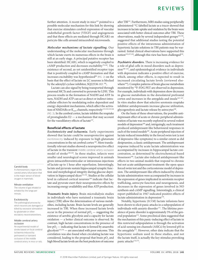
Figure 2 | Lactate-mediated metabolic coupling and signalling between neurons and astrocytes. a | Lactate is formed by astrocytes through two pathways: glycogenolysis and glycolysis. Both processes are triggered by activity‑dependent neuronal signals: noradrenaline (NA), vasoactive intestinal peptide (VIP), adenosine (ADO) and K+ promote glycogenolysis, whereas glucose uptake and lactate production (aerobic glycolysis) are triggered by glutamate, ammonium (NH4), nitric oxide (NO) and K+ (REFS 24,30,41,43,81,88–90,92). Lactate is released by astrocytes through three means: via transmembrane monocarboxylate transporters (MCT1 and MCT4), via a high‑capacity cation channel and via pannexins27,42,82. Lactate exerts metabolic effects and activates signalling cascades in neurons through two main mechanisms: through transmembrane transport via MCT2 and by acting on specific receptors. A G protein-coupled receptor called hydrocarboxylic acid receptor 1 (HCAR1) has been identified that binds lactate and is negatively coupled to adenylyl cyclase (AC)79. A second, unidentified receptor type, positively coupled to AC, is present on noradrenergic neurons in the locus coeruleus80. The processing of lactate as a consequence of its
uptake into the neuronal cytoplasm through MCT2 transporters results in the production of NADH and ATP. The conversion of lactate to pyruvate by lactate dehydrogenase 1 (LDH1) produces NADH, which affects the redox state of the neuron. The increase in NADH positively modulates the activity of NMDA receptors (NMDARs), resulting in enhanced Ca2+ currents, the activation of intracellular signalling cascades and the induction of the expression of plasticity‑ associated genes — for example, those encoding activity‑regulated cytoskeleton‑associated protein (ARC), early growth response protein 1 (EGR1) and brain‑derived neurotrophic factor (BDNF)113. Pyruvate formed from lactate enters the mitochondria and fuels ATP production. ATP supports the energy demands of active neurons and provides a signal that modulates the activity of ATP‑dependent potassium (KATP) channels, resulting in depolarization122,123,146. The possibility that pyruvate may act directly to activate KATP channels has been suggested62. b | The physiological processes involving lactate in the nervous system and the cellular mechanisms and molecular interactors through which lactate exerts these physiological effects (see text for details). pO2, partial pressure of oxygen.
R E V I E W S
238 | APRIL 2018 | VOLUME 19 www.nature.com/nrn
© 2018
Macmillan
Publishers
Limited,
part
of
Springer
Nature.
All
rights
reserved. ©
2018
Macmillan
Publishers
Limited,
part
of
Springer
Nature.
All
rights
reserved.

Complex IThe first step in the mitrochondrial respiratory chain; it removes two electrons from NADH and operates their transfer to ubiquinone.
TransaccelerationA property of monocarboxylate transporters whereby the presence of extracellular monocarboxylates (such as pyruvate) stimulates transporter efflux of the substrate (for example, lactate).
Warburg effectAlso called aerobic glycolysis. The metabolic pathway of glucose that results in the production of lactate in the presence of physiological concentrations of oxygen.
This is clearly the case in skeletal muscle, where lactate is shuttled between white and red fibres12,13. Intracellular lactate has also been described to shuttle between the cytosol and mitochondria16,23.
More than two decades ago, studies showed that glutamate stimulated aerobic glycolysis in primary astrocyte cultures and triggered lactate release24. This effect was mediated by sodiumdriven glutamate reuptake, a wellcharacterized function of astrocytes. Ex vivo analys es of protein expression and localization suggested that lactate is produced in astrocytes and transferred to neurons25,26. Specifically, neurons were found to express lactate dehydrogenase 1 (LDH1; the form present in lactateconsuming tissues such as the myocardium), whereas LDH5 (the form predominantly expressed in lactateproducing tissues such as skeletal muscle) is exclusively present in astrocytes25. Furthermore, the monocarboxylate transporters (MCTs) that operate transmembrane lactate transport showed a cellspecific distribution, with the lowaffinity MCT1 and MCT4 present in astrocytes and the highaffinity MCT2 present in neurons27. This cellspecific distribution is not sufficient alone to argue for a predominant traffic of lactate from astrocytes to neurons, as the interconversion of lactate and pyruvate is driven by the redox state of cells. Astrocytes present a much higher NADH:NAD+ ratio than do neurons28, indicating that astrocytes have a highly reducing status, which thermodynamically favours the conversion of pyruvate to lactate in these cells.
Overall, these observations gave rise to the astrocyte– neuron lactate shuttle (ANLS) model, whereby synaptically released glutamate triggers glucose uptake and lactate production by astrocytes for the use of neurons24,25 (FIG. 2). Over the following two decades, experiments from several laboratories have supported the ANLS model7,9,22,24,28–51 (for reviews, see REFS 4,5,52–54).
In further support of the ANLS model, the existence of a lactate gradient between astrocytes and neurons was recently demonstrated in vivo44. Twophoton microscopy of genetically encoded nanosensors was used to visualize changes in lactate levels. This approach revealed that, upon simultaneous transacceleration55 of MCTs in both astrocytes and neurons, by adding exogenous pyruvate (see REFS 56,57 for details on transacceleration), lactate decreases in astrocytes and then, after a delay, increases in neurons. Furthermore, glutamate reuptake into astrocytes triggers activitydependent glucose import from the circulation into the brain parenchyma both ex vivo and in vivo8,9,58,59. Electrophysiological evidence also indicates that lactate released by astrocytes and taken up by neurons is necessary to sustain neuronal activity60–62 (see FIG. 3 for example).
Challenges to the ANLS. Until the ANLS was described, the unanimous view was that glucose was transferred directly from capillaries to neurons as their exclusive energy substrate. The ANLS model introduced: a mechanism for the coupling between neuronal activity and energy delivery; the first evidence that astrocytes take up
Box 2 | Cell-specific expression of genes for glucose metabolism in the brain
As demonstrated by the transcriptional profiling of astrocytes acutely isolated using fluorescence-activated cell sorting (FACS), these cells are characterized by high expression of glycolytic genes, such as the gene encoding 6-phosphofructo-2- kinase/fructose-2,6-bisphosphate 3 (PFKFB3), a key positive regulator of glycolysis34. Furthermore, the M2 form of pyruvate kinase (PKM2) is also highly expressed in astrocytes, as in cancer cells, where aerobic glycolysis predominates193. In fact, PKM2 is necessary to regulate glycogen fluxes to feed glycolysis in cells such as cancer cells and astrocytes that are metabolically defined as sites of the Warburg effect193.
Furthermore, neurons express low levels of PFKFB3, implying a low glycolytic activity, and the M1 form of pyruvate kinase predominates in neurons, indicative of oxidative activity19,34 (FIG. 1b). Another distinguishing metabolic difference is the relatively low activity of pyruvate dehydrogenase (PDH) in astrocytes compared with neurons194, implying a lower flux of pyruvate into the tricarboxylic acid (TCA) cycle and a corresponding prevalence of lactate production in astrocytes (FIG. 1b). This low activity of PDH in astrocytes is due to its high degree of phosphorylation, which renders the enzyme less active20,194. Consistent with this observation, transcriptomic analysis indicates a high expression in astrocytes of pyruvate dehydrogenase kinase isoform 4 (PDK4), which phosphorylates PDH20,34. The reverse profile for both PDH and PDK4 is observed in neurons, consistent with a high flux of pyruvate into the TCA cycle and its associated oxidative phosphorylation in these cells20,34 (FIG. 1b).
In addition, glycogen, the stored form of glucose, is contained almost exclusively in astrocytes3. Interestingly, neurons express the gene encoding glycogen synthase, a key enzyme in glycogen metabolism; however, the expressed glycogen synthase protein is continuously degraded by an ubiquitin–proteasome process, thus preventing glycogen accumulation in neurons195. Manipulations or pathological conditions in which glycogen accumulates in neurons lead to neuronal demise195,196. For example, Lafora body disease, a form of juvenile myoclonal epilepsy, is associated with a toxic accumulation of glycogen in neurons197,198.
Interestingly, studies in cultured neurons and astrocytes showed that PFKFB3 undergoes a similar proteasome-driven degradation process in neurons, further highlighting the inability of neurons to upregulate glycolysis. In fact, glucose taken up by neurons is mainly processed through the pentose phosphate pathway (PPP) to generate reducing equivalents in the form of NADPH, which are used for scavenging reactive oxygen species produced by the intense oxidative activity of neurons32 (FIG. 1). In testimony to the importance of glucose processing through the PPP rather than glycolysis in neurons, inhibition of the proteasome-mediated degradation of PFKFB3 results specifically in neuronal death by oxidative stress32. Indeed, this intervention reduces the flux of glucose through the PPP, thus decreasing the production of reducing equivalents such as NADPH54.
R E V I E W S
NATURE REVIEWS | NEUROSCIENCE VOLUME 19 | APRIL 2018 | 239
© 2018
Macmillan
Publishers
Limited,
part
of
Springer
Nature.
All
rights
reserved. ©
2018
Macmillan
Publishers
Limited,
part
of
Springer
Nature.
All
rights
reserved.

Fast glucose transportFacilitated transmembrane transport of glucose via specific transporters.
glucose and use it to produce lactate via aerobic glycolysis (reviewed elsewhere5,52,53); and support for the idea that lactate could adequately fuel neurons, as was already suggested by Henry McIlwain in the 1950s (BOX 1).
Nevertheless, as is often the case for new proposals in science, the ANLS model was met with a degree of scepticism by some, and over the years has been challenged, mostly on theoretical grounds63–65. Most resistance came from the interpretation that the ANLS implied that all circulating glucose enters exclusively into
astrocytes and operates at all synapses; however, neither of these interpretations is correct. The original article describing the ANLS24 shows that glucose also enters neurons. Moreover, it is unlikely that ANLS operates at all neurons; indeed, ANLS probably does not operate at GABAergic synapses. GABA reuptake into astrocytes does not promote aerobic glycolysis66, and GABAergic neurons may instead rely on their own glycolytic activity to sustain their energy needs67.
Nevertheless, a few challenges have arisen from experimental reports that warrant discussion. For example, two reports have claimed that glucose is taken up preferentially, if not exclusively, by neurons. The first showed that a fluorescent glucose analogue, IR 2deoxyglucose 800 (IRDye 800CW 2DG; which has a molecular mass of ~1,300 Da, compared with 180 Da for glucose), was preferentially taken up by activated neurons68. Limiting the interpretation of this study, however, IRDye 800CW 2DG has been shown to be endocytosed as a complex with the glucose transporter69 — a process that is unrelated to fast glucose transport. In the second report, the phosphorylation of infused fluorodeoxyglucose (FDG) was used as a marker of glucose utilization70. The ratio of FDG6phosphate (FDG6P) to Nacetylasparate (NAA; a neuronal marker) was compared between brain homogenate and a preparation of neuronal terminals from rats with or without injection of the epileptogenic agent bicuculline. The FDG6P:NAA ratio in the nerve terminal preparation was similar to that in the brain homogenate (which contained neurons and glia). As such, the study’s authors concluded that, during activation, neurons directly take up glucose, challenging the role of the ANLS. However, these observations would indicate that all glucose is taken up by neurons — a notion that is not supported experimentally6,7. Moreover, technical issues (such as the use of neuronal terminal preparations, which undergo membrane disruption and resealing followed by density gradient centrifugation during their preparation and thus are likely subject to changes in substrate concentrations) and the use of bicuculline, which does not reflect a physiological activation, make it difficult to draw firm conclusions from this study.
By contrast, the astrocytic reuptake of glutamate has been demonstrated to be required for glucose utilization (as predicted in the ANLS model) in the whisker–barrel cortex pathway in vivo. Downregulation of astrocyte specific glutamate transporters markedly reduced glucose utilization71 in the barrel corresponding to a mechanically stimulated whisker58,72. In the same system, and under basal conditions, glucose uptake (measured using the fluorescent, non metabolizable glucose analogue, 6deoxyN(7nitrobenz2 oxa 1,3 diazol4yl)aminoglucose (6NBDG), a probe validated as a marker of fast glucose transport)73–75 by astrocytes and neurons occurred at the same rate. By contrast, during whisker stimulation, glucose uptake by astrocytes, but not by neurons, in the barrel rapidly increased7. Another in vivo imaging study showed that the 18FFDG positron emission tomography (PET) signal is driven by astroglial glutamate transport9.
Nature Reviews | Neuroscience
a
b
Glucose-containing mediumAstrocyte
Neuron
LDH Lactate
Glucose-containing medium
LDH LactateOxamate
c
d
Lactate-containing medium
LDH Lactate
–60
–64
Neu
rona
l mem
bran
epo
tent
ial (
mV
) a
b
c
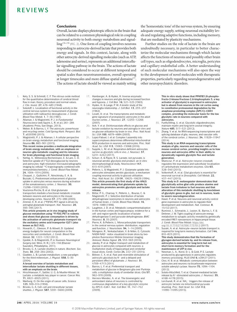
Figure 3 | Lactate transfer from astrocytes to neurons modulates the excitability of pyramidal cells. Double patch recordings from a pyramidal cell and a neighbouring astrocyte in the CA1 region of the hippocampus62. a | In a glucose-containing medium, lactate is formed and released by astrocytes. b | Inhibition of lactate dehydrogenase (LDH) activity in astrocytes by oxamate results in the hyperpolarization of the neighbouring pyramidal cell. c | In the presence of lactate in the medium, the same inhibition of LDH by oxamate does not affect the membrane potential of the pyramidal cell, as lactate is readily available. d | Approximate membrane potentials corresponding to the pyramidal cells shown in the other figure parts are schematized here.
R E V I E W S
240 | APRIL 2018 | VOLUME 19 www.nature.com/nrn
© 2018
Macmillan
Publishers
Limited,
part
of
Springer
Nature.
All
rights
reserved. ©
2018
Macmillan
Publishers
Limited,
part
of
Springer
Nature.
All
rights
reserved.
Energy chargeAn index that measures the energy status of cells. It is related to ATP, ADP and AMP concentrations.
Furthermore, in hippocampal slices, 6NBDG was taken up predominantly by astrocytes at a much higher rate than by neurons29. In primary mixed cultures, glutamate stimulated glucose uptake into astrocytes and, interestingly, also inhibited the transport of glucose into neurons by triggering an AMPA receptormediated influx of sodium into these cells76. This latter observation suggests that glutamate signalling may actually redistribute glucose towards astrocytes during activation. Together with the much higher cytosolic NADH:NAD+ ratio (indirectly indicating higher glycolytic flux) observed in astrocytes relative to neurons in hippocampal slices, these results converge towards a predominant glucose uptake into astrocytes28.
In another study in hippocampal slices, inhibitors of the neuronal lactate transporter MCT2, under basal conditions, lowered the neuronal NADH:NAD+ ratio, implying lactate consumption in these cells77. Yet, during activation of hippocampal granulecell synapses, the rise in the neuronal NADH:NAD+ ratio was insensitive to MCT2 inhibition — an observation that was taken to suggest that the active neurons did not require imported lactate, challenging the ANLS77. However, the NADH:NAD+ ratio in active neurons remained much lower than that in resting astrocytes28,77, which is inconsistent with the notion that neurons exhibit glycolysis more than do astrocytes.
Another indirect argument against the ANLS is a stoichiometric one based on the theoretical notion that the energy for sustaining glutamate reuptake by astrocytes does not require glucose uptake, as glutamate can enter the TCA cycle and thus fuel its own uptake64. However, this contention is not consistent with experimental evidence. First, as noted above, impairing astrocytic glutamate uptake reduces glucose uptake into the brain in vivo58,72. Second, daspartate, a substrate that is taken up by astrocytic glutamate transporters but is not metabolized in the TCA cycle, stimulates glucose uptake into astrocytes just as glutamate does24. Third, glutamate uptake into astrocytes markedly decreases the energy charge of these cells78. Thus, glutamate uptake into astrocytes and glucose utilization are tightly linked.
In summary, the ANLS does not discount glucose utilization by neurons (particularly under basal conditions) and probably does not operate equally at all synapses of the brain. Some lactate produced is probably not used by neurons and may instead be released into the circulation64. Moreover, given that lactate receptors have been identified in the brain79,80 (FIG. 2), lactate itself may in part act as a gliotransmitter on cognate receptors5. Finally, activated neurons might initially use and oxidize lactate from the extracellular space that is subsequently replenished through the ANLS5.
Extending the ANLS model. Several more recent studies have provided further evidence for the ANLS and have even extended the model. In addition to glutamate, certain other neuronderived agents promote glucose uptake, lactate production and lactate release by astrocytes (FIG. 2). Experiments using genetically encoded nanosensors demonstrated that K+ and NH4
+ ions, which increase in the extracellular space with neuronal activity, drive aerobic
glycolysis in astrocytes30,43. Physiological concentrations of nitric oxide (NO), another signal released by neurons, were also recently shown to promote lactate release from astrocytes81. Cortical astrocytes of anaesthetized mice were also observed to release lactate rapidly in response to local field stimulation42. Similarly, there may be further conduits for lactate release beyond MCT1 and MCT4. Neuronal depolarization or physiological increases in extracellular K+ induce rapid release of lactate through a lactate permeable, highcapacity ion channel42. Moreover, pannexin and connexin hemichannels can also extrude lactate82.
The ANLS seems to be evolutionarily conserved, as it has been demonstrated in vivo with cellular resolution in Drosophila melanogaster36. Indeed, inhibition of the ANLS by genetic knockout of selective glycolytic enzymes in glia results in neurodegeneration36, suggesting that the ANLS is necessary for proper neuronal function in flies. Related to the evolutionary conservation of the ANLS, a well coordinated transfer of lactate from astrocytes to neurons in D. melanogaster was recently shown to involve the formation of lipid droplets and to be important for neuroprotection. Briefly, the authors of the study showed that glial lactate fuelled neuronal lipogenesis when reactive oxygen species were formed in neurons owing to mitochondrial dysfunction. These neuronal lipids are subsequently transported and stored in glia as lipid droplets, and this neuronal lipogenesis and subsequent lipid storage in glia were found to be important for neuro protection in flies83,84. Also in line with evolutionary conservation of the ANLS, a compartmentation between glia and neurons that enables metabolite exchange was reported in the honey bee retina in the 1980s85.
The ANLS model has recently been extended to metabolic exchanges between oligodendrocytes and axons by in vivo studies showing that in mice, lactate released by oligodendrocytes is required to maintain axonal function33,86. Genetic disruption of lactate transfer from oligodendrocytes to axons resulted in axonal dysfunction. Furthermore, decreased expression of the gliaspecific lactate transporter MCT1 has been observed in the spinal cord of mice expressing SOD1G93A (a model of amyotrophic lateral sclerosis (ALS)) as well as in the motor cortices of individuals with ALS49,87.
Lactate production from astrocytic glycogen is another example of neuron–glia metabolic coupling. Neuronderived molecules such as noradrenaline (NA), vasoactive intestinal peptide (VIP) and adenosine, as well as activity dependent increases in extracellular K+, all promote glycogen mobilization in astrocytes41,88–92, where glycogen is exclusively localized within the brain3 (FIG. 2). As described below, transfer of glycogenderived lactate is critically involved in neuronal plasticity39,40,93.
Overall, work by several groups over almost three decades has provided strong evidence of neuron–glia metabolic coupling. The transfer of lactate from astrocytes to neurons is one example of a larger palette of metabolic transactions between astrocytes and neurons that also includes the exchange of ATP and serine94–97 as well as the release of agents that promote the reducing capacity of neurons37,98,99 (the discussion of which is outside the scope of this Review).
R E V I E W S
NATURE REVIEWS | NEUROSCIENCE VOLUME 19 | APRIL 2018 | 241
© 2018
Macmillan
Publishers
Limited,
part
of
Springer
Nature.
All
rights
reserved. ©
2018
Macmillan
Publishers
Limited,
part
of
Springer
Nature.
All
rights
reserved.

Memory consolidationA category of processes whereby a brain converts short-term memories into long-term memories (that is, stabilizing a memory trace after its initial acquisition).
Lactate as a signalling moleculeLactate, neuronal plasticity and memory. Studies initiated in the 1990s by Gibbs and Hertz100 showed that glycogenolysis was necessary for memory consolidation in a neonatal chick model of passive avoidance learning, which, in this species, involves the intermediate medial mesopallium (which corresponds to the cortex in mammals100,101). In addition to providing energy substrates for neurons, the breakdown of glycogen in astrocytes
was proposed to produce glutamine as a precursor for glutamate and GABA neurotransmission100. Given the evidence identifying astrocytes as lactate producers, a possible role of lactate transfer from astrocytes to neurons in memory consolidation was investigated. In the rat hippocampus, pharmacological inhibition of glycogen phosphorylase using DAB (1,4dideoxy 1,4iminodarabinitol) inhibited the production of lactate, as determined by microdialysis, during single trial learning tasks including inhibitory avoidance39. Blockade of lactate production from astrocytic glycogen also inhibited in vivo hippocampal longterm potentiation (LTP) and memory consolidation. By contrast, co injection of lactate with DAB (negating the inhibitory effects of DAB on lactate concentrations) rescued LTP and memory consolidation. Similarly, downregulation of the astrocytic MCTs (MCT1 and MCT4) using targeted antisense oligonucleotides prevented the transfer of lactate from astrocytes to neurons and resulted in amnesia that was rescued by lactate. Consistent with these findings, MCT1deficient mice exhibit impaired memory in the inhibitory avoidance task102. By contrast, downregulation of the neuronal lactate transporter MCT2 also prevented learning but was not rescued by lactate, as it could not be transported into neurons39. Thus, the transfer of lactate from astrocytes to neurons is necessary for LTP and memory consolidation (FIG. 4).
Lactate has similarly been shown to be necessary for memory consolidation in other learning tasks in rodents, including spatial working memory40 and conditioned place preference for cocaine (the latter behaviour involving the basolateral amygdala)103–105. Recently, astrocyte derived llactate was shown to facilitate synchrony of the amygdala with the anterior cingulate cortex and decision making106.
One of the physiological triggers for glycogenolysis in the brain is NA released by fibres originating in the locus coeruleus (LC)107,108. Activation of βadrenergic receptors by NA has a key role in the consolidation of inhibitory avoidance memory; this type of memory consolidation is inhibited by bilateral hippocampal injection of the βadrenergic antagonist propranolol109. Interestingly, the amnestic effect of propranolol is prevented by injecting lactate into the hippocampus93, implicating NAevoked glycogenolysis in memory consolidation. This implication is also consistent with the role of the LC–cortical noradrenergic projection in memory consolidation110,111. Using 3D immersive virtual reality constructed from electron microscopy stacks, more than 50% of the glycogen granules in astrocytic profiles were observed to localize near monoaminergic — probably noradrenergic — boutons, with the remaining 25% close to postsynaptic compartments. This intracellular distribution renders glycogen readily available to modulate synaptic functions112.
Why is glycogenderived lactate released by astrocytes necessary for neuronal plasticity and memory? A straightforward answer may be that lactate meets the increased energy demands of neurons under going plasticity processes such as cytoskeletal rearrangements, protein trafficking or increased gene expression.
Nature Reviews | Neuroscience
24 h• ↑ Latency to move into dark compartment• ↑ Expression of ARC, pCREBs and pCFL1
Memory intact
Short latency to move into dark compartment
Memory impaired
a
b
MCT1/4
Training Test
Training Test
–15 min 0 24 h
Intrahippocampalinjection of DAB
• ↑ Latency to move into dark compartment• ↑ Expression of ARC, pCREBs and pCFL1
Memory intact
Training Test
–15 min 0 24 h
Intrahippocampalinjection of DAB and lactate
NeuronAstrocyte
Glycogen
LactateGP
DAB
MCT2
↑ ARC↑ pCREBs↑ pCFL1Injected
lactate
Figure 4 | Lactate transfer from astrocytes to neurons in memory consolidation. a | One-trial learning for inhibitory avoidance. Rats are placed in a box with two compartments: one well illuminated and one dark. Rodents rapidly move into the dark compartment. On the day of training, rodents receive a mild electric shock as they enter the dark compartment. After 24 hours, the animals are retested; their latency to move into the dark compartment is considerably longer, and the expression of molecules involved in plasticity (such as activity‑regulated cytoskeleton‑associated protein (ARC), phosphorylated cAMP‑responsive element‑binding proteins (pCREBs) and phosphorylated cofilin 1 (pCFL1)) is increased, suggesting that the memory of the trained shock is intact. When 1,4‑dideoxy‑1,4‑imino‑d‑arabinitol (DAB), an inhibitor of glycogen phosphorylase (GP; the key enzyme involved in glycogenolysis), is injected bilaterally into the dorsal hippocampus 15 minutes before training, the latency to move into the dark compartment tested 24 hours later remains short, meaning that memory consolidation has been impaired39. If lactate is co‑injected with DAB, thus bypassing the inhibition of DAB, memory consolidation is rescued39. b | A schematic summarizing the mechanisms underlying the results observed in the one‑trial learning for inhibitory avoidance. The production of lactate in astrocytes can be blocked by DAB, and this impairs the formation of memories; however, injection of lactate directly into the hippocampus bypasses the need for lactate production and rescues memory formation. MCT, monocarboxylate transporter.
R E V I E W S
242 | APRIL 2018 | VOLUME 19 www.nature.com/nrn
© 2018
Macmillan
Publishers
Limited,
part
of
Springer
Nature.
All
rights
reserved. ©
2018
Macmillan
Publishers
Limited,
part
of
Springer
Nature.
All
rights
reserved.
Glycogen phosphorylaseThe enzyme that catalyses the rate-limiting step in glycogenolysis.
Inhibitory avoidanceA behavioural task that is commonly used to investigate learning and memory processes in rodents and that is based on contextual fear conditioning.
Nucleus solitary tractA major sensory nucleus in the dorsal medulla that receives cardiovascular, visceral, respiratory, gustatory and orotactile information.
However, the observation that glucose only marginally rescues the effect of blocking glycogenolysis with DAB on memory consolidation suggests that lactate has an energy independent function in this context39.
Consistent with an energyindependent action of lactate on neuronal plasticity, lactate, but not glucose or pyruvate, induces the expression of several plasticity related genes such Arc, Egr1 and Bdnf 113 in cultured mouse cortical neurons. Lactate exerts this action starting at concentrations as low as 2.5 and 5 mM by amplifying NMDA receptor (NMDAR)mediated currents and increasing intracellular calcium concentrations113. The molecular mechanism or mechanisms of this interaction between lactate and NMDARs are still unknown, but it relies on changes in neuronal redox state and the binding of glycine to its regulatory site on the NMDAR. For example, exposure of cultured neurons to NADH, which is formed by the intracellular conversion of lactate to pyruvate by LDH, mimics the effects of lactate on plasticity113 (FIG. 2). Conversely, the oxidizing agent DTNB (5,5´dithiobis(2nitrobenzoic acid)) antagonizes the potentiating effect of lactate on NMDAR signalling113.
Further supporting a role for lactate in plasticity, aerobic glycolysis is high throughout the human brain during early development (with a peak at around 5 years), when synaptic growth rate is prominent114. During adulthood, aerobic glycolysis becomes restricted to brain areas where expression of neotenic genes (that is, genes typically expressed during early development, such as those related to synapse formation and growth) persists, suggesting that aerobic glycolysis in the brain supports processes related to synaptic plasticity. Recently, a marked decrease in aerobic glycolysis was shown by brain PET imaging in humans during ageing115, consistent with a decrease in plasticity with ageing116.
Lactate and neuronal excitability. Experimental evidence suggests that lactate also strongly affects neuronal excitability62. In single neurons from the subthalamic nucleus, and in pyramidal cells in the CA1 hippocampal subregion, intracellular application of lactate depolarized the patched neurons. However, when oxamate, an inhibitor of LDH, was coapplied, neurons became hyperpolarized. By contrast, following application of pyruvate (a metabolite downstream of LDH; FIG. 1), oxamate had no effect. These results indicate that conversion of lactate to pyruvate is necessary for the depolarizing effect of lactate. Furthermore, double recordings from pyramidal cells and neighbouring astrocytes revealed that preventing lactate formation in astrocytes (by inhibiting LDH activity with oxamate) results in hyperpolarization of neighbouring pyramidal neurons, an effect that is rescued by extracellularly applied lactate, indicating that the hyperpolarization was due to a reduction in lactate release from astrocytes62. Therefore, lactate released by astrocytes modulates neuronal excitability (FIG. 3). Interestingly, these effects were not observed in inhibitory fastspiking cells in the hippocampus, supporting the view that the astrocytetoneuron transfer of lactate may operate predominantly, if not exclusively, at excitatory synapses.
Consistent with these observations, preventing the intercellular trafficking of lactate (using carbenoxolone, which blocks connexin 30 and connexin 43 gap junctions) across the astrocytic network in mouse hippocampal slices inhibited excitatory synaptic activity61. A recent study showed that the delivery of lactate from the astrocytic network to orexin neurons is necessary to maintain the daily cycles of wakefulness51, emphasizing the involvement of astrocyte–neuron metabolic coupling across the sleep–wake cycle117. Moreover, wholecell recordings showed that lactate transport into neurons of the rat nucleus solitary tract constitutively supports excitatory synaptic transmission, even in the presence of glucose118. The researchers concluded that “[...]‘onsite’ astrocyte–neuron lactate transport to presynaptic and postsynaptic elements is necessary for the integrity of excitatory synaptic transmission” (REF. 118).
Several other examples of the effects of lactate on the excitability of specific neuronal populations have been reported. One study showed that lactate is a necessary and sufficient energy substrate to maintain the spontaneous activity of orexin neurons in hypothalamic slices through a mechanism involving ATPdependent potassium (KATP) channels119. Similarly, LC noradrenergic neurons are excited by lactate80. Unlike what is observed in orexin neurons, this effect does not depend on transport of lactate into LC neurons; rather, it seems to depend on an asyet unidentified ‘lactate receptor’ that is positively coupled to cAMP formation80. This putative lactate receptor is distinct from another G proteincoupled receptor for lactate previously described in the nervous system: hydroxycarboxylic acid receptor 1 (HCAR1). However, HCAR1 is coupled to an inhibitory Gα subunit and therefore probably does not affect the excitatory action of lactate on LC neurons79. In fact, lactate reduces the excitability of cortical neurons in primary cultures (as monitored by calcium imaging), as demonstrated by a decrease in the basal spontaneous calcium spiking frequency, and pharmacological analysis has indicated that this action of lactate is mediated by HCAR1 (REF. 120).
In summary, the excitability of several populations of neurons is modulated by lactate, with most results indicating that lactate increases excitability. Different responses to lactate probably depend on the signalling pathways that the monocarboxylate activates as well as on the intrinsic properties of the target neurons. Further studies are necessary to draw a definitive picture of the acute effects of lactate on neuronal excitability.
The truly novel message of these studies revealing roles of lactate in neuronal plasticity and neuronal excitability is that lactate should no longer be considered simply as an energy substrate for neurons, but rather as an important signalling molecule that can regulate cellular processes independent of its metabolic effects.
Lactate and integrated physiological responses. The brain can be considered as the ultimate organ for homeostatic regulations. It monitors a large number of physiological functions of the body through subtle processes that rely on efficient sensing mechanisms in various brain regions, particularly the hypothalamus and the brainstem.
R E V I E W S
NATURE REVIEWS | NEUROSCIENCE VOLUME 19 | APRIL 2018 | 243
© 2018
Macmillan
Publishers
Limited,
part
of
Springer
Nature.
All
rights
reserved. ©
2018
Macmillan
Publishers
Limited,
part
of
Springer
Nature.
All
rights
reserved.
EuglycaemiaPhysiological concentration of glucose in the blood.
Ependymal cellsA glial cell type that lines the spinal cord and the ventricular system of the brain and that is involved in the creation, secretion and circulation of cerebrospinal fluid.
Subfornical organOne of the highly vascularized circumventricular organs of the brain localized on the ventral surface of the fornix. It does not possess a blood–brain barrier.
Macrophage polarizationThe process by which macrophages express different functional programmes in response to microenvironmental signals.
In turn, neuronal and neuroendocrine pathways operate counterregulatory responses that ensure the maintenance of physiological variables. Recently, it has become clear that lactate — either in circulation or formed in the brain by glial cells — signals in several neuronal circuits involved in homeostatic functions. A detailed review of all these physiological processes is beyond the scope of this article, which instead focuses on physiological regulation involving the action of lactate on neuronal mechanisms. BOX 3 includes some details of how lactate and other metabolic intermediates signal in other cellular systems.
One of the main physiological variables relevant for energy homeostasis, food intake and reward is blood glucose121. Neurons in the ventromedial hypothalamus (VMH) are major sensors of circulating glucose levels and operate counterregulatory neuroendocrine and metabolic responses122. However, VMH neurons do not only respond to glucose; VMH neurons that are sensitive to astrocytederived lactate and that contribute to the maintenance of systemic euglycaemia have also been identified122–125. The signalling mechanisms triggered by lactate in these neurons include changes in redox and cellular energy states, which are mediated by NADH and ATP, respectively.
Glialcellderived lactate also operates as a signalling molecule in the regulation of plasma osmolarity126. Astrocytes and ependymal cells in the subfornical organ are in direct contact with the cerebrospinal fluid at the surface of the fourth ventricle. They express Nax channels, which monitor sodium levels in the cerebrospinal fluid. Increases in sodium transport through the Nax channels activate the α2 subunit of Na+/K+ATPase, which, in analogy with the ANLS127, is fuelled by the glycolytic metabolism of glucose and results in lactate production126,128. A similar link between Na+/K+ATPase activity and lactate formation has also been observed in injured peripheral nerves129. Here, endothelin receptor type B is coupled to a similar Nax channel (as in the subfornical organ), resulting in endothelinmediated activation of the Na+/K+ATPase and lactate release, which promotes peripheral nerve regeneration129.
Respiratory control is operated by subtle regulatory mechanisms, mainly through chemosensory neuronal circuits in the brainstem and in the carotid body that detect changes in pH and pO2. Lactate seems to have a key signalling role in respiratory control. Pharmacological disruption of the transfer of lactate from astrocytes to neurons in the retrotrapezoid nucleus (RTN) of the rat brainstem increased minute ventilation, tidal volume and breathing frequency130. The signalling mechanism of lactate on RTN neurons involves a reduction in extracellular pH. These results were interpreted to indicate that astrocytederived lactate in the RTN contributes to the chemosensory signal that reflects ventilation130.
Lactate levels in the circulation also increase during moderatetovigorous exercise and can reach concentrations close to 10 mM in humans131. MCTs are expressed on endothelial cells that make up brain capillaries27, and lactate uptake into the brain parenchyma has been demonstrated by magnetic resonance spectroscopy (MRS)132 and correlates with increasing plasma lactate levels133–135. In humans, the increase in plasma lactate that occurs during exercise or following intravenous administration is oxidized by the brain131, and this is associated with a reduction in glucose utilization in the brain135–137. This observation suggests a preferential use of lactate as an energy substrate for the brain, also shown by MRS in vitro and in vivo20,46,48,138 (reviewed elsewhere139). In vivo lactate use by the brain is related to neuronal activity140; for example, during whisker stimulation in rats, lactate produced from 13Clabelled glucose infused in the circulation increases locally in the corresponding (active) somatosensory cortex141.
These observations are particularly interesting when considered alongside the effects of lactate on neuronal plasticity and excitability reviewed above. For example, in rats, exercise results in increased expression of brainderived neurotrophic factor (BDNF) in the hippo campus142. In humans, increases in blood lactate levels during exercise increase excitability of the primary motor cortex as evaluated by a decrease in the motor threshold evoked by single pulses of transcranial magnetic stimulation143,144. Thus, the link between the beneficial effects of exercise, plasma levels of lactate and the actions of lactate on the nervous system deserves
Box 3 | Signalling role of lactate in other cellular systems
The role of lactate as a signalling molecule, in addition to serving metabolic functions, is well established in various cellular systems. For example, lactate regulates gene transcription in muscle cells199, acts as a signalling molecule for cancer cell migration200 and participates in immune and inflammatory responses and in stem cell differentiation201–203 (reviewed elsewhere204).
At sites of inflammation, lactate is transported into CD8+ T cells and CD4+ T cells via monocarboxylate transporter 1 (MCT1) and the sodium-coupled monocarboxylate transporter 2 (SLC5A12; also known as SMCT2), respectively, and renders these immune cells unresponsive to various chemokines202. This action of lactate results in the accumulation of T cells at the inflammatory site. Lactate has additional modulatory effects on immune-competent cells: it inhibits the cytolytic function of cytotoxic CD8+ T cells205 and promotes the expression of interleukin-17, a pro-inflammatory cytokine, by CD4+ T cells204.
In tumours, lactate is taken up by tumour-associated macrophages, where it promotes macrophage polarization201 and stabilizes the transcription factor hypoxia-inducible factor 1α (HIF1α), which in turn promotes expression of the angiogenic factor vascular endothelial growth factor (VEGF)14. In endothelial cells, the HIF1α-stabilizing effects require lactate uptake through MCT1. Lactate-evoked angiogenesis can be inhibited by targeting these transporters206. Lactate can also contribute to angiogenesis by promoting the differentiation of vasculogenic stem cells203.
Lactate is not the only energy substrate that exerts signalling actions. Other intermediate metabolites, including succinate and α-ketoglutarate204, regulate cellular functions by acting as intercellular signals, notably in the immune and vascular systems and in cell-differentiation processes. For example, succinate is angiogenic and, in equilibrium with α-ketoglutarate, influences the pluripotency of embryonic stem cells207.
It is tempting to speculate that molecules produced by energy-related metabolic pathways that are vital for maintaining cell integrity evolved early on to become intercellular signalling molecules in simple cellular systems and maintained this function through evolution204. It must be hypothesized that the emergence of a role as intercellular signals for metabolic intermediates has been contingent upon the appearance of signal-transduction operators, such as specific receptors that could recognize them on target cells. Molecules such as glutamate, adenosine, lactate and ATP that are now established intercellular signalling molecules in the nervous system are also key molecular intermediates of energy metabolism.
R E V I E W S
244 | APRIL 2018 | VOLUME 19 www.nature.com/nrn
© 2018
Macmillan
Publishers
Limited,
part
of
Springer
Nature.
All
rights
reserved. ©
2018
Macmillan
Publishers
Limited,
part
of
Springer
Nature.
All
rights
reserved.
Carotid bodyA chemosensory organ at the carotid artery bifurcation that is the major sensor of blood oxygen in mammals.
Minute ventilationThe volume of gas inhaled or exhaled by the lungs per minute.
ExcitotoxicityThe pathological process by which neurons are damaged or killed by excessive stimulation by glutamate or other excitatory neurotransmitters.
Middle cerebral artery occlusionAn experimental model of stroke based on focal cerebral ischaemia induced by permanent or transient occlusion of the middle cerebral artery in mice or rats.
further attention. A recent study in mice145 pointed to a possible molecular mechanism for this link by showing that exercise stimulates cerebral expression of vascular endothelial growth factor (VEGF) and angiogenesis and that these effects are mediated through HCAR1 on pericytelike cells around intracerebral microvessels.
Molecular mechanisms of lactate signalling. Our understanding of the molecular mechanisms through which lactate exerts its numerous effects in the brain is still at an early stage. A principal putative receptor has been identified: HCAR1, which is negatively coupled to cAMP production and decreases excitability79,120. The existence of second, asyet unidentified receptor type that is positively coupled to cAMP formation and that increases excitability was hypothesized80 (FIG. 2) on the basis that the effect of lactate on LC neurons is blocked by the adenylyl cyclase inhibitor, SQ22536 (REF. 80).
Lactate can also signal by being transported through neuronal MCTs and converted to pyruvate by LDH. This process results in the formation of NADH and ATP. In turn, NADH and ATP can act as direct or indirect intracellular effectors by modulating redoxdependent and energydependent mechanisms, which affect the activation of NMDARs or KATP channels, respectively113,122,123,146 (FIG. 2). In the cerebral cortex, lactate inhibits the re uptake of prostaglandin E2 — a mechanism that may account for the vasodilatory effects of lactate147.
Beneficial effects of lactateExcitotoxicity and ischaemia. Early experiments showed that lactate could be neuroprotective against excitotoxicity induced by exposure to high glutamate concentrations in the rat cerebral cortex148. More translationally relevant studies showed a neuroprotective effect of lactate in the transient middle cerebral artery occlusion model of stroke149,150. In these studies, infarcts were smaller and neurological scores improved in animals given intracerebroventricular or intravenous injections of lactate up to 1 hour after reperfusion. Interestingly, lactate and pyruvate sustain hippocampal synaptic function and morphological integrity during glucose deprivation in hippocampal slices151,152. Studies at the cellular level in cultured cortical neurons146 indicate that lactate and pyruvate exert their neuroprotective effects by increasing energy availability and thus ATP production.
Traumatic brain injury. Brain microdialysis studies in individuals who have sustained a traumatic brain injury (TBI) allow the determination of various metabolites, including lactate. Brain lactate levels are generally increased in TBI. When these increased lactate levels occur in the presence of normal pO2 — suggesting the existence of aerobic glycolysis and a capacity for lactate oxidation — a better clinical outcome is observed. By contrast, high lactate concentrations in the presence of low pO2 — indicating that lactate is formed by anaerobic glycolysis153,154 — are associated with poor outcome. This correlation was also found when circulating lactate was considered, leading to the proposal that brain pO2 and high blood lactate levels are the best predictors of outcome
after TBI155. Furthermore, MRS studies using peripherally administered 13Clabelled lactate as a tracer showed that increases in lactate uptake and oxidation by the brain were associated with better clinical outcomes after TBI. These observations, made by several independent groups155,156, suggested that additional studies testing the potential positive effects of the intravenous administration of hypertonic lactate solutions in TBI patients may be warranted. Initial clinical observations have supported this notion155,157,158, although this view has been challenged159.
Psychiatric disorders. There is increasing evidence for a role of glial cells in mood disorders such as depression160–163, and epidemiological evidence in individuals with depression indicates a positive effect of exercise, which, among other effects, is expected to result in increased circulating lactate levels (reviewed elsewhere164). Complex patterns of brain glucose metabolism monitored by 18FFDG PET are observed in depression. For example, individuals with depression show decreases in glucose metabolism in the dorsolateral prefrontal cortex and increases in the amygdala and insula165–167. In vitro studies show that selective serotoninreuptake inhibitor antidepressants increase glucose utilization, glycogenolysis and lactate release by astrocytes168.
On the basis of these observations, the possible antidepressant effect of acute or chronic peripheral administration of lactate was recently explored in several rodent models of depression169 and, intriguingly, such treatment produced antidepressantlike behavioural responses in each of the tested models169. Acute peripheral injection of lactate reduced immobility in the forced swim test (a test of depressivelike symptoms) to a similar extent as did desipramine, a classic antidepressant. The anti depressant response induced by acute lactate administration was accompanied by increases in hippocampal lactate concentration, as determined by electrochemicalbased biosensors169. Lactate also induced antidepressantlike effects in two animal models that respond to chronic but not acute antidepressant treatment: the openspace forced swim test and the corticosterone model of depression. The antidepressantlike effects induced by chronic lactate administration were accompanied by increases in the expression of genes implicated in serotonin receptor trafficking, astrocyte function and neurogenesis, and decreases in the expression of genes involved in NO synthesis and cAMP signalling. Interestingly, a clinical report published in 1947 indicated positive effects of lactate administration in depression170.
Notably, hypertonic (0.5 M) lactate infusions have been shown to elicit panic attacks in a subpopulation of individuals with anxiety disorder171. The lifetime prevalence of panic disorder is approximately 5% of the general population172. Some preclinical data suggested that the mechanism of this panicinducing effect of lactate in this restricted subpopulation is through the activation of acidsensing ion channels (ASICs) by lowered pH in the amygdala173. However, other data indicate that the hypertonic sodium used in these studies, and not the lactate itself, is actually the key stimulus provoking panic attacks174,175.
R E V I E W S
NATURE REVIEWS | NEUROSCIENCE VOLUME 19 | APRIL 2018 | 245
© 2018
Macmillan
Publishers
Limited,
part
of
Springer
Nature.
All
rights
reserved. ©
2018
Macmillan
Publishers
Limited,
part
of
Springer
Nature.
All
rights
reserved.
ConclusionsOverall, lactate displays pleiotropic effects in the brain that can be related to a common physiological role in coupling neuronal activity to both energy metabolism and signalling176–178 (FIG. 2). One form of coupling involves neurons responding to astrocytederived lactate that provides both energy and signals. In this context, lactate, along with other astrocytederived signalling molecules (such as ATP, adenosine and serine), represents an additional intercellular signalling pathway in the brain. The actions of lactate should be considered to occur at different temporal and spatial scales than neurotransmission, overall operating at longer timescales and more diffuse spatial domains177. The actions of lactate should be viewed as mainly setting
the ‘homeostatic tone’ of the nervous system, by ensuring adequate energy supply, setting neuronal excitability levels and regulating adaptive functions, including memory, that are mediated by plasticity mechanisms.
Further studies on the role of lactate in the brain are undoubtedly necessary, in particular to better characterize the molecular mechanisms through which lactate affects the functions of neurons and possibly other brain cell types, such as oligodendrocytes, microglia, pericytes and capillary endothelial cells. A better understanding of such molecular mechanisms will also open the way to the development of novel molecules with therapeutic properties, particularly regarding neurodegenerative and other neuropsychiatric disorders.
1. Kety, S. S. & Schmidt, C. F. The nitrous oxide method for the quantitative determination of cerebral blood flow in man; theory, procedure and normal values. J. Clin. Invest. 27, 476–483 (1948).
2. Sokoloff, L. Localization of functional activity in the central nervous system by measurement of glucose utilization with radioactive deoxyglucose. J. Cereb. Blood Flow Metab. 1, 7–36 (1981).
3. Allaman, I. & Magistretti, P. J. in Fundamental Neuroscience (eds Squire, L. R. et al.) 261–284 (Academic Press, San Diego, 2013).
4. Weber, B. & Barros, L. F. The astrocyte: powerhouse and recycling center. Cold Spring Harb. Perspect. Biol. 7, a020396 (2015).
5. Magistretti, P. J. & Allaman, I. A cellular perspective on brain energy metabolism and functional imaging. Neuron 86, 883–901 (2015).This recent review provides a multiscale integration of brain energy metabolism, with an emphasis on neuron–glia metabolic coupling and its relevance for brain physiology and functional brain imaging.
6. Nehlig, A., Wittendorp-Rechenmann, E. & Lam, C. D. Selective uptake of [14C]2-deoxyglucose by neurons and astrocytes: high-resolution microautoradiographic imaging by cellular 14C-trajectography combined with immunohistochemistry. J. Cereb. Blood Flow Metab. 24, 1004–1014 (2004).
7. Chuquet, J., Quilichini, P., Nimchinsky, E. A. & Buzsaki, G. Predominant enhancement of glucose uptake in astrocytes versus neurons during activation of the somatosensory cortex. J. Neurosci. 30, 15298–15303 (2010).
8. Voutsinos-Porche, B. et al. Glial glutamate transporters mediate a functional metabolic crosstalk between neurons and astrocytes in the mouse developing cortex. Neuron 37, 275–286 (2003).
9. Zimmer, E. R. et al. [18F]FDG PET signal is driven by astroglial glutamate transport. Nat. Neurosci. 20, 393–395 (2017).This article presents an in vivo imaging study of glucose metabolism using 18F-FDG PET in rodents and shows that glucose consumption is driven by the activation of astrocytic glutamate transport via the excitatory amino acid transporter GLT1 (also known as SLC1A2).
10. Howarth, C., Gleeson, P. & Attwell, D. Updated energy budgets for neural computation in the neocortex and cerebellum. J. Cereb. Blood Flow Metab. 32, 1222–1232 (2012).
11. Gjedde, A. & Magistretti, P. in Youmans Neurological Surgery (ed. Winn, H. R.) 123–146 (Elsevier Saunders, Philadelphia, 2011).
12. Brooks, G. A. Lactate shuttles in nature. Biochem. Soc. Trans. 30, 258–264 (2002).
13. Gladden, L. B. Lactate metabolism: a new paradigm for the third millennium. J. Physiol. 558, 5–30 (2004).A broad overview of lactate metabolism and intercellular lactate shuttling in different tissues, with an emphasis on the brain.
14. Hirschhaeuser, F., Sattler, U. G. & Mueller-Klieser, W. Lactate: a metabolic key player in cancer. Cancer Res. 71, 6921–6925 (2011).
15. Warburg, O. On the origin of cancer cells. Science 123, 309–314 (1956).
16. Brooks, G. A. Cell–cell and intracellular lactate shuttles. J. Physiol. 587, 5591–5600 (2009).
17. Hamberger, A. & Hyden, H. Inverse enzymatic changes in neurons and glia during increased function and hypoxia. J. Cell Biol. 16, 521–525 (1963).
18. Hyden, H. & Lange, P. W. A kinetic study of the neuronglia relationship. J. Cell Biol. 13, 233–237 (1962).
19. Lovatt, D. et al. The transcriptome and metabolic gene signature of protoplasmic astrocytes in the adult murine cortex. J. Neurosci. 27, 12255–12266 (2007).
20. Itoh, Y. et al. Dichloroacetate effects on glucose and lactate oxidation by neurons and astroglia in vitro and on glucose utilization by brain in vivo. Proc. Natl Acad. Sci. USA 100, 4879–4884 (2003).
21. Lopez-Fabuel, I. et al. Complex I assembly into supercomplexes determines differential mitochondrial ROS production in neurons and astrocytes. Proc. Natl Acad. Sci. USA 113, 13063–13068 (2016).
22. Supplie, L. M. et al. Respiration-deficient astrocytes survive as glycolytic cells in vivo. J. Neurosci. 37, 4231–4242 (2017).
23. Schurr, A. & Payne, R. S. Lactate, not pyruvate, is neuronal aerobic glycolysis end product: an in vitro electrophysiological study. Neuroscience 147, 613–619 (2007).
24. Pellerin, L. & Magistretti, P. J. Glutamate uptake into astrocytes stimulates aerobic glycolysis: a mechanism coupling neuronal activity to glucose utilization. Proc. Natl Acad. Sci. USA 91, 10625–10629 (1994).This article presents the first study of the ANLS model, demonstrating that glutamate uptake by astrocytes promotes aerobic glycolysis and lactate release.
25. Bittar, P. G., Charnay, Y., Pellerin, L., Bouras, C. & Magistretti, P. J. Selective distribution of lactate dehydrogenase isoenzymes in neurons and astrocytes of human brain. J. Cereb. Blood Flow Metab. 16, 1079–1089 (1996).
26. Laughton, J. D. et al. Metabolic compartmentalization in the human cortex and hippocampus: evidence for a cell- and region-specific localization of lactate dehydrogenase 5 and pyruvate dehydrogenase. BMC Neurosci. 8, 35 (2007).
27. Pierre, K. & Pellerin, L. Monocarboxylate transporters in the central nervous system: distribution, regulation and function. J. Neurochem. 94, 1–14 (2005).
28. Mongeon, R., Venkatachalam, V. & Yellen, G. Cytosolic NADH-NAD+ redox visualized in brain slices by two-photon fluorescence lifetime biosensor imaging. Antioxid. Redox Signal. 25, 553–563 (2016).
29. Jakoby, P. et al. Higher transport and metabolism of glucose in astrocytes compared with neurons: a multiphoton study of hippocampal and cerebellar tissue slices. Cereb. Cortex 24, 222–231 (2014).
30. Bittner, C. X. et al. Fast and reversible stimulation of astrocytic glycolysis by K+ and a delayed and persistent effect of glutamate. J. Neurosci. 31, 4709–4713 (2011).
31. Barros, L. F. et al. Preferential transport and metabolism of glucose in Bergmann glia over Purkinje cells: a multiphoton study of cerebellar slices. Glia 57, 962–970 (2009).
32. Herrero-Mendez, A. et al. The bioenergetic and antioxidant status of neurons is controlled by continuous degradation of a key glycolytic enzyme by APC/C–Cdh1. Nat. Cell Biol. 11, 747–752 (2009).
This in vitro study shows that PFKFB3 (6-phospho-fructo-2-kinase/fructose-2,6-bisphosphate 3; a key activator of glycolysis) is expressed in astrocytes but is absent from neurons in the rat cortex owing to constitutive proteasomal degradation by the anaphase-promoting complex (APC/C)–CDH1 complex, providing the molecular basis for the low glycolytic rate in neurons compared with astrocytes.
33. Funfschilling, U. et al. Glycolytic oligodendrocytes maintain myelin and long-term axonal integrity. Nature 485, 517–521 (2012).
34. Zhang, Y. et al. An RNA-sequencing transcriptome and splicing database of glia, neurons, and vascular cells of the cerebral cortex. J. Neurosci. 34, 11929–11947 (2014).This study is an RNA-sequencing transcriptome analysis of glia, neurons and vascular cells of the mouse cerebral cortex, providing insights as to how neurons and astrocytes differ in their ability to dynamically regulate glycolytic flux and lactate generation.
35. Mamczur, P. et al. Astrocyte–neuron crosstalk regulates the expression and subcellular localization of carbohydrate metabolism enzymes. Glia 63, 328–340 (2015).
36. Volkenhoff, A. et al. Glial glycolysis is essential for neuronal survival in Drosophila. Cell Metab. 22, 437–447 (2015).This study in D. melanogaster demonstrates that glycolytically active glial cells produce alanine and lactate from trehalose to fuel neurons and that alteration of this metabolic shuttling by knockdown of glycolytic genes in glia, but not in neurons, leads to severe neurodegeneration.
37. Hasel, P. et al. Neurons and neuronal activity control gene expression in astrocytes to regulate their development and metabolism. Nat. Commun. 8, 15132 (2017).
38. Ruminot, I., Schmalzle, J., Leyton, B., Barros, L. F. & Deitmer, J. W. Tight coupling of astrocyte energy metabolism to synaptic activity revealed by genetically encoded FRET nanosensors in hippocampal tissue. J. Cereb. Blood Flow Metab. https://doi.org/10.1177/0271678X17737012 (2017).
39. Suzuki, A. et al. Astrocyte–neuron lactate transport is required for long-term memory formation. Cell 144, 810–823 (2011).This study demonstrates that the formation of glycogen-derived lactate by, and its release from, astrocytes is essential for long-term but not short-term memory formation and for the maintenance of LTP in vivo.
40. Newman, L. A., Korol, D. L. & Gold, P. E. Lactate produced by glycogenolysis in astrocytes regulates memory processing. PLoS ONE 6, e28427 (2011).
41. Choi, H. B. et al. Metabolic communication between astrocytes and neurons via bicarbonate-responsive soluble adenylyl cyclase. Neuron 75, 1094–1104 (2012).
42. Sotelo-Hitschfeld, T. et al. Channel-mediated lactate release by K+-stimulated astrocytes. J. Neurosci. 35, 4168–4178 (2015).
43. Lerchundi, R. et al. NH4+ triggers the release of
astrocytic lactate via mitochondrial pyruvate shunting. Proc. Natl Acad. Sci. USA 112, 11090–11095 (2015).
R E V I E W S
246 | APRIL 2018 | VOLUME 19 www.nature.com/nrn
© 2018
Macmillan
Publishers
Limited,
part
of
Springer
Nature.
All
rights
reserved. ©
2018
Macmillan
Publishers
Limited,
part
of
Springer
Nature.
All
rights
reserved.

44. Machler, P. et al. In vivo evidence for a lactate gradient from astrocytes to neurons. Cell Metab. 23, 94–102 (2016).Using the genetically encoded fluorescence resonance energy transfer (FRET) sensor Laconic in combination with two-photon microscopy, this study provides the first in vivo evidence for a lactate gradient from astrocytes to neurons.
45. Mazuel, L. et al. A neuronal MCT2 knockdown in the rat somatosensory cortex reduces both the NMR lactate signal and the BOLD response during whisker stimulation. PLoS ONE 12, e0174990 (2017).
46. Bouzier-Sore, A. K. et al. Competition between glucose and lactate as oxidative energy substrates in both neurons and astrocytes: a comparative NMR study. Eur. J. Neurosci. 24, 1687–1694 (2006).
47. van Hall, G. et al. Blood lactate is an important energy source for the human brain. J. Cereb. Blood Flow Metab. 29, 1121–1129 (2009).
48. Wyss, M. T., Jolivet, R., Buck, A., Magistretti, P. J. & Weber, B. In vivo evidence for lactate as a neuronal energy source. J. Neurosci. 31, 7477–7485 (2011).
49. Lee, Y. et al. Oligodendroglia metabolically support axons and contribute to neurodegeneration. Nature 487, 443–448 (2012).
50. Saab, Aiman, S. et al. Oligodendroglial NMDA receptors regulate glucose import and axonal energy metabolism. Neuron 91, 119–132 (2016).
51. Clasadonte, J., Scemes, E., Wang, Z., Boison, D. & Haydon, P. G. Connexin 43-mediated astroglial metabolic networks contribute to the regulation of the sleep–wake cycle. Neuron 95, 1365–1380.e5 (2017).
52. Barros, L. F. & Weber, B. CrossTalk proposal: an important astrocyte-to-neuron lactate shuttle couples neuronal activity to glucose utilisation in the brain. J. Physiol. 596, 347–350 (2018).
53. Barros, L. F. & Deitmer, J. W. Glucose and lactate supply to the synapse. Brain Res. Rev. 63, 149–159 (2010).
54. Bolanos, J. P., Almeida, A. & Moncada, S. Glycolysis: a bioenergetic or a survival pathway? Trends Biochem. Sci. 35, 145–149 (2010).
55. Halestrap, A. P. Monocarboxylic acid transport. Compr. Physiol. 3, 1611–1643 (2013).
56. Garcia, C. K., Goldstein, J. L., Pathak, R. K., Anderson, R. G. & Brown, M. S. Molecular characterization of a membrane transporter for lactate, pyruvate, and other monocarboxylates: implications for the Cori cycle. Cell 76, 865–873 (1994).
57. Fishbein, W. N., Foellmer, J. W., Davis, J. I., Fishbein, T. M. & Armbrustmacher, P. Clinical assay of the human erythrocyte lactate transporter. I. Principles, procedure, and validation. Biochem. Med. Metab. Biol. 39, 338–350 (1988).
58. Cholet, N. et al. Local injection of antisense oligonucleotides targeted to the glial glutamate transporter GLAST decreases the metabolic response to somatosensory activation. J. Cereb. Blood Flow Metab. 21, 404–412 (2001).
59. Gurden, H., Uchida, N. & Mainen, Z. F. Sensory-evoked intrinsic optical signals in the olfactory bulb are coupled to glutamate release and uptake. Neuron 52, 335–345 (2006).
60. Morgenthaler, F. D., Kraftsik, R., Catsicas, S., Magistretti, P. J. & Chatton, J. Y. Glucose and lactate are equally effective in energizing activity-dependent synaptic vesicle turnover in purified cortical neurons. Neuroscience 141, 157–165 (2006).
61. Rouach, N., Koulakoff, A., Abudara, V., Willecke, K. & Giaume, C. Astroglial metabolic networks sustain hippocampal synaptic transmission. Science 322, 1551–1555 (2008).
62. Sada, N., Lee, S., Katsu, T., Otsuki, T. & Inoue, T. Targeting LDH enzymes with a stiripentol analog to treat epilepsy. Science 347, 1362–1367 (2015).This study demonstrates that lactate shuttling from astrocytes to neurons controls the excitability of excitatory neurons in the subthalamic nucleus and the hippocampus.
63. Dienel, G. A. Brain lactate metabolism: the discoveries and the controversies. J. Cereb. Blood Flow Metab. 32, 1107–1138 (2012).
64. Dienel, G. A. Lack of appropriate stoichiometry: strong evidence against an energetically important astrocyte–neuron lactate shuttle in brain. J. Neurosci. Res. 95, 2103–2125 (2017).
65. Bak, L. K. & Walls, A. B. CrossTalk opposing view: lack of evidence supporting an astrocyte-to-neuron lactate shuttle coupling neuronal activity to glucose utilisation in the brain. J. Physiol. 596, 351–353 (2018).
66. Chatton, J. Y., Pellerin, L. & Magistretti, P. J. GABA uptake into astrocytes is not associated with significant metabolic cost: implications for brain imaging of inhibitory transmission. Proc. Natl Acad. Sci. USA 100, 12456–12461 (2003).
67. Peng, L., Zhang, X. & Hertz, L. High extracellular potassium concentrations stimulate oxidative metabolism in a glutamatergic neuronal culture and glycolysis in cultured astrocytes but have no stimulatory effect in a GABAergic neuronal culture. Brain Res. 663, 168–172 (1994).
68. Lundgaard, I. et al. Direct neuronal glucose uptake heralds activity-dependent increases in cerebral metabolism. Nat. Commun. 6, 6807 (2015).
69. Kovar, J. L., Volcheck, W., Sevick-Muraca, E., Simpson, M. A. & Olive, D. M. Characterization and performance of a near-infrared 2-deoxyglucose optical imaging agent for mouse cancer models. Anal. Biochem. 384, 254–262 (2009).
70. Patel, A. B. et al. Direct evidence for activity-dependent glucose phosphorylation in neurons with implications for the astrocyte-to-neuron lactate shuttle. Proc. Natl Acad. Sci. USA 111, 5385–5390 (2014).
71. Sokoloff, L. et al. The [14C]deoxyglucose method for the measurement of local cerebral glucose utilization: theory, procedure, and normal values in the conscious and anesthetized albino rat. J. Neurochem. 28, 897–916 (1977).
72. Voutsinos-Porche, B. et al. Glial glutamate transporters and maturation of the mouse somatosensory cortex. Cereb. Cortex 13, 1110–1121 (2003).
73. Speizer, L., Haugland, R. & Kutchai, H. Asymmetric transport of a fluorescent glucose analogue by human erythrocytes. Biochim. Biophys. Acta 815, 75–84 (1985).
74. Kim, W. H., Lee, J., Jung, D. W. & Williams, D. R. Visualizing sweetness: increasingly diverse applications for fluorescent-tagged glucose bioprobes and their recent structural modifications. Sensors 12, 5005–5027 (2012).
75. Aller, C. B., Ehmann, S., Gilman-Sachs, A. & Snyder, A. K. Flow cytometric analysis of glucose transport by rat brain cells. Cytometry 27, 262–268 (1997).
76. Porras, O. H., Loaiza, A. & Barros, L. F. Glutamate mediates acute glucose transport inhibition in hippocampal neurons. J. Neurosci. 24, 9669–9673 (2004).
77. Díaz-García, C. M. et al. Neuronal stimulation triggers neuronal glycolysis and not lactate uptake. Cell Metab. 26, 361–374.e4 (2017).
78. Magistretti, P. J. & Chatton, J. Y. Relationship between L-glutamate-regulated intracellular Na+ dynamics and ATP hydrolysis in astrocytes. J. Neural Transm. 112, 77–85 (2005).
79. Morland, C. et al. The lactate receptor, G-protein-coupled receptor 81/hydroxycarboxylic acid receptor 1: expression and action in brain. J. Neurosci. Res. 93, 1045–1055 (2015).
80. Tang, F. et al. Lactate-mediated glia–neuronal signalling in the mammalian brain. Nat. Commun. 5, 3284 (2014).
81. San Martin, A., Arce-Molina, R., Galaz, A., Perez-Guerra, G. & Barros, L. F. Nanomolar nitric oxide concentrations quickly and reversibly modulate astrocytic energy metabolism. J. Biol. Chem. 292, 9432–9438 (2017).
82. Karagiannis, A. et al. Hemichannel-mediated release of lactate. J. Cereb. Blood Flow Metab. 36, 1202–1211 (2016).
83. Liu, L., MacKenzie, K. R., Putluri, N., Maletic-Savatic, M. & Bellen, H. J. The glia–neuron lactate shuttle and elevated ROS promote lipid synthesis in neurons and lipid droplet accumulation in glia via APOE/D. Cell Metab. 26, 719–737.e6 (2017).
84. Nave, K. A., Tzvetanova, I. D. & Schirmeier, S. Glial cell evolution: the origins of a lipid store. Cell Metab. 26, 701–702 (2017).
85. Tsacopoulos, M., Coles, J. A. & van de Werve, G. The supply of metabolic substrate from glia to photoreceptors in the retina of the honeybee drone. J. Physiol. 82, 279–287 (1987).
86. Saab, A. S., Tzvetanova, I. D. & Nave, K. A. The role of myelin and oligodendrocytes in axonal energy metabolism. Curr. Opin. Neurobiol. 23, 1065–1072 (2013).
87. Morrison, B. M. et al. Deficiency in monocarboxylate transporter 1 (MCT1) in mice delays regeneration of peripheral nerves following sciatic nerve crush. Exp. Neurol. 263, 325–338 (2015).
88. Magistretti, P. J., Morrison, J. H., Shoemaker, W. J., Sapin, V. & Bloom, F. E. Vasoactive intestinal polypeptide induces glycogenolysis in mouse cortical slices: a possible regulatory mechanism for the local control of energy metabolism. Proc. Natl Acad. Sci. USA 78, 6535–6539 (1981).
89. Hof, P. R., Pascale, E. & Magistretti, P. J. K+ at concentrations reached in the extracellular space during neuronal activity promotes a Ca2+-dependent glycogen hydrolysis in mouse cerebral cortex. J. Neurosci. 8, 1922–1928 (1988).
90. Sorg, O. & Magistretti, P. J. Characterization of the glycogenolysis elicited by vasoactive intestinal peptide, noradrenaline and adenosine in primary cultures of mouse cerebral cortical astrocytes. Brain Res. 563, 227–233 (1991).
91. Sorg, O., Pellerin, L., Stolz, M., Beggah, S. & Magistretti, P. J. Adenosine triphosphate and arachidonic acid stimulate glycogenolysis in primary cultures of mouse cerebral cortical astrocytes. Neurosci. Lett. 188, 109–112 (1995).
92. Ruminot, I. et al. NBCe1 mediates the acute stimulation of astrocytic glycolysis by extracellular K+. J. Neurosci. 31, 14264–14271 (2011).
93. Gao, V. et al. Astrocytic β2-adrenergic receptors mediate hippocampal long-term memory consolidation. Proc. Natl Acad. Sci. USA 113, 8526–8531 (2016).
94. Lalo, U. et al. Exocytosis of ATP from astrocytes modulates phasic and tonic inhibition in the neocortex. PLoS Biol. 12, e1001747 (2014).
95. Papouin, T., Dunphy, J., Tolman, M., Foley, J. C. & Haydon, P. G. Astrocytic control of synaptic function. Phil. Trans. R. Soc. B 372, 20160154 (2017).
96. Papouin, T. et al. Synaptic and extrasynaptic NMDA receptors are gated by different endogenous coagonists. Cell 150, 633–646 (2012).
97. Wolosker, H., Balu, D. T. & Coyle, J. T. Astroglial versus neuronal d-serine: check your controls! Trends Neurosci. 40, 520–522 (2017).
98. Cerdan, S. et al. The redox switch/redox coupling hypothesis. Neurochem. Int. 48, 523–530 (2006).
99. Dringen, R. & Hirrlinger, J. Glutathione pathways in the brain. Biol. Chem. 384, 505–516 (2003).
100. Gibbs, M. E., Anderson, D. G. & Hertz, L. Inhibition of glycogenolysis in astrocytes interrupts memory consolidation in young chickens. Glia 54, 214–222 (2006).
101. O’Dowd, B. S., Gibbs, M. E., Ng, K. T., Hertz, E. & Hertz, L. Astrocytic glycogenolysis energizes memory processes in neonate chicks. Brain Res. Dev. Brain Res. 78, 137–141 (1994).
102. Tadi, M., Allaman, I., Lengacher, S., Grenningloh, G. & Magistretti, P. J. Learning-induced gene expression in the hippocampus reveals a role of neuron–astrocyte metabolic coupling in long term memory. PLoS ONE 10, e0141568 (2015).
103. Boury-Jamot, B. et al. Disrupting astrocyte–neuron lactate transfer persistently reduces conditioned responses to cocaine. Mol. Psychiatry 21, 1070–1076 (2016).
104. Zhang, Y. et al. Inhibition of lactate transport erases drug memory and prevents drug relapse. Biol. Psychiatry 79, 928–939 (2016).
105. Boutrel, B. & Magistretti, P. J. A role for lactate in the consolidation of drug-related associative memories. Biol. Psychiatry 79, 875–877 (2016).
106. Wang, J. et al. Astrocytic l-lactate signaling facilitates amygdala–anterior cingulate cortex synchrony and decision making in rats. Cell Rep. 21, 2407–2418 (2017).
107. Foote, S. L., Bloom, F. E. & Aston-Jones, G. Nucleus locus ceruleus: new evidence of anatomical and physiological specificity. Physiol. Rev. 63, 844–914 (1983).
108. Magistretti, P. J. & Morrison, J. H. Noradrenaline- and vasoactive intestinal peptide-containing neuronal systems in neocortex: functional convergence with contrasting morphology. Neuroscience 24, 367–378 (1988).
109. McGaugh, J. L. Consolidating memories. Annu. Rev. Psychol. 66, 1–24 (2015).
110. Aston-Jones, G. & Waterhouse, B. Locus coeruleus: from global projection system to adaptive regulation of behavior. Brain Res. 1645, 75–78 (2016).
111. Roozendaal, B. & McGaugh, J. L. Memory modulation. Behav. Neurosci. 125, 797–824 (2011).
112. Cali, C. et al. Three-dimensional immersive virtual reality for studying cellular compartments in 3D models from EM preparations of neural tissues. J. Comp. Neurol. 524, 23–38 (2016).
R E V I E W S
NATURE REVIEWS | NEUROSCIENCE VOLUME 19 | APRIL 2018 | 247
© 2018
Macmillan
Publishers
Limited,
part
of
Springer
Nature.
All
rights
reserved. ©
2018
Macmillan
Publishers
Limited,
part
of
Springer
Nature.
All
rights
reserved.

113. Yang, J. et al. Lactate promotes plasticity gene expression by potentiating NMDA signaling in neurons. Proc. Natl Acad. Sci. USA 111, 12228–12233 (2014).This study provides evidence that lactate stimulates expression of synaptic-plasticity-related genes in neurons through a mechanism involving redox changes and potentiation of NMDAR activity and its downstream signalling cascade through extracellular signal-regulated kinase 1and 2 (ERK1/2).
114. Goyal, M. S., Hawrylycz, M., Miller, J. A., Snyder, A. Z. & Raichle, M. E. Aerobic glycolysis in the human brain is associated with development and neotenous gene expression. Cell Metab. 19, 49–57 (2014).This study in human brain provides evidence that aerobic glycolysis is very active throughout the brain during childhood, persists in neotenous adult brain regions and spatially correlates with gene expression related to synapse formation and neurite growth.
115. Goyal, M. S. et al. Loss of brain aerobic glycolysis in normal human aging. Cell Metab. 26, 353–360.e3 (2017).
116. Petralia, R. S., Mattson, M. P. & Yao, P. J. Communication breakdown: the impact of ageing on synapse structure. Ageing Res. Rev. 14, 31–42 (2014).
117. Petit, J. M. & Magistretti, P. J. Regulation of neuron–astrocyte metabolic coupling across the sleep-wake cycle. Neuroscience 323, 135–156 (2016).
118. Nagase, M., Takahashi, Y., Watabe, A. M., Kubo, Y. & Kato, F. On-site energy supply at synapses through monocarboxylate transporters maintain excitatory synaptic transmission. J. Neurosci. 34, 2605–2617 (2014).
119. Parsons, M. P. & Hirasawa, M. ATP-sensitive potassium channel-mediated lactate effect on orexin neurons: implications for brain energetics during arousal. J. Neurosci. 30, 8061–8070 (2010).
120. Bozzo, L., Puyal, J. & Chatton, J. Y. Lactate modulates the activity of primary cortical neurons through a receptor-mediated pathway. PLoS ONE 8, e71721 (2013).
121. Devarakonda, K. & Mobbs, C. V. Mechanisms and significance of brain glucose signaling in energy balance, glucose homeostasis, and food-induced reward. Mol. Cell. Endocrinol. 438, 61–69 (2016).
122. Mobbs, C. V., Kow, L. M. & Yang, X. J. Brain glucose-sensing mechanisms: ubiquitous silencing by aglycemia vs. hypothalamic neuroendocrine responses. Am. J. Physiol. Endocrinol. Metab. 281, E649–E654 (2001).
123. Ainscow, E. K., Mirshamsi, S., Tang, T., Ashford, M. L. & Rutter, G. A. Dynamic imaging of free cytosolic ATP concentration during fuel sensing by rat hypothalamic neurones: evidence for ATP-independent control of ATP-sensitive K+ channels. J. Physiol. 544, 429–445 (2002).
124. Borg, M. A., Tamborlane, W. V., Shulman, G. I. & Sherwin, R. S. Local lactate perfusion of the ventromedial hypothalamus suppresses hypoglycemic counterregulation. Diabetes 52, 663–666 (2003).
125. Lam, T. K., Gutierrez-Juarez, R., Pocai, A. & Rossetti, L. Regulation of blood glucose by hypothalamic pyruvate metabolism. Science 309, 943–947 (2005).
126. Hiyama, T. Y. & Noda, M. Sodium sensing in the subfornical organ and body-fluid homeostasis. Neurosci. Res. 113, 1–11 (2016).
127. Pellerin, L. & Magistretti, P. J. Glutamate uptake stimulates Na+,K+-ATPase activity in astrocytes via activation of a distinct subunit highly sensitive to ouabain. J. Neurochem. 69, 2132–2137 (1997).
128. Shimizu, H. et al. Glial NaX channels control lactate signaling to neurons for brain [Na+] sensing. Neuron 54, 59–72 (2007).
129. Tu, N. H. et al. Na+/K+-ATPase coupled to endothelin receptor type B stimulates peripheral nerve regeneration via lactate signalling. Eur. J. Neurosci. 46, 2096–2107 (2017).
130. Erlichman, J. S. et al. Inhibition of monocarboxylate transporter 2 in the retrotrapezoid nucleus in rats: a test of the astrocyte–neuron lactate-shuttle hypothesis. J. Neurosci. 28 4888–4896 (2008).
131. Dalsgaard, M. K. Fuelling cerebral activity in exercising man. J. Cereb. Blood Flow Metab. 26, 731–750 (2006).This review article provides an overview of in vivo evidence that increasing blood lactate levels through physical exercise leads to its utilization by the brain at the expense of glucose utilization.
132. Hassel, B. & Brathe, A. Cerebral metabolism of lactate in vivo: evidence for neuronal pyruvate carboxylation. J. Cereb. Blood Flow Metab. 20, 327–336 (2000).
133. Boumezbeur, F. et al. The contribution of blood lactate to brain energy metabolism in humans measured by dynamic 13C nuclear magnetic resonance spectroscopy. J. Neurosci. 30, 13983–13991 (2010).
134. Ide, K. & Secher, N. H. Cerebral blood flow and metabolism during exercise. Prog. Neurobiol. 61, 397–414 (2000).
135. Kemppainen, J. et al. High intensity exercise decreases global brain glucose uptake in humans. J. Physiol. 568, 323–332 (2005).This human study provides evidence that increasing blood lactate levels through (moderate-to-vigorous) exercise linearly shifts cerebral metabolism from using glucose to using other energy substrates.
136. Smith, D. et al. Lactate: a preferred fuel for human brain metabolism in vivo. J. Cereb. Blood Flow Metab. 23, 658–664 (2003).
137. Gonzalez-Alonso, J. et al. Brain and central haemodynamics and oxygenation during maximal exercise in humans. J. Physiol. 557, 331–342 (2004).
138. Bouzier-Sore, A. K., Voisin, P., Canioni, P., Magistretti, P. J. & Pellerin, L. Lactate is a preferential oxidative energy substrate over glucose for neurons in culture. J. Cereb. Blood Flow Metab. 23, 1298–1306 (2003).
139. Rodrigues, T. B., Valette, J. & Bouzier-Sore, A. K. 13C NMR spectroscopy applications to brain energy metabolism. Front. Neuroenerg. 5, 9 (2013).
140. Serres, S., Bezancon, E., Franconi, J. M. & Merle, M. Ex vivo analysis of lactate and glucose metabolism in the rat brain under different states of depressed activity. J. Biol. Chem. 279, 47881–47889 (2004).
141. Sampol, D. et al. Glucose and lactate metabolism in the awake and stimulated rat: a 13C-NMR study. Front. Neuroenerg. 5, 5 (2013).This nuclear magnetic resonance (NMR) study demonstrates that, during whisker stimulation in rats, an increase in lactate produced from plasma 13C-labelled glucose occurs in the corresponding somatosensory cortex, implying that neuronal activity increases brain glucose uptake and intracortical lactate production.
142. Cotman, C. W., Berchtold, N. C. & Christie, L. A. Exercise builds brain health: key roles of growth factor cascades and inflammation. Trends Neurosci. 30, 464–472 (2007).
143. Coco, M. et al. Elevated blood lactate is associated with increased motor cortex excitability. Somatosens. Mot. Res. 27, 1–8 (2010).
144. Singh, A. M. & Staines, W. R. The effects of acute aerobic exercise on the primary motor cortex. J. Mot. Behav. 47, 328–339 (2015).
145. Morland, C. et al. Exercise induces cerebral VEGF and angiogenesis via the lactate receptor HCAR1. Nat. Commun. 8, 15557 (2017).
146. Jourdain, P. et al. L-Lactate protects neurons against excitotoxicity: implication of an ATP-mediated signaling cascade. Sci. Rep. 6, 21250 (2016).
147. Gordon, G. R., Choi, H. B., Rungta, R. L., Ellis-Davies, G. C. & MacVicar, B. A. Brain metabolism dictates the polarity of astrocyte control over arterioles. Nature 456, 745–749 (2008).
148. Ros, J., Pecinska, N., Alessandri, B., Landolt, H. & Fillenz, M. Lactate reduces glutamate-induced neurotoxicity in rat cortex. J. Neurosci. Res. 66, 790–794 (2001).
149. Berthet, C. et al. Neuroprotective role of lactate after cerebral ischemia. J. Cereb. Blood Flow Metab. 29, 1780–1789 (2009).
150. Berthet, C., Castillo, X., Magistretti, P. J. & Hirt, L. New evidence of neuroprotection by lactate after transient focal cerebral ischaemia: extended benefit after intracerebroventricular injection and efficacy of intravenous administration. Cerebrovasc. Dis. 34, 329–335 (2012).
151. Izumi, Y., Benz, A. M., Katsuki, H. & Zorumski, C. F. Endogenous monocarboxylates sustain hippocampal synaptic function and morphological integrity during energy deprivation. J. Neurosci. 17, 9448–9457 (1997).
152. Izumi, Y., Benz, A. M., Zorumski, C. F. & Olney, J. W. Effects of lactate and pyruvate on glucose deprivation in rat hippocampal slices. Neuroreport 5, 617–620 (1994).
153. Sala, N. et al. Cerebral extracellular lactate increase is predominantly nonischemic in patients with severe traumatic brain injury. J. Cereb. Blood Flow Metab. 33, 1815–1822 (2013).
154. Carpenter, K. L., Jalloh, I. & Hutchinson, P. J. Glycolysis and the significance of lactate in traumatic brain injury. Front. Neurosci. 9, 112 (2015).
155. Glenn, T. C. et al. Energy dysfunction as a predictor of outcome after moderate or severe head injury: indices of oxygen, glucose, and lactate metabolism. J. Cereb. Blood Flow Metab. 23, 1239–1250 (2003).
156. Gallagher, C. N. et al. The human brain utilizes lactate via the tricarboxylic acid cycle: a 13C-labelled microdialysis and high-resolution nuclear magnetic resonance study. Brain 132, 2839–2849 (2009).
157. Bouzat, P. et al. Cerebral metabolic effects of exogenous lactate supplementation on the injured human brain. Intensive Care Med. 40, 412–421 (2014).
158. Ichai, C. et al. Half-molar sodium lactate infusion to prevent intracranial hypertensive episodes in severe traumatic brain injured patients: a randomized controlled trial. Intensive Care Med. 39, 1413–1422 (2013).
159. Dienel, G. A., Rothman, D. L. & Nordstrom, C. H. Microdialysate concentration changes do not provide sufficient information to evaluate metabolic effects of lactate supplementation in brain-injured patients. J. Cereb. Blood Flow Metab. 36, 1844–1864 (2016).
160. Rajkowska, G. & Miguel-Hidalgo, J. J. Gliogenesis and glial pathology in depression. CNS Neurol. Disord. Drug Targets. 6, 219–233 (2007).
161. Rajkowska, G. Postmortem studies in mood disorders indicate altered numbers of neurons and glial cells. Biol. Psychiatry 48, 766–777 (2000).
162. Banasr, M. et al. Glial pathology in an animal model of depression: reversal of stress-induced cellular, metabolic and behavioral deficits by the glutamate-modulating drug riluzole. Mol. Psychiatry 15, 501–511 (2010).
163. Banasr, M. & Duman, R. S. Glial loss in the prefrontal cortex is sufficient to induce depressive-like behaviors. Biol. Psychiatry 64, 863–870 (2008).
164. Elsayed, M. & Magistretti, P. J. A. New outlook on mental illnesses: glial involvement beyond the glue. Front. Cell. Neurosci. 9, 468 (2015).
165. Konarski, J. Z. et al. Relationship between regional brain metabolism, illness severity and age in depressed subjects. Psychiatry Res. 155, 203–210 (2007).
166. Drevets, W. C. Neuroimaging and neuropathological studies of depression: implications for the cognitive-emotional features of mood disorders. Curr. Opin. Neurobiol. 11, 240–249 (2001).
167. Dunlop, B. W. & Mayberg, H. S. Neuroimaging-based biomarkers for treatment selection in major depressive disorder. Dialogues Clin. Neurosci. 16, 479–490 (2014).
168. Allaman, I., Fiumelli, H., Magistretti, P. J. & Martin, J. L. Fluoxetine regulates the expression of neurotrophic/growth factors and glucose metabolism in astrocytes. Psychopharmacology 216, 75–84 (2011).
169. Carrard, A. et al. Peripheral administration of lactate produces antidepressant-like effects. Mol. Psychiatry 23, 392–399 (2018).This study demonstrates that peripheral administration of lactate produces antidepressant -like effects in different animal models of depression that respond to acute and chronic antidepressant treatment.
170. Lowenbach, H. & Greenhill, M. H. The effect of oral administration of lactic acid upon the clinical course of depressive states. J. Nerv. Ment. Dis. 105, 343–358 (1947).
171. Vollmer, L. L., Strawn, J. R. & Sah, R. Acid-base dysregulation and chemosensory mechanisms in panic disorder: a translational update. Transl Psychiatry 5, e572 (2015).
172. Grant, B. F. et al. The epidemiology of DSM-IV panic disorder and agoraphobia in the United States: results from the National Epidemiologic Survey on Alcohol and Related Conditions. J. Clin. Psychiatry 67, 363–374 (2006).
173. Ziemann, A. E. et al. The amygdala is a chemosensor that detects carbon dioxide and acidosis to elicit fear behavior. Cell 139, 1012–1021 (2009).
174. Peskind, E. R et al. Sodium lactate and hypertonic sodium chloride induce equivalent panic incidence, panic symptoms, and hypernatremia in panic disorder. Biol. Psychiatry 44, 1007–1016 (1998).
175. Molosh, A. I. et al. Changes in central sodium and not osmolarity or lactate induce panic-like responses in a model of panic disorder. Neuropsychopharmacology 35, 1333–1347 (2010).
R E V I E W S
248 | APRIL 2018 | VOLUME 19 www.nature.com/nrn
© 2018
Macmillan
Publishers
Limited,
part
of
Springer
Nature.
All
rights
reserved. ©
2018
Macmillan
Publishers
Limited,
part
of
Springer
Nature.
All
rights
reserved.
176. Mosienko, V., Teschemacher, A. G. & Kasparov, S. Is L-lactate a novel signaling molecule in the brain? J. Cereb. Blood Flow Metab. 35, 1069–1075 (2015).
177. Barros, L. F. Metabolic signaling by lactate in the brain. Trends Neurosci. 36, 396–404 (2013).
178. Bergersen, L. H. Lactate transport and signaling in the brain: potential therapeutic targets and roles in body–brain interaction. J. Cereb. Blood Flow Metab. 35, 176–185 (2015).
179. Schurr, A. Cerebral glycolysis: a century of persistent misunderstanding and misconception. Front. Neurosci. 8, 360 (2014).
180. McIlwain, H. Substances which support respiration and metabolic response to electrical impulses in human cerebral tissues. J. Neurol. Neurosurg. Psychiatry 16, 257–266 (1953).
181. Dolivo, M. & Larrabee, M. G. Metabolism of glucose and oxygen in a mammalian sympathetic ganglion at reduced temperature and varied pH. J. Neurochem. 3, 72–88 (1958).
182. Schurr, A., West, C. A. & Rigor, B. M. Lactate-supported synaptic function in the rat hippocampal slice preparation. Science 240, 1326–1328 (1988).
183. Brown, A. M., Wender, R. & Ransom, B. R. Metabolic substrates other than glucose support axon function in central white matter. J. Neurosci. Res. 66, 839–843 (2001).
184. Kadekaro, M., Crane, A. M. & Sokoloff, L. Differential effects of electrical stimulation of sciatic nerve on metabolic activity in spinal cord and dorsal root ganglion in the rat. Proc. Natl Acad. Sci. USA 82, 6010–6013 (1985).
185. Larrabee, M. G. Partitioning of CO2 production between glucose and lactate in excised sympathetic ganglia, with implications for brain. J. Neurochem. 67, 1726–1734 (1996).
186. Hassel, B., Sonnewald, U. & Fonnum, F. Glial-neuronal interactions as studied by cerebral metabolism of [2-13C]acetate and [1-13C]glucose: an ex vivo 13C NMR spectroscopic study. J. Neurochem. 64, 2773–2782 (1995).
187. Bouzier-Sore, A. K., Serres, S., Canioni, P. & Merle, M. Lactate involvement in neuron-glia metabolic interaction: 13C-NMR spectroscopy contribution. Biochimie 85, 841–848 (2003).
188. Zilberter, Y., Zilberter, T. & Bregestovski, P. Neuronal activity in vitro and the in vivo reality: the role of energy homeostasis. Trends Pharmacol. Sci. 31, 394–401 (2010).
189. Abi-Saab, W. M. et al. Striking differences in glucose and lactate levels between brain extracellular fluid and plasma in conscious human subjects: effects of hyperglycemia and hypoglycemia. J. Cereb. Blood Flow Metab. 22, 271–279 (2002).
190. Reinstrup, P. et al. Intracerebral microdialysis in clinical practice: baseline values for chemical markers during wakefulness, anesthesia, and neurosurgery. Neurosurgery 47, 701–710 (2000).
191. Hui, S. et al. Glucose feeds the TCA cycle via circulating lactate. Nature 551, 115–118 (2017).
192. Cremer, J. E., Cunningham, V. J., Pardridge, W. M., Braun, L. D. & Oldendorf, W. H. Kinetics of blood–brain barrier transport of pyruvate, lactate and glucose in suckling, weanling and adult rats. J. Neurochem. 33, 439–445 (1979).
193. Shulman, R. G. & Rothman, D. L. The glycogen shunt maintains glycolytic homeostasis and the Warburg effect in cancer. Trends Cancer 3, 761–767 (2017).
194. Halim, N. D. et al. Phosphorylation status of pyruvate dehydrogenase distinguishes metabolic phenotypes of cultured rat brain astrocytes and neurons. Glia 58, 1168–1176 (2010).
195. Vilchez, D. et al. Mechanism suppressing glycogen synthesis in neurons and its demise in progressive myoclonus epilepsy. Nat. Neurosci. 10, 1407–1413 (2007).
196. Duran, J. et al. Deleterious effects of neuronal accumulation of glycogen in flies and mice. EMBO Mol. Med. 4, 719–729 (2012).
197. Magistretti, P. J. & Allaman, I. Glycogen: a Trojan horse for neurons. Nat. Neurosci. 10, 1341–1342 (2007).
198. Duran, J., Gruart, A., Garcia-Rocha, M., Delgado-Garcia, J. M. & Guinovart, J. J. Glycogen accumulation underlies neurodegeneration and autophagy impairment in Lafora disease. Hum. Mol. Genet. 23, 3147–3156 (2014).
199. Hashimoto, T., Hussien, R., Oommen, S., Gohil, K. & Brooks, G. A. Lactate sensitive transcription factor network in L6 cells: activation of MCT1 and mito-chondrial biogenesis. FASEB J. 21, 2602–2612 (2007).
200. Baumann, F. et al. Lactate promotes glioma migration by TGF-β2-dependent regulation of matrix metalloproteinase-2. Neuro Oncol. 11, 368–380 (2009).
201. Colegio, O. R. et al. Functional polarization of tumour-associated macrophages by tumour-derived lactic acid. Nature 513, 559–563 (2014).
202. Haas, R. et al. Lactate regulates metabolic and pro-inflammatory circuits in control of T cell migration and effector functions. PLoS Biol. 13, e1002202 (2015).
203. Milovanova, T. N. et al. Lactate stimulates vasculogenic stem cells via the thioredoxin system and engages an autocrine activation loop involving hypoxia-inducible factor 1. Mol. Cell. Biol. 28, 6248–6261 (2008).
204. Haas, R. et al. Intermediates of metabolism: from bystanders to signalling molecules. Trends Biochem. Sci. 41, 460–471 (2016).
205. Fischer, K. et al. Inhibitory effect of tumor cell-derived lactic acid on human T cells. Blood 109, 3812–3819 (2007).
206. Sonveaux, P. et al. Targeting the lactate transporter MCT1 in endothelial cells inhibits lactate-induced HIF-1 activation and tumor angiogenesis. PLoS ONE 7, e33418 (2012).
207. Carey, B. W., Finley, L. W., Cross, J. R., Allis, C. D. & Thompson, C. B. Intracellular α-ketoglutarate maintains the pluripotency of embryonic stem cells. Nature 518, 413–416 (2015).
208. Tholey, G., Roth-Schechter, B. F. & Mandel, P. Activity and isoenzyme pattern of lactate dehydrogenase in neurons and astroblasts cultured from brains of chick embryos. J. Neurochem. 36, 77–81 (1981).
AcknowledgementsResearch in P.J.M.’s laboratory has been supported over the years by the Swiss National Science Foundation, King Abdullah University of Science and Technology (KAUST; Saudi Arabia), the University of Lausanne (UNIL; Switzerland), École Polytechnique Fédérale de Lausanne (EPFL; Switzerland), Centre Hospitalier Universitaire Vaudois (CHUV; Switzerland), the National Centre for Competence in Research (NCCR) Synapsy and the Préfargier Foundation. The authors thank H. Fiumelli and F. Barros for comments on the manuscript.
Author contributionsP.J.M. wrote the manuscript. P.J.M. and I.A. researched data for the article, made substantial contributions to the discus-sion of content and reviewed and edited the manuscript before submission.
Competing interestsThe authors declare no competing interests.
Publisher’s noteSpringer Nature remains neutral with regard to jurisdictional claims in published maps and institutional affiliations.
Reviewer informationNature Reviews Neuroscience thanks S. Kasparov, K.-A. Nave, A. S. Saab, and the other anonymous reviewer(s), for their contribution to the peer review of this work.
R E V I E W S
NATURE REVIEWS | NEUROSCIENCE VOLUME 19 | APRIL 2018 | 249
© 2018
Macmillan
Publishers
Limited,
part
of
Springer
Nature.
All
rights
reserved. ©
2018
Macmillan
Publishers
Limited,
part
of
Springer
Nature.
All
rights
reserved.