lack of topography in the ventral nucleus of the lateral lemniscus
TRANSCRIPT

Lack of Topography in the Ventral Nucleusof the Lateral LemniscusKAREN K. GLENDENNING1* AND K.A. HUTSON2
1Program in Neuroscience, Florida State University, Tallahassee, Florida 32306–12702Department of Psychology, University of Toledo, Toledo, Ohio 43606
KEY WORDS ventral nucleus lateral lemniscus; cat; 2-DG; tonotopicity; VLL; fluorescent dyes
ABSTRACT In contrast to the ease of finding tonotopicity in other nuclei, both anatomical andelectrophysiological methods have failed to demonstrate a clear and simple tonotopic map within theventral nucleus of the lateral lemniscus (VLL). The present study was undertaken in cat with thehope that methods not used previously in studies of VLL might succeed in demonstrating anorderliness in its exiting fibers (i.e., efferents) or its incoming fibers (i.e., afferents). Since the sameorganization of ascending frequencies present in the cochlea is maintained in these fibers as well asin all main auditory nuclei, demonstration of a similar organization of frequencies in VLL would beevidence of the cochleo- or tono-topicity of this nucleus. Using triple injection of 3 differentfluorescent dyes in inferior colliculus to study efferents, orderly and tonotopic cell-labeling is foundin each of the brainstem auditory nuclei, with the notable exception of VLL. Instead, labeling of cellclusters, each cluster containing a small number of cells, is found randomly distributed throughoutVLL in all 3 of its spatial dimensions. Using the 2-deoxyglucose (2-DG) method, during stimulationat 6 different frequencies, afferent orderliness, indeed, tonotopicity is found in all major brainstemauditory nuclei, again with the notable exception of VLL. Rather, each frequency evokes 2-DG labelthroughout VLL. In agreement with the results based on electrophysiological methods, therefore,the anatomical methods used here also yield no evidence of tonotopicity in VLL. Thus, if there isorderliness in VLL’s efferents or afferents, it is based on an auditory dimension incommensuratewith frequency. Microsc. Res. Tech. 41:298–312, 1998. r 1998 Wiley-Liss, Inc.
INTRODUCTIONSensory and motor systems maintain an orderly
representation from their site of origin up through theircortical representation. Further, virtually all of theactivity of the central nervous system comes to berepresented contralateral to its site of origin or termina-tion. Thus, in the motor and somatosensory systems,the parts of the body are represented as a homunculusin the contralateral hemisphere. In the visual system,the visual fields are carefully maintained and invertedas well as contralateralized. In the auditory system, atonotopic pattern is maintained throughout the systemwhich is contralateralized above brainstem levels. Thatis, there is an organization of tones as they affect thebasilar membrane in the cochlea, which results in anorderly spatial organization of ascending frequencies.And, as in other systems, this organization (in this casea tonotopic or cochleotopic map) is maintained through-out all levels of the system.
That is, the central auditory system consists of 8main nuclei located in the brainstem, the inferiorcolliculus in the midbrain, the medial geniculate bodyin the thalamus, and finally the auditory cortex (seeFig. 1). All but one of these nuclei is known to contain atleast one tonotopic or cochleotopic representation. Inves-tigators of auditory function assume that the presenceof a tonotopic organization allows nuclei at higherlevels to direct their activities to an aspect of hearingother than that of frequency analysis.
However, the ventral nucleus of the lateral lemniscus(VLL) appears to be an exception to this tightly main-
tained organization. It is curious as to why this nucleusdefies the standard of tonotopicity. Rather, it appearsinstead to jumble the frequencies, and bring disorderinto a well-organized system.
The purpose of the present paper is to investigatewhether the organization of the frequencies in theventral nucleus of the lateral lemniscus (VLL) hassomehow been overlooked. The VLL consists of cellbodies scattered throughout the ascending fibers of thelateral lemniscus (Fig. 2). VLL remains a functionalenigma despite increasing knowledge of its connectionsand cellular physiology.
Unlike other brainstem auditory nuclei, VLL receivesmostly contralateral afferent input directly from thecochlear nucleus. More importantly, unlike other nucleiin the ascending auditory system, VLL is virtuallyavoided by the ipsilateral efferents of the medial andlateral superior olives. Since these nuclei are known tobe involved in sound localization (e.g., Boudreau andTsuchitani, 1968, 1970; Glendenning et al., 1985; for areview see Irvine, 1986), it is almost as if VLL wereavoiding adulteration of its activity with activity basedon sound direction.
Although the unit responses in VLL to tones and,hence, the auditory allegiance of its afferents and
Contract grant sponsor: NIH; Contract grant numbers: N507726 and DC00197.*Correspondence to: Prof. Karen K. Glendenning, Program in Neuroscience,
Department of Psychology, Florida State University, Tallahassee, FL 32306–1270.
Received 16 September 1997; Accepted in revised form 26 January 1998
MICROSCOPY RESEARCH AND TECHNIQUE 41:298–312 (1998)
r 1998 WILEY-LISS, INC.

efferents, have been demonstrated several times byelectrophysiological methods, a strict cochleotopic order-liness within it, at least in cat, has not been found byelectrophysiological methods (e.g., Aitkin et al., 1970;Guinan et al., 1972a, b). Nor has a strict cochleotopicorderliness in VLL been found by anatomical methods(e.g., Adams, 1979; Friauf, 1992; Glendenning et al.,1990; Willard and Martin, 1983).
The present study was undertaken with neuroana-tomical tract-tracing methods in the hope that theymight succeed in demonstrating an orderliness withinVLL not possible with the necessarily small samples ofunits typical of electrophysiological methods. To thatend, both orthograde and retrograde tracing techniqueswere used to label VLL via its afferents or efferents.First, the method of tone-evoked 2-deoxyglucose label-ing was used as an orthograde method to visualize thepossible preservation of tonotopic organization withinVLL’s afferents. Second, the method of triple retrograde
axonal transport of fluorescent dyes was used to exam-ine the possible cochleotopic organization within VLL’sefferents to the inferior colliculus. A single injectiondemonstrates the presence of one frequency and adouble injection can demonstrate the representation oftwo different frequencies. However, only a triple injec-tion can illustrate that a repeating pattern, if present,is truly tonotopic. This demonstration would entail thatthe middle frequency is always surrounded by a higherand a lower frequency.
MATERIALS AND METHODSIn the series of tract-tracing experiments described
here, two separate methods were employed to investi-gate the possible tonotopicity of VLL. For labeling ofVLL’s afferents, 14C 2-deoxyglucose (2-DG) evoked bypure tone stimulation was used. Stimulation by tones ofdifferent frequencies in conjunction with 2-DG tests can
Fig. 1. Schematic of the ten major nuclei of the auditory systemand the route of information arriving from the left ear. The cochlearnucleus is the first major auditory nucleus of the central auditorysystem. It receives input (afferents ) from the ear and cochlea on thesame side (ipsilateral; in this case left). The cochlear nucleus consistsof 3 subdivisions [the dorsal cochlear nucleus (DCN), anteroventralcochlear nucleus (AVCN), and the posteroventral cochlear nucleus(PVCN)]. The cochlear nucleus projects to nuclei in the superiorolivary complex (the LSO, MSO, and MTB) as well as to the contralat-eral inferior colliculus (IC). The cochlear nucleus projections to thesuperior olivary complex are to the same side (ipsilateral), to theopposite side (contralateral), or to both sides (bilateral; this complexityis not illustrated). The cochlear nucleus projections to the inferiorcolliculus are only to the contralateral side. These projections synapsein and/or drop off collaterals in the ventral nucleus of the laterallemniscus (VLL) and dorsal nucleus of the lateral lemniscus (DLL) on
their way to the inferior colliculus. In spite of the complexity of thecrossed and uncrossed ascending auditory fibers, all of the nucleiillustrated, with the exception of VLL, have been shown to have atonotopic map. That is, they maintain the same map of frequenciespresent in the cochlea ( i.e., a cochleotopic or tonotopic map). Soundlocalization has been shown to occur at the superior olivary complex(Boudreau and Tsuchitani, 1968, 1970; for a review see Irvine, 1986).Fibers ascend directly to the inferior colliculus (IC) from the superiorolivary complex and from VLL and DLL. At the level of IC soundlocalization has become completely contralateralized so that damageto the right IC would result in a loss of sound localization ability in theleft hemifield of auditory space (and vice versa). Fibers of theascending auditory system do not cross above midbrain levels so thatthe contralateralization of auditory space is uncompromised above themidbrain.
299LACK OF TOPOGRAPHY IN VLL

demonstrate tonotopicity by showing a sequential ar-rangement of frequencies.
To label VLL’s ascending efferents, triple retrogradelabeling from the inferior colliculus was used. Use ofthree different labels in different frequency regions ofthe inferior colliculus can, again, demonstrate tonotopi-city by showing a sequential organization of the differ-ent frequencies.
Method for 2-Deoxyglucose Labelingof VLL Afferents
We selected for examination the appropriate casesfrom our library of over 40 2-DG cases with tone-stimulation. The 14C 2-deoxyglucose (2-DG) method ofSokoloff et al. (1977) and Kennedy et al. (1975) has nowgained wide acceptance for the study of the functionalneuroanatomy of the central nervous system (i.e., Hand,1981; Kennedy et al., 1975). The usefulness of themethod depends, in part, on the property of deoxyglu-cose to follow the glucose metabolic pathway. Becauseactive neurons require extra energy and demand moreglucose, a high amount of 2-DG label in a nucleus in thecentral nervous system is interpreted as a sign of a highlevel of neural activity. For example, a coincidencebetween 2-DG label and actively discharging auditoryunits has been demonstrated (e.g., Nudo and Masterton,
1986; Serviere and Webster, 1981; Serviere et al., 1984;Theurich et al., 1984; Webster et al., 1984).
The 14C 2-deoxyglucose was administered systemati-cally to cats with one previously deafened ear. Briefly,an intravenous injection of 15 µCi of [14C]-2-DG per100-g body weight was administered while the animalwas being exposed to one or another auditory frequency.For a 45-minute period, the animals were exposed tothe 2-DG and to one of three frequencies: either a low(1, 2, or 4 kHz), middle (8 or 16 kHz), or high (32 kHz)continuous tone delivered at 85 dB SPL. Some animalswere exposed to the 2-DG and white noise delivered at85dB SPL. The animals were then overdosed withpentobarbital and without perfusion their brains wereremoved and quick-frozen in methylbutane cooled withdry ice. The brains were sectioned at 30 µm on acryostat maintained at -15° to 220°C. The sectionswere picked up on coverslips and immediately dried ona hot plate at 60°C under a jet of dry nitrogen. Thesections were first exposed to Kodak SB-5 X-ray film(for 4–7 days) and then to fine-grain Kodak XTL-2 film(for 35–60 days) in light-tight cassettes. Then the filmwas developed in Kodak X-ray developer and fixed. Theimages of brainstem auditory nuclei were studied forheterogeneity (e.g., Nudo and Masterton, 1986) whilethe sections themselves were fixed in formalin andstained with cresyl violet.
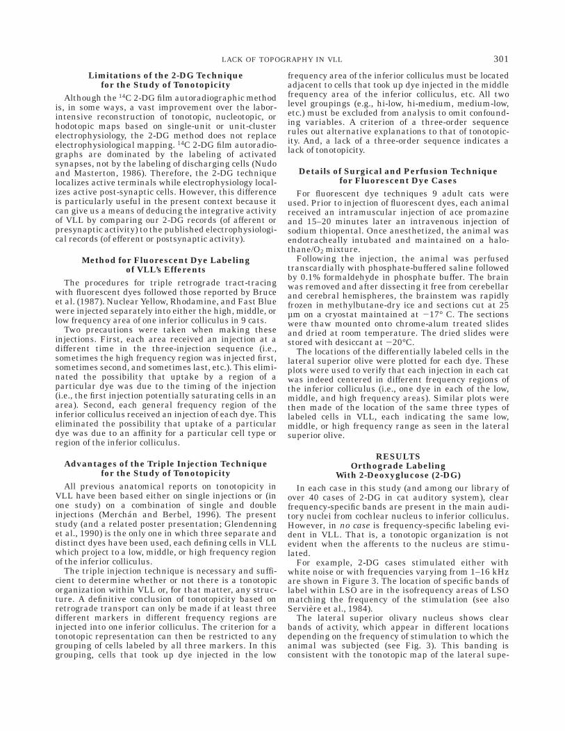
Fig. 2. A cresyl violet stain of caudal (left)and intermediate (right) coronal sections ofVLL. Note the density of fibers in the caudalsection and the clustering of cells in the morerostral section. The dense fibers comprise thelateral lemniscus and they carry informationfrom the contralateral cochlear nucleus (whichsynapse here) and bilaterally from the supe-rior olivary complex (which largely bypassingVLL). The fibers are partly responsible for theclustering of the cells. Both the fibers and thecell clusters make VLL a nucleus difficult tostudy by electrophysiological techniques.
300 K. K. GLENDENNING
Limitations of the 2-DG Techniquefor the Study of Tonotopicity
Although the 14C 2-DG film autoradiographic methodis, in some ways, a vast improvement over the labor-intensive reconstruction of tonotopic, nucleotopic, orhodotopic maps based on single-unit or unit-clusterelectrophysiology, the 2-DG method does not replaceelectrophysiological mapping. 14C 2-DG film autoradio-graphs are dominated by the labeling of activatedsynapses, not by the labeling of discharging cells (Nudoand Masterton, 1986). Therefore, the 2-DG techniquelocalizes active terminals while electrophysiology local-izes active post-synaptic cells. However, this differenceis particularly useful in the present context because itcan give us a means of deducing the integrative activityof VLL by comparing our 2-DG records (of afferent orpresynaptic activity) to the published electrophysiologi-cal records (of efferent or postsynaptic activity).
Method for Fluorescent Dye Labelingof VLL’s Efferents
The procedures for triple retrograde tract-tracingwith fluorescent dyes followed those reported by Bruceet al. (1987). Nuclear Yellow, Rhodamine, and Fast Bluewere injected separately into either the high, middle, orlow frequency area of one inferior colliculus in 9 cats.
Two precautions were taken when making theseinjections. First, each area received an injection at adifferent time in the three-injection sequence (i.e.,sometimes the high frequency region was injected first,sometimes second, and sometimes last, etc.). This elimi-nated the possibility that uptake by a region of aparticular dye was due to the timing of the injection(i.e., the first injection potentially saturating cells in anarea). Second, each general frequency region of theinferior colliculus received an injection of each dye. Thiseliminated the possibility that uptake of a particulardye was due to an affinity for a particular cell type orregion of the inferior colliculus.
Advantages of the Triple Injection Techniquefor the Study of Tonotopicity
All previous anatomical reports on tonotopicity inVLL have been based either on single injections or (inone study) on a combination of single and doubleinjections (Merchan and Berbel, 1996). The presentstudy (and a related poster presentation; Glendenninget al., 1990) is the only one in which three separate anddistinct dyes have been used, each defining cells in VLLwhich project to a low, middle, or high frequency regionof the inferior colliculus.
The triple injection technique is necessary and suffi-cient to determine whether or not there is a tonotopicorganization within VLL or, for that matter, any struc-ture. A definitive conclusion of tonotopicity based onretrograde transport can only be made if at least threedifferent markers in different frequency regions areinjected into one inferior colliculus. The criterion for atonotopic representation can then be restricted to anygrouping of cells labeled by all three markers. In thisgrouping, cells that took up dye injected in the low
frequency area of the inferior colliculus must be locatedadjacent to cells that took up dye injected in the middlefrequency area of the inferior colliculus, etc. All twolevel groupings (e.g., hi-low, hi-medium, medium-low,etc.) must be excluded from analysis to omit confound-ing variables. A criterion of a three-order sequencerules out alternative explanations to that of tonotopic-ity. And, a lack of a three-order sequence indicates alack of tonotopicity.
Details of Surgical and Perfusion Techniquefor Fluorescent Dye Cases
For fluorescent dye techniques 9 adult cats wereused. Prior to injection of fluorescent dyes, each animalreceived an intramuscular injection of ace promazineand 15–20 minutes later an intravenous injection ofsodium thiopental. Once anesthetized, the animal wasendotracheally intubated and maintained on a halo-thane/O2 mixture.
Following the injection, the animal was perfusedtranscardially with phosphate-buffered saline followedby 0.1% formaldehyde in phosphate buffer. The brainwas removed and after dissecting it free from cerebellarand cerebral hemispheres, the brainstem was rapidlyfrozen in methylbutane-dry ice and sections cut at 25µm on a cryostat maintained at 217° C. The sectionswere thaw mounted onto chrome-alum treated slidesand dried at room temperature. The dried slides werestored with desiccant at 220°C.
The locations of the differentially labeled cells in thelateral superior olive were plotted for each dye. Theseplots were used to verify that each injection in each catwas indeed centered in different frequency regions ofthe inferior colliculus (i.e., one dye in each of the low,middle, and high frequency areas). Similar plots werethen made of the location of the same three types oflabeled cells in VLL, each indicating the same low,middle, or high frequency range as seen in the lateralsuperior olive.
RESULTSOrthograde Labeling
With 2-Deoxyglucose (2-DG)In each case in this study (and among our library of
over 40 cases of 2-DG in cat auditory system), clearfrequency-specific bands are present in the main audi-tory nuclei from cochlear nucleus to inferior colliculus.However, in no case is frequency-specific labeling evi-dent in VLL. That is, a tonotopic organization is notevident when the afferents to the nucleus are stimu-lated.
For example, 2-DG cases stimulated either withwhite noise or with frequencies varying from 1–16 kHzare shown in Figure 3. The location of specific bands oflabel within LSO are in the isofrequency areas of LSOmatching the frequency of the stimulation (see alsoServiere et al., 1984).
The lateral superior olivary nucleus shows clearbands of activity, which appear in different locationsdepending on the frequency of stimulation to which theanimal was subjected (see Fig. 3). This banding isconsistent with the tonotopic map of the lateral supe-
301LACK OF TOPOGRAPHY IN VLL

rior olivary nucleus as determined by electrophysiologi-cal methods (e.g., Boudreau and Tsuchitani, 1970;Goldberg and Brown, 1968, 1969; Guinan et al., 1972a,b; Tsuchitani and Boudreau, 1969). Consistent with thebanding, the lateral superior olivary nucleus is acti-vated throughout following stimulation with whitenoise (top row, Fig. 3).
In contrast to the 2-DG results in the lateral superiorolivary nucleus, it can be seen that no specific bands orareas of increased activity appear in VLL (Fig. 4). Three
typical cases of VLL following stimulation at threedifferent frequencies show labeling throughout VLL.That is, regardless of the frequency of the stimulatingtone, the 2-DG labeling in VLL is virtually homoge-neous and is present throughout VLL’s length andbreadth. This is in marked contrast to the localizedfrequency-specific 2-DG labeling in the lateral superiorolivary nucleus. Therefore, the 2-DG evidence suggeststhat there are no special or frequency-specific regionsamong the afferent terminals within VLL.
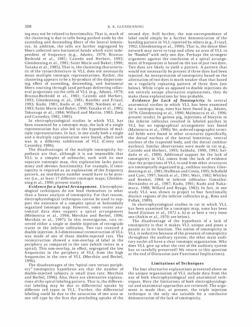
Fig. 3. Direct prints of 2-deoxyglucose la-beling in the lateral superior olivary nucleusof 4 cats subjected to a continuous tone of 85dB SPL. Four separate cases, each with adifferent frequency or white noise are shown:The lateral superior olivary nucleus ipsilat-eral to the stimulation (left column) andcontralateral (right column) are shown foreach case. White areas indicate active syn-apses. Note the differences in the location ofthe white areas. That is, a white noise case(top) shows activity throughout the lateralsuperior olivary nucleus. The higher fre-quency case (16 kHz: second from top photos)has the most activity in the top medial part ofthe S-shaped nucleus. The middle frequencycase (4 kHz; third from top photos) has mostactivity in the more ventral and lateral part ofthe S. And finally, the lowest frequency case (1kHz; bottom) has the most activity in themost lateral limb on the side ipsilateral to thestimulation.
302 K. K. GLENDENNING

Retrograde LabelingWith Fluorescent Dyes in IC
The results with fluorescent dyes support the lack oflocalized labeling found in VLL with 2-DG. Furthersupport for the lack of tonotopicity in VLL again comesfirst from an examination of the control nucleus, thelocation of retrograde label in the lateral superiorolivary nucleus. The retrograde transport of label to thelateral superior olive was located in either of three
discrete areas, either the low, middle, or high frequencyarea, of the lateral superior olive corresponding to thesite of the injection of the fluorescent dye in the inferiorcolliculus. In contrast, the retrograde transport to VLLfrom the same inferior colliculus injections resulted inno discernible pattern.
Three of the nine fluorescent dye cases are shown inFigures 5–7. In each case, the differential labeling inLSO shows that each of the three inferior colliculus
Fig. 4. Direct prints of 2-deoxyglucose la-beling in VLL of 3 cats subjected to a continu-ous tone of 85 dB SPL. Three separate caseseach with a different frequency are shown: alow frequency (2 K; top), a middle frequency(8 K; middle), and a high frequency (32 K;bottom). A caudal section (left column) androstral section (right column) are shown foreach case. As in Figure 3, the white areaindicates the active synapses in VLL follow-ing stimulation by different frequencies. How-ever, unlike the lateral superior olivarynucleus (in Fig. 3), the VLL is active (white)throughout, regardless of the frequency ofstimulation. Unlike the change of the activeregion in all other auditory nuclei , there isnot a particular area of VLL which becomesmore active depending on the frequency ofstimulation. That is, no frequency-specific la-bel appears in VLL. Rather, the entire nucleusappears homogeneously labeled regardless ofthe frequency of the stimulation.
303LACK OF TOPOGRAPHY IN VLL

injection sites are separate and discrete, effectivelylabeling three different areas of LSO according to theirfrequency allegiance. However, in VLL, cells labeledwith the same dye do not appear in separate zones.Instead, they appear in small clusters throughout thelength and breadth of VLL, regardless of the dimension(i.e., dorso-ventral, medial-lateral, or rostral-caudal).
An injection (Fig. 5) that results in extensive labelingin the lateral (low frequency) limb of the lateral supe-rior olivary nucleus, results in label throughout therostral-caudal extent of VLL. There does appear to be abanding pattern in the most caudal sections of VLL butthe banding consists of only 2 markers (low and highfrequency). Since the third (middle frequency) label isabsent from these bands, it cannot be said to be anaccurate reflection of a tonotopic arrangement butrather is the result of other considerations (see Discus-sion). The other two cases illustrated show a clearsegregation of frequencies in the lateral superior oli-vary nucleus and also in the dorsal nucleus of thelateral lemniscus but not in VLL (Figs. 6, 7).
One can argue that the labeling in VLL is not entirelyrandom in its distribution, in that small clusters ofsimilarly labeled cells abound and they can be arbi-trarily gathered together in triads comprised of oneeach of the three dyes (i.e., indicating a low, middle, andhigh frequency representation within each triad, e.g.,see Figs. 5–7). However, this selective grouping resultsin a very large number of triads within each VLL andeven these triads vary between left and right VLLs aswell as from section to section within each VLL.
That is, there is no pattern to the uptake in VLL inspite of the clear segregation of uptake in the lateralsuperior olivary nucleus. The lack of a pattern isconsistent in all of the nine cases that received tripleinjections of fluorescent dyes.
In sum, consistent with anterograde labeling of VLL,retrograde labeling of VLL’s efferents also does notreveal evidence of frequency-specific areas in VLL. Inspite of the clear evidence of anatomical segregation ofthe retrograde labeling in the lateral superior olivarynucleus, no evidence of a similar segregation is found inVLL. Hence, retrograde labeling of VLL’s efferents isconsistent with the orthograde 2-DG labeling. Bothsuggest that there is no frequency-specificity withinVLL.
DISCUSSIONThe main conclusion from the present study is that
while both orthograde and retrograde tract-tracingtechniques show clear evidence of tonotopicity in otherbrainstem auditory nuclei, they fail to show suchevidence for VLL. From the present results, it isdifficult to contend that VLL is either tonotopicallyorganized or that it consists of tonotopically organizedsubnuclei. In turn, it can be concluded that VLL lacks afrequency map.
Tonotopic Organization of BrainstemAuditory Nuclei
Tonotopicity is a basic principle of organization in theauditory pathway. For example, as one ascends theauditory system, starting at the level of the cochlearnucleus, tonotopicity has been found in all of its threedivisions (e.g., Bourk et al., 1981; Irvine, 1992; Master-
ton and Imig, 1984; Rose, 1960; Saldana, 1993; Saldanaet al., 1989). At the level of the nuclei of the superiorolivary complex, tonotopicity is also the rule (e.g.,Casseday and Covey, 1983; Elverland, 1978; Henkeland Spangler, 1983). At the level of the midbrain,tonotopicity is found in the dorsal nucleus of the laterallemniscus (e.g., Kelly et al., 1995; Merchan et al., 1994;Zook and Kawamura , 1995), and in the inferior collicu-lus (e.g., Clopton and Windfield, 1973; Kelly et al., 1991,Malmierca et al., 1995; Merzenich and Reid, 1974; Roseet al., 1963; Saldana and Merchan, 1992; Semple andAitkin, 1979, 1981). VLL is also situated in the mid-brain. But again, unlike the other auditory midbrainnuclei, VLL is unique in its lack of a tonotopic represen-tation.
The potential significance of the lack of tonotopicityin VLL may be gained by comparing it with two otherauditory nuclei. One, the medial superior olive, has astrong tonotopic map but has been shown to lacktonotopicity under certain conditions. The other, thedorsal nucleus of the lateral lemniscus, of all theauditory nuclei is the one nucleus most closely associ-ated with VLL. This association is due to its proximityto and its connections with VLL. Yet the dorsal nucleusof the lateral lemniscus has some striking differencesfrom VLL. A comparison follows of VLL, first with themedial superior olive, and then with the dorsal nucleusof the lateral lemniscus.
Some Similarities of VLLand the Medial Superior Olive
A nucleus somewhat similar to VLL in its lack oftonotopicity (only at high frequencies) is the medialsuperior olive. The 2-DG activity of the medial superiorolive at high frequencies shows an entirely differentpattern than the specific frequency labeling at lowerfrequencies (Masterton et al., 1986) . The medial supe-rior olive is strongly tonotopic at low frequencies.However, at frequencies from 8–32 kHz and above themedial superior olive is activated throughout (Master-ton et al., 1986). This overall activation in the medialsuperior olive occurs only at these higher frequencies.On the basis of these results, it has been suggested thatwhile the cells of the medial superior olive receivehigh-frequency stimulation, they do not relay it tohigher levels (Masterton et al., 1986).
This non-specific labeling of both the medial superiorolive (at high frequencies) and VLL (at all frequencies)appears to be the result of active inhibitory synapses.That is, dense 3H-strychnine label is found throughoutthe extent of the medial superior olive and VLL (e.g.,Baker and Glendenning, 1992; Glendenning and Baker,1988; 1991; Riquelme et al., 1998). Both neurophysi-ological and biochemical evidence support the view thatstrychnine binding occurs at the glycine receptor (e.g.,Young and Snyder, 1974a, b). For example, there is astrong correlation between the location of high densitystrychnine binding sites and areas in which glycine orstrychnine is electrophysiologically active (e.g., Alt-mann et al., 1972; Caspary et al., 1979; Felpel, 1977;Kelly and Renand, 1973; Zarbin et al., 1981). And thebinding of 3H-strychnine to tissue sections has beenshown to have both the pharmacologic and kineticcharacteristics associated with the synaptic glycinereceptor (e.g., Muller and Snyder, 1978; Young and
304 K. K. GLENDENNING

Fig. 5. A case (88–85) with a triple injection of 3 fluorescent dyes,each in a separate frequency region of the right inferior colliculus. Inthis and Figures 6 and 7, the injections in the inferior colliculus areillustrated at top from caudal (left) to rostral (right). Fast Blue (bluesymbols) is in the low frequency region, Rhodamine (red symbols) inthe middle frequency region, and Nuclear Yellow (yellow symbols) inthe high frequency region of the inferior colliculus. Plots of the labeledcells in the right lateral superior olivary nucleus and dorsal nucleus ofthe lateral lemniscus in this and Figures 6 and 7 indicate thesegregation of the three frequencies; low frequencies (blue dots) are
represented in the lateral limb, middle frequencies (red symbols) inthe middle limb, and high frequencies (yellow symbols) in the mediallimbs of the lateral superior olive. Plots of the labeled cells in the rightVLL show clusters of similarly labeled cells throughout (caudal left,rostral right). Note the lack of an orderly pattern. In this and Figures 6and 7, each area is labeled as if only one dye was injected in that area(i.e., high frequency area is always yellow, etc.). This is done forillustrative purposes only since each region received each dye; seeMaterials and Methods.
Fig. 6. A second case (88–62) showing triple-label retrogradetract-tracing of VLL’s efferents to inferior colliculus (see caption of Fig.5). Top: Injection sites of 3 different fluorescent dyes within rightcolliculus. Middle: Obvious topography of efferents of the lateralsuperior olivary nucleus. Bottom: Three sections through the dorsal
nucleus of the lateral lemniscus with low frequencies representeddorsolaterally, highest frequencies ventromedially. Notice the jumbledtopography of VLL’s efferents. Also note several alternating blue, red,and yellow symbol clusters within VLL.

Snyder, 1973, 1974a,b). Further, the strychnine bindingsite has been localized on the isolated glycine receptor(Graham et al., 1983). For these reasons the strychninelabel in MSO and VLL would seem to indicate arelatively high density of glycine receptors in the cellsof both nuclei (e.g., Adams and Wenthold, 1987; Aoki etal., 1988; Helfert et al., 1989; Luque et al., 1995; SaintMarie and Baker, 1990; Saint Marie et al., 1989;Wenthold et al., 1987; Winer et al., 1995).
Since the non-specific 2-DG labeling of both themedial superior olive (at high frequencies) and VLL (atall frequencies) appears to be the result of activeinhibitory synapses, a lack of specific label of 2-DGwould seem to indicate that some activity in each ofthese nuclei (in this case frequency) is not sent on tohigher levels of the auditory system. It is well known byother techniques that the medial superior olive relayslittle high frequency information to the inferior collicu-lus (e.g., Brunso-Bechtold et al., 1981; Guinan et al.,1972b). Therefore the different pattern in 2-DG label-ing in the medial superior olive at low and highfrequencies is consistent with electrophysiological andanatomical evidence (e.g., Masterton et al., 1986).
A similar argument can be made for the nonspecific2-DG labeling in VLL. That is, the lack of tonotopiclabeling in VLL regardless of frequency indicates thatthe VLL probably relays little information to the infe-rior colliculus of any frequency whatsoever. From thiswe can conclude that the primary function of VLL is
neither to encode frequencies nor to relay this informa-tion. The question remains as to what information VLLdoes send on to the inferior colliculus.
Comparison of VLL and the Dorsal Nucleusof the Lateral Lemniscus
The histochemistry of and afferents to VLL are incontrast to those found in the dorsal nucleus of thelateral lemniscus. Thus, while VLL contains manyglycinergic cells, the dorsal nucleus of the laterallemniscus contains mostly GABAergic cells (e.g., Ad-ams and Mugnaini, 1984; Glendenning and Baker,1988; Gonzalez-Hernandez et al., 1996; Moore andMoore, 1987; Peruzzi et al., 1997; Pollak, 1997; Robertsand Ribak, 1987; Thompson et al., 1985; Vater et al.,1992; Winer et al., 1995; Wu and Kelly, 1996; Yang andPollak, 1997). As a result, VLL presumably exerts aninhibitory influence both on the dorsal nucleus of thelateral lemniscus and on the inferior colliculus (e.g.,Aoki et al., 1988; Glendenning and Baker, 1988; Mooreand Moore, 1987; Roberts and Ribak, 1987; Saint Marieand Baker, 1990; Thompson et al., 1985; Vater et al.,1992; Winer et al., 1995).
Further, VLL receives primarily glutamate-immuno-reactive terminals (from the contralateral ventral co-
Fig. 7. A third case (88–84) with more extensive injection sites inright inferior colliculus (top). The cells representing high, middle, andlow frequencies within the lateral superior olivary nucleus are not asclearly segregated as in Figure 6. The middle frequencies (indicated byred symbols) have considerable overlap with the low frequencies(indicated by blue symbols) in one section. Nevertheless, the plots of
the lateral superior olivary nucleus allow a parcellation of the nucleuson the basis of frequency. There is also a segregation in the frequencyrepresentation in DLL (with low frequencies represented dorsolater-ally and high frequencies ventromedially). However, in VLL theresults are the same as those reported previously. That is, there is noconsistent pattern among the clustering of cells with the same label.
306 K. K. GLENDENNING

chlear nucleus; Suneja et al., 1995), while the dorsalnucleus of the lateral lemniscus receives glutamate,GABA, and some glycine immunoreactive terminals(from the superior olivary complex; e.g., Schwartz andEager, 1992; Wu and Kelly, 1996; Yang and Pollak,1997). Thus, the cochlear input to VLL is primarilyexcitatory while the input to the dorsal nucleus of thelateral lemniscus is mixed, both excitatory and inhibi-tory.
Electrophysiological evidence supports the chemistryshowing that VLL plays a role in shaping the neuronalresponse properties of the dorsal nucleus of the laterallemniscus. For example, VLL has an influence on theinitial phasic discharge of the neurons of the dorsalnucleus of the lateral lemniscus (e.g., Covey and Casse-day, 1991). VLL has also been shown to limit phase-locking of higher modulation rates in anterior parts ofthe dorsal nucleus of the lateral lemniscus (e.g., Yangand Pollak, 1997). Thus, VLL appears to shape at leastsome of the responses of the dorsal nucleus of thelateral lemniscus.
A further contrast between VLL and the dorsalnucleus of the lateral lemniscus is apparent in acomparison of their connections. Both nuclei receiveinput from a variety of sources (e.g., Covey and Casse-day, 1986; Glendenning et al., 1981; Glendenning andMasterton, 1983; Glendenning et al., 1992; Schwartz,1992; Zook and Casseday, 1985). However, the afferentallegiance of VLL is primarily to the contralateralventral cochlear nucleus and not to the superior olivarycomplex. In contrast, the afferent allegiance of thedorsal nucleus of the lateral lemniscus is to the superiorolivary complex and not to the cochlear nucleus. VLL,then, is more directly influenced by direct cochlearinput, while the dorsal nucleus of the lateral lemniscusis more directly in line with the sound localizationnuclei of the superior olivary complex. The difference inthe source of the afferent information they receiveundoubtedly influences the functions of each of thesenuclei and may serve as the basis of the inhibitory effectof VLL on DLL and subsequent shaping of DLL’sresponses.
Afferents to VLL From the Cochlear Nucleus.The afferents VLL receives from the cochlear nucleusarise from a variety of cell types. For example, octopus,multipolar, spherical, and globular bushy cells in theventral cochlear nucleus project to VLL (e.g., Friauf andOstwald, 1988; Glendenning et al., 1981; Glendenningand Kavanagh, 1986; Huffman and Covey, 1995;Schofield and Cant, 1997; Smith et al., 1991, 1993). Ithas been shown that the type of cell projecting to VLLcan be determined in part by the thickness of its axon(Schofield and Cant, 1997). A count made of the numberof axons reaching VLL based on HRP techniques deter-mined that it is mainly the globular and multipolarcells of the cochlear nucleus that project to VLL (Glen-denning and Kavanagh, 1986). It was found that atleast 38% of the axons of these cells reach as high asVLL while only 12% of the axons of spherical cells andfewer axons of octopus cells reached that high (Glenden-ning and Kavanagh, 1986).
The projections VLL receives from different nucleiand from the different cell types of the ventral cochlearnucleus are a source of cochleotopic information. Yet atthe level of VLL the projections and, thus, the cochleo-
topic information is shuffled (e.g., Schofield and Cant,1997). Again, the question remains as to the purpose ofthis reshuffling. Perhaps VLL has a function which isbetter served by ignoring frequency information (seethe following). In fact, frequency information mayinterfere with some aspects of hearing (i.e., identifica-tion of the source or location of a sound).
Hypotheses of Organization of VLLAt least four hypotheses have been put forward to
explain VLL’s unique organization. These are: (1) Thereis a single tonotopic organization albeit more looselyorganized than in other auditory nuclei (e.g., Aitkin etal., 1970; Friauf, 1992; Malmeirca et al., 1997; Merchanand Berbel, 1996; Merchan et al., 1997; Metzner andRadtke-Schuller, 1987; Willard and Martin, 1983); (2)There are multiple tonotopic representations (e.g., Coveyand Casseday, 1986, 1991); (3) There is a complex spiralarrangement (e.g., Merchan and Berbel, 1996; Willardand Martin, 1983); and finally (4) there is no tonotopicorganization (e.g., Adams, 1979; Glendenning et al.,1990; Guinan et al., 1972 a, b; present study). Thesehypotheses are not necessarily mutually exclusive. Forexample, the hypothesis of a spiral arrangement is notinconsistent with that of a single or multiple represen-tation as discussed in the following.
Evidence for a Single Tonotopic Organization.Conclusions vary in anatomical studies in which VLLhas been examined for a tonotopic map. Some suggestthat VLL is not unlike other auditory nuclei in having asingle tonotopic representation.Anatomical studies sup-porting this view have been done in at least threespecies (opossums: Willard and Martin, 1983; and rats:Merchan and Berbel, 1996). However, while supportingthe existence of a tonotopic map, these studies areconsistent in the consensus that the apparent tonotopicorganization of VLL is not as precise as in otherauditory nuclei.
Conclusions also vary in electrophysiological studiesin which VLL has been examined for a tonotopic map.Electrophysiological studies in two species show a loosetonotopic organization. For example, a progression ofbest frequencies was found through VLL in bats(Metzner and Radtke-Schuller, 1987), and in cats,although it was much less clear than the progressionthrough the dorsal nucleus of the lateral lemniscus(Aitkin et al., 1970).
A disadvantage of the single tonotopicity hypothesisis that the tonotopicity found in VLL in these studieswas, at best, loose. It was also notably different from thetonotopicity found in other structures. Other disadvan-tages are those that are inherent in anatomical andelectrophysiological methods per se (see below).
Evidence for a Pattern of Multiple Representa-tions. Other anatomical and electrophysiological stud-ies failing to find a single tonotopic map in VLL havesuggested that tonotopicity, if present, can only beexplained by multiple representations (e.g., Covey andCasseday, 1986, 1991; Friauf, 1992).
An anatomical study using c-fos as a marker failed tofind a single tonotopic representation in VLL andproposed that multiple representations are present(Friauf, 1992). The cytoarchitecture of VLL lends itselfto this view, at least in part, because of the clustering ofneurons throughout most of VLL (Fig. 2). The cluster-
307LACK OF TOPOGRAPHY IN VLL
ing may not be related to functionality. That is, much ofthe clustering is due to cells being pushed aside by theascending and descending fibers of the lateral lemnis-cus. In addition, the cells are further segregated byfibers collected into horizontal bands which exist inde-pendent of frequency (e.g., Adams, 1979; Brunso-Bechtold et al., 1981; Caicedo and Herbert, 1993;Glendenning et al., 1981; Saint Marie and Baker; 1990;Tanaka et al., 1985). That is, the clustering characteris-tic of the cytoarchitecture in VLL does not necessarilymean multiple tonotopic representations. Rather, theclustering appears to be a by-product of the dispersion-ing effect of ascending, descending, and horizontalfibers coursing through (and perhaps delivering collat-eral projections to) the cells of VLL (e.g., Adams, 1979;Brunso-Bechtold et al., 1981; Caicedo and Herbert,1993; Glendenning et al., 1981; Kandler and Friauf,1993; Kudo, 1981; Kudo et al., 1990; Nordeen et al.,1983; Saint Marie and Baker, 1990; Tanaka et al., 1985;Tokunaga et al., 1984; Willard and Martin, 1983; Zookand Casseday, 1982, 1985).
In electrophysiological studies in which VLL hasbeen examined for a tonotopic map, the lack of a singlerepresentation has also led to the hypothesis of mul-tiple representations. In fact, in one study both a singleand a multiple representation were found, albeit eachwas in a different subdivision of VLL (Covey andCasseday, 1986).
The disadvantages of the multiple tonotopicity hy-pothesis are that, although it is not impossible thatVLL is a complex of subnuclei, each with its ownseparate tonotopic map, this explanation lacks parsi-mony and obvious functionality. For example, if tono-topicity is required as an explanation of the frequencypattern, an inordinate number would have to be pres-ent (i.e., at least 17 different tonotopic maps; Glenden-ning et al., 1990; present study).
Evidence for a Spiral Arrangement. Electrophysi-ological techniques do not lend themselves to otherthan a linear analysis of tonotopicity. For that reason,electrophysiological techniques cannot be used to sup-port the existence of a complex spiral or helicoidallyorganized tonotopic map. However, some recent ana-tomical data does support a spiral arrangement(Melmeirca et al., 1994; Merchan and Berbel, 1996;Merchan et al., 1997). In this investigation, rats re-ceived either a single or double injection of retrogradetracer in the inferior colliculus. Two rats received adouble injection. A 3-dimensional reconstruction of VLLwas made of one of these double-injected rats. Thereconstruction showed a non-overlap of label in theperiphery as compared to the core (which twists in aspiral). This non-overlap, in effect, segregated the lowfrequencies in the periphery of VLL from the highfrequencies in the core of VLL (Merchan and Berbel,1996).
The disadvantages of the ‘‘spiral core versus periph-ery’’ tonotopicity hypothesis are that the number ofdouble-injected subjects is small (two rats: Merchanand Berbel, 1996). Also, there are alternative explana-tions of the spiral labeling pattern. That is, the differen-tial labeling may be due to differential uptake bydifferent cell types in VLL. Further, the differentiallabeling could be due to the saturation of one area orone cell type by the first dye precluding uptake of the
second dye. Still further, the non-correspondence oflabel could simply be a further demonstration of thebanding pattern in VLL reported by others (e.g., Friauf,1992; Glendenning et al., 1990). That is, the dense fibernetwork may serve to trap and allow an area of VLL tobe ‘‘flooded’’ with only one dye. Perhaps the strongestargument against the conclusion of a spiral arrange-ment of frequencies is based on the use of just two dyes.Two dyes are likely to yield a pattern. A pattern thatwould not necessarily be present if three dyes had beeninjected. An interpretation of tonotopicity based on thealternation of two dyes is much weaker than that basedon a regularly repeating pattern of three dyes (seebelow). While triple as opposed to double injections donot entirely escape alternative explanations, they domake these explanations far less probable.
Evidence for Lack of Tonotopicity. In severalanatomical studies in which VLL has been examinedfor a tonotopic map, none has been found (e.g., Adams,1979; Glendenning et al., 1990; Malmierca et al., 1996;present study). In guinea pig, injections of biocytin inthe inferior colliculus resulted in labeled patches inVLL but no topographical arrangement was found(Malmierca et al., 1996). Yet, ordered topographic termi-nal fields were found in other structures (specifically,the dorsal nucleus of the lateral lemniscus, ventralnucleus of the trapezoid body, and the dorsal cochlearnucleus). Similar observations were made in rat (e.g.,Caicedo and Herbert, 1993; Saldana, 1990, 1993; Sal-dana et al., 1989). Additional support for the lack oftonotopicity in VLL comes from the lack of evidencethat the projections of VLL to and from other structuresare tonotopically organized (e.g., cochlear nucleus: Glen-denning et al., 1981; Huffman and Covey, 1995; Schofieldand Cant, 1997; Smith et al., 1991; Warr, 1982; Whitleyand Henkel, 1984; or inferior colliculus: Gonzalez-Hernandez et al., 1987; Kudo, 1981; Kudo and Naka-mura, 1988; Willard and Ryugo, 1983). In fact, in onestudy VLL was shown to project to four functionallydistinct regions of the inferior colliculus (e.g., Ross andPollak, 1989).
In electrophysiological studies in cat in which VLLhas been examined for a tonotopic map, none has beenfound (Guinan et al., 1972 a, b) or at best a very looseone (Aitkin et al., 1970; see below).
The disadvantage of the hypothesis of a lack oftonotopicity is that it makes VLL unique and poses apuzzle as to its function. The notion of tonotopicity inVLL is seductive because of the presence of tonotopicitythroughout the auditory system; the other main audi-tory nuclei all have a clear tonotopic organization. Whydoes VLL give up what the rest of the auditory systemhas so carefully preserved? We return to this questionat the end of Discussion (see Functional Implications).
Limitations of TechniquesThe four alternative explanations presented above on
the unique organization of VLL include data from theuse of both electrophysiological and anatomical tech-niques. Here the limitations of both electrophysiologi-cal and anatomical approaches are reviewed. The argu-ment is made that, at present, the triple injectiontechnique is the only one suitable for a conclusivedemonstration of the lack of tonotopicity.
308 K. K. GLENDENNING

Limitations of Electrophysiological Techniquesfor the Study of Tonotopicity. One limitation ofelectrophysiological studies is that they depend on datacollected from one or more penetrations of a straightelectrode. Thus, a clear delineation of a tonotopic mapwould require a penetration of the electrode orthogonalto the predicted direction of increasing or decreasingfrequencies. Any twists, bulges, bends, etc,. in the mapwould elude detection by this technique. An apparentsequence of frequencies widely separated could be aloosely organized, seemingly single tonotopic map or aninadequately sampled repeating frequency of multipleor spiral tonotopic maps. That is, electrophysiologicaltechniques do not allow a distinction to be easily madeas to whether a sequence consists of multiple represen-tations, a spiral organization, or a loose single organiza-tion.
Limitations of Anatomical Techniques for theStudy of Tonotopicity. As discussed above, there aretwo main limitations to anatomical studies using retro-grade transport. First, the representation of tonotopic-ity in VLL is dependent on the area of the inferiorcolliculus that was injected. Second, while two injec-tions (each with a unique dye) is preferable to a singleinjection, double injections result in an overlappingand/or a repeating pattern, neither of which necessarilyhave to do with the presence of a tonotopic organization(i.e., different markers may have more or less affinityfor different cell types; injection of one marker couldsaturate a region and decrease the likelihood of uptakeof a second marker, etc.; see above).
Functional ImplicationsWhy does VLL alone among major auditory nuclei
seem to eschew a spatial segregation of frequencies,despite the availability of this information in the affer-ents it receives from the contralateral ventral cochlearnucleus. VLL’s lack of tonotopicity suggests that it isadding a new dimension to the contralateralization ofsound. Support for this view comes from the anatomyshowing the avoidance of the fibers ascending from thesuperior olivary complex for higher centers, almost as ifthey do not want to be contaminated by or interferewith the input and processing occurring in VLL.
That is, the absence of tonotopicity within VLLimplies that it may perform a function orthogonal to, orincommensurate with, the frequency of sound stimula-tion. Why and how does a nucleus ignore or disorder itsafferent supply only to reorder it again in its efferentprojection to the colliculus?
Let us make the assumption that the cells of the VLLreceive little or no input from other nuclei, or that ifthey do receive input from other nuclei, they ignoremost of that from auditory nuclei. One could thenenvision a possible autonomy of the neurons in VLL.Such an autonomy could be contributed to by a pace-maker-like activity of the neurons (Covey and Casse-day, 1991). These then could supply a timing mecha-nism for hearing.
In fact, there is some evidence that VLL has an effecton the timing or temporal characteristics of acousticstimuli (e.g., Covey and Casseday, 1991). In bats, VLLcresponds to a broad range of frequencies with an almostconstant latency regardless of the frequency of thesound (Covey and Casseday, 1991). According to this
view, the constant latency neurons in VLLc give upfrequency specificity in order to achieve a high degree oftemporal precision (Covey and Casseday, 1991). Thisview is in agreement with another study of bat in whicha subdivision of VLL has been shown to have character-istics suited for precise timing (e.g., low variability oflatency, unique tuning and timing properties, hightemporal resolution, lack of all spontaneous activity,broad tuning curves, and a limit of one spike at theonset of the stimulus; Haplea et al., 1994).
However, if VLL is involved with timing properties itseems that the timing precision of VLL may not bepassed on to higher levels. That is, although VLLprojects strongly and widely throughout the inferiorcolliculus, few inferior colliculus units have a timingprecision comparable to that in VLLc (Haplea et al.,1994). Instead, the precise synchrony of a subdivision ofVLL in bats seems to be converted to a different,non-synchronized representation in the inferior collicu-lus (Haplea et al., 1994).
The timing hypothesis of VLL function is consistentwith a hypothesis of the autonomy of VLL neurons(Covey and Casseday, 1991). Another hypothesis of VLLfunction is that the nucleus is bombarded by multiplesources and would therefore be involved in multipleaspects of audition. A study, again in bat, suggests thatVLL is involved in multiple aspects of auditory process-ing (Ross and Pollak, 1989). Consistent with this viewis the suggestion by others that the diversity of connec-tions in VLL contributes to the multiple response typesrecorded in VLL (e.g., Aitkin et al., 1970; Covey, 1993;Covey and Casseday, 1991; Guinan et al., 1972 a,b).Further support of this view comes from the wide-spread projection pattern of VLL to the inferior collicu-lus. This branching projection suggests that the role ofVLL is to contribute to multiple functions. In fact, in thebat VLL has been shown to project to four functionallydistinct regions of the inferior colliculus (Ross andPollak, 1989).
The present study makes no attempt to distinguishbetween the hypotheses presented as to the possiblefunction of VLL. VLL may have a role in the timing ofauditory information or in multiple functions, withtiming being just one of these multiple functions. Atiming vs. multiple function role of VLL may be basedin part on whether VLL pays little attention (the timinghypothesis) or a great deal of attention (the multiplefunction hypothesis) to the input it receives from othersources. Since the present study was not designed toexamine the influence of the afferent information on theactivity of cells in VLL nor to label potential colocaliza-tions to the VLL from brainstem nuclei, we proposethese alternatives to others for future study.
It should be noted that the hypothesis of precisiontiming is not necessarily inconsistent with that ofmultiple functions. VLL may do both. Thus, VLL maybe multifunctional with timing as a special priority.This precision timing may be a specialized feature inthe bat for echolocation or it may be important in allmammals for temporal processing, distinguishing anecho, filtering out noise, etc. At the same time, VLL mayserve multiple functions other than those dependent onfrequency. In any case, VLL appears to be in a uniqueposition to both modify and fine tune the ‘‘frequencydependent system.’’
309LACK OF TOPOGRAPHY IN VLL

ACKNOWLEDGMENTSThe authors thank Anna Williams for her histological
assistance. This work was supported in part by NIHgrant DC00197.
REFERENCESAdams, J. C. (1979) Ascending projections to the inferior colliculus. J.
Comp. Neurol., 183:519–538.Adams, J. C., and Mugnaini, E. (1984) Dorsal nucleus of the lateral
lemniscus: A nucleus of GABAergic projection neurons. Brain Res.Bull., 13:585–590.
Adams, J. C., and Wenthold, R. J. (1987) Immunostaining of ascendingauditory pathways with glycine antiserum. Assoc. Res. Otolaryngol.Abstract, 10:63.
Aitkin, L. M., Anderson, D. J., and Brugge, J. F. (1970) Tonotopicorganization and discharge characteristics of single neurons in thenuclei of the lateral lemniscus of the cat. J. Neurophysiol., 33:421–440.
Altmann, H., ten Bruggencate, G., and Sonnhof, U. (1972) Differentialstrength of action of glycine and GABA in hypoglossus nucleus.Pfluegers Arch., 331:90–94.
Aoki, E., Semba, R., Keino, H., Kato, K., and Kashiwamata, S. (1988)Glycine-like immunoreactivity in the rat auditory pathway. BrainRes., 442:63–71.
Baker, B. N., and Glendenning, K. K. (1992) Distribution of kainatereceptors in hindbrain auditory nuclei of the cat. Assoc. Res.Otolaryngol. Abstract, 15:57.
Boudreau, J. C., and Tsuchitani, C. (1968) Binaural interaction in thecat superior olive S segment. J. Neurophysiol., 31:442–454.
Boudreau, J. C., and Tsuchitani, C. (1970) Cat superior olive S-segment cell discharge to tonal stimulation. In: Contributions toSensory Physiology, Vol. 4. W. D. Neff, ed. Academic Press, NewYork, pp. 143–213.
Bourk, T. R., Meilcarz, J. P., and Norris B. E. (1981) Tonotopicorganization of the anteroventral cochlear nucleus of the cat. HearRes., 4:215–241.
Bruce, L. L., McHaffie, J. G., and Stein, B. E. (1987) The organizationof trigeminotectal and trigeminothalamic neurons in rodents: Adouble-labeling study with fluorescent dyes. J. Comp. Neurol.,262:315–330.
Brunso-Bechtold, J. K., Thompson, G. C., and Masterton, R. B. (1981)HRP study of the organization of auditory afferents ascending tocentral nucleus of inferior colliculus in cat. J. Comp. Neurol.,197:705–722.
Caicedo, A., and Herbert, H. (1993) Topography of descending projec-tions from the inferior colliculus to auditory brainstem nuclei in therat. J. Comp. Neurol., 328:377–392.
Caspary, D.M., Havey, D.C., and Faingold, C.L. (1979) Effects ofmicroiontophoretically applied glycine and GABA on neuronal re-sponse patterns in the cochlear nuclei. Brain Res., 172:179–185.
Casseday, J. H., and Covey, E. (1983) Laminar projections to theinferior colliculus as seen from injections of wheat germ agglutinin-horseradish peroxidase in the superior olivary complex of the cat.Soc. Neurosci. Abstract, 9:766.
Clopton, B. M., and Windfield, J. A. (1973) Tonotopic organization ofthe inferior colliculus of the rat. Brain Res., 56:355–358.
Covey, E. (1993) Response properties of single units in the dorsalnucleus of the lateral lemniscus and paralemniscal zone of anecholocating bat. J. Neurophysiol., 69:842–859.
Covey, E., and Casseday, J. H. (1986) Connectional basis for frequencyrepresentation in the nuclei of the lateral lemniscus of the bat,Eptesicus fuscus. J. Neurosci., 6:2926–2940.
Covey, E., and Casseday, J. H. (1991) The monaural nuclei of thelateral lemniscus in an echolocating bat: Parallel pathways foranalyzing temporal features of sound. J. Neurosci., 11:3456–3470.
Elverland, H. H. (1978) Ascending and intrinsic projections of thesuperior olivary complex in the cat. Exp. Brain Res., 32:117–134.
Felpel, L.P. (1977) Effects of strychnine, bicuculline and picrotoxin oninhibition of hypoglossal motoneurons. J. Neurosci. Res., 3:289–294.
Friauf, E. (1992) Tonotopic order in the adult and developing auditorysystem of the rat as shown by c-fos immunocytochemistry. Eur. J.Neurosci., 4:798–812.
Friauf, E., and Ostwald, J. (1988) Divergent projections of physiologi-cally characterized rat ventral cochlear nucleus neurons as shownby intra-axonal injection of horseradish peroxidase. Exp. Brain Res.,73:263–284.
Glendenning, K. K., and Baker, B. N. (1988). Neuroanatomical distri-bution of receptors for three potential inhibitory neurotransmitters
in the brainstem auditory nuclei of the cat. J. Comp. Neurol.,275:288–308.
Glendenning, K. K., and Baker, B. N. (1991) Neurochemical basis ofthe acoustic chiasm. Assoc. Res. Otolaryngol. Abstract, 14:32.
Glendenning, K. K., and Kavanagh, G. (1986) Origins of trapezoidbody and lateral lemniscus fibers in cat brainstem. Assoc. Res.Otolaryngol. Abstract, 9:38.
Glendenning, K. K. and Masterton, R. B. (1983) Acoustic Chiasm:Efferent projections of the lateral superior olive. J. Neurosci.,3:1521–1537.
Glendenning, K. K., Brunso-Bechtold, J. K., Thompson, G. C., andMasterton, R. B. (1981) Ascending auditory afferents to the nuclei ofthe lateral lemniscus. J. Comp. Neurol., 197:673–704.
Glendenning, K. K., Hutson, K. A., Nudo, R. J., and Masterton, R. B.(1985) Acoustic Chiasm II: Anatomical basis of binaurality in lateralsuperior olive of cat. J. Comp. Neurol., 232:261–285.
Glendenning, K. K., Masterton, R. B., Hutson, K. A., and Nudo, R. J.(1990) Ventral nucleus of the lateral lemniscus: Nontonotopic orga-nization. Anat. Rec. Abstract, 226:37A.
Glendenning, K. K., Baker, B. N., Hutson, K. A, and Masterton, R. B.(1992) Acoustic chiasm V: Inhibition and excitation in the ipsilateraland contralateral projections of LSO. J. Comp. Neurol., 319:100–122.
Goldberg, J. M., and Brown, P. B. (1968) Functional organization of thedog superior olivary complex: An anatomical and electrophysiologi-cal study. J. Neurophysiol., 31:639–656.
Goldberg, J. M., and Brown, P. B. (1969) Response of binaural neuronsof dog superior olivary complex to dichotic tonal stimuli: Somephysiological mechanisms of sound localization. J. Neurophysiol.,32:613–636.
Gonzalez-Hernandez, T., Meyer, G., Ferres-Torres, R., Castaneyra-Perdomo, A., and Perez-Delgada, M. M. (1987) Afferents connectionsof the inferior colliculus in the albino mouse. J. Hirnforsch.,28:315–324.
Gonzalez-Hernandez, T., Mantolan-Sarmeinto, B., Gonzalez-Gonzalez,B., and Perez-Gonzalez, H. (1996) Sources of GABAergic input to theinferior colliculus of the rat. J. Comp. Neurol., 372:309–326.
Graham, D., Pfeiffer, F., and Betz, H. (1983) Photoaffinity-labeling ofthe glycine receptor of rat spinal cord. Eur. J. Biochem., 131:519–525.
Guinan, J. J., Jr., Guinan, S. S., and Norris, B. E. (1972a) Singleauditory units in the superior olivary complex: I. Responses tosounds and classifications based on physiological properties. Int. J.Neurosci., 4:101–120.
Guinan, J. J., Jr., Norris, B. E., and Guinan, S. S. (1972b) Singleauditory units in the superior olivary complex: II. Locations of unitcategories and tonotopic organization. Int. J. Neurosci., 4:147–166.
Hand, P.J. (1981) The 2-deoxyglucose method. In: NeuroanatomicalTract-Tracing Methods. L. Heimer, and M.J. Robards, eds. PlenumPress, New York, pp. 511–538.
Haplea, S., Covey, E., and Casseday, J. H. (1994) Frequency tuningand response latencies at three levels in the brainstem of theecholocating bat, Eptesicus fuscus. J. Comp. Physiol. A, 174:671–683.
Helfert, R. H., Bonneau, J. M., Wenthold, R. J., and Altschuler, R. A.(1989) GABA and glycine immunoreactivity in the guinea pigsuperior olivary complex. Brain Res., 501:269–286.
Henkel, C. K., and Spangler, K. M. (1983) Organization of the efferentprojections of the medial superior olivary nucleus in the cat asrevealed by HRP and autoradiographic tracing methods. J. Comp.Neurol., 221:416–428.
Huffman, R. F., and Covey, E. (1995) Origin of ascending projections tothe nuclei of the lateral lemniscus in the big brown bat, Eptesicusfuscus. J. Comp. Neurol., 357:532–545.
Irvine, D. R. F. (1986) The auditory brain. In: Progress in SensoryPhysiology, Vol. 7. D. Ottoson, ed. Springer-Verlag, New York, pp.1–279.
Irvine, D. R. F. (1992) Physiology of the auditory brainstem. In: TheMammalian Auditory Pathway: Neurophysiology. A. Popper, andR. R. Fay, eds. Springer-Verlag, New York, pp. 153–231.
Kandler, K., and Friauf, E. (1993) Pre- and postnatal development ofefferent connections of the cochlear nucleus in the rat. J. Comp.Neurol., 328:161–184.
Kelly, J. B., Glenn, S. L., and Beaver, C. L. (1991) Sound frequency andbinaural response properties of single neurons in rat inferiorcolliculus. Hear. Res., 56:273–280.
Kelly, J. B., Liscum, A., Van Adel, B., and Ito, M. (1995) Retrogradelabeling in the rat’s dorsal nucleus of the lateral lemniscus fromfrequency specific regions of the central nucleus of the inferiorcolliculus. Assoc. Res. Otolaryngol. Abstract, 18:40.
Kelly, J.S., and Renaud, L.P. (1973) On the pharmacology of the
310 K. K. GLENDENNING

glycine receptors on the cuneothalamic relay cells in the cat. Br. J.Pharmacol., 48:387–395.
Kennedy, C., Des Rosiers, M. H., Jehle, J. W., Reivich, M., Sharp, F.,and Sokoloff, L. (1975) Mapping of functional neural pathways byautoradiographic survey of local metabolic rate with [14C]deoxyglu-cose. Science, 187:850–853.
Kudo, M. (1981) Projections of the nuclei of the lateral lemniscus in thecat: An autoradiographic study. Brain Res., 221:57–69.
Kudo, M., and Nakamura, Y. (1988) Organization of the laterallemniscal fibers converging onto the inferior colliculus in the cat: Ananatomical review. In: Auditory Pathway, Structure and Function.J. Syka, and R. B. Masterton, eds. Plenum Press, New York, pp.171–183.
Kudo, M., Nakamura, Y., Tokuno, H., and Kitao, Y. (1990) Auditorybrainstem in the mole (Mogera): Nuclear configurations and theprojections to the inferior colliculus. J. Comp. Neurol., 298:400–412.
Luque, J. M., Nelson, N., and Richards, J. G. (1995) Cellular expres-sion of glycine transporter 2 Messenger RNA exclusively in rathindbrain and spinal cord. Neuroscience, 64:525–535.
Malmierca, M. S., Rees, A., Le Beau, F. E. N., and Bjaalie, J. G. (1995)Laminar organization of frequency-specific local axons within andbetween the inferior colliculi of the guinea pig. J. Comp. Neurol.,357:124–144.
Malmierca, M. S., Le Beau, F. E. N., and Rees, A. (1996) Thetopographical organization of descending projections from the cen-tral nucleus of the inferior colliculus in guinea pig. Hear. Res.,93:167–180.
Malmeirca, M.S., Merchan, M.A., and Bajo, V.M. (1997) The ventralnecleus of the lateral lemniscus in cat is tonotopically organized.Assoc. Res. Otolaryngol. Abstract, 654:164.
Masterton, R. B., and Imig, T. 1984. Neural mechanisms for soundlocalization. Ann. Rev. Physiol., 46:275–287.
Masterton, R. B., Nudo, R. J., Baker, B. N., and Glendenning, K. K.(1986) Functional anatomy of the medial superior olive in cat. Anat.Rec. Abstract, 214:84A.
Masterton, R. B., Nudo, R., Moreland-Granger, E., and Frost, S. (1986)Acoustic chiasm: 2-DG studies of hindbrain auditory nuclei in cat.Assoc. Res. Otolaryngol. Abstract, 9:8.
Merchan, M. A., and Berbel, P. (1996) Anatomy of the ventral nucleusof the lateral lemniscus in rats: A nucleus with a concentric laminarorganization. J. Comp. Neurol., 372:245–263.
Merchan, M. A., Saldana, E., and Plaza, I. (1994) Dorsal nucleus of thelateral lemniscus in the rat: Concentric organization and tonotopicprojection to the inferior colliculus. J. Comp. Neurol., 342:259–278.
Merchan, M. A., Malmierca, M.S., Bajo, V.M. and Bjaalie, J.G. (1997)The nuclei of the lateral lemniscus: Old views and new perspectives.In: Acoustical Signal Processing in the Central Auditory System. J.Syka, ed. Plenum Press, New York, pp. 211–226.
Merzenich, M. M., and Reid, M. D. (1974) Representation of thecochlea within the inferior colliculus of the cat. Brain Res., 77:397–415.
Metzner W., and Radtke-Schuller, S. (1987) The nuclei of the laterallemniscus in the rufous horseshoe bat, Rhinolophus rouxi. J. Comp.Physiol A, 160:395–411.
Moore, J. K., and Moore, R. Y. (1987) Glutamic acid decarboxylase-likeimmunoreactivity in brainstem auditory nuclei of the rat. J. Comp.Neurol., 260:157–174.
Muller, W. E., and Snyder, S. H. (1978) Strychnine binding associatedwith synaptic glycine receptors in rat spinal cord membranes: Ionicinfluences. Brain Res., 147:107–116.
Nordeen, K. W., Killackey, J. P., and Kitzes, L. M. (1983) Ascendingauditory projections to the inferior colliculus in the adult gerbil,Meriones unguiculatus. J. Comp. Neurol., 214:131–143.
Nudo, R. J., and Masterton, R. B. (1986) Stimulation-induced [14C]2-deoxyglucose labeling of synaptic activity in the central auditorysystem. J. Comp. Neurol., 245:553–565.
Peruzzi, D., Bartlett, E., Smith, P. H., and Oliver, D. L. (1997) Amonosynaptic GABAergic input from the inferior colliculus to themedial geniculate body in rat. J. Neurosci., 17:3766–3777.
Pollak, G. D. (1997) Roles of GABAergic inhibition for the binauralprocessing of multiple sound sources in the inferior colliculus. Ann.Otol. Rhinol. Laryngol. (Suppl.), 168:44–54.
Riquelme, R., Merchan, M.A., Ottersen, O.P. (1998) GABA and Glycinein the ventral nucleus of the lateral lemniscus:An immunocytochemi-cal and in situ hybridization study in rat. Assoc. Res. Otolaryngol.Abstract, 370:93.
Roberts, R. C., and Ribak, E. C. (1987) GABAergic neurons and axonterminals in the brainstem auditory nuclei of the gerbil. J. Comp.Neurol., 258:267–280.
Rose, J. E. (1960) Organization of frequency sensitive neurons in thecochlear nuclear complex of the cat. In: Neural Mechanisms of the
Auditory and Vestibular Systems. G. L. Rasmussen, and W. Windle,eds. Charles C. Thomas, Springfield, IL, pp. 116–136.
Rose, J. E., Greenwood, D. D., Goldberg, J. M., and Hind, J. E. (1963)Some discharge characteristics of single neurons in the inferiorcolliculus of the cat. I. Tonotopical organization, relation of spike-counts to tone intensity, and firing patterns of single elements. J.Neurophysiol., 26:314–319.
Ross, L. S., and Pollak, G. D. ( 1989) Differential ascending projectionsto aural regions in the 60 kHz contour of the mustache bat’s inferiorcolliculus. J. Neurosci., 9:2819–2834.
Saint Marie, R. L., and Baker, R. A. (1990) Neurotransmitter-specificuptake and retrograde transport of [3H]glycine from the inferiorcolliculus by ipsilateral projections of the superior olivary complexand nuclei of the lateral lemniscus. Brain Res., 524:244–253.
Saint Marie, R. L., Ostapoff, E.-M., Morest, D. K., and Wenthold, R. J.(1989) Glycine-immunoreactive projection of the cat lateral superiorolive: Possible role of midbrain ear dominance. J. Comp. Neurol.,279:382–396.
Saldana, E. (1990) The rat colliculo-olivary projection is tonotopic. Soc.Neurosci. Abstract, 16:716.
Saldana, E. (1993) Descending projections from the inferior colliculusto the cochlear nuclei in mammals. In: The Mammalian CochlearNuclei: Organization and Function. M. A. Merchan, J. M. Juiz, D. A.Godfrey, and E. Mugnaini, eds. Plenum, New York, pp. 153–165.
Saldana, E., Bajo, V. M., Lopez, D. E., and Malmierca, M. S. (1989) Isthe descending projection from the central nucleus of the inferiorcolliculus to the cochlear nuclear complex tonotopically organised?Eur. J Neurosci. (Suppl.), 2:262.
Saldana, E., and Merchan, M. A. (1992) Intrinsic and commissuralconnections of the rat inferior colliculus. J. Comp. Neurol., 319:417–437.
Schofield, B. R. and Cant, N. M. (1997) Ventral nucleus of the laterallemniscus in guinea pigs: Cytoarchitecture and inputs from thecochlear nucleus. J. Comp. Neurol., 379:363–385.
Schwartz, I. R. (1992) The superior olivary complex and laterallemniscal nuclei. In: The Mammalian Auditory Pathway: Neuro-anatomy. D. B. Webster, A. N. Popper, and R. R. Fay, eds. Springer-Verlag, New York, pp. 117–167.
Schwartz, I. R., and Eager, P. R. (1992) Differential distribution ofcalcium binding proteins and neuronal surface markers and theirrelationship to GABA immunoreactive cells in the superior olivarycomplex and lateral lemniscal nuclei of the gerbil. Assoc. Res.Otolaryngol. Abstract,, 15:59.
Semple, M. N., and Aitkin, L. M. (1979) Representation of soundfrequency and laterality by units in central nucleus of cat inferiorcolliculus. J. Neurophysiol., 42:1626–1639.
Semple, M. N., and Aitkin, L. M. (1981) Integration and segregation ofinput to the cat inferior colliculus. In: Neuronal Mechanisms ofHearing. J. Syka, and L. Aitkin, eds. Plenum, New York, pp.155–161.
Serviere, J., and Webster, W. R. (1981) A combined electrophysiologicaland 2-[14C]-deoxyglucose study of the frequency organization of theinferior colliculus of the cat. Neurosci. Lett., 27:113–118.
Serviere, J., Webster, W. R., and Calford, M. C. (1984) Isofrequencylabeling revealed by a combined [14C]-2-deoxyglucose, electrophysi-ological, and horseradish peroxidase study of the inferior colliculusof the cat. J. Comp. Neurol., 228:463–477.
Smith, P. H., Joris, P. X., Carney, L. H., and Yin T. C. T. (1991)Projections of physiologically characterized globular bushy cellaxons from the cochlear nucleus of the cat. J. Comp. Neurol.,304:387–407.
Smith, P. H., Joris, P. X., and Yin, T. C. T. (1993) Projections ofphysiological characterized spherical bushy cell axons from thecochlear nucleus of the cat: Evidence for delay lines to the medialsuperior olive. J. Comp. Neurol., 331:245–260.
Sokoloff, L., Reivich, M., Kennedy, D. C., Rosiers, M. H., Patlak, C. S.,Pettigrew, K. D., Sakurada, O., and Shinahara, M. (1977) The 14Cdeoxyglucose method for the measurement of local cerebral glucoseutilization: Theory, procedure and normal values in conscious andanesthetized albino rat. J. Neurochem., 28:897–916.
Suneja, S. K., Benson, C. G., Gross, J., and Potashner, S. J. (1995)Evidence for glutamatergic projections from the cochlear nucleus tothe superior olive and the ventral nucleus of the lateral lemniscus. J.Neurochem., 64:161–171.
Tanaka, K., Otani, K., Tokunaga, A., and Sugita, S. (1985) Theorganization of neurons in the nucleus of the lateral lemniscusprojecting to the superior and inferior colliculi in the rat. Brain Res.,341:252–260.
Theurich, M., Muller, C. M., and Scheich, H. (1984) 2-deoxyglucoseaccumulation parallels extracellularly recorded spike activity in theavian auditory neostriatum. Brain Res., 322:157–161.
311LACK OF TOPOGRAPHY IN VLL

Thompson, G. C., Cortez, A. M., and Lam, D. M-K. (1985) Localizationof GABA immunoreactivity in the auditory brainstem of guinea pigs.Brain Res., 339:119–122.
Tokunaga, A., Sugita, S., and Otani, K. (1984) Auditory and nonauditory subcortical afferents to the inferior colliculus in the rat. J.Hirnforsch., 25:461–472.
Tsuchitani, C., and Boudreau, J. C. (1969) Stimulus level of dichoti-cally presented tones and cat superior olive S-segment cell dis-charge. J. Acoust. Soc. Am., 46:979–988.
Vater, M., Habbicht, H., Kossl, M., and Grothe, B. (1992) Thefunctional role of GABA and glycine in monaural and binauralprocessing in the inferior colliculus of horseshoe bats. J. Comp.Physiol. A, 171:541–553.
Warr, W. B. (1982) Parallel ascending pathways from the cochlearnucleus: Neuroanatomical evidence of functional specialization. In:Contributions to Sensory Physiology, Vol. 7. D. W. Neff, ed. AcademicPress, New York, pp. 1–38.
Webster, W. R., Serviere, J., Crewther, D., and Crewther, S. (1984)Iso-frequency 2-DG contours in the inferior colliculus of the awakemonkey. Exp. Brain Res., 56:425–437.
Wenthold, R. J., Huie, D., Altschuler, R. A., and Reeks, K. A. (1987)Glycine immunoreactivity localized in the cochlear nucleus andsuperior olivary complex. Neuroscience, 22:897–912.
Whitley, J. M., and Henkel, C. K. (1984) Topographical organization ofthe inferior collicular projection and other connections of the ventralnucleus of the lateral lemniscus in the cat. J. Comp. Neurol.,229:257–270.
Willard, F. H., and Martin, G. F. (1983) The auditory brainstem nucleiand some of their projections to the inferior colliculus in the NorthAmerican opossum. Neuroscience, 10:1203–1232.
Willard, F. H., and Ryugo, D. K. (1983) Anatomy of the central auditorysystem. In: The Auditory Psychobiology of the Mouse. J. F. Willott,ed. Charles C. Thomas, Springfield, IL, pp. 201–304.
Winer, J. A., Larue, D. T., and Pollak, G. D. (1995) GABA and glycine in
the central auditory system of the mustache bat: Structural sub-strates for inhibitory neuronal organization. J. Comp. Neurol.,355:317–353.
Wu, S. H., and J. B. Kelly (1996) In vitro brain slice studies of the rat’sdorsal nucleus of the lateral lemniscus. III. synaptic pharmacology.J. Neurophysiol., 75:1271–1282.
Yang, L., and Pollak, G. D. (1997) Differential response properties toamplitude modulated signals in the dorsal nucleus of the laterallemniscus of the mustache bat and the roles of GABAergic inhibi-tion. J. Neurophysiol., 77:324–340.
Young, A. B., and Snyder, S. H. (1973) Strychnine binding associatedwith glycine receptors of the central nervous system. Proc. Natl.Acad. Sci. U.S.A., 70:2832–2836.
Young, A. B., and Snyder, S. H. (1974a) Strychnine binding in ratspinal cord membranes associated with the synaptic glycine recep-tor: cooperativity of glycine interactions. Mol. Pharmacol., 10:790–809.
Young, A. B., and Snyder, S. H. (1974b) The glycine synaptic receptor:Evidence that strychnine binding is associated with the ionicconductance mechanism. Proc. Natl. Acad. Sci. U.S.A., 71:4002–4005.
Zook, J. M, and Casseday, J. H. (1982) Origin of ascending projectionsto the inferior colliculus in the mustache bat, Pteronotus parnellii. J.Comp Neurol., 207:14–28.
Zook, J. M., and Casseday, J. H. (1985) Ascending projections of thecochlear nucleus in the mustache bat, Pteronotul parnellii. J. Comp.Neurol., 207:14–28.
Zook, J. M., and Kawamura, N. (1995) Intrinsic and descendingprojections of the dorsal nucleus of the lateral lemniscus in thegerbil and mouse. Assoc. Res. Otolaryngol. Abstract, 18:40.
Zarbin, M.A., Wamsley, J. K., and Kuhar, M. J. (1981) Glycinereceptor: Light microscopic autoradiographic localization with [3H]strychnine. J. Neurosci., 1:532–547.
312 K. K. GLENDENNING