laboratory 7 - university of pennsylvaniacal.vet.upenn.edu/projects/parasit06/website/lab7...
TRANSCRIPT

Laboratory 7 pg. 1
LABORATORY
7
The ArachnidsIntroduction:
Adult arachnids are eight-legged arthropods with anterior body segments fused into acephalothorax bearing walking legs, sensory structures and the feeding apparatus, which iscapable only of fluid feeding. Arachnids develop by a pattern of punctuated growth and moltingsimilar to simple metamorphosis in insects. A number of specialized forms have evolved adependency on feeding upon vertebrate animal blood or tissue fluid and may be temporary orpermanent inhabitants of the skin or other tissues in wild and domestic animals and humans.Such arachnids may act as transmitters of pathogens (eg. the Lyme disease agent), as agents ofdermatosis (mange, scabies) and systemic disease (eg. tick paralysis) and as sources of bloodloss and annoyance sufficient to impact production in food animals and the general welfare ofcompanion animals and their owners.
Objectives:
The order Acarina of the class Arachnida includes the ticks and mites and thus, manyimportant ectoparasites of domestic animals. On completing this exercise, we would like for youto:
1.) Be able to recognize on sight the important families of burrowing and non-burrowing miteparasites.
2.) Be able to recognize on sight the tick families Ixodidae and Argasidae (the hard and softticks respectively).
3.)Be familiar enough with the morphological characters of ticks to use the pictorial key (Figure7) to identify a tick specimen to the genus level.
Checklist of Objectives:
Be able to recognize a representative mite from each of the following 5 families:
9 Dermanyssidae 9 Chyletidae
9 Psoroptidae 9 Sarcoptidae
9 Demodicidae
Be able to:
9 Use the pictorial key to identify an unknown tick specimen to the genus level.
9 Recognize Rhipicephalus, Ixodes, Dermacentor, Ambylomma ticks without using a key.

Laboratory 7 pg. 2
At the Bench
1) Slides from the student slide box:
A. Dermanyssus gallinae (Family Dermanyssidae) – Student Slide #91 (Foreyt, pg. 150). Thisis the chicken mite, also called the “red mite” of poultry. The “red mite” is a lairectoparasite, visiting the birds only to feed. The “red” in its name refers to the mite’scolor when engorged with blood. This mite may also attack mammals if birds are notavailable.
B. Psoroptes ovis (Family Psoroptidae ) – Student Slide #87 (Foreyt, pg 99). Found on sheepand cattle, this is the cause of “sheep scab” (Psoroptic mange). Other members of thegenus cause mange in horses and rabbits. Note the elongate legs (compared to Sarcoptesslide #88). Legs I, II, and IV bear a segmented pretarsus (Figure 2).
C. Sarcoptes scabei (Family Sarcoptidae)– Student Slide #88 (Foreyt pg 38). Host-adaptedphysiologic races of this mite species are found on all domestic animals as well as onhumans. It causes sarcoptic mange (or “scabies” in humans). Note the small size and theglobular body shape with very short legs. The coxae of legs II and III are widelyseparated. In contrast to Psoroptes, the pretarsi of legs I and II are in the form of simple(unsegmented) stalk with terminal suckers. There are long trailing setae or hairs (Figure3).
D. Demodex canis (Family Demodicidae) – Student Slide #90 (Foreyt pg. 38). This is theubiquitous follicular mite of dogs. This mite, although usually a harmless commensalorganism, can cause mange (demodectic mange) especially in immuno-compromisedanimals. Student slide #90 is a section of skin showing the effects of demodectic mange. Note the thickened dermis, you may or may not see a mite in this section.
E. Dermacentor variabilis – The American Dog Tick – Student Slide #96 This three-host tick isornate with the scutum more or less covered with irregular white markings. Note the“short” mouthparts (not much longer than the basis capitulium - see figure 6). The shortmouthparts and the ornate scutum are enough to identify this tick when it is removedfrom a dog, cat, horse or human in Pennsylvania.
F. Rhipicephalus sanguineus – The Brown Dog Tick – Student Slide #95 (Foreyt pg. 44) Thisinornate (no markings), three-host tick is another common parasite of dogs. All of its lifestages occur on dogs. Like D. variabilis it has short mouth parts. Note the festoons(indentations along the posterior margin), these are common in many ticks and may be afeature used to differentiate between ticks. The short mouthparts and the inornate scutumare enough to identify this tick when it is removed from a dog in Pennsylvania.
G. Rhipicephalus (Boophilus) annulatus larva - Cattle Tick - Student Slide #93 Note that ticklarvae have only 6 legs. The larval ticks may not key out using the key in Figure 7 whichwas developed for adult ticks. The genus Boophilus was recently made a subgenus ofRhipicephalus, but the label of slide #93 and the key in Fig 7 do not reflect this.
2) Tick keying exercise
There are unidentified ticks on the center bench. Take one and try your hand at keying it to thegenus level with the pictorial key (Figure 7). NOTE: Views of the capitulae are dorsal. For
Laboratory 7 pg. 3
locations of diagnostic structures (palpi, basis capituli, eyes, festoons, anal pore, etc.) refer toFigure 6. (Please return the specimen as soon as you are finished).
TIP: Don’t mistake an engorged hard tick for a soft tick.
Also on the center bench are dishes with the 4 tick species common in Pennsylvania/NJ. You must be able to recognize this ticks without a key. Since these ticks carry differentpathogens, knowing the tick species will prepare you to deal with subsequent disease in the pet.Being able to respond immediately to an owner’s questions about the tick they remove from theirpet will save you time (and impress the client). The key provided in Fig. 7a will allow you totell these ticks apart, but the key will not be available on any test.
Demonstrations
Check list material:
NON-BURROWING MITES
1) Family Dermanyssidae
These are tick-like mites with an ovoid body shape. They have a pair of spiracles between thethird and fourth coxae. In life, they use their long legs to move about both on the host and in itsnest or bedding. Many are lair ectoparasites.
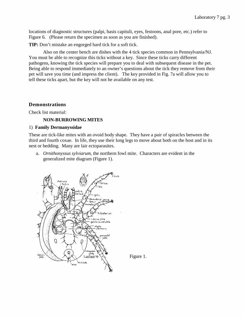
a. Ornithonyssus sylviarum, the northern fowl mite. Characters are evident in thegeneralized mite diagram (Figure 1).
Figure 1.

Laboratory 7 pg. 4
2) Fam. Cheyletiellidae
This family has palptibial claws curved ventrally and usually greatly enlarged. Parasites ofbirds and small mammals.
a. Cheyletiella parasitivorax – (Foreyt, pg. 39). This is the “rabbit mite”. Other membersof this genus can be found on dogs and cats. Note: The body has a “waist”; the legs end incombs, and the large palpi have pincers on their ends.
3) Fam. Psoroptidae
Legs III with long terminal setae, legs IV may be reduced, usually with claws; males withanal suckers. Skin parasites of mammals.
a. Psoroptes ovis –(Foreyt, pg 99) Found on sheep and cattle, this is the cause of“sheep scab” (Psoroptic mange). Other members of the genus cause mange in horses andrabbits. Note the elongate legs (compared to Sarcoptes). Legs I, II, and IV bear asegmented pretarsus (Figure 2).
Figure 2.
BURROWING MITES
4) Fam. Sarcoptidae
Rounded or sac-like; legs short.
a. Sarcoptes scabei –(Foreyt pg 38). Host- adapted physiologic races of this mitespecies are found on all domestic animals as well as on humans. It causes sarcopticmange (or “scabies” in humans). Note the small size and the globular body shape withvery short legs. The coxae of legs II and III are widely separated. In contrast toPsoroptes, the pretarsi of legs I and II are in the form of simple (unsegmented) stalk withterminal suckers. There are long trailing setae or hairs (Figure 3).
Segmented
pretarsus

Laboratory 7 pg. 5
Figure 3.
5) Fam. Demodicidae
Elongated, annulated, worm-like species found in hair follicles, and the surface glands
and ducts of vertebrates.
a. Demodex canis – (Foreyt pg. 38).
This is the ubiquitous follicular mite of dogs. Note the elongate shape of the
body and the 4 pairs of stumpy legs (Figure 4). This mite, although usually a
harmless commensal organism, can cause mange (demodectic mange) especially
in immuno-compromised animals.
Figure 4.
Unsegmented
pretarsus
Setae

Laboratory 7 pg. 6
Ticks
6) Ixodes scapularis – The deer tick (AKA: the black-legged tick) – This is the vector ofBorrelia burgdorferi (the agent of Lyme disease) in the eastern and midwestern United States. To learn more about Lyme disease, check out the following Web sites:http://www3.niaid.nih.gov/topics/lymeDisease/PDF/LymeDisease.pdf andhttp://www.cdc.gov/ncidod/dvbid/lyme/
Note the following about I. scapularis.
i. The elongate mouthparts with tips or palpi converging (in the female).
ii. The preanal groove (characteristic of genus).
iii. The inornate (plain brown) scutum
iv. The prominent, posteriorly-directed spine on coxa I
The long mouthparts and the inornate scutum are enough to identify this tick when it isremoved from a dog, cat, human, or horse in Pennsylvania.
7) Amblyomma americanum – The Lone Star Tick – This is an ornate tick. The male haswhite spots on its back (mainly along the posterior margin of the scutum), while the female hasa single white spot (“lone star”) on its scutum. Note the long mouthparts and the eyes on thelateral margin of the scutum. Ticks’ eyes are simply translucent patches of cuticle overlyingphotoreceptors. The long mouthparts and the ornate scutum are enough to identify this tickwhen it is removed from a dog, cat, human, or horse in Pennsylvania.
8) Dermacentor variabilis – The American Dog Tick – This three-host tick is also ornate withthe scutum more or less covered with irregular white markings. The larval and nymphal stagesof this tick are found on rodents and other small mammals and the adults on a variety ofmiddle-sized to large mammals including dogs and humans. Note also the rectangular basiscapitulum and the festoons on the posterior margin (see Fig. 6). The short mouthparts and theornate scutum are enough to identify this tick when it is removed from a dog, cat, human, orhorse in Pennsylvania.
Figure 6 .

Laboratory 7 pg. 7
9) Rhipicephalus sanguineus – The Brown Dog Tick – This inornate, three-host tick isanother common parasite of dogs. All of its life stages occur on dogs. Note that the basiscapitulum is laterally produced (roughly hexagonal in shape). There are eyes, and festoonsmay be visible along the posterior margin of the body. The short mouthparts and the inornatescutum are enough to identify this tick when it is removed from a dog in Pennsylvania.
Other related mites:
1.) Pneumonyssus caninum – Family Dermanyssidae
This mite lives in the nasal cavity and sinuses of dogs. Note the familycharacteristics: its tick-like appearance with ovoid body.
2.) Chorioptes bovis – Fam. Psoroptidae
(Foreyt, pg. 99) This mite is found on sheep, cattle, goats and horses. Itresembles Psoroptes sp. Since it causes little disease in sheep it should bedistinguished from Psoroptes. Although you are not responsible for differentiatingthese 2 mites it can be done by noting the pretarsi of Chorioptes spp. are short andunsegmented, and there are suckers on legs I, II and III in contrast to Psoroptes spp.
3.) Otodectes cyanotis – Fam. Psoroptidae
(Foreyt, pg. 39) This is the most common mite ectoparasite of dogs and cats, and itnormally lives in the ear. It resembles Psoroptes and Chorioptes in its generalappearance (body-shape and legs). The pretarsi are unsegmented.
4.) Notoedres cati – Fam. Sarcoptidae
(Foreyt, pg. 54). This mange mite of the cat is similar in appearance to Sarcoptesbut is smaller. Sarcoptes is rare on cats.
5.) Knemidocoptes – Fam. Sarcoptidae
The “scaly-leg” mite of poultry. This mite also resembles Sarcoptes in shape but thelegs have claw-like structures instead of suckers (Sarcoptes is not found on poultry).
Other Ticks:
Try keying these ticks out using the key in Figure 7.
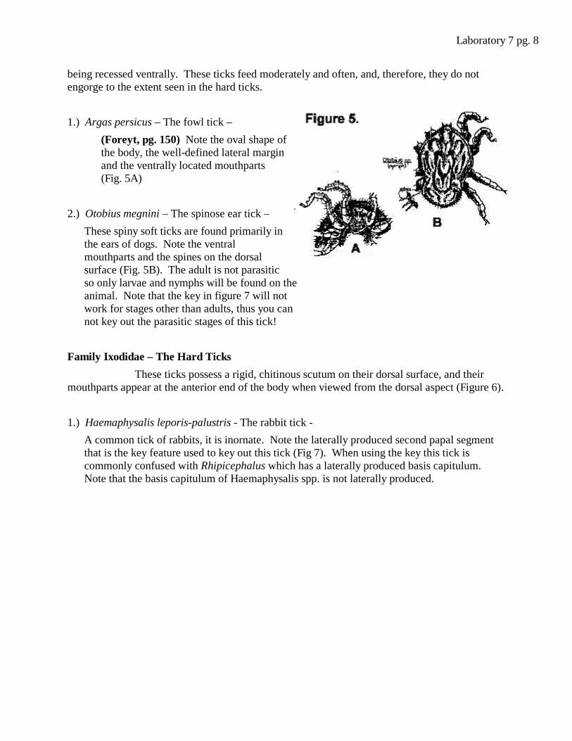
Family Argasidae – The Soft Ticks
Ticks of this family lack the scutum (the hard shield-like plate on the dorsal surface)and have a leathery cuticle. The mouthparts are not visible from the dorsal side (Figure 5),
Laboratory 7 pg. 8
being recessed ventrally. These ticks feed moderately and often, and, therefore, they do notengorge to the extent seen in the hard ticks.
1.) Argas persicus – The fowl tick –
(Foreyt, pg. 150) Note the oval shape ofthe body, the well-defined lateral marginand the ventrally located mouthparts(Fig. 5A)
2.) Otobius megnini – The spinose ear tick –
These spiny soft ticks are found primarily inthe ears of dogs. Note the ventralmouthparts and the spines on the dorsalsurface (Fig. 5B). The adult is not parasiticso only larvae and nymphs will be found on theanimal. Note that the key in figure 7 will notwork for stages other than adults, thus you cannot key out the parasitic stages of this tick!
Family Ixodidae – The Hard Ticks
These ticks possess a rigid, chitinous scutum on their dorsal surface, and theirmouthparts appear at the anterior end of the body when viewed from the dorsal aspect (Figure 6).
1.) Haemaphysalis leporis-palustris - The rabbit tick -
A common tick of rabbits, it is inornate. Note the laterally produced second papal segmentthat is the key feature used to key out this tick (Fig 7). When using the key this tick iscommonly confused with Rhipicephalus which has a laterally produced basis capitulum. Note that the basis capitulum of Haemaphysalis spp. is not laterally produced.
Laboratory 7 pg. 9
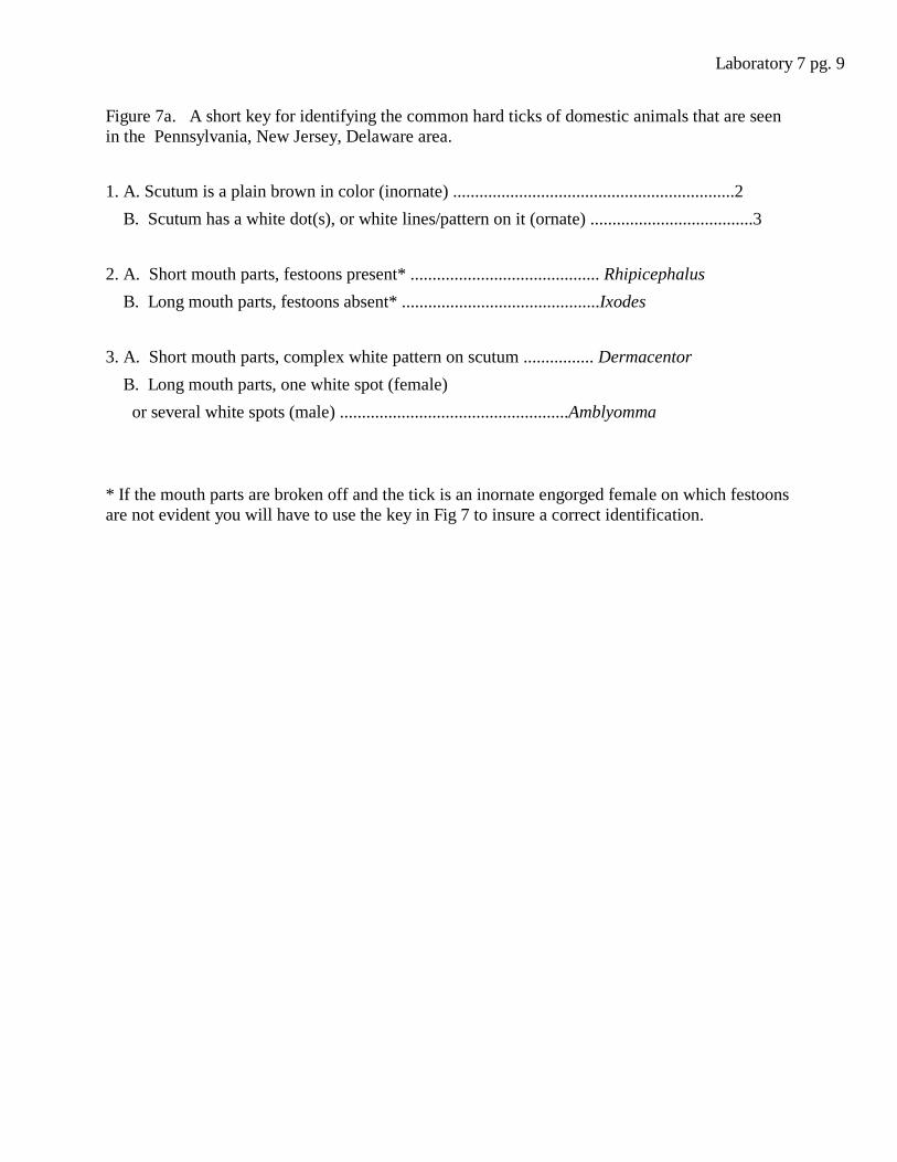
Figure 7a. A short key for identifying the common hard ticks of domestic animals that are seenin the Pennsylvania, New Jersey, Delaware area.
1. A. Scutum is a plain brown in color (inornate) ................................................................2
B. Scutum has a white dot(s), or white lines/pattern on it (ornate) .....................................3
2. A. Short mouth parts, festoons present* ........................................... Rhipicephalus
B. Long mouth parts, festoons absent* .............................................Ixodes
3. A. Short mouth parts, complex white pattern on scutum ................ Dermacentor
B. Long mouth parts, one white spot (female)
or several white spots (male) ....................................................Amblyomma
* If the mouth parts are broken off and the tick is an inornate engorged female on which festoonsare not evident you will have to use the key in Fig 7 to insure a correct identification.

Laboratory 7 pg. 10