laboratoire gdndtique moldculaire docteur roux,
TRANSCRIPT
Proc. Nat!. Acad. Sci. USAVol. 86, pp. 6676-6680, September 1989Genetics
Genome organization of the anaerobic pathogenClostridium perfringens
(genome evolution/pulsed-field gel electrophoresis/indirect end-labefing/gangrene/virulence)
BRUNO CANARD AND STEWART T. COLELaboratoire de Gdndtique Moldculaire Bacterienne, Institut Pasteur, 28, rue du Docteur Roux, 75724 Paris Cedex 15, France
Communicated by Jesse C. Rabinowitz, May 22, 1989
ABSTRACT A physical map of the genome of Clostiidiumperfringens, an important human pathogen, has been estab-lished by pulsed-field gel electrophoresis. Recognition sites forsix rare-cutting endonucleases were situated on a single circularchromosome of approximately 3.6 million base pairs thusdefining 50 arbitrary genetic intervals of between 10 and 250kilobase pairs. This considerably facilitated the chromosomallocalization of some 24 genes and loci for which probes wereavailable and allowed the construction of the genome map of aclostridial species.
The genus Clostridium consists of a diverse collection ofobligately anaerobic Gram-positive bacteria that all sporulatebut have a dG+dC content varying from 24 to 55% (1).Several of its members are ofbroad biotechnological interestwhereas others produce some of the deadliest toxins known.Clostridium perfringens, a common pathogen of man anddomestic animals, is responsible for a number of clinicalconditions ranging from relatively mild food poisoning to thepotentially life-threatening gas gangrene. Clinical isolateshave been classified into five serotypes, A-E, differing intheir tropisms and toxigenicity (2).
Genetic studies ofthis medically important bacterium havebeen hampered by the lack of classical tools, such as trans-ducing phages, and by difficulty in introducing recombinantDNA molecules. To circumvent these problems and, inparticular, to complement studies on bacterial virulencefactors and their role in pathogenesis, we decided to con-struct a physical map of the C. perfringens genome using avariety of techniques based around pulsed-field gel electro-phoresis (PFGE) (3-6).
In the second phase of the project, a gene map comprisingmore than 24 genes and chromosomal loci was superimposedon the physical map and it is now possible to locate rapidlyany cloned C. perfringens gene to a genetic interval of 50-250kilobase pairs (kb).
MATERIALS AND METHODSDNA Preparation. C. perfringens strain CPN50 was grown
to midexponential phase in TYG broth (7, 8). Washed cells(109 cells per ml) were mixed with an equal volume of 1%molten low-melting-point agarose (BRL) and the mixture wasallowed to solidify in 80-pI rectangular molds. Genomic DNAwas prepyred in the agarose plugs as described (9). Briefly,plugs wefe incubated for 16 hr at 37°C with gentle shaking ina solution containing 6mM Tris HCl (pH 7.6), 1 M NaCl, 100mM EDTA, 0.5% sarkosyl, and lysozyme (1 mg/ml) and thenplugs were deproteinized by incubating in ESP [0.5 MEDTA/0.5% sarkosyl/proteinase K (2 mg/ml), pH 8.5] for 48hr at 50°C. Prior to restriction endonuclease digestion, the
plugs, which had been stored in 0.5 M EDTA (pH 8.5), werewashed in TE buffer (10 mM Tris-HCl/1 mM EDTA, pH 8.0).
Restriction Digests. Apa I, Avi II, Ksp I, Mlu I, Nar I, NruI, Sma I, Sph I, and Stu I were provided by BoehringerMannheim, whereas Fsp I, Nae I, Not I, Sac II, and Xma Iwere purchased from New England Biolabs. DNA samples inagarose (40 Al) were digested with 20 units of enzyme for 4hr at 370C (250C for Sma I) in 200 Al of the buffer recom-mended by the manufacturer. In our hands, Avi II- and KspI digested DNA in agarose better than their isoschizomersFsp I and Sac II.
Electrophoresis Conditions. Most of the PFGE was per-formed by field inversion (5) using a COPROG model 3000microprocessor (Production Fabrication Controle, Paris) tocontrol the pulse duration and ramp. In some experimentssamples were subjected to contour-clamped homogeneouselectric field (CHEF) electrophoresis (6) or orthogonal fieldgel electrophoresis (OFAGE) (4) using an apparatus pur-chased from Pharmacia L.K.B. Biotechnology.
Chilled agarose plugs were inserted into the wells of a 1%agarose gel (25 X 20 cm) containing 0.66x TBE buffer (lxTBE is 89mM Tris/89mM boric acid/2 mM EDTA, pH 8.3).Gels were kept at constant temperature (80C) and the runningbuffer (0.66x TBE) was recirculated during electrophoresis,which was performed for 36 hr at 240 V using a ramp of 0.33-+60 s in the forward direction and 0.11 -* 20 s in the reverse.After electrophoresis, gels were stained with ethidium bro-mide and photographed using a Polaroid-Land model MP-4camera. The sizes of DNA fragments were estimated bycomparison with concatamerized A genomes or yeast chro-mosomes (10).
Southern Blot Hybridizations. After electrophoresis gelswere UV-irradiated for 2 min to fragment DNA, denatured in0.5 M NaOH/1.5 M NaCl for 45 min, and then neutralized in0.5 M Tris-HCI/1.5 M NaCl, pH 7.5 for 45 min. DNA wastransferred to Hybond C-extra filters (Amersham) as de-scribed (11) and fixed by baking for 2 hr at 80'C. Hybridiza-tion with 32P-labeled probes, produced by random priming(12), and washing were as described (13). When heterologousprobes were used, filters were hybridized and washed at220C.Other Techniques. Standard cloning procedures (14) were
employed in conjunction with dephosphorylated plasmidvectors pUC18 and pMTL22p (15, 16). Plaque hybridizationwas performed as described (17).
RESULTSEstimation of the Genome Size. The principal aims of this
project were to determine the size of the genome of a type-AC. perfringens strain associated with human disease byPFGE and to use the information obtained to establish aphysical map to facilitate the localization ofcloned genes. We
Abbreviations: PFGE, pulsed-field gel electrophoresis; Mb,megabase pair(s).
6676
The publication costs of this article were defrayed in part by page chargepayment. This article must therefore be hereby marked "advertisement"in accordance with 18 U.S.C. §1734 solely to indicate this fact.
Dow
nloa
ded
by g
uest
on
Dec
embe
r 11
, 202
1
Proc. Natl. Acad. Sci. USA 86 (1989) 6677
reasoned that since the genomic DNA was composed of 75%dA+dT, restriction enzymes with dG+dC-rich recognitionsites of six nucleotides, or more, should cleave on averageevery 60-100 kb and thereby generate a small number oflargerestriction fragments. Furthermore, we calculated that if therestriction-site distribution was near random and the genomesize was typical of a eubacterium [3-5 megabase pairs (Mb)],then the use of five or six appropriate enzymes wouldgenerate a map with arbitrary genetic intervals of 50-200 kb,defined by the distance between sites. This would be ideal forthe rapid localization of genetic loci for which hybridizationprobes were available.
Consequently, DNA was prepared from strain CPN50,originally isolated from a patient with septicemia (7, 8), andscreened with a battery of restriction enzymes. Analysis ofthe cleavage products by PFGE showed that Apa I(GGGCCC), Fsp I (TGCGCA), Mlu I (ACGCGT), Nru I(TCGCGA), Sac II (CCGCGG), and Sma I (CCCGGG)reproducibly generated a small number of large DNA frag-ments. The patterns obtained with Nru I and Sac II werevirtually identical, indicating close linkage and similar dis-tribution of the recognition sites. Of the other enzymestested, it was found that Nae I (GCCGGC) and Nar I(GGCGCC) always gave incomplete digests, Sph I (GCAT-GC) and Stu I (AGGCCT) generated too many fragments, andNot I (GCGGCCGC) had no sites in the genome.
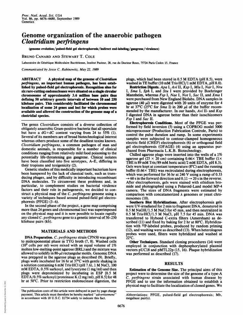
Typical results obtained by cleavage with the six chosenenzymes are presented in Fig. 1. The fragments generated bysingle digests ranged from 5 to 16 in number and from 0.03 to2.2 Mb in size. By comparison with appropriate molecularweight markers, fragment sizes could be estimated accuratelyafter PFGE and these are summarized in Table 1. For thelarger fragments (>0.3 Mb), sizes were also determined afterorthogonal field gel electrophoresis or contour-clamped ho-mogeneous electric field electrophoresis (4, 6). However, asreported by others (18), the ethidium bromide fluorescence offragments larger than 0.6 Mb was often stoichiometricallydisproportional (see Fig. 1, lanes 1 and 8). The genome size
A B
1 2 3 4 5 6 7 8 9
kb
1000
800 -
680650
200
kb
970
- 485
48.5
FIG. 1. PFGE separation of restriction fragments of C. perfrin-gens genomic DNA. (A) Samples separated on a field-inversion gelwere digested with Mlu I and the following endonucleases. Lanes: 1,alone; 2, Sma I; 3, Apa I; 4, Sac II; 5, FspI; 6, NruI. (B) Samplesdigested with Apa I (lane 7) or Mlu I (lane 8) were resolved byorthogonal field gel electrophoresis. The deduced sizes of the Mlu Ifragments and the positions of the A size markers (lane 9) areindicated.
Table 1. Restriction fragments of C. perfringens genomic DNAFragment size, kb
Apa I Fsp I M1u I Nru I Sac II Sma I650 450 1200 2280 1640 1460530 450 830 405 405 440440 340 680 260 320 410420 230 650 260 260 405410 230 200 160 260 170360 225 100 230 170320 210 70 160 170200 210 45 100 120140 190 70 120100 180 45 11040 170 7535 160 45
1401108550
Total 3645 3430 3560 3580 3490 3695Mean fragment size from five independent measurements was
rounded offto the nearest 5 kb; fragments <35 kb were not detected.
deduced from the various single digests was 3.58 ± 0.2 Mb(mean ± SEM) (Table 1).To determine the relative order of the sites, double digests
were performed and these yielded valuable information aboutphysical relationships. However, it was difficult to attributeunambiguously the smaller restriction fragments (<100 kb) toa precise genomic location so a complementary mappingapproach was employed.Mapping by Indirect End-Labeling. Olson and coworkers
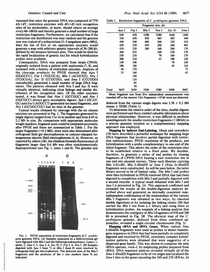
(19) have described a powerful technique for mapping largeDNA fragments that involves partial digestion with restric-tion endonucleases, PFGE resolution of the products, andhybridization with a probe complementary to one end of theinitial fragment. This allows the order of the restriction sitesto be established relative to a fixed point. We decided,therefore, to generate a series of end probes by cloningfragments of CPN50 DNA bearing a rare restriction site atone end into plasmid vectors. Three such libraries carryingMlu I-EcoRI, Mlu I-HindIII or Sma I (Xma I)-HindIIIfragments were constructed. As will be seen below, the latterlibrary proved to be of limited value. The Mlu I end probeswere then hybridized to PFGE-resolved DNA that had beendigested to completion with Mlu I and partially digested witha second enzyme. A typical result obtained with Mlu I andApa I is presented in Fig. 2A. This approach confirmed andextended the results of the double-digestion analysis de-scribed above and generated an internally consistent map.Independent confirmation of the relationships of the variousMlu I fragments was obtained in two ways, by classicaldouble digestions or by isolating the linking clones (20) thatcarried the Mlu I site from a A library and using them ashybridization probes. An example of a linking analysis thatdemonstrates the contiguity ofMlu I fragments of650 and 200kb is presented in Fig. 2B. The physical map of the C.perfringens genome, deduced from these combined ap-proaches, revealed a single circular chromosome (Fig. 3).Mapping rRNA and tRNA Genes. When various Sma
I-HindIII fragments were used as probes to detect homolo-gous sequences in DNA that had been partially or completelydigested and resolved by PFGE, extremely complex hybrid-ization patterns were obtained that were suggestive of adispersed gene family. This was shown to comprise the ninerRNA operons, rrnA-I, by employing probes prepared fromrRNA. DNA sequence analysis revealed virtually all of theSma 1-HindIII fragments to be of rrn origin and localized theSma I sites in the genes encoding the 16S and 23S rRNAs. In
Genetics: Canard and Cole
Dow
nloa
ded
by g
uest
on
Dec
embe
r 11
, 202
1
Proc. Natl. Acad. Sci. USA 86 (1989)
kb
A
1 2 3 4 5 6 7 8
650 %%
530- _ so "!
4j3g60 *. v .
3
FIG. 2. (A) Indirect end-labeling atgenomic DNA. Samples (1 Ag) were digestI then partially digested with serially dilute1, no enzyme; 2-8, 0.02, 0.05, 0.1, 0.respectively. After PFGE analysis, DNA vwith a 32P-labeled Mlu I-EcoRI end priindicated; the map deduced covers the Mh2.3 Mb to position 3.0 Mb in the clockwLinking analysis of 0.20- and 0.65-Mb Ashown in Fig. LA was hybridized with a 2.'which carries the Mlu I site 300 base Fexplains the relatively weak hybridization(Additional bands in lane 12 are due to inc
some cases the HindIII sites were fourunique sequences and one of these wato the gyrA gene, coding for the large sby homology with its counterpart inAdditional mapping studies establisicluster of rare sites, Sac II-Sac II-SnI-Nru I, in each rrn operon (R in Fispecific for the 5' or 3' ends of the 16'we could demonstrate that seven of
2
B transcribed clockwise and the remaining two were tran-scribed in a counter-clockwise direction. When hybridization
9 10 11 12 13 14 was performed with probes prepared from total tRNA, thekb pattern was indistinguishable from that obtained with rRNA650 probes, indicating tight linkage between the corresponding
genes.Mapping Genes for Virulence Factors. It is generally be-
lieved that the pathogenicity of C. perfringens is due to thesecretion of toxins or virulence factors that attack the host
4W.tissue. The genes for several putative virulence factors thathave been cloned in this laboratory and elsewhere (22, 23)were used as hybridization probes to test the hypothesis that
200 a particular area of the genome might be associated withbacterial virulence. In this way the chromosomal locations of
30 the plc and pfo genes encoding the a and 6 toxins and thenanH and nagH genes encoding a sialidase and 83-
ialysis of C. perfringens N-acetylglucosaminidase, respectively, were established.,ed to completion with Mlu With the exception ofnanH, three ofthe four virulence factordApa I as follows: Lanes: genes were localized to a 200-kb region of the chromosome.2, 0.4, 0.6, and 4 units, Interestingly, nanH is situated on the same Fsp I fragment asvas blotted and hybridized the lysogenic bacteriophage'29.*obe. Fragment sizes are MappingHoskepn.ge V e s du I fragment from position Mapping Housekeeping Genes. Very few genes involved inise direction (Fig. 3). (B) the basic metabolism of clostridial species have been iso-W1u I fragments. The gel lated. Consequently, to map some of the genes for house-7-kb EcoRI linking probe, keeping functions, a series of heterologous probes derived)airs from one end. This from cloned B. subtilis or Escherichia coli genes was em-on the 0.2-Mb fragment. ployed. Although many of these did not cross-hybridize,omplete Sac II digestion.) probably due to differences in base composition, a numberid to be associated with gave positive results. Ofparticular interest was the gyrB geneiS shown to correspond of B. subtilis (24) that detected homologous sequences on asubunit ofDNA gyrase, 40-kb Fsp I-Sma I (Fig. 4) fragment known to carry the gyrABacillus subtilis (21). gene and this suggests that gyrA and gyrB are tightly linked
hed the presence of a as is the case in B. subtilis (21, 24). By using this heterologousna I-Sma I-NruI-Sma approach, genes have been tentatively localized for theig. 3). By using probes following cellular components: DNA gyrase (gyrA and gyrB),S and 23S rRNA genes, RNA polymerase core enzyme (rpoA, rpoB, and rpoC), o3the rrn operons were (rpoD), ATP synthase (atpD), and elongation factor Tu,
3.6 / 0
M
FIG. 3. Physical and gene map of the chromo-some of C. perfringens strain CPN50. Restrictionsites forApa I (A), Fsp I (F), Mlu I (M), Sac 11 (C),and Sma I (S) are positioned on the outside of thecircle; R denotes the rare site cluster, Sac 11 (2),Sma 1 (2), Nru I, Sma I, and Nru I, which occurs ineach RNA operon. The scale in 0.1-Mb intervals isshown on the inside as well as known genes orgenetic loci identified by cross hybridization withheterologous probes. Known clostridial genes wereas follows: gyrA, DNA gyrase; nagH, /3-N-acetylglucosaminidase; nanH, sialidase; pfo, per-fringolysin or 0 toxin; plc, phospholipase C or atoxin; rrnA-I, rRNA operons; att029, integrationsite for 429; att059, integration site for )59. Othergenetic loci are as follows: atpD, P subunit of ATPsynthase; gyrB, DNA gyrase; rpoA, a subunit ofRNA polymerase; rpoB, /3 subunit of RNA poly-merase; rpoC, 8' subunit of RNA polymerase;rpoD, a43 subunit of RNA polymerase; tufA, B,elongation factor Tu.
6678 Genetics: Canard and Cole
Dow
nloa
ded
by g
uest
on
Dec
embe
r 11
, 202
1

Proc. Natl. Acad. Sci. USA 86 (1989) 6679
A B1 2 3 4
kb 1 2 3 4
--40-
FIG. 4. Mapping DNA gyrase genes. (A) Hybridization with a C.perfringens gyrA-specific probe. Samples were digested with Sma I,
Sma I plus Apa I, Sma I plus Fsp I, or Sma I plus Nru I, in lanes 1-4,respectively. (B) Same blot was hybridized with a probe derived fromthe B. subtilis gyrB gene (24).
which seems to be encoded by two genes (tufA and tufB; ref.25).
DISCUSSIONC. perfringens strain CPN50, associated with human disease,has been shown to possess a single circular chromosome ofabout 3.58 Mb by PFGE. More than 100 restriction sites and24 genetic loci have been located on the genome that isslightly larger in size than that of the other Gram-positivebacterium for which physical mapping data have been pub-lished, Staphylococcus aureus at 2.86 Mb (18). B. subtilis, adistant relative and fellow member of the Bacillaceae, has agenome estimated at 5 Mb by classical genetic techniques(26), although, surprisingly, no PFGE data are available forthis most intensively studied and thoroughly mapped Gram-positive microorganism.A striking similarity between C. perfringens and B. subtilis
can be seen in the number, 9 and 10, respectively, andorganization of the rRNA operons, rrn, as in both bacteriathese are clustered in a region representing about one-third ofthe genome (Fig. 3; refs. 26 and 27). More importantly, in B.subtilis the origin of replication is linked to the DNA gyrasegenes, gyrA and gyrB, which precede the rrnO operon (21,28). In this study, we have demonstrated linkage betweengyrA, gyrB, and rrnA and, by analogy, it is tempting tospeculate that the C. perfringens origin of replication isadjacent. If this is the case, there are 7 rrn operons locatedon the clockwise side of the origin, compared to 9 in B.subtilis (27) and 2 in an anticlockwise position. Furthersimilarities can be seen in the compact organization of thetRNA and rRNA genes near the putative origin and this maybe significant with respect to the sporulation and germinationprocesses that both undergo (29).One of our goals was to determine whether genes likely to
be involved in bacterial virulence were clustered in a preciseregion ofthe chromosome. As shown in Fig. 3, theplc andpfogenes encoding the major cytotoxic determinants secreted byC. perfringens are located near the putative origin of repli-cation. Furthermore, an operon coding for a third possiblevirulence factor, the f-N-acetylglucosaminidase is also situ-ated nearby. Although this does not identify a "virulencedomain," it does suggest that this region may merit furtherinvestigation.
It is highly significant that the genes for the a and 0 toxins,which are produced by all serotypes of C. perfringens (2), arelocated near the presumed origin of replication, as this areashould be among the most conserved regions of the genome.
PFGE studies of genomic DNA from the five major classesof C. perfringens strains suggest that the bulk of the chro-mosome is the same in all isolates but that the 1.2-Mb MIu Ifragment is prone to genetic variation (unpublished results).This could in part be explained by bacteriophage-mediatedrearrangements and it is noteworthy that the attachment sitefor the lysogenic phage q429 (8) is on this fragment in CPN50.Strikingly, the only other genetic marker mapped to thisregion is nanH encoding sialidase, a potential virulence factor(23). These findings suggest that this particular area of thegenome is susceptible to genetic variability and this might, inpart, account for the different host specificities displayed byvarious clinical isolates of C. perfringens.
Finally, the physical map constructed in this study, to ourknowledge, is not only the first such map for a clostridialspecies but also the first detailed map of any Gram-positivebacterium. Good examples of the utility and value ofgenomemapping are provided by pioneering work with the Gram-negative organism E. coli (20, 30). It is clear that thisapproach will not only have important ramifications forbacterial genetics but also considerably advance studies onphylogeny and molecular evolution.
We thank Thierry Gamier for advice, encouragement, and the Alibrary, Christine Petit and Jacqueline Levilliers for help with con-tour-clamped homogeneous electric field and orthogonal field gelelectrophoresis analyses, and all the individuals, too numerous toname, who kindly provided probes. B.C. was supported by apredoctoral fellowship from the Centre National de la RechercheScientifique. This work was funded by Grants 85008 and 880588 fromthe Institut National de la Santd et de la Recherche Mddicale.
1. Cato, E. P., George, W. L. & Finegold, S. M. (1986) inBergey's Manual of Systematic Bacteriology, eds. Sneath,S. P. H. A., Mair, S. N., Sharpe, M. E. & Holt, J. G. (Wil-liams & Wilkins, Baltimore), pp. 1141-1200.
2. Finegold, S. (1977) Anaerobic Bacteria in Human Disease(Academic, New York).
3. Schwartz, D. C. & Cantor, C. R. (1984) Cell 37, 67-75.4. Carle, G. F. & Olson, M. V. (1985) Proc. Natl. Acad. Sci. USA
82, 3756-3760.5. Carle, G. F., Frank, M. & Olson, M. V. (1986) Science 232,
65-68.6. Chu, G., Vollrath, D. & Davis, R. W. (1986) Science 234,
1582-1585.7. Gamier, T. & Cole, S. T. (1986) J. Bacteriol. 168, 1189-11%.8. Brefort, G., Magot, M., lonesco, H. & Sebald, M. (1977)
Plasmid 1, 52-66.9. Smith, C. L., Warburton, P. E., Gaal, A. & Cantor, C. R.
(1986) in Genetic Engineering, eds. Setlow, J. K. & Hollaen-der, A. (Plenum, New York), Vol. 8, pp. 45-70.
10. Anand, R. (1986) Trends Genet. 2, 278-283.11. Southern, E. M. (1975) J. Mol. Biol. 98, 503-517.12. Feinberg, A. P. & Vogelstein, B. (1984) Anal. Biochem. 137,
266-267.13. Wahl, G. M., Stern, M. & Stark, G. R. (1979) Proc. NatI.
Acad. Sci. USA 76, 3683-3687.14. Silhavy, T. J., Berman, M. L. & Enquist, L. (1984) Experi-
ments with Gene Fusions (Cold Spring Harbor Lab., ColdSpring Harbor, NY).
15. Yanisch-Perron, C., Vieira, J. & Messing, J. (1985) Gene 33,113-119.
16. Chambers, S. P., Prior, S. E., Barstown, D. A. & Minton,N. P. (1988) Gene 68, 139-149.
17. Benton, W. D. & Davis, R. W. (1977) Science 196, 180-182.18. Weil, M. D. & McClelland, M. (1989) Proc. Natl. Acad. Sci.
USA 86, 51-55.19. Graham, M. Y., Otani, T., Boime, I., Olson, M. V., Carle,
G. F. & Chaplin, D. D. (1987) Nucleic Acids Res. 15, 4437-4448.
20. Smith, C. L., Econome, J. G., Schutt, A., Kleo, S. & Cantor,C. R. (1987) Science 236, 1448-1453.
21. Moriya, S., Ogasawara, N. & Yoshikawa, H. (1985) NucleicAcids Res. 13, 2251-2265.
Genetics: Canard and Cole
Dow
nloa
ded
by g
uest
on
Dec
embe
r 11
, 202
1

6680 Genetics: Canard and Cole
22. Tweten, R. K. (1988) Infect. Immun. 56, 3228-3234.23. Roggentin, P., Rothe, B., Lottspeich, F. & Schauer, R. (1988)
FEBS Lett. 238, 31-34.24. Lampe, M. F. & Bott, K. F. (1985) J. Bacteriol. 162, 78-84.25. Sela, S., Yogev, D., Razin, S. & Bercovier, H. (1989) J.
Bacteriol. 171, 581-584.26. Piggot, P. J. & Hoch, J. A. (1985) Microbiol. Rev. 49,158-179.
Proc. Nati. Acad. Sci. USA 86 (1989)
27. Jarvis, E. D., Wisdom, R. L., La Fauci, G., Setoguchi, Y.,Richter, I. R. & Rudner, R. (1988) Genetics 120, 625-635.
28. Henckes, G., Vannier, F., Seiki, M., Ogasawara, N., Yoshi-kawa, H. & Seror-Laurent, S. J. (1982) Nature (London) 299,268-271.
29. Vold, B. S. (1985) Microbiol. Rev. 49, 71-80.30. Kohara, Y., Akiyama, K. & Isono, K. (1987) Cell 50, 495-508.
Dow
nloa
ded
by g
uest
on
Dec
embe
r 11
, 202
1