lab unit 1 histology of bone tissue - mc3cb.com · lab unit 1 histology of bone tissue. general...
TRANSCRIPT
Lab Unit 1
Histology of Bone Tissue
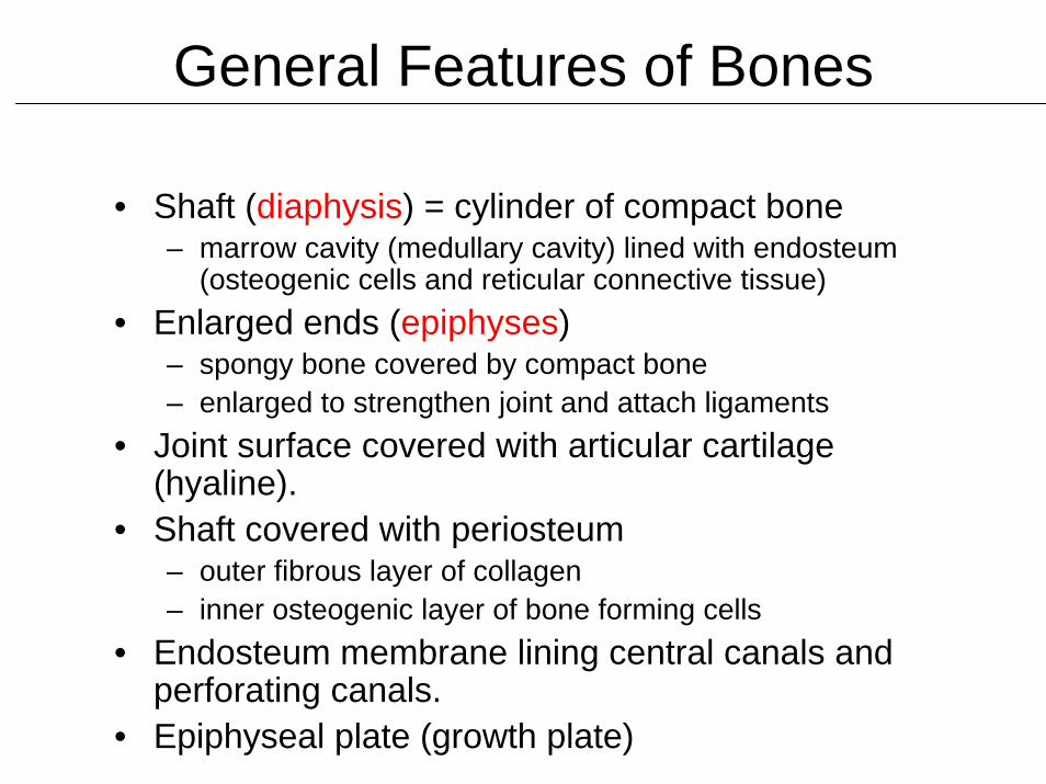
General Features of Bones
• Shaft (diaphysis) = cylinder of compact bone– marrow cavity (medullary cavity) lined with endosteum
(osteogenic cells and reticular connective tissue)• Enlarged ends (epiphyses)
– spongy bone covered by compact bone– enlarged to strengthen joint and attach ligaments
• Joint surface covered with articular cartilage (hyaline).
• Shaft covered with periosteum– outer fibrous layer of collagen – inner osteogenic layer of bone forming cells
• Endosteum membrane lining central canals and perforating canals.
• Epiphyseal plate (growth plate)
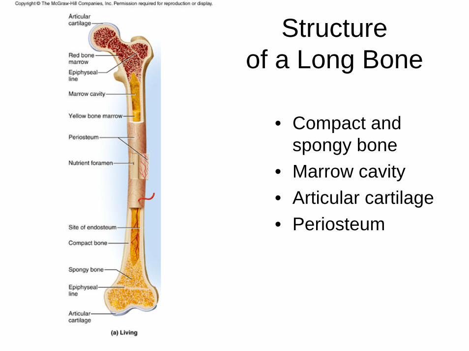
Structureof a Long Bone
• Compact and spongy bone
• Marrow cavity• Articular cartilage• Periosteum
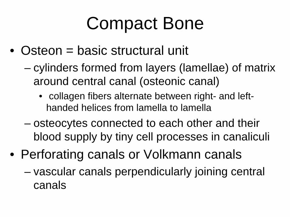
Compact Bone• Osteon = basic structural unit
– cylinders formed from layers (lamellae) of matrix around central canal (osteonic canal)
• collagen fibers alternate between right- and left-handed helices from lamella to lamella
– osteocytes connected to each other and their blood supply by tiny cell processes in canaliculi
• Perforating canals or Volkmann canals– vascular canals perpendicularly joining central
canals
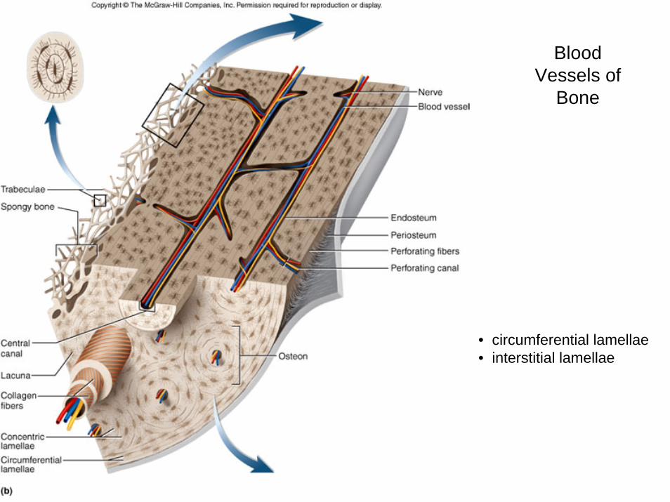
BloodVessels of
Bone
• circumferential lamellae• interstitial lamellae
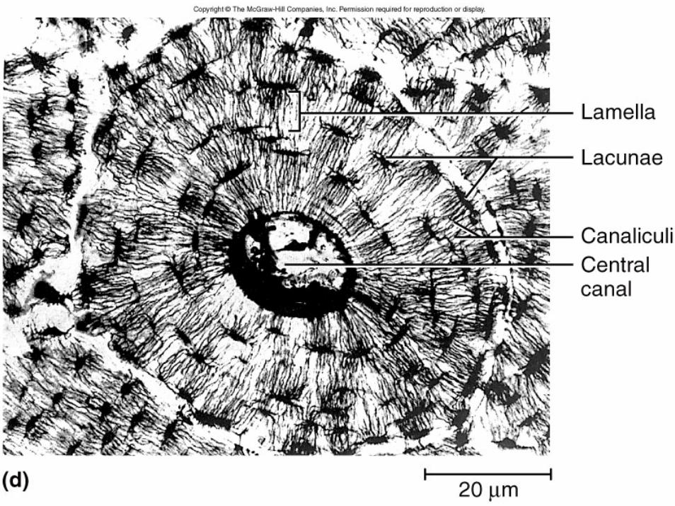
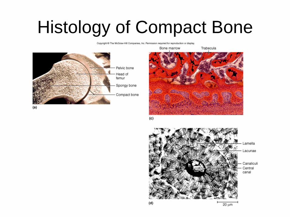
Histology of Compact Bone
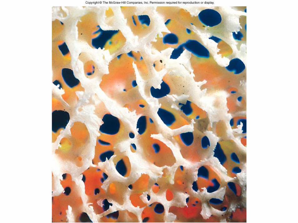
Spongy Bone
• Spongelike appearance formed by plates of bone called trabeculae– spaces filled with red bone marrow
• Trabeculae have few osteons or central canals– no osteocyte is far from blood of bone
marrow• Provides strength with little weight
– trabeculae develop along bone’s lines of stress
CO 7
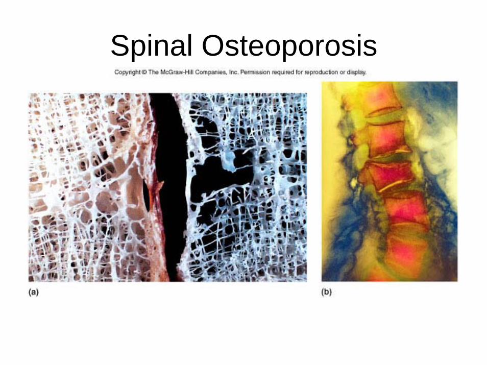
Spinal Osteoporosis
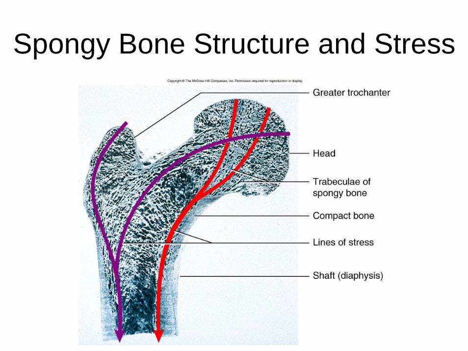
Spongy Bone Structure and Stress
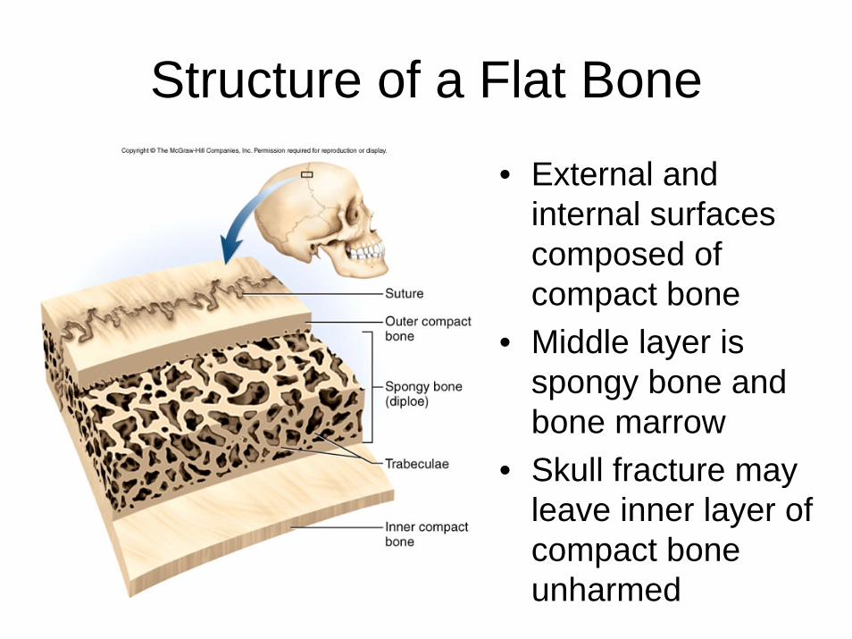
Structure of a Flat Bone
• External and internal surfaces composed of compact bone
• Middle layer is spongy bone and bone marrow
• Skull fracture may leave inner layer of compact bone unharmed
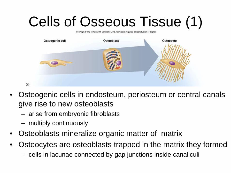
Cells of Osseous Tissue (1)
• Osteogenic cells in endosteum, periosteum or central canals give rise to new osteoblasts– arise from embryonic fibroblasts – multiply continuously
• Osteoblasts mineralize organic matter of matrix • Osteocytes are osteoblasts trapped in the matrix they formed
– cells in lacunae connected by gap junctions inside canaliculi
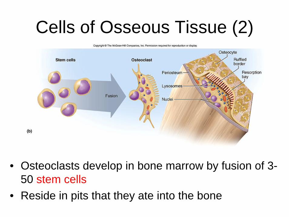
Cells of Osseous Tissue (2)
• Osteoclasts develop in bone marrow by fusion of 3-50 stem cells
• Reside in pits that they ate into the bone
Matrix of Osseous Tissue• Dry weight = 1/3 organic and 2/3 inorganic matter• Organic matter
– collagen, glycosaminoglycans, proteoglycans and glycoproteins• Inorganic matter
– 85% hydroxyapatite– 10% calcium carbonate– other minerals (fluoride, potassium, magnesium)
• Combination provides for strength and resilience– composite– minerals resist compression; collagen resists tension– bone adapts by varying proportions
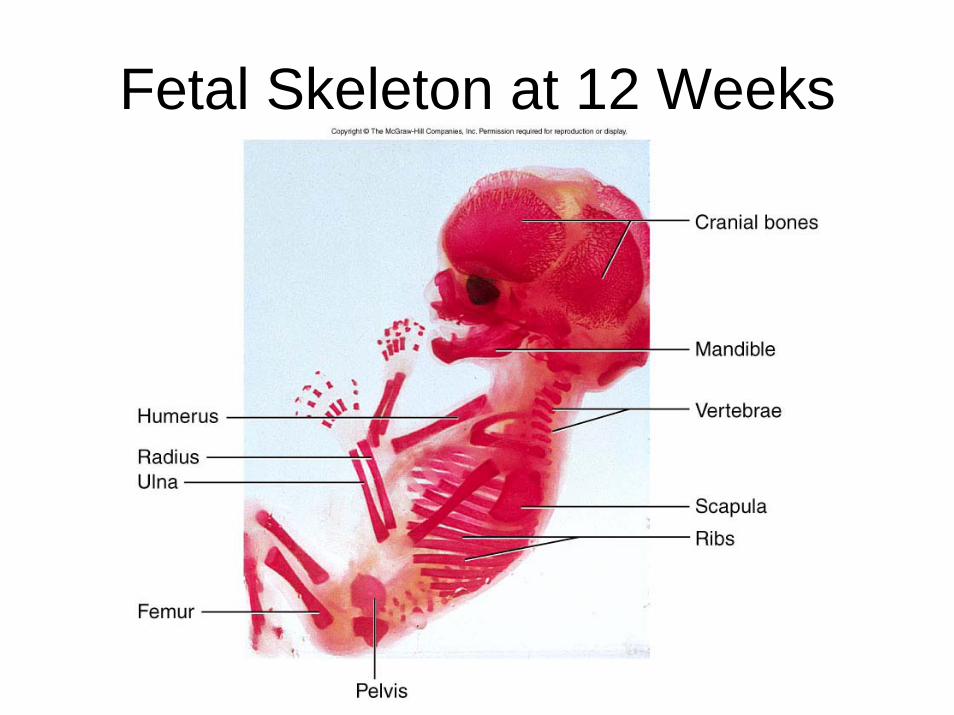
Fetal Skeleton at 12 Weeks
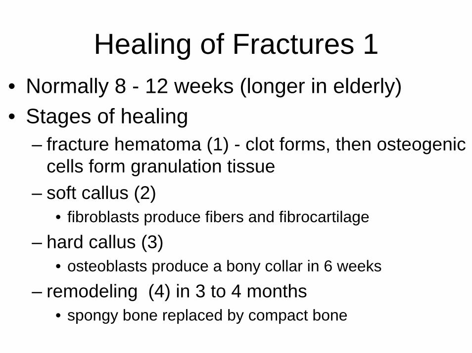
Healing of Fractures 1• Normally 8 - 12 weeks (longer in elderly)• Stages of healing
– fracture hematoma (1) - clot forms, then osteogenic cells form granulation tissue
– soft callus (2)• fibroblasts produce fibers and fibrocartilage
– hard callus (3)• osteoblasts produce a bony collar in 6 weeks
– remodeling (4) in 3 to 4 months • spongy bone replaced by compact bone
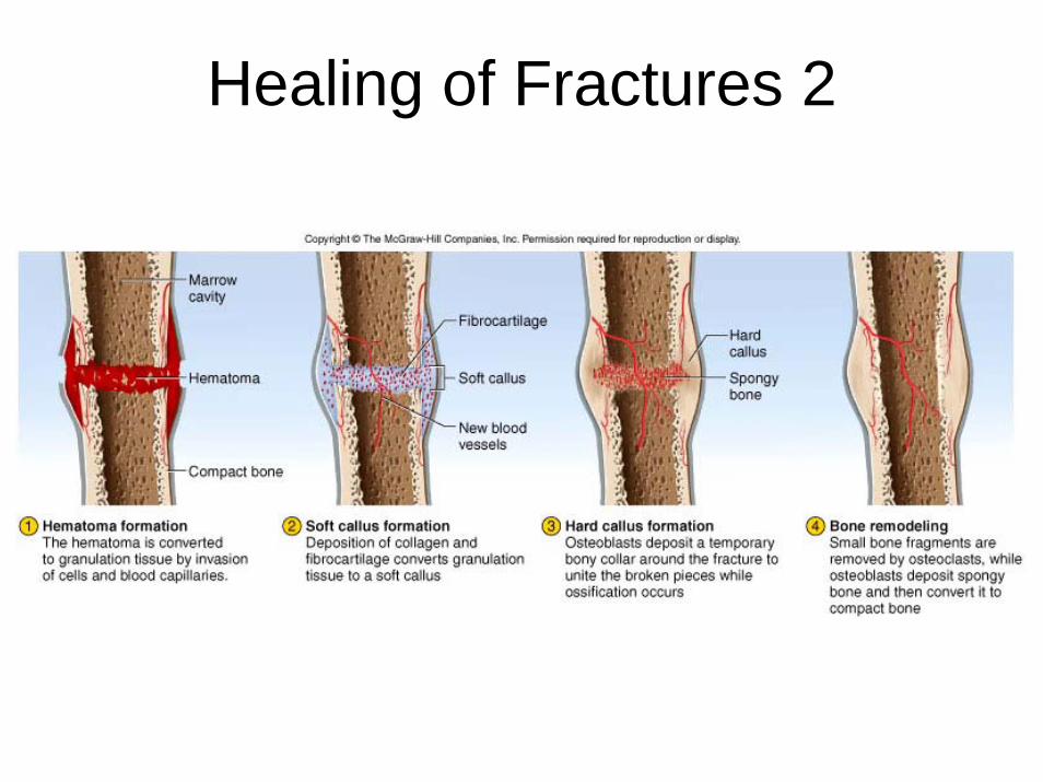
Healing of Fractures 2

Bone Marrow
• In medullary cavity (long bone) and among trabeculae (spongy bone)
• Red marrow like thick blood– reticular fibers and immature cells– Hemopoietic (produces blood cells)– in vertebrae, ribs, sternum, pelvic girdle
and proximal heads of femur and humerus in adults
• Yellow marrow– fatty marrow of long bones in adults
• Gelatinous marrow of old age– yellow marrow replaced with reddish jelly