lab exercise 12 - houston community college
TRANSCRIPT

Lab Exercise 12:
DNA to Protein
Evelyn I. Milian Instructor
BIOLOGY I

BIOLOGY I. Lab 12: DNA to Proteins
Activities for this Lab Exercise
1. Brief introduction to Interactive online DNA tutorial – Students are strongly encouraged to use this and other websites for review
2. Conclusion of Lab Exercise 12 from lab manual
3. Building a DNA molecule using a kit provided by the instructor
4. Building your own DNA double helix from scratch
– This will be fun!!!
Evelyn I. Milian, Instructor 2

BIOLOGY I. Lab 12: DNA to Proteins
DNA STRUCTURE AND REPLICATION:
Some Websites for Review
• DNA structure interactive tutorial:
http://www.umass.edu/molvis/tutorials/dna/
• DNA replicating song (creative and funny!):
http://www.youtube.com/watch?v=dIZpb93NYlw
• DNA structure: http://www.youtube.com/watch?v=qy8dk5iS1f0
• Molecular visualization of DNA:
http://www.youtube.com/watch?v=4PKjF7OumYo
• DNA replication brief videos:
– http://www.youtube.com/watch?v=teV62zrm2P0&feature=related
– http://www.youtube.com/watch?v=AGUuX4PGlCc&feature=related
– http://www.youtube.com/watch?v=z685FFqmrpo&feature=related
Evelyn I. Milian, Instructor 3
BIOLOGY I. Lab 12: DNA to Proteins
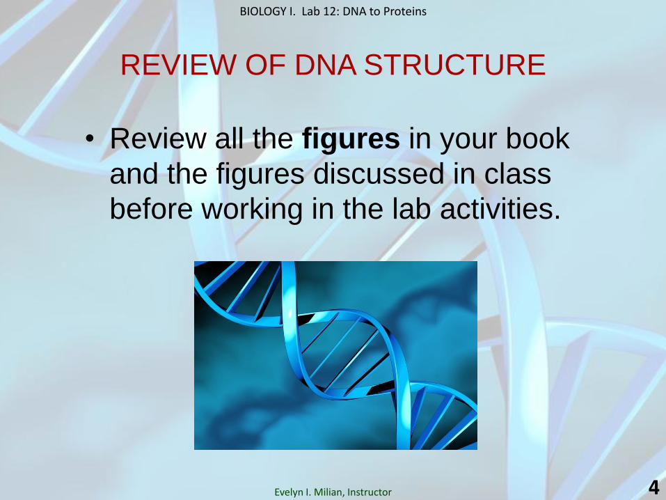
REVIEW OF DNA STRUCTURE
• Review all the figures in your book
and the figures discussed in class
before working in the lab activities.
Evelyn I. Milian, Instructor 4

BIOLOGY I. Lab 12: DNA to Proteins
NUCLEIC ACIDS: Nucleotide Structure
5 Evelyn I. Milian, Instructor

BIOLOGY I. Lab 12: DNA to Proteins
NUCLEIC ACIDS:
DNA
• Nucleotides (top) are composed of a deoxyribose (in DNA) sugar molecule linked to a phosphate group and to a nitrogenous base. The two nucleotides shown here are linked by hydrogen bonds between their complementary bases.
• The ladderlike form of DNA’s double helix (bottom) is made up of many nucleotides, with the repeating sugar-phosphate combination forming the backbone and the complementary bases the rungs.
6

BIOLOGY I. Lab 12: DNA to Proteins
NUCLEIC ACIDS: DNA
• DNA is a double helix in which the two polynucleotide strands twist about each other.
a) Hydrogen bonds (dotted lines) occur between the complementarily paired bases: A is always paired with T, and G is always paired with C (a purine with a pyrimidine).
b) Space-filling model of DNA.
Evelyn I. Milian, Instructor 7

BIOLOGY I. Lab 12: DNA to Proteins
Watson and Crick’s
Model of DNA
a. A space-filling model of DNA.
b. The two strands of the
molecule are antiparallel—
that is, the sugar-phosphates
are oriented in different
directions: The 5’ end of one
strand is opposite the 3’ end
of the other strand.
c. Diagram of DNA double
helix shows that the
molecule resembles a
twisted ladder or a spiral.
The bases are joined by
hydrogen bonds.
Evelyn I. Milian, Instructor 8

BIOLOGY I. Lab 12: DNA to Proteins
The Structure of DNA:
The Watson and Crick Model
• The two sugar-phosphate chains run in opposite directions. This orientation permits the complementary bases to pair.
• The pairs of nitrogenous bases
in a DNA double helix are held
together by hydrogen bonds,
as shown here.
• Adenine (A) pairs with
thymine (T), and guanine (G)
pairs with cytosine (C).
• The A-T pair has two hydrogen
bonds, the G-C pair has three.
Evelyn I. Milian, Instructor 9

BIOLOGY I. Lab 12: DNA to Proteins
The Structure of DNA: The Watson and Crick Model
• The two upright strands, composed of the sugar deoxyribose (D) and phosphate groups (P), are held together by hydrogen bonds between complementary bases. Adenine (A) always pairs with thymine (T), and guanine (G) always pairs with cytosine (C). Each strand can thus provide the information needed for the formation of a new DNA molecule.
• The DNA molecule is twisted into a double helix. The two sugar-phosphate strands run in opposite (antiparallel) directions. Each new strand grows from the 5’ (“five prime”) end toward the 3’ end.
Evelyn I. Milian, Instructor 10

BIOLOGY I. Lab 12: DNA to Proteins
Evelyn I. Milian, Instructor 11

BIOLOGY I. Lab 12: DNA to Proteins
A Simplified View of DNA Replication: The Basic Concept
• In this simplification, a short segment of DNA has been untwisted into a
structure that resembles a ladder. The rails of the ladder are the sugar-
phosphate backbones of the two DNA strands; the rungs are the pairs
of nitrogenous bases. Simple shapes symbolize the four kinds of
bases. Dark blue represents DNA strands present in the parent molecule;
light blue represents free nucleotides and newly synthesized DNA.
Evelyn I. Milian, Instructor 12

BIOLOGY I. Lab 12: DNA to Proteins
DNA Replication: Basic Steps
• After the DNA double helix unwinds (by helicase), each old strand serves as a template for the formation of the new strand.
• Complementary nucleotides available in the cell pair with those of the old strand and then are joined together to form a new strand.
• After replication is complete, there are two daughter DNA double helices. Each one is composed of an old strand and a new strand.
• Each daughter double helix has the same sequence of base pairs as the parental double helix had before unwinding occurred.
Evelyn I. Milian, Instructor 13

BIOLOGY I. Lab 12: DNA to Proteins
DNA REPLICATION: Incorporation of a Nucleotide into a DNA Strand
• DNA polymerase catalyzes the addition of a nucleoside triphosphate to the 3’ end of a growing DNA strand (a nucleoside triphosphate has a nitrogenous base, a pentose sugar, and three phosphate groups).
• When a nucleoside triphosphate bonds to the sugar in a growing DNA strand, it loses two phosphates. Hydrolysis of the phosphate bonds provides the energy for the reaction.
Evelyn I. Milian, Instructor 14

BIOLOGY I. Lab 12: DNA to Proteins
Building a DNA Model Using a Kit
• Use the kit provided by your instructor to build a model
of the DNA double helix molecule. The kit contains all
the parts you will need to build a DNA molecule model.
• Follow the instructions provided.
• Ask your instructor if you have questions.
Evelyn I. Milian, Instructor 15

BIOLOGY I. Lab 12: DNA to Proteins
Model of the DNA Molecule
Evelyn I. Milian, Instructor 16
Sugar-Phosphate Backbone:
• Red spheres = phosphates
• Black = deoxyribose (sugar)
• Yellow straws = connectors
Bases and Hydrogen Bonds:
• Red straws = adenine
• Blue straws = thymine
• Gray straws = guanine
• Green straw = cytosine
• White spheres = hydrogen bonds

BIOLOGY I. Lab 12: DNA to Proteins
Build Your Own DNA Double Helix from Scratch!!!
• Work in small groups of 4-6 students.
• Build your own DNA double helix using the materials supplied by your instructor. * Suggestion: Make your DNA molecule about 6 – 8 nucleotide pairs long, enough to fit the poster board. BE CREATIVE!!!
• You must build and join ALL the components of the DNA nucleotides, clearly labeled, or write a legend identifying them by colors. The DNA molecule should look like one of the figures we have discussed in class, but please do not use a “ribbon” pattern to represent the phosphates and sugars; you must build all the parts and join them together.
– Sugars (deoxyribose)
– Phosphates
– Nitrogenous bases: adenine, thymine, guanine, cytosine
– Hydrogen bonds – You can draw them if they are too difficult to build.
– 5’ to 3’ direction in one strand; 3’ to 5’ direction in the second strand
• Work as a team: ALL the students in each small group must contribute to building the molecule; distribute tasks wisely.
• Be creative and have fun!!!
• This activity will probably be considered as a BONUS for your LAB TEST #2.
Evelyn I. Milian, Instructor 17

BIOLOGY I. Lab 12: DNA to Proteins
References
• Audesirk, Teresa; Audesirk, Gerald & Byers, Bruce E. (2005). Biology: Life on Earth. Seventh Edition.
Pearson Education, Inc.-Prentice Hall. NJ, USA.
• Brooker, Robert J.; Widmaier, Eric P.; Graham, Linda E.; Stiling, Peter D. (2008). Biology. The
McGraw-Hill Companies, Inc. NY, USA.
• Campbell, Neil A.; Reece, Jane B., et al. (2011). Campbell Biology. Ninth Edition. Pearson Education,
Inc.-Pearson Benjamin Cummings. CA, USA.
• Cowan, Marjorie Kelly; Talaro, Kathleen Park. (2009). Microbiology A Systems Approach. Second
Edition. The McGraw-Hill Companies, Inc. NY, USA. www.mhhe.com/cowan2e
• Ireland, K.A. (2011). Visualizing Human Biology. Second Edition. John Wiley & Sons, Inc. NJ, USA.
• Mader, Sylvia S. (2010). Biology. Tenth Edition. The McGraw-Hill Companies, Inc. NY, USA.
• Martini, Frederic H.; Nath, Judi L. (2009). Fundamentals of Anatomy & Physiology. Eighth Edition.
Pearson Education, Inc. – Pearson Benjamin Cummings. CA, USA.
• Solomon, Eldra; Berg, Linda; Martin, Diana W. (2008). Biology. Eighth Edition. Cengage Learning. OH,
USA.
• Starr, Cecie. (2008). Biology: Concepts and Applications , Volume I. Thompson Brooks/Cole. OH, USA.
• Tortora, Gerard J.; Derrickson, Bryan. (2006). Principles of Anatomy and Physiology. Eleventh Edition.
John Wiley & Sons, Inc. NJ, USA. www.wiley.com/college/apcentral.
• Tortora, Gerard J.; Funke, Berdell R.; Case, Christine L. (2010). Microbiology An Introduction. Tenth
Edition. Pearson Education, Inc.-Pearson Benjamin Cummings; CA, USA. www.microbiologyplace.com.
Evelyn I. Milian - Instructor 18