lab 5 – connective tissue connective tissue · learning objectives. 1. be able to identify the...
TRANSCRIPT

Connective TissueLab 5 – Connective Tissue
IUSM – 2016
I. IntroductionII. Learning ObjectivesIII. KeywordsIV. Slides
A. Types of Connective Tissue1. Mesenchyme2. Connective Tissue Proper
a. Loose/Areolari. Elastic fibersii. Reticular fibers
b. Densei. Irregularii. Regular
3. Specialized CTa. Adiposeb. Cartilage (Lab 6)c. Bone (Lab 6/7)d. Blood (Lab 8)
B. Resident and Wandering Cells1. Lymphocytes2. Plasma cells3. Macrophages4. Mast cells5. Eosinophils
V. SummarySEM of mesenchymal stem cell. Steve Gschmeissner.

Connective Tissue (CT)
1. Forms the stroma of most organs, serving to connectand support the other primary tissue types.
2. Derived from embryonic mesenchyme.
3. Unlike the other tissue types which are composedprimarily of cells, CT consists of only a few dispersed,inconspicuous cells within a prominent extracellularmatrix (ECM).
• Fibroblasts are the principal resident cells ofconnective tissue, responsible for its synthesis andmaintenance.
• ECM is tissue-specific and composed of proteinfibers (collagen, reticular, and elastic) and groundsubstance (amorphous gel-like substance).
4. Function and classification of CT is primarily basedupon the composition and organization of theextracellular matrix and its functions.
5. Within connective tissue, several types of cells,primarily leukocytes (white blood cells), can be found;some are long-lived in the tissue (resident cells) whileothers are transient and short-lived (wandering cells).
Lab 5 – Connective TissueIUSM – 2016
I. IntroductionII. Learning ObjectivesIII. KeywordsIV. Slides
A. Types of Connective Tissue1. Mesenchyme2. Connective Tissue Proper
a. Loose/Areolari. Elastic fibersii. Reticular fibers
b. Densei. Irregularii. Regular
3. Specialized CTa. Adiposeb. Cartilage (Lab 6)c. Bone (Lab 6/7)d. Blood (Lab 8)
B. Resident and Wandering Cells1. Lymphocytes2. Plasma cells3. Macrophages4. Mast cells5. Eosinophils
V. Summary
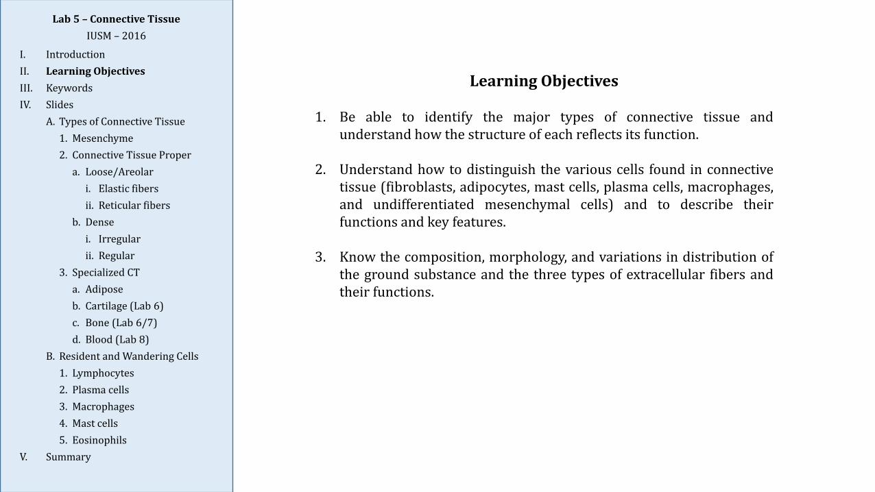
Learning Objectives
1. Be able to identify the major types of connective tissue andunderstand how the structure of each reflects its function.
2. Understand how to distinguish the various cells found in connectivetissue (fibroblasts, adipocytes, mast cells, plasma cells, macrophages,and undifferentiated mesenchymal cells) and to describe theirfunctions and key features.
3. Know the composition, morphology, and variations in distribution ofthe ground substance and the three types of extracellular fibers andtheir functions.
Lab 5 – Connective TissueIUSM – 2016
I. IntroductionII. Learning ObjectivesIII. KeywordsIV. Slides
A. Types of Connective Tissue1. Mesenchyme2. Connective Tissue Proper
a. Loose/Areolari. Elastic fibersii. Reticular fibers
b. Densei. Irregularii. Regular
3. Specialized CTa. Adiposeb. Cartilage (Lab 6)c. Bone (Lab 6/7)d. Blood (Lab 8)
B. Resident and Wandering Cells1. Lymphocytes2. Plasma cells3. Macrophages4. Mast cells5. Eosinophils
V. Summary

Keywords
Brown adipose tissueCollagen fibersConnective tissue properDense irregular CTDense regular CTElastin (elastic) fibersFibroblasts
Loose/areolar CTMacrophagesMast cellsMesenchymePlasma cellsReticulin (reticular) fibersWhite adipose tissue
Lab 5 – Connective TissueIUSM – 2016
I. IntroductionII. Learning ObjectivesIII. KeywordsIV. Slides
A. Types of Connective Tissue1. Mesenchyme2. Connective Tissue Proper
a. Loose/Areolari. Elastic fibersii. Reticular fibers
b. Densei. Irregularii. Regular
3. Specialized CTa. Adiposeb. Cartilage (Lab 6)c. Bone (Lab 6/7)d. Blood (Lab 8)
B. Resident and Wandering Cells1. Lymphocytes2. Plasma cells3. Macrophages4. Mast cells5. Eosinophils
V. Summary
look here formesenchyme
look here formesenchyme
Slide 91: Hamster Embryo, H&ELab 5 – Connective Tissue
IUSM – 2016
I. IntroductionII. Learning ObjectivesIII. KeywordsIV. Slides
A. Types of Connective Tissue1. Mesenchyme2. Connective Tissue Proper
a. Loose/Areolari. Elastic fibersii. Reticular fibers
b. Densei. Irregularii. Regular
3. Specialized CTa. Adiposeb. Cartilage (Lab 6)c. Bone (Lab 6/7)d. Blood (Lab 8)
B. Resident and Wandering Cells1. Lymphocytes2. Plasma cells3. Macrophages4. Mast cells5. Eosinophils
V. Summary
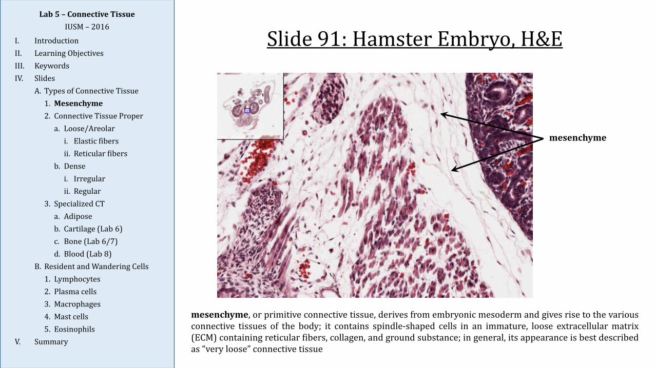
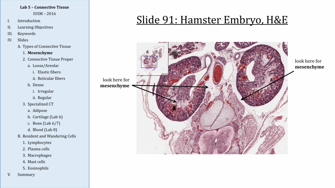
Slide 91: Hamster Embryo, H&E
mesenchyme, or primitive connective tissue, derives from embryonic mesoderm and gives rise to the variousconnective tissues of the body; it contains spindle-shaped cells in an immature, loose extracellular matrix(ECM) containing reticular fibers, collagen, and ground substance; in general, its appearance is best describedas “very loose” connective tissue
mesenchyme
Lab 5 – Connective TissueIUSM – 2016
I. IntroductionII. Learning ObjectivesIII. KeywordsIV. Slides
A. Types of Connective Tissue1. Mesenchyme2. Connective Tissue Proper
a. Loose/Areolari. Elastic fibersii. Reticular fibers
b. Densei. Irregularii. Regular
3. Specialized CTa. Adiposeb. Cartilage (Lab 6)c. Bone (Lab 6/7)d. Blood (Lab 8)
B. Resident and Wandering Cells1. Lymphocytes2. Plasma cells3. Macrophages4. Mast cells5. Eosinophils
V. Summary
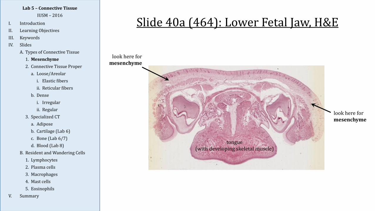
Slide 40a (464): Lower Fetal Jaw, H&E
tongue(with developing skeletal muscle)
look here formesenchyme
look here formesenchyme
Lab 5 – Connective TissueIUSM – 2016
I. IntroductionII. Learning ObjectivesIII. KeywordsIV. Slides
A. Types of Connective Tissue1. Mesenchyme2. Connective Tissue Proper
a. Loose/Areolari. Elastic fibersii. Reticular fibers
b. Densei. Irregularii. Regular
3. Specialized CTa. Adiposeb. Cartilage (Lab 6)c. Bone (Lab 6/7)d. Blood (Lab 8)
B. Resident and Wandering Cells1. Lymphocytes2. Plasma cells3. Macrophages4. Mast cells5. Eosinophils
V. Summary
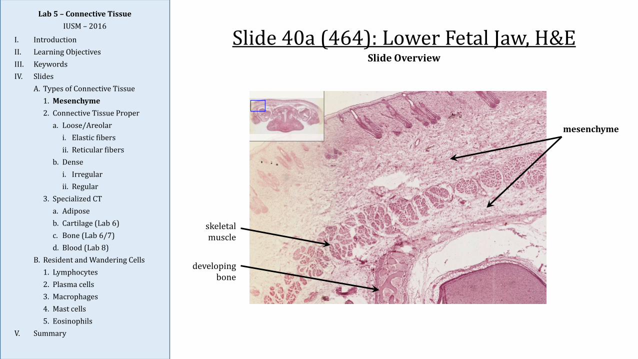
Slide 40a (464): Lower Fetal Jaw, H&E
skeletal muscle
mesenchyme
developing bone
Lab 5 – Connective TissueIUSM – 2016
I. IntroductionII. Learning ObjectivesIII. KeywordsIV. Slides
A. Types of Connective Tissue1. Mesenchyme2. Connective Tissue Proper
a. Loose/Areolari. Elastic fibersii. Reticular fibers
b. Densei. Irregularii. Regular
3. Specialized CTa. Adiposeb. Cartilage (Lab 6)c. Bone (Lab 6/7)d. Blood (Lab 8)
B. Resident and Wandering Cells1. Lymphocytes2. Plasma cells3. Macrophages4. Mast cells5. Eosinophils
V. Summary
Slide Overview
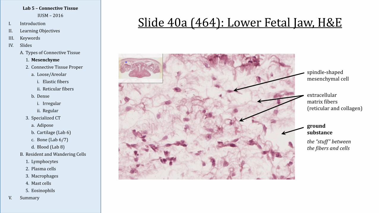
Slide 40a (464): Lower Fetal Jaw, H&E
spindle-shaped mesenchymal cell
ground substancethe “stuff” between the fibers and cells
extracellularmatrix fibers(reticular and collagen)
Lab 5 – Connective TissueIUSM – 2016
I. IntroductionII. Learning ObjectivesIII. KeywordsIV. Slides
A. Types of Connective Tissue1. Mesenchyme2. Connective Tissue Proper
a. Loose/Areolari. Elastic fibersii. Reticular fibers
b. Densei. Irregularii. Regular
3. Specialized CTa. Adiposeb. Cartilage (Lab 6)c. Bone (Lab 6/7)d. Blood (Lab 8)
B. Resident and Wandering Cells1. Lymphocytes2. Plasma cells3. Macrophages4. Mast cells5. Eosinophils
V. Summary
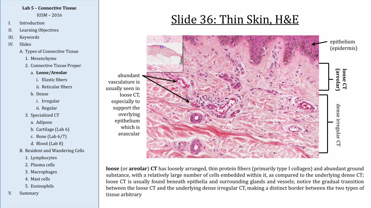
Slide 36: Thin Skin, H&E
loose (or areolar) CT has loosely arranged, thin protein fibers (primarily type I collagen) and abundant groundsubstance, with a relatively large number of cells embedded within it, as compared to the underlying dense CT;loose CT is usually found beneath epithelia and surrounding glands and vessels; notice the gradual transitionbetween the loose CT and the underlying dense irregular CT, making a distinct border between the two types oftissue arbitrary
Lab 5 – Connective TissueIUSM – 2016
I. IntroductionII. Learning ObjectivesIII. KeywordsIV. Slides
A. Types of Connective Tissue1. Mesenchyme2. Connective Tissue Proper
a. Loose/Areolari. Elastic fibersii. Reticular fibers
b. Densei. Irregularii. Regular
3. Specialized CTa. Adiposeb. Cartilage (Lab 6)c. Bone (Lab 6/7)d. Blood (Lab 8)
B. Resident and Wandering Cells1. Lymphocytes2. Plasma cells3. Macrophages4. Mast cells5. Eosinophils
V. Summary
dense irregular CTloose CT (areolar)
epithelium(epidermis)
abundant vasculature is
usually seen in loose CT,
especially to support the
overlying epithelium
which is avascular

Slide 36: Thin Skin, H&E
the principal cells of connective tissue proper are fibroblasts which synthesize and maintain the ECMcomponents (both the fibers and ground substance); they generally appear elongated with an ovoid, condensednucleus with one or two nucleoli (if visible); their thin cytoplasmic processes not readily seen; however, theymay become “activated” and appear more ovoid with a more extensive basophilic cytoplasm (lots of rER) duringperiods of growth or wound repair (note: the term fibrocyte is sometimes used to refer to “inactive” fibroblasts)
what is this pigmented
inclusion within these
epithelial cells of the skin?
fibroblast
eosinophilic collagenprotein fibers
unstained spaces are composed of ground substance
Lab 5 – Connective TissueIUSM – 2016
I. IntroductionII. Learning ObjectivesIII. KeywordsIV. Slides
A. Types of Connective Tissue1. Mesenchyme2. Connective Tissue Proper
a. Loose/Areolari. Elastic fibersii. Reticular fibers
b. Densei. Irregularii. Regular
3. Specialized CTa. Adiposeb. Cartilage (Lab 6)c. Bone (Lab 6/7)d. Blood (Lab 8)
B. Resident and Wandering Cells1. Lymphocytes2. Plasma cells3. Macrophages4. Mast cells5. Eosinophils
V. Summary

Slide 4a (464): Areolar Connective TissueLab 5 – Connective Tissue
IUSM – 2016
I. IntroductionII. Learning ObjectivesIII. KeywordsIV. Slides
A. Types of Connective Tissue1. Mesenchyme2. Connective Tissue Proper
a. Loose/Areolari. Elastic fibersii. Reticular fibers
b. Densei. Irregularii. Regular
3. Specialized CTa. Adiposeb. Cartilage (Lab 6)c. Bone (Lab 6/7)d. Blood (Lab 8)
B. Resident and Wandering Cells1. Lymphocytes2. Plasma cells3. Macrophages4. Mast cells5. Eosinophils
V. Summary
elastic fiber(thin)
fibroblast
collagen fiber(thick)
while collagen fibers provide strength to a tissue, elastic (or elastin) fibers are found interwoven in varyingamounts in the ECM of most connective tissues providing stretch and recoil (e.g., in skin and lung); the fibersare produced by fibroblasts or smooth muscle cells and are eosinophilic but are usually only seen withspecialized stains; they appears as fine, thin, and relatively straight fibers
Slide 100: Aorta, Van Gieson & ElasticLab 5 – Connective Tissue
IUSM – 2016
I. IntroductionII. Learning ObjectivesIII. KeywordsIV. Slides
A. Types of Connective Tissue1. Mesenchyme2. Connective Tissue Proper
a. Loose/Areolari. Elastic fibersii. Reticular fibers
b. Densei. Irregularii. Regular
3. Specialized CTa. Adiposeb. Cartilage (Lab 6)c. Bone (Lab 6/7)d. Blood (Lab 8)
B. Resident and Wandering Cells1. Lymphocytes2. Plasma cells3. Macrophages4. Mast cells5. Eosinophils
V. Summary
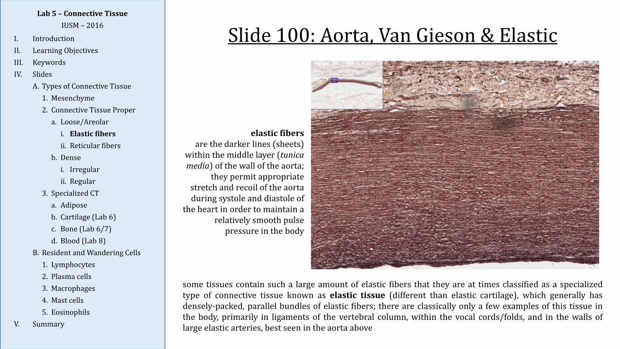
some tissues contain such a large amount of elastic fibers that they are at times classified as a specializedtype of connective tissue known as elastic tissue (different than elastic cartilage), which generally hasdensely-packed, parallel bundles of elastic fibers; there are classically only a few examples of this tissue inthe body, primarily in ligaments of the vertebral column, within the vocal cords/folds, and in the walls oflarge elastic arteries, best seen in the aorta above
elastic fibers are the darker lines (sheets)
within the middle layer (tunica media) of the wall of the aorta;
they permit appropriate stretch and recoil of the aorta during systole and diastole of
the heart in order to maintain a relatively smooth pulse
pressure in the body

Slide 99: Lymph Node, SilverLab 5 – Connective Tissue
IUSM – 2016
I. IntroductionII. Learning ObjectivesIII. KeywordsIV. Slides
A. Types of Connective Tissue1. Mesenchyme2. Connective Tissue Proper
a. Loose/Areolari. Elastic fibersii. Reticular fibers
b. Densei. Irregularii. Regular
3. Specialized CTa. Adiposeb. Cartilage (Lab 6)c. Bone (Lab 6/7)d. Blood (Lab 8)
B. Resident and Wandering Cells1. Lymphocytes2. Plasma cells3. Macrophages4. Mast cells5. Eosinophils
V. Summary
the stroma of certain organs – mainly in hematopoietic (e.g., bone marrow) and lymphatic tissues (excludingthe thymus) – contains abundant reticular fibers and specialized reticular cells, instead of the fibroblasts orsmooth muscle cells that typically make reticular fibers elsewhere; in these organs, the specialized stroma issometimes referred to as reticular connective tissue
look within the cortex or medulla of
the lymph node to find reticular fibers
cortex with lymphoid follicles
capsule of connective tissue
medulla
Slide Overview
Slide 99: Lymph Node, SilverLab 5 – Connective Tissue
IUSM – 2016
I. IntroductionII. Learning ObjectivesIII. KeywordsIV. Slides
A. Types of Connective Tissue1. Mesenchyme2. Connective Tissue Proper
a. Loose/Areolari. Elastic fibersii. Reticular fibers
b. Densei. Irregularii. Regular
3. Specialized CTa. Adiposeb. Cartilage (Lab 6)c. Bone (Lab 6/7)d. Blood (Lab 8)
B. Resident and Wandering Cells1. Lymphocytes2. Plasma cells3. Macrophages4. Mast cells5. Eosinophils
V. Summary
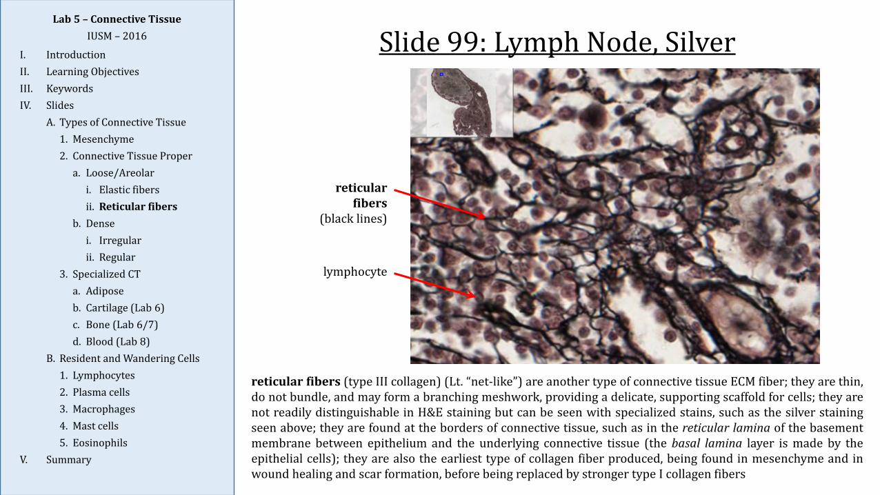
reticular fibers
(black lines)
lymphocyte
reticular fibers (type III collagen) (Lt. “net-like”) are another type of connective tissue ECM fiber; they are thin,do not bundle, and may form a branching meshwork, providing a delicate, supporting scaffold for cells; they arenot readily distinguishable in H&E staining but can be seen with specialized stains, such as the silver stainingseen above; they are found at the borders of connective tissue, such as in the reticular lamina of the basementmembrane between epithelium and the underlying connective tissue (the basal lamina layer is made by theepithelial cells); they are also the earliest type of collagen fiber produced, being found in mesenchyme and inwound healing and scar formation, before being replaced by stronger type I collagen fibers
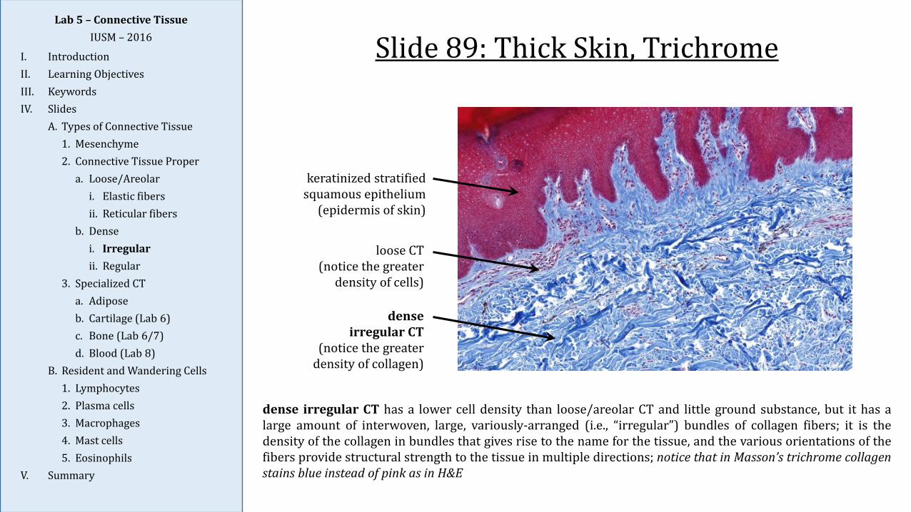
Slide 89: Thick Skin, Trichrome
dense irregular CT has a lower cell density than loose/areolar CT and little ground substance, but it has alarge amount of interwoven, large, variously-arranged (i.e., “irregular”) bundles of collagen fibers; it is thedensity of the collagen in bundles that gives rise to the name for the tissue, and the various orientations of thefibers provide structural strength to the tissue in multiple directions; notice that in Masson’s trichrome collagenstains blue instead of pink as in H&E
loose CT(notice the greater
density of cells)
dense irregular CT
(notice the greater density of collagen)
keratinized stratified squamous epithelium
(epidermis of skin)
Lab 5 – Connective TissueIUSM – 2016
I. IntroductionII. Learning ObjectivesIII. KeywordsIV. Slides
A. Types of Connective Tissue1. Mesenchyme2. Connective Tissue Proper
a. Loose/Areolari. Elastic fibersii. Reticular fibers
b. Densei. Irregularii. Regular
3. Specialized CTa. Adiposeb. Cartilage (Lab 6)c. Bone (Lab 6/7)d. Blood (Lab 8)
B. Resident and Wandering Cells1. Lymphocytes2. Plasma cells3. Macrophages4. Mast cells5. Eosinophils
V. Summary
Slide 16 (NW): Tendon, H&ELab 5 – Connective Tissue
IUSM – 2016
I. IntroductionII. Learning ObjectivesIII. KeywordsIV. Slides
A. Types of Connective Tissue1. Mesenchyme2. Connective Tissue Proper
a. Loose/Areolari. Elastic fibersii. Reticular fibers
b. Densei. Irregularii. Regular
3. Specialized CTa. Adiposeb. Cartilage (Lab 6)c. Bone (Lab 6/7)d. Blood (Lab 8)
B. Resident and Wandering Cells1. Lymphocytes2. Plasma cells3. Macrophages4. Mast cells5. Eosinophils
V. Summary
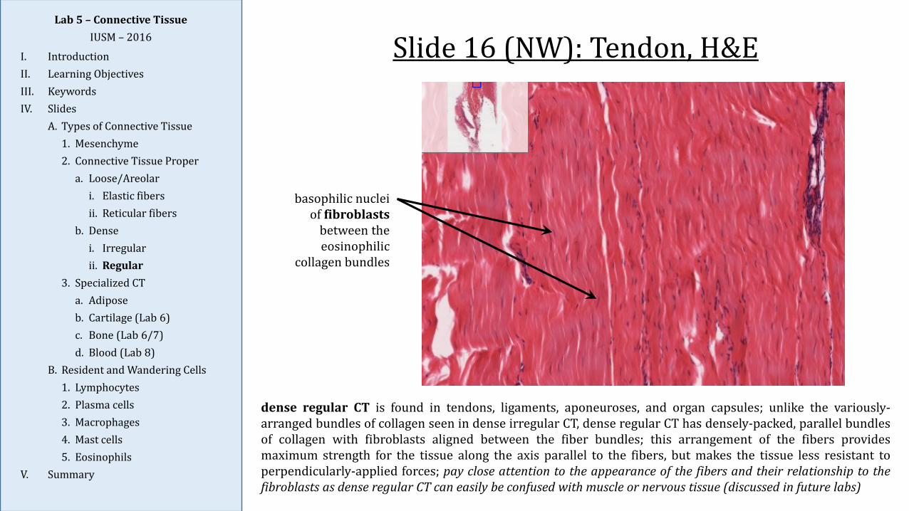
dense regular CT is found in tendons, ligaments, aponeuroses, and organ capsules; unlike the variously-arranged bundles of collagen seen in dense irregular CT, dense regular CT has densely-packed, parallel bundlesof collagen with fibroblasts aligned between the fiber bundles; this arrangement of the fibers providesmaximum strength for the tissue along the axis parallel to the fibers, but makes the tissue less resistant toperpendicularly-applied forces; pay close attention to the appearance of the fibers and their relationship to thefibroblasts as dense regular CT can easily be confused with muscle or nervous tissue (discussed in future labs)
basophilic nuclei of fibroblasts
between the eosinophilic
collagen bundles
Lab 5 – Connective TissueIUSM – 2016
I. IntroductionII. Learning ObjectivesIII. KeywordsIV. Slides
A. Types of Connective Tissue1. Mesenchyme2. Connective Tissue Proper
a. Loose/Areolari. Elastic fibersii. Reticular fibers
b. Densei. Irregularii. Regular
3. Specialized CTa. Adiposeb. Cartilage (Lab 6)c. Bone (Lab 6/7)d. Blood (Lab 8)
B. Resident and Wandering Cells1. Lymphocytes2. Plasma cells3. Macrophages4. Mast cells5. Eosinophils
V. Summary
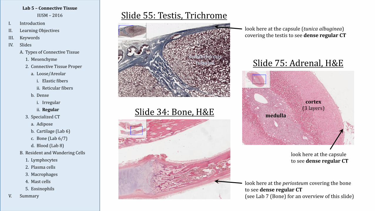
Slide 55: Testis, Trichrome
Slide 75: Adrenal, H&E
Slide 34: Bone, H&E
look here at the capsule (tunica albuginea) covering the testis to see dense regular CT
look here at the periosteum covering the bone to see dense regular CT(see Lab 7 (Bone) for an overview of this slide)
look here at the capsule to see dense regular CT
cortex(3 layers)
medulla
seminiferous tubules
Slide 92: Vessels, Elastic StainLab 5 – Connective Tissue
IUSM – 2016
I. IntroductionII. Learning ObjectivesIII. KeywordsIV. Slides
A. Types of Connective Tissue1. Mesenchyme2. Connective Tissue Proper
a. Loose/Areolari. Elastic fibersii. Reticular fibers
b. Densei. Irregularii. Regular
3. Specialized CTa. Adiposeb. Cartilage (Lab 6)c. Bone (Lab 6/7)d. Blood (Lab 8)
B. Resident and Wandering Cells1. Lymphocytes2. Plasma cells3. Macrophages4. Mast cells5. Eosinophils
V. Summary
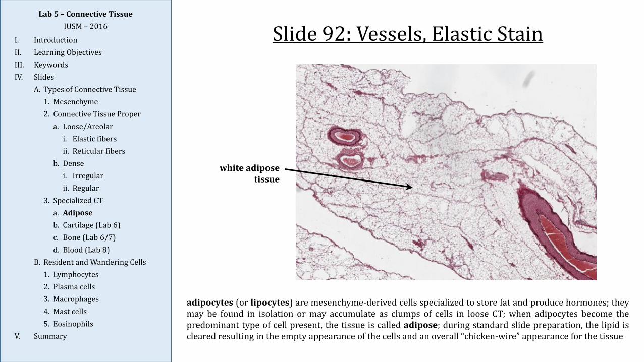
white adipose tissue
adipocytes (or lipocytes) are mesenchyme-derived cells specialized to store fat and produce hormones; theymay be found in isolation or may accumulate as clumps of cells in loose CT; when adipocytes become thepredominant type of cell present, the tissue is called adipose; during standard slide preparation, the lipid iscleared resulting in the empty appearance of the cells and an overall “chicken-wire” appearance for the tissue
Slide 92: Vessels, Elastic StainLab 5 – Connective Tissue
IUSM – 2016
I. IntroductionII. Learning ObjectivesIII. KeywordsIV. Slides
A. Types of Connective Tissue1. Mesenchyme2. Connective Tissue Proper
a. Loose/Areolari. Elastic fibersii. Reticular fibers
b. Densei. Irregularii. Regular
3. Specialized CTa. Adiposeb. Cartilage (Lab 6)c. Bone (Lab 6/7)d. Blood (Lab 8)
B. Resident and Wandering Cells1. Lymphocytes2. Plasma cells3. Macrophages4. Mast cells5. Eosinophils
V. Summary
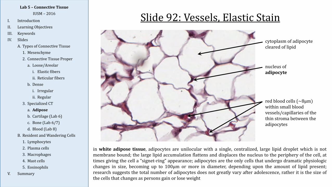
in white adipose tissue, adipocytes are unilocular with a single, centralized, large lipid droplet which is notmembrane bound; the large lipid accumulation flattens and displaces the nucleus to the periphery of the cell, attimes giving the cell a “signet-ring” appearance; adipocytes are the only cells that undergo dramatic physiologicchanges in size, becoming up to 100μm or more in diameter, depending upon the amount of lipid present;research suggests the total number of adipocytes does not greatly vary after adolescence, rather it is the size ofthe cells that changes as persons gain or lose weight
nucleus of adipocyte
red blood cells (~8μm)within small blood vessels/capillaries of the thin stroma between the adipocytes
cytoplasm of adipocyte cleared of lipid
Lab 5 – Connective TissueIUSM – 2016
I. IntroductionII. Learning ObjectivesIII. KeywordsIV. Slides
A. Types of Connective Tissue1. Mesenchyme2. Connective Tissue Proper
a. Loose/Areolari. Elastic fibersii. Reticular fibers
b. Densei. Irregularii. Regular
3. Specialized CTa. Adiposeb. Cartilage (Lab 6)c. Bone (Lab 6/7)d. Blood (Lab 8)
B. Resident and Wandering Cells1. Lymphocytes2. Plasma cells3. Macrophages4. Mast cells5. Eosinophils
V. Summary
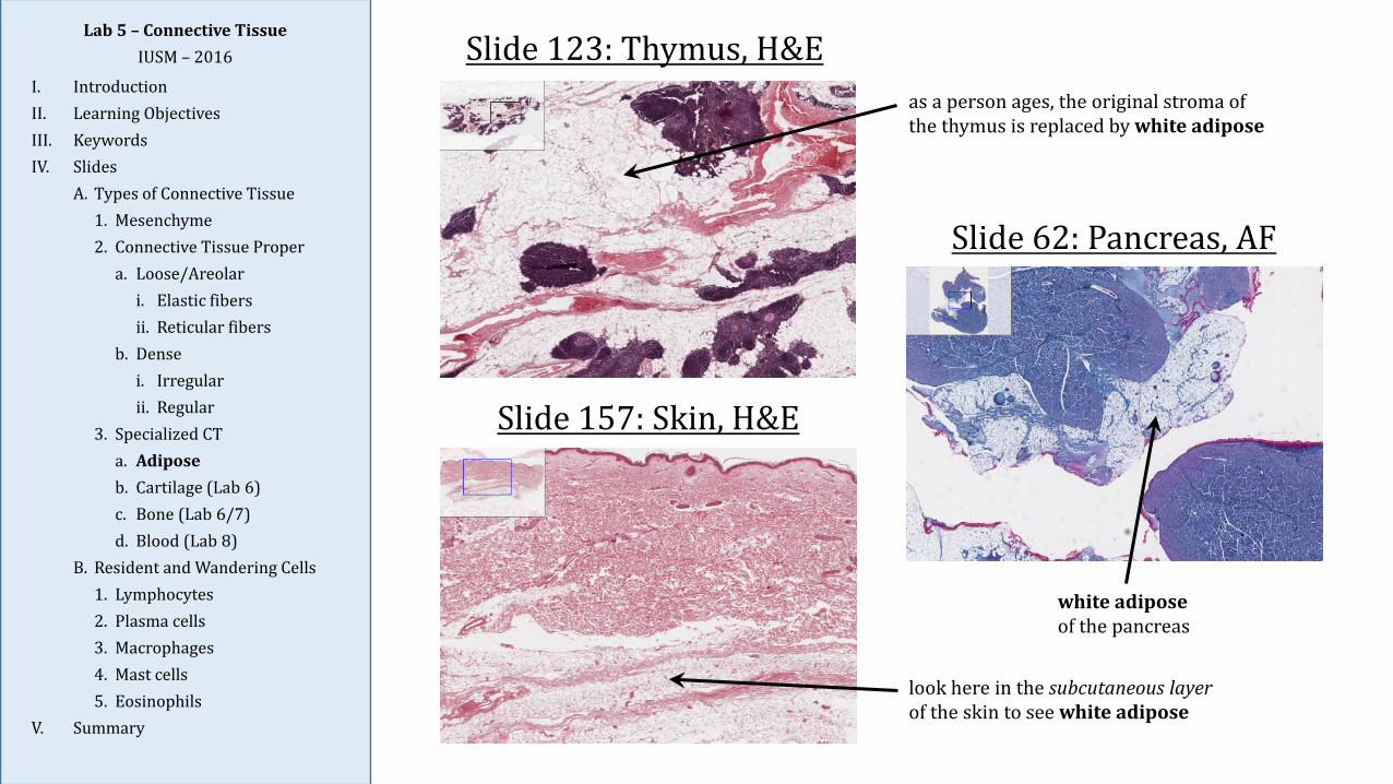
Slide 123: Thymus, H&E
Slide 157: Skin, H&E
Slide 62: Pancreas, AF
as a person ages, the original stroma of the thymus is replaced by white adipose
look here in the subcutaneous layer of the skin to see white adipose
white adiposeof the pancreas
Lab 5 – Connective TissueIUSM – 2016
I. IntroductionII. Learning ObjectivesIII. KeywordsIV. Slides
A. Types of Connective Tissue1. Mesenchyme2. Connective Tissue Proper
a. Loose/Areolari. Elastic fibersii. Reticular fibers
b. Densei. Irregularii. Regular
3. Specialized CTa. Adiposeb. Cartilage (Lab 6)c. Bone (Lab 6/7)d. Blood (Lab 8)
B. Resident and Wandering Cells1. Lymphocytes2. Plasma cells3. Macrophages4. Mast cells5. Eosinophils
V. Summary
Cells of Connective Tissue
1. Connective tissue cells are traditionally classified as either resident or wandering (transient)cells:
a. Resident cell populations are relatively fixed, stable populations of cells withinconnective tissues; they include fibroblasts, macrophages, and mast cells; these cellsserve to synthesize, maintain, and surveil the tissue.
b. Wandering cell populations are all the other white blood cell types which leave theblood and enter loose CT, which is the principal site of their activity as part ofimmune surveillance and response; these cells are especially present in the laminapropria (loose CT underlying the epithelium of a mucosa) of the respiratory and GItracts, where pathogens are readily encountered; wandering cells includelymphocytes, plasma cells, eosinophils, and neutrophils (however, neutrophils arerarely seen in connective tissue except during inflammatory conditions).
2. While white blood cells (leukocytes) will be explored more fully in Lab 8 (Blood), it isimportant to be familiar with those present in normal connective tissue and to appreciate thedifferences in appearance between leukocytes in the blood and those found in loose CT; thesedifferences are due in part to differences in how slides are prepared for tissue samples vs.blood smears, but are also due to changes that leukocytes undergo as they leave the circulationand enter into tissues (e.g., as monocytes become tissue macrophages).
3. When examining the slides, many cells encountered in CT will be difficult – if not impossible – toidentify with certainty; it is more important to know the characteristics for each of the respectivecell types studied and to find examples than it is to attempt to identify every cell seen (a fruitfulbut ultimately futile endeavor).
Lab 5 – Connective TissueIUSM – 2016
I. IntroductionII. Learning ObjectivesIII. KeywordsIV. Slides
A. Types of Connective Tissue1. Mesenchyme2. Connective Tissue Proper
a. Loose/Areolari. Elastic fibersii. Reticular fibers
b. Densei. Irregularii. Regular
3. Specialized CTa. Adiposeb. Cartilage (Lab 6)c. Bone (Lab 6/7)d. Blood (Lab 8)
B. Resident and Wandering Cells1. Lymphocytes2. Plasma cells3. Macrophages4. Mast cells5. Eosinophils
V. Summary
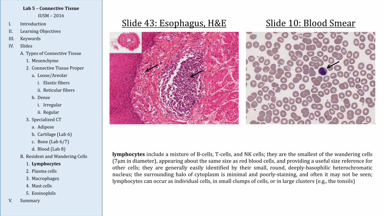
Slide 43: Esophagus, H&E Slide 10: Blood Smear
lymphocytes include a mixture of B-cells, T-cells, and NK cells; they are the smallest of the wandering cells(7μm in diameter), appearing about the same size as red blood cells, and providing a useful size reference forother cells; they are generally easily identified by their small, round, deeply-basophilic heterochromaticnucleus; the surrounding halo of cytoplasm is minimal and poorly-staining, and often it may not be seen;lymphocytes can occur as individual cells, in small clumps of cells, or in large clusters (e.g., the tonsils)
Lab 5 – Connective TissueIUSM – 2016
I. IntroductionII. Learning ObjectivesIII. KeywordsIV. Slides
A. Types of Connective Tissue1. Mesenchyme2. Connective Tissue Proper
a. Loose/Areolari. Elastic fibersii. Reticular fibers
b. Densei. Irregularii. Regular
3. Specialized CTa. Adiposeb. Cartilage (Lab 6)c. Bone (Lab 6/7)d. Blood (Lab 8)
B. Resident and Wandering Cells1. Lymphocytes2. Plasma cells3. Macrophages4. Mast cells5. Eosinophils
V. Summary
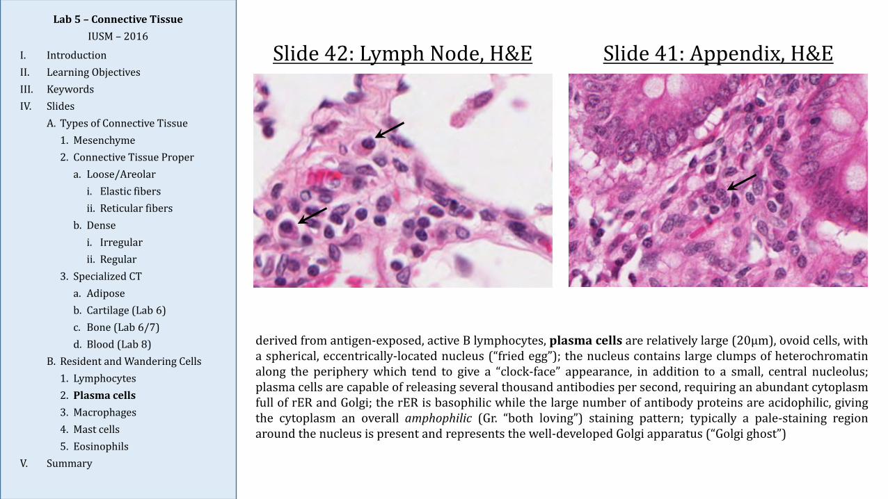
Slide 42: Lymph Node, H&E Slide 41: Appendix, H&E
derived from antigen-exposed, active B lymphocytes, plasma cells are relatively large (20μm), ovoid cells, witha spherical, eccentrically-located nucleus (“fried egg”); the nucleus contains large clumps of heterochromatinalong the periphery which tend to give a “clock-face” appearance, in addition to a small, central nucleolus;plasma cells are capable of releasing several thousand antibodies per second, requiring an abundant cytoplasmfull of rER and Golgi; the rER is basophilic while the large number of antibody proteins are acidophilic, givingthe cytoplasm an overall amphophilic (Gr. “both loving”) staining pattern; typically a pale-staining regionaround the nucleus is present and represents the well-developed Golgi apparatus (“Golgi ghost”)
Lab 5 – Connective TissueIUSM – 2016
I. IntroductionII. Learning ObjectivesIII. KeywordsIV. Slides
A. Types of Connective Tissue1. Mesenchyme2. Connective Tissue Proper
a. Loose/Areolari. Elastic fibersii. Reticular fibers
b. Densei. Irregularii. Regular
3. Specialized CTa. Adiposeb. Cartilage (Lab 6)c. Bone (Lab 6/7)d. Blood (Lab 8)
B. Resident and Wandering Cells1. Lymphocytes2. Plasma cells3. Macrophages4. Mast cells5. Eosinophils
V. Summary
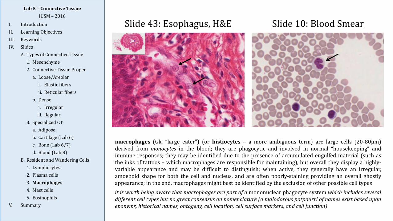
Slide 43: Esophagus, H&E Slide 10: Blood Smear
macrophages (Gk. “large eater”) (or histiocytes – a more ambiguous term) are large cells (20-80μm)derived from monocytes in the blood; they are phagocytic and involved in normal “housekeeping” andimmune responses; they may be identified due to the presence of accumulated engulfed material (such asthe inks of tattoos – which macrophages are responsible for maintaining), but overall they display a highly-variable appearance and may be difficult to distinguish; when active, they generally have an irregular,amoeboid shape for both the cell and nucleus, and are often poorly-staining providing an overall ghostlyappearance; in the end, macrophages might best be identified by the exclusion of other possible cell typesit is worth being aware that macrophages are part of a mononuclear phagocyte system which includes severaldifferent cell types but no great consensus on nomenclature (a malodorous potpourri of names exist based uponeponyms, historical names, ontogeny, cell location, cell surface markers, and cell function)
Lab 5 – Connective TissueIUSM – 2016
I. IntroductionII. Learning ObjectivesIII. KeywordsIV. Slides
A. Types of Connective Tissue1. Mesenchyme2. Connective Tissue Proper
a. Loose/Areolari. Elastic fibersii. Reticular fibers
b. Densei. Irregularii. Regular
3. Specialized CTa. Adiposeb. Cartilage (Lab 6)c. Bone (Lab 6/7)d. Blood (Lab 8)
B. Resident and Wandering Cells1. Lymphocytes2. Plasma cells3. Macrophages4. Mast cells5. Eosinophils
V. Summary
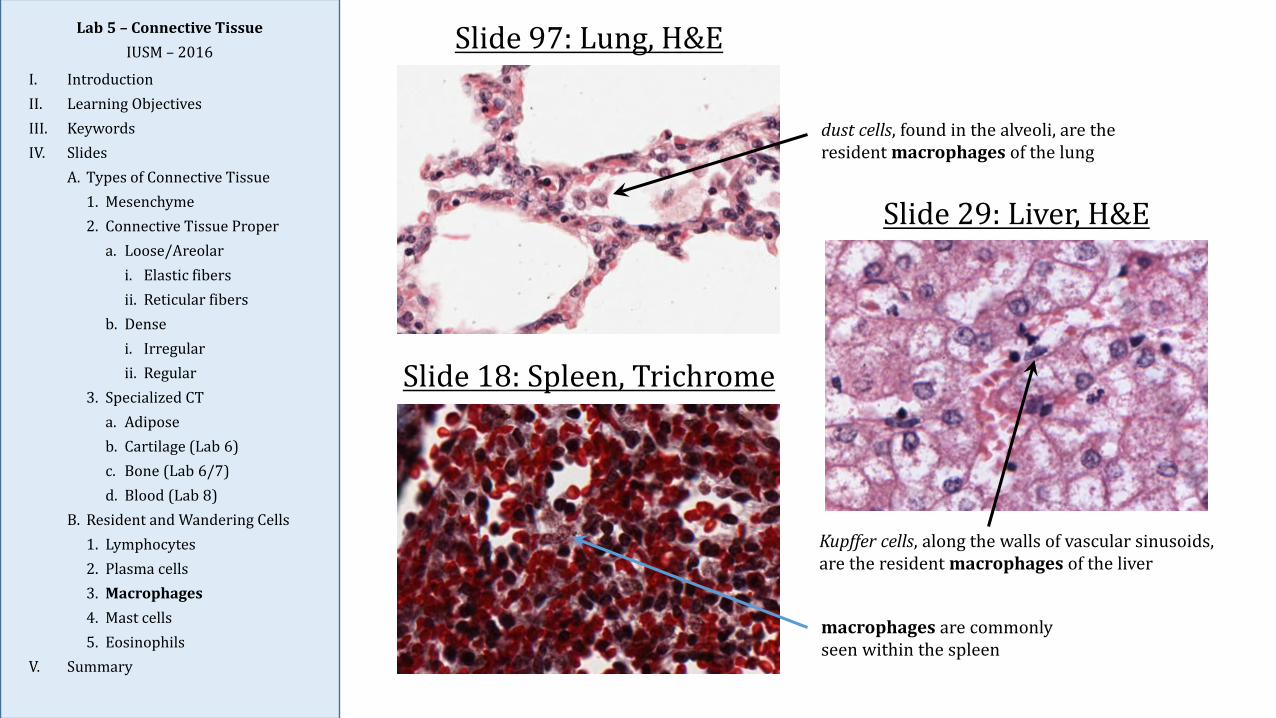
Slide 97: Lung, H&E
Slide 18: Spleen, Trichrome
Slide 29: Liver, H&E
dust cells, found in the alveoli, are the resident macrophages of the lung
Kupffer cells, along the walls of vascular sinusoids, are the resident macrophages of the liver
macrophages are commonly seen within the spleen
Lab 5 – Connective TissueIUSM – 2016
I. IntroductionII. Learning ObjectivesIII. KeywordsIV. Slides
A. Types of Connective Tissue1. Mesenchyme2. Connective Tissue Proper
a. Loose/Areolari. Elastic fibersii. Reticular fibers
b. Densei. Irregularii. Regular
3. Specialized CTa. Adiposeb. Cartilage (Lab 6)c. Bone (Lab 6/7)d. Blood (Lab 8)
B. Resident and Wandering Cells1. Lymphocytes2. Plasma cells3. Macrophages4. Mast cells5. Eosinophils
V. Summary
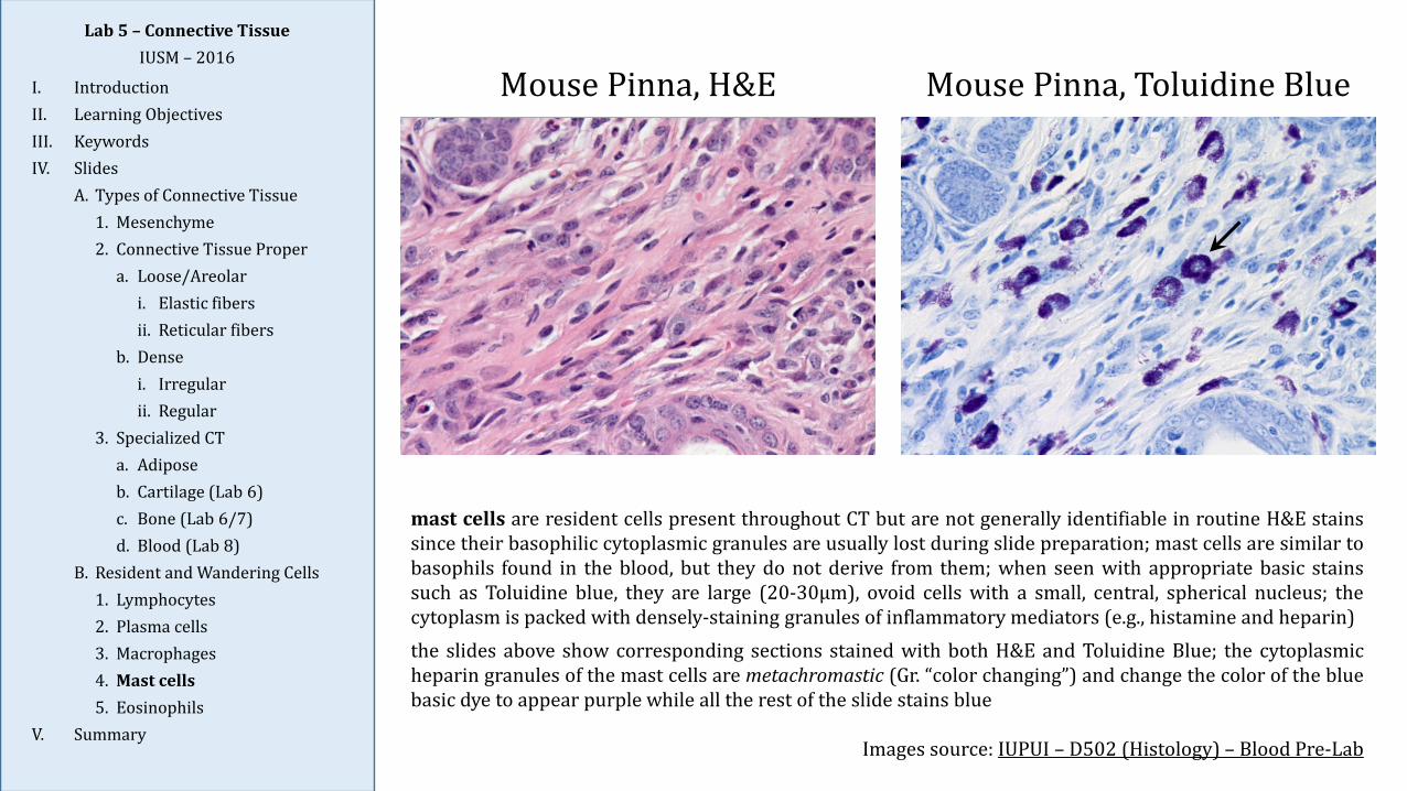
mast cells are resident cells present throughout CT but are not generally identifiable in routine H&E stainssince their basophilic cytoplasmic granules are usually lost during slide preparation; mast cells are similar tobasophils found in the blood, but they do not derive from them; when seen with appropriate basic stainssuch as Toluidine blue, they are large (20-30μm), ovoid cells with a small, central, spherical nucleus; thecytoplasm is packed with densely-staining granules of inflammatory mediators (e.g., histamine and heparin)the slides above show corresponding sections stained with both H&E and Toluidine Blue; the cytoplasmicheparin granules of the mast cells are metachromastic (Gr. “color changing”) and change the color of the bluebasic dye to appear purple while all the rest of the slide stains blue
Images source: IUPUI – D502 (Histology) – Blood Pre-Lab
Mouse Pinna, H&E Mouse Pinna, Toluidine Blue
Lab 5 – Connective TissueIUSM – 2016
I. IntroductionII. Learning ObjectivesIII. KeywordsIV. Slides
A. Types of Connective Tissue1. Mesenchyme2. Connective Tissue Proper
a. Loose/Areolari. Elastic fibersii. Reticular fibers
b. Densei. Irregularii. Regular
3. Specialized CTa. Adiposeb. Cartilage (Lab 6)c. Bone (Lab 6/7)d. Blood (Lab 8)
B. Resident and Wandering Cells1. Lymphocytes2. Plasma cells3. Macrophages4. Mast cells5. Eosinophils
V. Summary
Slide 113: Appendix, H&E Slide 10: Blood Smear
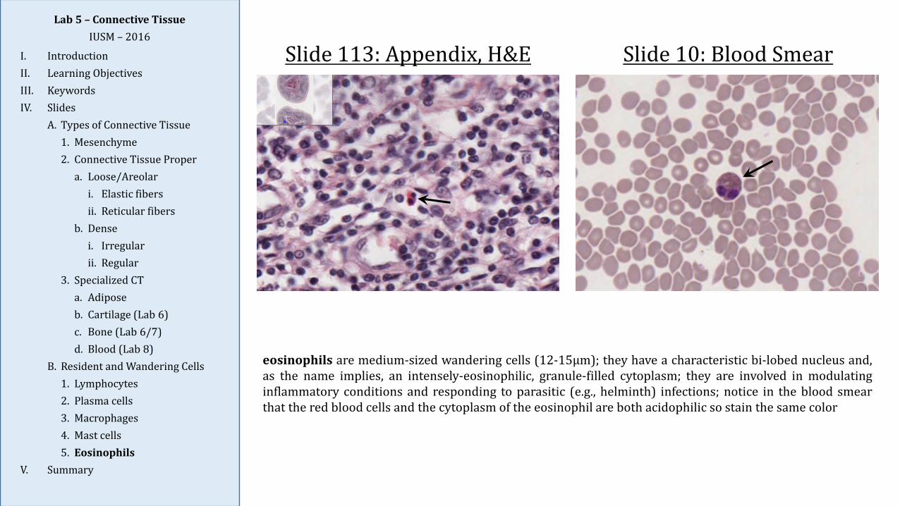
eosinophils are medium-sized wandering cells (12-15μm); they have a characteristic bi-lobed nucleus and,as the name implies, an intensely-eosinophilic, granule-filled cytoplasm; they are involved in modulatinginflammatory conditions and responding to parasitic (e.g., helminth) infections; notice in the blood smearthat the red blood cells and the cytoplasm of the eosinophil are both acidophilic so stain the same color

Lab 5 – Connective TissueIUSM – 2016
I. IntroductionII. Learning ObjectivesIII. KeywordsIV. Slides
A. Types of Connective Tissue1. Mesenchyme2. Connective Tissue Proper
a. Loose/Areolari. Elastic fibersii. Reticular fibers
b. Densei. Irregularii. Regular
3. Specialized CTa. Adiposeb. Cartilage (Lab 6)c. Bone (Lab 6/7)d. Blood (Lab 8)
B. Resident and Wandering Cells1. Lymphocytes2. Plasma cells3. Macrophages4. Mast cells5. Eosinophils
V. Summary
Common Confusion:Mesenchyme vs. Loose CT
Mesenchyme
Mesenchyme: primitive connective tissue found primarilyin embryonic tissue; mesenchymal cells are largelyundifferentiated and capable of forming the cells typical ofadult CT; can be considered “very loose” connective tissue
Look for: (1) angular or spindle-shaped cells with large,round/oval nuclei and prominent nucleoli; (2) ECM withabundant ground substance and very fine fibers; (3)mitotic figures may be visible
Loose/areolar connective tissue: connective tissue withprotein fibers of the ECM occurring singly, not in bundlesas in dense CT; occurs in mucosal membranes and oftenseen as “filler” tissue between other tissues such as muscleand epithelia
Look for: (1) nuclei of fibroblasts tend to be morecondensed and elongated; (2) ECM contains clearly visible,individual protein fibers; (3) other cells, such asmacrophages, mast cells, lymphocytes, and plasma cells,may be present; (4) blood vessels, lymphatic vessels, andnerves may be seenLoose CT
Lab 5 – Connective TissueIUSM – 2016
I. IntroductionII. Learning ObjectivesIII. KeywordsIV. Slides
A. Types of Connective Tissue1. Mesenchyme2. Connective Tissue Proper
a. Loose/Areolari. Elastic fibersii. Reticular fibers
b. Densei. Irregularii. Regular
3. Specialized CTa. Adiposeb. Cartilage (Lab 6)c. Bone (Lab 6/7)d. Blood (Lab 8)
B. Resident and Wandering Cells1. Lymphocytes2. Plasma cells3. Macrophages4. Mast cells5. Eosinophils
V. Summary
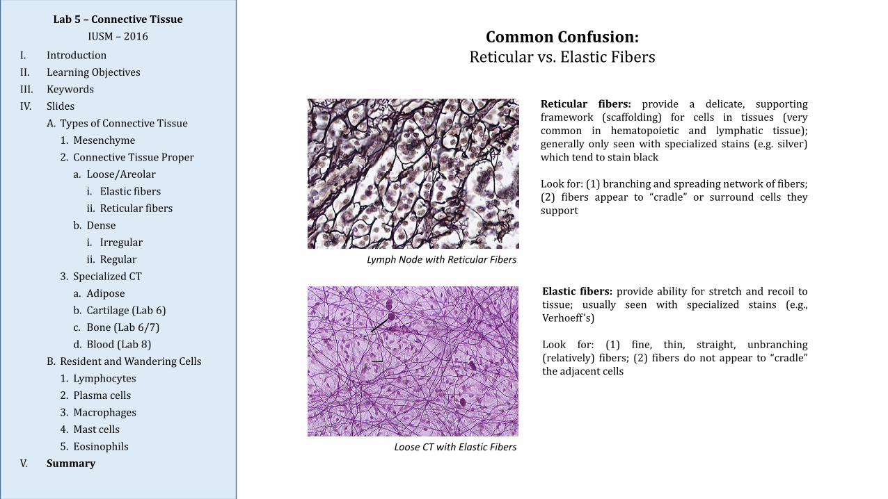
Common Confusion:Reticular vs. Elastic Fibers
Lymph Node with Reticular Fibers
Reticular fibers: provide a delicate, supportingframework (scaffolding) for cells in tissues (verycommon in hematopoietic and lymphatic tissue);generally only seen with specialized stains (e.g. silver)which tend to stain black
Look for: (1) branching and spreading network of fibers;(2) fibers appear to “cradle” or surround cells theysupport
Elastic fibers: provide ability for stretch and recoil totissue; usually seen with specialized stains (e.g.,Verhoeff ’s)
Look for: (1) fine, thin, straight, unbranching(relatively) fibers; (2) fibers do not appear to “cradle”the adjacent cells
Loose CT with Elastic Fibers
Summary
1. Connective tissue (CT) is one of the four primary tissue types; it forms the stroma thatsupports and connects the other types of tissues (parenchyma) to form organs: epitheliumoverlays it and muscle and nervous tissue are surrounded by it.
2. CT is composed primarily of a few dispersed, inconspicuous cells within a prominentextracellular matrix (ECM), which is primarily responsible for the properties andfunctions of CT; the ECM is composed of protein fibers (collagen, reticular, and elastic)and ground substance (amorphous gel-like substance rich in proteoglycans, with thespecific composition varying between types of CT (e.g., the ECM of CT proper differs fromthe ECM of cartilage or bone).
3. Connective tissue proper is categorized as loose or dense depending upon the relativeabundance of bundles of collagen protein fibers in the ECM:
Loose CT has loosely-arranged fibers with a relatively large numbers of cells andground substance present.
Dense CT has large bundles of fibers (collagen) with few cells and little groundsubstance.
4. Connective tissue cells are classified as resident cells (relatively stable, non-migratory;includes: fibroblasts, macrophages, adipocytes, mast cells, and stem cells) or wanderingcells (transient cells that have migrated from blood vessels; includes: plasma cells andother white blood cells).
Lab 5 – Connective TissueIUSM – 2016
I. IntroductionII. Learning ObjectivesIII. KeywordsIV. Slides
A. Types of Connective Tissue1. Mesenchyme2. Connective Tissue Proper
a. Loose/Areolari. Elastic fibersii. Reticular fibers
b. Densei. Irregularii. Regular
3. Specialized CTa. Adiposeb. Cartilage (Lab 6)c. Bone (Lab 6/7)d. Blood (Lab 8)
B. Resident and Wandering Cells1. Lymphocytes2. Plasma cells3. Macrophages4. Mast cells5. Eosinophils
V. Summary
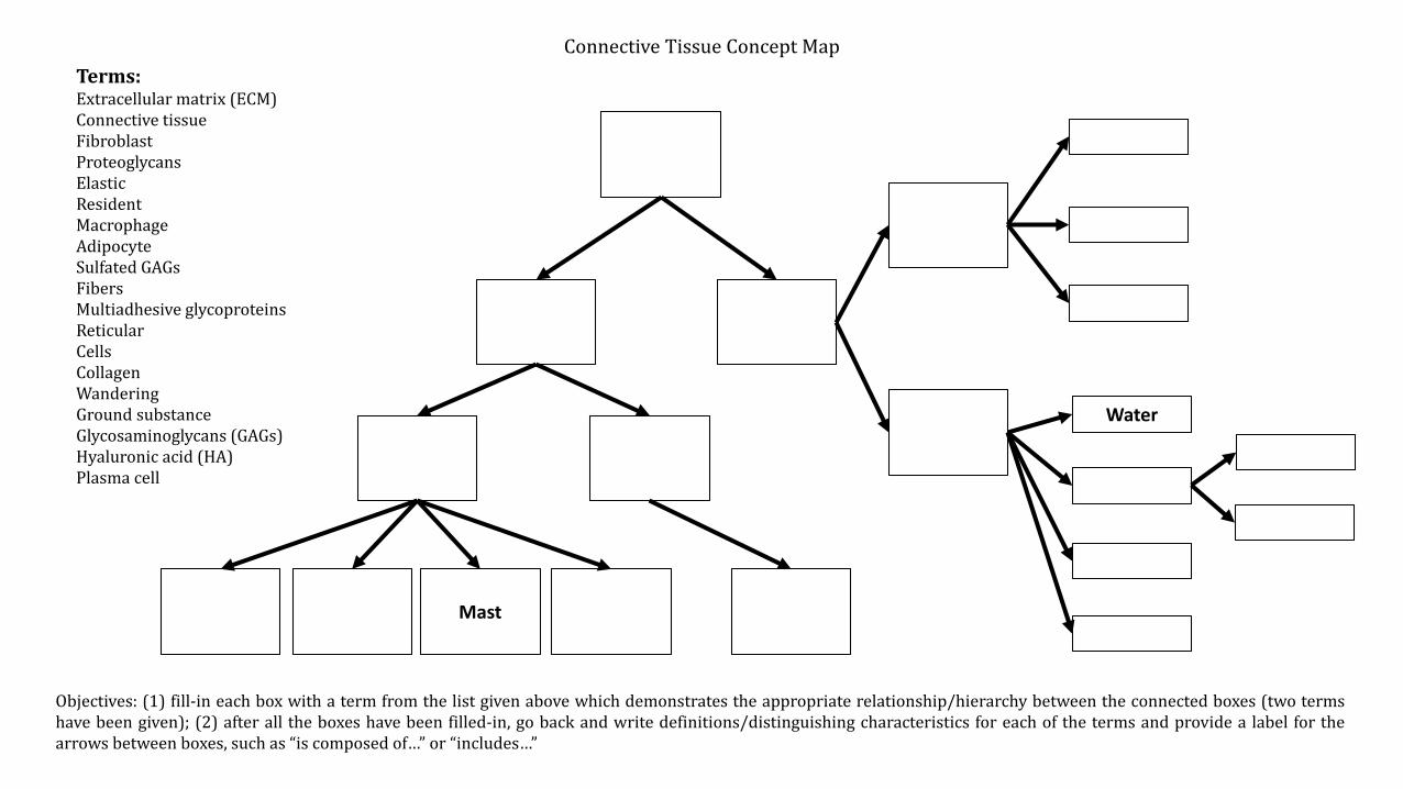
Connective Tissue Concept Map
Water
Mast
Terms:Extracellular matrix (ECM)Connective tissueFibroblastProteoglycansElasticResidentMacrophageAdipocyteSulfated GAGsFibersMultiadhesive glycoproteinsReticularCellsCollagenWanderingGround substanceGlycosaminoglycans (GAGs)Hyaluronic acid (HA)Plasma cell
Objectives: (1) fill-in each box with a term from the list given above which demonstrates the appropriate relationship/hierarchy between the connected boxes (two termshave been given); (2) after all the boxes have been filled-in, go back and write definitions/distinguishing characteristics for each of the terms and provide a label for thearrows between boxes, such as “is composed of…” or “includes…”