l1 interaction domains of papillomavirus l2 …jvi.asm.org/content/75/9/4332.full.pdf · l1...
TRANSCRIPT
JOURNAL OF VIROLOGY,0022-538X/01/$04.0010 DOI: 10.1128/JVI.75.9.4332–4342.2001
May 2001, p. 4332–4342 Vol. 75, No. 9
L1 Interaction Domains of Papillomavirus L2 Necessary forViral Genome Encapsidation
MARTIN M. OKUN,1 PATRICIA M. DAY,1 HEATHER L. GREENSTONE,1 FRANK P. BOOY,2
DOUGLAS R. LOWY,1 JOHN T. SCHILLER,1 AND RICHARD B. S. RODEN1,3*
Laboratory of Cellular Oncology, Division of Basic Sciences, National Cancer Institute, Bethesda, Maryland 208921;Department of Biochemistry, Wolfson Laboratory, Imperial College of Science, Technology and Medicine, London
SW7 2AY, England2; and Department of Pathology, Johns Hopkins University, Baltimore, Maryland 212053
Received 15 December 2000/Accepted 8 February 2001
BPHE-1 cells, which harbor 50 to 200 viral episomes, encapsidate viral genome and generate infectiousbovine papillomavirus type 1 (BPV1) upon coexpression of capsid proteins L1 and L2 of BPV1, but notcoexpression of BPV1 L1 and human papillomavirus type 16 (HPV16) L2. BPV1 L2 bound in vitro via itsC-terminal 85 residues to purified L1 capsomers, but not with intact L1 virus-like particles in vitro. However,when the efficiency of BPV1 L1 coimmunoprecipitation with a series of BPV1 L2 deletion mutants wasexamined in vivo, the results suggested that residues 129 to 246 and 384 to 460 contain independent L1interaction domains. An L2 mutant lacking the C-terminal L1 interaction domain was impaired for encapsi-dation of the viral genome. Coexpression of BPV1 L1 and a chimeric L2 protein composed of HPV16 L2residues 1 to 98 fused to BPV1 L2 residues 99 to 469 generated infectious virions. However, inefficientencapsidation was seen when L1 was coexpressed with either BPV1 L2 with residues 91 to 246 deleted or withBPV1 L2 with residues 1 to 225 replaced with HPV16 L2. Impaired genome encapsidation did not correlateclosely with impairment of the L2 proteins either to localize to promyelocytic leukemia oncogenic domains(PODs) or to induce localization of L1 or E2 to PODs. We conclude that the L1-binding domain located nearthe C terminus of L2 may bind L1 prior to completion of capsid assembly, and that both L1-binding domainsof L2 are required for efficient encapsidation of the viral genome.
Papillomaviruses are nonenveloped double-stranded DNAtumor viruses. Their capsid comprises 360 molecules of themajor capsid protein L1, arranged as 72 pentamers, or cap-somers, in a T57d icosahedral surface lattice (2). Expressionof L1 protein results in the self-assembly of virus-like particles(VLPs), which have the size, shape, and conformationalepitopes of virion capsids (14). Bovine papillomavirus type 1(BPV1) virions in the presence of low ionic strength and di-thiothreitol (DTT) (18) and human papillomavirus type 11(HPV11) and HPV33 VLPs in the presence of reducing agents(21, 25) are disassembled into capsomers. RecombinantHPV11 L1 protein with a Cys-to-Gly mutation in the C termi-nus of L1 forms pentamers but cannot assemble into capsid-like structures (18). Taken together, the data imply that bothionic and disulfide bonds mediate interpentamer binding in thepapillomavirus capsid.
Virions also contain L2, the minor capsid protein (5). Thenumber of L2 molecules per capsid has been estimated atbetween 12 (30) and 36 (5) molecules per virion. If L2 iscoexpressed with L1, the L2 protein is coassembled into VLPswith a stoichiometry similar to that seen in authentic virions(15). Three-dimensional reconstruction of cryo-electron mi-crographs of quench-frozen BPV virions has revealed the cap-sid architecture to 9-A resolution. This analysis detected aprotein density within the central cavity of the pentavalent
capsomers, suggesting that L2 may be associated with these 12vertex capsomers (30).
Rodent fibroblasts maintain the BPV1 genome at 50 to 200episomes/cell (17, 33). These cells express the nonstructuralviral proteins, but no virus is produced because the cells do notexpress the capsid proteins (1). However, expression of L1 andL2 in trans causes encapsidation of viral episomes and forma-tion of infectious virions (26, 36, 37). L2 is not absolutelyrequired for generation of pseudovirions in vitro (29) or in vivo(31). However, L2 enhances DNA encapsidation in vivo by.50-fold (23, 26, 36, 37). DNA encapsidation may also beenhanced by nucleotides 1506 to 1625 of the BPV1 genome(34), as well as by E2 (a virally encoded transcription/replica-tion factor) in some systems that generate pseudovirions (35)but not in others (31). L2 colocalizes with the promyelocyticleukemia protein (PML) in subnuclear domains called PMLoncogenic domains (PODs) or nuclear domain-10 (3). Further,while BPV1 E2 and L1 exhibit a diffuse nuclear localization inits absence, L2 causes E2 (both the full-length E2TA and shortrepressor form E2TR) and L1 to traffic to PODs (3, 10). Thislocalization partially, or completely, overlaps with the site ofHPV11 DNA replication (27). Interestingly, overexpressedHPV11 E2 is associated with the nuclear matrix (38), andHPV5 E2 is associated with RNA splicing factors in subnuclearfoci (16). L2 binds directly to two regions of BPV1 E2 in vitroand attenuates E2-mediated transcription but not viral repli-cation (10).
In the present study, we have sought to characterize theinteraction between L1 and L2 during virion formation andspecifically to determine (i) which domains of L2 mediate itsbinding to L1; (ii) at what stage during capsid assembly L2
* Corresponding author. Mailing address: Department of Pathology,Room 656, The Ross Research Building, 720 Rutland Ave., Baltimore,MD 21205. Phone: (410) 502-5161. Fax: (410) 614-3548. E-mail: [email protected].
4332
on Septem
ber 17, 2018 by guesthttp://jvi.asm
.org/D
ownloaded from

binds to L1; and (iii) whether the L1 interaction domains in L2contribute to virion assembly.
MATERIALS AND METHODS
Generation of vectors and recombinant viruses. The full-length BPV1 L2 geneand a PCR-amplified fragment (L2D384-469) comprising L2 nucleotides 1 to1149 were inserted between the BamHI and EcoRI sites of Bluescript II SK(2)phagemid (Stratagene Cloning Systems, La Jolla, Calif.), downstream from theT3 RNA polymerase promoter, to generate Bluescript II pSK(2)L2 and Blue-script II pSK(2)L2D384-469, respectively. The baculovirus transfer vectors con-taining the BPV1 L1 or L2 genes were constructed as previously described. Thebaculovirus transfer vector was constructed by subcloning the XbaI-HindIII mi-nor capsid gene fragment into pFastBac1. Recombinant baculovirus was gener-ated using the BAC-TO-BAC Baculovirus Expression System under the manu-facturer’s recommended reaction conditions (Gibco BRL Life Technologies,Gaithersburg, Md.). Deletions were introduced into L2 by PCR as describedpreviously (22, 24), using the following oligonucleotides: L2D1-88 employedCGCGAGATCTACCATGGGATCCAGAGCTGTAAC and GCGCAGATCTTTAGGCATGTTTCCG; L2D173-469 employed CGCGAGATCTACCATGAGTGCACGAAAAAGAG and GCGCAGATCTTTAAACCGCTATGTCCTCG;L2D247-469 employed CGCGAGATCTACCATGAGTGCACGAAAAAGAGand GCGCAGATCTTTAGGCAATACTGCGGGGCGT; L2D395-469 em-ployed CGCGAGATCTACCATGAGTGCACGAAAAAGAG and GCGCAGATCTTTACTGAGTTGGAATGAGGC; and L2D461-469 employed CGCGAGATCTACCATGAGTGCACGAAAAAGAG and GCGCAGATCTTTACAACAAGGAGGGATGC. The oligonucleotides were used to PCR amplify BPV L2fragments which, after BglII digestion, were cloned into the BamHI site ofpSFV-1. Deletions L2D91-129 and L2D91-246 were amplified using 59CGCGGGATCCGCCCCTGCAATAGTC and CGCGGGATCCTCTAAATCACGTGGC, respectively, with 39 oligonucleotide GTAGTGTCATCGATAAC. ThePCR fragments were cloned into pSFV-1.BPV1 L2 using BamHI and ClaI.Chimeras H98B and H225B were generated using 59 oligonucleotides CGCGGGGCCCTAGTATAGGTGCGGGC and GCGCCCTAGGAGAAAACATTGAACTGAC, respectively, and 39 oligonucleotide CGTTTGCGTAGGGATGTAAT to amplify a fragment from pSFV-1.BPV1 L2. Chimera H98B wasgenerated by cloning an ApaI and XmaI-digested PCR fragment into pSFV-1.HPV16 L2. Chimera H225B was generated by cloning an AvrII- and XmaI-digested PCR fragment into pSFV-1.HPV16 L2. All constructs were confirmedby automated fluorescence sequence analysis (Seqwright). Recombinant SemlikiForest virus (SFV) expressing mutant L2 proteins was constructed as describedpreviously (23). The recombinant pSFV-1 clones containing either the L1 ormutant L2 genes and pHelper-2 plasmid were linearized using SpeI (or NruI forpSFV-1.NruI-based clones). The DNAs were phenol-chloroform extracted andethanol precipitated. To generate SFV RNA, 1 mg of each linearized pSFV-1clone and 1 mg of pHelper-2 were resuspended in 100-ml reaction mixturescontaining 1 mM ATP, 1 mM CTP, 1 mM UTP, 0.5 mM GTP, 1 mM RNAcapping analog m7G(59)ppp(59)G, 5 mM DTT, 100 U of human placental ribo-nuclease inhibitor, and 75 U of SP6 RNA polymerase in 13 SP6 reaction buffer.The reactions were incubated for 1 h at 37°C, and 2.5 ml was analyzed on a 0.8%agarose gel to assess the integrity of the SFV RNAs. A total of 107 BHK21 cellsreleased into suspension by trypsin treatment was mixed with 5 mg of SFV RNAand 5 mg of Helper-2 RNA in 0.8 ml of serum-free Glasgow’s minimal essentialmedium (GMEM). After transfer to a cuvette, the cells were electroporated (225V, 800 mF, low ohms; Life Technologies Electroporator) twice and plated out in25 ml of complete GMEM (5% fetal calf serum, 10% tryptose-phosphate broth,10 mM HEPES [pH 7.4], 13 nonessential amino acids, 100 U of penicillin/ml,and 100 mg of streptomycin/ml in GMEM). After incubation for 24 h the mediumwas harvested, clarified by centrifugation (1,000 3 g, 10 min), aliquoted, andstored at 280°C.
Preparative purification of intact and disassembled VLPs. Preparation ofintact particles was performed as described previously (15). Disassembled par-ticles were prepared by dialyzing 200 ml of intact particles at 4°C against 250 mlof disassembly buffer (10 mM Tris-HCl [pH 7.4], 3 mM DTT, 1 mM EDTA)containing 1 pellet of Complete protease inhibitor cocktail tablets (Roche). Thedialysate was clarified by centrifugation at 14,000 rpm for 15 min at 4°C. Proteinconcentrations were determined with a colorimetric protein assay (Bio-RadLaboratories, Hercules, Calif.). For transmission electron microscopy, sampleswere spotted on carbon-coated grids, negatively stained with 1% uranyl acetate,and examined with a Philips electron microscope (model EM 400T) at 36,0003magnification as previously described.
Cell-free L1-L2 binding assay. In vitro transcription of nonlinearized DNA at25 mg/ml with T3 RNA polymerase (Promega Corp., Madison, Wis.) was per-
formed for 120 min at 37°C in the recommended reaction buffer (Gibco BRLCorp.). Transcription products were precipitated with sodium acetate and eth-anol, dissolved in water to a concentration of 35 mg/ml, and then diluted 25-foldin the TNT rabbit reticulocyte lysate system (Promega Corp.) containing[35S]cysteine (.1,000 Ci/mmol; Amersham) for translation for 90 min at 30°C.Twenty-five microliters from the in vitro translation reaction mixture was incu-bated with 4 mg of purified particles in 100 ml of phosphate-buffered saline (PBS)for 2 h at 30°C. Samples were diluted to 500 ml with radioimmunoprecipitationassay (RIPA) buffer (50 mM Tris [pH 8.0], 150 mM NaCl, 1.0% NP-40, 0.5%sodium deoxycholate, 0.1% sodium dodecyl sulfate [SDS]) containing 1 mMphenylmethylsulfonyl fluoride, 1 U of Trasylol/ml, and 0.67 ml of rabbit preim-mune serum for 1 h of continuous rotation at 4°C. This was followed by anadditional 30 min of continuous rotation at 4°C in the presence of 30 ml ofprotein A-Sepharose CL-4B beads (Pharmacia Biotech, Uppsala, Sweden) sus-pended in RIPA buffer. After centrifugation at 16,000 3 g for 10 s, the super-natant was immunoprecipitated with 0.30 ml of rabbit polyclonal antiserum to L1,essentially as described previously (4). Nonradiolabeled full-length L2 and sixoverlapping L2 peptides were used to block in vitro-translated L2 binding to L1.Generation of the recombinant bacteria expressing these proteins, induction ofprotein expression, and purification were performed as described previously (24).Rabbit polyclonal antisera to L1, L2, peptide B, corresponding to amino acids 45to 173 of L2, and peptide F, corresponding to amino acids 384 to 469 of L2, weregenerated as described previously (24).
Immunofluorescent localization of BPV proteins. As described previously (3),recombinant SFV stocks expressing L1 and L2 were rendered infectious byincubation with 0.5 mg of chymotrypsin A4 (Boehringer Mannheim) per ml for30 min on ice and treatment with 0.5 mg of aprotinin (Sigma) per ml. Activatedvirus, diluted 1:100 in Dulbecco’s modification of PBS (D-PBS), was added toBPHE-1 cells on glass coverslips for 1 h at 37°C and then replaced with medium.After 6 h, the cells were washed in PBS and fixed for 10 min in 1% paraform-aldehyde–PBS. After blocking in 200 mM glycine, the cells were permeabilizedwith 0.1% Brij 58, and all incubations were performed at 4°C. L2 was detectedwith rabbit antiserum to full-length L2 (24); L1 was detected with monoclonalantibody (MAb) 5B6 (24); and E2 was detected with MAb B201 (E. Androphy,Tufts Medical Center). Hybridoma supernatants were diluted 1:100, and rabbitpolyclonal serum was diluted 1:1,000. Secondary antibody (fluoroscein isothio-cyanate [FITC] or Texas Red labeled) was used at 5 mg/ml and mounted withFluoromount mounting fluid (Southern Biotechnology Associates) on a glassslide. Fluorescence was examined using a Bio-Rad MRC 1024 laser-scanningconfocal system attached to a Zeiss Axioplan microscope. All images wereacquired with a Zeiss 633 N.A. 1.4 Planapo objective. Control slides withisotype-matched, irrelevant MAbs or preimmune serum were used to establishthat fluorescence between the green and red channels did not overlap and thatimmunofluorescent labeling was specific. The images were arranged and pseudo-colored using Adobe Photoshop.
Coimmunoprecipitation of L1 and L2 mutants. A total of 4 3 106 BHK21 cellswere coinfected with equivalent titers of recombinant SFV expressing L1 andmutant L2. The cells were harvested 24 h postinfection by scraping and collectedby centrifugation (600 3 g, 10 min). The cell pellet was resuspended in 1 ml oflysis buffer (1% NP-40, 0.5 M NaCl, 50 mM Tris-HCl [pH 8], and Complete[Roche] protease inhibitors) at 4°C. The lysates were sheared by sonication(Branson Sonifier 250, ice-cold water bath; maximum setting; 30 s) and clarifiedby centrifugation (10,000 3 g, 10 min, 4°C). Sequential immunoprecipitationswere performed for 1 h each with a 50-ml packed volume of protein A-Sepharoseper tube precoupled to 5 ml of (i) preimmune rabbit antiserum, (ii) rabbitanti-BPV L1 VLP (14), and (iii) rabbit anti-BPV1 L2-6His (24). The immuno-precipitates were washed three times with 1 ml of lysis buffer, and the boundproteins were eluted by boiling in gel sample buffer. The immunoprecipitateswere subjected to SDS–10% polyacrylamide gel electrophoresis (PAGE) andWestern blotting. Coimmunoprecipitated capsid proteins were detected with 1mg of L2-specific MAb C6 (20) or MAb 3A10 (12) per ml, peroxidase-linkedanti-mouse immunoglobulin G antibody (1:10,000), and chemiluminescent sub-strate (Kirkegaard & Perry Laboratories).
Analysis of genome encapsidation. A total of 107 BPHE-1 cells were coin-fected with recombinant SFVs expressing wild-type L1 alone or with each dele-tion mutant of L2. The BPHE-1 cells were harvested 30 h postinfection byscraping into the medium and collected by centrifugation. The cells were washedin PBS and lysed on ice by sonication (Branson sonifier, 1 min at power level 7)in 1 ml of PBS containing 1% (vol/vol) Nonidet P-40, 10 mg of aprotinin/ml, and100 U of DNase I/ml. The lysates were clarified by centrifugation (10,000 3 g, 10min, 4°C). Preimmune serum (10 ml) and protein A-Sepharose (50-ml packedvolume) were added to the clarified extract, and the sample was tumbled at 4°Cfor 1 h. The beads were removed by centrifugation for 1 min at 2,000 3 g. The
VOL. 75, 2001 L1 INTERACTION DOMAINS OF PAPILLOMAVIRUS L2 4333
on Septem
ber 17, 2018 by guesthttp://jvi.asm
.org/D
ownloaded from
supernatant was transferred, and 10 ml of rabbit anti-BPV1 L1/L2 VLP serumand protein A-Sepharose (50-ml packed volume) were added. The sample wastumbled at 4°C for 1 h, and the beads were recovered by centrifugation at2,000 3 g for 1 min. The beads were washed three times with 1 ml of D-PBScontaining 1% (vol/vol) Nonidet P-40, 10 mg of aprotinin/ml, and 100 U ofDNase I and RNase A per ml and incubated for 1 h at 37°C to allow digestionof accessible DNA. The beads were then washed twice more in buffer lackingnuclease and aprotinin. Encapsidated DNA was released from the beads byresuspension in 400 ml of 50 mM Tris-HCl (pH 8), 10 mM EDTA, 1 mM DTT,and 100 mg of proteinase K/ml. After 15 min of incubation at 37°C to allowdigestion of the capsid proteins, the sample was centrifuged for 1 min at 2,000 3g. The supernatant was transferred and extracted using buffered phenol and thenchloroform. DNA was precipitated from the supernatant by addition of 20 mg ofglycogen, 40 ml of 3 M sodium acetate (pH 5.2), and 1 ml of ethanol and coolingto 220°C overnight. The DNA was recovered by centrifugation (16,000 3 g, 10min), washed with 70% ethanol, and resuspended in Tris-EDTA. The DNA(uncut) was separated on a 0.8% agarose–Tris-acetate-EDTA gel and trans-ferred to a Nytran1 membrane. BPV1 DNA was detected by Southern blotting.Biotinylated probe was prepared by random priming of the EcoRI-BamHI largefragment of BPV-pML. The probe was detected using strepavidin-peroxidaseand enhanced chemiluminescence according to the manufacturer’s instructions(Pierce).
Generation of infectious BPV. The system used to generate infectious BPV1 invitro has been described previously (23). Briefly, 107 BPHE-1 cells were coin-fected with recombinant SFVs expressing wild-type L1 alone or with each dele-tion mutant of L2. The BPHE-1 cells were harvested 30 h postinfection byscraping into the medium and collected by centrifugation. The cells were resus-pended in 2 ml of D-PBS and lysed by sonication (Branson sonifier; 1 min atpower level 7) on ice. Monolayers of C127C mouse fibroblasts in 60-mm-diam-eter dishes were incubated with 1 ml of extract for 1 h at 37°C. The cells werewashed and the medium was replaced with Dulbecco’s MEM containing 10%fetal calf serum, 100 U of penicillin/ml, and 100 mg of streptomycin/ml. TheC127C cells were cultured for 3 weeks and then stained with 0.5% (wt/vol)methylene blue and 0.25% (wt/vol) carbol fuchsin in methanol.
RESULTS AND DISCUSSION
Coimmunoprecipitation of BPV1 L1 and deletion mutantsof L2. To determine regions of L2 that interact with L1, a seriesof deletion mutants of the BPV1 L2 gene was generated byPCR and cloned into vector pSFV-1 (19) (Fig. 1A). Defectiverecombinant SFVs encoding these deletion mutants were gen-erated (23). Coexpression of L1 and L2 leads to their coas-sembly into VLPs with a stoichiometry similar to that of virionsobtained from bovine papillomas (23). We therefore examinedthe interaction of wild-type BPV1 L1 with seven of the L2deletion mutants in BHK21 cells that had been coinfected withrecombinant SFV that express L1 and mutant L2 (19, 23).Sequential immunoprecipitations were performed using pre-immune rabbit serum first, then rabbit anti-BPV1 L1 VLPs(14), and finally rabbit anti-BPV1 L2–six-His (24). By SDS-PAGE and Western blotting with L2-specific antibody, no L2was detected upon immunoprecipitation with preimmune rab-bit serum (results not shown) or when L2 was expressed aloneand immunoprecipitated by rabbit anti-BPV1 L1 VLPs (Fig.1B, lane 2). All mutants expressed high levels of L2 (Fig. 1Dand E). The extra immunoreactive band at ;50 kDa in Fig. 1Clikely results from cross-reactivity of the secondary antibodywith the heavy chain of the rabbit antibody used to immuno-precipitate L1. When the L2 deletion mutants were coex-pressed with L1, only L2D173-469 failed to coimmunoprecipi-tate with L1 (Fig. 1B, lane 7) despite its high level expression(Fig. 1D, lane 7). L2D395-469, L2D247-469 (Fig. 1B, lanes 5and 6), and L2D91-246 (Fig. 1C, lane 3) coimmunoprecipitatedwith L1, although with reduced efficiency compared to full-length L2. The remaining L2 deletion mutants (L2D1-88,
L2D461-469, and L2D91-129) coimmunoprecipitated with L1to the same extent as wild-type L2 (Fig. 1B and C). Takentogether, the data imply that L2 may contain two independentL1 interaction domains. One L1 interaction domain is locatednear the C terminus of L2, between residues 395 and 460, andthe second is located between residues 129 and 246 of L2.Consistent with this possibility, studies of L2-specific MAbbinding to L1 and L2 VLPs suggest that both these regions ofL2 are internal to the capsid (20, 32).
L2 binds in vitro to L1 capsomers, but not to intact VLPs.Since many viruses inject their genome into preformed capsidstructures (7, 11) and L2 is required for efficient papillomavi-rus DNA encapsidation (23), we sought to determine if L2interacts with intact L1 capsids or with capsomers. To producecapsomers, L1 VLPs were purified from Sf9 insect cells in-fected with recombinant baculovirus expressing the L1 gene(Fig. 2A) and dialyzed against a disassembly buffer. As ex-pected (18, 21, 25), this procedure resulted in the disassemblyof intact VLPs into capsomers, as determined by electron mi-croscopy (Fig. 2B) and by gel filtration (Superose 6 sizingcolumn) (data not shown).
To examine whether L2 could interact with capsids or withcapsomers, BPV1 L2 translated in rabbit reticulocyte lysatesupplemented with [35S]cysteine was incubated with intact ordisassembled L1 VLPs and immunoprecipitated with rabbitpolyclonal antiserum that recognizes intact and disassembledL1, and the immunoprecipitates were subjected to SDS-PAGE(Fig. 2C). In vitro-translated L2 was not efficiently coprecipi-tated by anti-L1 serum in the presence of intact L1 VLPs or, asa control, in the absence of L1 (lanes 1 and 3). Upon extendedexposure, a trace amount of in vitro-translated L2 coprecipi-tated with VLPs (lane 3), but this signal probably results fromfree capsomers contaminating the VLP preparation (Fig. 2A).In the presence of disassembled L1 VLPs, by contrast, in vitro-translated L2 was efficiently coprecipitated by anti-L1 serum(lane 4). This coprecipitation was specifically blocked by non-radiolabeled L2 produced in Escherichia coli (lane 5) or by aset of overlapping L2 peptides (A to F) that together encom-pass the full L2 protein (lane 6). Peptides A through F corre-spond to the following L2 amino acids: A, 1 to 88; B, 45 to 173;C, 130 to 257; D, 216 to 340; E, 300 to 425; and F, 384 to 469(24). As additional specificity controls, in vitro-translated L2was not coprecipitated by antipolyoma serum in the presenceof disassembled polyoma VP1 VLPs (data not shown), andradiolabeled in vitro-translated luciferase was not coimmuno-precipitated by anti-L1 serum in the presence of disassembledL1 VLPs (lane 7). The finding that L2 bound to capsomers, butnot to intact L1 VLPs, implies that L2 binds to L1 prior tocompletion of capsid assembly.
Determination of the L2 region that mediates binding toL1 in vitro. Since the set of six overlapping L2 peptides (Athrough F, described in reference 24) were able to block co-immunoprecipitation of radiolabeled L2 in the presence ofdisassembled L1 (Fig. 2C, lane 6), the peptides were testedindividually in an in vitro binding assay for their ability to blockL2 binding (Fig. 3A). Only peptide F, corresponding to theC-terminal 85 amino acids of L2 (amino acids 384 to 469),blocked L2 binding (Fig. 3A, lane 6). To exclude the possibilitythat the F domain blocks L1 binding by forming an inactivehetero-oligomer with full-length L2, L2 and peptide F were
4334 OKUN ET AL. J. VIROL.
on Septem
ber 17, 2018 by guesthttp://jvi.asm
.org/D
ownloaded from

FIG. 1. Coimmunoprecipitation of BPV1 L1 and L2 deletion mutants. (A) Schematic diagram of BPV1 L2 deletion mutants and chimeras withHPV16 L2. (B and D) BHK21 cells were infected with recombinant SFV expressing BPV1 L1 (lanes 1 and 3 to 7) and full-length L2 (lanes 1 and2), L2D1-88 (lane 3), L2D461-469 (lane 4), L2D395-469 (lane 5), L2D247-469 (lane 6), or L2-173–469 (lane 7). (C and E) BHK21 cells were infectedwith recombinant SFV expressing BPV1 L1 (lanes 1, 3, and 4) and full-length L2 (lanes 1 and 2), L2D91-246 (lane 3), or L2D91-129 (lane 4). Afterincubation for 24 h, the cells were harvested and sequential immunoprecipitation was performed using preimmune serum first (results not shown),then rabbit antiserum to L1 VLPs (B and C), and finally rabbit antiserum to full-length L2 (D and E). The presence of L2 in immunoprecipitateswas determined by Western blotting using L2-specific MAb C6 (B and C) or 3A10 (D and E).
VOL. 75, 2001 L1 INTERACTION DOMAINS OF PAPILLOMAVIRUS L2 4335
on Septem
ber 17, 2018 by guesthttp://jvi.asm
.org/D
ownloaded from

coincubated and then immunoprecipitated with a rabbit anti-serum raised against peptide B (residues 45 to 173 of BPV1L2) (24). However, there was no evidence of coprecipitation ofpeptide F with full-length L2 in the immunoprecipitated com-plexes when probed by Western blotting with a rabbit anti-
FIG. 2. In vitro binding of L2 to L1 pentamers, but not to intactVLPs. BPV1 L1 VLPs disassemble into component capsomers afterdialysis in disassembly buffer. BPV1 L1 VLPs purified from recombi-nant baculovirus-infected insect cells was left intact or was disassem-bled via dialysis against disassembly buffer (10 mM Tris-HCl [pH 7.4],1 mM EDTA, 3 mM DTT) at 4°C overnight. Samples of intact BPV1L1 VLPs (A) and disassembled BPV1 L1 VLPs (B) were examined viaelectron microscopy at 36,0003 magnification (bar, 100 nm). IntactVLPs and pentamer are indicated with large and small arrows, respec-tively. (C) In vitro-transcribed L2 mRNA was translated in rabbitreticulocyte lysate supplemented with [35S]cysteine. The in vitro trans-lation product was incubated with purified L1 VLPs or capsomer andthen immunoprecipitated and subjected to SDS-PAGE and autora-diography. Lane 1, in vitro-translated L2 immunoprecipitated withanti-L1 serum; lane 2, in vitro-translated L2 immunoprecipitated withanti-L2 serum; lane 3, in vitro-translated L2 incubated with intact L1VLPs and then immunoprecipitated with anti-L1 serum; lane 4, invitro-translated L2 incubated with disassembled L1 VLPs and thenimmunoprecipitated with anti-L1 serum; lane 5, in vitro-translated L2incubated with disassembled L1 VLPs and nonradiolabeled L2 with aC-terminal six-histidine tag and then immunoprecipitated with anti-L1serum; lane 6, in vitro-translated L2 incubated with disassembled L1VLPs and nonradiolabeled overlapping L2 peptides (A through F)with C-terminal six-histidine tags; lane 7, in vitro-translated luciferaseincubated with disassembled L1 VLPs and then immunoprecipitatedwith anti-L1 serum. Molecular masses (in kilodaltons) are shown.
FIG. 3. L2 C-terminal 85 amino acids are required for L1 bindingin vitro, but not in vivo. (A) Six overlapping peptides of L2 (A throughF), expressed as six-His fusion proteins in E. coli, were tested forblocking of L1 binding to in vitro-translated L2. In vitro-translated35S-labeled L2 and disassembled L1 VLPs were incubated either withpeptides A through F (lanes 1 to 6) or buffer (lane 7). Samples wereimmunoprecipitated with L1 antiserum, and immunoprecipitates weresubjected to SDS-PAGE and autoradiography. (B) In vitro-translated35S-labeled L2 and L2D384-469 were immunoprecipitated with anti-L2serum (lanes 1 and 4) or immunoprecipitated with anti-L1 serum ineither the presence (lanes 3 and 6) or absence (lanes 2 and 5) ofdisassembled L1 VLPs. Immunoprecipitates were subjected to SDS-PAGE and autoradiography. The positions of L2 and L2D384-469 areindicated. Molecular masses (in kilodaltons) are shown. (C) Four mi-crograms each of intact L1/L2D384-469 VLPs (lane 1), in vitro-disas-sembled L1/L2D384-469 VLPs (lane 2), intact L1/L2 VLPs (lane 3),and in vitro-disassembled L1/L2 VLPs (lane 4) were immunoprecipi-tated with MAb 5B6 (lanes 1 to 4) and then subjected to SDS-PAGEand analyzed by Western blotting using rabbit antiserum to BPV1 L2residues 45 to 173 and peroxidase-conjugated protein A. The positionsof L2 and L2D384-469 are indicated. (D) Four micrograms each ofpurified L1/L2D384-469 VLPs (lanes 3 and 4) were immunoprecipi-tated with rabbit antiserum to L2 residues 45 to 173 (lanes 1 and 3) orpreimmune serum (lanes 2 and 4) and then subjected to SDS-PAGEand analyzed by Western blotting with BPV1 L1 mouse monoclonalantibody 837 and peroxidase-conjugated protein A. The position of L1is indicated.
4336 OKUN ET AL. J. VIROL.
on Septem
ber 17, 2018 by guesthttp://jvi.asm
.org/D
ownloaded from

serum raised against peptide F (results not shown). To confirmthat the L1-binding domain lies within the C-terminal 85amino acids of L2, an L2 C-terminal deletion mutant lackingamino acids 384 to 469 (L2D384-469) was subjected to in vitrotranscription and translation. In vitro binding of L2D384-469 todisassembled L1 VLPss was dramatically reduced when com-pared to binding by full-length L2 (Fig. 3B). Thus, in vitrobinding of L2 to L1 requires sequences found at or near the Cterminus of L2. This location is consistent with these sequencescorresponding to the more C terminal domain of the two L1interacting domains identified in Fig. 1.
C-terminal L1-binding domain is not necessary in vivo forincorporation of L2 within VLPss. To determine if the C-terminal 85 amino acids of L2 are necessary for L2 to associatewith L1 in vivo, Sf9 cells were doubly infected with baculovirusencoding L1 and either L2D384-469 or full-length L2, andVLPss were purified (15). Both L2 and L2D384-469 copurifiedwith L1 in VLPss. There was no visible difference in VLPspreparations and their disassembly using either BPV L1 only(Fig. 2A and B), BPV1 L1/L2, or L1/L2D384–469 (data notshown) as assessed by transmission electron microscopy. Whenpurified L1/L2D384-469 and L1/L2 VLPss were immunopre-cipitated with L1-specific antibody, both L2 proteins coprecipi-tated with L1, although L2D384-469 did so ;3-fold less effi-ciently than did full-length L2 (Fig. 3C, lanes 1 and 3),consistent with the results obtained without VLPs purification(Fig. 1B). Analogous results were obtained when the purifiedVLPss were immunoprecipitated with antiserum to residues 45to 172 of BPV1 L2, which neutralizes BPV infection and there-fore presumably recognizes L2 epitopes exposed on the surfaceof intact virions and VLPs (Fig. 3D, lanes 1 and 3) (24). Bycontrast, when disassembled VLPss were subjected to L1 im-munoprecipitation, full-length L2, but not L2D384-469, co-precipitated with L1 (Fig. 3C, lanes 2 and 4). The finding forfull-length L2 is consistent with a previous report that L2remains associated with pentamers upon disassembly ofHPV33 L1 and L2 VLPss (25). Since a subset of L2 interac-tions with L1 are salt labile in vitro (25), perhaps the weakcoimmunoprecipitation of L2 with capsomers reflects the rel-atively harsh washing conditions used (RIPA buffer). The re-sults also suggest that the C terminus of L2 is required to forman interaction with L1 that is stable in vitro. However, wecannot exclude the possibility that L2D384-469 is more proneto degradation than L2 upon disassembly of these insect cell-derived VLPss, although L2D384-469 and full-length L2 exhib-ited similar stability upon translation in vitro (Fig. 3B). Thehigher stability of the association of L2D384-469 with VLPsscompared to capsomers may be due to retention of the L2within the closed structure of the VLPss. Consistent with thisidea, antibody studies suggest that L2 is predominantly locatedwithin the capsid rather than exposed on the surface (9, 13, 20,24).
Role of the C-terminal L1 interaction domain of L2 in ge-nome encapsidation. To assess the contribution to virion as-sembly of the L1 interaction domain at the C terminus of L2,we exploited a culture system that generates infectious papil-lomavirus (23). BPHE-1 cells harbor 50 to 200 episomal copiesof the BPV1 genome per cell (33), but they produce no virusbecause the L1 and L2 genes are not expressed. However,expression of L1 and L2 in these cells via recombinant defec-
tive SFV vectors results in the generation of infectious BPV.BPHE-1 cells were coinfected with recombinant SFV express-ing BPV1 L1 and mutant L2 (23). Thirty hours postinfection,the cells were harvested, the capsid proteins were immunopre-cipitated and treated with DNase I to eliminate nonencapsu-lated genomes, and the quantity of DNase I-resistant BPV1genome present in the immunoprecipitates was assessed bySouthern blotting (Fig. 4). As expected, DNase I-resistant BPVgenomes were recovered from immunoprecipitates of BPHE-1cells expressing both L1 and L2 of BPV1, but not from cellsexpressing BPV1 L1 and HPV16 L2, which do not coassembleinto VLPss, or from cells expressing only BPV1 L1 (Fig. 4A,lane 1, and Fig. 4C, lanes 1 and 2). The predominant size of theBPV1 genome in the immunoprecipitates derived fromBPHE-1 cells expressing L1 and L2 was consistent with thesupercoiled form of BPV1 DNA.
When the ability of BPV1 L2 mutants to substitute for full-length L2 was assessed, equivalent quantities of the BPV1genome were found to be encapsidated by full-length BPV1 L2and L2D461-469 (Fig. 4A, lanes 2 and 6). However, BPV1genome encapsidation was dramatically reduced for L2D395-469 and L2D247-469 and was absent from L2D173-469 (Fig.4A, lanes 3 to 5). These results closely correlate with therelative efficiency with which the L2 mutants coprecipitatedwith VLPss (Fig. 1B, lanes 4 to 7). As the L2D395-469 mutantencapsidated the viral genome less efficiently than the L2D461-
FIG. 4. Analysis of BPV1 genome encapsidation in BPHE-1 cellscoexpressing BPV1 L1 and L2 deletion mutants or HPV16-BPV1chimeras. (A) BPHE-1 cells were coinfected with recombinant SFVexpressing BPV1 L1 (lanes 1 to 6) and L2 (lane 2), L2D173-469 (lane3), L2D247-469 (lane 4), L2D395-469 (lane 5), or L2D461-469 (lane 6).(B) BPHE-1 cells were coinfected with recombinant SFV expressingBPV1 L1 (lanes 1 to 4) and L2 (lane 2), L2D91-129 (lane 3), or L2D91-246 (lane 4). (C) BPHE-1 cells were coinfected with recombinant SFVexpressing BPV1 L1 (lanes 1 to 4) and L2 (lane 1), HPV16 L2 (lane 2),H225B (lane 3), or H98B (lane 4). Thirty hours postinfection, the cellswere harvested and the capsid proteins were immunoprecipitated fromlysates using rabbit anti-BPV1 VLPs. The immunoprecipitates weretreated with DNase I to eliminate nonencapsulated genomes, and thequantity of DNase I-resistant BPV1 genome present in the immuno-precipitates was assessed by Southern blotting. The predominant sizeof BPV1 DNA in immunoprecipitates derived from BPHE-1 cells ex-pressing L1 and L2 is consistent with the supercoiled form of the viralgenome.
VOL. 75, 2001 L1 INTERACTION DOMAINS OF PAPILLOMAVIRUS L2 4337
on Septem
ber 17, 2018 by guesthttp://jvi.asm
.org/D
ownloaded from

469 mutant or wild-type L2, the data suggest that the L1 in-teraction domain at the C terminus of L2 is necessary forefficient viral genome encapsidation. In addition, the C termi-nus of L2 may contain residues required to interact with BPV1episomes in vivo (26), perhaps to the identified packaging en-hancement sequence between nucleotides 1506 and 1625 (34).
BPV1 infectivity was absent from crude extracts of BPHE-1cells expressing L1 and L2D395-469 (Fig. 5, plate 2), L2D247-469, or L2D173-469 (results not shown), as assayed by focaltransformation of mouse C127C cells (6). As we have recentlydemonstrated that the C-terminal 9 amino acids are necessaryfor infection but not virion assembly (R. B. S. Roden et al.,unpublished data), no infectious particles would have beenexpected using these larger C-terminal deletions.
Our previous studies in BPHE-1 cells had demonstrated that(i) L2 is targeted to PODs in the absence of other papilloma-virus proteins and DNA; and (ii) L1 and E2 (a sequence-specific papillomavirus DNA-binding protein) are targeted toPODs in the presence, but not in the absence, of L2 (3). Inaddition, Zhao et al. observed that E2 expression enhanced invivo DNA encapsidation 6- to 10-fold (35), and direct interac-tion between L2 and E2 in vitro has recently been demon-strated (10). Further, Swindle et al. found partial or completeoverlap of PML with HPV11 E1, E2, the host replication factorRP-A, and bromodeoxyuridine incorporation and the viral rep-lication origin, consistent with viral DNA amplification at this
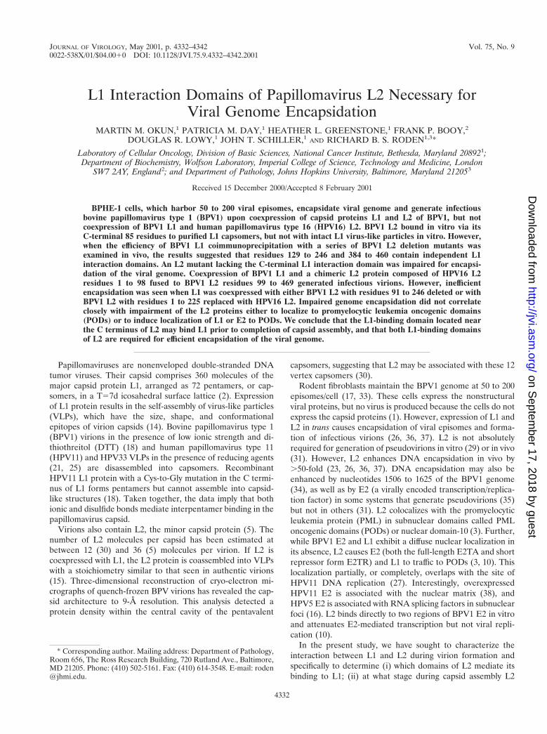
subnuclear domain (28). To exclude the possibility that thePOD-related L2 functions were compromised by deletion ofthe C-terminal L1 interaction domain, thus preventing viral ge-nome encapsidation, the intracellular localization of L2D395-469 and L2D247-469 with respect to PML (data not shown), L1(Fig. 6), and E2 (Fig. 7) were examined by immunofluorescentstaining. L2D395-469 trafficked to PODs, colocalized with PML(data not shown), and recruited E2, as did full-length L2 (Fig.6A to C and 7A to C). Therefore the inability of L2D395-469to encapsidate the BPV1 genome when coexpressed with L1does not result from improper subnuclear localization of L2 orimpairment of its recruitment of E2. We also determined thatL2D395-469 caused L1 to traffic to PODs. After examiningmany coexpressing cells, it is our impression that the colocal-ization was to a lesser degree than that with L1 and full-lengthL2 (Fig. 6) or L2D460-469 (results not shown). This result isconsistent with the deletion of the L1 interaction domain at theC terminus of L2. However, this conclusion must be consideredtentative, as the degree of colocalization of L1 and L2 wasquite variable, even for wild-type L2 (as previously reported),and could not be quantified.
Central domain of L2 functions in L1 binding and viralgenome encapsidation. Two strategies were adopted to explorethe role of the second L1-binding domain of L2 in viral ge-nome encapsidation. In the first, BPV1 L1 was coexpressedwith L2D91-129 or with L2D91-246 in BPHE-1. Wild-type lev-els of genome encapsidation were observed when BPV1 L1was coexpressed with L2-91-129 (Fig. 4B, lane 3), consistentwith the efficient coimmunoprecipitation of L1 and L2D91-129(Fig. 1C, lane 4). By contrast, viral genome encapsidation wasdrastically reduced when BPV1 L1 and L2D91-246, which co-immunoprecipitated only weakly (Fig. 1C, lane 3), were coex-pressed (Fig. 4B, lane 4). Coexpression of L1 and L2D91-246 inBPHE-1 failed to produce infectious virions, consistent withthe poor function of this mutant in genome encapsidation andL1 binding (Fig. 5, plate 3). L2D91-246 functioned similarly tofull-length L2 with respect to POD localization and its redis-tribution of endogenous E2 to PODs (Fig. 7), suggesting thatimpairment of these functions does not account for inefficientgenome encapsidation by this mutant. However, this mutantalso appeared to cause localization of L1 to PODs to a lesserdegree than full-length L2 (Fig. 6). This is consistent with thelow efficiency of coimmunoprecipitation of L2D91-246 with L1(Fig. 1C, lane 3) and suggests that L1 localization to PODsresults from its direct interaction with L2.
In a second approach to determine the role of the central L1interaction domain of L2 in genome encapsidation, we tookadvantage of the inability of HPV16 L2 to coimmunoprecipi-tate with BPV1 L1. To further define this L1 interaction do-main of BPV1 L2, two chimeras were constructed: chimeraH98B, which comprised residues 1 to 98 of HPV16 fused inframe with residues 99 to 469 of BPV1 L2, and chimera
FIG. 5. Ability of BPV1 L2 deletion mutants and HPV16-BPV1chimeras to generate infectious virions when coexpressed in BPHE-1cells. BPHE-1 cells were coinfected with recombinant SFV expressingBPV1 L1 (plates 1 to 6) and L2 (plate 1), L2D395-469 (plate 2),L2-91–246 (plate 3), HPV16 L2 (plate 4), H98B (plate 5), or H225B(plate 6). Thirty hours postinfection, the cells were harvested and lysedby sonication. Mouse C127C monolayers in 60-mm-diameter petridishes were infected with lysates and maintained for 3 weeks inDMEM containing 10% FCS. The plates were stained with 0.5%(wt/vol) methylene blue and 0.25% (wt/vol) carbol fuchsin in methanolto highlight transformed foci.
FIG. 6. Immunolocalization of BPV1 L2 deletion mutants and an HPV16-BPV1 chimera with respect to BPV1 L1 in SFV-infected BPHE-1cells. The cells were coinfected with the L1 and various L2 SFVs, fixed, and stained with antiserum against the L2 protein, detected with goatanti-rabbit Texas Red (panels A, D, G, and J), and MAb 5B6 directed against the L1 protein, detected with FITC-labeled goat anti-mouse antibody(panels B, E, H, and K). The digital superimposition of the two images is shown in panels C, F, I, and L. Colocalization is evident in the mergedimage as shown in yellow. The distribution of proteins for wild-type L2 is shown in panels A, B, and C. L2D395-469 is shown in panels D to F.L2D91-246 is shown in panels G to I. Chimera H225B is shown in panels J to L.
4338 OKUN ET AL. J. VIROL.
on Septem
ber 17, 2018 by guesthttp://jvi.asm
.org/D
ownloaded from

VOL. 75, 2001 L1 INTERACTION DOMAINS OF PAPILLOMAVIRUS L2 4339
on Septem
ber 17, 2018 by guesthttp://jvi.asm
.org/D
ownloaded from
H225B, which comprised residues 1 to 225 of HPV16 fused inframe with residues 226 to 469 of BPV1 L2. As expected fromthe presence of the L1 interaction of BPV1 L2 at the C ter-minus of each chimera, both H98B and H225B coimmunopre-cipitated with BPV1 L1 (not shown). However, H225B failedto promote encapsidation of the BPV1 genome when coex-pressed with BPV1 L1 in BPHE-1 cells, whereas H98B dem-onstrated significant activity (Fig. 4C, lanes 3 and 4). Thechimeras were also tested for their ability to generate infec-
tious virions when coexpressed with BPV1 L1 in BPHE-1 cells.When the focus-forming activity on C127C monolayers wasexamined, extracts of BPHE-1 cells expressing L1 and H225Bresulted in only a few foci, whereas H98B generated manyhundreds (Fig. 5, plates 5 and 6). H98B induced approximatelyhalf as many foci as wild-type BPV1 L2, although the platesshown in Fig. 5 were deliberately overloaded to allow detectionof low-level virion production (as for H225B). Notably, H98Bexpression was about twofold less than BPV1 L2 expression
FIG. 7. Immunolocalization of BPV1 L2 deletion mutants with respect to endogenous E2 in SFV-infected BPHE-1 cells. The cells wereinfected with the deletion mutant L2 SFVs and colocalized with respect to the endogenous E2 expressed in the BPHE-1 cells. L2 was detected withthe rabbit polyclonal antiserum and Texas Red-labeled goat anti-rabbit antibody (A, D, and G). E2 was detected with MAb B201 and FITC-labeledgoat anti-mouse antibody (B, E, and H). The merged images are shown in panels C, F, and I. The cells infected with wild-type L2 are shown inpanels A to C. L2D395-469 is shown in panels D to F. L2D91-246 is shown in panels G to I.
4340 OKUN ET AL. J. VIROL.
on Septem
ber 17, 2018 by guesthttp://jvi.asm
.org/D
ownloaded from

(results not shown). Overall, the results with the chimeric L2ssuggest that there is a type-restricted interaction of L1 with aBPV1 L2 domain that includes amino acids 99 to 225, whichcorrelates with the earlier results defining an L1 interactiondomain located between L2 residues 129 and 246.
Since H225B functioned poorly in virion assembly, the sub-cellular localization of this L2 chimeric protein was examined.H225B trafficked to PODs as well as BPV1 L2 (Fig. 6) andHPV16 L2 (results not shown). Furthermore, H225B causedBPV1 E2 to traffic to PODs in the same manner as BPV1 L2(data not shown). H225B also caused BPV1 L1 to colocalize inPODs (Fig. 6), consistent with the presence of the C-terminalL1 interaction domain of BPV1 L2 at the C terminus of thischimera. The immunostaining studies suggest that the poorfunctioning of H225B in virion assembly results from neitherimproper subnuclear localization nor inability to cause E2 andL1 to localize in PODs. Interestingly, BPV1 L1 colocalizedwith H225B to an even greater degree than did wild-type BPV1L2. We speculate that H225B is partially functional, causingaccumulation of L1 in PODs, but the BPV1 L1-H225B com-plex is unable to encapsidate the viral DNA and form virionsthat can exit PODs. By contrast, wild-type BPV1 L2 brings L1to PODs, thereby forming virions that then can exit the PODs,perhaps accounting for the stronger colocalization with BPV1L1 and H225B compared to that with BPV1 L2.
In summary, we have demonstrated that the C-terminal re-gion of L2 (residues 384 to 460) interacts with L1 both in vivoand in vitro. This domain interacts in vitro with L1 pentamers,but not intact L1 VLPs, suggesting that L2 may be incorpo-rated into virions prior to completion of capsid assembly. L2possesses a second independent L1 interaction domain locatedbetween residues 129 and 246. This interaction was detected invivo, but not in vitro. Both L1 interaction domains are neces-sary for L2 to efficiently encapsidate the BPV1 genome inBPHE-1 cells.
ACKNOWLEDGMENTS
We are most grateful to the late Jian Zhou for providing monoclonalantibody C6, to A. Bennett Jenson for monoclonal antibody 3A10, andto Elliot Androphy for monoclonal antibody B201. We thank CarlOlson for the bovine papilloma. We are also grateful to Jon Yewdell,Jack Bennink, and the Laboratory of Viral Diseases (NIH) for the useof their confocal microscope.
This work was supported by National Cancer Institute intramuralfunding, the Richard TeLinde endowment, the Cancer Research In-stitute (RBSR), and the Cancer Research Foundation of America(RBSR).
REFERENCES
1. Baker, C. C., and P. M. Howley. 1987. Differential promoter utilization bythe bovine papillomavirus in transformed cells and productively infectedwart tissues. EMBO J. 6:1027–1035.
2. Baker, T. S., W. W. Newcomb, N. H. Olson, L. M. Cowsert, C. Olson, andJ. C. Brown. 1991. Structures of bovine and human papillomaviruses. Anal-ysis by cryoelectron microscopy and three-dimensional image reconstruction.Biophys. J. 60:1445–1456.
3. Day, P. M., R. B. Roden, D. R. Lowy, and J. T. Schiller. 1998. The papillo-mavirus minor capsid protein, L2, induces localization of the major capsidprotein, L1, and the viral transcription/replication protein, E2, to PMLoncogenic domains. J. Virol. 72:142–150.
4. Delos, S. E., T. P. Cripe, A. D. Leavitt, H. Greisman, and R. L. Garcea. 1995.Expression of the polyomavirus minor capsid proteins VP2 and VP3 inEscherichia coli: in vitro interactions with recombinant VP1 capsomeres.J. Virol. 69:7734–7742.
5. Doorbar, J., and P. H. Gallimore. 1987. Identification of proteins encoded bythe L1 and L2 open reading frames of human papillomavirus la. J. Virol. 61:2793–2799.
6. Dvoretzky, I., R. Shober, S. K. Chattopadhyay, and D. R. Lowy. 1980. Aquantitative in vitro focus assay for bovine papilloma virus. Virology 103:369–375.
7. Fujisawa, H., and M. Morita. 1997. Phage DNA packaging. Genes Cells 2:537–545.
8. Grande, M. A., I. van der Kraan, B. van Steensel, W. Schul, H. de The, H. T.van der Voort, L. de Jong, and R. van Driel. 1996. PML-containing nuclearbodies: their spatial distribution in relation to other nuclear components.J. Cell. Biochem. 63:280–291.
9. Heino, P., B. Skyldberg, M. Lehtinen, I. Rantala, B. Hagmar, J. W. Kreider,R. Kirnbauer, and J. Dillner. 1995. Human papillomavirus type 16 capsidsexpose multiple type-restricted and type-common antigenic epitopes. J. Gen.Virol. 76:1141–1153.
10. Heino, P., J. Zhou, and P. F. Lambert. 2000. Interaction of the papilloma-virus transcription replication factor, E2, and the viral capsid protein, L2.Virology 276:304–314.
11. Homa, F. L., and J. C. Brown. 1997. Capsid assembly and DNA packaging inherpes simplex virus. Rev. Med. Virol. 7:107–122.
12. Jin, X. W., L. M. Cowsert, W. P. Pilacinski, and A. B. Jenson. 1989. Iden-tification of L2 open reading frame gene products of bovine papillomavirustype 1 using monoclonal antibodies. J. Gen. Virol. 70:1133–1140.
13. Kawana, K., H. Yoshikawa, Y. Taketani, K. Yoshiike, and T. Kanda. 1999.Common neutralization epitope in minor capsid protein L2 of human pap-illomavirus types 16 and 6. J. Virol. 73:6188–6190.
14. Kirnbauer, R., F. Booy, N. Cheng, D. R. Lowy, and J. T. Schiller. 1992.Papillomavirus L1 major capsid protein self-assembles into virus-like parti-cles that are highly immunogenic. Proc. Natl. Acad. Sci. USA 89:12180–12184.
15. Kirnbauer, R., J. Taub, H. Greenstone, R. Roden, M. Durst, L. Gissmann,D. R. Lowy, and J. T. Schiller. 1993. Efficient self-assembly of human pap-illomavirus type 16 L1 and L1–L2 into virus-like particles. J. Virol. 67:6929–6936.
16. Lai, M. C., B. H. Teh, and W. Y. Tarn. 1999. A human papillomavirus E2transcriptional activator. The interactions with cellular splicing factors andpotential function in pre-mRNA processing. J. Biol. Chem. 274:11832–11841.
17. Law, M. F., D. R. Lowy, I. Dvoretzky, and P. M. Howley. 1981. Mouse cellstransformed by bovine papillomavirus contain only extrachromosomal viralDNA sequences. Proc. Natl. Acad. Sci. USA 78:2727–2731.
18. Li, M., P. Beard, P. A. Estes, M. K. Lyon, and R. L. Garcea. 1998. Intercap-someric disulfide bonds in papillomavirus assembly and disassembly. J. Virol.72:2160–2167.
19. Liljestrom, P., and H. Garoff. 1991. A new generation of animal cell expres-sion vectors based on the Semliki Forest virus replicon. Bio/Technology 9:1356–1361.
20. Liu, W. J., L. Gissmann, X. Y. Sun, A. Kanjanahaluethai, M. Muller, J.Doorbar, and J. Zhou. 1997. Sequence close to the N-terminus of L2 proteinis displayed on the surface of bovine papillomavirus type 1 virions. Virology227:474–483.
21. McCarthy, M. P., W. I. White, F. Palmer-Hill, S. Koenig, and J. A. Suzich.1998. Quantitative disassembly and reassembly of human papillomavirustype 11 viruslike particles in vitro. J. Virol. 72:32–41.
22. Roden, R. B., A. Armstrong, P. Haderer, N. D. Christensen, N. L. Hubbert,D. R. Lowy, J. T. Schiller, and R. Kirnbauer. 1997. Characterization of ahuman papillomavirus type 16 variant-dependent neutralizing epitope. J. Vi-rol. 71:6247–6252.
23. Roden, R. B., H. L. Greenstone, R. Kirnbauer, F. P. Booy, J. Jessie, D. R.Lowy, and J. T. Schiller. 1996. In vitro generation and type-specific neutral-ization of a human papillomavirus type 16 virion pseudotype. J. Virol. 70:5875–5883.
24. Roden, R. B., E. M. Weissinger, D. W. Henderson, F. Booy, R. Kirnbauer,J. F. Mushinski, D. R. Lowy, and J. T. Schiller. 1994. Neutralization ofbovine papillomavirus by antibodies to L1 and L2 capsid proteins. J. Virol.68:7570–7574.
25. Sapp, M., C. Volpers, M. Muller, and R. E. Streeck. 1995. Organization ofthe major and minor capsid proteins in human papillomavirus type 33 virus-like particles. J. Gen. Virol. 76:2407–2412.
26. Stauffer, Y., K. Raj, K. Masternak, and P. Beard. 1998. Infectious humanpapillomavirus type 18 pseudovirions. J. Mol. Biol. 283:529–536.
27. Swindle, C. S., and J. A. Engler. 1998. Association of the human papilloma-virus type 11 E1 protein with histone H1. J. Virol. 72:1994–2001.
28. Swindle, C. S., N. Zou, B. A. Van Tine, G. M. Shaw, J. A. Engler, and L. T.Chow. 1999. Human papillomavirus DNA replication compartments in atransient DNA replication system. J. Virol. 73:1001–1009.
29. Touze, A., and P. Coursaget. 1998. In vitro gene transfer using humanpapillomavirus-like particles. Nucleic Acids Res. 26:1317–1323.
30. Trus, B. L., R. B. Roden, H. L. Greenstone, M. Vrhel, J. T. Schiller, and F. P.Booy. 1997. Novel structural features of bovine papillomavirus capsid re-vealed by a three-dimensional reconstruction to 9 Å resolution. Nat. Struct.Biol. 4:413–420.
31. Unckell, F., R. E. Streeck, and M. Sapp. 1997. Generation and neutralizationof pseudovirions of human papillomavirus type 33. J. Virol. 71:2934–2939.
VOL. 75, 2001 L1 INTERACTION DOMAINS OF PAPILLOMAVIRUS L2 4341
on Septem
ber 17, 2018 by guesthttp://jvi.asm
.org/D
ownloaded from

32. Volpers, C., M. Sapp, P. J. Snijders, J. M. Walboomers, and R. E. Streeck.1995. Conformational and linear epitopes on virus-like particles of humanpapillomavirus type 33 identified by monoclonal antibodies to the minorcapsid protein L2. J. Gen. Virol. 76:2661–2667.
33. Zhang, Y. L., A. Lewis, Jr., M. Wade-Glass, and R. Schlegel. 1987. Levels ofbovine papillomavirus RNA and protein expression correlate with variationsin the tumorigenic phenotype of hamster cells. J. Virol. 61:2924–2928.
34. Zhao, K. N., I. H. Frazer, W. Jun Liu, M. Williams, and J. Zhou. 1999.Nucleotides 1506–1625 of bovine papillomavirus type 1 genome can enhanceDNA packaging by L1/L2 capsids. Virology 259:211–218.
35. Zhao, K. N., K. Hengst, W. J. Liu, Y. H. Liu, X. S. Liu, N. A. McMillan, andI. H. Frazer. 2000. BPV1 E2 protein enhances packaging of full-length
plasmid DNA in BPV1 pseudovirions. Virology 272:382–393.36. Zhao, K. N., X. Y. Sun, I. H. Frazer, and J. Zhou. 1998. DNA packaging by
L1 and L2 capsid proteins of bovine papillomavirus type 1. Virology 243:482–491.
37. Zhou, J., D. J. Stenzel, X. Y. Sun, and I. H. Frazer. 1993. Synthesis andassembly of infectious bovine papillomavirus particles in vitro. J. Gen. Virol.74:763–768.
38. Zou, N., B. Y. Lin, F. Duan, K. Y. Lee, G. Jin, R. Guan, G. Yao, E. J.Lefkowitz, T. R. Broker, and L. T. Chow. 2000. The hinge of the humanpapillomavirus type 11 E2 protein contains major determinants for nuclearlocalization and nuclear matrix association. J. Virol. 74:3761–3770.
4342 OKUN ET AL. J. VIROL.
on Septem
ber 17, 2018 by guesthttp://jvi.asm
.org/D
ownloaded from