l1 drives hsc aging and affects prognosis of chronic
TRANSCRIPT
LETTER OPEN
L1 drives HSC aging and affects prognosis of chronicmyelomonocytic leukemia
Signal Transduction and Targeted Therapy (2020) 5:205 ; https://doi.org/10.1038/s41392-020-00279-4
Dear Editor,Telomere attrition is one of the hallmark of aging. Late-
generation Terc knockout mice exhibit impaired hematopoiesis,1
while the underling mechanisms remain poorly understood.Retrotransposon long interspersed element-1 (L1) is the onlyhuman retrotransposable elements capable of autonomous retro-transposition, and evolutionarily inactive. Recent studies reportedthat L1 is derepressed during the aging process with redistributionand reorganization of the heterochromatin.2 Considering thattelomere shortening can cause chromosome instability andrearrangements,3 we speculate that L1 may play a role in impairedhematopoiesis in telomere dysfunctional mice.As expected, we found that the expression of L1 were
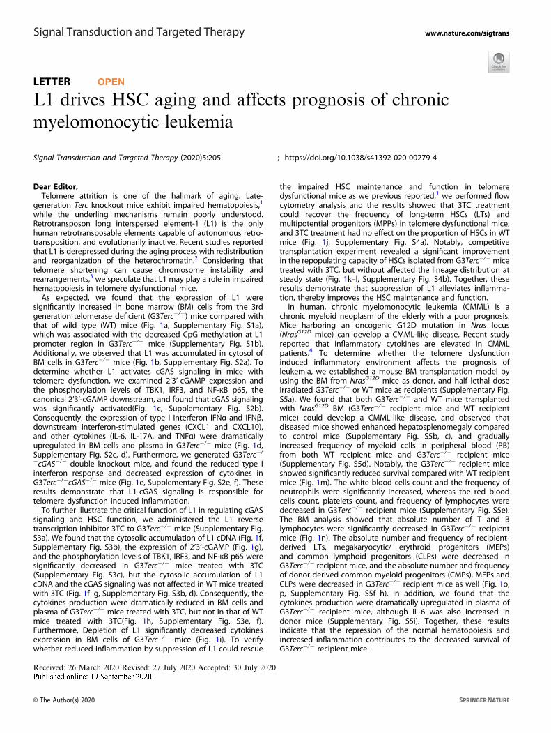
significantly increased in bone marrow (BM) cells from the 3rdgeneration telomerase deficient (G3Terc−/−) mice compared withthat of wild type (WT) mice (Fig. 1a, Supplementary Fig. S1a),which was associated with the decreased CpG methylation at L1promoter region in G3Terc−/− mice (Supplementary Fig. S1b).Additionally, we observed that L1 was accumulated in cytosol ofBM cells in G3Terc−/− mice (Fig. 1b, Supplementary Fig. S2a). Todetermine whether L1 activates cGAS signaling in mice withtelomere dysfunction, we examined 2’3’-cGAMP expression andthe phosphorylation levels of TBK1, IRF3, and NF-κB p65, thecanonical 2’3’-cGAMP downstream, and found that cGAS signalingwas significantly activated(Fig. 1c, Supplementary Fig. S2b).Consequently, the expression of type I interferon IFNα and IFNβ,downstream interferon-stimulated genes (CXCL1 and CXCL10),and other cytokines (IL-6, IL-17A, and TNFα) were dramaticallyupregulated in BM cells and plasma in G3Terc−/− mice (Fig. 1d,Supplementary Fig. S2c, d). Furthermore, we generated G3Terc−/
−cGAS−/− double knockout mice, and found the reduced type Iinterferon response and decreased expression of cytokines inG3Terc−/−cGAS−/− mice (Fig. 1e, Supplementary Fig. S2e, f). Theseresults demonstrate that L1-cGAS signaling is responsible fortelomere dysfunction induced inflammation.To further illustrate the critical function of L1 in regulating cGAS
signaling and HSC function, we administered the L1 reversetranscription inhibitor 3TC to G3Terc−/− mice (Supplementary Fig.S3a). We found that the cytosolic accumulation of L1 cDNA (Fig. 1f,Supplementary Fig. S3b), the expression of 2’3’-cGAMP (Fig. 1g),and the phosphorylation levels of TBK1, IRF3, and NF-κB p65 weresignificantly decreased in G3Terc−/− mice treated with 3TC(Supplementary Fig. S3c), but the cytosolic accumulation of L1cDNA and the cGAS signaling was not affected in WT mice treatedwith 3TC (Fig. 1f–g, Supplementary Fig. S3b, d). Consequently, thecytokines production were dramatically reduced in BM cells andplasma of G3Terc−/− mice treated with 3TC, but not in that of WTmice treated with 3TC(Fig. 1h, Supplementary Fig. S3e, f).Furthermore, Depletion of L1 significantly decreased cytokinesexpression in BM cells of G3Terc−/− mice (Fig. 1i). To verifywhether reduced inflammation by suppression of L1 could rescue
the impaired HSC maintenance and function in telomeredysfunctional mice as we previous reported,1 we performed flowcytometry analysis and the results showed that 3TC treatmentcould recover the frequency of long-term HSCs (LTs) andmultipotential progenitors (MPPs) in telomere dysfunctional mice,and 3TC treatment had no effect on the proportion of HSCs in WTmice (Fig. 1j, Supplementary Fig. S4a). Notably, competitivetransplantation experiment revealed a significant improvementin the repopulating capacity of HSCs isolated from G3Terc−/− micetreated with 3TC, but without affected the lineage distribution atsteady state (Fig. 1k–l, Supplementary Fig. S4b). Together, theseresults demonstrate that suppression of L1 alleviates inflamma-tion, thereby improves the HSC maintenance and function.In human, chronic myelomonocytic leukemia (CMML) is a
chronic myeloid neoplasm of the elderly with a poor prognosis.Mice harboring an oncogenic G12D mutation in Nras locus(NrasG12D mice) can develop a CMML-like disease. Recent studyreported that inflammatory cytokines are elevated in CMMLpatients.4 To determine whether the telomere dysfunctioninduced inflammatory environment affects the prognosis ofleukemia, we established a mouse BM transplantation model byusing the BM from NrasG12D mice as donor, and half lethal doseirradiated G3Terc−/− or WT mice as recipients (Supplementary Fig.S5a). We found that both G3Terc−/− and WT mice transplantedwith NrasG12D BM (G3Terc−/− recipient mice and WT recipientmice) could develop a CMML-like disease, and observed thatdiseased mice showed enhanced hepatosplenomegaly comparedto control mice (Supplementary Fig. S5b, c), and graduallyincreased frequency of myeloid cells in peripheral blood (PB)from both WT recipient mice and G3Terc−/− recipient mice(Supplementary Fig. S5d). Notably, the G3Terc−/− recipient miceshowed significantly reduced survival compared with WT recipientmice (Fig. 1m). The white blood cells count and the frequency ofneutrophils were significantly increased, whereas the red bloodcells count, platelets count, and frequency of lymphocytes weredecreased in G3Terc−/− recipient mice (Supplementary Fig. S5e).The BM analysis showed that absolute number of T and Blymphocytes were significantly decreased in G3Terc−/− recipientmice (Fig. 1n). The absolute number and frequency of recipient-derived LTs, megakaryocytic/ erythroid progenitors (MEPs)and common lymphoid progenitors (CLPs) were decreased inG3Terc−/− recipient mice, and the absolute number and frequencyof donor-derived common myeloid progenitors (CMPs), MEPs andCLPs were decreased in G3Terc−/− recipient mice as well (Fig. 1o,p, Supplementary Fig. S5f–h). In addition, we found that thecytokines production were dramatically upregulated in plasma ofG3Terc−/− recipient mice, although IL-6 was also increased indonor mice (Supplementary Fig. S5i). Together, these resultsindicate that the repression of the normal hematopoiesis andincreased inflammation contributes to the decreased survival ofG3Terc−/− recipient mice.
Received: 26 March 2020 Revised: 27 July 2020 Accepted: 30 July 2020
www.nature.com/sigtransSignal Transduction and Targeted Therapy
© The Author(s) 2020
1234567890();,:

To determine whether improvement of HSC by L1 inhibitionis beneficial in prognosis of CMML, we administered 3TC toG3Terc−/− recipient mice and WT recipient mice (SupplementaryFig. S6a), and found that the 3TC treatment extended the survivalof G3Terc−/− recipient mice, but not WT recipient mice (Fig. 1q).
The expression of cytokines in plasma of G3Terc−/− recipient micetreated with 3TC were significantly decreased (Supplementary Fig.S6b). Interestingly, we found that 3TC could not relieve thehepatosplenomegaly, high proportion of myeloid cells in PB andhigh absolute number of myeloid cells in BM from G3Terc−/−
Letter
2
Signal Transduction and Targeted Therapy (2020) 5:205

recipient mice (Fig. 1r, Supplementary Fig. S6c-e), and the absolutenumber and frequency of donor-derived hematopoietic stem/progenitor cells (HSPCs) were comparable between 3TC treatedand untreated G3Terc−/− recipient mice (Fig. 1s, SupplementaryFig. S6f). Notably, the absolute number and frequency of recipient-derived LTs, MEPs and megakaryocyte progenitors (MKPs) weredramatically increased, and the PLTs count was rescued to normallevel in G3Terc−/− recipient mice with 3TC treatment (Fig. 1t,Supplementary Fig. S6g, h). To explore whether 3TC treatmentalso contributes to the repression of donor cell (NrasG12D), wechecked the mRNA expression and CpG methylation level of L1 indiseased mice. The results showed decreased CpG methylation atL1 promoter region in endogenous G3Terc−/− BM cells, but not inendogenous WT BM cells, the donor-derived (NrasG12D) BM cells inWT and G3Terc−/− recipient mice (Supplementary Fig. S7a). Theexpression level of L1 in different groups were consistent with theCpG methylation levels (Supplementary Fig. S7b). Altogether,these results indicate that the suppression of L1 by 3TC treatmentin CMML mice reduces telomere dysfunction induced inflamma-tion and attenuates the impaired hematopoiesis, therebyextended survival of the diseased mice, probably due to theimprovement of HSPCs maintenance.In summary, we found that L1 activation is responsible for the
cGAS signaling induced inflammatory responses in G3Terc−/−
mice. 3TC treatment attenuated the aging-associated decline ofHSC maintenance and function, thereby extended the survival ofG3Terc−/− recipient mice transplanted with oncogenic NrasG12D
BM. Our findings suggest that reverse transcriptase inhibition mayserve as a new therapeutic strategy for patients suffering fromage-related disorders.
ACKNOWLEDGEMENTSWe thank the Dr. Jian Mao (School of Medicine, Hangzhou Normal University) fortechnical assistance. This work was supported by Grants 2016YFA0100602,2017YFA0103302, 2018YFA0109300 from the National Key Research and Develop-ment Program of China; Grants 81525010, 91749203, 81871116, 81501214, 91749117,81770155, and 81771502 from the National Natural Science Foundation of China;Grants LQ14C070002 from the Natural Science Foundation of Zhejiang Province ofChina; Grant 2018GZR110103002 from Innovative Team Program of GuangzhouRegenerative Medicine and Health Guangdong Laboratory and Grant 2017ZT07S347from the Program for Guangdong Introducing Innovative and Enterpreneurial Teams.This work was supported by the Science Foundation for Distinguished YoungScholars of Guangdong Province (2019B151502008) to Hu Wang.
AUTHOR CONTRIBUTIONSY.W., JP.Z., H.W., Y.L., JY.W., and LJ.X. carried out the experiments. Y.W., JP.Z., H.W., JinYW., J.M.S., ZF.S., and ZY.J. analyzed the data. Y.W., JP.Z., H.W., J.M.S., and ZY.J. wrotethe paper.
ADDITIONAL INFORMATIONThe online version of this article (https://doi.org/10.1038/s41392-020-00279-4)contains supplementary material, which is available to authorized users.
Competing interests: The authors declare no competing interests.
Ying Wang1, Jin-ping Zheng2, Ying Luo3, Junyi Wang1, Lingjie Xu1,Jinyong Wang4, John M. Sedivy5, Zhangfa Song6, Hu Wang1,3 and
Zhenyu Ju1,31Key Laboratory of Aging and Cancer Biology of Zhejiang Province,Institute of Aging Research, School of Medicine, Hangzhou NormalUniversity, Hangzhou 311121, China; 2Department of Public Health
and Preventive Medicine, Changzhi Medical College, Changzhi,Shanxi 046000, P. R. China; 3Key Laboratory of Regenerative
Medicine of Ministry of Education, Guangzhou Regenerative Medicineand Health Guangdong Laboratory, Institute of Aging and
Regenerative Medicine, Jinan University, Guangzhou 510632, China;4CAS Key Laboratory of Regenerative Biology and Guangdong
Provincial Key Laboratory of Stem Cell and Regenerative Medicine,Guangzhou Institutes of Biomedicine and Health, Chinese Academyof Sciences, Guangzhou, China; 5Department of Molecular Biology,
Cell Biology and Biochemistry, Brown University, Providence, RI02903, USA and 6Department of Colorectal Surgery, Sir Run Run
Shaw Hospital, Zhejiang University, Hangzhou, ChinaThese authors contributed equally: Ying Wang, Jin-ping Zheng
Correspondence: Ying Wang ([email protected]) orZhangfa Song ([email protected]) orHu Wang ([email protected]) or
Zhenyu Ju ([email protected])
REFERENCES1. Ju, Z. et al. Telomere dysfunction induces environmental alterations limiting
hematopoietic stem cell function and engraftment. Nat. Med. 13, 742–747 (2007).2. De Cecco, M. et al. Genomes of replicatively senescent cells undergo global epi-
genetic changes leading to gene silencing and activation of transposable ele-ments. Aging Cell. 12, 247–256 (2013).
Fig. 1 L1 drives HSC aging and affects prognosis of chronic myelomonocytic leukemia. a Quantitative real-time PCR (Q-PCR) analysis of therelative mRNA expression level of L1 in BM of WT and G3Terc−/− mice (n= 4, left). The protein levels of L1 (ORF1p) was determined by westernblot in BM of WT and G3Terc−/− mice. The graph represents the relative ORF1p protein abundance (right). b Q-PCR analysis of the relativecDNA levels of L1 in BM cytoplasmic fraction of WT and G3Terc−/− mice (n= 3). c 2’3’-cGAMP levels were determined by LC-MS/MS in BM ofWT and G3Terc−/− mice (n= 3). d Q-PCR analysis of the relative mRNA expression levels of IFNα and IFNβ in WT and G3Terc−/− mice (n= 5).e Q-PCR analysis of the relative mRNA expression levels of IFNα and IFNβ in G3Terc−/− and G3Terc−/−cGAS−/− mice (n= 3). f Q−PCR analysis ofthe relative cDNA levels of L1 in BM cytoplasmic fraction of WT or G3Terc−/− mice treated with or without 3TC (n= 3). g 2’3’-cGAMP levelswere determined by LC-MS/MS in BM of WT or G3Terc−/− mice treated with or without 3TC (n ≥ 5). h Q-PCR analysis of the relative mRNAexpression levels of IFNα and IFNβ in BM of WT or G3Terc−/− mice treated with or without 3TC (n= 3). i Q-PCR analysis of relative mRNAexpression levels of L1, IFNα, IFNβ, and IL-6 in control and L1 depleted BM cells of G3Terc−/− mice (n= 5). j Numbers of LT (CD34−Flt3−LSK), ST(CD34+Flt3− LSK) and MPP (CD34+Flt3+ LSK) per million BM cells of WT, or G3Terc−/− mice treated with or without 3TC (n= 5). k Percentage ofdonor-derived PB cells of G3Terc−/− mice treated with or without 3TC at the indicated time points in competitive transplantation assay (n ≥ 3).l Percentage of donor-derived LT in BM of G3Terc−/− mice and 3TC-treated G3Terc−/− mice 16 weeks after transplantation (n ≥ 4). m Survivalcurve of WT recipient mice and G3Terc−/− recipient mice following transplantation (n= 20). n Absolute number of M, T, and B cells in BM ofWT recipient mice and G3Terc−/− recipient mice (n ≥ 8). o Absolute number of LT, ST, MPP, GMP (granulocyte/monocyte progenitorCD34+CD16/32+Sca1−c-Kit+ Lin−), CMP (common myeloid progenitor, CD34+CD16/32−Sca1−c-Kit+Lin−), MEP (megakaryocyte /erythrocyteprogenitor, CD34−CD16/32−Sca1−c-Kit+Lin−), and CLP (common lymphoid progenitor, Flt3+ IL-7R+c-KitmidSca1midLin−) in recipient-derivedBM of WT recipient mice and G3Terc−/− recipient mice (n ≥ 7). p Absolute number of LT, ST, MPP, GMP, CMP, MEP, and CLP in donor-derived BMof WT recipient mice and G3Terc−/− recipient mice (n ≥ 5). q Survival curve of WT recipient or G3Terc−/− recipient mice treated with or without3TC following transplantation (n ≥ 10). r Absolute number of M, T, and B cells in BM of G3Terc−/− recipient mice treated with or without 3TC(n ≥ 7). s Absolute number of LT, ST, MPP, GMP, CMP, MEP, MKP (IL-7R−CD41+c-Kit+ Lin−) and CLP in donor-derived BM of G3Terc−/− recipientmice treated with or without 3TC (n ≥ 6). t Absolute number of LT, ST, MPP, GMP, CMP, MEP, MKP, and CLP in recipient-derived BM of G3Terc−/−
recipient mice treated with or without 3TC (n ≥ 7). *p < 0.05; **p < 0.01; ***p < 0.001; ****p < 0.0001
Letter
3
Signal Transduction and Targeted Therapy (2020) 5:205

3. Murnane, J. P. & Sabatier, L. Chromosome rearrangements resulting from telomeredysfunction and their role in cancer. Bioessays 26, 1164–1174 (2004).
4. Niyongere, S. et al. Heterogeneous expression of cytokines accounts for clinicaldiversity and refines prognostication in CMML. Leukemia 33, 205–216 (2019).
Open Access This article is licensed under a Creative CommonsAttribution 4.0 International License, which permits use, sharing,
adaptation, distribution and reproduction in anymedium or format, as long as you giveappropriate credit to the original author(s) and the source, provide a link to the Creative
Commons license, and indicate if changes were made. The images or other third partymaterial in this article are included in the article’s Creative Commons license, unlessindicated otherwise in a credit line to the material. If material is not included in thearticle’s Creative Commons license and your intended use is not permitted by statutoryregulation or exceeds the permitted use, you will need to obtain permission directlyfrom the copyright holder. To view a copy of this license, visit http://creativecommons.org/licenses/by/4.0/.
© The Author(s) 2020
Letter
4
Signal Transduction and Targeted Therapy (2020) 5:205