l-cysteine ameliorated testicular toxicity induced …l-cysteine ameliorated testicular toxicity...
TRANSCRIPT
European Journal
of Biological Research Research Article
European Journal of Biological Research 2015; 5 (2): 1-8
L-cysteine ameliorated testicular toxicity induced
by acrylamide in rats
Hossam El-Din M. Omar1*, Sary Kh. Abd-elghafar
2, Imhemed O. Fiedan
1
and Emad A. Ahmed1
1Department of Zoology, Faculty of Science, Assiut University, Egypt;
2Department of Pathology and Clinical
Pathology, Faculty of Veterinary Medicine, Assiut University, Egypt,
*Corresponding author: Prof. Hossam El-Din M. Omar; Physiology Laboratory, Department of Zoology,
Faculty of Science, Assiut University, Assiut, 71516, Egypt; e-mail: [email protected]
ABSTRACT
The general public is exposed to acrylamide from industrial manufacturing, laboratory work, foods rich in carbohydrates that have been cooked at high-temperature, and through cigarette smoke. The present experiment was conducted to investigate the reproductive toxicity of acrylamide exposure in male rats and the role of L-cysteine supplementation in amelioration of this toxicity. Forty eight adult male albino rats (weighing 120-140 g) were divided into four groups (16 rats/group). Group I - negative control group, drank tap water, group II - positive control, drank tap water that contains acrylamide (25 mg/kg body weight) for 28 days and group III drank tap water that contains acrylamide (25 mg/kg body weight) and L-cysteine (100 mg/kg body weight). Four rats from each group were killed at 7, 14, 21 and 28 days time intervals from the beginning of the experiment. In general, exposure to acrylamide induced a significant elevation in testes lipid peroxides and nitric oxide levels and a signi-ficant reduction in the level of glutathione and the activity of superoxide dismutase and catalase in all
periods of experiment. However, plasma testoste-rone was significantly decreased in acrylamide treated rats with congestion and interstitial edema, necrosis, calcification and degeneration of sperma-togenic cells in the seminiferous tubules and for-mation of spermatid gaint cells. Co-treatment of rats with L-cysteine reduced the changes in oxidative stress parameters and improved the pathological changes in testis. Therefore, supplementation of L-cysteine can be useful when there is a risk of acrlamide toxicity. Keywords: Acrylamide, Testis, Oxidative stress, L-cysteine, Testosterone. 1. INTRODUCTION Acrylamide (ACR) is formed through the Maillard reaction during the heating process by interactions of amino acids, especially asparagine, with reducing sugars like glucose [1, 2]. There is variance in literature about the levels of ACR in different foods and the potential risk from dietary exposure. The daily intakes of dietary ACR for the
Received: 26 February 2015; Revised submission: 16 April 2015; Accepted: 17 April 2015 TMKARPIŃSKI PUBLISHER
Copyright: © The Author(s) 2015. European Journal of Biological Research © Tomasz M. Karpiński 2015. This is an open-access article distributed under the terms of the Creative Commons Attribution License, which permits
unrestricted use, distribution, and reproduction in any medium, provided the original work is properly cited. www.journals.tmkarpinski.com/index.php/mmed
2 | Omar et al. Testicular toxicity induced by acrylamide
European Journal of Biological Research 2015; 5 (2): 1-8
general population are estimated to be in the range of 0.3-2.0 mg/kg body weight [3]. ACR can be reactive in three different ways, radical-mediated polymerization, and addition to thiol, hydroxyl, or amino groups result in alkylation of proteins or metabolized to an epoxide derivative, glycidamide being readily reactive toward DNA and other macromolecules [4-6]. The mechanism by which ACR exposure causes cellular dysfunction in experimental animals and humans is not completely clear. However, it is thought that oxidative stress was associated with ACR cytotoxicity. Reproduc-tive toxicity in rodents exposed to ACR includes alterations in gonadal and pituitary hormones associated with histopathological changes that includes formation of multinucleated giant cells, vacuolation and production of high numbers of apoptotic cells in the seminiferous tubules [7-11]. Acrylamide induced oxidative stress is capable of disrupting the steroidogenic capacity of Leydig cells [12] as well as the capacity of the germinal epithelium to differentiate into normal spermatozoa [13]. Fortunately, testes contain a complicated group of antioxidants and free radical scaven-gers to protect the spermatogenic and steroidogenic functions of testis from oxidative stress [14]. Kurebayashi and Ohno [15] reported that glutathione (GSH) precursors such as N-acetyl- L-cysteine and L-methionine increased the protec-tion against the cytotoxicity of ACR in isolated rat hepatocytes. Moreover, N-acetyl-L-cysteine as potent antioxidant protects tissues from ACR toxicity by inhibiting neutrophil infiltration, balan-cing the oxidant-antioxidant status, and regulating the generation of inflammatory mediators [8]. Also, α-lipoic acid protect cells from oxidative stress induced by ACR exposure via enhances cellular antioxidant defense capacity [9]. In this regard, the objectives of the present study were to measures the markers of oxidative stress in testis of rats exposed to ACR and to evaluate the protective role of L-cysteine as precursor of GSH against ACR toxicity. 2. MATERIALS AND METHODS Forty eight adult male albino rats (weighing 120-140 g) were purchased from the Animal House, Faculty of Medicine, Assiut University, Assiut,
Egypt. The animals were housed in cages at a controlled temperature (25±3°C) and ambient humi-dity (50-60%). Lights were maintained on a 12-h light-dark cycle. All animals received basal diet and water ad-libitum for one week as an adaptation period. Following one week of acclimatization, the rats were randomly divided into three groups (16 rats/group): • Group I: Negative control, fed on basal diet and
normal drinking water for 4 weeks. • Group II: Positive control, fed on basal diet and
drinking water that contains ACR (25 mg/kg body weight) according to Alturfan et al. [8].
• Group III: Fed on basal diet and drinking water that contains ACR (25 mg/kg body weight and L-cysteine (100 mg/kg body weight) according to Omar et al. [16].
Then, each week from the beginning of the experiment 4 rats from each group were killed under anesthesia with ether. The blood samples were collected directly from portal vein into centrifuge tubes for separation of serum by centrifugation at 3000 rpm for 15 minutes and were frozen at -20°C for subsequent biochemical analysis. Immediately after killing rats, small piece of testes were excised and fixed in formaline for histological studies, and the remnant was washed in cold saline, immersed in liquid nitrogen and stored at -20°C for biochemical assay. All animal experiments were carried out in accordance with Ethical Committee Acts. 2.1. Determination of oxidative stress biomarker Lipid peroxidation (LPO) products as TBARS content were determined according to the method of Ohkawa et al. [17]. Nitric oxide (NO) content was measured as nitrate concentration colori- metrically using the method of Ding et al. [18]. Glutathione (GSH) content was determined using the method of Beutler et al. [19]. The activity of superoxide dismutase (SOD) was determined basing on its ability to inhibit the autoxidation of epinephrine at alkaline medium according to the method of Misra and Fridovich [20]. The activity of catalase (CAT) was determined basing on its ability to decompose H2O2 to H2O and O2 according to Gregory and Fridovich [21]. Protein content in the spleen tissues was determined by the method of Lowry et al. [22].
3 | Omar et al. Testicular toxicity induced by acrylamide
European Journal of Biological Research 2015; 5 (2): 1-8
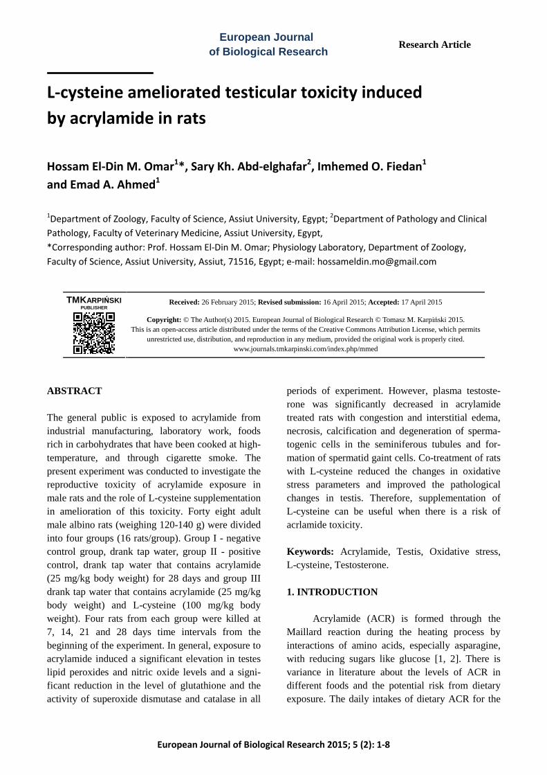
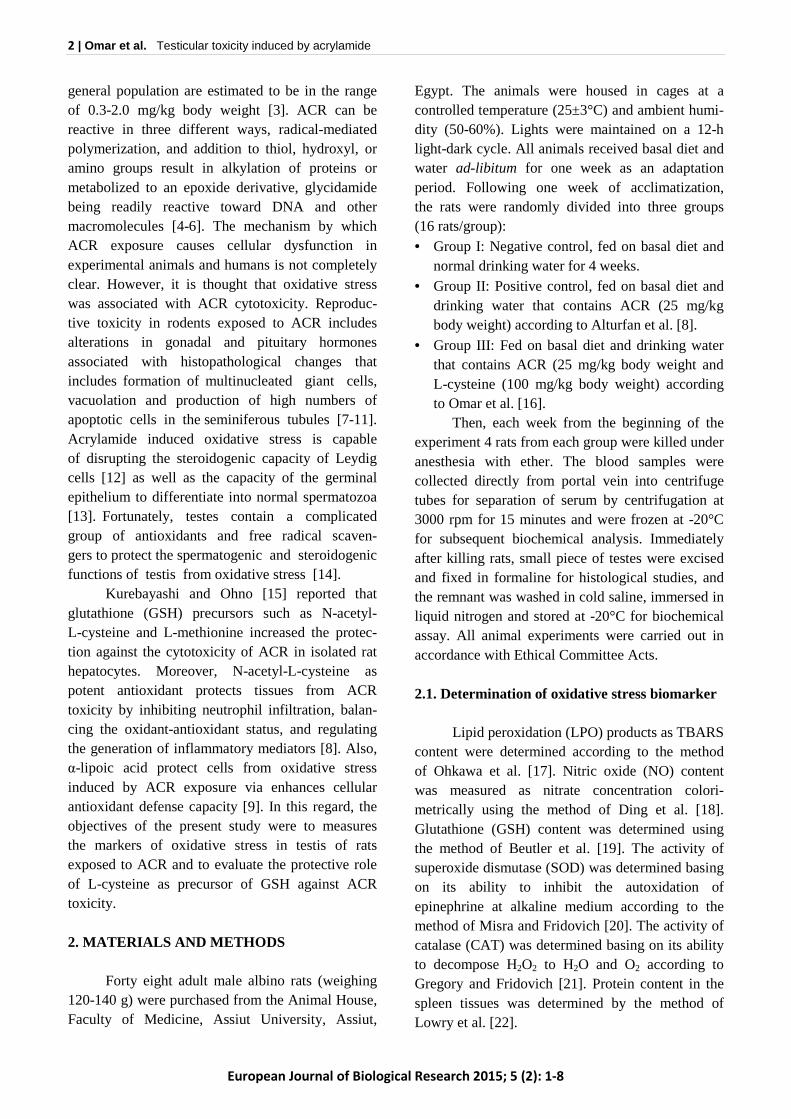
2.2. Estimation of testosterone Testosterone hormone in plasma was deter-mine by Enzyme Immunoassay Method (ELISA), Biocheck, Inc, 323 vintage Park Dr. Forster City, CA, USA, according to the kit manufacture instruc-tions. 2.3. Statistical analysis The data was expressed as mean ± SE. The results were analyzed statistically using column statistics and one-way analysis of variance with the Newman-Keuls multiple comparison test as a post-test. These analyses were carried out using the computer prism program for windows, version 6.0 (Graph pad software Inc., San Diego, California, USA). Differences between the groups were considered significant if P < 0.05, 0.01, or 0.001. 3. RESULTS Compared to control rats, ACR treated rats exhibited a significant decrease in plasma testo-sterone in 2nd, 3rd, and 4th weeks and LC treatment resulted in an increase in testosterone level especially in 2nd and 4th week (Fig. 1). In relation to control rats, ACR treated rats had greater level of LPO in testis especially in the 3rd week (P<0.001) and LC co-treatment failed to restore the elevation of LPO especially in 1st and 4th week (Fig. 2). Also, Fig. 2 showed that NO was significantly increased (P<0.001) in all periods of the experiment in comparison with control and LC treatment resulted in significant reduction in NO level (P<0.001).
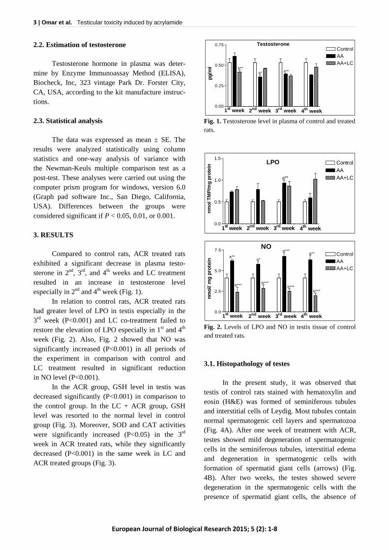
In the ACR group, GSH level in testis was decreased significantly (P<0.001) in comparison to the control group. In the LC + ACR group, GSH level was resorted to the normal level in control group (Fig. 3). Moreover, SOD and CAT activities were significantly increased (P<0.05) in the 3rd week in ACR treated rats, while they significantly decreased (P<0.001) in the same week in LC and ACR treated groups (Fig. 3).
Fig. 1. Testosterone level in plasma of control and treated rats.
Fig. 2. Levels of LPO and NO in testis tissue of control and treated rats.
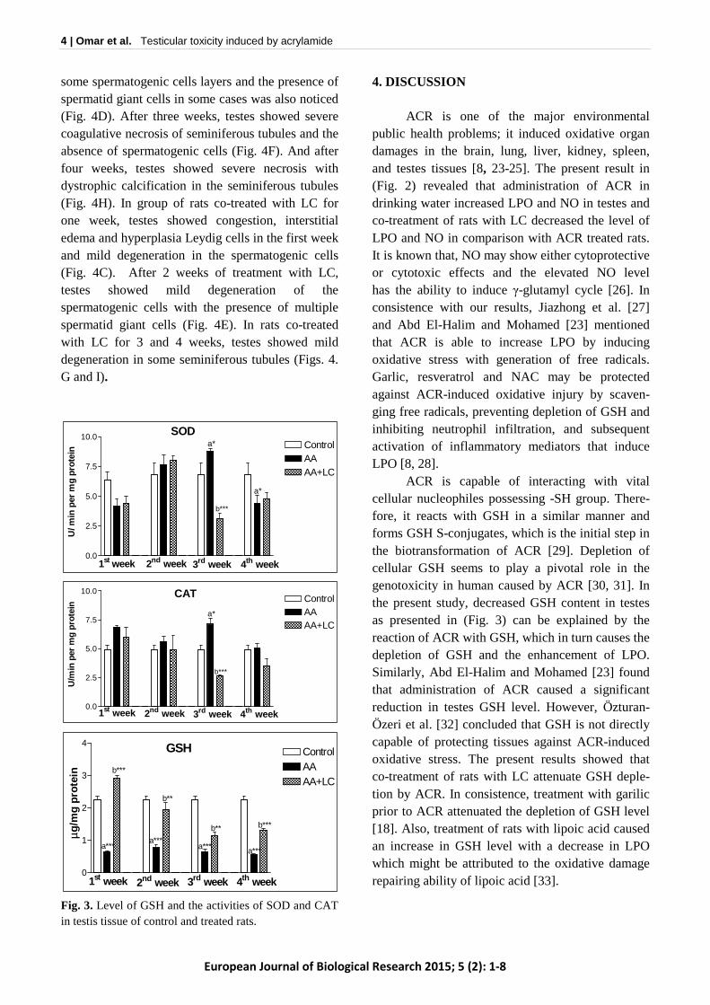
3.1. Histopathology of testes In the present study, it was observed that testis of control rats stained with hematoxylin and eosin (H&E) was formed of seminiferous tubules and interstitial cells of Leydig. Most tubules contain normal spermatogenic cell layers and spermatozoa (Fig. 4A). After one week of treatment with ACR, testes showed mild degeneration of spermatogenic cells in the seminiferous tubules, interstitial edema and degeneration in spermatogenic cells with formation of spermatid giant cells (arrows) (Fig. 4B). After two weeks, the testes showed severe degeneration in the spermatogenic cells with the presence of spermatid giant cells, the absence of
0.0
2.5
5.0
7.5ControlAAAA+LC
1st week 2nd week 3rd week 4th week
nmol
/ mg
prot
ein a**
b***
a*
b***b***
a***
b***
a**
NO
0.0
0.5
1.0
1.5ControlAAAA+LC
1st week 2nd week 3rd week 4th week
nm
ol T
MP
/mg
prot
ein
a**
LPO
0.00
0.25
0.50
0.75ControlAAAA+LC
pg/m
l
1st week 2nd week 3rd week 4th week
b**a* a**
Testosterone
4 | Omar et al. Testicular toxicity induced by acrylamide
European Journal of Biological Research 2015; 5 (2): 1-8
some spermatogenic cells layers and the presence of spermatid giant cells in some cases was also noticed (Fig. 4D). After three weeks, testes showed severe coagulative necrosis of seminiferous tubules and the absence of spermatogenic cells (Fig. 4F). And after four weeks, testes showed severe necrosis with dystrophic calcification in the seminiferous tubules (Fig. 4H). In group of rats co-treated with LC for one week, testes showed congestion, interstitial edema and hyperplasia Leydig cells in the first week and mild degeneration in the spermatogenic cells (Fig. 4C). After 2 weeks of treatment with LC, testes showed mild degeneration of the spermatogenic cells with the presence of multiple spermatid giant cells (Fig. 4E). In rats co-treated with LC for 3 and 4 weeks, testes showed mild degeneration in some seminiferous tubules (Figs. 4. G and I).
Fig. 3. Level of GSH and the activities of SOD and CAT in testis tissue of control and treated rats.
4. DISCUSSION ACR is one of the major environmental public health problems; it induced oxidative organ damages in the brain, lung, liver, kidney, spleen, and testes tissues [8, 23-25]. The present result in (Fig. 2) revealed that administration of ACR in drinking water increased LPO and NO in testes and co-treatment of rats with LC decreased the level of LPO and NO in comparison with ACR treated rats. It is known that, NO may show either cytoprotective or cytotoxic effects and the elevated NO level has the ability to induce γ-glutamyl cycle [26]. In consistence with our results, Jiazhong et al. [27] and Abd El-Halim and Mohamed [23] mentioned that ACR is able to increase LPO by inducing oxidative stress with generation of free radicals. Garlic, resveratrol and NAC may be protected against ACR-induced oxidative injury by scaven-ging free radicals, preventing depletion of GSH and inhibiting neutrophil infiltration, and subsequent activation of inflammatory mediators that induce LPO [8, 28]. ACR is capable of interacting with vital cellular nucleophiles possessing -SH group. There-fore, it reacts with GSH in a similar manner and forms GSH S-conjugates, which is the initial step in the biotransformation of ACR [29]. Depletion of cellular GSH seems to play a pivotal role in the genotoxicity in human caused by ACR [30, 31]. In the present study, decreased GSH content in testes as presented in (Fig. 3) can be explained by the reaction of ACR with GSH, which in turn causes the depletion of GSH and the enhancement of LPO. Similarly, Abd El-Halim and Mohamed [23] found that administration of ACR caused a significant reduction in testes GSH level. However, Özturan-Özeri et al. [32] concluded that GSH is not directly capable of protecting tissues against ACR-induced oxidative stress. The present results showed that co-treatment of rats with LC attenuate GSH deple-tion by ACR. In consistence, treatment with garilic prior to ACR attenuated the depletion of GSH level [18]. Also, treatment of rats with lipoic acid caused an increase in GSH level with a decrease in LPO which might be attributed to the oxidative damage repairing ability of lipoic acid [33].
0.0
2.5
5.0
7.5
10.0ControlAAAA+LC
U/ m
in p
er m
g pr
otei
n
a*
b***
a*
1st week 2nd week 3rd week 4th week
SOD
0.0
2.5
5.0
7.5
10.0ControlAAAA+LC
U/m
in p
er m
g pr
otei
n
1st week 2nd week 3rd week 4th week
CAT
a*
b***
0
1
2
3
4ControlAAAA+LC
µg/m
g pr
otei
n
1st week 2nd week 3rd week 4th week
GSH
a***
b***
a***
b**
a***
b**
a***
b***

5 | Omar et al. Testicular toxicity induced by acrylamide
European Journal of Biological Research 2015; 5 (2): 1-8
Fig. 4. Photomicrographs of control testis formed of seminiferous tubules and interstitial cells of leydig. A. Most tubules contain normal spermatogenic cells layers and spermatozoa in control rats. B-I testis of ACR and ACR plus L-cysteine treated groups at different weeks of treatment. B. mild degeneration of spermatogenic cells with formation of spermatid giant cells in the seminiferous tubules (arrows). C. Congestion interstitial edema and hyperplasia of Leydig cells (arrows). D. Severe degeneration in the spermatogenic cells with presence of spermatid giant cells (arrows). E. Mild degeneration in the spermatogenic cells (arrows). F. Severe coagulative necrosis of seminiferous tubules and absence of spermatogenic cells (arrows). G. Moderate degeneration of the spermatogenic cells and presence of multiple spermatid gaint cells (arrows). H. Severe necrosis (star) with dystrophic calcification in the seminiferous tubules (arrows). I. mild degeneration in some seminiferous tubules (arrows). H&E X400.
Superoxide radical may oxidize SH groups and undergo dismutation to form H2O2 and singlet oxygen [34]. This change in the redox status of the cell may modulate gene expression directly or via the transcription factors that are redox-regulated, and may lead to apoptosis, cell proliferation, or transformation (29). In the current study, (Fig. 3) showed alteration in the testicular SOD and CAT activities depending on the period of treatments. Also, it showed that co-treatments of rats with LC ameliorate these changes by different levels. These results are in agreement with Abd El-Halim and Mohamed [23] who found that administration of ACR caused a significant reduction in the activity of SOD in testes tissues. The reduction in antioxi-dant enzyme activities was increased with increas-
ing doses of ACR [35]. Treatment with Curcuma longa L. powder and garilic prior to ACR attenuated the reduction of SOD activity [23, 36], and admi-nistration of catechin and neem leaves extracts significantly enhanced the hepatic CAT activity [37]. Testosterone level in plasma of rats treated with ACR was significantly decreased, and co-treatment with LC elevates this decrease especially in the 4th week of treatment (Fig. 1). In this aspect, administration of ACR caused a significant reduc-tion of serum testosterone level as reported by many authors [9, 38, 39]. This significant reduction of testosterone may be a result of direct damage of ACR on the Leydig cells [40]. The previous opinion was confirmed histopathologicaly in the present
A C
D E F
G H I
B

6 | Omar et al. Testicular toxicity induced by acrylamide
European Journal of Biological Research 2015; 5 (2): 1-8
study by congestion and interstitial edema, necrosis, calcification and degeneration of spermatogenic cells in the seminiferous tubules with formation of spermatid gaint cells (Fig. 4 A, C, E & G). More-over, ACR may affect the endocrine function of the testes by altering the androgen biosynthesis of interstitial cells in the testes [41] or inducing the enzymes activity of hepatic biotransformation, which is capable of metabolically transforming androgens into products with low androgen receptor binding activity [42]. Song et al. [43] found that ACR can directly damage Leydig cells and affect the endocrine function of the testis. Moreover, Yang et al. [44] found that ACR induces histopathological lesions such as formation of multinucleated giant cells, vacuolation and production of high numbers of apoptotic cells in the seminiferous tubules of the rat. In the present study, treatment with LC along with ACR resulted in moderate attenuation of the histopathological changes in testes that were induced by ACR. From these observations, it can be concluded that LC ameliorates the toxicity of ACR in rat testes by alleviating LPO and NO through scavenging of free radicals, and enhancing the activity of SOD and CAT and GSH level. AUTHORS CONTRIBUTION All authors contributed equally in planning, conduct, data analysis, and editing the work. The final manuscript has been read and approved by all authors. TRANSPARENCY DECLARATION The author declares no conflicts of interest. NOTES This paper was presented in the 48th Annual Conference of Physiology and Pathology of Reproduction and simultaneously 40th Joint Congress of Veterinary and Human Medicine, Zurich. Switzerland, 11th-13th February 2015, Abstract No 74.
REFERENCES
1. Boettcher MI, Schettgen T, Kutting B, Pischetsrie-der M, Angerer J. Mercapturic acids of acrylamide and glycidamide as biomarkers of the internal exposure to acrylamide in the general population. Mutat Res. 2005; 580: 167-176.
2. Mottram DS, Wedzicha BL, Dodson AT. Acryl-amide is formed in the Maillard reaction. Nature. 2002; 419: 448-449.
3. Dybing E, Farmer PB, Andersen M, Fennell TR, Lalljie SPD, Müller DJG, et al. Human exposure and internal dose assessments of acrylamide in food. Food Chem Toxicol. 2005; 43: 365-410.
4. Park J, Kamendulis LM, Friedman MA, Klaunig JE. Acrylamide-induced cellular transformation. Toxicol Sci. 2002; 65: 177-183.
5. Besaratinia A, Pfeifer GP. DNA adduction and mutagenic properties of acrylamide. Mutat Res. 2005; 580: 31-40.
6. Paulsson B, Rannug A, Henderson AP, Golding BT, Törnqvist M, Warholm M. In vitro studies of the influence of glutathione transferases and epoxide hydrolase on the detoxification of acrylamide and glycidamide in blood. Mutat Res. 2005; 580: 53-59.
7. Hamdy SM, Bakeer HM, Eskande EF, Sayed ON. Efect of acrylamide on some hormones and endo-crine tissues in male rats. Hum Exp Toxicol. 2011; 5: 1-9.
8. Alturfan EI, Beceren A, Şehirli AQ, Demiralp ZE, Şener G, Omurtag GZ. Protective effect of N-acetyl-L-cysteine against acrylamide-induced oxidative stress in rats. Turk J Vet Anim Sci. 2008; 36(4): 438-445.
9. Lebda M, Gad S, Gaafar H. Effects of lipoic acid on acrylamide induced testicular damage. Mater Sociomed. 2014, 26(3): 208-212.
10. Khalil WKB, Ahmed HH, Hanan F, Aly HF, Eshak MG. Toxicological effects of acrylamide on testi-cular function and immune genes expression profile in rats. J Pharm Sci Rev Res. 2014; 24(1): 143-151.
11. Yang HJ, Lee SH, Jin Y, Choi JH, Han DU, Chae C, et al. Toxicological effects of acrylamide on rat testicular gene expression profile. Reprod Toxicol. 2005; 19(4): 527-534.
12. Hales DB, Allen JA, Shankara T, Janus P, Bucks S, Diemer T, Hales RH. Mitochondrial function in Leydig cell steroidogenesis. Ann NY Acad Sci. 2005; 1061: 120-134.
13. Naughton CK, Nangia AK, Agarwal A. Patho-physiology of varicoceles in male infertility. Hum Reprod Update. 2001; 7: 473-781.

7 | Omar et al. Testicular toxicity induced by acrylamide
European Journal of Biological Research 2015; 5 (2): 1-8
14. Aitken RJ and Roman SD. Antioxidant systems and oxidative stress in the testes. Oxid Med Cell Longev. 2008; 1(1): 15-24.
15. Kurebayashi H, Ohno Y. Metabolism of acrylamide to glycidamide and their cytotoxicity in isolated rat hepatocytes: protective effects of GSH precursors. Arch Toxicol 2006; 80: 820-828.
16. Omar HM, Ahmed EA, Abdel-Ghafar S Kh, Ragab MMS, Nasser AY. Hepatoprotective effects of vitamin C, DPPD, and L-cysteine against cisplatin-induced oxidative stress in male rats. J Biol Earth Sci. 2012; 2(1): B28-B36.
17. Ohkawa H, Ohishi N, Yagi K. Assay for lipid peroxides in animal tissue by thiobarbaturic acid reaction. Anal. Biochem. 1979; 95: 351-358.
18. Ding AH, Nathan CF, Stuchr DJ. Release of reactive nitrogen intermediate and reactive oxygen intermediate from mouse peritoneal macrophages Comparison of activating cytokines and evidence for independent production. J Immunol. 1988; 141: 2407-2412.
19. Beutler E, Duron O, Kelly BM. Improved method for the determination of blood glutathione. J Lab Clin Meth. 1963; 61: 882-888.
20. Misra HP, Fridovich I. The role of superoxide anion in the autoxidation of epinephrine and a simple assay for superoxide dismutase. J Biol Chem. 1972; 247: 3170-3175.
21. Gregory EM, Fridovich I. Visualization of catalase on acrylamide gels. Anal Biochem. 1974; 58: 57-62.
22. Lowry OH, Rosebrough NJ, Farr AL, Randall RJ. Protein measurement with the Folin phenol reagent. J Biol Chem. 1951; 41: 1863-1870.
23. Abd El-Halim SS, Mohamed MM. Garlic powder attenuates acrylamide-induced oxidative damage in multiple organs in rat. J Appl Sci Res. 2012; 8: 168-173.
24. Taha N, Korshom M, Mandour AW, Sadek K. Effects of garlic and acrylamide on some antioxidant enzymes. Global J Med Plant Res. 2013; 1: 190-194.
25. Venkatasubbaiah K, Venkataswamy DM, Suresh KS, Rao KJ. Acrylamide induced oxidative stress in rat and chick embtyonic liver. Indo Am J Pharmac Res. 2014; 6: 2791-2798.
26. Kuo PC, Abe KY, Schroeder RA. Interleukin-1-induced nitric oxide production modulates gluta-thione synthesis in cultured rat hepatocytes. Am J Physiol. 1996; 271: 851-862.
27. Jiazhong J, Yong ZU, James EK. Induction of oxidative stressing rat brain by acrylonitrile. Toxicol Sci. 1998; 46: 333-341.
28. Nursal GK, Levent S, Ozer E, Feriha S, Serap M, Keyer-Usal S, Goksel. Long term administration of aqueous garlic extract (AGE) alleviates liver fibrosis and oxidative damage induced by biliary obstruction in rats. Life Sci. 2005; 76: 2593-2606.
29. Awad ME, Abdel-Rahman MS, Hassan SA. Acryl-amide toxicity in isolated rat hepatocytes. Toxicol In Vitro. 1998; 12: 699-704.
30. Lamy E, Völkel Y, Roos PH, Kassie F, Mersch-Sundermann V. Ethanol enhanced the genotoxicity of acrylamide in human, metabolically competent HepG2 cells by CYP2E1 induction and glutathione depletion. Int J Hyg Environ Health. 2007; 1-2: 74-81.
31. Zhang X, Cao J, Jiang L, Geng C, Zhong L. Protec-tive effect of hydroxytyrosol against acrylamide-induced cytotoxicity and DNA damage in HepG2 cells. Mutat Res. 2009; 664: 64-68.
32. Özturan-Özeri E, Ucar G, Helvacoglu F, Ery AC, Rkaydin-Aldemir D, Turkoglu S. Effect of acryl-amide treatment on arginase activities and nitric oxide levels in rat liver and kidney. Acta Med Mediter. 2014; 30: 375-382.
33. Prahalathan C, Selvakumar E, Varalakshmi P. Lipoic acid modulates adriamycin-induced testicular toxi-city. Reprod Toxicol. 2006; 211: 54-59.
34. Andreev YA, Kushnareva YV, Starkov AA. Meta-bolism of reactive oxygen species in mitochondria. Biokhimiya. 2005; 70: 246-264.
35. Swamy MV, Subbaiah KV, Aumau B, Kamala K, Rao KJ, Raju KT. Toxic effect of acrylamide on body weight, the study of antioxidants and histoarchitecture of heart in the developing chick embryo. Indian J Appl Res. 2013; 3: 27-30.
36. Abd EL-Halim SS, EL-Adawi AS. Modulation of acrylamide-induced oxidative damage in rat tissues by Curcuma longa L. Med J Cairo Univ. 2008; 76: 639-547.
37. Mansour MK, Ibrahim EM, El-Kholy MM, El-Madawy SA. Antioxidant and histopathological effect of catechin and neem leaves extract in acrylamide toxicity of rats. Egypt. J Comp Path Clinic Path. 2008; 21: 290-313.
38. Abd El-Mottaleb EM, Rashed AYM. Some studies on acrylamide intoxication in male albino rats. Egypt. J Comp Path Clinic Path. 2008; 21: 222-245.
39. Yassa HA, George SM, Refaiy AE, Refaiy M, Abdel Moneim EM. Camellia sinensis (green tea) extract attenuate acrylamide induced testicular damage in albino rats. Environ Toxicol. 2013; 29: 1155-1161.
8 | Omar et al. Testicular toxicity induced by acrylamide
European Journal of Biological Research 2015; 5 (2): 1-8
40. Tag El-Din HA, Abbas HE, El-Kashoury AI. Experi-mental studies of dicofol reproductive toxicity on male albino rats. Bull Fac Pharm Cairo Univ. 2003; 40: 179-188.
41. Fowler A, Mistry P, Goering L. Mechanisms of meta-induced cell injury. Res Comm Chem Pathol Pharmacol. 1987; 28: 689.
42. Sonderfen AJ, Arlotto MP, Dutton DR, McMillen SK, Parkinson A. Regulation of testosterone hydro-xylation by rat liver microsomal cytochrome P-450. Arch Biochem Biophys. 1987; 255: 27-41.
43. Song HX, Wang R, Geng ZM, Cao SX, Liu TZ. Subchronic exposure top acrylamide effects repro-duction and testis endocrine functions if rats. Zhonghua Nan Ke Xue. 2008; 14: 406-910.
44. Yang HJ, Lee SH, Jin Y, Choi1 JH, Han CH, Lee MH. Genotoxicity and toxicological effects of acrylamide on reproductive system in male rats. J Vet Sci. 2005; 6: 103-109.