kuopion yliopiston julkaisuja g. - a.i....
TRANSCRIPT

Lentiviral Vector for Gene Transfer
A Versatile Tool for Regulated Gene Expression, GeneSilencing and Progenitor Cell Therapies
Doctoral dissertation
To be presented by permission of the Faculty of Natural and Environmental
Sciences of the University of Kuopio for public examination in
Auditorium, Tietoteknia building, University of Kuopio,
on Saturday 19th April 2008, at 1 p.m.
Department of Biotechnology and Molecular MedicineA.I. Virtanen Institute for Molecular Sciences
University of Kuopio
JONNA KOPONEN
JOKAKUOPIO 2008
KUOPION YLIOPISTON JULKAISUJA G. - A.I. VIRTANEN -INSTITUUTTI 60KUOPIO UNIVERSITY PUBLICATIONS G.
A.I. VIRTANEN INSTITUTE FOR MOLECULAR SCIENCES 60
Distributor : Kuopio University Library P.O. Box 1627 FI-70211 KUOPIO FINLAND Tel. +358 17 163 430 Fax +358 17 163 410 http://www.uku.fi/kirjasto/julkaisutoiminta/julkmyyn.html
Series Editors: Research Director Olli Gröhn, Ph.D. Department of Neurobiology A.I . Virtanen Institute for Molecular Sciences
Research Director Michael Courtney, Ph.D. Department of Neurobiology A.I . Virtanen Institute for Molecular Sciences
Author’s address: Department of Biotechnology and Molecular Medicine A.I . Virtanen Institute for Molecular Sciences University of Kuopio, Bioteknia 1 P.O. Box 1627 FI-70211 KUOPIO FINLAND E-mail : [email protected]
Supervisors: Professor Seppo-Ylä-Herttuala, M.D., Ph.D. Department of Biotechnology and Molecular Medicine A.I . Virtanen Institute for Molecular Sciences University of Kuopio
Docent Jarmo Wahlfors, Ph.D. Department of Biotechnology and Molecular Medicine A.I . Virtanen Institute for Molecular Sciences University of Kuopio
Reviewers: Docent Ari Hinkkanen, Ph.D. Department of Biochemistry and Pharmacy Åbo Akademi University
Pauli ina Lehtolainen, Ph.D. Centre for Cardiovascular Biology and Medicine University College London
Opponent: Professor Akseli Hemminki, M.D., Ph.D. Molecular Cancer Biology Program and Institute of Biomedicine, Biomedicum Helsinki University of Helsinki
ISBN 978-951-27-0619-8ISBN 978-951-27-1101-7 (PDF)ISSN 1458-7335
KopijyväKuopio 2008Finland

Koponen, Jonna. Lentiviral Vector for Gene Transfer – A Versatile Tool for Regulated GeneExpression, Gene Silencing and Progenitor Cell Therapies. Kuopio University PublicationsG. – A.I.Virtanen Institute for Molecular Sciences 60. 2008. 71 p.
ISBN 9789512706198ISBN 9789512711017 (PDF)ISSN 14587335
ABSTRACT
Gene therapy holds promise to improve the treatment options of both inherited and acquireddiseases like cardiovascular diseases. There is still a need for optimal gene delivery vectors forenhanced efficacy and safety. The aim of this research was to apply the humanimmunodeficiency virus1 (HIV1) derived lentiviral vector (LV) for different approaches of genetherapy. LVs have the ability to integrate into the host cell genome and are thus suitable forapplications requiring longterm expression of the therapeutic gene. However, in suchapplications, there is a need to regulate the level of therapeutic protein expression. During thisresearch, a LV system was developed and its efficacy tested for the capacity to adjust theamount of protein expressed or to switch the expression on and off by the addition of theantibiotic, doxycycline. This study demonstrates the ability to finetune the expression of a LVdelivered therapeutic gene by adjusting the concentration of doxycycline within a range whichcan be achieved by oral administration. It also shows the functionality of the system in vivo inrat brain. Another approach for therapeutic gene regulation is to utilize an endogenous,pathophysiological stimulus of the target tissue. We designed a series of vectors exploiting anovel approach, oxidative stress induced gene regulation. This is an alluring concept forcardiovascular gene therapy applications, since oxidative stress plays a role in a number ofcardiovascular diseases. Our results showed that antioxidant response elements introducedinto LVs can be used for oxidative stress induced gene expression. Also, we studied whetherLVs can be applied in a gene knockdown approach exploiting a small hairpin RNA (shRNA) –based method. Our results demonstrate efficient, longterm gene silencing by LVshRNA bothin cell culture and mouse brain. As a potential therapeutic application for LVs, we studied theirability to transduce cord blood (CB) derived progenitor cells and found that these cells could beefficiently transduced by LVs. CB is a unique source for hematopoietic stem cells and otherprogenitor cells, which can be exploited for novel cell therapy approaches. We also assessedthe therapeutic potential of progenitor cells in a nude mouse model of hindlimb ischemia. Wedid not detect engraftment of progenitor cells into the target tissue. However, our results showenhanced regeneration of the ischemic muscle by progenitor cell injections. Based on theseresults, we suggest that progenitor cells may be beneficial in the recovery of injured tissue byindirect mechanisms. Taken together, this study demonstrates the applicability of HIV1 basedvectors as a basic research tool and a potential gene therapy vector, particularly for ex vivoapproaches such as progenitor cell therapies.
National Library of Medicine Classification: QU 470, QU 475, QZ 52, QW 168.5.H6, QU 325Medical Subject Headings: Gene Therapy; Gene Transfer Techniques; Genetic Vectors;Lentivirus; HIV1; Gene Expression Regulation; Gene Silencing; Doxycycline; Brain; Rats;Oxidative Stress; Umbilical Cord; Blood; Stem Cells; Transduction, Genetic; CardiovascularDiseases/therapy; Ischemia; Muscle, Skeletal; Mice


ACKNOWLEDGEMENTS
This study was done at the Department of Molecular Medicine, A.I.Virtanen Institute, Universityof Kuopio, during the years 2000 – 2008. To carry out this work, I have been helped bynumerous people. It is their contribution I wish to acknowledge.
Firstly, I would like to thank my supervisor Prof. Seppo YläHerttuala for his guidance and theopportunity to be a part of his research group. I feel privileged that I have been able to doresearch in a setting with great facilities. My second supervisor, Docent Jarmo Wahlfors,deserves thanks for helping me get started with the design of the lentiviral vectors.
I owe my sincere thanks to the official reviewers of this thesis, Docent Ari Hinkkanen andPauliina Lehtolainen, PhD, for their careful revision and valuable comments in improving thethesis. I am thankful to Roseanne Girnary, PhD, for kindly reviewing the language of the thesis.
These studies would not have been completed without collaboration. I am grateful to Prof.Hermann Bujard and Prof. Wolfgang Hillen for providing the improved tetracyclinetransactivator. I want to thank people from the Finnish Red Cross Blood Service for sharingtheir expertise on cord blood progenitor cell techniques. I especially thank Tuija Kekarainen forher important role in this research, and for her friendship. Prof. Jari Koistinaho and Tarja Malmdeserve thanks for helping with the mouse brain injection techniques. I wish to thank DocentAnnaLiisa Levonen for rewarding cowork and for sharing her professional outlook on scientificmatters.
I deeply value the indispensable effort of my coauthors. I owe my special thanks to HannaKankkonen for introducing the lentivirus techniques to me. Without her knowledge andexperience many things would have been more complicated. During my thesis work, I waspleased to supervise the MSc theses of Petri Mäkinen and Hanna Hurttila. I wish to sincerelythank the momentous and skillful contribution of these two coauthors in my thesis work. I amthankful to Suvi Heinonen for performing the mouse ischemia surgery of this research, and forher friendship during the years we have shared an office. AnnaMari Kärkkäinen, a coauthorand a friend, deserves warm thanks. In addition to coauthors, many people have participatedin the unpublished animal experiments presented in this thesis. I want to thank JohannaMarkkanen, Tuomas Rissanen, Marcin Gruchala, Tommi Heikura and Juha Rutanen for theirkind and unselfish help. The skilled assistance of all the SYHgroup lab technicians has beenessential for this research to be carried out. I specially want to emphasize the invaluable help ofMervi Nieminen and Anne Martikainen. Also, I am grateful to Marja Poikolainen and HelenaPernu for their goodhearted secretarial help.
Since research is teamwork, I want to acknowledge all the former and present SYHgroupmembers for creating a prosperous team. I have been privileged to work as a member of thisteam, together with researchers who own a vast variety of expertise and research interests. Itruly appreciate this. I am thankful for all the generous help that I have received for numerousissues during these years. Besides being a top class research group, I will remember the SYHgroup as a group full of fun people. I thank these people for the enjoyable times!

During my PhD –study years, I have been more than happy to have relaxing leisure activities toboot my personal hard drive. I congratulate myself for having nonacademic interests and thankall my friends who have been involved in these truly essential moments!
I owe thanks to my parents Maarit and Antero and sister Tiina for all their support and help.
I am fortunate enough to share my life with four extreme dudes, who I simply admire. I want toacknowledge my fourlegged physical and mental coaches, Otto and Sisu, for always having aspiritraising effect on me. I envy their attitude. Finally, my warmest thanks and highestappreciation go to Juha and little Veikka.
Kuopio, April 2008
Jonna Koponen
This study was supported by grants from the Academy of Finland, The National TechnologyAgency of Finland (TEKES), Finnish Cultural Foundation of Northern Savo, Aarne KoskeloFoundation, OrionFarmos Research Foundation, Emil Aaltonen Foundation, Kuopio UniversityFoundation, Aarne and Aili Turunen Foundation, and Instrumentarium Foundation.
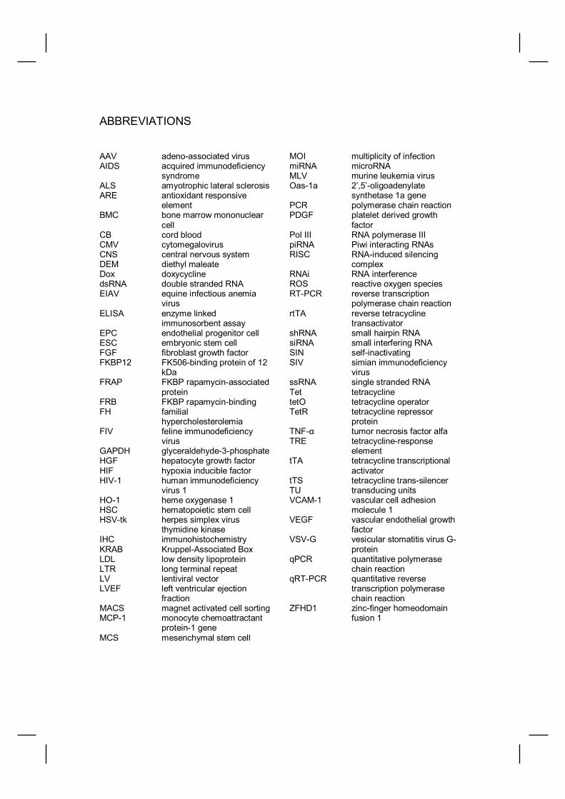
ABBREVIATIONS
AAV adenoassociated virusAIDS acquired immunodeficiency
syndromeALS amyotrophic lateral sclerosisARE antioxidant responsive
elementBMC bone marrow mononuclear
cellCB cord bloodCMV cytomegalovirusCNS central nervous systemDEM diethyl maleateDox doxycyclinedsRNA double stranded RNAEIAV equine infectious anemia
virusELISA enzyme linked
immunosorbent assayEPC endothelial progenitor cellESC embryonic stem cellFGF fibroblast growth factorFKBP12 FK506binding protein of 12
kDaFRAP FKBP rapamycinassociated
proteinFRB FKBP rapamycinbindingFH familial
hypercholesterolemiaFIV feline immunodeficiency
virusGAPDH glyceraldehyde3phosphateHGF hepatocyte growth factorHIF hypoxia inducible factorHIV1 human immunodeficiency
virus 1HO1 heme oxygenase 1HSC hematopoietic stem cellHSVtk herpes simplex virus
thymidine kinaseIHC immunohistochemistryKRAB KruppelAssociated BoxLDL low density lipoproteinLTR long terminal repeatLV lentiviral vectorLVEF left ventricular ejection
fractionMACS magnet activated cell sortingMCP1 monocyte chemoattractant
protein1 geneMCS mesenchymal stem cell
MOI multiplicity of infectionmiRNA microRNAMLV murine leukemia virusOas1a 2’,5’oligoadenylate
synthetase 1a genePCR polymerase chain reactionPDGF platelet derived growth
factorPol III RNA polymerase IIIpiRNA Piwi interacting RNAsRISC RNAinduced silencing
complexRNAi RNA interferenceROS reactive oxygen speciesRTPCR reverse transcription
polymerase chain reactionrtTA reverse tetracycline
transactivatorshRNA small hairpin RNAsiRNA small interfering RNASIN selfinactivatingSIV simian immunodeficiency
virusssRNA single stranded RNATet tetracyclinetetO tetracycline operatorTetR tetracycline repressor
proteinTNF tumor necrosis factor alfaTRE tetracyclineresponse
elementtTA tetracycline transcriptional
activatortTS tetracycline transsilencerTU transducing unitsVCAM1 vascular cell adhesion
molecule 1VEGF vascular endothelial growth
factorVSVG vesicular stomatitis virus G
proteinqPCR quantitative polymerase
chain reactionqRTPCR quantitative reverse
transcription polymerasechain reaction
ZFHD1 zincfinger homeodomainfusion 1


LIST OF ORIGINAL PUBLICATIONS
I Jonna K. Koponen, Hanna Kankkonen, Jani Kannasto, Thomas Wirth,Wolfgang Hillen, Hermann Bujard and Seppo YläHerttuala.Doxycyclineregulated lentiviral vector system with a novel reversetransactivator rtTA2SM2 shows a tight control of gene expression in vitro andin vivo.Gene Therapy 2003 Mar;10(6):45966.
II Hanna Hurttila, Jonna K. Koponen, Emilia Kansanen, HennaKaisa Jyrkkänen,Annukka M. Kivelä, Riina Kylätie, Seppo YläHerttuala and AnnaLiisaLevonen.Oxidative stress inducible lentiviral vectors for gene therapy.Gene Therapy in press
III Jonna K. Koponen, Tuija Kekarainen, Suvi E. Heinonen, Anita Laitinen,Johanna Nystedt , Jarmo Laine and Seppo YläHerttuala.Umbilical cord bloodderived progenitor cells enhance muscle regeneration inmouse hindlimb ischemia model.Molecular Therapy 2007 Dec;15(12):21727.
IV Petri I. Mäkinen, Jonna K. Koponen*, AnnaMari Kärkkäinen*, Tarja M. Malm,Kati H. Pulkkinen, Jari Koistinaho, Mikko P. Turunen and Seppo YläHerttuala.Stable RNA interference: comparison of U6 and H1 promoters in endothelialcells and in mouse brain.Journal of Gene Medicine 2006 Apr;8(4):43341.
V Jonna K. Koponen*, AnnaMari Turunen*, and Seppo YläHerttuala.Escherichia coli DNA contamination in AmpliTaq Gold polymerase interfereswith TaqMan analysis of lacZ.Molecular Therapy 2002 Mar;5(3):2202.
* Authors with equal contribution. Also some unpublished data is presented.


CONTENTS
INTRODUCTION… … … … … … … … … … … … … … … … … … … … … … … … ... 13
REVIEW OF THE LITERATURE… … … … … … … … … … … … … … … … … … 14
GENE THERAPY … … … … … … … … … … … … … … … … … … … … … … … … … … 14General concept… … … … … … … … … … … … … … … … … … … … … … … … … ... 14Cardiovascular diseases… … … … … … … … … … … … … … … … … … … … … … . 14Other targets… … … … … … … … … … … … … … … … … … … … … … … … … … … . 14
GENE TRANSFER VECTORS… … … … … … … … … … … … … … … … … … … … .. 15Overview of vectors… … … … … … … .… … … … … … … … … … … … … … … … … 15
Principles of gene transfer… … … … … … … … … … … … … … … … … .15Nonviral gene transfer vectors… … … … … … … … … … … … … … … .. 15Viral gene transfer vectors… … … … … … … … … … … … … … … … … . 16Vectors based on DNA viruses… … … … … … … … … … … … … … … . 16Vectors based on RNA viruses… … … … … … … … … … … … … … … . 16
Lentiviral HIV1 derived vectors… … … … … … … … … … … … … … … … … … … . 17HIV1 biology and genome… … … … … … … … … … … … … … … … … 17The HIV1 life cycle… … … … … … … … … … … … … … … … … … … … . 18The development of HIV1 derived gene transfer vectors… … … … 21Applications of HIV1 derived gene transfer vectors… … … … … … . 22
CELL THERAPIES FOR CARDIOVASCULAR DISEASES… … … … … … … … . 25General concept… … … … … … … … … … … … … … … … … … … … … … … … … ... 25Cell types and sources… … … … … … … … … … … … … … … … … … … … … … … 25Cell therapy combined with gene therapy… … … … … … … … … … … … … … … 27Clinical trials and future prospects… … … … … … … … … … … … … … … … … … 28
GENE TRANSFER VECTORS WITH REGULATED GENE EXPRESSION… ..28General concept… … … … … … … … … … … … … … … … … … … … … … … … … ... 28Tetracyclineregulated gene expression… … … … … … … … … … … … … … … .. 29Steroid hormone receptor based regulated gene expression… … … … … … . 31Rapamycinregulated gene expression … … … … … … … … … … … … … … … .. 31Physiologically regulated gene expression… … … … … … … … … … … … … … .. 32Radiation induced gene expression… … … … … … … … … … … … … … … … … .. 32
GENE EXPRESSION KNOCKDOWN BY RNA INTERFERENCE (RNAi)… … 33Introduction to RNAi… … … … … … … … … … … … … … … … … … … … … … … … 33The mechanism of RNAi… … … … … … … … … … … … … … … … … … … … … … . 33shRNA delivery by gene transfer vectors… … … … … … … … … … … … … … … . 34RNAi applications… … … … … … … … … … … … … … … … … … … … … … … … … . 35
AIMS OF THE STUDY… … … … … … … … … … … … … … … … … … … … … … ..37

MATERIALS AND METHODS… … … … … … … … … … … … … … … … … … … .38
SUMMARY OF THE MATERIALS AND METHODS… … … … … … … … … … … ..38Methods of assessing HIV1 lentiviral vector efficacy in animal modelsof cardiovascular gene therapy (unpublished studies)… … … … … … … … … .. 43
General methodology… … … … … … … … … … … … … … … … … … … . 43Intraluminal and periadventitial approach for lentiviral vector administration into rabbit carotid artery… … … … … … … … … … … .. 43Intramuscular injection of lentiviral vector in rabbit skeletal muscle… … … … … … … … … … … … … … … … … … … … … ...43Intramyocardial injection of lentiviral vector in porcine myocardium… … … … … … … … … … … … … … … … … … … .. 44
RESULTS AND DISCUSSION… … … … … … … … … … … … … … … … … … … 45
Doxycycline regulated lentiviral vector system (I)… … … … … … … … … … … .. 45Oxidative stress inducible lentiviral vector (II)… … … … … … … … … … … … … . 46The performance of lentiviral HIV1 derived vector incardiovascular gene therapy applications (unpublished data)… … … … … … . 47Cordblood derived progenitor cells in a mouse model forskeletal muscle ischemia (III)… … … … … … … … … … … … … … … … … … … … . 51A lentiviral vector for gene silencing by RNAi (IV)… … … … … … … … … … … .. 54A realtime quantitative PCR approach for the analysis of lacZmarker gene expression (V)… … … … … … … … … … … … … … … … … … … … ... 56
CONCLUSIONS AND FUTURE PERSPECTIVES… … … … … … … … … … ..57
REFERENCES… … … … … … … … … … … … … … … … … … … … … … … … … .. 59
APPENDIX: Original publications IV

13
INTRODUCTION
The concept of gene therapy, an approach totreat disease by either modifying the geneexpression or correction of abnormal genes,has been around since the first gene therapyapplications were introduced in the early1980s. By administration of DNA rather thana drug, many different diseases are currentlybeing investigated as candidates for genetherapy. This has been influenced by therapidly increasing knowledge of the humangenome and its regulatory mechanisms.However, the success of clinical therapies isstill limited due to the lack of optimal genetransfer vectors. Rather than aiming at asingle vector that is suitable for all genetictherapies, different vectors with qualitiestailored for each application is the objective.The most important features andrequirements should be taken into account.These include the vector tissue tropism, theduration of gene expression, the possiblegenomic integration ability, the feasibility toswitch off gene expression or to regulate itsexpression, the expected immune responseselicited by the vector, the possible need torepeated vector administrations, and safetyand ethical considerations. Adenoviralvectors have been extensively andsuccessfully used both in experimental andclinical settings and may be considered as
the standard vector of choice for manyapplications that need shortterm therapeuticgene expression. However, for therapiesrequiring longterm therapeutic geneexpression, there is not such a standardvector. Also, when longterm expression ofthe therapeutic gene is desired, distinctsafety and efficacy concerns need to beconsidered, such as the ability to regulatetherapeutic gene expression within thetherapeutic window, to switch off expressionwhen required and the possibility ofinsertional mutagenesis in the case ofintegrating vectors. The increased data onHIV1 molecular biology has been applied togene therapy research to enable HIV1 to beused as a gene therapy vector with a featureof stable integration into the target cellgenome. With the latest generation HIV1vectors, only a minute proportion of the viralgenome is exploited, both in the vector andthe production system, resulting in a vectorwhich does not transfer any viral genes, thusattenuating safety concerns. This thesis hasfocused on the development and applianceof HIV1 derived lentiviral gene transfervectors for regulated expression, genesilencing and progenitor cell therapies. Also,the efficacy of LV in animal models ofcardiovascular diseases is evaluated.

14
REVIEW OF THE LITERATURE
GENE THERAPY
General concept
The basic concept of gene therapy is toinsert genes into the somatic cells of anindividual in order to treat a disease, eitherinherited or acquired. Hereditary diseasestargeted by gene therapy usually aim at thecorrection of the function of one abnormalgene. However, in acquired diseases theactivity of several genes is disturbed and thedisease caused by these combined effectsmakes the gene therapy approaches of suchdiseases less straightforward.
Cardiovascular diseases
Despite major advances in therapies,cardiovascular diseases are still the leadingcause of death in the Western world and aretherefore attractive targets for gene therapy.Gene therapy approaches have beendirected to hyperlipidemias, promotion oftherapeutic angiogenesis in myocardium andskeletal muscle, postangioplasty restenosis,hypertension, heart failure, the prevention ofthrombosis and the protection of vascular bypass grafts (reviewed by YläHerttuala et al.,2000, Rissanen et al., 2007, Vincent et al.,2007).
To date, the promotion of blood vesselgrowth, that is, therapeutic angiogenesis,has been the most studied aspect ofcardiovascular gene therapy. Gene transferfor therapeutic angiogenesis has beentargeted to both myocardial and lower limbischemia, which are induced byatherosclerosis. Genes for vascularendothelial growth factors (VEGFs) (Mack etal., 1998, Gowdak et al., 2000, Arsic et al.,2003, Rutanen et al., 2004, Stewart et al.,2006), fibroblast growth factors (FGFs)(Giordano et al., 1996, Ueno et al., 1997,
Rissanen et al., 2003a), plateletderivedgrowth factors (PDGFs) (Richardson et al.,2001, Cao et al., 2003, Li et al., 2005b), andangiopoietins (Arsic et al., 2003, Cho et al.,2005) have been the mostly usedtherapeutic genes. Several clinical trials fortherapeutic angiogenesis have been carriedout (Rissanen et al., 2007).
For genetic cardiovascular diseases, genetherapy is a conceivable treatment optionespecially for familial hypercholesterolemia(FH), which is caused by the lack offunctional LDLreceptor. This results inserious hyperlipidemia, especially inindividuals whose both alleles are defective.Promising results have been attained withLDLreceptor gene transfer targeted to theliver in animal models (Pakkanen et al.,1999, Kankkonen et al., 2004, Lebherz et al.,2004).
Other targets
Other genetic disorders are also potentialcandidates for gene therapy. Probably themost known gene therapy studies are thosedirected to the primary immunodeficiencydisorder SCID/ADA (Blaese et al., 1995,Muul et al., 2003). Other candidates withpublished gene therapy research includecystic fibrosis (Flotte et al., 2007), inheritedmetabolic disorders like phenylketonuria(Ding et al., 2006), lysosomal storagedisorders like Gaucher’s disease (Sands etal., 2006), hematological disorders likehemophilias, hemoglobinopathies, anemiasand thalassemias (Nathwani et al., 2005)and muscular dystrophies (Foster et al.,2006).
Cancer gene therapy covers a number ofalluring treatment options for different typesof cancer. These applications can be dividedinto three subgroups: immunotherapy,oncolytic therapy and gene transfer therapy.Immunotherapy covers the studies in whicha cancer vaccine is produced by engineering

15
cancer cells to be more recognizable by theimmune system. This can occur by the invitro transfer of gene producing moleculeswhich are proinflammatory (Simons et al.,2006). Oncolytic gene therapy vectors areviruses which are modified to infect cancercells and induce cell death through thepropagation of the virus, expression ofcytotoxic proteins and cell lysis (Rein et al.,2005). The gene transfer concept involvesthe transfer of suicide genes (genes thatcause cellular death when expressed)(Rasmussen et al., 2002), antiangiogenesisgenes (Ohlfest et al., 2005) and cellularstasis genes (Eastham et al., 2000). Suicidegene therapy, utilizing herpes simplex virusthymidine kinase (HSVtk) gene transfer to atumor followed by ganciclovir treatment, hasshown potential in the treatment of themalignant brain tumor, glioblastoma(Immonen et al., 2004).
In addition to genetic disorders andglioblastoma, there are a number of otherpathologies which make the brain animportant gene therapy target tissue. Genetherapy treatments have been engineeredfor neurodegenerative disorders likeAlzheimer’s disease, amyotrophic lateralsclerosis, Parkinson’s disease (Cardone,2007) and for multiple sclerosis (Martino,2003), CNS injuries (Murray et al., 2001),epilepsy (Noe' et al., 2007) andcerebrovascular diseases like stroke (Jacobset al., 2005).
Gene therapy has also been studied for thetreatment of viral infections, mostly for theHIV1 infection (Dropulic et al., 2006). Interms of endocrine and metabolic disorders,diabetes is probably the most abundantlystudied (D'Anneo et al., 2006).
GENE TRANSFER VECTORS
Overview of vectors
Principles of gene transfer
Gene transfer aims at the delivery of nucleicacids across the cell membrane and into thenucleus of target cells. These genes areintroduced into the cells in vectors. Theefficiency of therapeutic gene transfer isdependent on the ability of the vector todeliver the gene into target cells and on thetransgene expression level. Different targettissues and cells require vectors with distinctproperties. Also, the vector choice isdependent on the application, for example,on the desired duration of expression of thetherapeutic gene. The development ofoptimal gene transfer vectors is one of thekey issues in determining the applicability ofgene therapy in clinical settings.
Nonviral gene transfer vectors
The concept of nonviral gene transfer coversplasmid vectors or oligonucleotides whichare introduced into the cells either as nakedDNA or by chemical or physical approaches.Early experiments suggested that a simpleinjection of naked DNA produced remarkablegene transfer efficiency in the muscle (Wolffet al., 1990), liver (Hickman et al.,1994) andskin (Choate et al.,1997). However, genetransfer by naked plasmids has not provenefficient enough for in vivo applications. Interms of chemical approaches, DNA isformulated into condensed particles byusing, for example; cationic lipids (Liu et al.,2003a) or polymers (Neu et al., 2005) ascarriers. These compounds are useful forenhanced gene transfer efficiency in vitro.Physical approaches for gene transferutilizing mechanical (particle bombardmentor gene gun), electric (electroporation),ultrasonic, hydrodynamic or laserbasedenergy to penetrate the cell membrane havebeen explored (Gao et al., 2007). Although

16
these methods may be efficient in vitro, theyhave not shown remarkable potency in vivo.In conclusion, nonviral vectors have notbeen able to improve upon the performanceof viral vectors to date.
Viral gene transfer vectors
In viral vectors, parts of the native viralgenome have been deleted and replaced bygenetic elements needed for the expressionof the therapeutic gene. Genetic engineeringhas meant that viral vectors do not carry thegenetic elements needed for the formation ofall the essential components of a virusparticle such as viral structural proteins andenzymes. Therefore, they are not able toreplicate and are not infectious. Elementsfor viral vectors are provided in trans by virusproducing systems such as helper constructsor packaging cell lines, increasing the safetyof the vectors. The tropism of viral vectors,that is the ability to transduce cells ofdifferent tissue types or animal species, maybe modified by coating the viral particle withenvelope proteins from another virus withknown specificity.
Vectors based on DNA viruses
Adenoviral vectors are nonenveloped,double stranded DNA vectors, which delivergenes efficiently into a wide variety of cellsboth in vitro and in vivo, and are the mostwidely used viral vectors so far. Wildtypehuman adenoviruses are a general cause ofbenign respiratory and other infections inhumans. Approximately 50 serotypes ofadenovirus have been identified and genetherapy vectors derived from serotypes 2and 5 are most commonly used. Adenoviralvectors are able to transduce both dividingand nondividing cells. Their genomeremains extrachromosomal in the host cellresulting in a transient expression of thetherapeutic gene. Conditionally replicatingadenoviral vectors have shown promise incancer gene therapy (Carette et al., 2007,
Ranki et al., 2007). However, a majorproblem of adenoviral vectors is theirimmunogenicity and toxicity (Liu et al.,2003b). In fact, on one occasion, genetherapy using adenoviral vector deliverycaused the death of a patient involved in aclinical trial for the treatment of ornithinetranscarbamylase deficiency due to a hugeimmune response triggered by the vector(Marshall, 1999).
Adenoassociated virus (AAV) derivedvectors are singlestranded DNA vectors.The prototype of AAV gene therapy vectorsis based on serotype 2. However, recentdata from mouse experiments has shownthat vectors derived from AAV serotype 8show superior tropism for the liver (Nakai etal., 2005) and those from serotype 6 forcardiac and skeletal muscles (Gregorevic etal., 2004). Also, serotype 9 vectors havebeen shown to transduce the myocardiummore efficiently than serotype 8 vectors(Inagaki et al., 2006). AAV vectors are ableto transduce both dividing and quiescentcells and although they remainextrachromosomal, longterm geneexpression is achieved. In one clinical trial,the duration of therapeutic gene expressionfor up to several years has been reported(Jiang et al., 2006). Native AAV is aparvovirus that is nonpathogenic in humans.In addition, AAV vectors are considered tobe rather low in immunogenicity. However, amajor drawback is the cumbersome virusproduction procedure, which is extremelydifficult to upscale (Xiao et al., 1998).
Other less frequently used gene transfervectors derived from DNA viruses are thosefrom baculovirus (Lehtolainen et al., 2002),herpex simplex virus (Gao et al., 2006) andEpsteinBarr virus (Hellebrand et al., 2006).
Vectors based on RNA viruses
Retroviral vectors are based on RNAviruses. The most extensively used retroviral

17
vectors are those derived from oncoviruses,such as murine leukemia virus (MLV) orlentiviruses (LV), such as humanimmunodeficiency virus1 (HIV1), simian(SIV), equine (EIAV) or feline (FIV)immunodeficiency viruses (reviewed by(Sinn et al. 2005)). Retroviral vectors carrytheir genetic information in the form of singlestranded RNA (ssRNA). In the target cell,viral RNA is reversetranscribed into doublestranded DNA, which is then integrated intothe host cell genome resulting in longtermtransgene expression. The prototype ofretroviral gene transfer vectors is derivedfrom MLV (Mann et al., 1983). MLV vectorsare only able to transduce dividing cells andthey have been used for both ex vivo and invivo applications. In clinical trials, MLVvectors have been used for the treatment ofcancer, inherited and acquired monogenicdisorders and AIDS. However, in a trial forthe treatment of Xlinked SCID patients withMLV vector gene transfer to hematopoieticstem cells ex vivo, vector induced leukemiaswere reported raising safety concerns(HaceinBeyAbina et al., 2003). In contrast,there have been no reports of insertionalmutagenesis in ADA/SCID patients treatedwith MLV vector gene transfer tohematopoietic stem cells (Aiuti et al., 2007).Thus, the risks of insertional mutagenesismay depend on the vector system, thetargeted cell types, the site of integration, thetransgene and the underlyingimmunodeficiency, as suggested by themolecular analysis of the three affectedpatients’ cells from the XSCID trial (HaceinBeyAbina et al., 2003).
In contrast to MLV vectors, lentiviral vectors(LVs) are able to transduce both quiescentand dividing cells, which is an advantage formany experimental and clinical settings. Ofthe lentiviruses used for gene transfer, HIV1derived vectors are the most advanced andowing to speciesspecific restrictions, it islikely that they are more efficient than animalLVs for the transduction of many types of
human cells. HIV1 derived vectors aredescribed in detail in the next chapter.
Lentiviral HIV1 derived vectors
HIV1 biology and genome
In the late 1970s and early 1980s, a newsyndrome, with symptoms of immunologicdysfunction, was discovered in United Statesand Europe. A connective laboratory findingwas the depletion of CD4+ Tlymphocytes inaffected individuals. The disease was termedacquired immunodeficiency syndrome(AIDS). Later, a new retrovirus was isolatedfrom both AIDS patients and infected,asymptomatic individuals from various riskgroups. The new retrovirus causing a slow,progressive disease affecting the immunesystem and exhibiting morphologic andgenetic characteristics typical of thelentivirus genus (Lentivirinae), was namedhuman immunodeficiency virus (HIV) (Coffinet al., 1986) and subsequently HIV1. Otherlentiviruses include HIV2 and nonhumanlentiviruses such as the felineimmunodeficiency virus (FIV) of cats, simianimmunodeficiency virus (SIV) of monkeys,bovine immunodeficiency virus (BIV) ofcattle, equine infectious anemia virus (EIAV)of horses, Maedi/Visna virus and caprinearthritis encephalitis virus of sheep andgoats.
Retroviral virion particles are spherical inshape and surrounded by a lipid membranebilayer envelope with projections ofglycoproteins. There is a spherical layer ofprotein under the membrane and an internalnucleocapsid whose shape varies from virusto virus. The members of the lentivirus genusare complex retroviruses with themorphology of cylindrical or conical cores.
Typically, all retroviruses carry three majorgenes that are critical for retroviral replicationand assembly, gag, pol and env. The morecomplex retroviruses contain accessory

18
genes that are essential or contribute toefficient virus replication and persistence.HIV1 encodes six additional genes: tat, rev,vif, vpu, vpr, and nef. (Figure 1 and Table1). The HIV1 virion has a diameter of ~110nm. The viral SU and TM glycoproteins areinserted into the lipid membrane surroundingthe nucleocapsid. Proteins within the innershell of a mature virion are cleavageproducts of the Pr55gag and Pr160gagpol
polyproteins. The condensed inner core isformed by the capsid protein (CA), p24.Between inner core and the lipid membraneis the matrix protein (MA), p17, whichremains associated with the lipid membrane.The virion core contains two copies of thesinglestranded genomic RNA to which the
NC protein is bound. Also packaged into thevirion are the host transfer RNA; tRNA3
Lys,and the viral proteins RT, PR, IN, Vif andVpr. (Haseltine, 1991)
The HIV1 life cycle
The HIV1 replication cycle, started with theviral genome integrated into a hostchromosome, leads to expression of viralgene products, production of new virusparticles, infection of a new cell andreintegration of the viral genome. The HIV1life cycle may be split into 15 steps (Frankelet al., 1998). These are illustrated in Figure2 and are described below.
Figure 1. Diagram of the HIV1 genome and virion structure. The genome is flanked by longterminal repeat (LTR). Nine genes (gag, pol, env, tat, rev, vif, vpr, vpu and nef) encode 15proteins, see Table 1 for descriptions. Modified from Frankel et al., 1998.

19
Figure 2. The HIV1 life cycle. Details for the numbered steps can be found in the text. Picturemodified from Frankel et al., 1998.
Viral transcripts are expressed from thepromoter located in the 5’ long terminalrepeat (LTR) (1), with Tat greatlyenhancing the rate of transcription. ViralRNAs are then transported from thenucleus into the cytoplasm where they canbe translated or packaged (2). This step isregulated by Rev. Some viral RNAs aretranslated by ribosomes in the cytoplasmto form Gag and GagPol polyproteins,which localize to the cell membrane (3).The Env mRNA is translated at theendoplasmic reticulum and formscomplexes with the coexpressed HIV1cellsurface receptor CD4. The virion coreparticle is constructed from the Gag andGagPol polyproteins which are laterprocessed into subunits (see Table1),accessory proteins Vif, Vpr and Nef, andthe genomic RNA (4). The immature virionbegins to bud from the cell surface. Toprovide surface (SU) and transmembrane(TM) proteins for the virion outer
membrane, the Env polyprotein must bereleased from the complexes it has formedwith CD4. Vpu assists this process bypromoting CD4 degradation (5). Env istransported to the cell surface, where itmust be protected from binding to CD4 (6).Nef promotes endocytosis and degradationof surface CD4 (7). As the virion particlebuds and is released from the host cellsurface (8), it undergoes maturationinvolving proteolytic processing of the Gagand GagPol polyproteins by protease (PR)and Vif (9). After budding, the mature virionis ready to infect another cell. This isinduced by interactions between surfaceprotein SU and CD4 receptor and CC orCXC chemokine coreceptors of the targetcell (10). After binding, the TM undergoesa conformational change that promotesviruscell membrane fusion therebyallowing entry of the core into the cell (11).The virion core is then uncoated to exposea viral nucleoprotein complex containing

20
the viral proteins matrix (MA), reversetranscriptase (RT), integrase (IN), Vpr andviral RNA (12). During the microtubulebased nuclear transport of this preintegration complex, the viral singlestranded RNA genome is reverse
transcribed into doublestranded RNA (13).The viral replication cycle is completed byIN catalyzing the integration of the viralDNA into a host chromosome (14).
Table 1. HIV1 genes, gene products and their function. Modified from (Ramezani et al., 2002)
Gene Encoded protein(s) Function
Regulatory genes
tat Tat Transactivation of geneexpressionNuclear export of late mRNAs
rev Rev Promotion of polysomalbinding to RREcontainingRNAs
Accessory genes
vif Vif Enhancement of virustransmissionNuclear transport of viralnucleoprotein complex
vpr VprInduction of G2 arrest individing cellsCD4 degradationvpu Vpu Virus maturation and releaseCD4 and MHC1 downregulationnef Nef Enhancement of virusreplication
Structural genes polyproteinscleaved into subunits
Formation of viral particlesgag
Pr55gag:matrix MA (p17), capsidCA (p24), nucleocapsidNC (p9), p6
Packaging of viral genomicRNA
Reverse transcription
Integrationpol
Pr160gagpol:protease PR (p10),reverse transcriptase RT(p61/p52), integrase IN(p31) Virus maturation
env
gp160:surface SU (gp120),transmembrane TM(gp41)
Binding and entry into the hostcell

21
The development of HIV1 derivedgene transfer vectors
Like in any other viral gene transfer vectors,the generation of replicationdefective LVsrequires splitting the cisacting sequences(vector sequences) needed for the transferand expression of a transgene in target cellsand the transacting sequences (packagingsequences) encoding the essential viralstructural and enzyme proteins, ontoseparate genetic units. The tropism of viralvectors is broadened by pseudotyping; viaencapsidation of the viral particle with theenvelope of another virus. LVs are mostlypseudotyped with vesicular stomatitis virusGprotein (VSVG), which is pantropic andhighly stable. The transfer vector plasmid iscotransfected with the packaging andenvelope plasmids into a cell line wherevirions are produced. Virions are assembledof viral proteins encapsidating thereplicationdefective transfer vector RNA.
The HIV1 derived transfer vector cisactingsequences include viral LTRs, the primerbinding site, the packaging signal, the Revresponsive element, and an internalpromoter linked to a transgene of interestconstituting a transcriptional unit (Naldini etal., 1996). The genetic elements derivedfrom HIV1 are required for viralencapsidation, reverse transcription andintegration. Like MLV retroviral vectors, HIV1 vectors do not transfer viral codingsequences into target cells, meaning thatcells transduced with the HIV1 vector do notexpress any viral proteins.
HIV1 transfer vectors have been modifiedby introducing various internal promotersdriving transgene expression, and by theinclusion of genetic elements such as thecentral DNA flap and the posttranscriptionalelement. The central DNA flap is a 99nucleotidelong overlap formed after nativeHIV1 reverse transcription and it is involved
in the import of the HIV1 preintegrationcomplex into the nucleus. The sequence forthis element, the central polypurine tract(cPPT), was omitted from early generationHIV1 vectors but has been routinelyincluded in current vector designs becauseof its beneficial effect on gene transferefficiency (Follenzi et al., 2000). Thewoodchuck hepatitis virus posttranscriptional regulatory element (WPRE)sequence is also commonly included incurrent HIV1 vectors. This element hasbeen shown to enhance transgeneexpression from several types of promoters(Deglon et al., 2000) by augmenting mRNA3’end processing and polyadenylation.
The HIV1 vector packaging systems havebeen extensively developed. The firstgeneration packaging systems comprisedthree expression plasmids: the transfervector, the plasmid for VSVG envelopeprotein production and the packagingconstruct (Naldini et al., 1996). In thissystem, only two of the nine native HIV1genes, vpu and env, were deleted.Subsequently, it was shown that none of thefour HIV1 accessory genes vif, vpr, vpu ornef were required for efficient production ofVSVG pseudotyped vector particles(Zufferey et al., 1997). Therefore, thesecondgeneration packaging system onlyutilized HIV1 gag, pol, rev and tat genes,which further attenuated the potential for thegeneration of replicationcompetent viruses.
For the currently used thirdgeneration HIV1vector system, several additionalmodifications have been made to ensure thesafety of these vectors. Firstly, they havebeen modified to selfinactivate (SIN) bydeleting the promoter sequences of the U3region of the 3’LTR (Miyoshi et al., 1998).Since the U3 region of the 3’LTR serves as atemplate for the U3 regions of both LTRs,the provirus carries the deletion in both LTRsafter reverse transcription. As a result, the

22
LTRs of the integrated vector are almostcompletely inactivated. The inability totranscribe fulllength vector RNA minimizesthe chance of replicationcompetent virusgeneration and reduces the potential ofoncogene activation by promoter insertionalmutagenesis. To avoid the reconstitution ofdeleted U3 sequences by homologousrecombination with intact 5’ LTR during viralvector production, the U3 region of the 5’LTR is replaced with a heterologouspromoter, usually a cytomegalovirus (CMV)promoter. Since the LTR promoter isdependent on Tat interaction, the use of theCMV promoter allows tat gene independentproduction of viral vectors. Therefore, fromthe thirdgeneration HIV1 vector packagingsystem, tat is deleted. This has enabledfurther refinement of the packaging systemfor increased safety, such as the expressionof gagpol and rev genes from two separatenonoverlapping plasmids. With 40% of thewildtype virus genome (three out of ninegenes) left, the parental virus can not bereconstituted from such an extensivelydeleted packaging system. Also, in theabsence of overlapping viral sequences therisk of recombination events betweencomponents of the viral production system isabolished, further limiting the possibility toyield replicationcompetent vectors. To date,replicationcompetent vector production hasnot been associated with the production ofHIV1 lentiviral vectors.
Applications of HIV1 derived genetransfer vectors
HIV1 derived gene transfer vectors showefficient delivery, integration and longtermexpression of transgenes in both dividingand nondividing cells, thus making themexcellent vehicles for basic studies of geneoverexpression and knockdown. As such,they represent an attractive tool for mostpotential targets of gene therapy, whetherthe targets are early precursors or terminallydifferentiated cells. While third generation
HIV1 vectors are able to transduce virtuallyall types of cells in vitro, it seems that theaccessory protein, Vpr, is important for thetransduction of macrophages andhepatocytes (Naldini et al., 1996, Kafri et al.,1997). Also, although HIV1 vectors do notrequire cell division, like the native HIV1virus, they are unable to successfullytransduce T lymphocytes during the G0 stageof the cell cycle. This is due to blocks at thelevels of reverse transcription and nuclearimport. However, HIV1 vectors mediateefficient stable transduction of many celltypes which are poorly transduced by othervectors. For example, gene transfer toprogenitor and stem cells is one of the mostimportant applications of HIV1 derivedvectors.
Embryonic stem cells (ESCs) are cellsderived from the inner cell mass of an earlyembryo. They can be maintained in anundifferentiated state indefinitely and can begenetically manipulated in vitro withoutlosing their differentiation potential. Thisunique property of ESCs suggests that theymay provide a useful tool to analyzedevelopmental pathways and are apromising cell source for transplantationtherapies. Efficient genetic manipulation ofESCs is critical for both development,biology research and for maximizing thetherapeutic potential of ESCs. HIV1 derivedvectors have been shown to efficiently drivetransgene expression in mouse (Kosaka etal., 2004) and human (Gropp et al., 2003)ESCs. ´
Of all blood cell types, only hematopoieticstem cells (HSC) can selfrenew, persistthroughout a lifetime and reconstitute thewhole lymphohematopoietic system of anindividual. HIV1 LVs can efficientlytransduce ex vivo mouse (MoreauGaudry etal., 2001), nonhuman primate (Horn et al.,2002) and human (Miyoshi et al., 1999)HSCs in the absence of cytokine stimulationand cell cycle induction. This is important

23
because culture conditions which facilitatethe proliferation of HSCs without the loss oftheir stem cell capacity have not beenidentified. HIV1 derived LVs efficientlytransduce human CD34+ cells, aheterogenous population of HSCs andprogenitor cells. The LVtransduced CD34+
cells are capable of engraftment and multilineage differentiation in NOD/SCID (nonobese diabetic/severe combinedimmunodeficient) mice (Miyoshi et al., 1999).Such genetically modified cells can bepassed to secondary transplants (Woods etal., 2000) which further confirms thetransduction of true HSCs and not only themultipotent progenitor cells.
The stereotactic injection of HIV1 LV wasthe model initially used to illustrate the abilityof these vectors to transduce nondiving cellsin vivo (Naldini et al., 1996). Numerousstudies have reported successful longlastingand efficient transgene expression interminally differentiated neurons of rodentbrain after a single injection of only a fewmicroliters of high titer (magnitude of 109
TU/ml) vector stock. In addition to neurons,LVs are able to transduce most celltypeswithin the CNS in vivo, including astrocytes,oligodendrocytes, adult neuronal stem cellsand glioma cells (Jakobsson et al., 2003,Consiglio et al., 2004, Miletic et al., 2004).LVs have a property of highly efficientretrograde transport providing access to awide area of the brain after a single injection,thus enabling potential therapy for widelydisseminating neurological disorders. Also,the delivery of ex vivo LV transduced HSCstrafficking to the CNS has been exploited.Promising therapeutic effects of HIV1 LVmediated gene transfer has beendocumented in animal models of Alzheimer’sdisease (Dodart et al., 2005), Huntington’sdisease (de Almeida et al., 2001),Parkinson’s disease (Kordower et al., 2000),amyotrophic lateral sclerosis (ALS, Raoul etal., 2005a) and lysosomal storage diseases(Biffi et al., 2004). Also, LV gene transfer has
been utilized in the development of newanimal models of Huntington’s (Regulier etal., 2003) and Parkinson’s disease (LoBianco et al., 2002). In these models LVgene transfer has been used to induceoverexpression of the mutated form ofprotein present in these diseases.
The liver is an important target tissue forgene therapy because of the numerousgenetic defects that cause defects in liverfunction resulting in severe disorders suchas hemophilia A and B and FH. Also, theliver is a target of chronic virus infectionssuch as hepatitis B and C. Despite theregeneration capacity of the liver,hepatocytes divide only occasionally in theadult. Several studies have reported that LVscan transduce nondividing rodent andhuman hepatocytes, both ex vivo and in vivo(Kafri et al., 1997, Nguyen et al., 2002,VandenDriessche et al., 2002, Follenzi et al.,2004). However, mouse studies have shownhigher LV gene transfer efficiency inneonates and after partial hepactomy (Parket al., 2000, Ohashi et al., 2002, Park et al.,2003), suggesting that proliferatinghepatocytes are more prone to LVtransduction. Some properties of thearchitecture of the hepatic lobule or thetightness of the endothelial barrier in hepaticblood vessels may be influenced by livergrowth or regeneration, thus favouring viralentry. Alternatively, it has been suggestedthat the HIV1 accessory protein Vpr, absentfrom later generation LVs, can enhancehepatocyte transduction (Kafri et al., 1997).However, in a study of LV mediated LDLreceptor gene transfer in rabbit model of FH,a long term therapeutic effect withouthepactomy was reported (Kankkonen et al.,2004). Although only a modest gene transferefficiency of 0.01% of the liver cells wasachieved, the results showed a significant(44%) decrease in the serum cholesterollevel of the treatment group at a one yeartimepoint compared to controls. These

24
results support further research of LVmediated liver gene therapy.When the early generations of HIV1 LVswere developed, high expectations of theirperformance in in vivo gene therapyapplications were raised. So far, theseexpectations have been fulfilled only in thetargets of the central nervous system (CNS),lymphohematopoietic system and to alesser extent in the liver. More work isneeded to evaluate the true utility of LVs intargeting tissues such as skeletal muscleand the myocardium. The first clinical trialutilizing HIV1 LV for the treatment of HIV1infection is currently in process (Levine et al.,2006). In this study, an antisense approachagainst the HIV1 envelope was utilized byex vivo transduction of the patients’ Tcells.A LV with wildtype LTRs was used andtherefore, expression of the antisensesequence was upregulated upon the wildtype HIV1 infection of the vector bearingcell. The results demonstrate safe andefficient gene delivery and good persistencein vivo and also, an improvement of theimmune function in four out of five patients.However, the use of LV in patients infectedwith wildtype HIV1 presents a problem, thepotential of the wildtype virus to infect a cellmodified by the vector. As a result, the wildtype virus infection would mobilize the vectorgenome by packaging it and transferring it tonew cell. For HIV1 patients, such a spreadof the vector might actually be beneficial.However, it poses complex biosafety andethical problems and should be avoided. Thepatients from this trial were monitored forover one year (Levine et al., 2006). Only alongterm followup after at least three yearswill reveal the true safety of such treatment.Nonetheless, based on the results from thefirst clinical trial with LV, it possesses thepotential to be used for the therapiesinvolving prior ex vivo genetic modification ofcells of the lymphohematopoietic system.
A few years ago, lentiviral HIV1 derivedvectors made a breakthrough in the
generation of transgenic animals (reviewedin Park, 2007). Previously, transgenesis hasbeen achieved by pronuclear injection ofnaked DNA. This is a rather inefficient andtricky technique requiring a clearly visibleoocyte pronucleus mostly inapplicable tospecies other than mouse. Also, mousetransgenesis utilizing MLV retroviral vectorsfailed as a result of transgene silencingduring development (Cherry et al., 2000).HIV1 LVs have been successfully used togenerate transgenic mice and rats by thetransduction of singlecell embryos, earlyblastocysts or embryonic stem cells (Lois etal., 2002, Pfeifer et al., 2002). In theseexperiments, LV mediated transgenesisresulted in very high embryo viability with80% of mice carrying the provirus. UnlikeMLV retroviral vectors, HIV1 LVs appear toescape epigenetic silencing. The reason forthis remains unknown but might be linked todifferent integration site preferences. MLVvectors have been found to integratepredominantly close to transcriptional startregions and CpG islands (Wu et al., 2003).In contrast, LVs, studied to date, integrateacross the entire transcribed gene regionwith no preference to the proximity to thetranscriptional start site. Also, LV genomescontain fewer CpG dinucleotides susceptibleto cytosine methylation than the oncoretroviral vectors, which may partially explainthe finding that they are less prone tosilencing. Successful LVmediatedtransgenesis has also been extended tolarger animal species including cattle(Hofmann et al., 2004) and pig (Hofmann etal., 2003). In addition to offering models ofhuman diseases, especially large transgenicanimals may find applications in futurebioindustry for example as producers ofhuman proteins for drug use or as a potentialsource of humanized organs fortransplantation.

25
CELL THERAPIES FORCARDIOVASCULAR DISEASES
General concept
Among treatment options for cardiovasculardiseases, there is a definite need foralternative therapies, particularly foradvanced and severe disease. Experimentalstudies have indicated that progenitor orstem cells derived from different sourcespossess regenerative capacity in the heartand vasculature, which has raisedexpectations of clinically applicable celltherapy for tissue repair in cardiovasculardiseases. The initial concept for thisresearch was based on the cell plasticityhypothesis, which suggests that progenitorcells can transdifferentiate in vivo acrossgenerally agreed tissue lineage boundaries.The concept of plasticity has, however, beenchallenged by data proposing that HSCs arecommitted to differentiate into cells ofhematopoietic lineages and do not own thecapacity to transdifferentiate (Wagers et al.,2002). Cell fusion has since been proposedas an alternative explanation for observedtransdifferentiation events. On the otherhand, by secretion of paracrine factors,progenitor cells might affect vasculogenesis,tissue repair and remodelling without theneed to undergo transdifferentiation or cellfusion. Also, stem cell niches have beenidentified from myocardium. The concept ofstem cell niche covers the local tissueenvironments of surrounding cells which areimportant for the regulation of stem cellscontrolling and balancing selfrenewal anddifferentiation (Moore et al., 2006, Morrisonet al., 2008). There is evidence of nestingcardiac stem cells and progenitors that areconnected structurally and functionally tomyocytes and fibroblasts by junctional and
adhesion proteins, such as connexins andcadherins (Urbanek et al., 2006). A novelfascinating mechanism proposed to play arole in cell therapy is the putative stimulationof endogenous tissue repair pathways whichmight contribute to the regeneration of stemcell niches (Mazhari et al., 2007).
With the evolving experimental data, theconcept of cell therapy for cardiovasculardiseases has shifted from the original idea ofprogenitor cells taking part in theregeneration of injured myocardium orskeletal muscle or playing a part in theinduction of angiogenesis by the directinvolvement of progenitor cells into newlyforming vessels. Instead, a broaderhypothesis suggests that cell therapy mightin fact facilitate complementary aspects oftissue repair (Figure 3). These effects mightinclude augmentation of cell survival (forexample, in limiting apoptosis), tissueoxygenation by angiogenesis orimprovement in positive tissue remodelling.The most potent target diseases for celltherapy include myocardial infarction,ischemic cardiomyopathy and peripheralvascular disease causing skeletal muscleischemia in lower limbs.
Cell types and sources
Several sources of progenitor cells forcardiovascular cell therapy exist in adults,including unfractioned or fractionedhematopoietic and mesenchymal stem cellsfrom bone marrow, circulating progenitorcells, skeletal myoblasts and residentprogenitor cells for example, from adiposetissue. Also, cord blood HSCs andcardiomyocytes derived from embryonicstem cells have been used in animal models.

26
Figure 3. A working hypothesis of cell therapy for myocardial and skeletal muscle regeneration.Cell therapy can have a favourable impact on tissue healing by alternative mechanismspresented in the figure. Stem and progenitor cell numbers and functional capacity areinfluenced by a patient’s age, gender, cardiovascular risk factors and underlying disease statewhich all contribute to the natural response in the injured tissue and also to preparation ofautologous cell preparations for therapy. Figure modified from Wollert et al., 2005.
For cardiovascular gene therapy, bonemarrow has been proposed as a source ofhematopoietic, vasculogenic andmesenchymal stem cells. Initial experimentalevidence suggested a significant degree ofmyocardial regeneration by theadministration of lineage negative ckit+ bonemarrow mononuclear cells (BMCs) into amurine model of myocardial infarction (Orlicet al., 2001). However, this has beenquestioned by subsequent studies showinglittle or no tissue integration of these BMCsin similar animal models (Balsam et al.,2004, Murry et al., 2004). These findingschallenged the paradigm of BMCtransdifferentiation, although did not excludethe possibility that such cells could potentiatemyocardial repair by other mechanisms.Bone marrow mesenchymal stem cells
(MSCs) are a component of marrow stroma.They are selfrenewing, clonal precursors,which expand easily in culture, exhibitmultipotency and have also been shown todifferentiate to cardiomyocytes and vascularcells (Jiang et al., 2002). Endothelialprogenitor cells (EPCs) have been proposedto induce angiogenesis and reendothelization in the models of ischemiaand vascular injury (Madeddu et al., 2004,Nowak et al., 2004). Mechanisms suggestedfor EPCmediated angiogenesis includeintegration of the EPC into newly formedmicro and macrovessels and the secretionof growth, survival and cellmodulatoryfactors. EPCs have been identified andenriched from bone marrow and peripheralblood by the expression of surface antigenssuch as CD31, CD133, VEGFR2 and Tie2.

27
Whether these cells, exhibiting endothelialplasticity, offer a significant therapeuticadvantage remains unclear in the absence ofconvincing data.
Skeletal myoblasts are satellite progenitorcells in muscle. In response to muscle injury,they are able to proliferate and fuse toregenerate new multinucleated cells. Thepotential advantage of using these cells incell therapy applications include theirautologous origin, ease of isolation, high invitro proliferative capacity, in vivo ischemictolerance and myocyte restricted lineagecommitment which limits the risk ofoncogenetic transformation (Deasy et al.,2004). In animal models of myocardialischemia, autologous skeletal myoblastsaugmented contractile function (Taylor et al.,1998) and findings from clinical studiessuggested that implanted myoblastsengrafted viably in scarred myocardium(Pagani et al., 2003). The enthusiasm formyoblast therapy has faded by the lack ofevidence for cardiomyocyte differentiation ofmyoblasts and further, due to arrhytmiasobserved in a clinical trial, presumablycaused by the inability of myoblasts tointegrate into the conduction system of theheart (Menasche et al., 2003). Recently, apopulation of myoendothelial cells withmultilineage capacity, including skeletal andcardiac muscle regenerative potential, hasbeen identified within human skeletal muscle(Zheng et al., 2007). Further research willshow whether these cells, showing myogenicand endothelial properties, can beenvisioned as a therapy for muscle diseases.
The ability of human embryonic stem cell(ESC) derived cardiomyocytes to surviveand integrate structurally and functionallyinto healthy and postinfarct cardiac tissuehas been demonstrated in animal models(Kehat et al., 2004, Laflamme et al., 2005,Xue et al., 2005, Caspi et al., 2007). Therecent breakthrough findings show thatmouse and human fibroblasts can be
reprogrammed to pluripotent ESClike cellsby the transfer of three to four transcriptionfactor genes (Takahashi et al., 2006,Takahashi et al., 2007, Wernig et al., 2007,Yu et al., 2007, Nakagawa et al., 2008). Theresulting induced pluripotent stem cells havethe potential to be used in future treatmentsfor cardiovascular diseases.
For cell therapy research of cardiovasculardiseases to date, bone marrow derivedprogenitor cells are the most commonlyused. An important issue complicating theinterpretation and comparison of bothexperimental and clinical data is theheterogeneity of cell preparations, since bothunfractioned mononuclear cells andfractioned preparations selected for CD34+
or CD133+ have been used.
Cell therapy combined with genetherapy
Gene therapy has been widely applied forthe therapy of cardiovascular diseases, mostpopularly in the concept of angiogenicgrowth factor therapy for ischemicmyocardium or skeletal muscle. Although thebiological effects of such growth factors arewell understood, these therapies have notproven efficient in clinical trials presumablydue to the limited efficacy of current genetransfer technology. One approach toimprove the delivery of growth factors mightbe the combination of cell and gene therapyto utilize progenitor cells as carriers. After atransgene is introduced, these engineeredprogenitor cells would home into the targetarea and secrete therapeutic proteins. Also,by gene transfer, the chemokine expressionprofile of the progenitor cell might be alteredto improve homing of endogenous progenitorcells into the injured area (Askari et al.,2003). Another approach using cell basedgene therapy is to engineer progenitor cellsto express a protein which is not secretedbut modifies the biology of the cell itself.Such a modification might aim at improving

28
cell survival by inhibiting apoptosis forexample (Mangi et al., 2003), or bystrengthening resistance to ischemia orscavenging free radicals.
Clinical trials and future prospects
Small clinical trials have focused on thesafety and feasibility of progenitor celltherapy in cardiovascular diseases, includingischemic cardiomyopathy (Fuchs et al.,2003, Perin et al., 2003, Tse et al., 2003),peripheral vascular disease (TateishiYuyama et al., 2002b) and myocardialinfarction (Strauer et al., 2002, Britten et al.,2003, FernandezAviles et al., 2004, Wollertet al., 2004). While the safety of cell therapyhas been demonstrated it has been difficultto compare data because of variations in themethods used. These include variations inroutes of cell delivery, preparation of cellsand chosen endpoint parameters for efficacyevaluation. To date, several randomized,controlled trials of intracoronary applicationof bone marrow cells for patients with acutemyocardial infarction have been reported(Chen et al., 2004, Bartunek et al., 2005,Erbs et al., 2005, Hendrikx et al., 2006,Janssens et al., 2006, Kang et al., 2006,Meyer et al., 2006, Cleland et al., 2007). Thechange in the left venctricular ejectionfraction (LVEF) after cell therapy has beenassessed by angiography, magneticresonance imaging or ultrasound andcompared to a control group. Statisticallysignificant LVEF improvements have beenobtained in some of the studies (Chen et al.,2004, Erbs et al., 2005, Hendrikx et al.,2006, Kang et al., 2006, Cleland et al.,2007). Nevertheless, it has been pointed outthat these improvements in myocardialfunction are not likely to be significant in aclinical context. In fact, experts have agreedthat before pursuing further clinical trials abetter understanding of the mechanisms ofprogenitor cell mediated therapy, themethods used to produce the therapeutic cellpreparations, the application route, the
timing of treatment and patient selectionneeds to be attained (Bartunek et al., 2006).This will show the true potential of progenitorcell therapy as a clinical treatment forcardiovascular diseases.
GENE TRANSFER VECTORSWITH REGULATED GENEEXPRESSION
General concept
In terms of both experimental and clinicalgene therapy applications, one of the keyissues is the ability to regulate theexpression of a therapeutic gene, in order toproduce levels of protein within a therapeuticwindow, and to switch off the expression ifdesired. Regulated gene expression vectorsare based on the insertion of sequencesbinding to transcriptional activatorspreceding the minimal promoter. Theseactivators will bind, and thus, activate geneexpression when a particular inducercompound is present. Binding is eitherachieved subsequent to a conformationalchange in the activator or byheterodimerization of two distinct factors,one that is responsible for specific DNAbinding and the other for transcriptionactivation. Regulated gene expressionsystems consist of at least two separateexpression cassettes: one that contains thetranscriptional activator under the control ofeither a constitutive or a tissue specificpromoter, and the other contains atransgene under the control of the regulated,transcriptional activator responsive promoter.To deliver these expression cassettes intotarget cells, either two separate genetransfer vectors are used or, all the elementsare combined into a single vector. To date,tetracyclinedependent gene regulationsystems are the most utilized and advanced.These and some other commonly appliedsystems are introduced in the followingsections. A direct comparison of different

29
regulation systems is difficult and therefore,each system should be selected according tothe requirements of the particularapplication. When regulatable geneexpression is used in clinical applications,the pharmacology of the inducer drug playsa key role in selection of the system.
Tetracyclineregulated geneexpression
Tetracycline (Tet) –regulated transcriptionalexpression systems have been adapted fromstudies of the E.coli tetracycline resistancemechanism (Hillen et al., 1994). In bacteria,the TetR protein inhibits the transcription ofgenes in the tetracyclineresistance operonpresent in the Tn10 transposon by dockingto the Tet operator (tetO) sequences, in theabsence of tetracycline. Therefore, thetranscription of the genes needed tometabolize Tet is inhibited by TetR in theabsence of Tet. When Tet is present, it isbound by TetR, inducing a conformationalchange, which results in the release of TetRfrom tetO, allowing transcription. In the Tetoff system (Figure 4), TetR is modified tofunction as a transcriptional activator byfusion with a viral protein domain VP16, aeukaryotic transactivator derived fromherpes simplex virus type 1. As a result,TetR is converted from a transcriptionalrepressor to an activator, and is termed theTet transactivator (tTA). The tTA isexpressed independently of the tetracyclineresponse element (TRE, a framework ofseven tandem sequences upstream of aminimal CMV promoter) driven transgeneencoding cassette. In the absence of Tet thetTA binds to the TRE, and activatestranscription of the transgene from anotherwise silent minimal promoter. When Tet
is present, it binds to tTA, which is thenreleased from tetO and transcription is nolonger continued. Thus, Tetoff systemdriven gene expression is switched on andoff in the absence or presence of theinducer, respectively. As an inducing drug,the Tet analog doxycycline (Dox) is oftenused.
When mutations were randomly introducedinto the TetR part of the tTA a separateprotein was produced which exhibitedopposite binding properties and led to thedevelopment of the Teton system (Figure4). The mutant of tTA, termed rtTA, triggerstranscription by activating the TRE in thepresence of the inducer Tet. The Tetongene regulation system, that utilizes rtTA,has been widely applied, but the system issomewhat limited with respect to theprecision of regulation. The rtTA has beenfound to also show some degree of affinityfor tetO sequences in the absence of Tet,therefore inducing a low basal level of geneexpression in the off state. To furtherimprove the Teton system, the inventors ofthe system screened for mutant rtTAmolecules in yeast to obtain a transactivatorwith more specific binding properties. Onemutant with superior features was identifiedand this novel transcriptional activator wasnamed rtTA2SM2. The Teton system withrtTA2SM2 showed negligible basalexpression and was fully induced with a 10fold lower Dox concentration than rtTA(Urlinger et al., 2000). To date, the rtTA2SM2has been successfully used in a wide varietyof applications (Lamartina et al., 2002,Chenuaud et al., 2004, Vogel et al., 2004,Pluta et al., 2005).

30
Figure 4. Components of the Tetoff and Teton gene expression regulation system. Bothsystems consist of two transcriptional units: one for the expression of transactivator (tTA orrtTA) and the other has the regulated transgene under the control of the transactivator bindingpromoter. The transactivator consists of the tetracycline operon (TetO) binding domain fused tothe transactivation domain VP16 originating from HSV virus. In the Tetoff system the TetObinding domain, tetracycline repressor (TetR) is able to bind in the absence of tetracycline or itsderivative, doxycycline (Dox) and thus, transcription is activated by the VP16 domain of the tTAin the absence of Dox. When Dox is present, it binds to the tTA and induces a conformationalchange, releasing tTA from TetO and deactivating transcription. In the Teton system thebinding properties of TetR are mutated for reversed binding properties. Thus, the resultingtransactivator, rtTA induces transcription in the presence of Dox.
In addition to its tight regulation and thepossibility to strictly adjust the transgeneexpression level dosedependently, thetetracyclineregulated gene expressionsystem has several advantages over othersystems. The inducer drug has been used asan antibiotic for decades and thus, has wellcharacterized pharmacological properties ina clinical setting. Doxycycline is nontoxic atdoses required for gene induction and thetissue concentrations needed are achievedby oral administration of the drug.Doxycycline is lipophilic and hence efficientlyabsorbed by cells. It is also rapidlymetabolized and cleared from the bodymaking it an ideal drug for rapid induction
and repression of gene expression. Thecomponents of the Teton system recognizeunique sequences of DNA, thus reducing therisk of side effects. However, because thetransactivator protein has both bacterial andviral protein components, an immuneresponse is possible. In fact, theimmunogenicity of rtTA2SM2 transactivatorhas been reduced by humanized amino acidcodon optimization (Urlinger et al., 2000).

31
Steroid hormone receptor basedregulated gene expression
Steroid hormones can easily cross epithelialbarriers and plasma membranes.Endogenously, steroid hormones bind totheir receptors in the cytoplasm and thesecomplexes are then translocated to thenucleus where they bind to DNA to regulategene expression. Thus, steroid hormonereceptor ligands bear the potential to beutilized as soluble drugs for the regulation oftransgenes in therapy applications.
A gene regulation system exploiting atruncated form of the ligandbinding domainof the human progesterone receptor hasbeen developed (Wang et al., 1994). Thissystem relies on the ability of the modifiedligandbinding domain to bind to syntheticantiprogestins as agonists. An antiprogestindependent, sitespecific chimerictranscription factor was generated by linkingthe modified ligandbinding domain to aheterologous DNAbinding domain, yeastGAL4, and to a transcription activationdomain, NF p65 subunit. In the presenceof an antiprogestin, such as mifepristone, thechimeric transactivator binds to its targetsequence, the 17mer GAL4 sequencepositioned upstream of the minimalpromoter, to activate transcription of thetransgene. Mifepristone regulated geneexpression has been used both in in vitroand in vivo applications (Wang et al., 1997,Oligino et al.,1998, Burcin et al., 1999). Anadvantage of the antiprogestin generegulation system is that it mostly comprisesof modified human proteins and thus, shouldbe less immunogenic. However, inducers ofthe system are generally able to activatenative steroid hormone receptors, whichcreates a risk of side effects. In fact,mifepristone is a drug which is used toinduce abortion in women, although with adose well above that is sufficient fortransgene regulation. These concentrationsof mifepristone are similar to those in
estrogen replacement therapies used duringmenopause (Wang et al., 1994) and thus,known to have biological effect.
Another strategy to utilize steroid hormoneregulation is the use of a nonmammaliansteroid hormone receptor, such as theecdysone receptor. The ecdysone receptoris of insect origin, and is involved intriggering metamorphosis in Drosophila.Ecdysone regulated gene expressionsystems consist of a chimeric transactivatorprotein composed of the VP16 activationdomain fused to the ecdysone receptor withaltered DNAbinding specificity and with theability to hetorodimerize with the retinoid Xreceptor. The response element, which iscombined to the transgene, is synthetic andnot recognized by natural nuclear receptors.The presence of an ecdysone analog elicitsheterodimerization of the transactivatorleading to transgene expression. Ecdysoneregulated gene expression has beensuccessfully applied in animal models (Johnset al., 1999, Karns et al., 2001, Galimi et al.,2005). As gene expression inducers,ecdysteroids have certain advantagescompared to human steroids. Ecdysteroidshave short halflives, which aids in precisegene induction. Also, they are consideredrelatively nontoxic and do not appear toaffect mammalian physiology (No et al.,1996). However, a disadvantage of thesystem is that expression of insect proteinsin vivo may induce an immune response inthe host.
Rapamycinregulated geneexpression
Rapamycinregulated gene expressionsystems are based on the ability of theimmunosuppressant drug rapamycin todimerize two cellular proteins, immunophilinFKBP12 (FK506binding protein of 12 kDa)and FRAP (FKBP rapamycinassociatedprotein). The system utilizes two fusionproteins. The first is a fusion of the FKBP12

32
domain and a synthetic DNAbinding domainZFHD1 (zincfinger homeodomain fusion 1).The second protein is a fusion of a domain ofFRAP, called FRB (FKBP rapamycinbinding) and a transcriptional activationdomain of the p65 subunit from the NF .Rapamycin induced dimerization is able toconstitute a functional transcriptionalactivator which binds to the ZFHD1recognition site located upstream of thetransgene cassette containing a minimalpromoter and thus, induce transcriptionwhen rapamycin is present (Rivera et al.,1996). Rapamycinregulated geneexpression has been applied in animalmodels for erythropoietin expression andcancer therapy (Crittenden et al., 2003,Rivera et al., 2005, Nguyen et al., 2007). Anadvantage of the rapamycinregulated geneexpression system is that it is free ofbacterial and viral protein components.However, the immunosuppressive activity ofthe inducer drug rapamycin limits itspotential use in clinical applications.
Physiologically regulated geneexpression
Instead of controlling transgene expressionby dosing of a drug, gene expression may beregulated by an endogenous physiologicalstimulus. A prototype of such a “vigilantvector” is the hypoxia regulated vector,which has potential in gene therapy ofmyocardial and skeletal muscle ischemia. Inits current form, hypoxiainduced generegulation is achieved by an oxygensensitive transcription activator, which iscomposed of the oxygen degradationdomain of hypoxiainducible factor 1 fusedto the transcriptional activation domain p65and to the DNA binding domain GAL4. Thisengineered transcription factor is degradedin normoxic conditions, but in hypoxia it isable to bind to its GAL4 target sequence,placed upstream of the transgene, andactivate transcription (Tang et al., 2005).Other pathophysiological stimuli which might
be utilized in therapeutic transgeneregulation are vascular shear stress andoxidative stress. Oxidative stress has beenimplicated to play a role in a number ofcardiovascular pathologies includinghypertension, atherosclerosis, myocardialinfarction, reperfusion injury and restenosis(Levonen et al., in press) and thus, oxidativestress induced gene regulation could beexploited in a variety of potential genetherapy applications. Therapeutic geneexpression regulation by a physiologicalstimulus typical for the targeted pathology isan alluring concept and presumably, will beincreasingly applied in future therapies.
Radiation induced gene expression
Genetic radiotherapy was introduced basedon the finding that a specific sequence of theearly growth factor response1 genepromoter, called the CArG element,mediates ionizing radiationinduced genetranscription (Hallahan et al., 1995). Thisapplication to cancer therapy is based on avector where tumor necrosis factor (TNF
) expression is under the control of theCArG elements and combines both thespatial and temporal control of therapeuticgene expression. By using an adenoviralvector, radiation induced TNF expressionhas resulted in high intratumoral TNFconcentration and produced antitumoreffects in animal models of human cancersand has been tested in phase I clinical trialsfor the treatment of advanced solid tumors(reviewed by Mezhir et al., 2006).

33
GENE EXPRESSION KNOCKDOWN BY RNA INTERFERENCE(RNAi)
Introduction to RNAi
The Nobel Prize awarded discovery in 1998,showed that long doublestranded RNA(dsRNA) could silence gene expression inthe nematode Caenorhabditis elegans (Fireet al.,1998). This was followed by the findingin 2001, that showed that short syntheticRNAs could induce the same phenomenonin mammalian cells (Elbashir et al., 2001).These findings have led to an increasingamount of information of the role of smallRNAs in gene regulation and the subsequentuse of RNAi based therapeutics inpreliminary clinical trials. To date, severalsmall regulatory RNAs have beendiscovered. These include short interferingRNAs (siRNAs), microRNAs (miRNAs) andPiwiinteracting RNAs (piRNAs). In additionto uncovering new mechanisms of genesilencing and revolutionizing theunderstanding of endogenous mechanismsof gene regulation, the discovery of RNAihas provided a powerful new tool forbiological research and a potentialtherapeutic approach for the in vivoinactivation of proteins linked to humandisease progression and pathology.
The mechanism of RNAi
In the RNAguided genesilencing pathwaydiscovered thus far, dsRNA is the triggermolecule. Long dsRNA can derive fromvarious sources, such as simultaneoussense and antisense transcription of specificgenomic loci or from viral replicationintermediates. However, the predominantform of dsRNA in mammalian cells is derivedfrom endogenously expressed miRNAs.miRNAs are derived from imperfectly pairednoncoding hairpin RNA structures and areencoded within introns. They are transcribed
by a RNA polymerase II enzyme, processedin the nucleus by the Drosha enzymecomplex and exported to the cytoplasm asprecursor molecules called premicroRNAs.In the cytoplasm, the premicroRNAs arefurther shortened and processed by anRNAse III endonuclease enzyme calledDicer to produce imperfectly matched,doublestranded miRNAs. Similarly, Dicerprocesses long, perfectly matched dsRNAsinto shorter siRNAs. In the next step, a multienzyme complex, including the Argonaute 2–protein and the RNAinduced silencingcomplex (RISC), binds to miRNA or siRNAduplex and discards one strand, to form anactivated complex containing the guide orantisense strand. If complementarity of thestrands is imperfect, as with a miRNA andmRNA, the enzyme complex blockstranslation. When complementarity is perfector nearly perfect, such as with a siRNA andmRNA, the complex degrades the RNAstrand in a guided fashion. In both cases,translation of the target mRNA is inhibited,either by translational repression or mRNAcleavage. (Kim et al., 2007). The principle ofsiRNA and miRNA function in the cytoplasmof mammalian cells is presented in theFigure 5.
The goal of RNAibased researchapplications and therapies is to activateselective mRNA cleavage for efficient genesilencing. It is possible to harness theendogenous pathway in two ways: either byusing a gene transfer vector to express ashort hairpin RNA (shRNA) that resembles amicroRNA precursor, or by introducingsynthetic siRNAs into the cytoplasm thatreadily mimic the Dicer cleavage product. Todate, only synthetic siRNAs have beenutilized in clinical RNAi therapeutics, whilevector mediated shRNAbased approachesare widely used in basic research and preclinical gene therapy applications.

34
Figure 5. Mechanisms of siRNA and miRNA pathways in the cytoplasm of mammalian cells.Cytoplasmic dsRNAs or premiRNAs are processed by an enzyme complex consisting of Dicer,TAR RNAbinding protein (TRBP) and protein activator of protein kinase PKR (PACT) intosiRNAs or miRNAs which are loaded into Argonaute 2 (AGO2) and the RNAinduced silencingcomplex (RISC). The siRNA guide strand recognizes the target sites to direct mRNA cleavage,which is carried out by the catalytic domain of AGO2. In the case of the miRNApathway, themature miRNA recognizes target sites in the 3’ untranslated region (3’ UTR) of mRNAs to directtranslational inhibition and mRNA degradation in processing Pbodies that contain thedecapping enzymes DCP1 and DCP2. ORF: open reading frame. Figure modified from Kim etal., 2007.
shRNA delivery by gene transfervector
Vector mediated shRNA delivery is based onDNAbased cassettes expressing shRNAsfrom RNA polymerase III (Pol III) promoters(Brummelkamp et al., 2002). Thesepromoters include the human ribonuclease PRNA component H1 or the human or mouseU6 small nuclear RNA promoter. They havewell defined transcriptional initiation andprecise termination sites, and have thereforebecome a popular choice for vector basedgene silencing. The minimal shRNAexpression cassette includes a Pol IIIpromoter, directly followed by at least 19nucleotides of the sense target sequence, aloop of 410 nucleotides, the complementaryantisense target sequence, and a stretch of
four to six uridylates as a terminator (Figure6). The duplex shRNAs are substrates fornuclear export by the exportin5 pathway,and they are further processed by Dicer toyield functional siRNA duplexes. In contrast,the siRNAs enter the RISC directly. Themost critical factor in determining thesuccess of RNAi mediated gene silencing isthe choice of target sequence. Despite thepresence of different algorithms predictingthe performance of a particular sequence,not all predicted sequences result in efficientsilencing. Thus, each siRNA or shRNA mustbe tested independently. The expressioncassettes for shRNAs may be introducedinto cells within a plasmid vector or, forimproved efficiency and stable expression,within a viral vector such as an adenoviralvector (Shen et al., 2003), a retroviral vector

35
Figure 6. A diagram of a polymerase III (Pol III) driven shRNA hairpin expression cassetteused in gene transfer vectors. Expression cassette consists of a Pol III promoter, a hairpinforming sequence and a termination sequence of thymidines (T) and the resulting shRNAmolecule after transcription.
(Barton et al., 2002), a lentiviral vector(Rubinson et al., 2003) or an AAVvector(Han et al., 2004). The LVmediatedapproach has proven efficient in the deliveryof shRNAs into stem cells and in thedevelopment of geneknockdown in preimplantation mouse embryos (Rubinson etal., 2003, Tiscornia et al., 2003).
RNAi applications
In addition to in vitro applications of RNAi ithas also been exploited in transgenesisusing lentiviral vectors (Singer et al., 2006).In comparison to the standard method ofgenerating knockdown rodents, that is theinduction of targeted deletions in the mousegenome via homologous recombination, LVmediated RNAi has proven efficient in bothlossoffunction analysis and theestablishment of disease models. As atherapeutic approach, RNAi has enormouspotential in a vast range of pathologies. Viraldiseases are potent targets of RNAi, and theefficacy of this approach has beendemonstrated against the HIV infection bysuccessful targeting of most viral transcriptsin cells infected with HIV (reviewed by Rossi,2006). Clinical trials for the treatment ofrespiratory syncytial virus infection and ofHIV infection were scheduled to begin inyear 2007. Also, RNAi therapeuticapproaches directed to ocular disease andagerelated macular degeneration, are
currently in progress. These trials are basedon the intraocular delivery of siRNAstargeting VEGF and VEGFreceptor 1. Forcancer therapy, various molecular pathwaysmay be intervened by the employment ofRNAi, and antiproliferative or proapoptoticeffects have been reported in cell culturestudies or animal models (reviewed by Pai etal., 2006). Suggested targets for cancertherapy include gene products involved incarcinogenesis, e.g. cell cycle regulators,molecules of oncogenesis pathways and cellsenescence factors. Also, molecules crucialfor tumorhost interactions, factors involvedin tumor resistance to chemo orradiotherapy and molecules related to DNArepair mechanisms represent potentialtargets for RNAi mediated cancer therapy.RNAi based gene therapy has also beenstudied in animal models ofneurodegenerative diseases. For example,the delivery of a shRNA against mutantataxin1 by AAV vector into the brain of micerepresenting the pathology ofspinocerebellar ataxia type 1, led to abeneficial effect on neuronal cells (Xia et al.,2004). Also, in a mouse model of ALS, LVmediated delivery of shRNA against themutant superoxide dismutase 1 led to longterm, stable gene silencing along withimproved survival of motor neurons and adelayed onset of the disease phenotype inmice (Ralph et al., 2005, Raoul et al.,2005a).

36
Although RNAi holds a clear potential fortherapeutic applications, the currenttechnology has its limitations. Due tosimilarities in nucleic acid sequences, RNAimediated therapy may mediate offtargeteffects (Jackson et al., 2006). Also, nonspecific immune stimulation, such asinterferon response and dendritic cellactivation through tolllike receptors, hasbeen observed (Judge et al., 2005).However, it is likely that a greaterunderstanding of the selection criteria foroptimal siRNA or shRNA sequence will atleast partly overcome these issues. As in thecase of gene therapy, the potential successof RNAi therapy is much dependent on thedevelopment of improved delivery methods,relating both to synthetic siRNA delivery andtransfer vector based shRNA delivery.

37
AIMS OF THE STUDY
The aim of this study was to develop HIV1derived lentiviral gene transfer vectors forgene therapy, mainly for applications of genetherapy for cardiovascular diseases. SinceLVs are able to integrate into the host cellgenome, there is a requirement for regulatedtherapeutic gene expression. Therefore,approaches to regulate LVmediated geneexpression were explored in this study.During the recent years two noveltechnologies, RNA interference andprogenitor cell therapy, have emerged. Weaimed at combining these approaches tolentiviral gene transfer technology.
More specifically, the following objectiveswere addressed:
The design of an exogenouslyregulated lentiviral vector system (I)
For regulated gene expression byadministration of an orally available drug,can the novel transactivator rtTA2SM2 for theTeton system be successfully applied to theHIV1 derived vector system?
The design of an oxidative stressinduced lentiviral vector (II)
Can the antioxidant response element (ARE)sequence be utilized for an oxidative stressinducible gene transfer vector? Which of thestudied AREelements are the most potentand what is the most effective combination?Is the HIV1 derived lentiviral vector suitablefor oxidative stress induced gene expressionapplications?
Longterm therapeutic geneexpression in cardiovascular genetherapy applications (unpublished)
Is the HIV1 based lentiviral vector effectivefor in vivo gene transfer to the rabbit carotidartery by intraluminal or periadventitialapproach, rabbit skeletal muscle byintramuscular injection and to the porcinemyocardium by intramyocardial injection?
Cord blood derived progenitor celltherapy in skeletal muscle ischemia(III)
Do HIV1 derived lentiviral vectors efficientlytransduce different subfractions of humanCB derived progenitor cells? Do CBprogenitor cells possess therapeuticpotential in the mouse model of skeletalmuscle ischemia? Does lentiviraltransduction of the VEGFD C growthfactor gene to progenitor cells enhance theirtherapeutic potential?
The design of a lentiviral vector forgene silencing by RNAinterference(IV)
May HIV1 derived lentiviral vector beutilized for longterm gene silencing byRNAi? Which RNA polymerase III promoteris more powerful for efficient silencing; U6 orH1?
Realtime quantitative PCR approach for the analysis of lacZmarker gene expression (V)
Can a realtime quantitative PCR method bedeveloped for the accurate quantification oflacZ marker gene transcription?

38
MATERIALS AND METHODS
SUMMARY OF THE MATERIALSAND METHODS
The materials and methods used in thisstudy are summarized in the following tablesand described in detail in the original
publications (I, IIIV) and in the manuscript(II). The methods used for assessinglentiviral vector efficacy in animal modelsthat are not intended for publishing aredescribed below.
Table 2. A summary of the prime methods used for this study.
Principal method Description Used in
DNA cloning Vector construction I, II, IVFuGENER transfection ICalcium phosphate transfection II, III, IVProduction of lentiviral vectorsUltracentifugation for virus concentration I, II, III, IV,
up.Titer determination by p24 ELISA I, II, III, IVAnalysis of lentiviral vector
quality Titer determination by FACS IIIFACS III, IVFluorescence microscopy III, IVLuminescent galactosidase assay ILuciferase assay IIChemical induction of oxidative stress byDEM or 15dPGJ2
II
hCB cell subfraction isolation III
Cell biology methods
Progenitor cell differentiation assays IIIPCR I, II, III, up.DNAbased analysis methods Southern blot IVRTPCR I, III, up.RNAbased analysis methods Quantitative realtime PCR (qPCR) IV, VImmunocytochemistry III, IVImmunohistochemistry III, IV, up.ELISA I, II, III, IVWestern blot II
Immunological methods
MACS III
Tissue sample processing Fixation, embedding, sectioning, liquidnitrogen freezing I, III, IV
Xgal staining for galactosidase activity I, up.Hematoxylineosin staining IIIMorphometric analysis III
Other analysis methods ontissue samples
Miles assay up.
Abbreviations used: up.: unpublished results, FACS: fluorescence activated cell sorting, ELISA: enzymelinked immunosorbent assay, DEM: diethyl maleate, 15 dPGJ2: 15deoxy1214prostaglandin J2, PCR:polymerase chain reaction, MACS: magnet activated cell sorting

39
Table 3. Plasmids used in this study.
Plasmid Description Origin Used in
pCMVG VSVG envelope expression plasmid T. Friedmann, USCD, LaJolla, CA, USA
I, II, III,IV
pMDLg/pRRE HIV1 GagPol encoding packagingplasmid
I. Verma, Salk Institute,La Jolla, CA, USA
I, II, III,IV
pRSVRev HIV1 Rev encoding packaging plasmid I. Verma I, II, III,IV
pHIVCS Third generation HIV1 vector plasmid,SINvector due to deletion in 3’ LTR I. Verma I
pUHrT611 hCMVrtTA2SM2 encoding plasmid H. Bujard, ZMBH,Heidelberg, Germany I
pHIVrtTA2 SM2 HIVCS based vector encoding rtTA2SM2 J. Koponen, University ofKuopio, Finland I
pTRE2LacZPlasmid with lacZ cDNA under the controlof tetracycline responsive elementcontaining promoter
Clontech I
pHIVTRELacZ pHIVCS based vector with TRELacZ J. Koponen I
pGL3Promoter Plasmid luciferase cDNA under theminimal SV40 promoter Promega II
pGLhNQO1,pGLhGCLM,pGLmHO1
Luciferase reporter constructs withantioxidant response elements (AREs) ofrespective genes in combinations of of 1,2 and 3 copies of each element
H. Hurttila, University ofKuopio, Finland II
pLVhNQO1,pLVhGCLM,pLVmHO1
Lentiviral plasmid vectors of ARE reporterconstructs, vectors with 1 and 2 copies ofeach element
H. Hurttila II
pLVPGKGFP
Third generation HIV1 SINvector withcPPT and WPRE elements. Human PGKpromoter driving the expression of theGFP marker gene.
L. Naldini, IRCC, Turin,Italy III
pLVPGKVEGFD C
As pLVPGKGFP, but GFP replaced withVEGFD C cDNA
P. Mäkinen, University ofKuopio, Finland III
pBluescriptII Cloning vector Stratagene IVpLVPGK
NGFRAs pLVPGKGFP, but GFP replaced with
NGFR marker gene cDNA L. Naldini IV
pLVU6shGFP,pLVH1shGFP,pLVU6shGFPH1shGFP
shGFP vectors with U6 or H1 promoter orboth, based on pLVPGK NGFR P. Mäkinen IV
pLVU6shGAPDH,pLVH1shGAPDH,pLVU6shGAPDHH1shGAPDH
shGAPDH vectors with U6 or H1promoter or both, based on pLVPGK
NGFRP. Mäkinen IV
pRRLThird generation HIV1 SINvector withcPPT and WPRE elements. CMVpromoter for transgene expression.
W. Osborne, University ofWashington, Seattle, WA,USA
up.
pRRL VEGFD C
pRRLbased vector encoding VEGFD C P. Mäkinen up.
pRRLLacZ pRRL based vector encoding lacZ P. Mäkinen up.
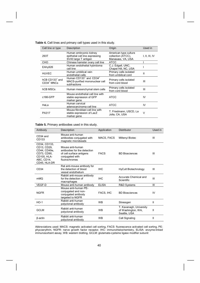
40
Table 4. Cell lines and primary cell types used in this study.
Cell line or type Description Origin Used in
293THuman embryonic kidneyepithelial cell line expressingSV40 large T antigen
American type culturecollection (ATCC),Manasass, VA, USA
I, II, III, IV
CHO Chinese hamster ovary cell line ATCC I
EAhy926 Human endothelial hybridomacell line
C.J. Edgell, UNC,Chapel Hill, NC, USA I
HUVEC Human umbilical veinendothelial cells
Primary cells isolatedfrom umbilical cord II
hCB CD133+ andCD34+ MNCs
Human CD133+ and CD34+
MACSpurified mononuclear cellsubfractions
Primary cells isolatedfrom cord blood III
hCB MSCs Human mesenchymal stem cells Primary cells isolatedfrom cord blood III
c166GFPMouse endothelial cell line withstable expression of GFPmarker gene
ATCC IV
HeLa Human cervicaladenocarcinoma cell line ATCC IV
PA317Mouse fibroblast cell line withstable expression of LacZmarker gene
T. Friedmann, USCD, LaJolla, CA, USA V
Table 5. Primary antibodies used in this study.
Antibody Description Application Distributor Used in
CD34 andCD133
Mouse antihumanantibodies conjugated withmagnetic microbeads
MACS, FACS Miltenyi Biotec III
CD34, CD133,CD13, CD29,CD44, CD49e,CD73, CD90,CD105, HLAABC, CD14,CD45, HLADR
Mouse antihumanantibodies for the detectionof cell surface antigensconjugated withfluorochromes
FACS BD Biosciences III
CD34Rat antimouse antibody forthe detection of bloodvessel endothelium
IHC HyCult Biotechnology III
mMQRabbit antimouse antibodyfor the detection ofmacrophages
IHC Accurate Chemical andScientific III
VEGFD Mouse antihuman antibody ELISA R&D Systems III
NGFR
Mouse antihuman PEconjugated and nonconjugated antibodytargeted to NGFR
FACS, IHC BD Biosciences IV
HO1 Rabbit antihumanpolyclonal antibody WB Stressgen II
GCLM Rabbit antihumanpolyclonal antibody WB
T. Kavanagh, Universityof Washington, WA,Seattle, USA
II
actin Rabbit antihumanpolyclonal antibody WB Cell Signaling II
Abbreviations used: MACS: magnetic activated cell sorting, FACS: fluorescence activated cell sorting, PE:phycoerythrin, NGFR: nerve growth factor receptor, IHC: immunohistochemistry, ELISA: enzymelinkedimmunosorbent assay, WB: western blotting, GCLM: glutamatecysteine ligase modifier subunit

41
Table 6. HIV1 lentiviral vectors and their properties used in this study.
Vector Promoter Transgene Additionalelements Origin Used
in
HIVrtTA2 SM2 CMV
Reversetetracyclinetransactivator rtTA2 SM2
J. Koponen,University ofKuopio,Finland
I
HIVTRELacZ
MinimalCMV withsevencopies ofTRE
LacZ markergene encoding for
galactosideJ. Koponen I
LVhNQO1Luc,LVhGCLMLuc,LVhGCLMHO1
AREelements (1or 2 copies)ofrespectivegenescombinedwithminimalSV40
Luciferase orheme oxygenase1
cPPT,WPRE
H. Hurttila,University ofKuopio,Finland
II
LVPGKGFP HumanPGK GFP marker gene cPPT,
WPRE
L.Naldini,IRCC, Turin,Italy
III
LVPGKVEGFD C
HumanPGK VEGFD C cPPT,
WPRE
P. Mäkinen,University ofKuopio,Finland
III
LVU6shGFP, LVH1shGFP, LVU6shGFPH1shGFP
Human U6or/and H1and PGK
small hairpin RNAtargeting GFP;shGFP, NGFRmarker gene
cPPT,WPRE P. Mäkinen IV
LVU6shGAPDH,LVH1shGAPDH,LVU6shGAPDHH1shGAPDH
Human U6or/and H1and PGK
small hairpin RNAtargeting mouseGAPDH;shGAPDH,
NGFR markergene
cPPT,WPRE P. Mäkinen IV
RRL VEGFD C CMV VEGFD C cPPT,WPRE P. Mäkinen up.
RRLLacZ CMV LacZ markergene
cPPT,WPRE P. Mäkinen up.
Abbreviations used: CMV: cytomegalovirus, TRE: tetracycline responsive element, hNQO1: humanNAD(P)H quinone oxidoreductase, hGCLM: human glutamatecysteine ligase modifier subunit, HO1:heme oxygenase 1, ARE: antioxidant response element, PGK: phosphoglycerate kinase, GFP: greenfluorescent protein, cPPT: central polypurine tract, WPRE: woodchuck hepatitis virus posttranscriptionalelement, NGFR: truncated form of nerve growth factor receptor, GAPDH: glyceraldehyde3phosphatedehydrogenase, up.: unpublished results

42
Table 7. PCRmethods used in this study.Target gene or region Application Used in
Betagalactosidase (lacZ) gPCR, RTPCR,qPCR
I, V, u.p.
Tetracycline reverse transactivator rtTA2 SM2 gPCR, RTPCR IhNQO1, hGCLM AREs PCR cloning IIhHO1 qPCR IIHuman chromosome 17 satellite region gPCR IIIHuman U6 and H1 promoter PCR cloning IVGreen fluorescent protein (GFP) qPCR IVRodent GAPDH qPCR IVMouse Oas1a, MCP1, VEGFA, PDGF qPCR IVEukaryotic ribosomal 18S RNA qPCR IV, VHuman VEGFD C RTPCR up.
Abbreviations used: gPCR: genomic PCR, RTPCR: reversetranscription PCR, qPCR: quantitative PCR,hNQO1: human NAD(P)H quinone oxidoreductase, hGCLM: human glutamatecysteine ligase modifiersubunit, ARE: antioxidant response element, hHO1: human heme oxygenase 1, Oas1a: 2’,5’oligoadenylate synthetase 1a, MCP1: monocyte chemoattractant protein1, VEGFA: vascular endothelialgrowth factor A, PDGF : platelet derived growth factor – , up: unpublished results
Table 8. Animal models used in this study.
Animal Model Used in n
Rat Intracerebral injection of lentiviral vector I 20
Mouse Hindlimb ischemia model, intramuscular injection of progenitorcells III 40
Intracerebral injection of lentiviral vector IV 28
Rabbit Intraluminal administration of lentiviral vector into carotidartery up. 4
Periadventitial administration of lentiviral vector into carotidartery 4
Intramuscular injection of lentiviral vector up. 9Pig Intramyocardial injection of lentiviral vector up. 3
Abbreviations used: n: number of animals, up: unpublished

43
Methods of assessing HIV1lentiviral vector efficacy in animalmodels of cardiovascular genetherapy (unpublished)
General methodology
Pilot animal studies to assess the efficacyof third generation lentiviral gene transfervector in previously established animalmodels are described below. For thesestudies RRLLacZ, RRLVEGFD C orLVPGKVEGFD C vector preparationswith viral titers of 7×1081×109 TU/ml wereused.
Intraluminal and periadventitialapproach for lentiviral vectoradministration into the rabbitcarotid artery
Intraluminal administration of the viralvector in to the rabbit carotid artery wasperformed as previously described(Gruchala et al., 2004). New ZealandWhite (NZW) rabbits (n=2) were used.Briefly, a segment of exposed carotidartery was isolated between atraumaticvascular clamps, and two cannulas wereintroduced into the vessel lumen throughproximal and distal arteriotomies. Thelumen of the vessel was flushed withsaline, and a volume of 100 l of RRLLacZ vector was administered into thevessel lumen and allowed to dwell for 20min. After the incubation, blood flow wasrestored. In a subgroup of rabbits (n=2),the carotid artery was mechanically injured4 days before gene transfer. Injury wasdone by introducing an angioplasty ballooncatheter into the artery and passing it threetimes to denude the endothelial layer anddisrupt the internal elastic lamina. Animalswere sacrificed 7 days after viral vectoradministration and artery samplescollected for lacZ gene expressiondetermination by Xgal, RTPCR and PCR.
Periadventitial administration of the viralvector was done with the extravascularmodel taking advantage of a silastic collarpositioned around the rabbit carotid artery(n=4) (Laitinen et al., 1997). Briefly, acollar (2 cm in length, Ark Therapeutics,Kuopio, Finland) was placed around theexposed carotid artery. The vector solution(300 l) was administered into the collarand tissue glue was used to seal the collar.The animals were sacrificed 10 (n=2) or 28(n=2) days after viral vector administrationand samples collected for lacZ geneexpression determination by Xgal, RTPCR and PCR.
Intramuscular injection of lentiviralvector in rabbit skeletal muscle
Intramuscular injection of the RRLLacZ LVin nonischemic NZW rabbitsemimembranosus thigh muscle (n=3) wasperformed as previously described(Rissanen et al., 2003a). A total volume of1 ml of vector stock was divided into 10separate injections. Seven days after theinjections, the animals were sacrificed andmuscle samples collected for lacZ geneexpression analysis by Xgal staining, PCRand RTPCR.
The angiogenic potency of RRLVEGFD C was studied by skeletal muscleinjections. Also, the potential gene transferefficiency enhancing effect of bupivacaine,a myotoxic local anaesthetic, was studiedby bupivacaine pretreatment of muscle (5mg/ml, 10 injections 0.1 ml each) 3 daysbefore vector injections. Lentiviral vectorsRRLLacZ (n=2, of which one rabbit waspretreated with bupivacaine) and RRLVEGFD C (n=4, of which 2 rabbits werepretreated with bupivacaine) wereinjected. Contrast enhanced ultrasoundimaging was used to asses muscleperfusion 7 and 21 days after vectoradministration. Animals were sacrificedafter 21 days and samples collected for X

44
gal staining, PCR and RTPCR. Toanalyse the angiogenic effect, sampleswere subjected to immunohistochemicalstaining of capillary vessels and vascularpermeability by the Miles assay aspreviously described (Rissanen et al.,2003a).
Intramyocardial injection oflentiviral vector in porcinemyocardium
The efficacy of RRLVEGFD C inporcine myocardium (n=3) was studied byutilizing a NOGA electromechanicalmapping and injection system offering apercutaneous route of intramyocardialinjection as previously described (Rutanenet al., 2004). Briefly, electromechanicalmapping of the left ventricle was performedwith the NOGA system and injectioncatheter. After mapping, ten myocardialinjections of the RRLVEGFD C vector(0.2 ml per injection, total volume 2 ml)were performed to the anterolateral wall.Six days after the injections, myocardialperfusion was studied with contrastechocardiography. The animals weresacrificed 14 days after vectoradministration and samples collected for aMiles permeability assay and histologicalanalyses.

45
RESULTS AND DISCUSSION
Doxycycline regulated lentiviralvector system (I)
A lentiviral vector system utilizing the revisedtetracycline transactivator, rtTA2SM2,showed a tight regulation of lacZ geneexpression with negligible basal level in thenoninduced stage. The gene expressionlevel could be adjusted by the dosing ofdoxycycline, and expression could berepeatedly switched on and off. Theseresults were obtained by in vitrocotransduction of two LVs, one encoding thertTA2SM2 and the other the lacZ markergene under the control of the tetracyclineresponsive promoter. By cotransducing CHOand EAhy926 cell lines at a low viralmultiplicity of infection (MOI 1 forHIV/TRE/LacZ and MOI 2 for HIV/rtTA2SM2), a 132 and 17 –fold induction in galactosidase activity, respectively, wasdetected. During longterm culture of cotransduced CHO cells, the lacZ gene couldbe repeatedly switched on and off by theaddition and withdrawal of doxycycline.Importantly, the induction level was dosedependent and full induction was achieved ata doxycycline concentration of 500 ng/ml,which can be obtained by oraladministration. LVs efficiently transduceseveral cell types of brain. Also, doxycyclineis known to penetrate the bloodbrain barrier.Therefore, as proof of the principle of the invivo functionality of the vector system,vectors were injected into the rat brain.Following cotransduction of viral vectors intothe rat striatum (n=20), RTPCR revealedthat doxycycline induced lacZ transcription in5 out of 7 rats and not in the 3 rats wheredoxycycline was not administred. Ten ratswere used for Xgal staining to study galactosidase activity in frozen brainsections. In all rats which receiveddoxycycline (n=5) Xgal positive cells weredetected. No positive cells were seen insamples from rats without doxycycline
treatment (n=5), further confirming thetightness of gene induction with our vectorsystem. DNA PCR confirmed the presenceof both vectors in the brain of every animal.However, this does not confirm thatcotransduction of both vectors targeted thesame cells.
The use of rtTA2SM2 for tight doxycyclinedependent regulation has also beenreported by others, for example, in thegeneration of conditionally transgenicanimals (Michalon et al., 2005, Katsantoni etal., 2007) and in in vivo gene transferapproaches with AAVvectors in mouse andnonhuman primate muscle and retina(Chenuaud et al., 2004, Stieger et al., 2006).However, doseresponse experiments havedemonstrated that tightness of control maybe partially lost at higher vector doses(Lamartina et al., 2002). These findingsstress the importance of specifying anoptimal vector dose for each application.
There are specific advantages achieved byintroducing the elements for doxycyclineregulated gene expression into two separatevectors. The first, is the ability to adjust theproportion of each transcriptional unit bysimply changing the ratio and amount ofvector particles applied for optimal generegulation. Secondly, in the two vectorsystem the transcriptional units are not inclose proximity and therefore, the regulatedpromoter is not disturbed by promoterinterference from an adjacent promoterconstitutively expressing the transactivator,an apparent disadvantage of the singlevector system (Bornkamm et al., 2005). Thetwo vector system enables the generation oftransgenic animals with induced transgeneexpression accomplished by first generatingseparate mouse lines that contain thetransactivator and target constructs, andthen producing double transgenic animals bybreeding. Since lentiviral vectors are provento be exceptionally powerful tools intransgenesis, our vector system shows the

46
potential to be exploited in the generation ofconditionally transgenic animals.
Despite the advantages of a twovectorsystem, there are numerous applicationswhere repeatable transfer of thetranscriptional units required for regulatedgene expression require the use of a singlevector. These include all in vivo genetransfer procedures. Vogel and coworkershave described a HIV1 based singlevectorsystem, in which the transcriptional unit forrtTA2SM2 has been inserted in the antisenseorientation and separated from theregulatable unit by the chicken globininsulator (Vogel et al., 2004). The authorsreport transgene expression regulation in therat striatum over two orders of magnitude bythe addition or withdrawal of doxycycline.The latest and most advanced versions ofthe Teton system where all components areon a single vector combine the rtTA2SM2with the use of a transrepressing factor,doxycyclinedependent transsilencer calledtTS. tTS is a fusion protein consisting of theKRAB (KruppelAssociated Box)transrepressing domain of the human Kid1protein fused to the wildtype TetR (Salucciet al., 2002, Lamartina et al., 2003,Epanchintsev et al., 2006). The KRABdomain can induce transcriptional silencingto within 3 kb of its binding site. In theabsence of doxycycline, tTS binds to thetetO target sequence and inhibits basaltranscription. As doxycycline is added, thetTS dissociates allowing rtTA2SM2 to bindand trigger activation. This design ispreferred in singlevector applications sincethe transrepressing factor, that ensures theinactivity of the regulated promoter in the offstate, abolishes any potential promoterinterference. It may also reduce interferencefrom genomic elements which come intoproximity after integration when integratingvectors are used to deliver the regulatorysystem. However, a disadvantage of usingthe tTS means the inclusion of an additionaltranscriptional unit, which complicates the
vector system and increases the size ofgenomic material to be inserted into thetransfer vectors.
Overall, particularly from a clinical point ofview, the doxycycline system has a clearpharmacological advantage over other generegulation systems like mifepristone orrapamycin. Namely, that doxycycline has ahalflife of 1422 h, has excellent tissuepenetration due to its lipophilic nature and iswell tolerated. The most serious side effect isa dosedependent photosensitivity that mayoccur in 13% of patients after prolongedadministration (Klein et al., 2001). The latestversions of the Teton system can be fullyinduced in rodents and monkeys bydoxycycline at doses comparable to thosecommonly used for antibiotic therapy(Lamartina et al., 2003). The major concernwith respect to the use of an antibioticinduced regulatory system is the possibilityof raising resistance to the antibiotic itself.Although tetracycline derivatives are lessused than in the past, they may stillrepresent the treatment of choice for certaininfectious diseases such as Lyme disease(Klein et al., 2001). Thus, the generation ofTetsystem compatible, nonantibioticdoxycycline analogs would be desirable.
Oxidative stress inducible lentiviralvector (II)
We studied if the antioxidant responseelement (ARE), a cisacting sequence foundin the promoters of the genes induced byoxidative stress, can be utilized for geneinduction in a transfer vector. The AREsequences of three genes, human NADPHquinine oxidoreductase 1 (NQO1), humanglutamatecysteine ligase modifier subunit(GCLM) and mouse heme oxygenase 1 (HO1), were subcloned into a reporter plasmidvector in combination with one, two or threecopies of the ARE sequence, upstream ofthe minimal promoter. After plasmidtransfections, oxidative stress was mimicked

47
by the addition of a chemical, either diethylmaleate (DEM) or 15deoxy12,14prostaglandin J2 (15dPGJ2), and luciferaseactivity of the cell cultures was analysed. Inall constructs, luciferase activity was inducedupon chemical induction of oxidative stress.However, there was some basal expressionof luciferase without induction and theexpression increased in relation to thenumber of ARE elements in the constructs.The basal expression level could be due toreactive oxygen species and oxidativeproducts of macromolecules produced bynormal cell respiration and thus cannot beavoided. Also, it should be noted that theoxygen levels in cell culture conditions (21%)are much higher than the naturally occurringlevels in most tissues (Roy et al., 2003), andit is likely that basal AREactivity would belower in an in vivo situation. In our study, formore efficient and longlasting expression,the reporter constructs with one and twocopies of AREs from hNQO1 or hGCLMgenes were cloned into LVs and theirefficacy for oxidative stress induced geneexpression studied in human umbilical veinendothelial cells (HUVECs). Endothelial cellsrepresent a potential target for antioxidantgene therapies (Levonen et al., in press) andwere therefore selected for these studies.Upon chemical induction of oxidative stress,a clear induction of luciferase activity wasseen with all lentiviral constructs. WhenHUVECs were transduced at the MOI of 5,the highest induction in luciferase activity(nine fold activity compared to the uninducedstate) was seen with a construct carrying twocopies of hGCLM ARE. Thus, this vectorwas used for the following studies. When theluciferase reporter gene was replaced withthe potential therapeutic gene, hemeoxygenase 1 (HO1), a 10fold induction ofgene expression was seen. This is asignificant result, as the most advancedhypoxiainduced vector system yield sevenfold induction in gene expression uponhypoxia in cell culture (Tang et al., 2005). Inaddition, we used TNF to mimic vascular
inflammation in HUVECs and studied theeffect of AREinduced HO1 therapeuticgene expression on the expression of theproinflammatory factor, VCAM1. Our resultsshowed that ARE induced HO1 geneexpression was able to decrease theexpression of VCAM1 and thus, the systemholds potential for the therapy of oxidativestress.
Based on our results the genetic AREelements, induced cytoprotective genes byoxidative stress, and can be exploited ingene transfer vectors. Since oxidative stresscan not be completely mimicked in cellculture conditions, the true performance ofour ARE vectors need to be defined inanimal models. Considering the lentiviralvector tropism for brain tissue, anexperimental model of stroke might beoptimal for these studies. Also, for thepromotion of cell survival in oxidativeconditions in progenitor cell therapyapproaches, our lentiviral ARE constructsmay be readily applicable. As oxidativestress plays a role in atherosclerosis,restenosis and ischemiareperfusion injury ofmyocardium and skeletal muscle, the AREinduced expression of therapeutic genesusing AAV vector, which has proved to bemore effective at the transduction of thesetissues, is an appealing concept.
The performance of lentiviral HIV1derived vector in cardiovasculargene therapy applications(unpublished data)
Most of the previous gene therapyapplications for cardiovascular diseaseshave used adenoviruses. Despite theirefficiency, only shortterm transgeneexpression is achieved; maximal proteinexpression is achieved after four days andthis declines to below detection level aftertwo weeks in most applications. Severalcardiovascular applications would benefitfrom longterm therapeutic gene expression.

48
Therefore, we tested the efficiency of theHIV1 derived, third generation selfinactivating LV to transduce skeletal muscle,blood vessel and myocardium and mediate atherapeutic effect. The results of these pilotstudies are summarized in Table 9.
The intraluminal approach may be performedsimultaneously with angioplasty and is apotential choice for the administration ofvectors for gene therapy of restenosis. Westudied the efficacy of LV in the blood vesselby intraluminal administration in a rabbitmodel (Gruchala et al., 2004). In a subgroupof rabbits, mechanical injury by ballooncatheter was performed simultaneously todisrupt the endothelial cell layer and theinternal elastic lamina of the blood vessel inorder to model balloon angioplastyrevascularization procedure. As a result, noLacZ gene expression was detected by Xgalstaining or RTPCR in arterial samples ofrabbits sacrificed seven days after vectoradministration into the lumen of intactarteries, whereas in the samples of denudedarteries LacZ gene expression could bedetected with RTPCR. However, galactosidase positive cells were notdetectable by Xgal staining, suggesting alow level of transgene expression onlydetectable with the sensitive RTPCRmethod. LacZ expression present only inarteries preconditioned with balloon
denudation may be due to the abolishmentof the endothelial cell layer or, inducedproliferation of endothelial and smoothmuscle cells due to injury, thus facilitatingthe viral transduction. In intraluminal genetransfer of the rabbit carotid artery, the useof AAV and adenoviral vectors have resultedin effective transduction (Gruchala et al.,2004). AAV vectors showed efficienttransduction of smooth muscle cells lastingfor at least 100 days and therefore, can beregarded as more potent arterial genetransfer vehicles than LVs for prolongedtherapeutic gene expression.
To study whether adventitial exposure of LVwould result in a significant gene transferefficiency into the blood vessel, we assesseda model of periadventitial administration ofvector making use of a silastic collar placedaround the rabbit carotid artery (Laitinen etal., 1997). Ten days after LV administration,transgene expression could be detected withRTPCR, while Xgal staining did not showthe presence of positive cells suggesting avery low efficacy of gene transfer. 28 daysafter LV administration, LacZ expression wasno longer detectable by RTPCR. Based onthese results, we concluded LV is not anideal vector for periadventitial arterial genetransfer requiring longterm transgeneexpression.

49
Tabl
e 9.
Pilo
t stu
dies
for t
he e
valu
atio
n of
LV
gene
tran
sfer
effi
cacy
for c
ardi
ovas
cula
r gen
e th
erap
y
Anim
alM
odel
Vect
orn
Follo
w
up (day
s)
Anal
ysis
met
hods
and
resu
lts fo
r LV
gene
tran
sfer
Car
otid
arte
ry, i
ntra
lum
inal
adm
inis
tratio
n (G
ruch
ala
et a
l 200
4)R
RL
LacZ
2 7
Xga
l sta
inin
g ()
, RT
PCR
()
Car
otid
arte
ry, i
ntra
lum
inal
adm
inis
tratio
n,pr
econ
ditio
ned
with
bal
loon
ang
iopl
asty
(Gru
chal
aet
al 2
004)
RR
LLa
cZ2
7X
gal s
tain
ing
(), R
TPC
R (+
)
Car
otid
arte
ry, p
eria
dven
tital
adm
inis
tratio
n w
ithsi
last
ic c
olla
r (La
itine
net
al 1
997)
RR
LLa
cZ2 2
10 28X
gal s
tain
ing
(), R
TPC
R (+
)X
gal s
tain
ing
(), R
TPC
R (
)
Skel
etal
mus
cle,
intra
mus
cula
r inj
ectio
n (R
issa
nen
et a
l 200
3)
RR
LLa
cZ
RR
LVE
GF
D
3 4 2
7 21 21
Xga
l sta
inin
g ()
, RT
PCR
(+),
PCR
(+)
Xga
l sta
inin
g ()
, RT
PCR
(),
PCR
(),
perfu
sion
()
RT
PCR
(),
PCR
(),
Mile
s as
say
(),
perfu
sion
(),
IHC
()
Rab
bit
Skel
etal
mus
cle,
intra
mus
cula
r inj
ectio
n,pr
econ
ditio
ned
with
bup
ivac
aine
(Ris
sane
net
al
2003
)
RR
LLa
cZ
RR
LVE
GF
D
1 2
21 21
Xga
l sta
inin
g ()
, RT
PCR
(),
PCR
(),
perfu
sion
()
RT
PCR
(),
PCR
(),
Mile
s as
say
(),
perfu
sion
(),
IHC
()
Pig
Intra
myo
card
ial i
njec
tion
(Rut
anen
et a
l 200
4)R
RL
VEG
FD
3
14pe
rfusi
on (
), M
iles
assa
y ()
, IH
C (
)
Abbr
evia
tions
use
d in
the
tabl
e: L
acZ:
g
alac
tosi
dase
, VEG
FD
: vas
cula
r end
othe
lial g
row
th fa
ctor
D, R
TPC
R: r
ever
se tr
ansc
riptio
n po
lym
eras
ech
ain
reac
tion,
IHC
: im
mun
ohis
toch
emis
try

50
The efficacy of LV gene transfer was studiedin rabbit skeletal muscle to assess itspotential to be utilized in longterm genetherapy applications in a rabbit model of hindlimb ischemia. Seven days afterintramuscular injections, LacZ geneexpression and LV vector presence could bedetected by RTPCR or genomic PCR.However, in tissue sections, transgeneexpression was not detectable by Xgalstaining. Three weeks after LV injections,transgene expression or vector presencecould not be detected with RTPCR orgenomic PCR. Also, in animals injected withLVVEGFD C, the effects typical for thisgrowth factor could not be detected,including enhanced blood perfusionassessed with ultrasound, increasedextravasated plasma proteins from newlyformed vessels within the tissue measuredby Miles assay or, promoted capillary vesselformation detected by immunohistochemistry(Rissanen et al., 2003b). In a subgroup ofanimals, we studied if LV transfer efficiencycould be increased by musclepreconditioning with injections of localanaesthetic, bupivacaine, which isconsidered myotoxic and therefore mightenhance proliferative activity of muscle cells(Wells, 1993). As studied 21 days after LVinjections, bupivacaine pretreatment ofmuscle was not seen to promote LVtransduction.
In our hands, LV mediated gene transfer inrabbit models of cardiovascular gene therapydid not show potency. Supporting ourfinding, there are reports suggesting thatrabbit cells are to some extent resistant totransduction by HIV1 derived, VSVGpseudotyped LVs in vitro (Hofmann et al.,1999, Ikeda et al., 2002). According to theresults of Hofmann et al, human, hamsterand pig cell lines are most prone to HIV1 LVtransduction, while mouse, rat and rabbitcells lines are less efficiently transduced,listed in order of reducing transductionefficiency. This is in line with our own
observations that human primary cells andcell lines are very efficiently transduced invitro even with low multiplicities of infection(MOI) of the viral vector, while mouse, ratand rabbit cell lines require higher MOIs forefficient transduction (Koponen JK andMäkinen P, unpublished data). Theseobservations may be explained by speciesspecific host cellular factors that limit thepostentry events of the viral vector, such asthe nuclear translocation (Hofmann et al.,1999). Based on the in vitro results showingHIV1 being more prone to the transductionof human cell lines, it is impossible to predictif LVs could mediate efficient gene transfer inhuman tissues.
The myocardium is an important targettissue for cardiovascular gene therapyincluding many applications where sustainedexpression of the therapeutic gene would beessential. We studied the efficacy of LV inpig myocardium by the intramyocardialapproach. We assessed a gene transfervector injection technique readily applicablefor clinical use, the NOGAcatheter assistedinjections with electromechanical mapping ofthe myocardium (Rutanen et al., 2004). Afterinjections of LV VEGFD C we searchedfor the effects of the growth factor previouslyseen in both skeletal muscle (Rissanen etal., 2003b) and myocardium (Rutanen et al.,2004). We were unable to see VEGFD C
mediated effect by ultrasound measurementof blood perfusion (6 days after injections),or by Miles assay for the detection ofextravasated proteins from newly formedcapillary vessel or induced capillaryangiogenesis in immunohistochemicalstainings (14 days after the injections).Therefore, we concluded LV gene transferinsufficient to trigger therapeutic effect in pigmyocardium.
As the pilot animal studies with LVs,performed in our previously establishedanimal models of cardiovascular genetherapy, and described in this thesis,

51
resulted in no positive LV transduced cells(as detected by histological methods) andwas inefficient in terms of the effect oftransgene expression, we concluded that thegene transfer efficiency was below what isrequired for a therapeutic effect. In fact, atleast in their current form, LVs seemcomparatively poor at delivering genes intothe liver and muscle. Despite someencouraging results originally obtained withHIV1 LVs (Kafri et al., 1997), furtherexperiments have generally revealed lowefficacies, at least in adult animals. In aprevious study from our research group, amodest LV gene transfer efficiency of lessthan 0.01% of the liver cells has beenreported in a rabbit model of FH (Kankkonenet al., 2004). However, in this model even alow level of LDL receptor gene transfer wassufficient to produce a longterm serumcholesterol reducing effect probably due tothe fact that even a low number of cells witha functional LDL receptor are adequate forsuch an effect. In terms of gene transferefficiency, it appears that adenoviral andAAV vectors are better suited for therapiestargeted to the liver or muscle, which seemsto apply to blood vessel as well (Gruchala etal., 2004). Nevertheless, LVs still showexcellent performance in gene delivery intothe central nervous system, retinal cells andinto the cells of lymphohematopoieticsystem. In relation to cardiovascular genetherapy, the ability of LVs to transduce HSCsmay hold therapeutic potential. For example,ex vivo genetic manipulation of progenitorcells of the monocyte lineage could beutilized to modify the effect of macrophagesin the development of atherosclerotic lesionsor restenosis or, to take advantage of therecruitment of macrophages into areas ofpathology where they could express thetherapeutic protein. Also, geneticmodification of stem and progenitor cells forcombined gene and cell therapy forcardiovascular diseases like myocardialischemia is a fascinating prospect for which
HIV1 LV would be a highly potent vector ofchoice.
Cord bloodderived progenitor cellsin a mouse model for skeletalmuscle ischemia (III)
Progenitor cell therapy is a novel approachwhich may be utilized for the treatment ofischemia in the myocardium and skeletalmuscle. Combining gene therapy with celltherapy applications allows for themodification of progenitor cell properties,such as cytokine release, surface receptorexpression and factors potentially promotingcell survival in conditions like ischemia oroxidative stress. LVs have enabled thegenetic modification of HSCs and embryonicstem cells that were nonpermissive to earliervectors, especially in the quiescent state.Cytokines, for example, stem cell factor andinterleukins, may be used to stimulate HSCsto proliferate and facilitate transduction.However, as it is important to precludeunwanted differentiation of the cells bycytokine induction, an efficient transductionmethod for unstimulated cells is essential. Ithas been reported that cytokine pretreatment of human CD34+ cells reducestheir longterm engraftment ability in miceindicating a defect in cell survival or homingto bone marrow (Ahmed et al., 2004, WulfGoldenberg et al., 2007). In our study, weassessed whether cord blood derived HSCsubfractions of CD34+ and CD133+ cells canbe efficiently transduced by LV without theneed for cytokine induced proliferation. At aLV MOI of 100, we achieved comparabletransduction efficiencies with and withoutcytokine induction of both CD34+ (57.0% and52.5%, respectively) and CD133+ (53.0%and 52.5%) cell subfractions as judged fromFACS detecting GFP marker genefluorescence. Also, we used cell cycleanalysis to confirm that cytokines inducedcell cycling in HSCs (data not shown). In themajority of published reports utilizing LVgene transfer into HSCs, cytokine stimulation

52
has been used. However, to excludecytokine induced effects, optimaltransduction conditions requiring minimalmodifications of the HSCs should bedetermined for each application. By a colonyforming assay, we were able to show thatthe ability of CD34+ and CD133+ cells todifferentiate into cells of the hematopoieticlineage was not altered by LV transduction,suggesting that their progenitor capacity wasnot affected. Also, we were able to efficientlytransduce MSCs and as detected byadipogenic and osteogenic differentiationassays, the transduced MSCs did retain theirdifferentiation ability.
Cord blood (CB) is an excellent source ofprogenitor cells in terms of a highpercentage of stem cells and the presence ofimmature HSCs derived from a youngindividual. We assessed the therapeuticpotential of two subpopulations of progenitorcells, CD133+ and MSCs, in a nude mousemodel of hind limb ischemia. Cells weretransduced with a LV vector encoding theGFP marker gene or the VEGFD C growthfactor gene. Due to the more primitive natureof CD133+ compared to CD34+ cells(Hemmoranta et al., 2006), the CD133+
subpopulation was selected to be tested inthe animal model. Also, since CD133+
antigen expression has been connected toendothelial progenitor cells (Gehling et al.,2000), and CB derived CD133+ cells havebeen reported to be capable of differentiationinto endothelial and cardiomyocytelike cellsin vitro (Bonanno et al., 2007), we wanted tosee if these cells could incorporate intonewly forming vessels or regeneratingmuscle. MSCs have been shown to becapable of multilineage differentiation, forexample into skeletal muscle cells (Pittengeret al., 1999) and therefore, might havetherapeutical potential in the regenerationprocess of ischemic muscle.
Because of the contradictory resultspublished earlier, mostly concerning cell
therapy of myocardial ischemia, we alsoanalysed whether progenitor cell therapymight mediate a therapeutic effect withoutcells directly participating in the regenerationprocess. As a model of progenitor cellmediated gene therapy, we also assessedwhether CB cell subfractions expressingVEGFD C could mediate an angiogeniceffect, previously shown to be induced bythis particular growth factor (Rissanen et al.,2003b, Rutanen et al., 2004, Kholova et al.,2007). In our model, we injected progenitorcell suspensions (total cell number ~1×10⁵)intramuscularly into the left hindlimb musclesof nude mice, for which unilateral hind limbischemia was induced by femoral arteryligation immediately before injections.Animals were sacrificed 3, 7 or 21 days afterthe operation. We were unable to show thepresence of progenitor cells byimmunohistochemistry at any of the timepoints, as studied by GFP or human cellmitochondrial or cytoskeleton specificantibodies in immunohistochemistry (datanot shown). By human chromosome 17 satellite region specific PCR, we were ableto show the presence of human geneticmaterial in the injected muscles three daysafter injection. However, seven days postinjection human DNA was no longer present.Also, PCRbased detection does not indicateif the cells are alive. Our result is in line withthe previous observation by Tateno et al.,who have shown that mouse bone marrow derived mononuclear cells disappeared frommouse ischemic muscle three days afterimplantation (Tateno et al., 2006). It isunknown, whether these observations aredue to the general inability of progenitor cellsto engraft or are attributed to ischemicconditions of the muscle, presumably limitingcell survival. By counting capillary vessels,we did not observe enhanced capillaryangiogenesis in mice groups injected withCD133+ cells or MSCs transduced with LVVEGFD C or LVGFP, when compared tothe control group. Since the typical effect ofVEGFD C was not seen, we concluded

53
that the injected cells did not secrete enoughgrowth factor to induce therapeuticangiogenesis. This might be due to a tooshort survival time of the cells in theischemic muscle. The direct incorporation ofbone marrow derived progenitor cells intonew or remodelling blood vessels in theischemic myocardium and skeletal musclehas been previously reported. Themagnitude of cellular incorporation highlyvaries between studies. Although a highoccurrence of capillaries containingtransplanted cells has been depicted insome studies (Asahara et al., 1997, Kocheret al., 2001, Kawamoto et al., 2003), only asingle transplanted cell in the circumferenceof the capillary vessel is required for thevessel to be counted as positive. This maylimit the interpretation of the true effect of cellincorporation into structures of a vascularbead. Furthermore, some studies havereported only small numbers of positivevessels, despite impressive improvements inblood perfusion (Tomita et al., 1999, Wanget al., 2001, Iba et al., 2002), suggesting theexistence of alternative mechanisms to thedirect cellular incorporation mediating theeffects.
In our study, we observed increasedregeneration of ischemic muscle byprogenitor cell injections (regeneratingmuscle area 86.7% 93.8% in progenitor cellinjected groups versus 73.2% in the controlgroup) as measured from the histologysections 21 days after the operation. Ourresults suggest that both CB CD133+ andMSC –progenitor cells increase theregeneration capacity of ischemic muscle bya mechanism unrelated to progenitor cellengraftment. As an alternative mechanismfor progenitor cell mediated therapeuticresponse in ischemic skeletal muscle, aparacrine effect has been suggested(Kinnaird et al., 2004, Ziegelhoeffer et al.,2004, Tateno et al., 2006). This might bemediated either by the factors released bythe progenitor cells themselves or by tissue
resident cells stimulated by the progenitorcells. Immunohistochemical staining formacrophages showed a higher count ofinfiltrated macrophages in the progenitor cellinjected muscles. In fact, impairedmacrophage recruitment has been reportedto cause delayed recovery of hind limbfunction, increased muscle atrophy andfibrosis in a hind limb ischemia model inCD4knockout mice (Stabile et al., 2003).Therefore, the regenerative effect ofprogenitor cell injection might be explainedby their attraction of monocytes resulting in ahigher number of infiltrating macrophages,which might further stimulate regenerationfor example by the release of cytokines.However, as we studied the effects of humanCB cells in mouse skeletal muscle ischemia,an immune deficient nude mouse model wasrequired, limiting the interpretation of issuesconcerning inflammatory mechanisms.
Overall, the mechanisms of progenitor cellmediated response in ischemic myocardiumand skeletal muscle require furtherevaluation. Therefore, new experimentalstudies are warranted. Clinical trials havebeen focused mostly on myocardial ischemia(Wollert et al., 2005) rather than skeletalmuscle ischemia (TateishiYuyama et al.,2002a, Ishida et al., 2005). Nevertheless,skeletal muscle ischemia might be a morepotent candidate for progenitor celltherapies, since there is no need forelectrical coupling of engrafting cells andbecause of the presumed easieradministration of the cell preparation in theskeletal muscle compared to themyocardium. Also, the most potent subtypesand sources of progenitor cells need to beidentified. CB is enriched with HSCs ascompared to other sources like bone marrowor peripheral blood. A worldwide CBbanking system provides rapid supply toHLAtyped CB tested for infectious agents asthe material can be shipped easily anywhere(Brunstein et al., 2006). This makes cordblood an optimal source for progenitor cell

54
preparations for clinical use. Thus, thepotential of CB derived cells in differenttherapies is worth studying.
A lentiviral vector for gene silencingby RNAi (IV)
A wide range of experimental andtherapeutic applications would benefit fromsustained RNAimediated gene silencing,which is not achieved by introducingsynthetic siRNA oligonucleotides. In thesesituations shRNAmediated silencingdelivered into a target cell within a transfervector is applicable. However, specificconstructs are required for each application.Pol III promoters have welldefinedtranscriptional start sites and they produceRNA transcripts lacking a polyadenylationtail and are therefore ideally suited for theproduction of shRNAs. We compared theefficiency of two widely used Pol IIIpromoters, the human U6 small nuclearpromoter and the human H1 promoter fortheir effects on gene silencing after LVmediated transfer of a shRNA against GFP.We chose the marker gene GFP as a targetgene because it is easily detected by FACSand fluorescence microscopy. Also, cell lineswith stable GFP expression, such as themouse endothelial cell line c166GFP, andGFP transgenic mice are commerciallyavailable. Our results showed that by LVmediated transduction, the U6 promoter ismore powerful than the H1 promoter for GFPmarker gene expression silencing in vitro ina GFP expressing endothelial cell line asjudged at the protein level by FACS (94%and 86% silencing for U6 and H1,respectively) and at the RNA level byquantitative RTPCR (88% and 71%). Similarresults were obtained in vivo, in mouse brainas revealed by immunohistochemistry. Also,the U6 promoter was found to be moreeffective than the H1 promoter in thesilencing of the GAPDH housekeeping geneas studied by RTqPCR (35% and 22%).Compared to the silencing effect of the U6
promoter driven vectors, we did not findenhanced silencing of GFP or GAPDH by avector with two shRNA cassettes driven byboth promoters. This might be due tomaximal loading of the small RNAguidedgenesilencing pathway in the transducedcells achieved by strong transcription fromthe U6 promoter. The fact that we reachedfar more efficient maximal silencing of GFPthan GAPDH gene transcription (88% versus35%) might be due to tight regulation andhigher expression levels of the GAPDHhousekeeping gene and more likely, a lessefficient RNAi target sequence. By assessingviral vector copy number in the genome oftransduced cells by southern blot we wereable to confirm the more powerful effect ofthe U6 LVs compared to the H1 LVs.Namely, in comparison to a single copy of aU6 LV, a higher copy number of H1 LV (510copies) was required for an equal 80%silencing effect. The powerful knockdowneffect obtained with a single integrated copyof U6 LV suggests that our vector design hasthe potential for in vivo applications andRNAi applications in cells which are noteasily transduced in vitro, including stem andprogenitor cells and thus, has theapplicability in the generation of transgenicmice.
We also studied longterm gene silencingmediated by LVs in vitro and in vivo. Sincethe silencing of the GFP marker gene doesnot lead to a loss of function effect, it is apotential target for studying longtermsilencing. Our in vitro results showed thatwith all constructs, silencing of GFP lastedup to 28 weeks and remained at constantlevel, as assessed by FACS. Further,stereotactic injection of LVU6shGFP or LVH1shGFP vectors into the brain of GFPtransgenic mice showed GFP gene silencingfor up to 9 months as studied byfluorescence microscopy of frozen brainsections. The merged images of GFPfluorescence and immunohistochemicalstainings with a fluorescent signal to detect

55
marker gene NGFR expression showed thecolocalization of diminished GFPfluorescence and the expression of NGFR,further confirming vectormediated GFPsilencing. This suggests the usefulness ofour vector in the field of brain research, forexample, in developing either models ortherapeutic tools for neurodegenerativediseases.
Originally, siRNA induced gene silencingwas introduced as a mechanism which canbe utilized without inducing a type 1interferon response when dsRNA sequencesshorter than 30 bp are used (Elbashir et al.,2001). However, it has been shown thatdsRNA molecules mediating RNAi maytrigger an interferon response by alternativepathways, like via Tolllike receptors (Seth etal., 2006). Therefore, the possibility ofevoking an interferon response needs to beevaluated for each application. In our study,we used RTqPCR to study the potentialinduction of the mouse Oas1a gene, whoseexpression is upregulated by an interferonresponse. We found that LVmediated RNAidid not induce Oas1a gene expression andthus did not evoke an interferon response.RNAi induced offtarget effects also need tobe assessed for each application. For this,we analyzed the expression levels of MCP1,VEGFA and PDGFR genes, which are allexpressed in endothelial cells. We foundthem unaffected by shRNA expressing LVtransductions. Despite the limited geneexpression array analysis, the unchangedexpression of these representative genessuggests a lack of widespread offtargeteffects by our vector design.
LVmediated shRNA delivery has beensuccessfully applied by others in variousapplications in vitro (Li et al., 2005a), in vivo(Raoul et al., 2005b, Pfeifer et al., 2006) andex vivo (Anderson et al., 2007, Ohmori et al.,2007) and also, in the generation of knockout mice (Tiscornia et al., 2003). Combiningthe efficient LVmediated generation of
transgenic mice with the shRNA approachresults in a feasible tool. In fact, we havealso used our U6 driven shRNA LVs in thegeneration of knockdown mice, and theanalysis of the generated mouse lines iscurrently in progress (Hämäläinen et al,unpublished data).
To date, the advantages of utilizing Pol IIIpromoters in inducing RNAi have beenclearly demonstrated. However, one majorlimitation of Pol III promoters is their lack oftissue specificity. Therefore, Pol II promotershave also been utilized in RNAi vectors. ThePol II based systems mimic miRNAbasedsilencing (Zeng et al., 2005), meaning thatthe transcript needs additional processing bythe cellular enzyme Drosha to produce ashRNA before Dicermediated processing toyield siRNAs. Because this system mimicsthe natural miRNA pathway, it might proveadvantageous. However, it adds a level ofcomplexity which may be detrimental insome settings. In addition, Pol II based RNAisystems may directly utilize regulatorysystems, such as the tetracycline systems.In fact, several systems have beendeveloped for both Pol II and Pol III basedRNAi regulation, either reversibly bydoxycycline or irreversibly by CreLox basedsystems (reviewed in Wiznerowicz et al.,2006). Conditional knockdown approachesare useful for therapeutic applications thatrequire longterm silencing, fordevelopmental research, when the effect ofknockdown of lethal genes is desired or, formodelling human pathologies in animalmodels, that is to induce and revert theshutoff of a disease affecting gene, such asan oncogene or tumor suppressor gene.Although using Pol II promoters for RNAi hascertain advantages, a more systematiccomparison of the levels of RNAi obtainedwith Pol IIIdriven shRNAs versus Pol IIdriven miRNAbased shRNA is needed.Finally, it is likely that for RNAi applicationsranging from screening of shRNA libraries to

56
genebased therapies, both systems may beuseful.
A realtime quantitative PCR approach for the analysis of lacZmarker gene expression (V)
Quantitative reverse transcription PCR (qRTPCR) is a method for quantitativedetermination of gene expression. The realtime method, a method monitoring the PCRreaction in the thermal cycler as itprogresses, has been extensively utilizedsince first discovered (Gibson et al., 1996).The realtime method utilizes 5’ exonucleaseactivity of TaqDNA polymerase enzymethrough the use of sequencespecificfluorogenic hydrolysis (TaqMan®chemistry).During the amplification stage of the PCRcycle, a probe that is specific to the amplifiedregion will bind, and as the DNApolymerasesynthesizes the complementary strand, its5’exonuclease activity degrades the boundprobe. As a result, once the fluorescentmolecules, that are tagged to the 5’end ofthe probe are released from the closeproximity of the 3’end fluorescent quenchingmolecules, a fluorescence signal isgenerated. This signal fluorescence can bemeasured and it is proportional to theamplified product amount, and thus, relativeto the amount of starting material. qPCR orqRTPCR methods taking advantage of thesequence specific probe are sensitive,specific and enable quantitativedetermination of traces of sample DNA orvery low levels of target gene expression.
We assessed whether a realtime qRTPCRmethod utilizing TaqMan®chemistry wouldbe suitable for the quantification of LacZmarker gene expression. This method wasintended for determining the LacZ geneexpression level in our doxycycline regulatedLVconstructs and for assaying gene transferefficiency and biodistribution of various viralvectors in animal models. However, despitethe appropriate design of primers and probe,
qRTPCR analysis constantly resulted in astrong amplification signal from the controlswhich did not include any sample. Further,we did not observe a signal in the negativecontrols for 18S RNA amplification, anendogenous control used to normalize theresults. Since the possibility of externalcontamination of reagents was also ruled outby testing several aliquots, we reasoned thatthe amplified genetic material was originatingfrom the commercial reagents used. TheLacZ gene, encoding for the bacterial galactosidase enzyme, origins from E.coli.Unfortunately, there is contaminatingbacterial DNA present in the Taq polymerase(Bottger, 1990, Rand et al., 1990), whichgave rise to the strong amplification signal inour qRTPCR method. The enzyme(AmpliTaq Gold DNA polymerase®) iscommercially synthesized in E.coli as arecombinant protein, and the incompletepurification of the enzyme results in traces ofbacterial DNA in the preparation. In ourhands, the amplification of the controlsample without sample resulted in a strongamplification signal with a relatively lowthreshold cycle (Ct=32), indicating aremarkable amount of contaminatingsequence. The resultant amplification of thenegative control could not be ruled out bymathematical elimination, since theamplification of low concentrations of targetDNA or RNA would presumably be maskedby the contamination at this level. Therefore,we concluded that our method was notapplicable for the intended use. Also, if thedetection of other bacterial genes isperformed with a comparable method,adequate controls should be included ineach run to rule out amplification originatingfrom the DNA polymerase. Today,commercial DNA polymerase enzymes ofgreater purity are available. In fact, Tondeuret al reported that the qRTPCR method forLacZ gene expression designed by us canbe successfully utilized by using apolymerase of higher purity, AmpliTaq GoldDNA polymerase low DNA®, or alternatively,

57
by treating the standard AmpliTaq Gold DNApolymerase® with DNAse enzyme prior touse (Tondeur et al., 2004). This observationenables the use of our qRTPCR design forthe quantification of LacZ gene expressionand detection of the LacZ encoding vectorDNA.
CONCLUSIONS AND FUTUREPERSPECTIVES
Studies included in this thesis indicate theversatile applicability of HIV1 derived LVs.In our studies, third generation LVs weresuccessfully used as a platform for regulatedgene expression induced by the orallyavailable drug, doxycycline or anendogenous pathophysiological stimulus,oxidative stress. The SINdesign of thirdgeneration LVs, whereby after vectorintegration into host cell genome nopromoter activities are left in the LTR regionsflanking the integrated sequence, means thatpossible promoter interference with theregulated promoter is abolished and, may beone key factor in successful transgeneregulation. In fact, inactive LTRs flanking thetransgene cassette might also operate asgenomic insulators and hinder theinterference of genomic promoters near theintegration site with transgene promoterregulation. Also, LVs exploited for RNAiresulted in powerful, longterm genesilencing. The in vivo performance of LV incardiovascular disease gene therapymodels, whereby the rabbit skeletal muscleand blood vessel and pig myocardium wereused as target tissues, did not prove to beefficient. However, as shown by us andothers, LVs are valued because of their hightransduction efficiency into the stem andprogenitor cells such as HCSs and ESCsand are thus, excellent candidates for genetransfer vectors in progenitor cell therapiesand the generation of transgenic animals. Inour studies, we also took advantage of thehigh transduction efficiency of LVs into thebrain tissue. In their current form, LVs
represent excellent tools for longtermgenetic modification of the cells of thelymphohematopoietic and central nervoussystem. From the viewpoint of experimentalresearch, third generation HIV1 LVs areeasy to produce by a simple cotransductionprotocol of four separate plasmids. However,for the clinical use of LVs, a stableproduction of vectors would be desirable.Safety issues regarding the use of HIV1 LVsin clinical treatments are of concern. Herein,it should be noticed that third generation LVsdo not transfer any viral genes into the hostcell genome and, the packaging system isdeleted of genes that are not absolutelynecessary for vector production. Given thesemodifications HIV1 LVs can be consideredas highly safe for experimental use.However, the use of integrating vectors inclinical gene therapy trials is under thespotlight because of the lymphomas thatoccurred in the trial for the treatment of Xlinked SCID. It can not be predicted if theseserious adverse effects could have beenruled out with the use of SINvectorpossessing inactive LTRs. It may beproposed that the risk of insertionalmutagenesis might have been reduced bythe use of SINvectors. The results of thefirst clinical trial with the HIV1 LV for thetreatment of the HIV1 infection suggest thatex vivo modification of autologous Tcells byLV is safe and efficient and transduced cellspersist in vivo. Moreover, based on thisstudy, LVs hold promise for efficient genetransfer into cells of the lymphohematopoietic system, and thus, may beutilized in a variety of therapies, for examplein combating viral infections, in the treatmentof bloodcell disorders such ashemoglobinopathies, storage and metabolicdiseases, immunodeficiencies and cancer. Inthe design of these therapies the possibilityto use the LV as a platform for regulatedtransgene expression is of an advantage.
The concept of progenitor and stem celltherapies has shifted from the original idea of

58
cells taking part in the regeneration processthrough mechanisms such astransdifferentiation or cell fusion, into abroader hypothesis that cell therapy mightfacilitate complementary aspects of tissuerepair. The beneficial effects, althougharguable, that have been observed in theclinical cell therapy trials for myocardialinfarction, may be explained by theinvolvement of indirect mechanisms such asthe secretion of paracrine factors by thetherapy cells, or by their effects mediatingendogenous regenerative pathways of thetissue. In our study, we demonstrated abeneficial effect on the regeneratingischemic skeletal muscle by the injection ofprogenitor cells. As we could not detectengraftment of the cells, we concluded thatthe effect was indirect and might beassociated with enhanced recruitment ofmacrophages into the ischemic muscle area.Thus, progenitor cell inducedimmunostimulatory processes may also playa role in cell therapies. In conclusion, abetter understanding of cell therapymechanisms and a critical evaluation of theresults from both experimental and clinicalstudies are crucial for the development ofnew therapies.
Finally, genetic therapies, either alone orcombined with cell therapies, hold promisefor the treatment of cardiovascular diseases.The development of suitable gene transfervectors for each specific application plays acritical role in this progress. Along with theevolving knowledge of the human genomeand its regulatory mechanisms and themolecular details of different viruses,improved gene transfer vectors will bedeveloped. It may be speculated that thefuture gene transfer vectors will not bedirectly based on any natural viruses butinstead, will be based on engineered,synthetic viruslike vectors mimickingcombined features of several differentviruses.

59
REFERENCES
Ahmed, F., Ings, S.J., Pizzey, A.R., Blundell, M.P.,Thrasher, A.J., Ye, H.T., Fahey, A., Linch, D.C.,and Yong, K.L. (2004). Impaired bone marrowhoming of cytokineactivated CD34+ cells in theNOD/SCID model. Blood 103, 20792087.
Aiuti, A., Cassani, B., Andolfi, G., Mirolo, M.,Biasco, L., Recchia, A., Urbinati, F., Valacca, C.,Scaramuzza, S., Aker, M. et al. (2007).Multilineage hematopoietic reconstitution withoutclonal selection in ADASCID patients treated withstem cell gene therapy. J. Clin. Invest. 117, 22332240.
Anderson, J., Li, M.J., Palmer, B., Remling, L., Li,S., Yam, P., Yee, J.K., Rossi, J., Zaia, J., andAkkina, R. (2007). Safety and efficacy of alentiviral vector containing three antiHIV genesCCR5 ribozyme, tatrev siRNA, and TAR decoyinSCIDhu mousederived T cells. Mol. Ther. 15,11821188.
Arsic, N., Zentilin, L., Zacchigna, S., Santoro, D.,Stanta, G., Salvi, A., Sinagra, G., and Giacca, M.(2003). Induction of functional neovascularizationby combined VEGF and angiopoietin1 genetransfer using AAV vectors. Mol. Ther. 7, 450459.
Asahara, T., Murohara, T., Sullivan, A., Silver, M.,van der Zee, R., Li, T., Witzenbichler, B.,Schatteman, G., and Isner, J.M. (1997). Isolationof putative progenitor endothelial cells forangiogenesis. Science 275, 964967.
Askari, A.T., Unzek, S., Popovic, Z.B., Goldman,C.K., Forudi, F., Kiedrowski, M., Rovner, A., Ellis,S.G., Thomas, J.D., DiCorleto, P.E., Topol, E.J.,and Penn, M.S. (2003). Effect of stromalcellderived factor 1 on stemcell homing and tissueregeneration in ischaemic cardiomyopathy. Lancet362, 697703.
Balsam, L.B., Wagers, A.J., Christensen, J.L.,Kofidis, T., Weissman, I.L., and Robbins, R.C.(2004). Haematopoietic stem cells adopt maturehaematopoietic fates in ischaemic myocardium.Nature 428, 668673.
Barton, G.M., and Medzhitov, R. (2002). Retroviraldelivery of small interfering RNA into primary cells.Proc. Natl. Acad. Sci. U. S. A. 99, 1494314945.
Bartunek, J., Dimmeler, S., Drexler, H.,FernandezAviles, F., Galinanes, M., Janssens,S., Martin, J., Mathur, A., Menasche, P., Priori, S.et al. (2006). The consensus of the task force of
the European Society of Cardiology concerningthe clinical investigation of the use of autologousadult stem cells for repair of the heart. Eur. HeartJ. 27, 13381340.
Bartunek, J., Vanderheyden, M., Vandekerckhove,B., Mansour, S., De Bruyne, B., De Bondt, P., VanHaute, I., Lootens, N., Heyndrickx, G., and Wijns,W. (2005). Intracoronary injection of CD133positive enriched bone marrow progenitor cellspromotes cardiac recovery after recent myocardialinfarction: feasibility and safety. Circulation 112,I17883.
Biffi, A., De Palma, M., Quattrini, A., Del Carro, U.,Amadio, S., Visigalli, I., Sessa, M., Fasano, S.,Brambilla, R., Marchesini, S., Bordignon, C., andNaldini, L. (2004). Correction of metachromaticleukodystrophy in the mouse model bytransplantation of genetically modifiedhematopoietic stem cells. J. Clin. Invest. 113,11181129.
Blaese, R.M., Culver, K.W., Miller, A.D., Carter,C.S., Fleisher, T., Clerici, M., Shearer, G., Chang,L., Chiang, Y., Tolstoshev, P. et al. (1995). Tlymphocytedirected gene therapy for ADA SCID:initial trial results after 4 years. Science 270, 475480.
Bonanno, G., Mariotti, A., Procoli, A., Corallo, M.,Rutella, S., Pessina, G., Scambia, G., Mancuso,S., and Pierelli, L. (2007). Human cord bloodCD133+ cells immunoselected by a clinicalgradeapparatus differentiate in vitro into endothelial andcardiomyocytelike cells. Transfusion 47, 280289.
Bornkamm, G.W., Berens, C., KuklikRoos, C.,Bechet, J.M., Laux, G., Bachl, J., Korndoerfer, M.,Schlee, M., Holzel, M., Malamoussi, A. et al.(2005). Stringent doxycyclinedependent control ofgene activities using an episomal onevectorsystem. Nucleic Acids Res. 33, e137.
Bottger, E.C. (1990). Frequent contamination ofTaq polymerase with DNA. Clin. Chem. 36, 12581259.
Britten, M.B., Abolmaali, N.D., Assmus, B.,Lehmann, R., Honold, J., Schmitt, J., Vogl, T.J.,Martin, H., Schachinger, V., Dimmeler, S., andZeiher, A.M. (2003). Infarct remodeling afterintracoronary progenitor cell treatment in patientswith acute myocardial infarction (TOPCAREAMI):mechanistic insights from serial contrastenhancedmagnetic resonance imaging. Circulation 108,22122218.

60
Brummelkamp, T.R., Bernards, R., and Agami, R.(2002). A system for stable expression of shortinterfering RNAs in mammalian cells. Science 296,550553.
Brunstein, C.G., and Wagner, J.E. (2006).Umbilical cord blood transplantation and banking.Annu. Rev. Med. 57, 403417.
Burcin, M.M., Schiedner, G., Kochanek, S., Tsai,S.Y., and O'Malley, B.W. (1999). Adenovirusmediated regulable target gene expression in vivo.Proc. Natl. Acad. Sci. U. S. A. 96, 355360.
Cao, R., Brakenhielm, E., Pawliuk, R., Wariaro, D.,Post, M.J., Wahlberg, E., Leboulch, P., and Cao,Y. (2003). Angiogenic synergism, vascular stabilityand improvement of hindlimb ischemia by acombination of PDGFBB and FGF2. Nat. Med. 9,604613.
Cardone, M. (2007). Prospects for gene therapy ininherited neurodegenerative diseases. Curr. Opin.Neurol. 20, 151158.
Carette, J.E., Graat, H.C., Schagen, F.H.,Mastenbroek, D.C., Rots, M.G., Haisma, H.J.,Groothuis, G.M., Schaap, G.R., Bras, J., Kaspers,G.J. et al. (2007). A conditionally replicatingadenovirus with strict selectivity in killing cellsexpressing epidermal growth factor receptor.Virology 361, 5667.
Caspi, O., Huber, I., Kehat, I., Habib, M., Arbel, G.,Gepstein, A., Yankelson, L., Aronson, D., Beyar,R., and Gepstein, L. (2007). Transplantation ofhuman embryonic stem cellderivedcardiomyocytes improves myocardial performancein infarcted rat hearts. J. Am. Coll. Cardiol. 50,18841893.
Chen, S.L., Fang, W.W., Ye, F., Liu, Y.H., Qian, J.,Shan, S.J., Zhang, J.J., Chunhua, R.Z., Liao, L.M.,Lin, S., and Sun, J.P. (2004). Effect on leftventricular function of intracoronary transplantationof autologous bone marrow mesenchymal stemcell in patients with acute myocardial infarction.Am. J. Cardiol. 94, 9295.
Chenuaud, P., Larcher, T., Rabinowitz, J.E.,Provost, N., Joussemet, B., Bujard, H., Samulski,R.J., Favre, D., and Moullier, P. (2004). Optimaldesign of a single recombinant adenoassociatedvirus derived from serotypes 1 and 2 to achievemore tightly regulated transgene expression fromnonhuman primate muscle. Mol. Ther. 9, 410418.
Cherry, S.R., Biniszkiewicz, D., van Parijs, L.,Baltimore, D., and Jaenisch, R. (2000). Retroviralexpression in embryonic stem cells and
hematopoietic stem cells. Mol. Cell. Biol. 20, 74197426.
Cho, C.H., Kim, K.E., Byun, J., Jang, H.S., Kim,D.K., Baluk, P., Baffert, F., Lee, G.M., Mochizuki,N., Kim, J. et al. (2005). Longterm and sustainedCOMPAng1 induces longlasting vascularenlargement and enhanced blood flow. Circ. Res.97, 8694.
Choate, K.A., and Khavari, P.A. (1997). Directcutaneous gene delivery in a human genetic skindisease. Hum. Gene Ther. 8, 16591665.
Cleland, J.G., Coletta, A.P., Abdellah, A.T., Nasir,M., Hobson, N., Freemantle, N., and Clark, A.L.(2007). Clinical trials update from the AmericanHeart Association 2006: OAT, SALT 1 and 2,MAGIC, ABCD, PABACHF, IMPROVECHF, andpercutaneous mitral annuloplasty. Eur. J. HeartFail. 9, 9297.
Coffin, J., Haase, A., Levy, J.A., Montagnier, L.,Oroszlan, S., Teich, N., Temin, H., Toyoshima, K.,Varmus, H., and Vogt, P. (1986). What to call theAIDS virus? Nature 321, 10.
Consiglio, A., Gritti, A., Dolcetta, D., Follenzi, A.,Bordignon, C., Gage, F.H., Vescovi, A.L., andNaldini, L. (2004). Robust in vivo gene transferinto adult mammalian neural stem cells bylentiviral vectors. Proc. Natl. Acad. Sci. U. S. A.101, 1483514840.
Crittenden, M., Gough, M., Chester, J., Kottke, T.,Thompson, J., Ruchatz, A., Clackson, T., Cosset,F.L., Chong, H., Diaz, R.M. et al. (2003).Pharmacologically regulated production oftargeted retrovirus from T cells for systemicantitumor gene therapy. Cancer Res. 63, 31733180.
D'Anneo, A., Rood, P., Bottino, R., Balamurugan,A.N., He, J., and Giannoukakis, N. (2006). Genetherapy for type 1 diabetes: is it ready for theclinic? Immunol. Res. 36, 8389.
de Almeida, L.P., Zala, D., Aebischer, P., andDeglon, N. (2001). Neuroprotective effect of aCNTFexpressing lentiviral vector in the quinolinicacid rat model of Huntington's disease. Neurobiol.Dis. 8, 433446.
Deasy, B.M., Li, Y., and Huard, J. (2004). Tissueengineering with musclederived stem cells. Curr.Opin. Biotechnol. 15, 419423.
Deglon, N., Tseng, J.L., Bensadoun, J.C., Zurn,A.D., Arsenijevic, Y., Pereira de Almeida, L.,Zufferey, R., Trono, D., and Aebischer, P. (2000).

61
Selfinactivating lentiviral vectors with enhancedtransgene expression as potential gene transfersystem in Parkinson's disease. Hum. Gene Ther.11, 179190.
Ding, Z., Georgiev, P., and Thony, B. (2006).Administrationroute and genderindependentlongterm therapeutic correction ofphenylketonuria (PKU) in a mouse model byrecombinant adenoassociated virus 8pseudotyped vectormediated gene transfer. GeneTher. 13, 587593.
Dodart, J.C., Marr, R.A., Koistinaho, M.,Gregersen, B.M., Malkani, S., Verma, I.M., andPaul, S.M. (2005). Gene delivery of humanapolipoprotein E alters brain Abeta burden in amouse model of Alzheimer's disease. Proc. Natl.Acad. Sci. U. S. A. 102, 12111216.
Dropulic, B., and June, C.H. (2006). Genebasedimmunotherapy for human immunodeficiency virusinfection and acquired immunodeficiencysyndrome. Hum. Gene Ther. 17, 577588.
Eastham, J.A., Grafton, W., Martin, C.M., andWilliams, B.J. (2000). Suppression of primarytumor growth and the progression to metastasiswith p53 adenovirus in human prostate cancer. J.Urol. 164, 814819.
Elbashir, S.M., Harborth, J., Lendeckel, W., Yalcin,A., Weber, K., and Tuschl, T. (2001). Duplexes of21nucleotide RNAs mediate RNA interference incultured mammalian cells. Nature 411, 494498.
Epanchintsev, A., Jung, P., Menssen, A., andHermeking, H. (2006). Inducible microRNAexpression by an allinone episomal vectorsystem. Nucleic Acids Res. 34, e119.
Erbs, S., Linke, A., Adams, V., Lenk, K., Thiele,H., Diederich, K.W., Emmrich, F., Kluge, R.,Kendziorra, K., Sabri, O., Schuler, G., andHambrecht, R. (2005). Transplantation of bloodderived progenitor cells after recanalization ofchronic coronary artery occlusion: first randomizedand placebocontrolled study. Circ. Res. 97, 756762.
FernandezAviles, F., San Roman, J.A., GarciaFrade, J., Fernandez, M.E., Penarrubia, M.J., dela Fuente, L., GomezBueno, M., Cantalapiedra,A., Fernandez, J., Gutierrez, O. et al. (2004).Experimental and clinical regenerative capability ofhuman bone marrow cells after myocardialinfarction. Circ. Res. 95, 742748.
Fire, A., Xu, S., Montgomery, M.K., Kostas, S.A.,Driver, S.E., and Mello, C.C. (1998). Potent and
specific genetic interference by doublestrandedRNA in Caenorhabditis elegans. Nature 391, 806811.
Flotte, T.R., Ng, P., Dylla, D.E., McCray, P.B.,Jr,Wang, G., Kolls, J.K., and Hu, J. (2007). Viralvectormediated and cellbased therapies fortreatment of cystic fibrosis. Mol. Ther. 15, 229241.
Follenzi, A., Ailles, L.E., Bakovic, S., Geuna, M.,and Naldini, L. (2000). Gene transfer by lentiviralvectors is limited by nuclear translocation andrescued by HIV1 pol sequences. Nat. Genet. 25,217222.
Follenzi, A., Battaglia, M., Lombardo, A., Annoni,A., Roncarolo, M.G., and Naldini, L. (2004).Targeting lentiviral vector expression tohepatocytes limits transgenespecific immuneresponse and establishes longterm expression ofhuman antihemophilic factor IX in mice. Blood103, 37003709.
Foster, K., Foster, H., and Dickson, J.G. (2006).Gene therapy progress and prospects: Duchennemuscular dystrophy. Gene Ther. 13, 16771685.
Frankel, A.D., and Young, J.A. (1998). HIV1:fifteen proteins and an RNA. Annu. Rev. Biochem.67, 125.
Fuchs, S., Satler, L.F., Kornowski, R., Okubagzi,P., Weisz, G., Baffour, R., Waksman, R.,Weissman, N.J., Cerqueira, M., Leon, M.B., andEpstein, S.E. (2003). Catheterbased autologousbone marrow myocardial injection in nooptionpatients with advanced coronary artery disease: afeasibility study. J. Am. Coll. Cardiol. 41, 17211724.
Galimi, F., Saez, E., Gall, J., Hoong, N., Cho, G.,Evans, R.M., and Verma, I.M. (2005).Development of ecdysoneregulated lentiviralvectors. Mol. Ther. 11, 142148.
Gao, Q., Sun, M., Wang, X., Zhang, G.R., andGeller, A.I. (2006). Longterm inducible expressionin striatal neurons from helper virusfree HSV1vectors that contain the tetracyclineinduciblepromoter system. Brain Res. 1083, 113.
Gao, X., Kim, K.S., and Liu, D. (2007). Nonviralgene delivery: what we know and what is next.AAPS J. 9, E92104.
Gehling, U.M., Ergun, S., Schumacher, U.,Wagener, C., Pantel, K., Otte, M., Schuch, G.,Schafhausen, P., Mende, T., Kilic, N. et al. (2000).In vitro differentiation of endothelial cells from

62
AC133positive progenitor cells. Blood 95, 31063112.
Gibson, U.E., Heid, C.A., and Williams, P.M.(1996). A novel method for real time quantitativeRTPCR. Genome Res. 6, 9951001.
Giordano, F.J., Ping, P., McKirnan, M.D., Nozaki,S., DeMaria, A.N., Dillmann, W.H., MathieuCostello, O., and Hammond, H.K. (1996).Intracoronary gene transfer of fibroblast growthfactor5 increases blood flow and contractilefunction in an ischemic region of the heart. Nat.Med. 2, 534539.
Gowdak, L.H., Poliakova, L., Wang, X., Kovesdi,I., Fishbein, K.W., Zacheo, A., Palumbo, R.,Straino, S., Emanueli, C., MarroccoTrischitta, M.et al. (2000). Adenovirusmediated VEGF(121)gene transfer stimulates angiogenesis innormoperfused skeletal muscle and preservestissue perfusion after induction of ischemia.Circulation 102, 565571.
Gregorevic, P., Blankinship, M.J., Allen, J.M.,Crawford, R.W., Meuse, L., Miller, D.G., Russell,D.W., and Chamberlain, J.S. (2004). Systemicdelivery of genes to striated muscles using adenoassociated viral vectors. Nat. Med. 10, 828834.
Gropp, M., Itsykson, P., Singer, O., BenHur, T.,Reinhartz, E., Galun, E., and Reubinoff, B.E.(2003). Stable genetic modification of humanembryonic stem cells by lentiviral vectors. Mol.Ther. 7, 281287.
Gruchala, M., Bhardwaj, S., Pajusola, K., Roy, H.,Rissanen, T.T., Kokina, I., Kholova, I., Markkanen,J.E., Rutanen, J., Heikura, T. et al. (2004). Genetransfer into rabbit arteries with adenoassociatedvirus and adenovirus vectors. J. Gene Med. 6,545554.
HaceinBeyAbina, S., von Kalle, C., Schmidt, M.,Le Deist, F., Wulffraat, N., McIntyre, E., Radford,I., Villeval, J.L., Fraser, C.C., CavazzanaCalvo,M., and Fischer, A. (2003). A serious adverseevent after successful gene therapy for Xlinkedsevere combined immunodeficiency. N. Engl. J.Med. 348, 255256.
Hallahan, D.E., Mauceri, H.J., Seung, L.P.,Dunphy, E.J., Wayne, J.D., Hanna, N.N.,Toledano, A., Hellman, S., Kufe, D.W., andWeichselbaum, R.R. (1995). Spatial and temporalcontrol of gene therapy using ionizing radiation.Nat. Med. 1, 786791.
Han, W., WindRotolo, M., Kirkman, R.L., andMorrow, C.D. (2004). Inhibition of human
immunodeficiency virus type 1 replication bysiRNA targeted to the highly conserved primerbinding site. Virology 330, 221232.
Haseltine, W.A. (1991). Molecular biology of thehuman immunodeficiency virus type 1. FASEB J.5, 23492360.
Hellebrand, E., Mautner, J., Reisbach, G.,Nimmerjahn, F., Hallek, M., Mocikat, R., andHammerschmidt, W. (2006). EpsteinBarr virusvectormediated gene transfer into human B cells:potential for antitumor vaccination. Gene Ther. 13,150162.
Hemmoranta, H., Hautaniemi, S., Niemi, J.,Nicorici, D., Laine, J., YliHarja, O., Partanen, J.,and Jaatinen, T. (2006). Transcriptional profilingreflects shared and unique characters for CD34+and CD133+ cells. Stem Cells Dev. 15, 839851.
Hendrikx, M., Hensen, K., Clijsters, C., Jongen, H.,Koninckx, R., Bijnens, E., Ingels, M., Jacobs, A.,Geukens, R., Dendale, P. et al. (2006). Recoveryof regional but not global contractile function bythe direct intramyocardial autologous bone marrowtransplantation: results from a randomizedcontrolled clinical trial. Circulation 114, I1017.
Hickman, M.A., Malone, R.W., LehmannBruinsma, K., Sih, T.R., Knoell, D., Szoka, F.C.,Walzem, R., Carlson, D.M., and Powell, J.S.(1994). Gene expression following direct injectionof DNA into liver. Hum. Gene Ther. 5, 14771483.
Hillen, W., and Berens, C. (1994). Mechanismsunderlying expression of Tn10 encodedtetracycline resistance. Annu. Rev. Microbiol. 48,345369.
Hofmann, A., Kessler, B., Ewerling, S., Weppert,M., Vogg, B., Ludwig, H., Stojkovic, M.,Boelhauve, M., Brem, G., Wolf, E., and Pfeifer, A.(2003). Efficient transgenesis in farm animals bylentiviral vectors. EMBO Rep. 4, 10541060.
Hofmann, A., Zakhartchenko, V., Weppert, M.,Sebald, H., Wenigerkind, H., Brem, G., Wolf, E.,and Pfeifer, A. (2004). Generation of transgeniccattle by lentiviral gene transfer into oocytes. Biol.Reprod. 71, 405409.
Hofmann, W., Schubert, D., LaBonte, J., Munson,L., Gibson, S., Scammell, J., Ferrigno, P., andSodroski, J. (1999). Speciesspecific, postentrybarriers to primate immunodeficiency virusinfection. J. Virol. 73, 1002010028.
Horn, P.A., Morris, J.C., Bukovsky, A.A., Andrews,R.G., Naldini, L., Kurre, P., and Kiem, H.P. (2002).

63
Lentivirusmediated gene transfer intohematopoietic repopulating cells in baboons. GeneTher. 9, 14641471.
Iba, O., Matsubara, H., Nozawa, Y., Fujiyama, S.,Amano, K., Mori, Y., Kojima, H., and Iwasaka, T.(2002). Angiogenesis by implantation of peripheralblood mononuclear cells and platelets intoischemic limbs. Circulation 106, 20192025.
Ikeda, Y., Collins, M.K., Radcliffe, P.A.,Mitrophanous, K.A., and Takeuchi, Y. (2002).Gene transduction efficiency in cells of differentspecies by HIV and EIAV vectors. Gene Ther. 9,932938.
Immonen, A., Vapalahti, M., Tyynela, K.,Hurskainen, H., Sandmair, A., Vanninen, R.,Langford, G., Murray, N., and YlaHerttuala, S.(2004). AdvHSVtk gene therapy with intravenousganciclovir improves survival in human malignantglioma: a randomised, controlled study. Mol. Ther.10, 967972.
Inagaki, K., Fuess, S., Storm, T.A., Gibson, G.A.,Mctiernan, C.F., Kay, M.A., and Nakai, H. (2006).Robust systemic transduction with AAV9 vectorsin mice: efficient global cardiac gene transfersuperior to that of AAV8. Mol. Ther. 14, 4553.
Ishida, A., Ohya, Y., Sakuda, H., Ohshiro, K.,Higashiuesato, Y., Nakaema, M., Matsubara, S.,Yakabi, S., Kakihana, A., Ueda, M. et al. (2005).Autologous peripheral blood mononuclear cellimplantation for patients with peripheral arterialdisease improves limb ischemia. Circ. J. 69, 12601265.
Jackson, A.L., Burchard, J., Schelter, J., Chau,B.N., Cleary, M., Lim, L., and Linsley, P.S. (2006).Widespread siRNA "offtarget" transcript silencingmediated by seed region sequencecomplementarity. RNA 12, 11791187.
Jacobs, A.H., Winkler, A., Castro, M.G., andLowenstein, P. (2005). Human gene therapy andimaging in neurological diseases. Eur. J. Nucl.Med. Mol. Imaging 32 Suppl 2, S35883.
Jakobsson, J., Ericson, C., Jansson, M., Bjork, E.,and Lundberg, C. (2003). Targeted transgeneexpression in rat brain using lentiviral vectors. J.Neurosci. Res. 73, 876885.
Janssens, S., Dubois, C., Bogaert, J., Theunissen,K., Deroose, C., Desmet, W., Kalantzi, M.,Herbots, L., Sinnaeve, P., Dens, J. et al. (2006).Autologous bone marrowderived stemcelltransfer in patients with STsegment elevation
myocardial infarction: doubleblind, randomisedcontrolled trial. Lancet 367, 113121.
Jiang, H., Pierce, G.F., Ozelo, M.C., de Paula,E.V., Vargas, J.A., Smith, P., Sommer, J., Luk, A.,Manno, C.S., High, K.A., and Arruda, V.R. (2006).Evidence of multiyear factor IX expression byAAVmediated gene transfer to skeletal muscle inan individual with severe hemophilia B. Mol. Ther.14, 452455.
Jiang, Y., Jahagirdar, B.N., Reinhardt, R.L.,Schwartz, R.E., Keene, C.D., OrtizGonzalez,X.R., Reyes, M., Lenvik, T., Lund, T., Blackstad,M. et al. (2002). Pluripotency of mesenchymalstem cells derived from adult marrow. Nature 418,4149.
Johns, D.C., Marx, R., Mains, R.E., O'Rourke, B.,and Marban, E. (1999). Inducible geneticsuppression of neuronal excitability. J. Neurosci.19, 16911697.
Judge, A.D., Sood, V., Shaw, J.R., Fang, D.,McClintock, K., and MacLachlan, I. (2005).Sequencedependent stimulation of themammalian innate immune response by syntheticsiRNA. Nat. Biotechnol. 23, 457462.
Kafri, T., Blomer, U., Peterson, D.A., Gage, F.H.,and Verma, I.M. (1997). Sustained expression ofgenes delivered directly into liver and muscle bylentiviral vectors. Nat. Genet. 17, 314317.
Kang, H.J., Lee, H.Y., Na, S.H., Chang, S.A.,Park, K.W., Kim, H.K., Kim, S.Y., Chang, H.J.,Lee, W., Kang, W.J. et al. (2006). Differentialeffect of intracoronary infusion of mobilizedperipheral blood stem cells by granulocyte colonystimulating factor on left ventricular function andremodeling in patients with acute myocardialinfarction versus old myocardial infarction: theMAGIC Cell3DES randomized, controlled trial.Circulation 114, I14551.
Kankkonen, H.M., Vahakangas, E., Marr, R.A.,Pakkanen, T., Laurema, A., Leppanen, P.,Jalkanen, J., Verma, I.M., and YlaHerttuala, S.(2004). Longterm lowering of plasma cholesterollevels in LDLreceptordeficient WHHL rabbits bygene therapy. Mol. Ther. 9, 548556.
Karns, L.R., Kisielewski, A., Gulding, K.M., Seraj,J.M., and Theodorescu, D. (2001). Manipulation ofgene expression by an ecdysoneinducible geneswitch in tumor xenografts. BMC Biotechnol. 1, 11.
Katsantoni, E.Z., Anghelescu, N.E., Rottier, R.,Moerland, M., Antoniou, M., de Crom, R.,Grosveld, F., and Strouboulis, J. (2007).

64
Ubiquitous expression of the rtTA2SM2 induciblesystem in transgenic mice driven by the humanhnRNPA2B1/CBX3 CpG island. BMC Dev. Biol. 7,108.
Kawamoto, A., Tkebuchava, T., Yamaguchi, J.,Nishimura, H., Yoon, Y.S., Milliken, C., Uchida, S.,Masuo, O., Iwaguro, H., Ma, H. et al. (2003).Intramyocardial transplantation of autologousendothelial progenitor cells for therapeuticneovascularization of myocardial ischemia.Circulation 107, 461468.
Kehat, I., Khimovich, L., Caspi, O., Gepstein, A.,Shofti, R., Arbel, G., Huber, I., Satin, J., ItskovitzEldor, J., and Gepstein, L. (2004).Electromechanical integration of cardiomyocytesderived from human embryonic stem cells. Nat.Biotechnol. 22, 12821289.
Kholova, I., Koota, S., Kaskenpaa, N., Leppanen,P., Narvainen, J., Kavec, M., Rissanen, T.T.,Hazes, T., Korpisalo, P., Grohn, O., and YlaHerttuala, S. (2007). Adenovirusmediated genetransfer of human vascular endothelial growthfactord induces transient angiogenic effects inmouse hind limb muscle. Hum. Gene Ther. 18,232244.
Kim, D.H., and Rossi, J.J. (2007). Strategies forsilencing human disease using RNA interference.Nat. Rev. Genet. 8, 173184.
Kinnaird, T., Stabile, E., Burnett, M.S., Shou, M.,Lee, C.W., Barr, S., Fuchs, S., and Epstein, S.E.(2004). Local delivery of marrowderived stromalcells augments collateral perfusion throughparacrine mechanisms. Circulation 109, 15431549.
Klein, N.C., and Cunha, B.A. (2001). New uses ofolder antibiotics. Med. Clin. North Am. 85, 125132.
Kocher, A.A., Schuster, M.D., Szabolcs, M.J.,Takuma, S., Burkhoff, D., Wang, J., Homma, S.,Edwards, N.M., and Itescu, S. (2001).Neovascularization of ischemic myocardium byhuman bonemarrowderived angioblasts preventscardiomyocyte apoptosis, reduces remodeling andimproves cardiac function. Nat. Med. 7, 430436.
Kordower, J.H., Emborg, M.E., Bloch, J., Ma, S.Y.,Chu, Y., Leventhal, L., McBride, J., Chen, E.Y.,Palfi, S., Roitberg, B.Z. et al. (2000).Neurodegeneration prevented by lentiviral vectordelivery of GDNF in primate models of Parkinson'sdisease. Science 290, 767773.
Kosaka, Y., Kobayashi, N., Fukazawa, T.,Totsugawa, T., Maruyama, M., Yong, C., Arata, T.,Ikeda, H., Kobayashi, K., Ueda, T., Kurabayashi,Y., and Tanaka, N. (2004). Lentivirusbased genedelivery in mouse embryonic stem cells. Artif.Organs 28, 271277.
Laflamme, M.A., Gold, J., Xu, C., Hassanipour, M.,Rosler, E., Police, S., Muskheli, V., and Murry,C.E. (2005). Formation of human myocardium inthe rat heart from human embryonic stem cells.Am. J. Pathol. 167, 663671.
Laitinen, M., Pakkanen, T., Donetti, E., Baetta, R.,Luoma, J., Lehtolainen, P., Viita, H., Agrawal, R.,Miyanohara, A., Friedmann, T. et al. (1997). Genetransfer into the carotid artery using an adventitialcollar: comparison of the effectiveness of theplasmidliposome complexes, retroviruses,pseudotyped retroviruses, and adenoviruses.Hum. Gene Ther. 8, 16451650.
Lamartina, S., Roscilli, G., Rinaudo, C.D.,Sporeno, E., Silvi, L., Hillen, W., Bujard, H.,Cortese, R., Ciliberto, G., and Toniatti, C. (2002).Stringent control of gene expression in vivo byusing novel doxycyclinedependent transactivators. Hum. Gene Ther. 13, 199210.
Lamartina, S., Silvi, L., Roscilli, G., Casimiro, D.,Simon, A.J., Davies, M.E., Shiver, J.W., Rinaudo,C.D., Zampaglione, I., Fattori, E. et al. (2003).Construction of an rtTA2(s)m2/tts(kid)basedtranscription regulatory switch that displays nobasal activity, good inducibility, and highresponsiveness to doxycycline in mice and nonhuman primates. Mol. Ther. 7, 271280.
Lebherz, C., Gao, G., Louboutin, J.P., Millar, J.,Rader, D., and Wilson, J.M. (2004). Gene therapywith novel adenoassociated virus vectorssubstantially diminishes atherosclerosis in amurine model of familial hypercholesterolemia. J.Gene Med. 6, 663672.
Lehtolainen, P., Tyynela, K., Kannasto, J.,Airenne, K.J., and YlaHerttuala, S. (2002).Baculoviruses exhibit restricted cell type specificityin rat brain: a comparison of baculovirus andadenovirusmediated intracerebral gene transfer invivo. Gene Ther. 9, 16931699.
Levine, B.L., Humeau, L.M., Boyer, J., MacGregor,R.R., Rebello, T., Lu, X., Binder, G.K., Slepushkin,V., Lemiale, F., Mascola, J.R. et al. (2006). Genetransfer in humans using a conditionally replicatinglentiviral vector. Proc. Natl. Acad. Sci. U. S. A.103, 1737217377.

65
Levonen, A.L., Vahakangas, E., Koponen, J.K.,and YlaHerttuala, S. Antioxidant gene therapy forcardiovascular disease: current status and futureperspectives. Circulation in press
Li, M.J., Kim, J., Li, S., Zaia, J., Yee, J.K.,Anderson, J., Akkina, R., and Rossi, J.J. (2005).Longterm inhibition of HIV1 infection in primaryhematopoietic cells by lentiviral vector delivery of atriple combination of antiHIV shRNA, antiCCR5ribozyme, and a nucleolarlocalizing TAR decoy.Mol. Ther. 12, 900909.
Li, X., Tjwa, M., Moons, L., Fons, P., Noel, A., Ny,A., Zhou, J.M., Lennartsson, J., Li, H., Luttun, A. etal. (2005). Revascularization of ischemic tissuesby PDGFCC via effects on endothelial cells andtheir progenitors. J. Clin. Invest. 115, 118127.
Liu, D., Ren, T., and Gao, X. (2003). Cationictransfection lipids. Curr. Med. Chem. 10, 13071315.
Liu, Q., and Muruve, D.A. (2003). Molecular basisof the inflammatory response to adenovirusvectors. Gene Ther. 10, 935940.
Lo Bianco, C., Ridet, J.L., Schneider, B.L., Deglon,N., and Aebischer, P. (2002). alpha Synucleinopathy and selective dopaminergicneuron loss in a rat lentiviralbased model ofParkinson's disease. Proc. Natl. Acad. Sci. U. S.A. 99, 1081310818.
Lois, C., Hong, E.J., Pease, S., Brown, E.J., andBaltimore, D. (2002). Germline transmission andtissuespecific expression of transgenes deliveredby lentiviral vectors. Science 295, 868872.
Mack, C.A., Patel, S.R., Schwarz, E.A.,Zanzonico, P., Hahn, R.T., Ilercil, A., Devereux,R.B., Goldsmith, S.J., Christian, T.F., Sanborn,T.A. et al. (1998). Biologic bypass with the use ofadenovirusmediated gene transfer of thecomplementary deoxyribonucleic acid for vascularendothelial growth factor 121 improves myocardialperfusion and function in the ischemic porcineheart. J. Thorac. Cardiovasc. Surg. 115, 16876;discussion 1767.
Madeddu, P., Emanueli, C., Pelosi, E., Salis, M.B.,Cerio, A.M., Bonanno, G., Patti, M., Stassi, G.,Condorelli, G., and Peschle, C. (2004).Transplantation of low dose CD34+KDR+ cellspromotes vascular and muscular regeneration inischemic limbs. FASEB J. 18, 17371739.
Mangi, A.A., Noiseux, N., Kong, D., He, H.,Rezvani, M., Ingwall, J.S., and Dzau, V.J. (2003).Mesenchymal stem cells modified with Akt prevent
remodeling and restore performance of infarctedhearts. Nat. Med. 9, 11951201.
Mann, R., Mulligan, R.C., and Baltimore, D.(1983). Construction of a retrovirus packagingmutant and its use to produce helperfreedefective retrovirus. Cell 33, 153159.
Marshall, E. (1999). Gene therapy death promptsreview of adenovirus vector. Science 286, 22442245.
Martino, G. (2003). Perspectives in gene therapyfor MS. Int. MS J. 10, 8488.
Mazhari, R., and Hare, J.M. (2007). Mechanismsof action of mesenchymal stem cells in cardiacrepair: potential influences on the cardiac stem cellniche. Nat. Clin. Pract. Cardiovasc. Med. 4 Suppl1, S216.
Menasche, P., Hagege, A.A., Vilquin, J.T.,Desnos, M., Abergel, E., Pouzet, B., Bel, A.,Sarateanu, S., Scorsin, M., Schwartz, K. et al.(2003). Autologous skeletal myoblasttransplantation for severe postinfarction leftventricular dysfunction. J. Am. Coll. Cardiol. 41,10781083.
Meyer, G.P., Wollert, K.C., Lotz, J., Steffens, J.,Lippolt, P., Fichtner, S., Hecker, H., Schaefer, A.,Arseniev, L., Hertenstein, B., Ganser, A., andDrexler, H. (2006). Intracoronary bone marrow celltransfer after myocardial infarction: eighteenmonths' followup data from the randomized,controlled BOOST (BOne marrOw transfer toenhance STelevation infarct regeneration) trial.Circulation 113, 12871294.
Mezhir, J.J., Smith, K.D., Posner, M.C., Senzer,N., Yamini, B., Kufe, D.W., and Weichselbaum,R.R. (2006). Ionizing radiation: a genetic switch forcancer therapy. Cancer Gene Ther. 13, 16.
Michalon, A., Koshibu, K., Baumgartel, K., Spirig,D.H., and Mansuy, I.M. (2005). Inducible andneuronspecific gene expression in the adultmouse brain with the rtTA2SM2 system. Genesis43, 205212.
Miletic, H., Fischer, Y.H., Neumann, H., Hans, V.,Stenzel, W., Giroglou, T., Hermann, M., Deckert,M., and Von Laer, D. (2004). Selectivetransduction of malignant glioma by lentiviralvectors pseudotyped with lymphocyticchoriomeningitis virus glycoproteins. Hum. GeneTher. 15, 10911100.
Miyoshi, H., Blomer, U., Takahashi, M., Gage,F.H., and Verma, I.M. (1998). Development of a

66
selfinactivating lentivirus vector. J. Virol. 72,81508157.
Miyoshi, H., Smith, K.A., Mosier, D.E., Verma,I.M., and Torbett, B.E. (1999). Transduction ofhuman CD34+ cells that mediate longtermengraftment of NOD/SCID mice by HIV vectors.Science 283, 682686.
Moore, K.A., and Lemischka, I.R. (2006). Stemcells and their niches. Science 311, 18801885.
MoreauGaudry, F., Xia, P., Jiang, G., Perelman,N.P., Bauer, G., Ellis, J., Surinya, K.H., Mavilio, F.,Shen, C.K., and Malik, P. (2001). Highlevelerythroidspecific gene expression in primaryhuman and murine hematopoietic cells with selfinactivating lentiviral vectors. Blood 98, 26642672.
Morrison, S.J., and Spradling, A.C. (2008). Stemcells and niches: mechanisms that promote stemcell maintenance throughout life. Cell 132, 598611.
Murray, M., and Fischer, I. (2001). Transplantationand gene therapy: combined approaches for repairof spinal cord injury. Neuroscientist 7, 2841.
Murry, C.E., Soonpaa, M.H., Reinecke, H.,Nakajima, H., Nakajima, H.O., Rubart, M.,Pasumarthi, K.B., Virag, J.I., Bartelmez, S.H.,Poppa, V. et al. (2004). Haematopoietic stem cellsdo not transdifferentiate into cardiac myocytes inmyocardial infarcts. Nature 428, 664668.
Muul, L.M., Tuschong, L.M., Soenen, S.L.,Jagadeesh, G.J., Ramsey, W.J., Long, Z., Carter,C.S., Garabedian, E.K., Alleyne, M., Brown, M. etal. (2003). Persistence and expression of theadenosine deaminase gene for 12 years andimmune reaction to gene transfer components:longterm results of the first clinical gene therapytrial. Blood 101, 25632569.
Nakagawa, M., Koyanagi, M., Tanabe, K.,Takahashi, K., Ichisaka, T., Aoi, T., Okita, K.,Mochiduki, Y., Takizawa, N., and Yamanaka, S.(2008). Generation of induced pluripotent stemcells without Myc from mouse and humanfibroblasts. Nat. Biotechnol. 26, 101106.
Nakai, H., Fuess, S., Storm, T.A., Muramatsu, S.,Nara, Y., and Kay, M.A. (2005). Unrestrictedhepatocyte transduction with adenoassociatedvirus serotype 8 vectors in mice. J. Virol. 79, 214224.
Naldini, L., Blomer, U., Gallay, P., Ory, D.,Mulligan, R., Gage, F.H., Verma, I.M., and Trono,
D. (1996). In vivo gene delivery and stabletransduction of nondividing cells by a lentiviralvector. Science 272, 263267.
Nathwani, A.C., Davidoff, A.M., and Linch, D.C.(2005). A review of gene therapy forhaematological disorders. Br. J. Haematol. 128, 317.
Neu, M., Fischer, D., and Kissel, T. (2005). Recentadvances in rational gene transfer vector designbased on poly(ethylene imine) and its derivatives.J. Gene Med. 7, 9921009.
Nguyen, M., HuanTu, G., GonzalezEdick, M.,Rivera, V.M., Clackson, T., Jooss, K.U., andHarding, T.C. (2007). Rapamycinregulated controlof antiangiogenic tumor therapy following rAAVmediated gene transfer. Mol. Ther. 15, 912920.
Nguyen, T.H., Oberholzer, J., Birraux, J., Majno,P., Morel, P., and Trono, D. (2002). Highly efficientlentiviral vectormediated transduction ofnondividing, fully reimplantable primaryhepatocytes. Mol. Ther. 6, 199209.
No, D., Yao, T.P., and Evans, R.M. (1996).Ecdysoneinducible gene expression inmammalian cells and transgenic mice. Proc. Natl.Acad. Sci. U. S. A. 93, 33463351.
Noe', F., Nissinen, J., Pitkanen, A., Gobbi, M.,Sperk, G., During, M., and Vezzani, A. (2007).Gene therapy in epilepsy: the focus on NPY.Peptides 28, 377383.
Nowak, G., Karrar, A., Holmen, C., Nava, S.,Uzunel, M., Hultenby, K., and SumitranHolgersson, S. (2004). Expression of vascularendothelial growth factor receptor2 or Tie2 onperipheral blood cells defines functionallycompetent cell populations capable ofreendothelialization. Circulation 110, 36993707.
Ohashi, K., Park, F., and Kay, M.A. (2002). Role ofhepatocyte direct hyperplasia in lentivirusmediated liver transduction in vivo. Hum. GeneTher. 13, 653663.
Ohlfest, J.R., Demorest, Z.L., Motooka, Y.,Vengco, I., Oh, S., Chen, E., Scappaticci, F.A.,Saplis, R.J., Ekker, S.C., Low, W.C., Freese, A.B.,and Largaespada, D.A. (2005). Combinatorialantiangiogenic gene therapy by nonviral genetransfer using the sleeping beauty transposoncauses tumor regression and improves survival inmice bearing intracranial human glioblastoma.Mol. Ther. 12, 778788.

67
Ohmori, T., Kashiwakura, Y., Ishiwata, A.,Madoiwa, S., Mimuro, J., and Sakata, Y. (2007).Silencing of a targeted protein in in vivo plateletsusing a lentiviral vector delivering short hairpinRNA sequence. Arterioscler. Thromb. Vasc. Biol.27, 22662272.
Oligino, T., Poliani, P.L., Wang, Y., Tsai, S.Y.,O'Malley, B.W., Fink, D.J., and Glorioso, J.C.(1998). Drug inducible transgene expression inbrain using a herpes simplex virus vector. GeneTher. 5, 491496.
Orlic, D., Kajstura, J., Chimenti, S., Jakoniuk, I.,Anderson, S.M., Li, B., Pickel, J., McKay, R.,NadalGinard, B., Bodine, D.M., Leri, A., andAnversa, P. (2001). Bone marrow cells regenerateinfarcted myocardium. Nature 410, 701705.
Pagani, F.D., DerSimonian, H., Zawadzka, A.,Wetzel, K., Edge, A.S., Jacoby, D.B., Dinsmore,J.H., Wright, S., Aretz, T.H., Eisen, H.J., andAaronson, K.D. (2003). Autologous skeletalmyoblasts transplanted to ischemiadamagedmyocardium in humans. Histological analysis ofcell survival and differentiation. J. Am. Coll.Cardiol. 41, 879888.
Pai, S.I., Lin, Y.Y., Macaes, B., Meneshian, A.,Hung, C.F., and Wu, T.C. (2006). Prospects ofRNA interference therapy for cancer. Gene Ther.13, 464477.
Pakkanen, T.M., Laitinen, M., Hippelainen, M.,Kallionpaa, H., Lehtolainen, P., Leppanen, P.,Luoma, J.S., Tarvainen, R., Alhava, E., and YlaHerttuala, S. (1999). Enhanced plasma cholesterollowering effect of retrovirusmediated LDLreceptor gene transfer to WHHL rabbit liver afterimproved surgical technique and stimulation ofhepatocyte proliferation by combined partial liverresection and thymidine kinaseganciclovirtreatment. Gene Ther. 6, 3441.
Park, F. (2007). Lentiviral vectors: Are they thefuture of animal transgenesis? Physiol. Genomics
Park, F., Ohashi, K., Chiu, W., Naldini, L., andKay, M.A. (2000). Efficient lentiviral transduction ofliver requires cell cycling in vivo. Nat. Genet. 24,4952.
Park, F., Ohashi, K., and Kay, M.A. (2003). Theeffect of age on hepatic gene transfer with selfinactivating lentiviral vectors in vivo. Mol. Ther. 8,314323.
Perin, E.C., Dohmann, H.F., Borojevic, R., Silva,S.A., Sousa, A.L., Mesquita, C.T., Rossi, M.I.,Carvalho, A.C., Dutra, H.S., Dohmann, H.J. et al.
(2003). Transendocardial, autologous bonemarrow cell transplantation for severe, chronicischemic heart failure. Circulation 107, 22942302.
Pfeifer, A., Eigenbrod, S., AlKhadra, S., Hofmann,A., Mitteregger, G., Moser, M., Bertsch, U., andKretzschmar, H. (2006). LentivectormediatedRNAi efficiently suppresses prion protein andprolongs survival of scrapieinfected mice. J. Clin.Invest. 116, 32043210.
Pfeifer, A., Ikawa, M., Dayn, Y., and Verma, I.M.(2002). Transgenesis by lentiviral vectors: lack ofgene silencing in mammalian embryonic stemcells and preimplantation embryos. Proc. Natl.Acad. Sci. U. S. A. 99, 21402145.
Pittenger, M.F., Mackay, A.M., Beck, S.C.,Jaiswal, R.K., Douglas, R., Mosca, J.D., Moorman,M.A., Simonetti, D.W., Craig, S., and Marshak,D.R. (1999). Multilineage potential of adult humanmesenchymal stem cells. Science 284, 143147.
Pluta, K., Luce, M.J., Bao, L., AghaMohammadi,S., and Reiser, J. (2005). Tight control oftransgene expression by lentivirus vectorscontaining secondgeneration tetracyclineresponsive promoters. J. Gene Med. 7, 803817.
Ralph, G.S., Radcliffe, P.A., Day, D.M., Carthy,J.M., Leroux, M.A., Lee, D.C., Wong, L.F.,Bilsland, L.G., Greensmith, L., Kingsman, S.M. etal. (2005). Silencing mutant SOD1 using RNAiprotects against neurodegeneration and extendssurvival in an ALS model. Nat. Med. 11, 429433.
Ramezani, A., and Hawley, T.S. (2002). Overviewof the HIV1 lentiviral vector system. In CurrentProtocols in Molecular Biology, (USA: John Wiley& Sons, Inc.) pp. 16.21.116.21.15.
Rand, K.H., and Houck, H. (1990). Taqpolymerase contains bacterial DNA of unknownorigin. Mol. Cell. Probes 4, 445450.
Ranki, T., Sarkioja, M., Hakkarainen, T., vonSmitten, K., Kanerva, A., and Hemminki, A.(2007). Systemic efficacy of oncolyticadenoviruses in imagable orthotopic models ofhormone refractory metastatic breast cancer. Int.J. Cancer 121, 165174.
Raoul, C., AbbasTerki, T., Bensadoun, J.C.,Guillot, S., Haase, G., Szulc, J., Henderson, C.E.,and Aebischer, P. (2005a). Lentiviralmediatedsilencing of SOD1 through RNA interferenceretards disease onset and progression in a mousemodel of ALS. Nat. Med. 11, 423428.

68
Raoul, C., AbbasTerki, T., Bensadoun, J.C.,Guillot, S., Haase, G., Szulc, J., Henderson, C.E.,and Aebischer, P. (2005b). Lentiviralmediatedsilencing of SOD1 through RNA interferenceretards disease onset and progression in a mousemodel of ALS. Nat. Med. 11, 423428.
Rasmussen, H., Rasmussen, C., Lempicki, M.,Durham, R., Brough, D., King, C.R., andWeichselbaum, R. (2002). TNFerade Biologic:preclinical toxicology of a novel adenovector with aradiationinducible promoter, carrying the humantumor necrosis factor alpha gene. Cancer GeneTher. 9, 951957.
Regulier, E., Trottier, Y., Perrin, V., Aebischer, P.,and Deglon, N. (2003). Early and reversibleneuropathology induced by tetracyclineregulatedlentiviral overexpression of mutant huntingtin in ratstriatum. Hum. Mol. Genet. 12, 28272836.
Rein, D.T., Breidenbach, M., Kirby, T.O., Han, T.,Siegal, G.P., Bauerschmitz, G.J., Wang, M.,Nettelbeck, D.M., Tsuruta, Y., Yamamoto, M. et al.(2005). A fibermodified, secretory leukoproteaseinhibitor promoterbased conditionally replicatingadenovirus for treatment of ovarian cancer. Clin.Cancer Res. 11, 13271335.
Richardson, T.P., Peters, M.C., Ennett, A.B., andMooney, D.J. (2001). Polymeric system for dualgrowth factor delivery. Nat. Biotechnol. 19, 10291034.
Rissanen, T.T., Markkanen, J.E., Arve, K.,Rutanen, J., Kettunen, M.I., Vajanto, I.,Jauhiainen, S., Cashion, L., Gruchala, M.,Narvanen, O. et al. (2003a). Fibroblast growthfactor 4 induces vascular permeability,angiogenesis and arteriogenesis in a rabbithindlimb ischemia model. FASEB J. 17, 100102.
Rissanen, T.T., Markkanen, J.E., Gruchala, M.,Heikura, T., Puranen, A., Kettunen, M.I., Kholova,I., Kauppinen, R.A., Achen, M.G., Stacker, S.A.,Alitalo, K., and YlaHerttuala, S. (2003b). VEGFDis the strongest angiogenic and lymphangiogeniceffector among VEGFs delivered into skeletalmuscle via adenoviruses. Circ. Res. 92, 10981106.
Rissanen, T.T., and YlaHerttuala, S. (2007).Current status of cardiovascular gene therapy.Mol. Ther. 15, 12331247.
Rivera, V.M., Clackson, T., Natesan, S., Pollock,R., Amara, J.F., Keenan, T., Magari, S.R., Phillips,T., Courage, N.L., Cerasoli, F.,Jr, Holt, D.A., andGilman, M. (1996). A humanized system for
pharmacologic control of gene expression. Nat.Med. 2, 10281032.
Rivera, V.M., Gao, G.P., Grant, R.L., Schnell,M.A., Zoltick, P.W., Rozamus, L.W., Clackson, T.,and Wilson, J.M. (2005). Longtermpharmacologically regulated expression oferythropoietin in primates following AAVmediatedgene transfer. Blood 105, 14241430.
Rossi, J.J. (2006). RNAi as a treatment for HIV1infection. BioTechniques Suppl, 2529.
Roy, S., Khanna, S., Bickerstaff, A.A.,Subramanian, S.V., Atalay, M., Bierl, M.,Pendyala, S., Levy, D., Sharma, N., Venojarvi, M.et al. (2003). Oxygen sensing by primary cardiacfibroblasts: a key role of p21(Waf1/Cip1/Sdi1).Circ. Res. 92, 264271.
Rubinson, D.A., Dillon, C.P., Kwiatkowski, A.V.,Sievers, C., Yang, L., Kopinja, J., Rooney, D.L.,Zhang, M., Ihrig, M.M., McManus, M.T. et al.(2003). A lentivirusbased system to functionallysilence genes in primary mammalian cells, stemcells and transgenic mice by RNA interference.Nat. Genet. 33, 401406.
Rutanen, J., Rissanen, T.T., Markkanen, J.E.,Gruchala, M., Silvennoinen, P., Kivela, A.,Hedman, A., Hedman, M., Heikura, T., Orden,M.R. et al. (2004). Adenoviral cathetermediatedintramyocardial gene transfer using the matureform of vascular endothelial growth factorDinduces transmural angiogenesis in porcine heart.Circulation 109, 10291035.
Salucci, V., Scarito, A., Aurisicchio, L., Lamartina,S., Nicolaus, G., Giampaoli, S., GonzalezPaz, O.,Toniatti, C., Bujard, H., Hillen, W., Ciliberto, G.,and Palombo, F. (2002). Tight control of geneexpression by a helperdependent adenovirusvector carrying the rtTA2(s)M2 tetracyclinetransactivator and repressor system. Gene Ther.9, 14151421.
Sands, M.S., and Davidson, B.L. (2006). Genetherapy for lysosomal storage diseases. Mol. Ther.13, 839849.
Seth, R.B., Sun, L., and Chen, Z.J. (2006).Antiviral innate immunity pathways. Cell Res. 16,141147.
Shen, C., Buck, A.K., Liu, X., Winkler, M., andReske, S.N. (2003). Gene silencing byadenovirusdelivered siRNA. FEBS Lett. 539, 111114.

69
Simons, J.W., and Sacks, N. (2006). Granulocytemacrophage colonystimulating factortransducedallogeneic cancer cellular immunotherapy: theGVAX vaccine for prostate cancer. Urol. Oncol.24, 419424.
Singer, O., Tiscornia, G., Ikawa, M., and Verma,I.M. (2006). Rapid generation of knockdowntransgenic mice by silencing lentiviral vectors. Nat.Protoc. 1, 286292.
Sinn, P.L., Sauter, S.L., and McCray, P.B.,Jr.(2005). Gene therapy progress and prospects:development of improved lentiviral and retroviralvectorsdesign, biosafety, and production. GeneTher. 12, 10891098.
Stabile, E., Burnett, M.S., Watkins, C., Kinnaird,T., Bachis, A., la Sala, A., Miller, J.M., Shou, M.,Epstein, S.E., and Fuchs, S. (2003). Impairedarteriogenic response to acute hindlimb ischemiain CD4knockout mice. Circulation 108, 205210.
Stewart, D.J., Hilton, J.D., Arnold, J.M., Gregoire,J., Rivard, A., Archer, S.L., Charbonneau, F.,Cohen, E., Curtis, M., Buller, C.E. et al. (2006).Angiogenic gene therapy in patients withnonrevascularizable ischemic heart disease: aphase 2 randomized, controlled trial ofAdVEGF(121) (AdVEGF121) versus maximummedical treatment. Gene Ther. 13, 15031511.
Stieger, K., Le Meur, G., Lasne, F., Weber, M.,Deschamps, J.Y., Nivard, D., MendesMadeira, A.,Provost, N., Martin, L., Moullier, P., and Rolling, F.(2006). Longterm doxycyclineregulatedtransgene expression in the retina of nonhumanprimates following subretinal injection ofrecombinant AAV vectors. Mol. Ther. 13, 967975.
Strauer, B.E., Brehm, M., Zeus, T., Kostering, M.,Hernandez, A., Sorg, R.V., Kogler, G., andWernet, P. (2002). Repair of infarcted myocardiumby autologous intracoronary mononuclear bonemarrow cell transplantation in humans. Circulation106, 19131918.
Takahashi, K., Tanabe, K., Ohnuki, M., Narita, M.,Ichisaka, T., Tomoda, K., and Yamanaka, S.(2007). Induction of pluripotent stem cells fromadult human fibroblasts by defined factors. Cell131, 861872.
Takahashi, K., and Yamanaka, S. (2006).Induction of pluripotent stem cells from mouseembryonic and adult fibroblast cultures by definedfactors. Cell 126, 663676.
Tang, Y.L., Tang, Y., Zhang, Y.C., Qian, K., Shen,L., and Phillips, M.I. (2005). Improved graft
mesenchymal stem cell survival in ischemic heartwith a hypoxiaregulated heme oxygenase1vector. J. Am. Coll. Cardiol. 46, 13391350.
TateishiYuyama, E., Matsubara, H., Murohara, T.,Ikeda, U., Shintani, S., Masaki, H., Amano, K.,Kishimoto, Y., Yoshimoto, K., Akashi, H. et al.(2002a). Therapeutic angiogenesis for patientswith limb ischaemia by autologous transplantationof bonemarrow cells: a pilot study and arandomised controlled trial. Lancet 360, 427435.
TateishiYuyama, E., Matsubara, H., Murohara, T.,Ikeda, U., Shintani, S., Masaki, H., Amano, K.,Kishimoto, Y., Yoshimoto, K., Akashi, H. et al.(2002b). Therapeutic angiogenesis for patientswith limb ischaemia by autologous transplantationof bonemarrow cells: a pilot study and arandomised controlled trial. Lancet 360, 427435.
Tateno, K., Minamino, T., Toko, H., Akazawa, H.,Shimizu, N., Takeda, S., Kunieda, T., Miyauchi,H., Oyama, T., Matsuura, K. et al. (2006). Criticalroles of musclesecreted angiogenic factors intherapeutic neovascularization. Circ. Res. 98,11941202.
Taylor, D.A., Atkins, B.Z., Hungspreugs, P., Jones,T.R., Reedy, M.C., Hutcheson, K.A., Glower, D.D.,and Kraus, W.E. (1998). Regenerating functionalmyocardium: improved performance after skeletalmyoblast transplantation. Nat. Med. 4, 929933.
Tiscornia, G., Singer, O., Ikawa, M., and Verma,I.M. (2003). A general method for geneknockdown in mice by using lentiviral vectorsexpressing small interfering RNA. Proc. Natl.Acad. Sci. U. S. A. 100, 18441848.
Tomita, S., Li, R.K., Weisel, R.D., Mickle, D.A.,Kim, E.J., Sakai, T., and Jia, Z.Q. (1999).Autologous transplantation of bone marrow cellsimproves damaged heart function. Circulation 100,II24756.
Tondeur, S., Agbulut, O., Menot, M.L., Larghero,J., Paulin, D., Menasche, P., Samuel, J.L.,Chomienne, C., and Cassinat, B. (2004).Overcoming bacterial DNA contamination in realtime PCR and RTPCR reactions for LacZdetection in cell therapy monitoring. Mol. Cell.Probes 18, 437441.
Tse, H.F., Kwong, Y.L., Chan, J.K., Lo, G., Ho,C.L., and Lau, C.P. (2003). Angiogenesis inischaemic myocardium by intramyocardialautologous bone marrow mononuclear cellimplantation. Lancet 361, 4749.

70
Ueno, H., Li, J.J., Masuda, S., Qi, Z., Yamamoto,H., and Takeshita, A. (1997). Adenovirusmediated expression of the secreted form of basicfibroblast growth factor (FGF2) induces cellularproliferation and angiogenesis in vivo. Arterioscler.Thromb. Vasc. Biol. 17, 24532460.
Urbanek, K., Cesselli, D., Rota, M., Nascimbene,A., De Angelis, A., Hosoda, T., Bearzi, C., Boni,A., Bolli, R., Kajstura, J., Anversa, P., and Leri, A.(2006). Stem cell niches in the adult mouse heart.Proc. Natl. Acad. Sci. U. S. A. 103, 92269231.
Urlinger, S., Baron, U., Thellmann, M., Hasan,M.T., Bujard, H., and Hillen, W. (2000). Exploringthe sequence space for tetracyclinedependenttranscriptional activators: novel mutations yieldexpanded range and sensitivity. Proc. Natl. Acad.Sci. U. S. A. 97, 79637968.
VandenDriessche, T., Thorrez, L., Naldini, L.,Follenzi, A., Moons, L., Berneman, Z., Collen, D.,and Chuah, M.K. (2002). Lentiviral vectorscontaining the human immunodeficiency virustype1 central polypurine tract can efficientlytransduce nondividing hepatocytes and antigenpresenting cells in vivo. Blood 100, 813822.
Vincent, K.A., Jiang, C., Boltje, I., and Kelly, R.A.(2007). Gene therapy progress and prospects:therapeutic angiogenesis for ischemiccardiovascular disease. Gene Ther. 14, 781789.
Vogel, R., Amar, L., Thi, A.D., Saillour, P., andMallet, J. (2004). A single lentivirus vectormediates doxycyclineregulated expression oftransgenes in the brain. Hum. Gene Ther. 15, 157165.
Wagers, A.J., Sherwood, R.I., Christensen, J.L.,and Weissman, I.L. (2002). Little evidence fordevelopmental plasticity of adult hematopoieticstem cells. Science 297, 22562259.
Wang, J.S., ShumTim, D., Chedrawy, E., andChiu, R.C. (2001). The coronary delivery ofmarrow stromal cells for myocardial regeneration:pathophysiologic and therapeutic implications. J.Thorac. Cardiovasc. Surg. 122, 699705.
Wang, Y., DeMayo, F.J., Tsai, S.Y., and O'Malley,B.W. (1997). Ligandinducible and liverspecifictarget gene expression in transgenic mice. Nat.Biotechnol. 15, 239243.
Wang, Y., O'Malley, B.W.,Jr, Tsai, S.Y., andO'Malley, B.W. (1994). A regulatory system for usein gene transfer. Proc. Natl. Acad. Sci. U. S. A. 91,81808184.
Wells, D.J. (1993). Improved gene transfer bydirect plasmid injection associated withregeneration in mouse skeletal muscle. FEBS Lett.332, 179182.
Wernig, M., Meissner, A., Foreman, R., Brambrink,T., Ku, M., Hochedlinger, K., Bernstein, B.E., andJaenisch, R. (2007). In vitro reprogramming offibroblasts into a pluripotent EScelllike state.Nature 448, 318324.
Wiznerowicz, M., Szulc, J., and Trono, D. (2006).Tuning silence: conditional systems for RNAinterference. Nat. Methods 3, 682688.
Wolff, J.A., Malone, R.W., Williams, P., Chong,W., Acsadi, G., Jani, A., and Felgner, P.L. (1990).Direct gene transfer into mouse muscle in vivo.Science 247, 14651468.
Wollert, K.C., and Drexler, H. (2005). Clinicalapplications of stem cells for the heart. Circ. Res.96, 151163.
Wollert, K.C., Meyer, G.P., Lotz, J., RingesLichtenberg, S., Lippolt, P., Breidenbach, C.,Fichtner, S., Korte, T., Hornig, B., Messinger, D. etal. (2004). Intracoronary autologous bonemarrowcell transfer after myocardial infarction: theBOOST randomised controlled clinical trial. Lancet364, 141148.
Woods, N.B., Fahlman, C., Mikkola, H.,Hamaguchi, I., Olsson, K., Zufferey, R., Jacobsen,S.E., Trono, D., and Karlsson, S. (2000). Lentiviralgene transfer into primary and secondaryNOD/SCID repopulating cells. Blood 96, 37253733.
Wu, X., Li, Y., Crise, B., and Burgess, S.M. (2003).Transcription start regions in the human genomeare favored targets for MLV integration. Science300, 17491751.
WulfGoldenberg, A., Eckert, K., and Fichtner, I.(2007). Cytokinepretreatment of CD34(+) cordblood stem cells in vitro reduces longterm cellengraftment in NOD/SCID mice. Eur. J. Cell Biol.
Xia, H., Mao, Q., Eliason, S.L., Harper, S.Q.,Martins, I.H., Orr, H.T., Paulson, H.L., Yang, L.,Kotin, R.M., and Davidson, B.L. (2004). RNAisuppresses polyglutamineinducedneurodegeneration in a model of spinocerebellarataxia. Nat. Med. 10, 816820.
Xiao, X., Li, J., and Samulski, R.J. (1998).Production of hightiter recombinant adenoassociated virus vectors in the absence of helperadenovirus. J. Virol. 72, 22242232.

71
Xue, T., Cho, H.C., Akar, F.G., Tsang, S.Y.,Jones, S.P., Marban, E., Tomaselli, G.F., and Li,R.A. (2005). Functional integration of electricallyactive cardiac derivatives from geneticallyengineered human embryonic stem cells withquiescent recipient ventricular cardiomyocytes:insights into the development of cellbasedpacemakers. Circulation 111, 1120.
YlaHerttuala, S., and Martin, J.F. (2000).Cardiovascular gene therapy. Lancet 355, 213222.
Yu, J., Vodyanik, M.A., SmugaOtto, K.,AntosiewiczBourget, J., Frane, J.L., Tian, S., Nie,J., Jonsdottir, G.A., Ruotti, V., Stewart, R., Slukvin,I.I., and Thomson, J.A. (2007). Induced pluripotentstem cell lines derived from human somatic cells.Science 318, 19171920.
Zeng, Y., Cai, X., and Cullen, B.R. (2005). Use ofRNA polymerase II to transcribe artificialmicroRNAs. Methods Enzymol. 392, 371380.
Zheng, B., Cao, B., Crisan, M., Sun, B., Li, G.,Logar, A., Yap, S., Pollett, J.B., Drowley, L.,Cassino, T. et al. (2007). Prospective identificationof myogenic endothelial cells in human skeletalmuscle. Nat. Biotechnol. 25, 10251034.
Ziegelhoeffer, T., Fernandez, B., Kostin, S., Heil,M., Voswinckel, R., Helisch, A., and Schaper, W.(2004). Bone marrowderived cells do notincorporate into the adult growing vasculature.Circ. Res. 94, 230238.
Zufferey, R., Nagy, D., Mandel, R.J., Naldini, L.,and Trono, D. (1997). Multiply attenuated lentiviralvector achieves efficient gene delivery in vivo. Nat.Biotechnol. 15, 871875.

Kuopio University Publications G. - A.I.Virtanen Institute G 43. Nairismägi, Jaak. Magnetic resonance imaging study of induced epileptogenesis in animal models of epilepsy. 2006. 77 p. Acad. Diss. G 44. Niiranen, Kirsi. Consequences of spermine synthase or spermidine/spermine N1-acetyltransferase deficiency in polyamine metabolism - Studies with gene-disrupted embryonic stem cells and mice. 2006. 72 p. Acad. Diss. G 45. Roy, Himadri. Vascular Endothelial Growth (VEGFs) - Role in Perivascular Therapeutic Angiogenesis and Diabetic Macrovascular Disease. 2006. 81 p. Acad. Diss. G 46. Räty, Jani. Baculovirus surface modifications for enhanced gene delivery and biodistribution imaging. 2006. 86 p. Acad. Diss. G 47. Tyynelä, Kristiina. Gene therapy of malignant glioma. Experimental and clinical studies. 2006. 114 p. Acad. Diss. G 48. Malm, Tarja. Glial Cells in Alzheimer's Disease Models. 2006. 118 p. Acad. Diss. G 49. Tuunanen, Pasi. Sensory Processing by Functional MRI. Correlations with MEG and the Role of Oxygen Availability. 2006. 118 p. Acad. Diss. G 50. Liimatainen, Timo. Molecular magnetic resonance imaging of gene therapy-induced apoptosis and gene transfer: a role for 1H spectroscopic imaging and iron oxide labelled viral particles. 2007. 81 p. Acad. Diss. G 51. Keinänen, Riitta et al. (eds.). The first annual post-graduate symposium of the graduate school of molecular medicine: winter school 2007. 2007. 65 p. Abstracts. G 52. Vartiainen, Suvi. Caenorhabditis elegans as a model for human synucleopathies. 2007. 94 p. Acad. Diss. G 53. Määttä, Ann-Marie. Development of gene and virotherapy against non-small cell lung cancer. 2007. 75 p. Acad. Diss. G 54. Rautsi, Outi. Hurdles and Improvements in Therapeutic Gene Transfer for Cancer. 2007. 79 p. Acad. Diss. G 55. Pehkonen, Petri. Methods for mining data from genome wide high-throughput technologies. 2007. 91 p. Acad. Diss. G 56. Hyvönen, Mervi T. Regulation of spermidine/spermine N'-acetyltransferase and its involvement in cellular proliferation and development of acute pancreatitis. 2007. 79 p. Acad. Diss. G 57. Gurevicius, Kestutis. EEG and evoked potentials as indicators of interneuron pathology in mouse models of neurological diseases. 2007. 76 p. Acad. Diss. G 58. Leppänen, Pia. Mouse models of atherosclerosis, vascular endothelial growth factors and gene therapy. 2007. 91 p. Acad. Diss.