knee
TRANSCRIPT
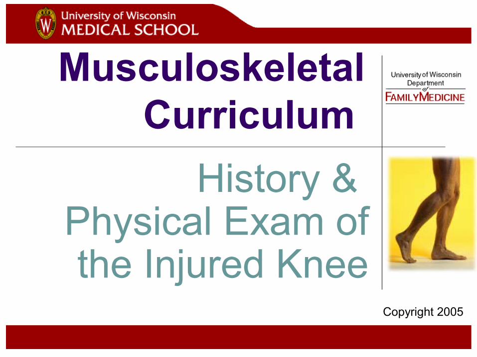
MusculoskeletalCurriculum
History & Physical Exam of the Injured Knee
Copyright 2005

2
Authors
Kathleen Carr, MD
Madison Residency [email protected]
Dennis Breen, MD
Eau Claire Residency [email protected]
Dan Smith, DO
3
Contributors
Marguerite Elliott, DO
Jeff Patterson, DO
Jerry Ryan, MD
4
Goal
Learn a standardized, evidence-based history and physical examination of patients with knee injuries
WHICH WILL:
Enable family medicine resident physicians to accurately diagnose common knee problems throughout the full age spectrum of patients seen in family medicine
5
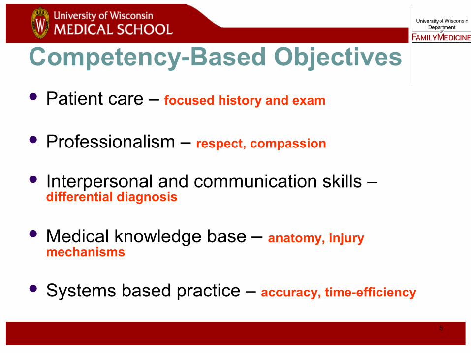
Competency-Based Objectives
Patient care – focused history and exam
Professionalism – respect, compassion
Interpersonal and communication skills – differential diagnosis
Medical knowledge base – anatomy, injury mechanisms
Systems based practice – accuracy, time-efficiency
6
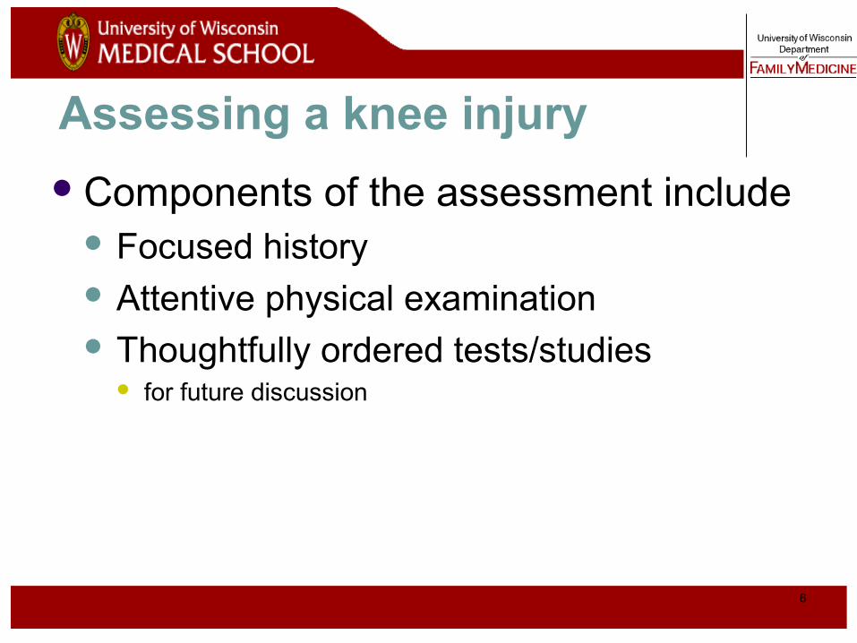
Assessing a knee injury
Components of the assessment include Focused history Attentive physical examination Thoughtfully ordered tests/studies
for future discussion
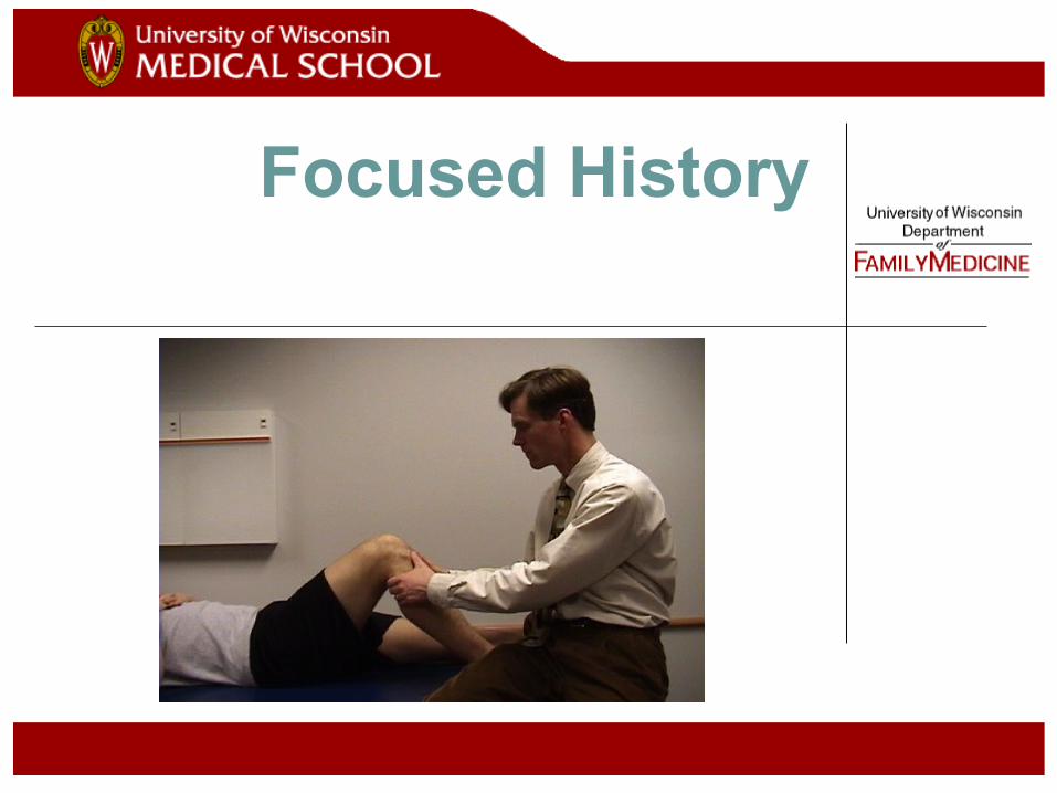
Focused History
8
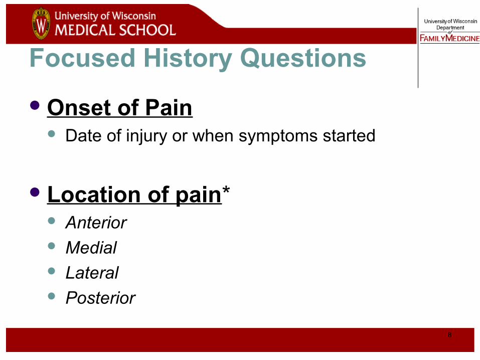
Focused History Questions
Onset of Pain Date of injury or when symptoms started
Location of pain* Anterior Medial Lateral Posterior
9
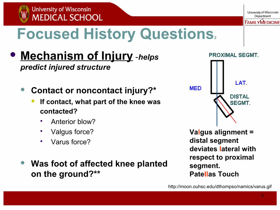
Focused History Questions2
Mechanism of Injury -helps predict injured structure
Contact or noncontact injury?* If contact, what part of the knee was
contacted? Anterior blow? Valgus force? Varus force?
Was foot of affected knee planted on the ground?**
Valgus alignment = distal segment deviates lateral with respect to proximal segment. Patellas Touch
http://moon.ouhsc.edu/dthompso/namics/varus.gif
10
Focused History Questions3
Injury-Associated Events* Pop heard or felt?
Swelling after injury (immediate vs delayed)
Catching / Locking
Buckling / Instability (“giving way”)
11
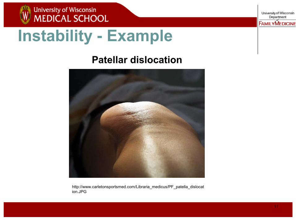
Instability - Example
http://www.carletonsportsmed.com/Libraria_medicus/PF_patella_dislocation.JPG
Patellar dislocation
12
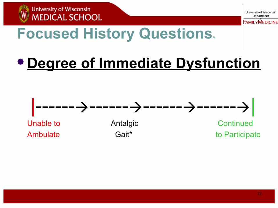
Focused History Questions4
Degree of Immediate Dysfunction
|------------------------|Unable to Antalgic Continued
Ambulate Gait* to Participate
13
Focused History Questions5
Aggravating Factors Activities, changing positions, stairs, kneeling
Relieving Factors/treatments tried Ice, medications, crutches
History of previous knee injury or surgery

14
Historical Clues to Knee Injury Diagnoses
Noncontact injury with “pop” ACL tear
Contact injury with “pop” MCL or LCL tear, meniscus tear, fracture
Acute swelling ACL tear, PCL tear, fracture, knee dislocation, patellar dislocation
Lateral blow to the knee MCL tear
Medial blow to the knee LCL tear
Knee “gave out” or “buckled” ACL tear, patellar dislocation
Fall onto a flexed knee PCL tear

Physical Exam
16
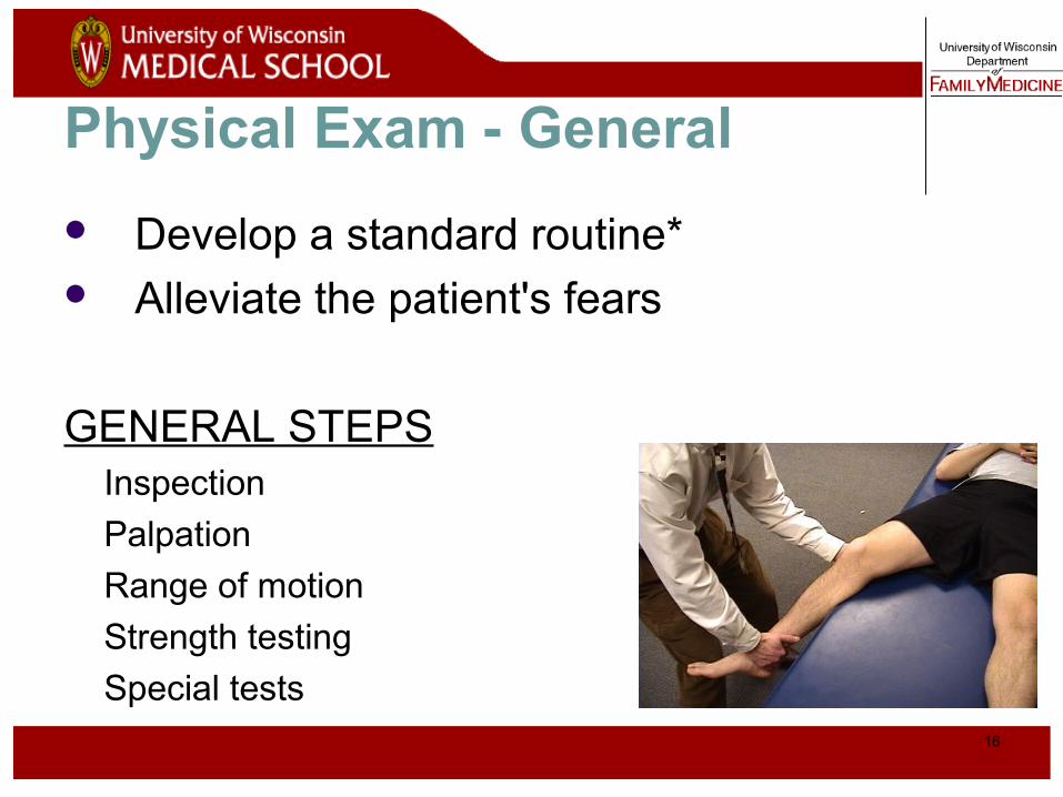
Physical Exam - General
Develop a standard routine* Alleviate the patient's fears
GENERAL STEPSInspection
Palpation
Range of motion
Strength testing
Special tests
17
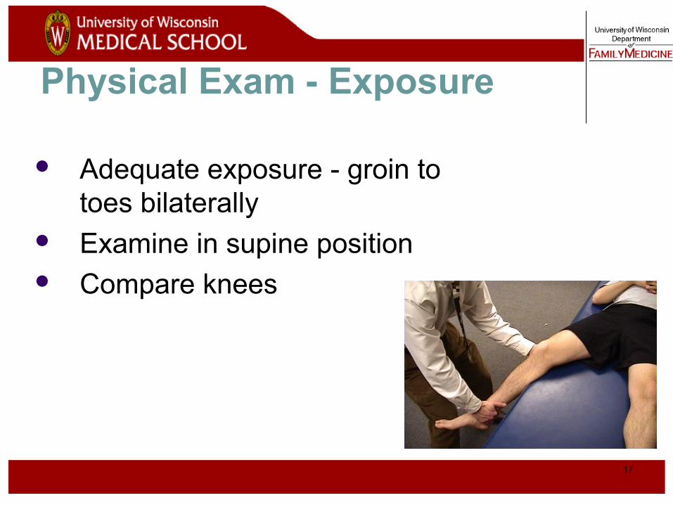
Physical Exam - Exposure
Adequate exposure - groin to toes bilaterally
Examine in supine position Compare knees
18
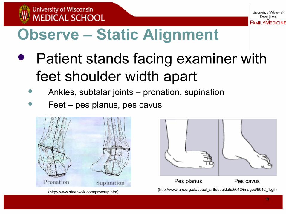
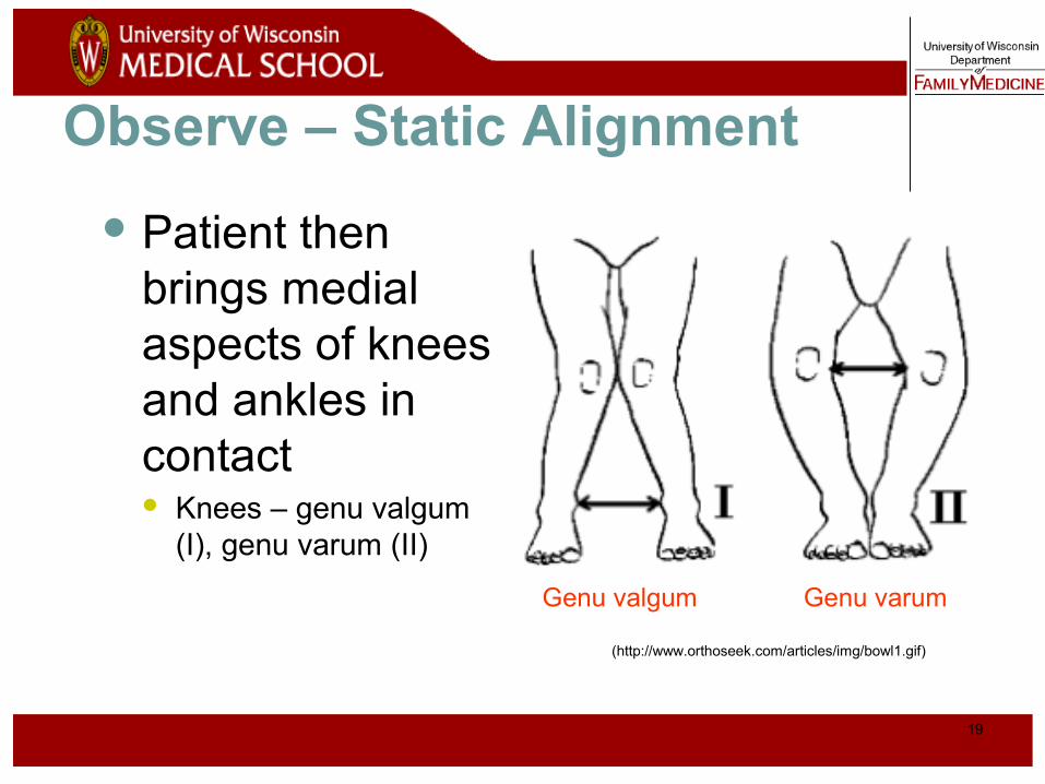
Observe – Static Alignment Patient stands facing examiner with
feet shoulder width apart Ankles, subtalar joints – pronation, supination Feet – pes planus, pes cavus
(http://www.steenwyk.com/pronsup.htm)
Pes planus Pes cavus
(http://www.arc.org.uk/about_arth/booklets/6012/images/6012_1.gif)
19
Patient then brings medial aspects of knees and ankles in contact Knees – genu valgum
(I), genu varum (II)
Observe – Static Alignment
(http://www.orthoseek.com/articles/img/bowl1.gif)
Genu valgum Genu varum
20
Observe – Dynamic Alignment
Pronation/Supination may be enhanced with ambulation
Antalgic gait indicates significant problem (anti = against, algic = pain)

21
Inspect Knee
Warmth Erythema Effusion*
Evidence of local traumaAbrasionsContusionsLacerations
Patella positionMuscle atrophy
22
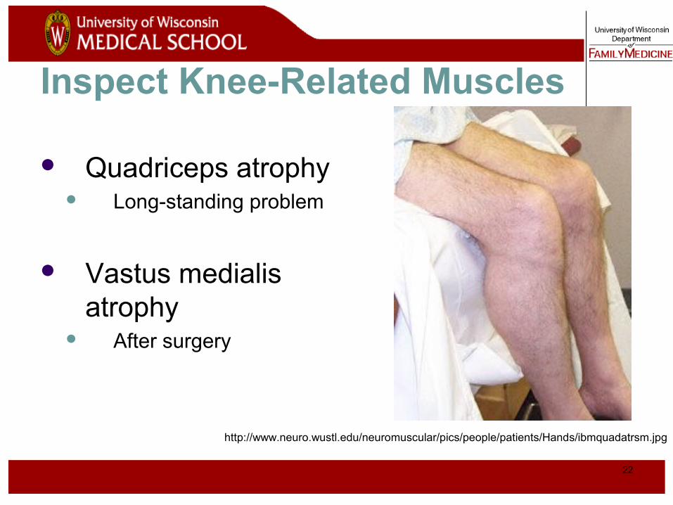
Inspect Knee-Related Muscles
Quadriceps atrophy Long-standing problem
Vastus medialis atrophy
After surgery
http://www.neuro.wustl.edu/neuromuscular/pics/people/patients/Hands/ibmquadatrsm.jpg
23
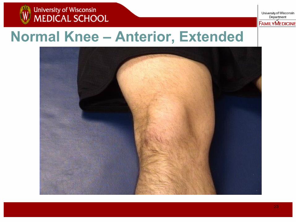
Normal Knee – Anterior, Extended
24
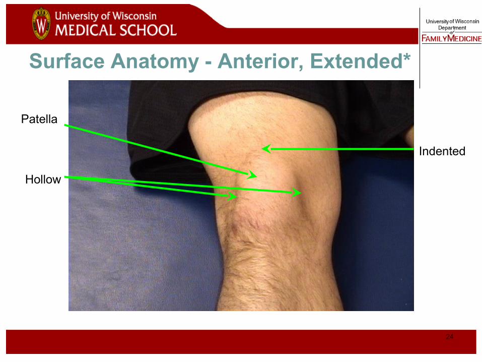
Surface Anatomy - Anterior, Extended*
Patella
Hollow
Indented

25
Normal Knee – Anterior, Flexed
26
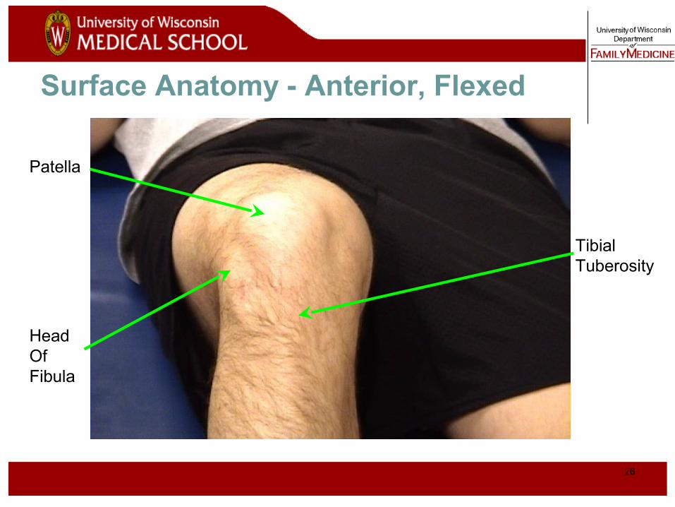
Surface Anatomy - Anterior, Flexed
Head OfFibula
Patella
TibialTuberosity
27
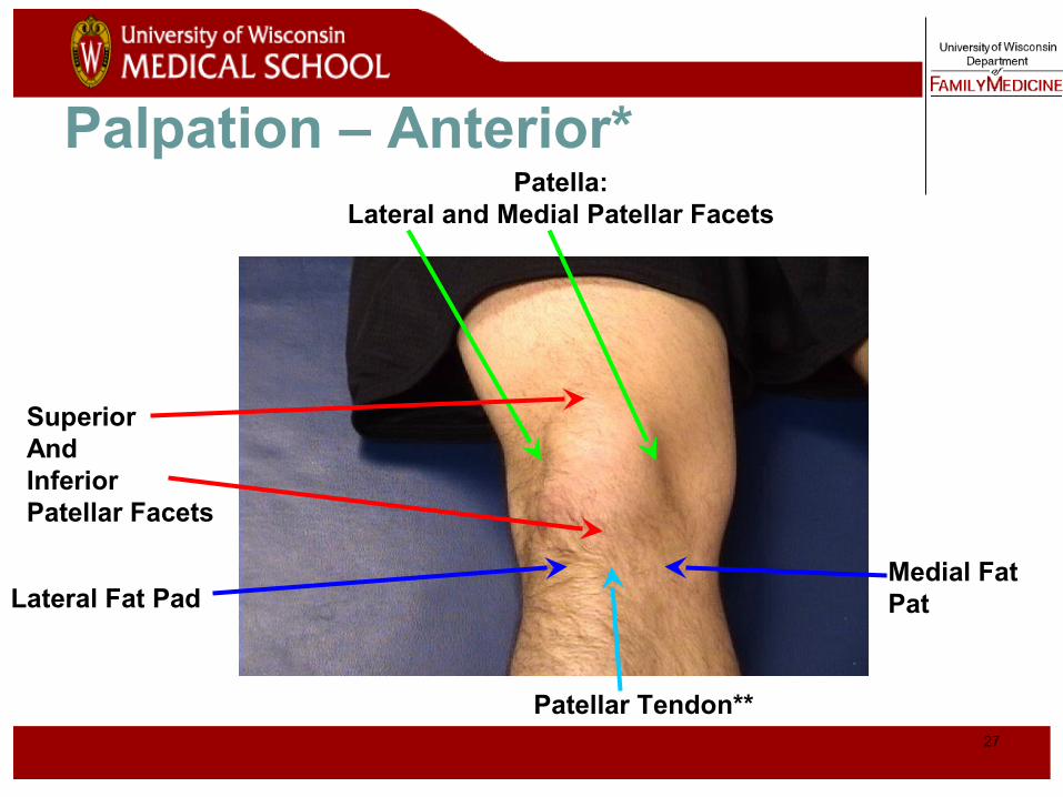
Palpation – Anterior*Patella:
Lateral and Medial Patellar Facets
Superior AndInferior Patellar Facets
Patellar Tendon**
Lateral Fat PadMedial Fat Pat
28
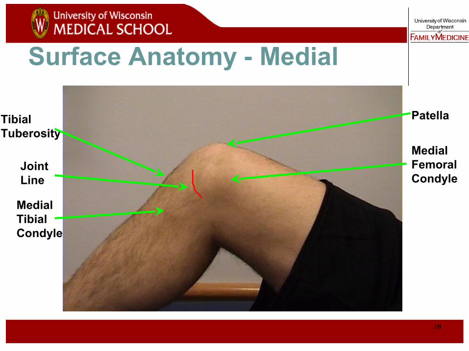
Surface Anatomy - Medial
Medial FemoralCondyle
Patella
JointLine
MedialTibial Condyle
TibialTuberosity

29
Palpation - Medial
Medial Collateral Ligament (MCL)*
Pes anserine bursa**
Medial joint line
30
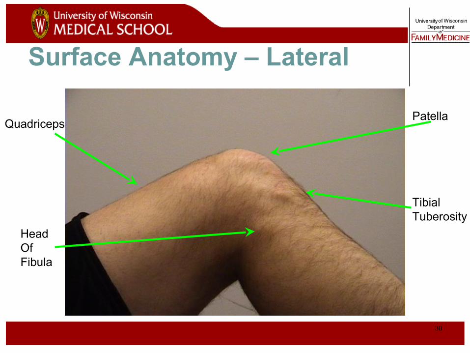
Surface Anatomy – Lateral
Patella
Head OfFibula
TibialTuberosity
Quadriceps
31
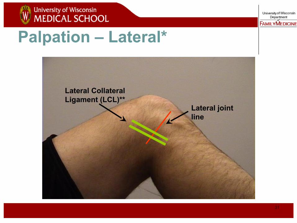
Palpation – Lateral*
Lateral joint line
Lateral Collateral Ligament (LCL)**
32
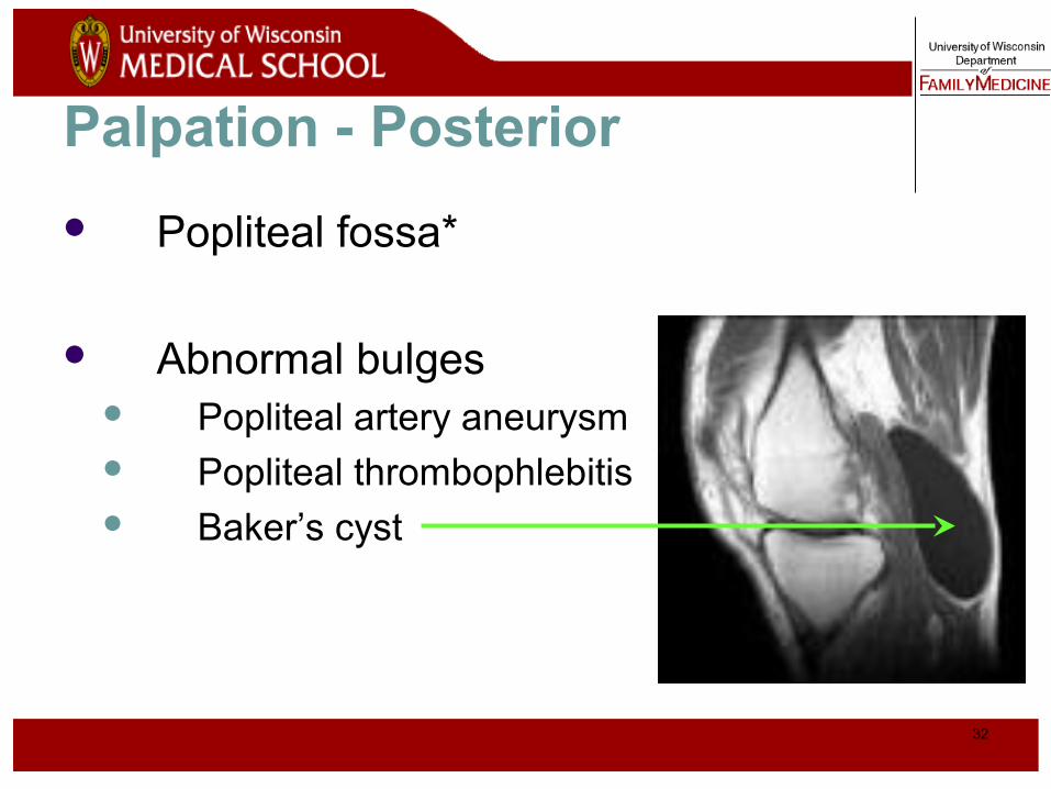
Palpation - Posterior
Popliteal fossa*
Abnormal bulges Popliteal artery aneurysm Popliteal thrombophlebitis Baker’s cyst

33
Range Of Motion Testing Extension Flexion
0º 135º
Describe loss of degrees of extension Example: “lacks 5 degrees of
extension”
Locking* = patient unable to fully extend or flex knee due to a mechanical blockage in the knee (i.e., loose body, bucket-handle meniscus tear)

34
Strength Testing
Test knee extensors (quadriceps) and knee flexors (hamstrings) Can test both with patient in seated position,
knees bent over edge of table Ask patient to extend/straighten knee against your
resistance Then ask patient to flex/bend knee against your
resistance
Compare to unaffected knee
35
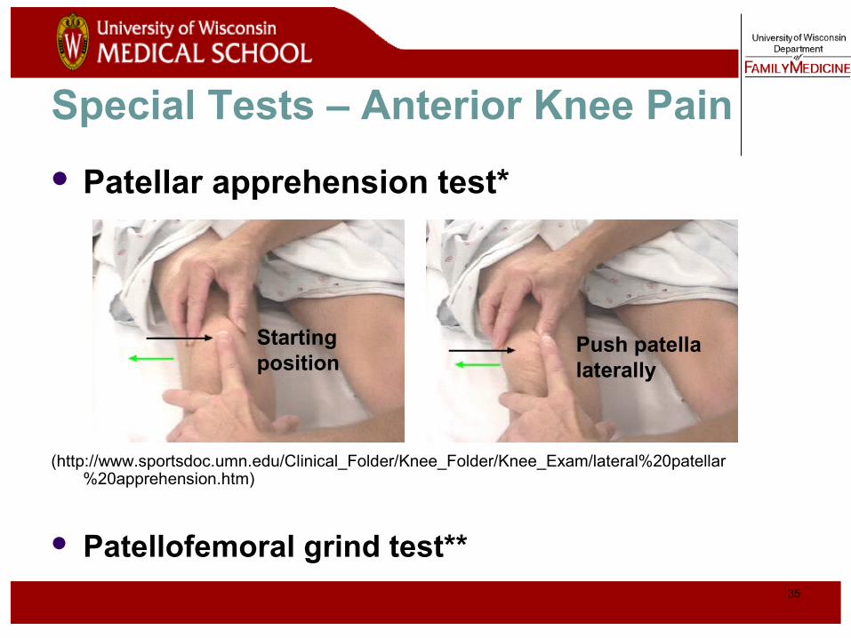
Special Tests – Anterior Knee Pain
Patellar apprehension test*
(http://www.sportsdoc.umn.edu/Clinical_Folder/Knee_Folder/Knee_Exam/lateral%20patellar%20apprehension.htm)
Patellofemoral grind test**
Starting position
Push patella laterally
36
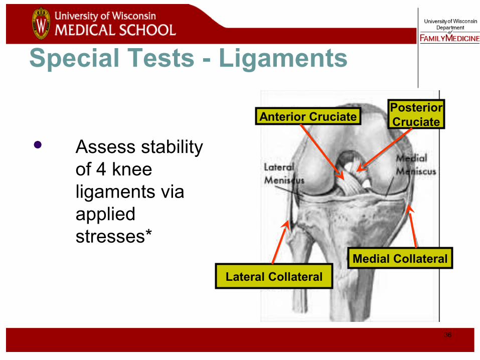
Special Tests - Ligaments
Assess stability of 4 knee ligaments via applied stresses*
Anterior CruciatePosteriorCruciate
Lateral Collateral
Medial Collateral

37
Stress Testing of Ligaments
Use a standard exam routine Direct, gentle pressure No sudden forces
Abnormal test 1. Excessive motion = laxity
What is NORMAL motion?*
2. Soft/mushy end point**
38
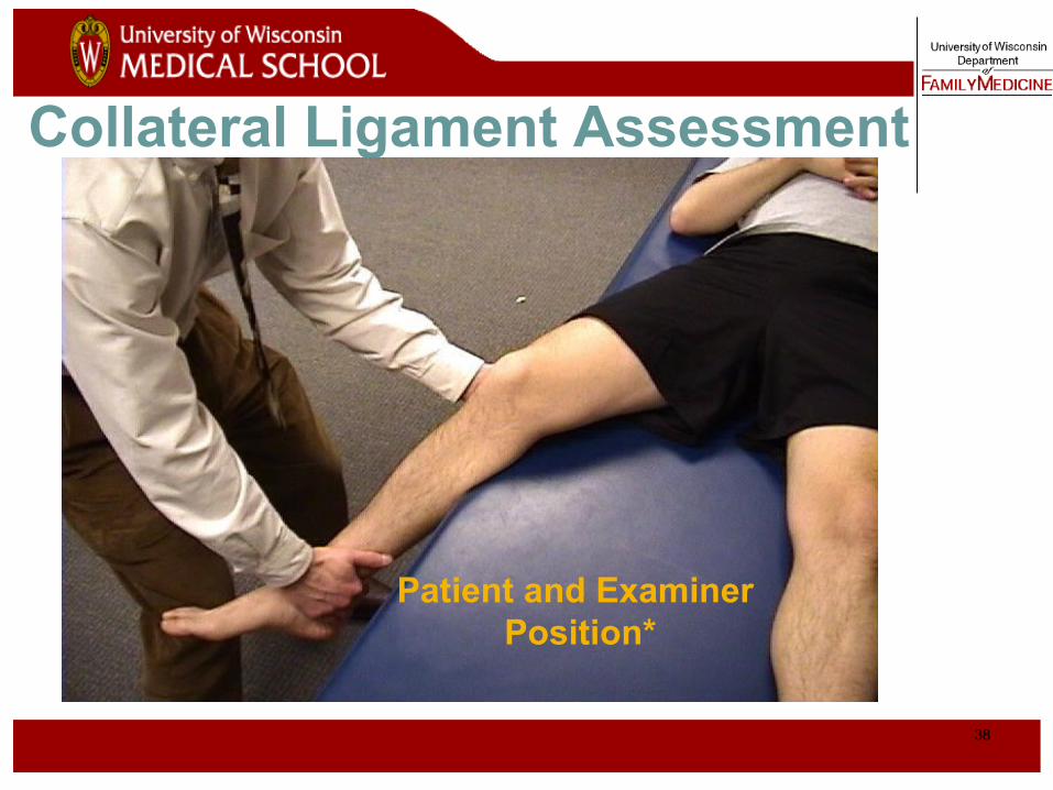
Collateral Ligament Assessment
Patient and Examiner Position*
39
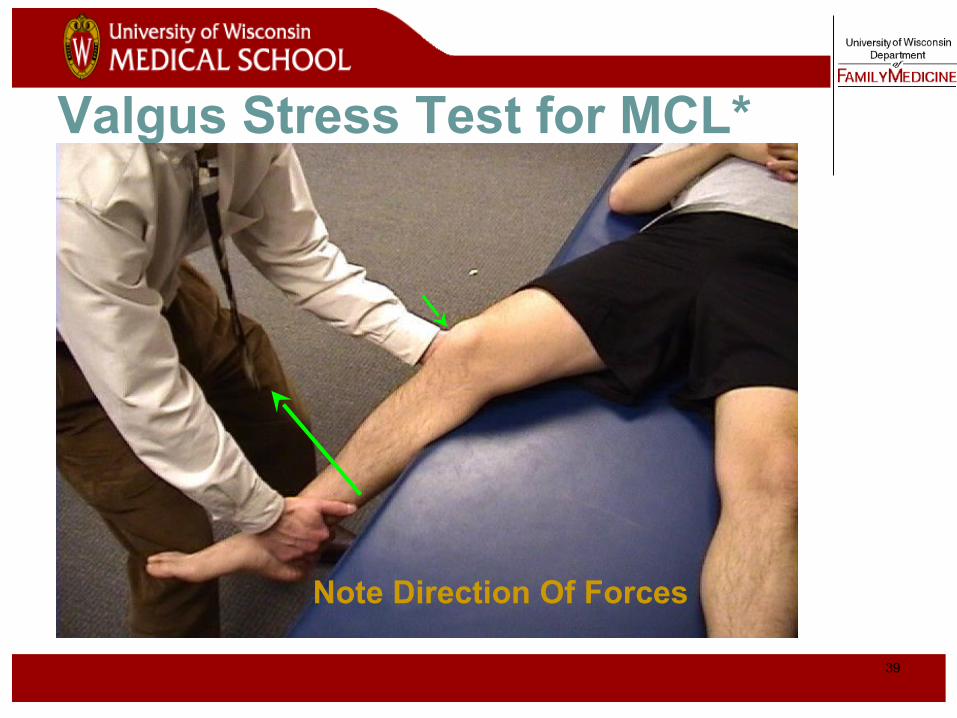
Valgus Stress Test for MCL*
Note Direction Of Forces
40
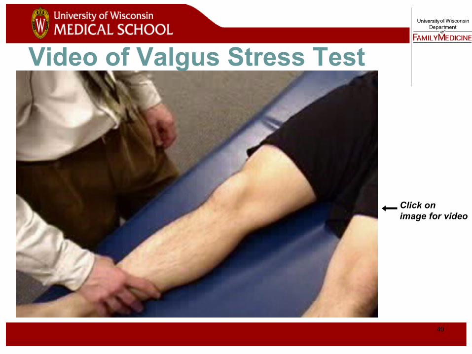
Video of Valgus Stress Test
Click on image for video
41
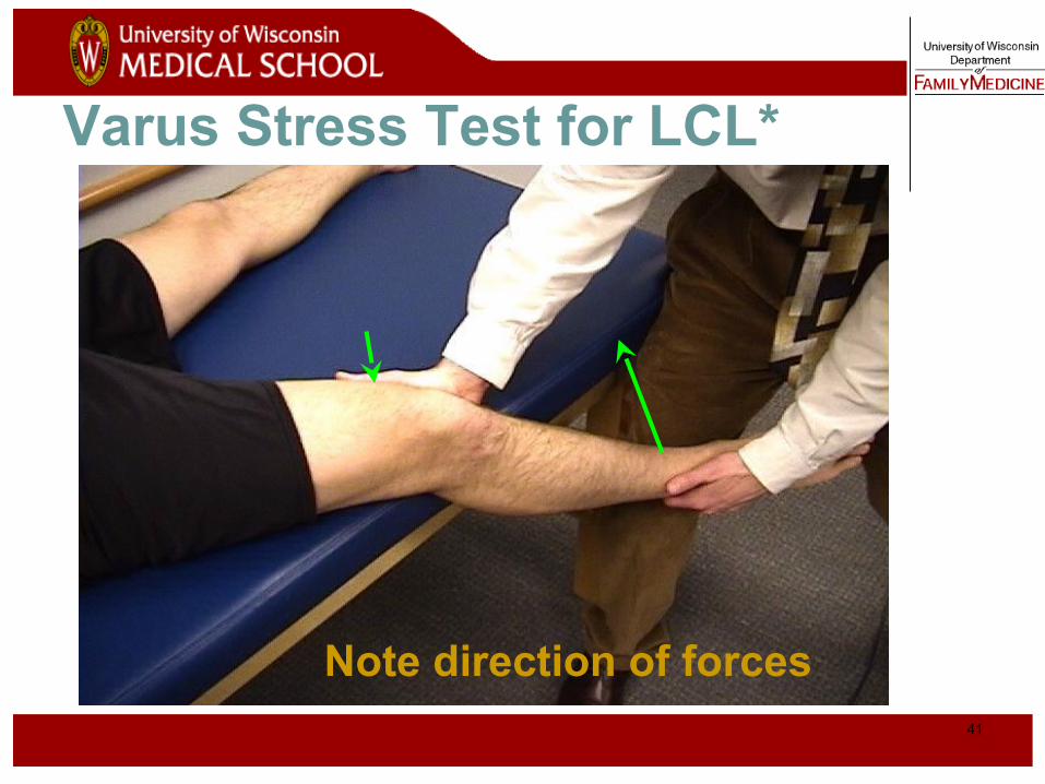
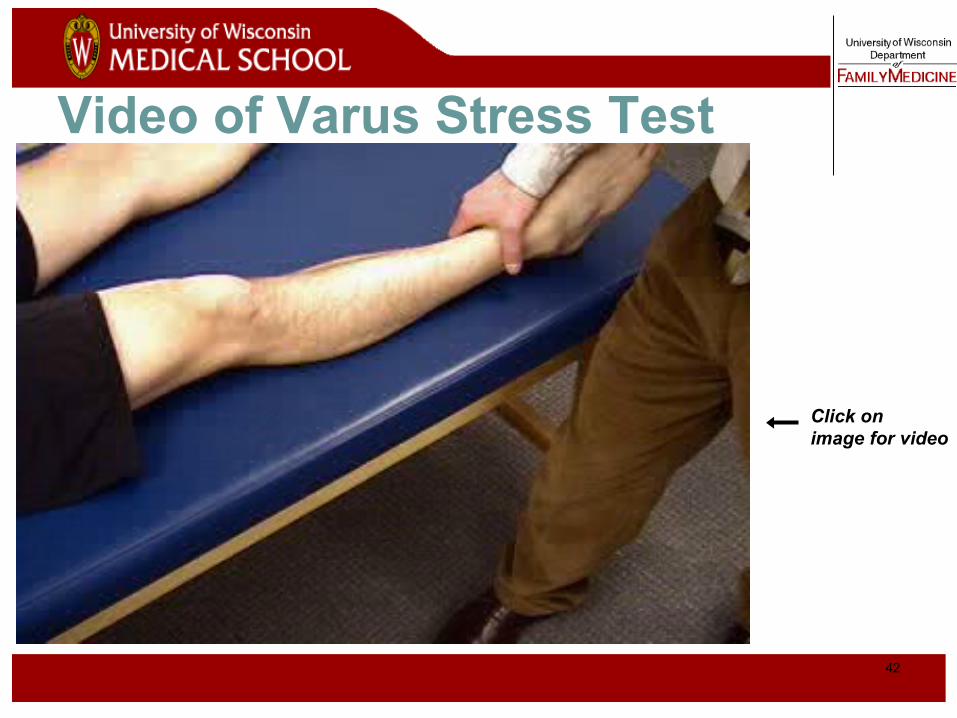
Varus Stress Test for LCL*
Note direction of forces
42
Video of Varus Stress Test
Click on image for video
43
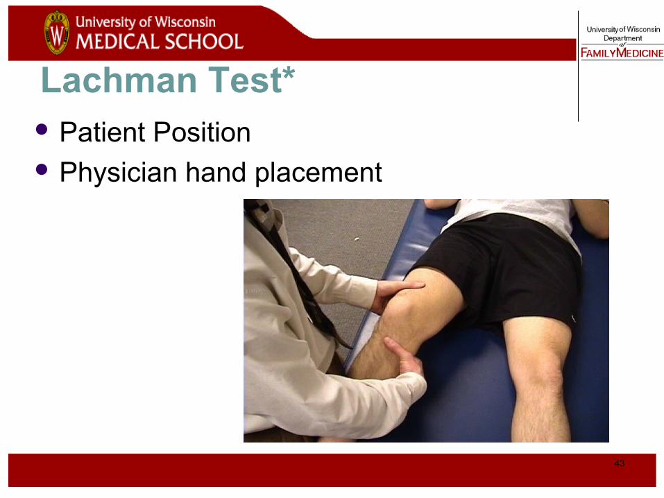
Lachman Test* Patient Position Physician hand placement
44
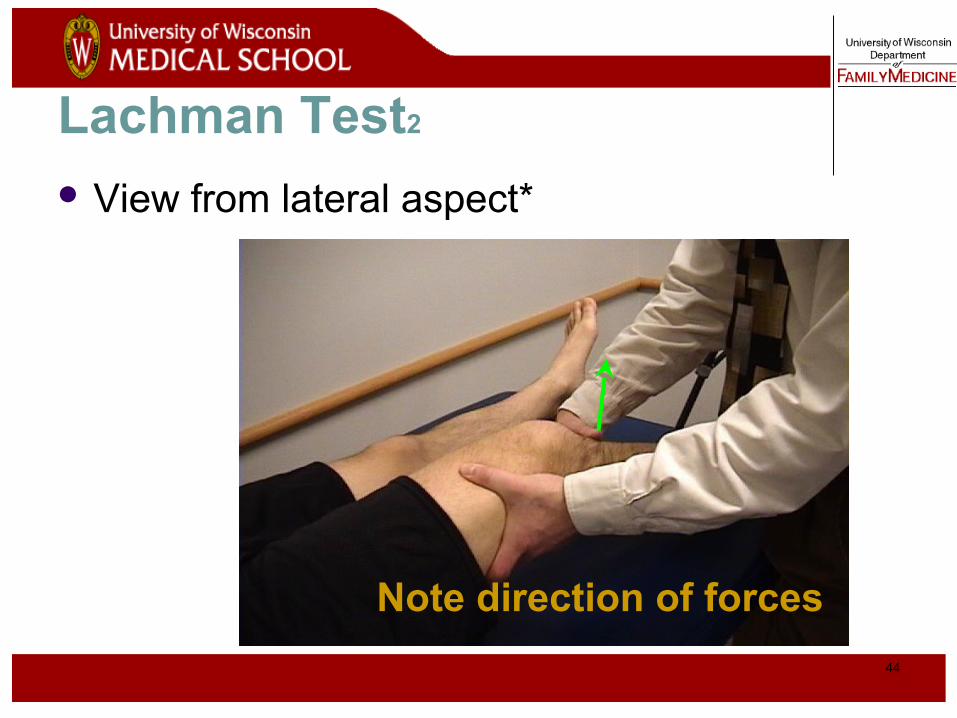
Lachman Test2
View from lateral aspect*
Note direction of forces
45
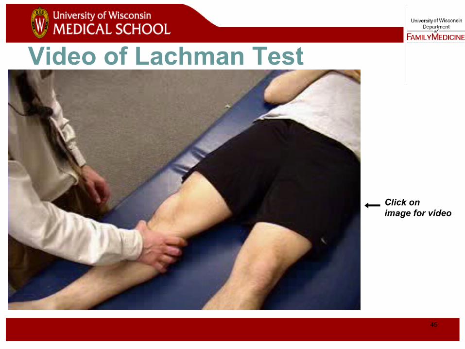
Video of Lachman Test
Click on image for video
46
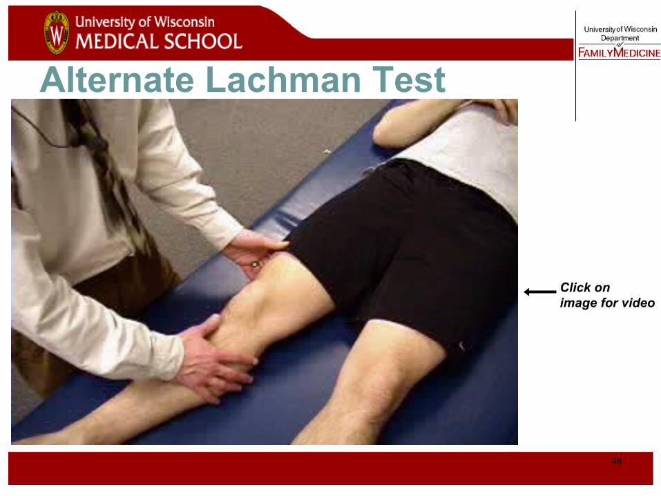
Alternate Lachman Test
Click on image for video
47
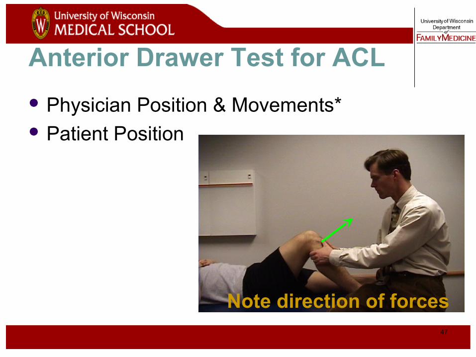
Anterior Drawer Test for ACL
Physician Position & Movements* Patient Position
Note direction of forces
48
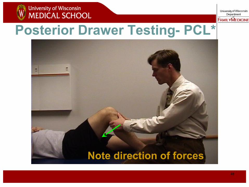
Posterior Drawer Testing- PCL*
Note direction of forces
49
Assess Meniscus – Knee Flexion
Most sensitive test is full flexion* Examiner passively flexes the knee or has patient
perform a full two-legged squat to test for meniscal injury
Joint line tenderness** Flexion of the knee enhances palpation of the
anterior half of each meniscus
50
Tests that we do not recommend routinely
Pivot-Shift* - for ACL tear
McMurray Test**- for meniscus tears

51
Review of Evidence – ACL*
Lachman Test Sens 87% Spec 93% Anterior Drawer Sens 48% Spec 87% Pivot Shift Test Sens 61% Spec 97%
(Jackson JL, et al.)
52
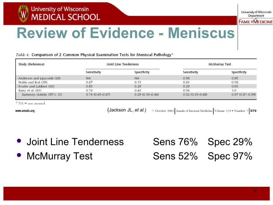
Review of Evidence - Meniscus
Joint Line Tenderness Sens 76% Spec 29% McMurray Test Sens 52% Spec 97%
(Jackson JL, et al.)

53
ReferencesCalmbach WL, Hutchens M. Evaluation of Patients Presenting with Knee Pain: Part I.
History, Physical Examination, Radiographs, and Laboratory Tests. Am Fam Physician 2003;68:907-12.
Ebell MH. A Tool for Evaluating Patients with Knee Injury. Family Practice Management. March 2005:67-70.
Jackson JL, O’Malley PG, Kroenke K. Evaluation of Acute Knee Pain in Primary Care.
Ann Intern Med. 2003;139:575-588. Malanga GA, Andrus S, Nadler SF, McLean J. Physical Examination of the Knee: A
Review of the Original Test Description and Scientific Validity of Common Orthopedic Tests. Arch Phys Med Rehabil 2003;84:592-603.
Solomon DH, Simel DL, Bates DW, Katz JN. Does this patient have a torn meniscus or
ligament of the knee? Value of the Physical Examination. JAMA 2001;286:1610-1620.
54
Video of Knee Exam
http://www.fammed.wisc.edu/our-department/media/musculoskeletal