knee injuries and imaging · the use of mri and clinical exam in evaluation of knee injuries...
TRANSCRIPT

16.11.2016
1
Ortopedisk avdeling
Knee Injuries and Imaging
Marc Jacob Strauss
1. Knee injuries and priority
2. Patellofemoral instability
Ortopedisk avdeling

16.11.2016
2
1 Knee injuries and priority
Ortopedisk avdeling
Differences in knee injuries
Contact vs non-contact
Low vs High energy
Concomitant injuries
Age (child or adult)
Ortopedisk avdeling

16.11.2016
3
Orthopedic Approach
• History
• Try to imagine injury mechanism
• Clinical examination
Ortopedisk avdeling
Orthopedic Approach
Assesment of potential injury/injuries
Imaging Verify suspected injury/injuries and rule-out others
Ortopedisk avdeling

16.11.2016
4
The Use of MRI and Clinical Exam in Evaluation of Knee Injuries
Ortopedisk avdeling
Ercin: Knee Surg Sports Traumatol Arthrosc (2012) 20:851–856
Importance of early diagnosis
Bucket Handle
Root lesion
Osteochondral lesions
Eminentia fracture / avulsion
Fractures
Knee dislocations
Ortopedisk avdeling

16.11.2016
5
Meniscus lesion
Ortopedisk avdeling
Bucket Handle tear
Ortopedisk avdeling
Locking of the knee Extention deficit Treatment: Suture if possible 75-80 % healing rate... Grant: Am J Sports Med 2012 40: 459

16.11.2016
6
Meniscal root tear
Ortopedisk avdeling
Timing of surgery? Still remains to be answered
Ostechondral lesion
Ortopedisk avdeling
Clinic: Pain and swelling Locking, possibly extention deficits Previous history of OCD or PF instability Treatment: Fixation of detached chondral lesion must be within 2 weeks of injury

16.11.2016
7
Avulsion fracture
Ortopedisk avdeling
Strub WM. The arcuate sign. Radiology. 2007;244 (2): 620-1.
Eminentia fracture Tibial spine avulsion Arcuate sign Avulsion fracture of the LCL and/or biceps tendon from the tip of the proximal fibula Suspect ACL tear (~80-90%) Treatment: Depending on grade of displacement Non-displaced conservative treatment Displaced Surgical treatment within 2 weeks
The arcuate sign
Knee dislocation
Very serious
Often involment of multiple structures
Vascular injury
Nerve injury
Radiograph and MRI and possibly CT angio
Important for preoperative planning
Treatment:
Surgery within 10-14 days after injury
Ortopedisk avdeling

16.11.2016
8
Summery 1 Indication for early Radiographs or MRI on suspicion for the following injuries...
• Bucket handle lesions
• Root lesion
• Osteochondral lesions
• Eminentia fracture / avulsion
• Fractures
• Knee dislocations
Most of these injuries require surgical intervention within 2 weeks of injury
Ortopedisk avdeling
2 Patellofemoral Instability
Ortopedisk avdeling

16.11.2016
9
Patellofemoral Dislocation
”Rule of thumb”
Approx. 50% of 1. time dislocation can manage with rehab.
Approx. 50% will experience redislocation/instability and should be examed by an orthopedic surgeon, and possibly surgically treated.
– Clinic
– Radiogtraphs
– MRI
Ortopedisk avdeling
Non-dysplasi 31 % re-dislocate
Dysplasi 69 % re-dislocate
Ortopedisk avdeling

16.11.2016
10
Patellofemoral Dysplasia
Ortopedisk avdeling
Anatomical factors
Bony abnormality
• Patella dysplasia
• Trochlea dysplasia
• Femoral antevertion
Soft tissue abnormality
• Mediale stabilisors
• Laterale retinacle
• Patella alta
• Hypermobile
Ortopedisk avdeling

16.11.2016
11
Patella Index
• Insall-Salvati
1.3 > pathologic
• Caton-Deschamp
1.2 > pathologic
Ortopedisk avdeling
Methods of Investigation
Tangentiale patellapictures Merchant’s view
45 graders fleksion MRI CT

16.11.2016
12
Tuberositas Tibia – Trochlea Groove
Ortopedisk avdeling
TT-TG > 20 mm = pathologic
Schoettle: The Knee 13 (2006) 26 – 31
Dysplasia
• Patella classification
– Wiberg (1941)
• Trochlea classification
– Dujour • 1987 Crossing sign
• 1998 Classification
Ortopedisk avdeling

16.11.2016
13
Patella Dysplasia
Wiberg classification
Ortopedisk avdeling
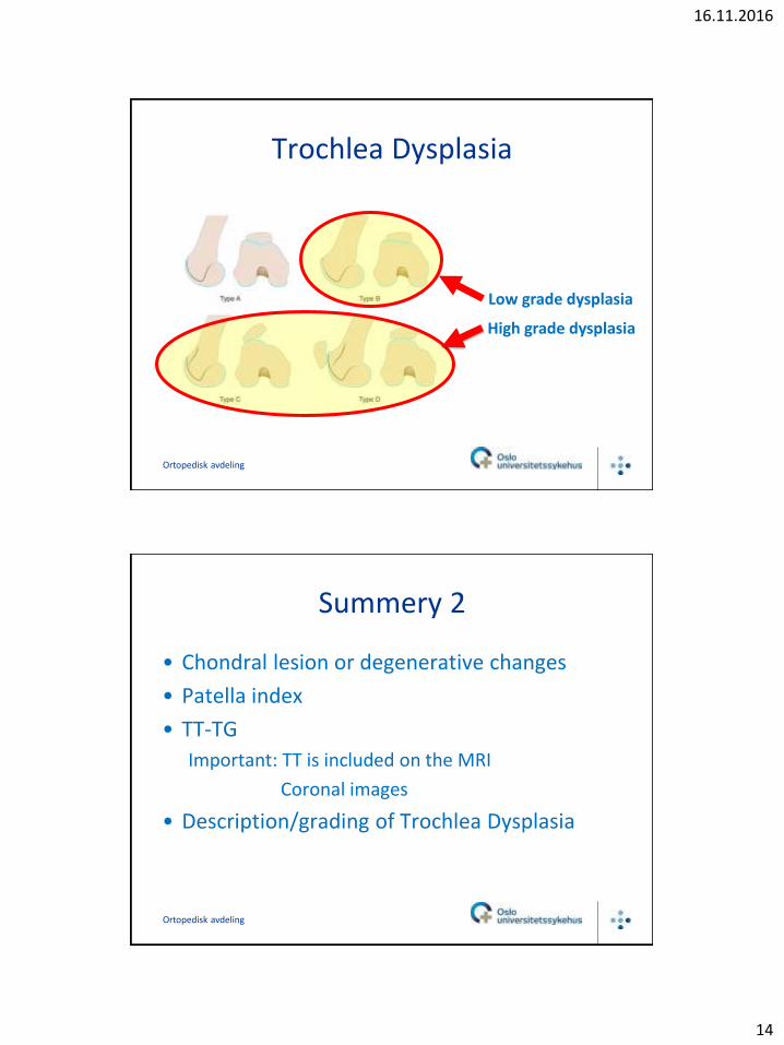
Trochlea Dysplasia
Dujour classification
– Type A-D
– Crossing sign
Ortopedisk avdeling
16.11.2016
14
Trochlea Dysplasia
Ortopedisk avdeling
Low grade dysplasia
High grade dysplasia
Summery 2
• Chondral lesion or degenerative changes
• Patella index
• TT-TG
Important: TT is included on the MRI
Coronal images
• Description/grading of Trochlea Dysplasia
Ortopedisk avdeling

16.11.2016
15
Thank you
Ortopedisk avdeling