kmj 2011.pdfmustafa al-mousawi nasser j hayat nawaf al-mutairi nebojsa rajacic sami asfar soad...
TRANSCRIPT
EDITORIALPostgraduate Residency Training Program in Kuwait - Courses to Competencies: A Shifting Paradigm 1Jasbir S Bajaj, Abdullatif A Al-Bader
REVIEW ARTICLEPediatric Obstructive Sleep Apnea Syndrome 6Tareq Al-Abdoulsalam, Manisha Witmans
ORIGINAL ARTICLES The Treatment of Chronic Monteggia Lesions and Chronic Traumatic Isolated Radial Head Dislocations 16
James W Roach, Hanna M Saltzman
Value of Abdominal Pressure Measurement in Neonatal Abdominal Surgical Emergencies 20
Ahmed M Kamal, Abdulrahman S Almulhim
Factors Associated with Patients Bypassing Primary Health Care Facilities in Saudi Arabia: A Cross-Sectional Study 26
Saad Abdullah Alghanim
Fine Needle Aspiration Cytology in the Diagnosis of Superficial Lymphadenopathy in Children and Adolescents:
An analysis of 869 cases 33
Shahed K Pathan, Sara Shirly George, Bahiyah E Haji, Taiba Ali Al-Ansary, Dilip K Das, Kusum Kapila The Effect of Oxygenation Technique in General Anesthesia on Postoperative Nausea and Vomiting 37
Hassan Karbasi, Ghodratollah Naseh, Kamyab Alizadeh
CASE REPORTS Laparoscopic Removal of a Foreign Body from the Intestine 41
Yousef A Hussain, Abdulrahman Faraj Almutairi, Mutlaq Al-Sihan
Autoimmune Polyendocrinopathy – Candidiasis – Ectodermal Dystrophy Syndrome Presenting as Unexplained
Chronic Interstitial Keratitis 44
Hassan Elsori, Hesham A Aziz
Ecthyma Gangrenosum with Pseudomonas Sepsis in a Previously Healthy Child 47
Mohamed Rifai
Laparoscopic Treatment of Gall Bladder Duplication 50
Syed Farooqh Hussain Belgrami, Ayman Elezeby, Vinod Kumar Grover
Persistent Left Superior Vena Cava Draining into the Left Atrium with a Large Primum Atrial Septal Defect 53
Mohammed Abdulaziz Al Jarallah, Muath Mohammed AlAnbai
Breastfeeding Malnutrition with Hypernatremic Dehydration: Case Report and Literature Review 57
Adnan Mahmoud Hariri, Nihad Gamal Omar, Ibrahim Abdel-Monsef Al-Sharkawi
Gastrointestinal Sarcoidosis: A Case Report 60
Salem Farhan Owayed, Jaffer Ismail Ali, Mubarak Al-Ajmi
MARCH 2011VOLUME 43 NUMBER 1
KMJKUWAIT MEDICAL JOURNAL
The Official Journal of The Kuwait Medical Association
KU ISSN 0023-5776 Continued inside

MARCH 2011Vol. 43 No. 1
C O N T E N T S
KUWAIT MEDICAL JOURNAL
Continued from cover
THE PUBLICATION OF ADVERTISEMENTS IN THE KUWAIT MEDICAL JOURNAL DOES NOT CONSTITUTE ANY GUARANTEE OR ENDORSEMENT BY THE KUWAIT MEDICAL ASSOCIATION OR THE EDITORIAL BOARD OF THIS JOURNAL, OF THE ADVERTISED PRODUCTS, SERVICES, OR CLAIMS MADE BY THE ADVERTISERS. THE PUBLICATION OF ARTICLES AND OTHER EDITORIAL MATERIAL IN THE JOURNAL DOES NOT NECESSARILY REPRESENT POLICY RECOMMENDATIONS OR ENDORSEMENT BY THE ASSOCIATION.
PUBLISHER: The Kuwait Medical Journal (KU ISSN-0023-5776) is a quarterly publication of THE KUWAIT MEDICAL ASSOCIATION. Address: P.O. Box 1202, 13013 Safat, State of Kuwait; Telephone: 1881181 Fax: 25317972, 25333276. E-mail : [email protected]: The Kuwait Medical Journal. All rights reserved. No part of this publication may be reproduced without written permission from the publisher. Printed in Kuwait.INSTRUCTIONS FOR AUTHORS: Authors may submit manuscripts prepared in accordance with the Uniform Requirements for Manuscripts Submitted to Biomedical Journals. These Requirements are published in each issue of the Kuwait Medical Journal.CHANGE OF ADDRESS: Notice should be sent to the Publisher six weeks in advance of the effective date. Include old and new addresses with mail codes.KUWAIT MEDICAL JOURNAL (previously The Journal of the Kuwait Medical Association) is added to the list of journals adhering to the “Uniform Requirements for Manuscripts Submitted to Biomedical Journals”, American College of Physicians, Independence Mall West, Sixth Street at Race, Philadelphia, PA 19106-1572, USA, and can be located at http://www.icmje.org/jrnlist.html
❈ ❈ ❈
SELECTED ABSTRACTS OF ARTICLES PUBLISHED ELSEWHERE BY AUTHORS IN KUWAIT 64
FORTHCOMING CONFERENCES AND MEETINGS 70
WHO-FACTS SHEET 81 1. Cardiovascular Diseases (CVDs)2. WHO Endorses New Rapid Tuberculosis Test3. New WHO Guidelines: TB Prevention for People with HIV4. Control of Neglected Tropical Diseases is Feasible: WHO5. Urgent Action Essential to Protect Malaria Therapies, Says WHO6. Chronic Obstructive Pulmonary Disease (COPD)
Open access for articles at: www.kma.org.kw/KMJ
Indexed and abstracted in:EMBASE (The Excerpta Medica Database)
Science Citation Index Expanded (also known as SciSearch®)Journal Citation Reports/Science Edition
IMEMR Current Contents (Index Medicus for the Eastern Mediterranean Region;available online at: www.emro.who.int/EMRJorList/online.aspx

Kuwait Medical Journal (KMJ)Published by the Kuwait Medical Association
Previously known as The Journal of the Kuwait Medical Association (Est. 1967)
Honorary President: Abdulaziz Al-Babtain
EDITORIAL BOARD Editor-in-Chief: Fuad Abdulla M Hasan, Kuwait Editor: Adel Khader Ayed, Kuwait International Editor: Pawan K Singal, Canada Associate Editors: Adel A Alzayed, Kuwait Ignacio Rodriguez, USA Michael Redmond, USA Mousa Khoursheed, Kuwait Mustafa M Ridha, Kuwait Nasser Behbehani, Kuwait Noura Al-Sweih, Kuwait
INTERNATIONAL ADVISORY BOARD
Ananda S Prasad, USAAnders Lindstrand, Sweden Andrew J Rees, UKBelle M Hegde, IndiaBengt Jeppsson, SwedenCharles A Dinarello, USAChristian Imielinski, PolandElizabeth Dean, CanadaFiona J Gilbert, UKFrank D Johnston, UKGeorge Russell, UKGraeme RD Catto, UK
Oleg Eremin, UKPeter RF Bell, UKPhilip M Moody, USARaymond M Kirk, UKSamuel Dagogo-Jack, USAS Muralidharan, IndiaStig Bengmark, SwedenTulsi D Chugh, IndiaWilliam A Tweed, CanadaWilliam B Greenough, USAZoheir Bshouty, Canada
REGIONAL ADVISORY BOARD
Abdulla BehbehaniAbeer K Al-BahoAlexander E OmuAli Al-MukaimiAli Al-SayeghAsmahan Al-ShubailiChacko MathewEiman M MokaddasFaisal A Al-Kandari
Habib Abul John F Greally Joseph C LongeneckerKamal Al-Shoumer Kefaya AM AbdulmalekKhalid Al-JarallahMazen Al EssaMohamed AA MoussaMousa Khadadah
Mustafa Al-Mousawi Nasser J HayatNawaf Al-MutairiNebojsa RajacicSami AsfarSoad Al-BaharSukhbir Singh UppalWaleed AlazmiWaleed A Aldhahi
EDITORIAL OFFICEEditorial Manager : Babichan K Chandy
Language Editor : Abhay U Patwari
EDITORIAL ADDRESSP.O. Box: 1202, 13013-Safat, Kuwait
Telephone: (00-965) 1881181(Ext. 201) - Fax: (00-965) 25317972, 25333276E-mail: [email protected]
Website: www.kma.org.kw/KMJ
Giuseppe Botta, ItalyJames W Roach, USAJan T Christenson, SwitzerlandJasbir S Bajaj, IndiaJohn V Forester, UKJulian Little, CanadaKostadin L Karagiozov, JapanLewis D Ritchie, UKMechael M Meguid, USAMohammed Zayer, SwedenNeva E Haites, UKNirmal K Ganguli, India
KUWAIT MEDICAL JOURNAL
i
INTRODUCTIONFormerly known as ‘The Journal of the Kuwait
Medical Association’, the Kuwait Medical Journal (KMJ) was established in the year 1967. It is the officialpublication of the Kuwait Medical Association and published quarterly and regularly in March, June, September and December.
AIMS AND SCOPEKMJ aims to publish peer-reviewed manuscripts
of international interest. Submissions on clinical, scientific or laboratory investigations of relevance tomedicine and health science come within the scope of its publication. Original articles, case reports, brief communications, book reviews, insights and letters to the editor are all considered. Review articles are solicited. Basic medical science articles are published under the section ‘Experimental Medicine’.
GENERALThe Kuwait Medical Journal is a signatory journal to
the Uniform Requirements for Manuscripts Submitted to Biomedical Journals, the fifth (1997) revision of adocument by the international Committee of Medical Journal Editors. A description of important features of this document is available on the Lancet website at http://www.thelancet.com. Alternatively, you may consult the following: N Engl J Med 1997; 336:307-315 or order the leaflet “Uniform Requirements for ManuscriptsSubmitted to Biomdical Journals” by writing to the Editor of the British Medical Journal (BMJ), BMA House, Tavistock Square, London WC1H 9JR, UK.
To present your original work for consideration, one complete set of the manuscript, written in English (only), accompanied by tables, and one set of figures(if applicable), should be submitted to the Editor. Authors should also provide the manuscript on an IBM compatible medium such as floppy, CD (in MS wordformat) or pen-drive, if not submitted through e-mail. The KMJ editorial office uses Microsoft ‘Office 2007’word processing and ‘Excel’ programs. Details of the type of computer used, the software employed and the disk system, if known, would be appreciated.
ELECTRONIC SUBMISSIONSA manuscript could be submitted through e-
mail as an attached word-document (.doc), together with a scanned copy of the signed consent letter of the author(s). The consent letter could otherwise be faxed to the journal office at (00965) 25317972 or25333276. Figures/photographs, photomicrogrphs etc, if any, need to be in .jpg/.jpeg/.bmp format with 300 dpi resolution and illustrations such as graphs,
KUWAIT MEDICAL JOURNAL (KMJ)Instructions for Authors
charts etc., as Excel files. All the figures includingillustrations should be saved as Fig. 1, Fig. 2, etc in running sequence and submitted as seperate attachments along with the manuscript. Incomplete/improper submissions will not be processed, and will be returned.
Following a peer review process, the corresponding author will be advised of the status; acceptance/recommendation for revision or rejection of the paper, in a formal letter sent through post and/or e-mail. A galley proof will be forwarded to the corresponding author through e-mail before publication of the accepted papers which must be returned to the journal office within 48hours with specific comments, if any. Corrections in thegalley proof, in case any, must be limited to typographical errors, or missing contents only.
ETHICAL CONSIDERATIONSWhere human investigations or animal experiments
are part of the study, the design of the work has to be approved by local ethics committee. A relevant statement of approval should be added in the ‘Subjects and Methods’ section of the manuscript.
PREPARATION OF THE MANUSCRIPT The manuscript should be typed as ‘normal text’ in
single column, with no hyphenation and no hard returns within paragraphs (use automatic word wrap) on A4 size (29.7 x 21 cm) paper in single column format, preferably in font size no.12. Cell format for paragraphs, artwork and/or special effects for the text and/or table(s) are not acceptable. Italics shall be used only for foreign/Latin expressions and/or special terminologies such as names of micro organisms. Maintain a minimum of 2 cm margin on both sides of the text and a 3 cm margin at the top and bottom of each page. No part of the text other than abbreviations and/or subtitles shall be written in upper case (ALL capital). Header/foot notes, end notes, lines drawn to separate the paragraphs or pages etc. are not acceptable. Do not submit articles written/saved in ‘Track-change’ mode
THE ORDER OF THE TEXTOriginal Articles: Title page, Abstract (in structured format for original articles) of no more than 250 words, Key Words (no more than five), followed byIntroduction, Subjects (or Materials) and methods, Results, Discussion, Conclusion, Acknowledgment/s (if any), References, Legends to figures, Tables, andFigures. Each section should begin on a new page.
Review Articles (solicited): Title Page, Abstract of no more than 250 words, Key Words (no more than

Instructions for Authors
ii
five), followed by Introduction, Methods/History(if applicable), Literature Review, Conclusion, Acknowledgment/s (if any), References, Legends to figures (if applicable), Tables, and Figures. Each sectionshould begin on a new page.
Case Studies: Title page followed by Abstract (a short summary of not more than 200 words), Key Words, Introduction, Case history/report, Discussion, Conclusion, Acknowledgment/s (if any), References, Legends to figures (if applicable), Tables, and Figures.
Manuscript should be paginated consecutively, commencing with the title page. Main headings, introduction, subjects and methods, etc., should be placed on separate lines.
THE TITLE PAGETitle page of the submitted manuscript should
provide a clear title of the study followed by full names of all authors, the highest academic degree and affiliations if any, the name and address of the institution/s where the work was done including the department, the name and complete address of the corresponding author to whom proofs and correspondences shall be sent, duly supported with contacts such as telephone, mobile/cell, fax and e-mail address.
STRUCTURED ABSTRACTA structured abstract of no more than 250 words
is required for studies under the section “Original Articles”. It must provide an overview of the entire paper, and should contain succinct statements on the following, where appropriate: Objective(s), Design, Setting, Subjects, Intervention(s), Main Outcome Measure(s), Result(s), and Conclusion(s). (See: Haynes RB, Mulrow CD, Huth AJ, Altman DG, Gardner MJ. More informative abstracts revisited. Annals of Internal Medicine 1990; 113:69-76). Abstract for all other category of submissions shall be a short summary not more than 200 words followed by Key words and the report or review.
KEY WORDSKey Words should be preferably MeSH terms, and
shall not duplicate words already in the manuscript title; MesH terms can be checked at: <http://www.nlm.nih.gov/mesh/>.
TABLESTables typed on separate pages using table format
should follow the list of references. All tables must be numbered consecutively and provided with appropriate titles. Contents of the table should be simple, and information therein not duplicated, but duly referred to, in the main text. Tables recording only a few values are not appreciated, since such information can be more accurately, usefully and concisely presented as a sentence or two in the text.
DESIGN OF THE WORKThis should be stated clearly. The rationale behind
the choice of sample size should be given. Those about to begin randomized controlled studies may wish to study the CONSORT statement (JAMA 1996; 276:637-639).
ILLUSTRATIONSPhotographs, Photomicrographs, line drawings,
transparencies, etc. must be of high quality and supplied in original (not photocopies or laser prints) of size 10 x 15 cm (4” x 6”). Regarding scanned image requirements, see ‘Electronic Submissions’. Photographs should fitwithin a print area of 164 x 235 mm. All the figuresmust be numbered serially (Fig 1, Fig 2 etc.) and the figure number written on the back side of each (incase of hard copy submission) and an arrow drawn to indicate the top edge. Figures where patient’s identity is not concealed, authors need to submit a written consent of the patient or of the patient’s guardian, in case of minors. Figure legends/titles should be listed separately after the ‘References’ section. If any of the tables, illustrations or photomicrographs have been published elsewhere previously, a written consent for re-production is required from the copyright holder along with the manuscript. Charts and drawings must be professionally done, duly titled and submitted in Excel format as separate files. When charts are submitted, thenumerical data on which they were based should be supplied.
ABBREVIATIONSExcept for units of measurement, abbreviations
should be defined on first use and then applied consistently throughout the article. Non-standard abbreviations or those appearing fewer than three times are not accepted. Use abbreviated units of measure, only when used with numbers. Abbreviations used as legends in tables and/or figures should be duly definedbelow the respective item.
NUMBERS AND UNITSMeasurements of length, height, weight and volume
must be reported in metric units (meter, kilogram, liter etc.) or their decimal multiples. Temperature should be given in degrees Celsius. Blood pressure in mmHg, and hematological and biochemical measurements in Système International (SI) units. For decimal values, use a point, and not a comma, e.g., 5.7. Use a comma for numbers > 10,000 (i.e., 103) and for numbers < 9999, do not use a comma (e.g., 6542).
DRUG NAMES Non-proprietary (generic) names of product
should be employed. If a brand name for a drug is used, the British or international non-proprietary (approved) name should be given in parentheses. The source of any new or experimental preparation should also be given.

KUWAIT MEDICAL JOURNAL
iii
REFERENCESIndicate references in the text in sequence using
Arabic numerals within square brackets and as superscripts (e.g.,[1, 3-5] etc). Do not quote additional data (like part of the title, year of publication etc.) from the references with citations in the text, unless very important. In the References section, list them in the same sequence as they appeared in the text. Include the names and initials of all authors if not more than six (≤ 6); when authorship exceeds six, use et al after three author names. Do not use automatic numbering, end notes or footnotes for references. References to manuscripts either in preparation or submitted for publication, personal communications, unpublished data, etc. are not acceptable.
The author’s name should be followed by the title of the article, the title of the journal abbreviated in the style of the Index Medicus, the year of publication, the volume number and the first and last page numbers. References to books should give the title of the book, followed by the place of publication, the publisher, the year and the relevant pages. Journal titles should be abbreviated according to the style in Index Medicus. References should be limited to those relating directly to the contents of the paper and should be set out in Vancouver style, as shown in the examples below.
EXAMPLESArticle
Burrows B, Lebowitz MD. The ß agonists dilemma (editorial). N Engl J Med 1992; 326:560-561.
BookRoberts NK. The cardiac conducting system
and His bundle electrogram. New York, Appleton-Century-Crofts, 1981; 49-56.
Book chapterPhilips SJ, Whisnam JP. Hypertension and stroke,
In: Laragh JH, Bremner BM, editors. Hypertension: pathophysiology, diagnosis, and management. 2nd Ed. New York: Raven Press; 1995. p 465-478.
WeblinksU.S. positions on selected issues at the third
negotiating session of the Framework Convention on Tobacco Control. Washington, D.C.: Committee on Government Reform, 2002. (Accessed June 4, 2003, at http://www.house.gov/reform/min/inves.tobacco/ index_accord.htm.)
AUTHORSHIP AND CONSENT FORM All authors must give their signed consent for
publication in a letter of submission, which should accompany the manuscript. This letter should contain the statement that “This manuscript (write the title) is an unpublished work which is not under consideration elsewhere and the results contained in this paper have not been published previously in whole or part, except in
abstract form. In consideration of the KMJ accepting my/our submission for publication, the author(s) undersigned hereby assign all copyrights ownership to the KMJ and shall have no right to withdraw its publication. It is expressly certified that I/we, have done/actively participated inthis study and agree to the accuracy of contents of this manuscript. It was conducted in accordance with current ethical considerations and meets with the committee’s approval. I/all of us agree to its publication in KMJ and to the authorship as expressed in this declaration and in the title page of our manuscript”. The participation of the authors must include: conception, design, analysis, interpretation, or drafting the article for critically important intellectual content. A change in authorship after initial submission of a manuscript should be duly supported with a documented request from the main author, duly endorsed by the author removed and/or-added. No changes in authorship will be permitted after acceptance for publication of a manuscript.
More than six authors are not appreciated for an article and if listed, the authors may be asked to justify the contribution of each individual author. For case reports, not more than three authors are acceptable. Regarding contributions of authors over the limit mentioned above, read the ‘Acknowledgment’ section.
ACKNOWLEDGMENTThe objective of this section is to disclose affiliations
with or association of any organization with a direct financial interest in the study. Otherwise, it will beconsidered as having no such interests. Contributions of others who have involved in the study, such as statisticians, radiologists etc. and/or those who have assisted in the preparation of the manuscript submitted could also be included in this section.
COPY RIGHTThe publisher reserves copyright on the Journal’s
contents. No part may be reproduced, translated or transmitted in any form by any means, electronic or mechanical, including scanning, photocopying, recording or any other information storage and retrieval system without prior permission from the publisher. The publisher shall not be held responsible for any inaccuracy of the information contained therein.
SUBMISSION OF A MANUSCRIPT
Manuscripts to be submitted to:The EditorKuwait Medical JournalP.O. Box: 1202Code-13013-SafatKuwaitTelephone (965) 1881181(Ext. 201) Fax (965) 25317972; 25333276E-mail: [email protected] Website: www.kma.org.kw/KMJ

KUWAIT MEDICAL JOURNAL 1March 2011
Address correspondence to:1Prof. Jasbir S Bajaj, Emeritus Professor and Chairman, Academic Council, National Academy of Medical Sciences, New Delhi -110029, India. MD, FRCP (Lond), FRCP (Ed), FAMS, DM (h c Karolinska), Hon D Sc (BHU), D Sc (h c MGR Med Univ), D Sc (h c GND Univ), Hon D Sc (Madras), Hon D Sc (Punjabi Univ), D Sc (h c Univ Health Sc Andhra) 2Secretary General
Editorial
Postgraduate Residency Training Program in Kuwait Courses to Competencies: A Shifting Paradigm
Kuwait Medical Journal 2011; 43 (1): 1-5
Jasbir S Bajaj1, Abdullatif A Al-Bader2
1Academic Council, National Academy of Medical Sciences, New Delhi, India2Kuwait Institute for Medical Specialization, Kuwait
PREAMBLE Recent years have witnessed a paradigm shift
in the tenets of postgraduate medical education. There is an evolving transformation from ‘courses’ to ‘competencies’. By definition, ‘competency isthe habitual and judicious use of communication, knowledge, technical skills, clinical reasoning, emotions, values, and reflection in daily practice for the benefit ofthe individual and the community being served’[1]. The defined competencies and their corresponding learningobjectives are complimentary. By definition and design,the competencies are generalized statements. In contrast, the corresponding learning objectives tend to delineate the scope and specificity of the competencies.Nevertheless, these objectives are neither prescriptive nor all-inclusive. Intrinsic flexibility is inherent in thecontour and contents of such learning objectives in each residency program.
The Mission envisioned in the Amiri Decree of the 16th July, 1984 was precisely articulated: To develop and enhance the level of Medical Graduates and train them in order to be qualified to practice inthe various medical specialties. The Decree further mandated the establishment of Kuwait Institute for Medical Specialization to serve as an institute of higher learning fully dedicated to postgraduate medical specialty training and educational programs in Kuwait. Hospital-based residency and fellowship training and educational programs are essential for strengthening the quality of health care delivery system. They also ensure continuity and progress.
Competency based learning objectives are an integral component of the new residency and fellowship training programs of Kuwait Institute for Medical Specialization (KIMS). These are aimed at promoting academic and professional interaction between the KIMS faculty, fellows and residents at various phases of education including clinical and
laboratory training. These have been made available to the faculty, training coordinators, preceptors, tutors, fellows and residents prior to the launch of new residency programs to enable them to carefully study and use them as a guideline to impart and acquire requisite knowledge and develop essential professional and behavior skills which are critical for the development of a competent, compassionate, and reflective professional[2]. They shall also serve as essential prerequisites for the development of valid criteria for formative and summative assessment as well as for program evaluation.
The integration of clinical training with basic science education must reflect sufficient emphasison fundamental scientific principles so as to enablethe residents to embark on lifelong learning through continuing application of scientific basis to clinicalpractice. It is only through a deep understanding and application of scientific principles that algorithm-based protocols can be transformed into evidence-based practice. This approach is being ensured in the residency program wherein learning of basic physical, chemical, mathematical, and biological sciences is embedded in the clinical, laboratory and diagnostic imaging practices that are so essential for a comprehensive plan for patient management. In the final analysis, a blend and balance between scientificfundamentals and behavioral foundations must characterize a reflective physician. In essence, thenew residency programs for education and training in various specialties provide the means to successfully achieve the defined competencies and outcomes asfirmly rooted in strong scientific foundations and thecultural ethos of Kuwait.
Fundamental new knowledge in biomedical sciences and development of new drugs and devices, along with emerging ethical demands and socio-economic developments continually pose a formidable

March 20112 Postgraduate Residency Training Program in Kuwait
challenge which must be responded to through quality assurance and quality development of health care delivery systems. Residency programs therefore must aim at developing requisite skills of scientificallyanalyzing published data with a view to fostering spirit of continuing academic enrichment. It is hoped that such a blend of scientific curiosity, reasoning andanalytical skills, embedded in the social, ethical and cultural matrix, shall provide a holistic approach.
TRAINING PROCESSPostgraduate residents shall follow a systematic
and structured training program, which includes generic and discipline-specific components oftraining. The training is essentially practice-based involving the personal participation of the residents in the services and responsibilities of patient care activities in the training institutions. The training program is designed to provide integrated practical and theoretical instruction. Postgraduate residency training shall interface with basic medical education and continuing medical education to facilitate continuing professional development. The training shall be structured and the trainee guided through supervision and regular appraisal and feedback. The training process shall ensure an increasing degree of independent responsibility as skills, knowledge and experience grow at each level. Every trainee shall have access to mentoring that provides feedback and counseling for enhancing quality of performance.
Knowledge, skills and attitudes essential to comprehending socio-economic, demographic and cultural determinants of causes, distribution, consequences, and natural course of illness including complications, as prevalent in the community must be discernable in every clinical encounter. The overall composition, structure and duration of training and professional development is being planned and implemented with clear definition of goals andexpected competencies as task-based outcomes at each stage of the program, and explanation of their relationship to basic medical education and health care delivery precisely defined. The capacityof the health care system needs to be effectively utilized for service based training purposes. The training provided shall be complementary and not subordinated to service demands.
CORE ELEMENTS OF RESIDENCY PROGRAMCompetency-based Learning Objectives
The major thrust inherent in the education and training programs is to ensure competency-based learning outcomes as defined in the goals andobjectives of each of the residency and fellowship programs. Competence includes scientificknowledge, basic clinical skills, logical analysis
and reasoning, as well as behavioral attributes based on beliefs and values. A reflective physicianis ever willing to use these skills humanely and with equanimity, patience and perseverance. All or most of these competencies aim at providing professional consultation and clinical management ensuring that all professional decisions are based on best evidence available through sources of knowledge and information that are independent, unbiased, and most importantly, when all such decisions are in the best interest of the patient. The Accreditation Council for Graduate Medical Education (ACGME) in the US, defined six areasof competence: patient care; medical knowledge; practice-based learning and improvement; systems-based practice; professionalism; and interpersonal skills and communication[3]. ACGME has also provided a Facilitator’s Guide, “An Introduction to Competency-based Residency Education”.
All or most of the competencies listed below are applicable to each of the KIMS postgraduate residency programs enabling the residents to demonstrate at the end of training program:
• Comprehensive knowledge of basic biomedical, clinical, behavioral and social sciences, clinical decision making process, public health policies, medical ethics and medical jurisprudence, and application of such knowledge in patient care, disease prevention, and health promotion
• Interpersonal and communication skills that ensure effective information exchange with individual patients and their families and teamwork with other health professionals, the scientific community and the public
• Patient care that is appropriate, effective and compassionate for dealing with health problems supported by patient and family counseling aimed at disease prevention and health promotion
• Ability for appraisal and utilization of new scientific knowledge to continuously update andimprove clinical practice
• Adherence to tenets of professionalism and quality assurance which would not only meet with professional standards but also with the expectations of patients and of society
• Abiding interest and ability to act as an advocate for the patients and their families
• Ability to function as supervisor, resource person and teacher in relation to colleagues, medical students, other health professionals and health care providers, inculcating amongst them a spirit of enquiry leading to planning design and development of research projects, both basic and translational in nature, aimed at enhancing quality of health care

KUWAIT MEDICAL JOURNAL 3March 2011
• Requisite knowledge of relevant issues of public health and national health policy especially related to health economics, resource allocations and practice of cost- effective health care
• Ability to act as an effective and efficientmember of health care delivery team, with the competence to assume leadership role as and when necessary
Professionalism and Professional Autonomy In accordance with the mandated mission,
the postgraduate training programs under KIMS are oriented to provide quality education and training aimed at strengthening professionalism and fostering professional autonomy, to enable the holders of Kuwait Board certified qualifications todemonstrate professional competencies of a high order and to act in the best interests of the patient and the public. Professionalism, in this context encompasses the knowledge, skills, and commitment towards lifelong learning, aimed at maintenance of acquired competencies and updating knowledge through available learning resources using common tools of information technology. Equally, if not more important, is the lifelong dedication to ethical behavior, consistent perusal of a set of values including adherence to professional code of conduct honoring social and cultural ethos and values, respect for patient dignity, personal and professional integrity, impeccable honesty, and mutual respect for other members of health care delivery team[4]. Several components of professionalism at best constitute a hidden curriculum. As Aristotle said, ‘virtue cannot be taught’. Likewise, professionalism can only be imbibed through role models such as preceptors, faculty and mentors.
The concept of professionalism is inherent in the Hippocratic Oath. However, it is now receiving increasing attention because of advances in technology as applied to initiation and maintenance of life process (in vitro fertilization; stem cell biology; organ transplantation etc.), termination of life (abortion, euthanasia), and influence ofcommerce and industry on medical practice. This is resulting in an erosion of public trust in the medical profession. Taking cognizance of these concerns, several international professional organizations have defined and elaborated on the concept ofprofessionalism. A multidimensional approach to medical professionalism in a changing world has been highlighted in the report of the Working Party of the Royal College of Physicians, London[5]. It is emphasized that medical professionalism lies at the heart of being a good doctor, and that the values that doctors enhance set a standard for what the patients expect as a result of a professional consultation or
intervention. At the core of the report are six themes: leadership, team work, education, appraisal, careers, and research.
A recent report of the American Academy of Pediatrics provides a ‘concrete overview of the ideal standards of behavior and professional practice to which pediatricians should aspire and by which students and residents can be evaluated’[6]. Likewise, the tenets of professionalism have been critically reviewed and a ‘Physician Charter’ has been produced as a result of several years of joint work by American Board of Internal Medicine, American College of Physicians - American Society of Internal Medicine, and following meetings with European Federation of Internal Medicine. The charter elaborates the rationale, and emphasizes three principles and ten commitments. The charter defines professionalism ‘as the basis ofmedicine’s contract with society’ and emphasizes three fundamental principles: principle of primacy of patient welfare; principle of patient autonomy; and principle of social justice[7].
The new residency program in Kuwait recognizes the significance of these trends and has endeavoredto internalize these concepts by incorporating most of the salient features of professionalism in the learning objectives.
Quality AssuranceAs the quality of professional education
and training during residency program is a key determinant of ultimate outcome of medical practice, quality assurance by assessing performance is not only an essential prerequisite but also critical component of the mandated mission of KIMS. It is essential to impart quality assurance and aim at its progressive enhancement by recognizing and defining the criteriaand standards of medical care. Professional criteria are an enforcer of standards of clinical and laboratory practices and must reflect an acceptable, achievable,and established level of care as determined by evidence-based laboratory and medical practice.
A core competency shared by all residency programs deals with quality assurance. Good record keeping and a sound database is an essential prerequisite for planning and design of quality assurance. Such records must provide data about patient’s clinical history, significant positive and negative findings in physicalexamination, abnormalities detected during laboratory and imaging investigations, provisional and finaldiagnosis, management planned, treatment prescribed / provided at each visit, follow-up and final outcome.Such data needs to be collected in a structured manner and must be accessible to independent observer(s) for professional audit aimed at quality assurance. Postgraduate residency programs must inculcate amongst the residents good professional practices that

March 20114
facilitate quality assurance. Online transmission of data from the hospitals with respect to each residency program on a regular basis to the central registry established at KIMS, shall enable in-house review of training programs and progress of residents.
Appraisal and Utilization of New ScientificKnowledge
It is an essential core competency in all residency programs and is intimately linked with the planning and design of research projects. Taking cognizance of information explosion especially with regards to new diagnostic tests and new clinical trials of drugs and devices, it has become increasingly important to impart the requisite competency to assess published findings of any study before translating themto practice of medicine and patient outcomes. It essentially involves ability to practice evidence-based medicine, the essential prerequisite being critical appraisal of published data which aims at responding to the following questions: Does the research design have internal validity? Is it relevant? What is the impact? Is it more cost-effective? At times, the terms efficacy and effectiveness are used as synonymous. These are not so. While efficacy reflects the impactof the intervention in a controlled environment of a clinical trial, effectiveness indicates the impact after the drug is released for marketing for general use by the practitioners and the patients.
While undertaking appraisal, it needs to be emphasized that there is a hierarchy of evidence: at the bottom are case reports, case series, cross-sectional studies, cohort studies, progressively advancing with increasing weightage to higher levels of hierarchy including non-randomized, randomized controlled trials, meta analysis and systematic reviews. Incorporating these learning outcomes in the residency program shall not only facilitate life-long learning but also enable the residents to become better research planners and investigators.
Assessment Methods Postgraduate medical training shall include
a process of assessment which would define andstate the methods used for assessment of trainees, including the criteria for passing examinations or other types of assessment. The faculty shall evaluate every resident’s performance in a timely manner during each rotation or similar training assignment and document this in a proforma at the completion of assignment. Essentially such assessment shall review competence in patient care, medical knowledge, interpersonal and communication skills, and professionalism. Assessment must emphasize formative in-training methods and ensure
constructive feedback. Such formative assessment shall be designed and administered in such a way that it does facilitate learning by making it accessible for review by the concerned resident. The reliability and validity of assessment methods shall be documented and evaluated and the use of external examiner(s) may be appropriate. Assessment principles, methods and practices must be clearly compatible with training objectives, competency-based outcomes and must promote learning and skill development.
In contrast to formative assessment, summative assessments are required for the purpose of critical decisions of promotion and graduation. They are typically made at the end of each residency year for progression or promotion to next year, and at the completion of program for certification. Summativeassessment must document and certify adequacy of training, in addition to certifying the competencies attained during the period of education and training. Such a certification carrying a stamp of authoritymust assure the society that specialists so certifiedare essentially qualified to carry on the tasks of theassigned discipline. Such an assessment must be made jointly with external examiners. If a resident in spite of requisite guidance fails to develop and demonstrate during the prescribed number of attempts the standards of skill, knowledge, and attitudes considered essential for obtaining certification, sucha certificate shall not be awarded by KIMS.
EvaluationThe responsibility and authority for organizing,
coordinating, managing and evaluating the individual learning settings and the education process is being clearly defined in terms of hierarchy, and designatedresponsibilities. Coordinated multi-site training within the chosen field of medicine shall be ensuredto gain meaningful exposure to different specialty areas and management protocols of the clinical discipline. Program evaluation must be documented for each training site and must monitor resident performance during assigned postings as well as at the examination for promotion / certification. Evaluation must also review faculty development including faculty participation in activities such as CME programs, pedagogic skills for developing teaching abilities, and in enhancing professionalism.
Evaluation based on generally accepted standards is an important incentive for improvement and for raising the quality of process of training and education, both when reorientation and reform are pursued, and also for promoting continuous improvement and development. For quality assurance and quality enhancement of postgraduate residency program, evaluation procedures shall
Postgraduate Residency Training Program in Kuwait
KUWAIT MEDICAL JOURNAL 5March 2011
include amongst others: Self-evaluation of Programs; Peer Review; Combination of Self-evaluation and External Peer Review; and Institutional Feedback. A program evaluation committee may identify not only the outstanding features of the program but also areas that are amenable to improvement.
Faculty Resource DevelopmentA well structured, competently organized and
academically coordinated Postgraduate Residency and Fellowship Program has been launched in Kuwait since September, 2010. Intensive preparatory efforts under the guidance of the KIMS Board of Trustees, and the close interaction of the Secretary General and Visiting Consultant with Members of the Medical Specialty Council, have resulted in a meaningful Program of Action.
The Chairpersons, Program Directors and Members of the Faculty Boards represent the leadership in all participating hospitals, and are expected to play key roles in the implementation of the Residency / Fellowship programs. The collegium of specialists, clinicians, consultants and biomedical scientists possess a wide range of academic qualifications and professional experience gainedboth in Kuwait as well as in other academic health centers all over the world. Most of them have also been involved in postgraduate medical education. Nevertheless perhaps for the first time, they havejoined together to collectively constitute the Faculty of the KIMS. This is a new leadership role of great challenge and greater opportunities. Furthermore, concepts such as competency-based learning outcomes, professionalism, quality assurance, and appraisal and utilization of new scientific knowledgeare embedded in the new residency program. To ensure a seamless transition, it is planned to conduct a series of workshops aimed to strengthen and facilitate Faculty Resource Development*.
EpilogueMedical knowledge, and professional
competencies constitute an essential and fundamental basis of quality of health care and physician performance. ‘The challenge for professional education is how to teach the complex ensemble of analytic thinking, skillful practice, and wise judgment upon which each profession rests’[8].
REFERENCES
1. Epstein RM, Hundert EM. Defining and AssessingProfessional Competence. JAMA 2002; 287:226-235.
2. Kuwait Institute for Medical Specialization. Mission and Educational Objectives: Residency & Fellowship Programs. Eds. Al-Bader AA, Bajaj JS. KIMS; 2010: p. 1-136.
3. ACGME Outcome Project: Accreditation Council for Graduate Medical Education Web site. Available at: http://www.acgme.org. Last updated May 22, 2008.
4. Arnold L. Assessing professional behavior: yesterday, today, and tomorrow. Acad Med 2002; 77:502-515.
5. Royal College of Physicians, London. Doctors in society. Medical professionalism in a changing world. Clin Med 2005; 5:S5-S40.
6. Fallat ME, Glover J, and the Committee on Bioethics. Professionalism in Pediatrics. Pediatrics 2007; 120:e1123- e1133.
7. ABIM Foundation, ACP-ASIM Foundation, and European Federation of Internal Medicine. Medical Professionalism in the New Millennium: A Physician Charter. Ann Int Med 2002; 136:243-246.
8. Sullivan WM. Work and integrity: the crisis and promise of professionalism in North America. 2nd ed. San Francisco, Calif: Jossey-Bass, 2005.
* Two Faculty Resource Development Worksops have been successfully held. (1 - 2 and 13 -14 December, 2010)

KUWAIT MEDICAL JOURNAL March 20116
Review Article
Kuwait Medical Journal 2011; 43 (1): 6-15
Pediatric Obstructive Sleep Apnea Syndrome
ABSTRACTSleep-related breathing disorder is a subgroup of the
sleep-related conditions that are characterized by disordered breathing during sleep. Obstructive sleep apnea syndrome (OSAS), the most important clinical entity within pediatric sleep-related breathing disorder, is a common medical problem in adults that is increasingly recognized in children. It occurs at all ages across the lifespan, from newborns to elderly. OSAS is prevalent in children, and anatomical obstruction, e.g., adenotonsillar hypertrophy, is the most common cause. However, the hypothesized mechanisms of OSAS are more complicated. Pediatric OSAS can present to different medical specialties with different presenting
symptoms. These can be snoring and breathing difficultiesduring the nighttime sleep, excessive sleepiness during the day, or behavioral and academic problems. Furthermore, history and physical examination alone are inadequate for determining the presence and severity of OSAS. Therefore, the diagnosis should be confirmed by a polysomnogram.If unrecognized and untreated, OSAS can lead to neurobehavioral, cardiovascular, metabolic, and growth sequelae in childhood and later on during adulthood.
The goals of this article are to review the up-to-date medical literature and describe the important aspects of pediatric OSAS to general practitioners and pediatricians.
KEY WORDS: apnea, complication, diagnosis, pediatrics, therapy
Tareq Al-Abdoulsalam, Manisha Witmans Department of Pediatric Respiratory and Sleep Medicine, Stollery Children’s Hospital, University of Alberta, Edmonton,
Canada
INTRODUCTIONSleep-related breathing disorder (SRBD) is a
subgroup of the sleep-related conditions that are characterized by disordered breathing during sleep[1]. SRBD is divided into central and obstructive types. Central SRBD includes disorders in which respiratory effort is diminished or absent due to central nervous system or cardiac dysfunction. Obstructive SRBD, on the other hand, is characterized by an obstruction of the upper airway resulting in continued or increased breathing effort but inadequate alveolar ventilation. Obstructive SRBD encompasses a range of disorders ranging from primary snoring, i.e., snoring without observed ventilation abnormalities, to obstructive sleep apnea syndrome (OSAS), the most important clinical entity within pediatric SRBD[2,3]. The first caseseries of pediatric OSAS was published in 1976[4]. Although there has been tremendous increases in knowledge related to pediatric OSAS much still remains unknown. OSAS is characterized by repeated episodes of prolonged upper airway obstruction during sleep despite continued or increased respiratory effort, resulting in complete or partial cessation of airflow,inadequate alveolar ventilation, and disrupted sleep. The resulting hypoxemia, hypercapnia, and arousals may consecutively result into significant OSAS-related morbidity, such as cardiovascular disease and
neurobehavioral deficits[5,6]. Therefore, the pediatric OSAS has become widely recognized over the last few decades as a likely cause of significant morbidity amongchildren, with increased health care utilization[7].
This article will review the epidemiology, pathophysiology, symptoms, diagnosis, treatment, and complications of pediatric OSAS.
LITERATURE REVIEWEpidemiology
The reported prevalence of pediatric OSAS in the literature varies greatly. This is mainly due to significantheterogeneity of the measures and diagnostic criteria used in the epidemiologic studies, gathered from around the world, to identify obstructive SRBD, and due to relatively small sample size of these studies[8]. When OSAS was defined by parent-reported symptoms(questionnaire), the prevalence was estimated to be 4 to 11%, and when it was defined by diagnosticcriteria, using objective tests such as ambulatory home study or in-lab polysomnogram (PSG), the prevalence ranged widely from 0.1 to 13%. However, most studies that had implemented objective tests in their designs had reported a figure between 1 to 4%[9-13]. Although these figures place OSAS between the commonconditions of childhood, the prevalence is still likely to be underestimated because the instrumentation
Address correspondence to:Tareq Al-Abdoulsalam, MBBCh, FAAP, FRCP(C), FCCP, P.O.Box: 34295, Adailiya, Postal code-73253, Kuwait, E-mail: [email protected]
KUWAIT MEDICAL JOURNAL 7March 2011
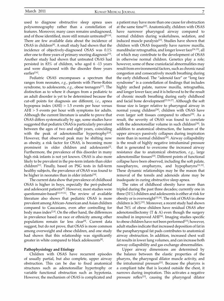
used to diagnose obstructive sleep apnea uses polysomnography rather than a constellation of features. Moreover, many cases remains undiagnosed, and of those identified, more still remain untreated[8,14]. There are few available data about the incidence of OSAS in children[8]. A small study had shown that the incidence of objectively-diagnosed OSAS was 0.1% after one to three years of primary snoring diagnosis[15]. Another study had shown that untreated OSAS had persisted in 83% of children, who aged 6 -13 years and were diagnosed with the disorder three years earlier[16].
Pediatric OSAS encompasses a spectrum that ranges from neonates, e.g., patients with Pierre-Robin syndrome, to adolescents, e.g., obese teenagers[17]. The distinction as to where it changes from a pediatric to an adult disorder is yet to be determined because the cut-off points for diagnosis are different, i.e., apnea hypopnea index (AHI) > 1.5 events per hour versus AHI > 5 events per hour with symptoms for adults. Although the current literature is unable to prove that OSAS differs systematically by age, some studies have suggested that pediatric OSAS is particularly prevalent between the ages of two and eight years, coinciding with the peak of adenotonsillar hypertrophy[2,8]. However, that observed peak may be shifting now as obesity, a risk factor for OSAS, is becoming more prominent in older children and adolescents[2]. Furthermore, the prevalence of this disorder in many high risk infants is not yet known. OSAS is also more likely to be prevalent in the pre-term infants than older children[11]. Finally, based on PSG tests in full-term healthy subjects, the prevalence of OSAS was found to be higher in neonates than in older infants[18].
The current data show that prevalence of childhood OSAS is higher in boys, especially the peri-pubertal and adolescent patients[8]. However, most studies were questionnaire-based[12,19-21]. The review of available literature also shows that pediatric OSAS is more prevalent among African-American and Asian children compared to Caucasians, even after controlling for body mass index[2,8]. On the other hand, the differences in prevalence based on race or ethnicity among other populations remain far less clear[8]. Current data suggest, but do not prove, that OSAS is more common among overweight and obese children, and one study had shown that this relationship was significantlygreater in white compared to black adolescents[8].
Pathophysiology and EtiologyChildren with OSAS have recurrent episodes
of usually partial, but also complete, upper airway obstruction. This can be due to fixed anatomicalstructures such as adenotonsillar hypertrophy or variable functional obstruction such as hypotonia. However, the mechanism of OSAS is complicated and
a patient may have more than one cause for obstruction at the same time[22]. Anatomically, children with OSAS have narrower pharyngeal airway compared to normal children during wakefulness, sedation, and induced muscle paralysis[23]. Studies have shown that children with OSAS frequently have narrow maxilla, mandibular retrognathia, and longer lower face[24, 25], all of which may contribute to the development of OSAS in otherwise normal children. Genetics play a role; however, some of these craniofacial abnormalities may result from environmental factors, such as chronic nasal congestion and consecutively mouth breathing during the early childhood. The “adenoid face” or “long face syndrome” is a constellation of findings that includeshighly arched palate, narrow maxilla, retrognathia, and longer lower face; and it is believed to be the result of chronic mouth breathing during early childhood and facial bone development[23,26,27]. Although the soft tissue size is larger relative to pharyngeal airway in normal young children[28], children with OSAS have even larger soft tissues compared to others[29]. As a result, the severity of OSAS was found to correlate with the adenotonsillar volume in such children[28]. In addition to anatomical obstruction, the lumen of the upper airways passively collapses during inspiration more than in normal children[30]. Airway collapsibility is the result of highly negative intraluminal pressure that is generated to overcome the increased airway resistance due to anatomical obstruction, e.g., large adenotonsillar tissues[23]. Different points of functional collapse have been observed, including the soft palate, nasopharynx, oropharynx, and hypopharynx[31,32]. These dynamic relationships may be the reason that removal of the tonsils and adenoids alone may be inadequate in curing obstructive sleep apnea.
The rates of childhood obesity have more than tripled during the past three decades; currently one in three (31%) children and adolescents is either at risk for obesity or is overweight[33,34]. The risk of OSAS in obese children is 36%[35]. Moreover, a recent study had shown that 76% of obese children have residual OSAS after adenotonsillectomy (T & A) even though the surgery resulted in improved AHI[36]. Imaging studies specificto obese children have not been performed[23]; however, adult studies indicate that increased deposition of fat in the parapharyngeal fat pads contributes to anatomical airway obstruction. In addition, increased chest wall fat results in lower lung volumes, and can increase both airway collapsibility and gas exchange abnormalities.
Upper airway dimensions are determined by the balance between the elastic properties of the pharynx, the pharyngeal dilator muscle activity, and the intraluminal pressure[23]. Because the pharynx is a compliant tube that is located outside the chest, it narrows during inspiration. This activates a negative pressure reflex[23], causing the pharyngeal dilator

March 20118
muscle to contract and compensate for the luminal narrowing. However, if the intraluminal pressure becomes increasingly negative, the pharynx may collapse and obstruct. In children with OSAS, the upper airway compliance is even higher compared to control subjects[31]. Their upper airways were also found to have lower muscle tone, decreased muscle response to hypercapnia and acute pressure changes, and impaired neural processing of respiratory load information during sleep[37-39]. This could be due to pathological changes to muscles, motor nerves, or sensory innervation of the upper airways that are chronically exposed to vibratory stress[23]. Overall, these data supported the conclusion that OSAS children are more prone to have functional upper airway obstruction than normal children thus suggesting that treatment of the anatomical obstruction alone may be woefully inadequate.
To understand the sequelae of pediatric OSAS, it is important to discuss the concept of cortical arousals and resultant sympathetic system activation. Cortical arousals, or increased brain activity without complete awakening, are believed to be necessary to alleviate airway obstruction by activation of pharyngeal dilator muscle. However, at the same time, it may increase the sympathetic output, and result in sleep disturbance[23]. Respiratory effort and hypercapnia, especially hypoxemic hypercapnia, are potent stimuli to reach arousal threshold[23,40], which varies according to the sleep stage, and is lowest during rapid eye movement (REM) stage. Although children with OSAS have normal overall ventilatory and arousal responses, and rely on arousal mechanism to sustain ventilation during sleep, they have high arousal threshold to respiratory work load and hypercapnia, compared to normal children[40,41]. The etiology of these findings isnot known, but it could be a stabilizing mechanism to decrease the sleep disturbance[23]. Moreover, if an obstructive event is associated with an arousal, the latter may cause a magnified ventilatory responsethat results into a significant functional airwayobstruction[42]. These factors may play a role in the lack of recognition of impairment in children who cannot report the symptoms compared to adults.
Clinical PresentationPrimary care physicians, including pediatricians
and general practitioners, are often the first healthcareprofessionals provided with the opportunity to recognize and diagnose pediatric OSAS. Many other medical practitioners, particularly otolaryngologists, developmental pediatricians, and pulmonologists, also see children with signs and symptoms of OSAS. In addition, because the clinical manifestations of pediatric OSAS include behavioral and academic problems, mental health professionals, including
psychologists and teachers may encounter children whose primary problem is OSAS. The symptoms of OSAS can be divided into nocturnal and diurnal symptoms (Table 1).
As in adults, loud, disruptive, continuous snoring is the most common symptom of OSAS in children; however, volume does not necessarily correlate with degree of obstruction. Parents may describe apneas
or breathing pauses, often followed by snorting or gasping. Because symptoms of OSAS may be worse during REM sleep, which is concentrated during the last third of the night, parents may not be awake to observe the most severe symptoms. However, some parents may observe and express considerable anxiety about these apnea episodes, and may report shaking the child during sleep to stimulate breathing or even bringing the child into the parental bed[43]. Parents may describe their child struggling to breathe with paradoxical chest wall movement, i.e., out-of-phase rib cage and abdominal movements. Children with OSAS are often described as having very restless and fitfulsleep, moving around frequently, and tossing and turning during sleep. They may sleep with their necks hyperextended, sitting upright with several pillows or other unusual positions in an attempt to reduce the airway obstruction. They may sweat excessively during sleep due to increased work of sleeping and high sympathetic system output. Secondary enuresis, or the recurrence of bedwetting in a child previously dry at night, may also occur in association with pediatric OSAS[44,45]. Very young children, or children incapable of making the ventilatory effort to generate noise of snoring, may present with failure to thrive or associated sequelae of OSAS as the first presentation.
Young children with sleep fragmentation are more likely to manifest daytime sleepiness behaviorally, through increased activity, aggression, poor concentration, and irritability rather than to act or complain of feeling “sleepy” as in adults. Excessive daytime sleepiness (EDS) was initially thought to be present in a small minority of children with OSAS; however, in more recent years, questionnaires indicated that the frequency of EDS may be higher, and revolve around 40 to 50%[46,47], particularly in obese adolescent.
Nocturnal Symptoms
Loud snoringObserved apneic pausesSnorting / gasping / chokingParadoxical chest wall movement
Restless sleep Abnormal sleeping positionDiaphoresis Secondary enuresis
Diurnal Symptoms
Daytime somnolenceDifficulty awakening in AMMorning headachesNasal congestion and hypo-nasal voiceMouth breathing and halitosis Frequent respiratory infections Poor AppetiteBehavioral / school problems
Table 1: Clinical symptoms of pediatric OSAS
Pediatric Obstructive Sleep Apnea Syndrome

KUWAIT MEDICAL JOURNAL 9March 2011
Children with OSAS may have difficulty awakeningin the morning despite having what appears to be adequate time in bed. Morning headaches may be a result of hypercapnia or hypoxemia. They may have recurrent otitis media and sinusitis, and poor appetite due to adenotonsillar hypertrophy and chronic nasal obstruction[2]. Behavioral and neurocognitive dysfunction as well as reduced academic achievement are well-recognized symptoms or complications of OSAS in children[48,49]. In addition, patients may present with attention deficit hyperactivity disorder (ADHD)-like symptoms, mood changes, and internalizing and externalizing behaviors[50,51].
Diagnosis and Evaluation
OSAS in a child is often suspected on the basis of parental complaints, but a high index of suspicion should remain for those at high risk. The patient’s evaluation should start with a thorough medical history to look for symptoms and complications of OSAS, and associated sleep disorders. The clinician should also look for risk factors for OSAS, which include prematurity; atopy, gastroesophageal reflux, andpassive smoking that are associated with chronic nasal congestion and obstruction (rhinitis); neuromuscular disease or genetic syndrome, e.g., cerebral palsy or
Down syndrome, which are associated with decreased muscular tone or abnormal upper airway anatomy[2]. The work up for OSAS also indicates the need for a general pediatric evaluation and a thorough evaluation of the upper airway anatomy. Starting with the nose, one should look for asymmetry of the nares, a large septal base and collapse of the nasal valves during inspiration, a deviated septum or enlargement of the inferior nasal turbinates. Next, the oropharynx should be examined for the position of the uvula in relation to the tongue. Mallampati scale[52] may help to evaluate this position. Although the scale is used in adults, its usefulness in pediatrics has not been determined. The size of the tonsils should be compared with the size of the airway; application of a standardized scale is useful[53]. The presence of a high and narrow hard palate and overlapping incisors are indicative of a small jaw and abnormal maxillo-mandibular development. The overall aspect of the face should also be appreciated for presence of characteristic adenoid face or long-face syndrome. This clinical evaluation provides important details of the upper airway anatomy and identifiesanatomical risk factors that can predispose a child to development of abnormal breathing during sleep.
Testing during sleep is the only way to confirmthe presence of OSAS[54]. In the context of habitual
Fig. 1: An epoch from a polysomnogram showing typical obstructive events (larger arrows), and arterial oxygen desaturation (small arrows)

March 201110
snoring in otherwise healthy children (usually albeit not universally defined as snoring loud enough to berecognized by parents at least three nights per week), clinical evaluation will usually yield 25 - 72% success rates in predicting who has OSAS[55]. This relatively poor predictive ability has, therefore, prompted the recognition and recommendation to refer symptomatic children for PSG to confirm or dispel the diagnosis ofOSAS[46,54,56,57]. PSG must always include monitoring of sleep-wake states through electroencephalography, electro-oculography, chin and leg electromyography, electrocardiography, body position, and appropriate monitoring of breathing effort, arterial oxygenation and carbon dioxide (Fig. 1). The PSG study should be performed overnight. Individual nap PSG studies are not very sensitive in predicting overnight PSG findings,and significantly underestimate the severity of OSAS[58]. Other potential diagnostic alternatives would involve the use of unattended overnight oximetry studies at home (Fig. 2); however, this test is reliable only in severe cases of OSAS[59,60]. Moreover, this test is only useful when the result is positive for OSAS due to its lower negative predictive value. More recently, the electrocardiogram signal or other tools providing non-invasive assessments of changes in autonomic nervous system tone have been proposed as potentially viable alternatives that when coupled with oximetry would reveal both hypoxic events and arousals[61,62]. However,
these techniques are not widely used in pediatric sleep laboratories or clinically and certainly merit further exploration.
The diagnosis of OSAS in children, however, still is a combination of clinical and objective measures. In the latest edition of the International Classificationof Sleep Disorders[1], the American Academy of Sleep Medicine specified the diagnostic criteria fordiagnosing pediatric OSAS, which necessitated the presence of OSAS symptoms, absence of other sleep disorders that may explain these symptoms, and a PSG confirmation of the OSAS diagnosis (Table 2).
TREATMENTOSAS in children is most commonly associated
with adenotonsillar hypertrophy, even when obesity is present, such that the currently recommended initial treatment consists of T&A[7]. Isolated tonsillectomy or adenoidectomy can be performed; however, these are not as effective as T&A[63,64]. In recent years, however, it has become apparent that not all children who undergo T&A for OSAS are cured[65,66]. The success rate for T&A in the context of OSAS is approximately 85%[67], and this figure may actually be lower, particularly amongobese children with OSAS, or among children with severe OSAS[22,36,66].
For children in whom T&A is not an option, or it does not lead to complete resolution and a residual
Fig. 2: A graphical summary from an unattended overnight oximetry study
Pediatric Obstructive Sleep Apnea Syndrome
KUWAIT MEDICAL JOURNAL 11March 2011
severity of OSAS considered moderate to severe, i.e., obstructive AHI > 5 events/hr, the only interventional option consists of the administration of positive airway pressure therapy, either continuous (CPAP) or bi-level positive airway pressure (BiPAP) therapy[68,69]. However, this treatment modality can be associated with difficulties, which are related to training thefamily and child and finding the appropriate maskinterface. One study that compared use of CPAP versus BiPAP showed no difference but the numbers were small and despite intensive support, there was a high dropout rate in using either CPAP or BiPAP[70]. The CPAP was found to be effective in decreasing the AHI and increasing the arterial oxygen saturation of the children with OSAS. However, children often need to be trained to tolerate the facial mask interface, sometimes with desensitization and sometimes with more intensive behavioral treatment involving psychologists. Moreover, intensive and
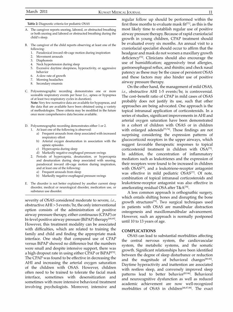
A. The caregiver reports snoring, labored, or obstructed breathing or both snoring and labored or obstructed breathing during the child’s sleep.
B. The caregiver of the child reports observing at least one of the following:1. Paradoxical inward rib-cage motion during inspiration2. Movement arousals3. Diaphoresis4. Neck hypertension during sleep5. Excessive daytime sleepiness, hyperactivity, or aggressive
behavior6. A slow rate of growth7. Morning headaches8. Secondary enuresis
C. Polysomnographic recording demonstrates one or more scoreable respiratory events per hour (i.e., apnea or hypopnea of at least two respiratory cycles in duration).Note: Very few normative data are available for hypopneas, and the data that are available have been obtained using a variety of methodologies. These criteria may be modified in the futureonce more comprehensive data become available.
D. Polysomnographic recording demonstrates either 1 or 2.1. At least one of the following is observed:
a) Frequent arousals from sleep associated with increased respiratory effort
b) Arterial oxygen desaturation in association with the apneic episodes
c) Hypercapnia during sleepd) Markedly negative esophageal pressure swings
2. Periods of hypercapnia, desaturation, or hypercapnia and desaturation during sleep associated with snoring, paradoxical inward rib-cage motion during inspiration, and at least one of the following:a) Frequent arousals from sleepb) Markedly negative esophageal pressure swings
E. The disorder is not better explained by another current sleep disorder, medical or neurological disorder, medication use, or substance use disorder.
regular follow up should be performed within the first three months to evaluate mask fit[71], as this is the most likely time to establish regular use of positive airway pressure therapy. Because of rapid craniofacial growth in young children, CPAP treatment should be evaluated every six months. An annual visit to a craniofacial specialist should occur to affirm that theheadgear and mask do not worsen a maxillary growth deficiency[72]. Clinicians should also encourage the use of humidification; aggressively treat allergies,gastroesophageal reflux, and rhinitis; and check nasalpatency as these may be the cause of persistent OSAS and these factors may also hinder use of positive airway pressure therapy.
On the other hand, the management of mild OSAS, i.e., obstructive AHI 1-5 events/hr, is controversial. The cost–benefit ratio of CPAP in mild cases of OSASprobably does not justify its use, such that other approaches are being advocated. One approach is the topical intranasal application of corticosteroids. In a series of studies, significant improvements in AHI andarterial oxygen saturation have been demonstrated in a cohort of children with OSAS or in children with enlarged adenoids[73,74]. Those findings are notsurprising considering the expression patterns of glucocorticoid receptors in the upper airways, which suggest favorable therapeutic responses to topical corticosteroid treatment in children with OSA[75]. In addition, the concentration of inflammatorymediators such as leukotrienes and the expression of their receptors were found to be increased in children with OSAS[76], and a leukotriene-receptor antagonist was effective in mild pediatric OSAS[77]. Of note, combination of topical intranasal corticosteroids and leukotriene-receptor antagonist was also effective in ameliorating residual OSA after T&A[78].
A less common approach is orthognathic surgery, which entails shifting bones and disrupting the bone growth structures[79]. Two surgical techniques used in patients with OSAS are mandibular distraction osteogenesis and maxillomandibular advancement. However, such an approach is normally postponed until 10 to 13 years of age.
COMPLICATIONS OSAS can lead to substantial morbidities affecting
the central nervous system, the cardiovascular system, the metabolic systems, and the somatic growth. Significant relationships have been identifiedbetween the degree of sleep disturbance or reduction and the magnitude of behavioral changes[80-82]. Daytime hyperactivity and inattention are associated with restless sleep, and conversely improved sleep patterns lead to better behavior[82-86]. Behavioral and neurocognitive dysfunction as well as reduced academic achievement are now well-recognized morbidities of OSAS in children[48,87,88]. The exact
Table 2: Diagnostic criteria for pediatric OSAS

March 201112
mechanisms by which OSAS elicits such neural deficitsremain relatively unclear. Most likely, both cortical arousals and episodic hypoxemia that characterize OSAS lead to alterations within the prefrontal cortex with resultant executive dysfunction[89,90]. However, not all children with OSAS actually manifest cognitive morbidities, suggesting that other factors may be playing a role in this process[7]. These factors may include obesity, genetics, and environmental risk factors.
Similar to adult OSAS, pediatric OSAS has been associated with a higher risk for cardiovascular morbidities, which include systemic hypertension and left ventricular changes[91,92]. The mechanisms mediating cardiac and blood pressure changes are most likely associated with the increases in sympathetic activity and reactivity that progressively develop in the context of OSAS[93]. In addition, there is an assumption of potential endothelial dysfunction in children with OSAS, as evidenced by increase in circulating levels of several adhesion molecules[94]. Moreover, it has been hypothesized that the intermittent hypoxemia, occurring during sleep of children with OSAS, may induce elevation of pulmonary artery pressure, and such event may lead to some degree of right ventricular dysfunction; however, this has not been systematically examined[95].
OSAS was also found to increase the risk for metabolic syndrome (Clustering of insulin resistance, dyslipidemia, hypertension, and obesity) in obese children[96]. Although the initial description of OSAS included failure to thrive, this is not the case nowadays, with only ≤ 5% of pediatric OSAS manifesting this problem[7,97]. On the other hand, treatment of OSAS in children resulted in weight gain even in obese patients[98]. The proposed mechanisms for somatic growth alterations in OSAS are decreased levels of insulin-like growth factor-I and insulin-like growth factor–binding proteins, and possibly decreased growth hormone release[99]. These could be the result of fragmented night time sleep due to frequent cortical arousals in children with OSAS.
CONCLUSIONOSAS is a common disorder in children, and it has
complex mechanism and etiology. It may manifest in different clinical case scenarios; hence, clinicians should have high index of suspicion in order to be able to diagnose this disorder. The diagnosis requires full clinical evaluation and an objective test for confirmation. The first-line treatment of pediatricOSAS is T&A; however, it may not be curative in children, in whom other treatment modalities should be considered. If unrecognized and untreated, pediatric OSAS can lead to significant medical complicationsand morbidities.
REFERENCES
1. Satecia M. The International Classification of SleepDisorders. Westchester, IL, 2005; 33-77.
2. Mindell J, Owens J. A Clinical Guide to Pediatric Sleep Diagnosis and Management of Sleep Problems. Philadelphia, Lippincott Williams & Wilkins 2010; 100-115.
3. Katz E, Marcus C. Diagnosis of Obstructive Sleep Apnea Syndrome in Infants and Children. In: Sheldon S, Ferber R, Kryger M, editors. Principles and Practice of Pediatric Sleep Medicine. 2nd edition. Philadelphia: Elsevier Saunders; 2005. p. 197-210.
4. Guilleminault C, Eldridge FL, Simmons FB, Dement WC. Sleep apnea in eight children. Pediatrics 1976; 58:23-30.
5. Gozal D. Obstructive sleep apnea in children: implications for the developing central nervous system. Semin Pediatr Neurol 2008; 15:100-106.
6. Bhattacharjee R, Kheirandish-Gozal L, Pillar G, Gozal D. Cardiovascular complications of obstructive sleep apnea syndrome: evidence from children. Prog Cardiovasc Dis 2009; 51:416-433.
7. Capdevila OS, Kheirandish-Gozal L, Dayyat E, Gozal D. Pediatric obstructive sleep apnea: complications, management, and long-term outcomes. Proc Am Thorac Soc 2008; 5:274-282.
8. Lumeng JC, Chervin RD. Epidemiology of pediatric obstructive sleep apnea. Proc Am Thorac Soc 2008; 5:242-252.
9. Ali NJ, Pitson DJ, Stradling JR. Snoring, sleep disturbance, and behaviour in 4-5 year olds. Arch Dis Child 1993; 68:360-366.
10. Ng DK, Kwok KL, Poon G, Chau KW. Habitual snoring and sleep bruxism in a paediatric outpatient population in Hong Kong. Singapore Med J 2002; 43:554-556.
11. Rosen CL, Larkin EK, Kirchner HL, et al. Prevalence and risk factors for sleep-disordered breathing in 8 to 11-year-old children: association with race and prematurity. J Pediatr 2003; 142:383-389.
12. Kaditis AG, Finder J, Alexopoulos EI, et al. Sleep-disordered breathing in 3,680 Greek children. Pediatr Pulmonol 2004; 37:499-509.
13. Anuntaseree W, Kuasirikul S, Suntornlohanakul S. Natural history of snoring and obstructive sleep apnea in Thai school-age children. Pediatr Pulmonol 2005; 39:415-420.
14. Chervin RD, Archbold KH, Panahi P, Pituch KJ. Sleep problems seldom addressed at two general pediatric clinics. Pediatrics 2001; 107:1375-1380.
15. Marcus CL, Hamer A, Loughlin GM. Natural history of primary snoring in children. Pediatr Pulmonol 1998; 26:6-11.
16. Anuntaseree W, Rookkapan K, Kuasirikul S, Thongsuksai P. Snoring and obstructive sleep apnea in Thai school-age children: prevalence and predisposing factors. Pediatr Pulmonol 2001; 32:222-227.
17. Nixon GM, Brouillette RT. Sleep. 8 paediatric obstructive sleep apnoea. Thorax 2005; 60:511-516.
18. Kato I, Franco P, Groswasser J, Kelmanson I, Togari H, Kahn A. Frequency of obstructive and mixed sleep apneas in 1,023 infants. Sleep 2000; 23:487-492.
Pediatric Obstructive Sleep Apnea Syndrome

KUWAIT MEDICAL JOURNAL 13March 2011
19. Delasnerie-Laupretre N, Patois E, Valatx JL, Kauffmann F, Alperovitch A. Sleep, snoring and smoking in high school students. J Sleep Res 1993; 2:138-142.
20. Liu X, Ma Y, Wang Y, et al. Brief report: An epidemiologic survey of the prevalence of sleep disorders among children 2 to 12 years old in Beijing, China. Pediatrics 2005; 115:266-268.
21. Bidad K, Anari S, Aghamohamadi A, Gholami N, Zadhush S, Moaieri H. Prevalence and correlates of snoring in adolescents. Iran J Allergy Asthma Immunol 2006; 5:127-132.
22. Guilleminault C, Huang YS, Glamann C, Li K, Chan A. Adenotonsillectomy and obstructive sleep apnea in children: a prospective survey. Otolaryngol Head Neck Surg 2007; 136:169-175.
23. Katz ES, D’Ambrosio CM. Pathophysiology of pediatric obstructive sleep apnea. Proc Am Thorac Soc 2008; 5:253-262.
24. Lofstrand-Tidestrom B, Thilander B, Ahlqvist-Rastad J, Jakobsson O, Hultcrantz E. Breathing obstruction in relation to craniofacial and dental arch morphology in 4-year-old children. Eur J Orthod 1999; 21:323-332.
25. Kawashima S, Niikuni N, Chia-hung L, et al. Cephalometric comparisons of craniofacial and upper airway structures in young children with obstructive sleep apnea syndrome. Ear Nose Throat J 2000; 79:499-502, 505-506.
26. Linder-Aronson S. Adenoids. Their effect on mode of breathing and nasal airflow and their relationship tocharacteristics of the facial skeleton and the denition. A biometric, rhino-manometric and cephalometro-radiographic study on children with and without adenoids. Acta Otolaryngol Suppl 1970; 265:1-132.
27. Peltomaki T. The effect of mode of breathing on craniofacial growth--revisited. Eur J Orthod 2007; 29:426-429.
28. Jeans WD, Fernando DC, Maw AR, Leighton BC. A longitudinal study of the growth of the nasopharynx and its contents in normal children. Br J Radiol 1981; 54:117-121.
29. Arens R, McDonough JM, Costarino AT, et al. Magnetic resonance imaging of the upper airway structure of children with obstructive sleep apnea syndrome. Am J Respir Crit Care Med 2001; 164:698-703.
30. Arens R, Sin S, McDonough JM, et al. Changes in upper airway size during tidal breathing in children with obstructive sleep apnea syndrome. Am J Respir Crit Care Med 2005; 171:1298-1304.
31. Isono S, Shimada A, Utsugi M, Konno A, Nishino T. Comparison of static mechanical properties of the passive pharynx between normal children and children with sleep-disordered breathing. Am J Respir Crit Care Med 1998; 157:1204-1212.
32. Donnelly LF, Surdulescu V, Chini BA, Casper KA, Poe SA, Amin RS. Upper airway motion depicted at cine MR imaging performed during sleep: comparison between young patients with and those without obstructive sleep apnea. Radiology 2003; 227:239-245.
33. Hedley AA, Ogden CL, Johnson CL, Carroll MD, Curtin LR, Flegal KM. Prevalence of overweight and obesity among US children, adolescents, and adults, 1999-2002. JAMA 2004; 291:2847-2850.
34. Kim Y, Lee S. Physical activity and abdominal obesity in youth. Appl Physiol Nutr Metab 2009; 34:571-581.
35. Marcus CL, Curtis S, Koerner CB, Joffe A, Serwint JR, Loughlin GM. Evaluation of pulmonary function and polysomnography in obese children and adolescents. Pediatr Pulmonol 1996; 21:176-183.
36. Mitchell RB, Kelly J. Outcome of adenotonsillectomy for obstructive sleep apnea in obese and normal-weight children. Otolaryngol Head Neck Surg 2007; 137:43-48.
37. Katz ES, White DP. Genioglossus activity in children with obstructive sleep apnea during wakefulness and sleep onset. Am J Respir Crit Care Med 2003; 168:664-670.
38. Huang J, Marcus C, Melendres C, Karamessinis L, Pepe M, Samuel J. Respiratory sensation during sleep in children with obstructive sleep apnea syndrome (abstract). Sleep 2007; 30:A67.
39. Marcus CL, Katz ES, Lutz J, Black CA, Galster P, Carson KA. Upper airway dynamic responses in children with the obstructive sleep apnea syndrome. Pediatr Res 2005; 57:99-107.
40. Marcus CL, Lutz J, Carroll JL, Bamford O. Arousal and ventilatory responses during sleep in children with obstructive sleep apnea. J Appl Physiol 1998; 84:1926-1936.
41. Marcus CL, Moreira GA, Bamford O, Lutz J. Response to inspiratory resistive loading during sleep in normal children and children with obstructive apnea. J Appl Physiol 1999; 87:1448-1454.
42. Katz ES, Marcus CL, White DP. Influence of airwaypressure on genioglossus activity during sleep in normal children. Am J Respir Crit Care Med 2006; 173:902-909.
43. Brouillette RT, Fernbach SK, Hunt CE. Obstructive sleep apnea in infants and children. J Pediatr 1982; 100:31-40.
44. Weider DJ, Sateia MJ, West RP. Nocturnal enuresis in children with upper airway obstruction. Otolaryngol Head Neck Surg 1991; 105:427-432.
45. Nowak KC, Weider DJ. Pediatric nocturnal enuresis secondary to airway obstruction from cleft palate repair. Clin Pediatr (Phila) 1998; 37:653-657.
46. Carroll JL, McColley SA, Marcus CL, Curtis S, Loughlin GM. Inability of clinical history to distinguish primary snoring from obstructive sleep apnea syndrome in children. Chest 1995; 108:610-618.
47. Chervin RD, Weatherly RA, Ruzicka DL, et al. Subjective sleepiness and polysomnographic correlates in children scheduled for adenotonsillectomy Vs other surgical care. Sleep 2006; 29:495-503.
48. Gozal D. Sleep-disordered breathing and school performance in children. Pediatrics 1998; 102:616-620.
49. Chervin RD, Clarke DF, Huffman JL, et al. School performance, race, and other correlates of sleep-disordered breathing in children. Sleep Med 2003; 4:21-27.
50. O’Brien LM, Gozal D. Sleep in children with attention deficit/hyperactivity disorder. Minerva Pediatr 2004;56:585-601.
51. Beebe DW. Neurobehavioral morbidity associated with disordered breathing during sleep in children: a comprehensive review. Sleep 2006; 29:1115-1134.
52. Mallampati SR, Gatt SP, Gugino LD, et al. A clinical sign to predict difficult tracheal intubation: a prospectivestudy. Can Anaesth Soc J 1985; 32:429-434.

March 201114
53. Friedman M, Tanyeri H, La Rosa M, et al. Clinical predictors of obstructive sleep apnea. Laryngoscope 1999; 109:1901-1907.
54. Clinical practice guideline: diagnosis and management of childhood obstructive sleep apnea syndrome. Pediatrics 2002; 109:704-712.
55. Kheirandish-Gozal L, Gozal D. The multiple challenges of obstructive sleep apnea in children: diagnosis. Curr Opin Pediatr 2008; 20:650-653.
56. Goldstein NA, Sculerati N, Walsleben JA, Bhatia N, Friedman DM, Rapoport DM. Clinical diagnosis of pediatric obstructive sleep apnea validated by polysomnography. Otolaryngol Head Neck Surg 1994; 111:611-617.
57. Wang RC, Elkins TP, Keech D, Wauquier A, Hubbard D. Accuracy of clinical evaluation in pediatric obstructive sleep apnea. Otolaryngol Head Neck Surg 1998; 118:69-73.
58. Saeed MM, Keens TG, Stabile MW, Bolokowicz J, Davidson Ward SL. Should children with suspected obstructive sleep apnea syndrome and normal nap sleep studies have overnight sleep studies? Chest 2000; 118:360-365.
59. Kirk VG, Bohn SG, Flemons WW, Remmers JE. Comparison of home oximetry monitoring with laboratory polysomnography in children. Chest 2003; 124:1702-1708.
60. Nixon GM, Kermack AS, Davis GM, Manoukian JJ, Brown KA, Brouillette RT. Planning adenotonsillectomy in children with obstructive sleep apnea: the role of overnight oximetry. Pediatrics 2004; 113:e19-25.
61. Pepin JL, Delavie N, Pin I, et al. Pulse transit time improves detection of sleep respiratory events and microarousals in children. Chest 2005; 127:722-730.
62. O’Brien LM, Gozal D. Potential usefulness of noninvasive autonomic monitoring in recognition of arousals in normal healthy children. J Clin Sleep Med 2007; 3:41-47.
63. Guilleminault C, Li KK, Khramtsov A, Pelayo R, Martinez S. Sleep disordered breathing: surgical outcomes in prepubertal children. Laryngoscope 2004; 114:132-137.
64. Guilleminault C, Li K, Quo S, Inouye RN. A prospective study on the surgical outcomes of children with sleep-disordered breathing. Sleep 2004; 27:95-100.
65. Tal A, Bar A, Leiberman A, Tarasiuk A. Sleep characteristics following adenotonsillectomy in children with obstructive sleep apnea syndrome. Chest 2003; 124:948-953.
66. Mitchell RB, Kelly J. Outcome of adenotonsillectomy for severe obstructive sleep apnea in children. Int J Pediatr Otorhinolaryngol 2004; 68:1375-1379.
67. Lipton AJ, Gozal D. Treatment of obstructive sleep apnea in children: do we really know how? Sleep Med Rev 2003; 7:61-80.
68. Marcus CL, Ward SL, Mallory GB, et al. Use of nasal continuous positive airway pressure as treatment of childhood obstructive sleep apnea. J Pediatr 1995; 127:88-94.
69. Massa F, Gonsalez S, Laverty A, Wallis C, Lane R. The use of nasal continuous positive airway pressure to treat obstructive sleep apnoea. Arch Dis Child 2002; 87:438-443.
70. Marcus CL, Rosen G, Ward SL, et al. Adherence to and effectiveness of positive airway pressure therapy in children with obstructive sleep apnea. Pediatrics 2006; 117:e442-451.
71. Guilleminault C, Pelayo R, Clerk A, Leger D, Bocian RC. Home nasal continuous positive airway pressure in infants with sleep-disordered breathing. J Pediatr 1995; 127:905-912.
72. Li KK, Riley RW, Guilleminault C. An unreported risk in the use of home nasal continuous positive airway pressure and home nasal ventilation in children: mid-face hypoplasia. Chest 2000; 117:916-918.
73. Brouillette RT, Manoukian JJ, Ducharme FM, et al. Efficacy of fluticasone nasal spray for pediatricobstructive sleep apnea. J Pediatr 2001; 138:838-844.
74. Berlucchi M, Salsi D, Valetti L, Parrinello G, Nicolai P. The role of mometasone furoate aqueous nasal spray in the treatment of adenoidal hypertrophy in the pediatric age group: preliminary results of a prospective, randomized study. Pediatrics 2007; 119:e1392-1397.
75. Goldbart AD, Veling MC, Goldman JL, Li RC, Brittian KR, Gozal D. Glucocorticoid receptor subunit expression in adenotonsillar tissue of children with obstructive sleep apnea. Pediatr Res 2005; 57:232-236.
76. Goldbart AD, Goldman JL, Li RC, Brittian KR, Tauman R, Gozal D. Differential expression of cysteinyl leukotriene receptors 1 and 2 in tonsils of children with obstructive sleep apnea syndrome or recurrent infection. Chest 2004; 126:13-18.
77. Goldbart AD, Goldman JL, Veling MC, Gozal D. Leukotriene modifier therapy for mild sleep-disordered breathing in children. Am J Respir Crit Care Med 2005; 172:364-370.
78. Kheirandish L, Goldbart AD, Gozal D. Intranasal steroids and oral leukotriene modifier therapy inresidual sleep-disordered breathing after tonsillectomy and adenoidectomy in children. Pediatrics 2006; 117:e61-66.
79. Guilleminault C, Li KK. Maxillomandibular expansion for the treatment of sleep-disordered breathing: preliminary result. Laryngoscope 2004; 114:893-896.
80. O’Brien LM, Holbrook CR, Mervis CB, et al. Sleep and neurobehavioral characteristics of 5 to 7-year-old children with parentally reported symptoms of attention-deficit/hyperactivity disorder. Pediatrics2003; 111:554-563.
81. Aronen ET, Paavonen EJ, Fjallberg M, Soininen M, Torronen J. Sleep and psychiatric symptoms in school-age children. J Am Acad Child Adolesc Psychiatry 2000; 39:502-508.
82. Chervin RD, Archbold KH. Hyperactivity and polysomnographic findings in children evaluated forsleep-disordered breathing. Sleep 2001; 24:313-320.
83. Stein MA, Mendelsohn J, Obermeyer WH, Amromin J, Benca R. Sleep and behavior problems in school-aged children. Pediatrics 2001; 107:E60.
84. Lavigne JV, Arend R, Rosenbaum D, et al. Sleep and behavior problems among preschoolers. J Dev Behav Pediatr 1999; 20:164-169.
85. Kaplan BJ, McNicol J, Conte RA, Moghadam HK. Sleep disturbance in preschool-aged hyperactive and nonhyperactive children. Pediatrics 1987; 80:839-844.
Pediatric Obstructive Sleep Apnea Syndrome

KUWAIT MEDICAL JOURNAL 15March 2011
86. Mulvaney SA, Goodwin JL, Morgan WJ, Rosen GR, Quan SF, Kaemingk KL. Behavior problems associated with sleep disordered breathing in school-aged children--the Tucson children’s assessment of sleep apnea study. J Pediatr Psychol 2006; 31:322-330.
87. Ali NJ, Pitson D, Stradling JR. Sleep disordered breathing: effects of adenotonsillectomy on behaviour and psychological functioning. Eur J Pediatr 1996; 155:56-62.
88. Weissbluth M, Davis AT, Poncher J, Reiff J. Signs of airway obstruction during sleep and behavioral, developmental, and academic problems. J Dev Behav Pediatr 1983; 4:119-121.
89. Beebe DW, Gozal D. Obstructive sleep apnea and the prefrontal cortex: towards a comprehensive model linking nocturnal upper airway obstruction to daytime cognitive and behavioral deficits. J Sleep Res 2002; 11:1-16.
90. Bass JL, Corwin M, Gozal D, et al. The effect of chronic or intermittent hypoxia on cognition in childhood: a review of the evidence. Pediatrics 2004; 114:805-816.
91. Enright PL, Goodwin JL, Sherrill DL, Quan JR, Quan SF. Blood pressure elevation associated with sleep-related breathing disorder in a community sample of white and Hispanic children: the Tucson Children’s Assessment of Sleep Apnea study. Arch Pediatr Adolesc Med 2003; 157:901-904.
92. Amin RS, Kimball TR, Bean JA, et al. Left ventricular hypertrophy and abnormal ventricular geometry in
children and adolescents with obstructive sleep apnea. Am J Respir Crit Care Med 2002; 165:1395-1399.
93. O’Brien LM, Gozal D. Autonomic dysfunction in children with sleep-disordered breathing. Sleep 2005; 28:747-752.
94. O’Brien LM, Serpero LD, Tauman R, Gozal D. Plasma adhesion molecules in children with sleep-disordered breathing. Chest 2006; 129:947-953.
95. Shiomi T, Guilleminault C, Stoohs R, Schnittger I. Obstructed breathing in children during sleep monitored by echocardiography. Acta Paediatr 1993; 82:863-871.
96. Waters KA, Sitha S, O’Brien LM, et al. Follow-up on metabolic markers in children treated for obstructive sleep apnea. Am J Respir Crit Care Med 2006; 174:455-460.
97. Everett AD, Koch WC, Saulsbury FT. Failure to thrive due to obstructive sleep apnea. Clin Pediatr (Phila) 1987; 26:90-92.
98. Roemmich JN, Barkley JE, D’Andrea L, et al. Increases in overweight after adenotonsillectomy in overweight children with obstructive sleep-disordered breathing are associated with decreases in motor activity and hyperactivity. Pediatrics 2006; 117:e200-208.
99. Nieminen P, Lopponen T, Tolonen U, Lanning P, Knip M, Lopponen H. Growth and biochemical markers of growth in children with snoring and obstructive sleep apnea. Pediatrics 2002; 109:e55.

KUWAIT MEDICAL JOURNAL March 201116
Original Article
Kuwait Medical Journal 2011; 43 (1): 16-19
The Treatment of Chronic Monteggia Lesions and Chronic Traumatic Isolated Radial Head Dislocations
ABSTRACT
Objectives: Isolated dislocations of the radiocapitellar joint or those associated with fractures of the ulna (Monteggia fracture-dislocations) are often missed in the emergency room. When left untreated radial head dislocations can cause pain, loss of motion, and progressive cubitus valgus. This report describes open reduction and annular ligament reconstruction to treat chronic radial head dislocations. Design: Retrospective chart review of 11 patients with chronic radial head dislocationsSetting: Texas Scottish Rite Hospital and Cook Fort Worth Children’s Hospital,USASubjects: Eleven patients had chronic radial head dislocations; six Monteggias, two intra-articular fracture-dislocations, and three isolated radial head dislocations Intervention: Four patients were left untreated. Seven patients had surgical reconstruction with open reduction of
the radiocapitellar joint and repair (1 patient) or reconstruction (6 patients) of the annular ligament. Three osteotomies were performed to straighten any ulnar malunion. Main Outcome Measures: Two of the four untreated patients demonstrated progressive cubitus valgus. Five of the seven operated patients had normal carrying angles and recovered full flexion and extension but lost 30% oftheir pronosupination. The two intra-articular fracture-dislocations had very poor results due to extremely stiff joints. Conclusions: We recommend surgical relocation and annular ligament reconstruction to treat chronic radial head dislocations. This reconstruction prevented cubitus valgus in patients with chronic radial head dislocations and restored elbow flexion. Patients with fracture-dislocations do notrespond as well.
KEY WORDS: Bell Tawse reconstruction, Monteggia
James W Roach1, Hanna M Saltzman2
1Department of Orthopedics, University of Pittsburgh Children’s Hospital, Pittsburgh, USA2Department of Orthopedics, University of Utah and Department of Pediatric Orthopedics, Shriners Hospitals for Children,
Salt Lake City, Utah, USA
Address correspondence to:James W Roach MD, Professor of Orthopedic Surgery, Department of Orthopedics, University of Pittsburgh, Children’s Hospital of Pittsburgh, 4401 Penn Ave Faculty Pavilion 4th Floor, Pittsburgh, PA 15224, USA. Tel: 1 412 692-7818, E-mail: [email protected]
INTRODUCTIONMonteggia fracture-dislocations or dislocations
of the radiocapitellar joint associated with fractures of the ulna often go unrecognized during the initial treatment of the ulnar fracture. Missed Monteggia fracture-dislocations are a common cause of chronic radial head dislocations and are often not discovered until months after the original injury. Traumatic radial head dislocation can occur in isolation from an ulnar fracture. When left untreated, both Monteggia lesions and isolated radial head dislocations can cause pain, limit the range of elbow motion, and produce progressive cubitus valgus[1]. Neuropathy of both the ulnar and radial nerves has been reported. The ulnar nerve can become stretched as the elbow gradually drifts into progressive valgus and the radial nerve
can become compressed as the radial head dislocates anteriorly[2,3].
When a Monteggia injury presents acutely, the dislocation and fracture are both reduced either with manipulation only or with surgical open reduction and internal fixation. Controversy exists about thebest treatment for children whose injuries are initially missed and then present late with a chronic radial head dislocation. Treatment of radial head dislocations presenting within a few weeks of the injury can often still be reduced with manipulation without surgery[4]. Dislocations that persist longer than 6 to 8 weeks usually require surgery[4]. Surgical reduction of a radial head dislocation is provided through an open reduction and often the annular ligament is reconstructed using a slip of the triceps tendon[5,6]. Additional procedures may

KUWAIT MEDICAL JOURNAL 17March 2011
be necessary and include ulnar osteotomies[7], radial osteotomies[8-10], gradual lengthening and angulation of the ulna with an external frame[9,11], or several combinations of these techniques. The necessity and effectiveness of these reconstructive strategies depend partly upon the child’s age and the elapsed time since the initial fracture[1]. This follow-up report elucidates the benefits of using open reductions and annularligament reconstructions, with osteotomies added as necessary, to treat chronic radial head dislocations.
SUBJECTS AND METHODSWe treated 11 patients with chronic radial head
dislocations. Ten patients were from the Texas Scottish Rite Hospital in Dallas, Texas and one was from the Cook - Fort Worth Children’s Medical Center in Fort Worth, Texas. This study was approved as chart review research. All patients were initially examined clinically and radiographically. All seven surgical patients and two of the four untreated patients were examined clinically and radiographically at follow-up.
RESULTSSix patients had Monteggia fracture-dislocations,
two had intra-articular fractures of the olecranon with dislocations of the radial head, and three had isolated radial head dislocations. Seven patients underwent surgery and four were followed but left untreated
Treatment Monteggia(n)
Radial head dislocations(n)
Intra-articular fracture dislocations with annular ligament disruption(n)
ObservationSurgeryTotal
246
213
022
Table 1: Mode of treatment undertaken
Surgical technique used Monteggia(n = 4)
Radial head dislocations(n = 1)
Intra-articular fracture dislocations(n = 2)
Open reductionUlnar osteotomyDirect repair of annular ligamentBell Tawse
4304
1001
2011
Table 2: Surgical technique adopted
(Table 1). All seven surgical patients underwent open reduction of the radiocapitellar joint, osteotomy of any ulnar angulation (3 patients), and either a direct repair (1 patient) or a reconstruction of the annular ligament (6 patients) (Table 2). Annular ligament reconstruction was accomplished as described by Bell Tawse (Fig. 1 - 5)[6]. The average age at the time of injury was seven years and one month; the youngest child at the time of injury was three years and nine months old and the oldest was 11 years old. At the time of presentation the average age was eight years and six months, ranging from four years to 16 years. The average delay in presentation was 17 months (range 3 to 60 months).
Four patients remained untreated, two because they were more than two years from the time of injury and two because their parents did not want surgery. Two of the four were followed; during the follow-up interval both demonstrated progressive cubitus valgus. The other two patients were lost to follow-up. The rest of the cohort (7 patients) had surgical reconstructions. Our preferred method of treatment was open reduction of the radiocapitellar joint together with
Fig. 1: Lateral radiograph of child with chronic Monteggia dislocation and ulnar malunion
Fig. 2: Reconstitution of annular ligament using strip of triceps tendon prior to final sutures

March 201118
repair or reconstruction of the annular ligament and this was completed on all seven patients. Osteotomies were performed as necessary to straighten any ulnar malunion. Among the radial head open reductions, two were done with direct repair of the annular ligament, four with annular ligament reconstruction using a slip of triceps tendon[6] and one with annular ligament augmentation using a autologous plantaris tendon. One patient had an ulnar osteotomy, and one had a radial neck osteotomy.
The average time of follow-up was two years and 10 months, ranging from 18 months to five years.Five of the seven operated patients recovered full flexion and extension of their elbow but experiencedan average loss of 30% of their pronosupination. All five of these patients had normal carrying angles andradiographically reduced radial heads. The remaining two operated patients, both of whom had intra-articular fractures of the olecranon together with dislocations of the radial heads, were left with very poor results due to extremely stiff joints. The two fracture-dislocation patients were fixed in neutral pronosupination andone had 30 degrees of flexion contracture with furtherflexion to 90 degrees and the other 60 degrees offlexion contracture with further flexion to 90 degrees.Both of the fracture-dislocation patients had attempts at surgical reductions performed at outside hospitals. At our facility the last fracture-dislocation patient had an attempted Bell-Tawse reconstruction together with a radial neck osteotomy, but the radiocapitellar joint redislocated and the radial head developed avascular necrosis.
DISCUSSIONWe believe the study provides useful information
regarding surgical reconstruction of radial head dislocations and the natural history of progressive cubitus valgus in those patients with dislocated radial heads who are left untreated. The surgical
reconstructions of our patients with chronic Monteggia fracture-dislocations were successful in maintaining reduction of the radial head and the surgeries were without any significant complications.This is in contrast to other series such as Rodgers et al, where radial head redislocations, nerve injuries, and substantial elbow stiffness were described[7].
The Bell-Tawse reconstruction can be performed safely and successfully with the expectation that postoperatively the patient will maintain a reduced radial head and will experience improved flexion
Fig. 3: Final repair of ligament
Fig. 4: Anterior-posterior radiograph at two years follow-up
Fig. 5: Lateral radiograph at two years follow-up
The Treatment of Chronic Monteggia Lesions and Chronic Traumatic Isolated ...

KUWAIT MEDICAL JOURNAL 19March 2011
with only a slight loss of full pronation. They typically achieve 40 degrees of pronation compared to the normal 60 degrees. The length of time from the acute injury and the child’s age at the time of reconstruction are both potentially important determinants of outcome. Ultimately the congruency between the capitellum and the reduced radial head will likely determine whether the range of motion is restored and whether the patient’s elbow will develop pain. Thus, if the child does not have sufficient remaining growthto remodel the joint surfaces or the shape change is too substantial because the dislocation has been present for too long, the outcome may not be better than the natural history of the untreated state. In our series, we arbitrarily recommended surgical reconstruction if the patient was within two years of the initial injury and was under age ten. Nakamura et al recommended reconstructions for children less than twelve years of age when they are within three years of their acute injury[1].
The most important lesson to be learned from this series is to avoid missing the acute Monteggia fracture-dislocation. Patients presenting with these chronic lesions remain common and the surgery to reconstruct the chronic radial head dislocation is much more difficult than the treatment of the acute Monteggiafracture-dislocation. Physicians who treat injured children must be reminded about the Monteggia fracture-dislocation and encouraged always to obtain radiographs which include the joint at each end of a fracture. Better awareness of the lesion by medical personnel and closer post-injury follow up will reduce the number of reconstructive surgeries for the Monteggia fracture-dislocation.
CONCLUSIONReconstruction played an important role in
preventing the further deformity of cubitus valgus caused by chronic Monteggia lesions. Patients who underwent reconstruction recovered flexionand extension of their elbows. However, in our experience the surgery diminished their total arc
of pronosupination. We view this particular loss of flexibility as a small trade-off when compared to thepotential detriment caused by cubitus valgus and thus recommend reconstructive surgery to treat chronic radial head dislocations.
REFERENCES
1. Nakamura K, Hirachi K, Uchiyama S, et al. Long-term clinical and radiographic outcomes after open reduction for missed Monteggia fracture-dislocations in children. J Bone Joint Surg Am 2009; 91:1394-1404.
2. Chen WS. Late neuropathy in chronic dislocation of the radial head. Report of two cases. Acta Orthop Scand 1992; 63:343-344.
3. Cho CH, Lee KJ, and Min BW. Tardy posterior interosseous nerve palsy resulting from residual dislocation of the radial head in a Monteggia fracture: a case report. J Med Case Reports 2009; 3:9300.
4. Dormans JP and Rang M. The problem of Monteggia fracture-dislocations in children. Orthop Clin North Am 1990; 21:251-256.
5. De Boeck H. Treatment of chronic isolated radial head dislocation in children. Clin Orthop Relat Res 2000; 380:215-219.
6. Bell Tawse AJ. The treatment of malunited anterior Monteggia fractures in children. J Bone Joint Surg Br 1965; 47:718-723.
7. Rodgers, WB, Waters PM, and Hall JE. Chronic Monteggia lesions in children. Complications and results of reconstruction. J Bone Joint Surg Am 1996; 78:1322-1329.
8. Hui JHP, Sulaiman AR, Lee HC, et al. Open reduction and annular ligament reconstruction with fascia of the forearm in chronic monteggia lesions in children. J Pediatr Orthop 2005; 25:501-506.
9. Lammens J, Mukherjee A, Eygen PV, et al. Forearm realignment with elbow reconstruction using the Ilizarov fixator. A case report. J Bone Joint Surg Br 1991;73:412-414.
10. Stoll TM, Willis RB, and Paterson DC. Treatment of the missed Monteggia fracture in the child. J Bone Joint Surg Br 1992; 74:436-440
11. Exner GU. Missed chronic anterior Monteggia lesion. Closed reduction by gradual lengthening and angulation of the ulna. J Bone Joint Surg Br 2001; 83:547-550.

KUWAIT MEDICAL JOURNAL March 201120
Original Article
Kuwait Medical Journal 2011; 43 (1): 20-25
Value of Abdominal Pressure Measurement in Neonatal Abdominal Surgical Emergencies
ABSTRACT
Objectives: To assess the predictive and prognostic value of abdominal pressure measurement in neonatal abdominal surgical emergencies Design: Case seriesSetting: Zagazig University Hospitals (Egypt) and King Fahd Hospital, Hofuf (KSA)Patients: Seventy nine neonatal abdominal surgical emergencies admitted and managed over a four-year period (Jan 2005 to Jan 2009)Intervention(s): Measurement of abdominal pressure through management periodMain Outcome Measure(s): Abdominal pressure was classified into pressure at presentation (T1), preoperative(T2), and postoperative pressure (T3). The levels of pressure were classified into (pA) below 12 mmHg, (pB) 12 - 20 mmHg,and (pC) more than 20 mmHg.Results: Seventy-nine neonates were included. There was
significant high abdominal pressure in jejunoileal atresia,necrotizing enterocolitis (NEC), and duodenal obstruction. In pyloric stenosis, there was no significant abdominalhypertension all over the management period. Temporary elevations occurred preoperatively (T1) in meconium ileus, cecal perforation, and jejunal stenosis. Significant associationwas found between (T1) and mortality, postoperative blood transfusion, need for mechanical ventilation, and sepsis. No correlation was found with blood need or sepsis. Postoperative complications had significant association withelevated postoperative abdominal pressure (T3). Conclusion: Intra-abdominal pressure (IAP) can be of importance in monitoring changes which accompany neonatal surgical emergencies. It can be of predictive and prognostic value in neonatal surgical emergencies. However, more controlled studies are needed to confirm thisconclusion.
KEY WORDS: abdominal pressure, neonates, surgical emergency
Ahmed M Kamal1, Abdulrahman S Almulhim2
1Pediatric Surgery Unit, Department of Surgery, King Fahd Hospital, Hufof, KSA2Department of Surgery, Faculty of Medicine, King Faisal University, Alhasa and Department of Surgery, King Fahd
Hospital, Hufof, KSA
Address correspondence to:Dr. Ahmed Mustafa Kamal, MD, Pediatric Surgery Unit, Department of Surgery, King Fahd Hospital, Hufof, Saudi Arabia, Mobile: 00966560878901, E-mail:[email protected], [email protected]
INTRODUCTIONAs long ago as 1950, Baggot, an anaesthetist from
Dublin, suggested that forcing distended bowel back into an abdominal cavity of limited size may kill the patient[1]. He conceived that the factors leading to the high mortality rate associated with abdominal dehiscence is not the dehiscence itself, but the emergency procedure to correct it that produces intra-abdominal hypertension (IAH).
This concept was ignored, but in the 1990s, however, generation of a large number of experimental and clinical studies promoted the notion that IAH syndrome is a common entity in critically ill surgical and traumatized patients[2].
In 2004, clinicians and research workers founded the World Society on Abdominal Compartment Syndrome (WSACS) and established consensus definitions forintra-abdominal pressure (IAP), IAH, and abdominal compartment syndrome (ACS)[3].
IAP was defined as steady-state pressure concealedwithin the abdominal cavity. It increases with diaphragmatic contraction (inspiration) and decreases with diaphragmatic relaxation (expiration). IAP is affected by the volume of solid organs and the intestines (which may be filled with air, liquid or fecal material),space-occupying lesions (ascites, blood, tumors), and the extensibility of the abdominal wall. Normal IAP is approximately 5 - 7 mmHg.
IAH was defined by a sustained or repeatedpathological elevation in IAP more of than 12 mmHg. This cut-off value was chosen because organ dysfunction becomes manifest in the majority of patients at an IAP of 12 mmHg or higher. ACS was defined as a sustainedIAP > 20 mmHg that is associated with a new organ dysfunction or failure.
The most common cause of IAH and ACS is severe abdominal trauma complicated by hemorrhage and coagulopathy. Other causes have been described,

KUWAIT MEDICAL JOURNAL 21March 2011
including ruptured abdominal aortic aneurysm, acute pancreatitis, pelvic fracture, mesenteric thrombosis, intra-abdominal abscess, laparoscopic pneumoperitoneum, liver transplantation, and closure of large ventral hernias[4]. Even non-abdominal causes, such as extraperitoneal trauma or shock, can lead to ACS when a large fluid resuscitation is required[5].
In addition to renal, pulmonary, and cardiovascular dysfunction, ACS also can lead to bowel ischemia, abdominal wall ischemia with wound healing problems, venous stasis with thrombosis, and increased intracranial pressure[6].
In a recent experimental study[7], it was concluded that even 10 mmHg of IAP for a 60-minutes period, which is generally the pressure and time needed for laparoscopic surgery, leads to a significant decreasein testicular blood flow, free radical production, andsubsequent testicular damage.
In neonates, abdominal pressure was found to increase after reduction of intestine in abdominal wall defects (exomphalos,and gastroschisis). It was mentioned that if primary closure is attempted without sufficient space in the abdominal cavity, potentialcomplications secondary to abdominal compartment syndrome may occur[8].
IAP may be monitored by direct and indirect methods. The direct method is measurement of intraperitoneal pressure by attaching a catheter placed into the peritoneal cavity to a saline manometer or a pressure transducer[9]. Direct method allow continuous monitoring of abdominal pressure, but it is invasive ,sophisticated and unsuitable in postoperative cases [10].
Kron and colleagues[11] first described thetechnique of using urinary bladder pressure as a means of assessing IAP. The urinary bladder is an extraperitoneal, intra-abdominal structure with a very compliant wall. Changes in intraperitoneal pressure are therefore reflected by a parallel change inintraluminal bladder pressures. It is the gold standard for measurement of IAP, but it lacks advantage of continuous monitoring[12].
In this work, we studied the issue of abdominal pressure with neonatal abdominal surgical emergencies. The aim of the work is to evaluate the diagnostic and prognostic value of abdominal pressure estimation in these cases.
PATIENTS AND METHODSThis study was done in pediatric surgery units of
general surgery departments in Zagazig university hospitals and King Fahd Hospital Hofuf over four years' period (from Jan. 2005 to Jan. 2009). Neonatal abdominal surgical emergency cases which were managed by laparotomy were included. Exclusion criteria included:
- Mortality before surgery- Mortality less than three days postoperatively- Management by modes other than laparotomy
(e.g, laparoscopy or drainage)- Abdominal stomas
Standard clinical, radiological, and laboratory investigations were done for every patient. Hemodynamic stabilization and homeostatic correction were according to individual circumstances. Introduction of venous lines, nasogastric tube, and Foley’s catheter were done, and through the latter, abdominal pressure was estimated for every case. Surgical correction was according to individual findings and was unified in similar cases.
Technique of abdominal pressure measurement:The rules for measurement of abdominal pressure
were standardized for all patients[3]: 1. Expressed in mmHg2. Measured at end-expiration3. Performed in the supine position4. Zeroed at the level of the mid-axillary line5. Performed with an instillation volume of 1 ml
/kg saline.6. Measured 30 – 60 s after instillation to allow
bladder detrusor muscle relaxation.
About 1 ml/kg of saline was instilled into the urinary bladder through the Foley catheter. The tubing of the collecting bag was clamped and a needle was inserted into the specimen-collecting port of the tubing proximal to the clamp and attached to a manometer. Bladder pressure measured in cm H2O is the height at which the level of the saline column stabilizes with the symphysis pubis as the zero point. The measurements were converted to mmHg by multiplying in 1/73[5].
Intra-abdominal pressure was measured for each patient:
- At presentation (T1)- Preoperative (after anesthesia and before
exploration) (T2)- Postoperative (T3)
Postoperative measurement was done by estimating pressure every six hours for three days with the mean value to be (T3). For comparison, abdominal pressure was classified into the following three levels: < 12 mmHg.(pA), 12 - 20 mmHg (pB) and > 20 mmHg (pC).
The data collected were: patients’ data, diagnosis, associated anomalies, complications, and mortality. The relation between these data and measured abdominal pressure was analyzed. The aim was to evaluate diagnostic and prognostic value of abdominal pressure in this context.

March 201122
This study was approved by the local ethical committee in both centeres, where the data were collected.
RESULTSSeventy-nine neonates with abdominal surgical
emergency were included in this study. Patient’s data were collected as in Table 1. Fifty-five male and 24female patients were managed. Fourty-five patientswere mature (57%), and associated anomalies were present in 24 cases (30%). 84%of patients presented before 20 days age. Operative time was between one and two hours in 41 (52%) cases. Blood transfusion was needed in 41 cases postoperatively. Postoperative sepsis was seen in 15 cases (19% ).
The distribution of cases is shown in Table 2. Twenty-four cases with jejunoileal atresia, 18 with pyloric stenosis, and 17 with necrotizing enterocolitis (NEC) were managed. Table 3 shows the associated anomalies in 24 cases included cryporchidism as the main one, followed by inguinal hernia.
In Tables 4 and 5, values of IAP were correlated to the diagnosis. There was significant rise in IAP injejunoileal atresia during all stages with significantelevations across T2 and T3. In NEC cases, IAP was high at all stages but with no significant changesacross. In malrotation, IAP increased significantlyacross T2 and T3. In pyloric stenosis, there was no significant rise of IAP all over the management period.Temporary elevations occurred at presentation (T1) in duodenal obstruction. Temporary preoperative (T2) increase occurred in meconium ileus, cecal perforation and jejunal stenosis .
In Table 6, relation correlates mortality, need for blood transfusion, mechanical ventilation, and
Patient data
SexMaleFemale
Gestation ageMaturePrematureDysmature
Associated anomaliesAbsentPresent
Age at presentation0 - 10 days11 - 20 days> 20 days
Surgery time< 60 minutes60 -120 minutes>120 minutes
Blood transfusionPre-operativePost-operative
Need for ventilatorPre-operativePost-operative
SepsisPre - operativePost - operative
n
5524
452014
5524
363013
29 419
1241
2 7
815
%
7030
572518
7030
463816
375211
1552
2.5 9
1019
Table 1 : Patient data ( N = 75 )
Abdominal surgical lesion
Jejunoileal atresiaPyloric stenosisNecrotizing enterocolitis (NEC)MalrotationMeconium ileusDuodenal obstructionCecal perforationJejunal stenosisTotal
n
241817 7 5 4 2 279
%
30 23 22 9 6 5 2.5 2.5100
Table 2 : Abdominal surgical cases
Anomaly
CryptorchidismInguinal herniaHypospadiasCardiac anomaliesDown’s syndrome
n
1613 7 4 3
%
2016 9 5 4
Table 3 : Associated extra-abdominal anomalies in 79 patients
AnomalyT1 T2 T3 Chi Square
pA pB pC pA pB pC pA pB pC
Jejuno-ileal atresia (n = 24)Pyloric stenosis (n = 18)NEC (n = 17)Malrotation (n = 7)Meconium ileus (n = 5)Duodenal obstruction (n = 4)Cecal perforation (n = 2)Jejunal stenosis (n = 2)Total
412 0 3 2 0 1 123
14 6 4 4 3 0 1 133
6 013 0 0 4 0 023
013 0 0 1 0 0 014
14 511 5 4 0 0 039
10 0 6 2 0 4 2 226
01100202116
8 7 5 3 3 1 0 128
16 012 4 0 3 0 035
HsNNsS
NsNsNsNsNs
Table 4 : Intra abdominal pressure (IAP) in relation to etiology and time
T1 (abdominal pressure at presentation), T2 (preoperative abdominal pressure), T3 (postoperative abdominal pressure).pA: pressure < 12 mmHg. pB: pressure 12 - 20 mmHg, pC: pressure > 20 mmHg, Hs: Highly significant (< 0.01), S: significant (< 0.05).,Ns: Non significant (> 0.05)
Value of Abdominal Pressure Measurement in Neonatal Abdominal Surgical Emergencies

KUWAIT MEDICAL JOURNAL 23March 2011
sepsis with abdominal pressure at presentation (T1). Significant correlation was found between(T1) and mortality, postoperative blood transfusion, mechanical vetilation either pre or posoperatively and postoperative sepsis. No correlation was found with preoperative blood need or preoperative sepsis.
Postoperative complications were also associated with coincidental increase in postoperative abdominal pressure (T3). The complications included mainly bronchopneumonia, sepsis and wound infection (Table 7).
DISCUSSIONPathologic conditions affecting the abdomen are
a significant cause of morbidity and mortality in theICU, but their importance is not widely recognized. Unless it is the primary reason for the ICU admission, the abdomen is often overlooked; and it is viewed by some as a “silent offender”[13].
Although the effects of high IAP on different organs and systems have been observed since 1860[14], it was not until recently that the consequences of the raised IAP, both in the trauma and non-trauma patient, were systematically studied.
Pediatric surgeons, in treating infants with abdominal wall defects or stage IV-S neuroblastoma,
have long been aware of the problems associated with IAH. They have been pioneers in the use of prosthetic silos for temporary abdominal decompression and in the clinical use of intra-abdominal pressure measurements for monitoring after difficult abdominalwall closure [15].
Markedly increased IAP occurs widely after extensive abdominal trauma. Many factors contribute to this phenomenon, such as accumulation of blood and clot, bowel edema or congestion resulting from injury to mesenteric vessels, and perihepatic or retroperitoneal packing after damage-control laparotomy[16].
Also, intra-abdominal pathologies, such as ileus and inflammatory lesions were known to elevate abdominalpressure[2], but the use of this elevation for diagnosis or prediction of results was not widely applied. In a study done by Kologlu[17], it was concluded that IAP can be used as a diagnostic parameter and predictor of complicated course associated with appendicitis. Also, recent studies found intimate association between IAH and septic shock[18].
Abdominal organ pressure would reflect IAPas expected by Pascal’s law. In accordance with this concept, measurement of the bladder pressure (BP) is currently the most widely applied method for IAP estimation[19]. Intragastric pressure measurement techniques have also been developed, but they were confounded by problems, mainly poor validation and frequent recalibration requirements[20].
Anomaly
Jejuno-ileal atresiaPyloric stenosisNEC.MalrotationMeconium ileusDuodenal obstructionCecal perforationJejunal stenosisOne way Anova
T1
14 ± 4.78.6 ± 316.7 ± 4.48.9 ± 2.810 ± 2.319 ± 2.210.5 ± 3.514.5 ± 2.1HS
T2
15.9 ± 4.48.2 ± 316 ± 4.813.4 ± 311.8 ± 1.917.8 ± 1.712 ± 1.412.5 ± 0.7HS
T3
19.5 ± 4.77 ± 1.317.8 ± 4.716.3 ± 3.49.8 ± 2.417 ± 3.28.5 ± 0.710.5 ± 3.5HS
One way Anova
HsNsNsHsNsNsNsNs
Table 5: Means ± SD of intra-abdominal pressure (IAP) in different surgical abdominal emergencies
T1(abdominal pressure at presentation); T2 (preoperative abdominal pressure), T3 (postoperative abdominal pressure)pA: pressure < 12 mmHg. pB: pressure 12 - 20 mmHg, pC: pressure > 20 mmHg, Hs: Highly significant (< 0.01), Ns: Non significant (> 0.05)
Patient’s data
Mortality (10)Blood transfusion
Preoperative (12)Postoperative (41)
Need for ventilationPreoperative (2) Postoperative (7)
SepsisPreoperative (8)Postoperative (15)
pA
0
59
00
30
pB
4
513
00
36
pC
6
219
27
29
p- value
S
NsS
SS
NsS
Table 6: T1 and patient data
pA: pressure < 12 mmHg. pB: pressure 12-20 mmHg. pC: pressure > 20 mmHg, S: significant (< 0.05) Ns: Non significant(> 0.05)
Complication
Mortality (10)Sepsis (15)Bronchopneumonia (9)Wound infection (12)abdominal sepsis (5)Fecal fistula (4)
pA
000000
pB
231401
pC
8128853
p-value
SSSSSS
Table 7: T3 and postoperative complications
pA: pressure < 12 mmHg. pB: pressure 12 - 20 mmHg. pC: pressure > 20 mmHg, S: significant (< 0.05)

March 201124
In a recent study, it was found that intravesical pressure closely correlates with intra-abdominal pressure in children, and a bladder-filling volume of1 ml/kg was recommended for the measurement of intra-abdominal pressure in children at risk of ACS[21].
There are no characteristic clinical findingsindicative of IAH. A distended abdomen is not always present, and abdominal diameter measurement is a poor indicator of presence of IAH[22]. In this work, IAH was present in duodenal obstruction which did not have any distension. Abdominal hypertension is the end results of inflammation, exudation, edema ofintestinal wall, and amount of fluid therapy. It wasconcluded in other studies that inflammation and modeof therapy are the most important factors contributing to IAH[17].
In our study, there was progressive increment in abdominal pressure with jejunoileal atresia and necrotizing enterocolitis. This reflects the inflammatoryreaction which accompanies these cases due to resection anastomosis done and presence of peritonitis. In contrast, pyloric stenosis cases revealed stationary low abdominal pressure all over management. Association of IAP and inflammation can be used toassess progress of abdominal cases before exploration, and for postoperative monitoring.
Also, we observed that mortality was associated with elevated abdominal pressure at presentation (T1) and postoperatively (T3). Moreover, abdominal hypertension at presentation (T1) was observed in cases which needed blood transfusion or mechanical ventilation. Initial abdominal pressure can be a guide to expected clinical course, but this needs to be fully standardized.
There was also significant association betweenmortality and complications with IAH. This elevated abdominal pressure can be attributed to the original pathology, or to surgical trauma, but mode of therapy especially fluid therapy cannot be excluded. Edema ofabdominal viscera was accused for causing IAH and even ACS after severe trauma with overzealous fluidtherapy[23].
The cause and effect relationship between IAH and deteriorating clinical condition is vague. It is not clear whether increased abdominal pressure is the cause or the original pathology is the trigger for deterioration. The most accepted explanation is that primary pathology causes IAH, and then elevated abdominal pressure adversely affects renal, cardiac, respiratory, and metabolic functions[24]. This means that IAH is not just a marker for the grave condition, but in itself is a unique pathology which needs to be managed.
In one experimental study[16], it was found that abdominal pressure above 20 mmHg was associated with massive bacterial translocation from the intestine. This would explain the role of abdominal hypertension
in targeting complications. Sepsis occurred in 15 cases, 12 of them had postoperative abdominal hypertension of more than 15 mmHg.
Also, IAH affects the respiratory system mainly by increasing elastance and decreasing compliance. Pulmonary edema is another complication of IAH and differentiation from cardiogenic pulmonary edema may be difficult. Moreover, there is a complexinterrelationship between IAH and intracranial hypertension (ICH). These effects were deduced in other studies and were to be related to postoperative complications[25]. In our study, IAH significantlycorrelated with mortality and complications, but the predictability of abdominal pressure measurement needs more controlled studies.
Wound infection was present in 12 cases. This can be explained by the effects of abdominal pressure on blood supply of the abdominal wall. In a study done in 1996[26], they concluded that there was impairment of blood supply to the abdominal wall and reduction of blood reaching the rectus sheath to 42% even under moderate IAH. They also concluded that complications of abdominal wound healing including fascial dehiscence and necrotizing fasciitis were more in patients with IAH[26].
CONCLUSIONIntra-abdominal hypertension can be of diagnostic
and monitoring value in neonatal abdominal surgical conditions. It would be of great value in predicting complications which accordingly can modify modes of therapy.
REFERENCES
1. Baggot MG. Abdominal blow-out: a concept. Curr Res Anaesth Analg 1951; 30:295-298.
2. Schein M, Wittmann DH, Aprahamian CC, Condon RE. The abdominal compartment syndrome: the physiological and clinical consequences of elevated abdominal pressure. J Am Coll Surg 1995; 180:745-753.
3. Malbrain ML, Cheatham ML, Kirkpatrick A, et al. Results from the international conference of experts on intra-abdominal hypertension and abdominal compartment syndrome. I. Definitions. Intensive CareMed 2006; 32:1722–1732.
4. Ivatury R. Etiology, pathophysiology and diagnosis for abdominal hypertension and abdominal compartment syndrome. Program and abstracts of the 85th Annual Clinical Congress of the American College of Surgeons. San. Francisco, Calif; October 1999 10-15.
5. Watson RA, Howdieshell TR. Abdominal compartment syndrome. South Med J 1998; 91:326-332.
6. Ivatury RR, Diebel L, Porter JM, Simon RJ. Intra-abdominal hypertension and the abdominal compartment syndrome. Surg Clin North Am 1997; 77:783-800.
Value of Abdominal Pressure Measurement in Neonatal Abdominal Surgical Emergencies

KUWAIT MEDICAL JOURNAL 25March 2011
7. İmamoğlu M, Çay A, Ünsal M, et al. The effects of increased intraabdominal pressure on testicular blood flow, oxidative stress markers, and morphology. JPediatr Surgery 2006; 41: 1118 – 1124.
8. Bone R C. Toward an epidemiology and natural history of SIRS (systematic inflammatory response syndrome).JAMA 1992; 286:3452-3455.
9. Richardson JD, Trinkle JK: Hemodynamic and respiratory alterations with increased intra-abdominal pressure. J Surg Res 1976; 20:401.
10. Engium SA, Kogon B, Jensen E, Isch J, Ballanoff C, Grosfeld JL. Gastric tonometry and direct intra-abdominal pressure monitoring in abdominal compartment syndrome. J Pediatr Surg 2002; 37:214–218.
11. Kron IL, Harman PK, Nolan SP. The measurement of intra-abdominal pressure as a criterion for exploration. Ann Surg 1984; 199:28.
12. Fusco MA, Martin RS, Chang MC. Estimation of intra-abdominal pressure by bladder pressure measurement: validity and methodology. J Trauma 2001; 50:297–302.
13. Liolios A, Oropello JM, Benjamin E. Gastrointestinal complications in the intensive care unit. Clin Chest Med 1999; 20:329-345.
14. Leppäniemi AK. Intra-abdominal pressure: the abdominal compartment syndrome. Program and abstracts of the 23rd International Symposium on Intensive Care and Emergency Medicine 2003; March 18-21.
15. Lee EW, Applebaum H. Abdominal expansion as a bridging technique in stage IV-S neuroblastoma with massive hepatomegaly. J Pediatr Surg 1994; 29:1470-1471.
16. Yagci G, Zeybek N, Kaymakcioglu N, Gorgulu S, Tas H, Aydogan MH. Increased intra-Abdominal pressure causes bacterial translocation in rabbits. J Chin Med Assoc 2005; 68:172–177.
17. Kologlu MB, Tanyel FC, Ocal T, Karaagaoglu E, senokac ME, Buyukpamukcu N. Intra abdominal
pressure: a parameter helpful for diagnosis and predicting a complicated course in children with appendicitis. J Pediatr Surg 2000; 35: 559-563.
18. Regueira T, Bruhn A, Hasbun P, et al. Intra-abdominal hypertension: Incidence and association with organ dysfunction during early septic shock. J Critical Care 2008; 23, 461–467.
19. Berti TJ, Lieber CE, Benjamin E. Determination of intra-abdominal pressure using a transurethral bladder catheter: clinical validation of the technique. Anesthesiology. 1989; 70:47-50.
20. Malbrain M. Intra-abdominal pressure: how to measure intra-abdominal pressure. Program and abstracts of the 23rd International Symposium on Intensive Care and Emergency Medicine 2003; March: 18-21.
21. Suominena P, Pakarinenb M, Rautiainena P, Mattilab I, Sairanenb H. Comparison of direct and intravesical measurement of intraabdominal pressure in children. J Pediatr Surgery 2006; 41: 1381–1385.
22. Sugrue M, Bauman A, Jones F. Clinical examination is an inaccurate predictor of intra-abdominal pressure. World J Surg 2002; 26:1428-1431.
23. Maxwell RA, Fabian TC, Croce MA. Secondary abdominal compartment syndrome: an underappreciated manifestation of severe hemorrhagic shock. J Trauma 1999; 47:995-999.
24. Wesely JR., Drongowski R, Coran AG. Intragastric pressure measurements: A guide for reduction and closure of silastic chimney in omphalocele and gastroschisis. J Pediatr Surg 1981;16:264-270.
25. Hong JJ, Cohn SM, Perez JM. Prospective study of the incidence and outcome of intra-abdominal hypertension and the abdominal compartment syndrome. Br J Surg 2002; 89:591-596.
26. Dieble L, Saxe J, Dulchavsky S. Effect of abdominal pressure on abdominal wall blood flow. Am Surg1992; 58:573-576.

KUWAIT MEDICAL JOURNAL March 201126
Original Article
Kuwait Medical Journal 2011; 43 (1): 26-32
Factors Associated with Patients Bypassing Primary Health Care Facilities in Saudi Arabia: A Cross-Sectional Study
ABSTRACT
Objectives: To determine the factors associated with patients’ decision to bypass primary health care (PHC) facilities and to identify respondents’ reasons for such health seeking behaviorDesign: Descriptive, cross-sectional studySetting: Outpatient clinics in two hospitals in Riyadh city, Saudi ArabiaSubjects: Eight hundred patients were administered a questionnaire to determine factors associated with bypassing PHC facilities and to explore patients’ reasons for doing so Main Outcome Measures: The percentages of patients who bypassed primary health care facilities, factors associated with bypassing, and patients’ perception about PHC services
Results: Respondents who were young, male or had a higher level of education were more likely to bypass PHC centers. Respondents who reported having poor health status or in severe pain or hospitalized in the past 12 months were more likely to bypass PHC doctors. Negative perceptions about quality, accessibility and availability of PHC services were reported as common reasons for bypassing.Conclusion: Bypassing PHC facilities is justified by thepatients’ health needs and their negative perceptions about quality, accessibility and availability of PHC services. Patients may continue to bypass PHC facilities until health resources are made readily available for them. If bypassing behavior continues in this vein, it will markedly increase the burden on specialty clinics and adversely affect patient outcomes.
KEY WORDS: bypassing, primary health care, Saudi Arabia
Saad Abdullah Alghanim Health and Hospital Administration ProgramKing Saud University, Riyadh, Saudi Arabia
Address correspondence to: Dr Saad A. Alghanim, Associate Professor, Health and Hospital Administration Program, P O Box 271373, Riyadh 11352, Saudi Arabia. Tel: (mobile) +966 559969759, Tel: (Fax) +966 1 4670564, E-mail: [email protected]
INTRODUCTIONThe phenomenon of bypassing, i.e., visiting
specialists without professional referrals, has been observed quite often in many countries irrespective of their prevailing health care system[1]. Today, the interest in “gatekeeping” is rapidly becoming the dominant model for the provision of health care where patients’ access to more specialized and more expensive services is regulated by their general practitioners (GPs). The goal of such regulations is to improve the efficiencyand quality of medical care, both at primary and secondary levels[2].
Previous research has indicated that patients, in systems with restricted access to specialist care, turn to emergency departments (ED) and outpatient clinics (OPD) in the private sector for advanced care[3]. The concern about the legitimacy of ‘bypassing’ appears to derive from the assumption that lay people are incapable of making an accurate diagnosis of their condition and, therefore, need professional advice for direction to the appropriate health facility. The consequences of skipping primary health care may
have negative impacts. For example, simple conditions are unnecessarily treated in a high-cost environment, scarce staff time is diverted from specialized areas into inappropriate care, outpatient clinics are congested by patients requiring primary care, and more complex cases requiring specialized care are crowded out by less technically demanding cases that could be cared for at lower levels[4].
In Saudi Arabia, the concept of Primary Health Care (PHC), recommended by the Alma Ata conference in 1978, was adopted and became a policy in an attempt to rationalize the use of health resources, relieve overcrowding in hospitals and contain unnecessary costs[5-7]. According to this “gatekeeping” system, patients in the public sector are required to be seen firstby PHC physicians who may refer them to hospitals for further diagnostic procedures, expert opinion or admission. However, in case of an emergency, a patient may bypass this usual channel and directly attend the hospital emergency department. Alternatively, patients may turn to the outpatient clinics in the private sector as long as the patient has the ability to pay. These clinics
KUWAIT MEDICAL JOURNAL 27March 2011
usually do not require appointments for consultation and patients can choose private hospitals or specialists for their convenience without any restriction.
Recent official health reports in Saudi Arabiaindicated that there were 53.6 million visits to the Ministry of Health (MoH) PHC centers, out of which approximately 3.6% of these visits resulted in officialreferrals to specialists[8]. Despite the importance of the issue of “bypassing”, there is no information about patients who seek health care from specialists at their own volition. Thus, research examining aspects of bypassing PHC centers in Saudi Arabia is a neglected area and the only available studies were rather old and centered on the effectiveness of the referral system[9-11].
It is important for health professionals and policy makers to understand the extent to which patients bypass PHC facilities, patients’ characteristics associated with such health seeking behavior and the reasons which may affect patients’ decision to bypass. Therefore, the main objectives of the present study were: 1) to identify factors associated with patients’ decision to bypass PHC facilities, 2) to identify patients’ reasons for bypassing and 3) to identify respondents’ perceptions about PHC services that might have played a role in such health seeking behavior.
SUBJECTS AND METHODSThis is a descriptive, cross-sectional study conducted
to investigate why patients bypass MoH PHC centers and seek help from specialists. To serve the purpose of this study, a systematic random sampling technique was used to represent the population attending two large hospitals in Riyadh city. The Cochran’s (1977) sample size equation[12] n = (p)(1-p)(Z)2/e2 yields that a sample size of 384, rounded up to 400, is required to represent large populations with 95% confidencelevel (Z = 1.96), an error rate (e) of 5% and a proportion of patients bypassing (p) equals 50%. Since we do not know the number of patients who bypass, p (prevalence) is used as 50% to achieve the maximum sample size. Accordingly, every 3rd patient (aged 18 years or older) who presented to outpatient clinics with new medical complaints was selected and requested to participate in the study. This sampling method was applied in each hospital until a sample size of 400 patients was reached. This procedure was adopted in order to ensure a satisfactory sample size and to allow for the possibility of spoiled, unreturned or incomplete questionnaires.
Patients who attended specialist clinics for new episodes without a referral from a health professional were labeled as “Bypassers” and patients were labeled as “Non-bypassers” if they were referred by health professionals. The study took place in Riyadh city from May to June 2010, after approval was obtained from the research center ethics committee.
The survey questionnaire was divided into four sections. The first section focused on personalcharacteristics of respondents and included questions on gender, age, educational level, marital status, monthly income, insurance coverage and whether the respondent is employed. The second section consisted of questions about health-related aspects such as the general health status of respondents (poor Vs good), presence of chronic illness (yes Vs no) and whether patients are in severe pain (yes Vs no). In addition, respondents were asked about their sources of referral and whether they were registered with a PHC center. In section three, respondents were asked to identify their perception about a number of aspects related to accessibility, availability and quality of PHC services which have been reported in the literature[13-16] and might have an impact on the bypassing decision. In this section, a five-point Likert scale ranging from “1 = very poor” to “5 = very good” was used to assess respondents’ perception. Reliability check showed that the scale has high internal consistency (Cronbach’s α = 0.819). In section four, respondents who bypassed their PHC facilities were requested to report their reasons for bypassing. In this section, respondents were instructed to mark as many reasons as applicable. Such reasons have been identifiedin some of the available literature[17-19].
In order to increase the content validity of the questionnaire, a review of the relevant literature was carried out, two academic staff reviewed the draft questionnaire and it was pilot-tested. On the basis of the suggestions of the reviewers and the outcome of the pilot study, the final questionnaire was reformulated.The respondents were assured of confidentiality andprovided with an explanation regarding the purpose of the study and the importance of their contribution. The subjects gave their consent to participate in the study. All questionnaires were distributed by well-trained postgraduate students and completed during respondents’ waiting times in the outpatient clinics.
In bivariate analyses, we tested for differences between bypassers (i.e., those who made a first visit toa medical specialist without first consulting their GPs)and non-bypassers (i.e., those who were referred by their GPs) using the χ2 test for categorical independent variables and the Student t test for continuous independent variables. A logistic regression analysis was used to assess factors associated with bypass. The independent variables included socio-demographic characteristics and health-related aspects. The multivariate-adjusted odds ratio (OR) and the corresponding 95% confidence intervals (CI) werecalculated. All tests were two-tailed with a statistical significance level of 0.05. The data for this study wereentered and analyzed using the Statistical Package for Social Sciences version 11 (SPSS Inc, Chicago, IL, USA).

March 201128
RESULTSOut of the 800 questionnaires which were
distributed, 780 (97.5%) were completed and valid for analyses. The remaining 20 (2.5%) questionnaires were excluded because of incompleteness. Based on the definition employed in this study, there were 395(50.6%) “bypassers” and 385 (49.4%) “non-bypassers”.
The vast majority of respondents were young with an average age of 35.8 ± 12.5 years (range, 18 - 78 years). More than three-quarters of respondents were Saudi citizens (76.3%). Males comprised of more than half of respondents (55%) and those who were married accounted for 68.7% of the study sample. Respondents who had an educational level of high school or above accounted for 63.7% of the total respondents and about two-thirds (66.4%) of the respondents were in employment. The majority of respondents (83.5%) had a monthly income of ≥ SR 5000 and the least majority (21.4%) reported having health insurance.
The significant associations between socio-demographic characteristics of respondents and “bypassing” behavior are shown in Table 1. The results indicate that young respondents (< 45 years) and Saudi citizens made a significantly higher percentage ofbypassing than their counterparts (p < 0.05). Similarly, males and those with higher level of education made a significantly higher percentage of bypassers than theircounterparts (p < 0.001). Likewise, respondents who perceived their health status as “poor”, those who had
chronic illness, those who had severe pain and those who were hospitalized in the past 12 months made a significantly higher percentage of the bypassers thantheir counterparts (p < 0.001).
Table 2 provides the adjusted odds ratios and 95% confidence intervals that quantify the associationbetween independent variables (socio-demographic and health-related factors) and the outcome variable (bypassing status: bypassers Vs non-bypassers). These estimates were obtained using the logistic regression analysis. The results indicate that younger respondents were two times more likely to bypass PHC centers than older ones (p < 0.05) and that males were about 3.5 times more likely to bypass than females (p < 0.001). Similarly, respondents who had a higher educational level were about three times more likely to bypass PHC centers than those who had a lower educational level (p < 0.001).
The results revealed that all aspects related to the health status were found to be statistically associated with bypassing. For example, patients who perceived their health status as poor and those who were in severe pain were more than 2.7 times more likely to bypass PHC centers than those who did not report such health problems (p < 0.001). Similarly, those who reported having chronic illness were about two times more likely to bypass than those who did not report such health condition (p < 0.05). Finally, respondents who had experienced an inpatient hospitalization in
Variables
Age (M = 35.8, SD = 12.5)< 45 years≥ 45 years
GenderMaleFemale
NationalitySaudiNon-Saudi
Level of educationLess than high schoolHigh school or above
Perceived health statusPoorGood
Having chronic illnessYesNo
Having severe painYesNo
Hospitalized in the past 12 monthsYesNo
Bypassing status
χ2
9.404
27.988
6.538
25.928
30.855
19.076
50.471
33.754
p-value
< 0.01
< 0.001
< 0.05
< 0.001
< 0.001
< 0.001
< 0.001
< 0.001
BypassersN = 395
(%)
52.338.4
58.038.7
46.757.8
37.156.3
62.741.7
63.444.8
64.049.4
74.244.8
Non-bypassersN = 385
(%)
27.761.6
42.061.3
53.342.2
62.943.7
37.358.3
36.655.2
36.050.6
25.855.2
Table 1: Profile of respondents and bypassing status (significant variables only)
Factors Associated with Patients Bypassing Primary Health Care Facilities in Saudi Arabia...

KUWAIT MEDICAL JOURNAL 29March 2011
the past 12 months were about five times more likelyto bypass PHC centers than those who were not hospitalized (p < 0.001).
Respondents who were labeled “bypassers” were asked about their reason for bypassing. Ten possible reasons were offered, and respondents could select any that applied. Over one-third (36.6%) of respondents reported that the PHC center could not deal with their health conditions. The second most often cited reason for bypassing was the “shortage of qualified healthstaff in the PHC facilities” and was reported by 34.3% of respondents, followed by the “shortage of diagnostic services” in the PHC facilities (28.3%). The surveyed patients reported other reasons for bypassing, such as “poor appointment system”, “unable to get referral”, “to avoid overcrowding at PHC center”, “non-availability of modern equipment” and because of inconvenient location of the PHC center” (Table 3).
Table 4 compares the perceptions of bypassers and non-bypassers across a variety of aspects of care provided in PHC facilities. The results indicated that, in general, bypassers were more likely to express more negative perceptions about these aspects than non-bypassers. In particular, bypassers made a significantlylower mean score of perception about accessibility to PHC services (such as opening hours and the appointment system) and the availability of services rendered in the PHC facilities (such as the provision of quality of care and the presence of qualified or friendlyhealth personnel).
DISCUSSIONThe results of this study indicate that the tendency
to bypass PHC doctors is widespread. Research, both in developed and developing countries (adopting the gatekeeping system), gives evidence that many visits to specialists were initiated by patients themselves and not referred by the primary care doctors[20-22]. These studies concluded that a considerable percentage of visits were “self-referral” and that patients had complaints which are potentially appropriate for primary care doctors. However, there is no agreement in the literature on what constitutes “bypassing” and the term is controversial[2].
In this study, the problem of bypassing seems to be driven by a number of factors, including patients’ characteristics, health-related aspects and patients’ perception about services provided in PHC facilities. Young and male respondents were more inclined to bypass their PHC doctors than their counterparts. This finding is consistent with other studies of bypassing[23]. However, these findings are expected given the factthat younger persons and males are more mobile than their counterparts in the community. Interestingly, patients with higher level of education were more likely to bypass the appropriate channel for referral. It was expected that such patients will comply with the referral rules. It is possible that these patients have better recognition of their medical conditions and on this basis they made assumptions about the type of facilities they required. Other authors reported that educated people may consider it as their individual right to visit a medical specialist without being dependent on the decision of their GPs[2].
The results of this study showed that respondents who reported having poor health status (i.e., having chronic illness or were in severe pain or were hospitalized in the past 12 months) were more likely to bypass PHC facilities. These findings agree withprevious research[24,25] which indicated that patients who have deteriorated health status or recently hospitalized were more likely to practice bypassing behavior and become frequent users of secondary
Characteristics
Age (years)45 years or oldera
Less than 45 yearsGender
Femalea
MaleEducational level
Less than High schoola
High school or above Perceived health status
Gooda
PoorHaving chronic illness
Noa
YesHaving severe pain
Noa
YesHospitalized in the past 12 months
Noa
Yes
Odds Ratio
1.002.01
1.003.54
1.00 3.07
1.00 2.80
1.00 1.91
1.00 2.71
1.00 4.90
ConfidenceInterval (95%)
(1.29 - 3.11)
(2.42 - 5.17)
(2.11 - 4.46)
(1.97 - 3.97)
(1.30-2.81)
(1.92 - 3.83)
(2.89 - 832)
p-value
< 0.01
< 0.001
< 0.001
< 0.001
< 0.001
< 0.001
< 0.001
Table 2: Characteristics associated with bypassing (significantvariables only)
Reasons for bypassing
PHC center could not deal with my health conditionShortage of qualified health staffShortage of diagnostic services in the PHC centerPoor appointment systemSlow services Unable to get referralTo avoid overcrowding at PHC centerLack of modern equipment at PHC centerInconvenient PHC center location Other reasons
n
141132109104100
8763625140
%a
36.634.328.327.026.022.616.416.113.210.4
Table 3: Respondents’ reasons for bypassing (N = 385)
a Reference category in the logistic regression model
a The numbers do not add up to 100% as patients might have more than one reason at a time.

March 201130
and tertiary care facilities in an attempt to get their health problems resolved. These findings may alsoquestion the referral system and its ability to direct patients to appropriate levels of care. In fact, the results of this study indicated that economic factors such as respondents’ income, insurance coverage and employment status were not significantly associatedwith bypassing behavior. These findings concur withprevious studies which reported that health seeking behavior is largely explained by people’s health needs[26] and their perception[27] about health rather than their socio-economic factors.
In the present study, it is likely that patients’ decisions to bypass and reasons for bypassing are partly based on their subjective perceptions about aspects of quality, accessibility and availability of services in PHC facilities. Previous studies in Saudi Arabia documented concerns about such issues. For example, poor access to PHC[28], lack of necessary diagnostic services[7], quality and quantity of health personnel[9] have been found to be important factors in seeking medical help at higher levels of care. The literature suggests that increasing access to high quality of primary care could potentially reduce unnecessary visits to hospital outpatient clinics[29], reduce overcrowding in
emergency departments[30], provide indigent patients with a less costly form of care for their immediate needs[31,32] and ensure continuity of care[33,34].
It was expected that the high rate of bypassing in this study is associated with not being registered with a PHC doctor, but the data did not support this expectation. In fact, the vast majority of bypassers were registered with PHC centers. It is possible, as has been indicated in previous studies from Saudi Arabia, that patients who were registered with a PHC do not necessarily have complete access to all services[35] or that patients may not be satisfied with them[36] or because PHC centers have insufficient facilitiesand there are delays in the results of investigative procedures[28]. However, about a quarter (n = 97, 25.2%) of respondents who were labeled as “bypassers” were not registered with primary care centers. This occurs despite the universal availability of primary care centers distributed throughout the Kingdom. Further investigation is needed on this subject in order to determine reasons for not registering with the firstlevel of care.
The findings reported here have several importantpolicy implications and suggest that the current PHC facilities are not meeting the expectations of
Aspects of health care
Convenience of opening hoursBypassers Non-bypassers
Quality of care provided in the PHCBypassers Non-bypassers
The appointment system in the PHCBypassers Non-bypassers
Waiting time to see the doctor in the PHCBypassers Non-bypassers
Presence of friendly PHC staffBypassers Non-bypassers
Sufficiency of health personnelBypassers Non-bypassers
Provision of information about healthBypassers Non-bypassers
Presence of qualified staff in the PHCBypassers Non-bypassers
The general environment at PHCBypassers Non-bypassers
Availability of medication in the PHCBypassers Non-bypassers
Patient services in the PHCBypassers Non-bypassers
Mean ± SDa
2.43 ± 0.913.06 ± 0.93
2.23 ± 0.852.89 ± 0.77
3.41 ± 0.893.52 ± 0.94
3.05 ± 0.883.16 ± 0.75
2.98 ± 0.663.20 ± 0.69
2.96 ± 0.973.04 ± 0.91
3.02 ± 0.593.04±0.67
2.80 ± 0.733.15 ± 0.79
3.16 ± 0.983.25 ± 0.88
2.92 ± 0.993.04 ± 0.87
2.62 ± 0.963.48 ± 0.79
t-test
0.291
37.124
9.698
65.230
2.468
4.824
0.001
17.917
2.215
4.235
13.037
p-value
< 0. 001
< 0.001
0.137
0.185
< 0.01
0.293
0.843
< 0.001
0.299
0.265
< 0.001
Table 4: Respondents’ perception about aspects of health care provided at PHC facilities
a 1= very poor, 2 = poor, 3 = fair, 4 = good, 5 = very good
Factors Associated with Patients Bypassing Primary Health Care Facilities in Saudi Arabia...
KUWAIT MEDICAL JOURNAL 31March 2011
the population or may not be sufficiently availableor responsive to meet routine health care needs. Therefore, extending the opening hours, increasing the availability of adequate diagnostic services and hiring qualified health personnel are important stepsin making these centers attractive and credible in the eyes of patients. In fact, this will ensure continuity of care and the use of preventive health services which other facilities may not be able to provide. Any strategy which aims to decrease the “bypassing” behavior would require the removal of accessibility and availability obstacles and improving the quality of care provided in PHC facilities.
Several limitations of this research are noteworthy. First, due to time and financialconstraints, the present study was limited to adult patients in Riyadh city only. Therefore, the study does not claim to be a representative of the entire population in Saudi Arabia and the results cannot be generalized. However, Riyadh is the largest city with many inhabitants coming from different parts of the Kingdom. Accordingly, it is fairly assumed that the population of this study has different socio-demographic characteristics which may represent the Saudi population. Second, the results reported here were based on information disclosed by the patients themselves and therefore, the results were subjective, which may undermine some of the findings reported here. However, subjectivemeasures have been used frequently in the medical literature to determine the utilization of various levels of care, including primary, secondary and tertiary health care[37,38]. In fact, other studies argued whether the use of health resources should be assessed in terms of clinical criteria such as signs or symptoms, duration or type of illness, and the level of skills needed for treatment, or whether it should be assessed by subjective criteria such as the patient’s perception about his or her physical, mental and psychological health status[39]. Further research is needed to elaborate on factors associated with bypassing. Third, due to non-availability of similar studies in Saudi Arabia, the sample size was calculated assuming the percentage of patient bypassing as 50%. But, since it provides us with maximum variability, the sample size is considered appropriate. Such estimation of the sample size has been reported in previous studies[40-42]. Finally, the methodology employed in this study, together with the types of data collected may have influenced theresults. Future research should attempt to address some of the concerns indicated in these limitations. Despite these limitations, the study provides valuable insight into the phenomenon of bypassing PHC centers and its underlying reasons which may pave the way for further research.
CONCLUSIONBypassing PHC facilities without formal referral is
a problem which undermines the effectiveness of the delivery of both specialized and primary care. Patients may continue to bypass the appropriate health care facilities until alternative services and facilities are made readily available for them. If bypassing continues in this vein, it will markedly increase the burden on specialty clinics and adversely affect health care of the population.
ACKNOWLEDGEMENTThe author would like to thank the Deanship
of Scientific Research at King Saud University forfinancing this study. The author would like also tothank the Master’s degree students for their valuable help in collecting data for this study.
REFFERENCES
1. Kinchen KS, Cooper LA, Levine D, Wang NY, Powe NR. Referral of patients to specialists: factors affecting choice of specialist by primary care physicians. Ann Fam Med 2004; 2:245-252.
2. Ward TR. Implementing a gatekeeper system to strengthen primary care in Egypt: pilot study. East Mediterr Health J 2010; 16:684-689.
3. Rasoulinezhad SA. Patient views for self-referral to specialists. Iranian J Publ Health 2007; 36:62-67.
4. Low A, de Coeyere D, Shivute N, Brandt LJ. Patient referral patterns in Namibia: identification of potentialto improve the efficiency of the health care system. Int JHealth Plann Manage 2001; 16:243-257.
5. Al-Rowais N, Khoja T, Al-Farra M, Al-Nahedh N. Primary health care physicians’ views on periodic health evaluation in Saudi Arabia. East Mediterr Health J 2000; 6:447-456.
6. Al-Yousuf M, Akerele TM, Al-Mazrou YY. Organization of the Saudi health system. East Mediterr Health J 2002; 8:645-653.
7. Al-Qahtani A, Al-Qahtani D. Utilization of diagnostic services in the primary care. Saudi Med J 2004; 25:1845-1848.
8. Ministry of Health. Health Statistical Year Book. Ministry of Health Riyadh, Saudi Arabia. 2009.
9. Khoja T, Al-Shehri A, Khawaja A. Patterns of referral from health centres to hospitals in Riyadh region. East Mediterr Health J 1997; 3:236-243.
10. Al-Mazrou YY, Al-Shammari SA, Siddique M, Jarallah JS. A preliminary report on the effect of referral system in four areas of the Kingdom of Saudi Arabia. Ann Saudi Med 1991; 11:663-668.
11. Khattab M, Abolfotouh M, Al-Khaldi Y, Khan M. Studying the referral system in one family practice centre in Saudi Arabia. Ann Saudi Med 1999; 19:167-170.
12. Cochran WG. Sampling techniques (3rd ed.). New York: John Wiley & Sons. 1977.
13. Kruk ME, Mbaruku G, McCord CW, Moran M, Rockers PC, Galea S. Bypassing primary care facilities for

March 201132
childbirth: a population-based study in rural Tanzania. Health Policy Plan 2009; 24:279-288.
14. Parkhurst JO, Ssengooba F. Assessing access barriers to maternal health care: measuring bypassing to identify health centre needs in rural Uganda. Health Policy Plan 2009; 24:377-384.
15. Saunders C, Bellamy GR, Menachemi N, Chukmaitov AS, Brooks RG. Bypassing the local rural hospital for outpatient procedures. J Rural Health 2009; 25:174-181.
16. Liu JJ, Bellamy G, Barnet B, Weng S. Bypass of local primary care in rural counties: effect of patient and community characteristics. Ann Fam Med 2008; 6:124-130.
17. Liu JJ, Bellamy GR, McCormick M. Patient bypass behavior and critical access hospitals: implications for patient retention. J Rural Health 2007; 23:17-24.
18. Varkevisser M, van der Geest SA. Why do patients bypass the nearest hospital? An empirical analysis for orthopaedic care and neurosurgery in the Netherlands. Eur J Health Econ 2007; 8:287-295.
19. Roh CY, Moon MJ. Nearby, but not wanted? The bypassing of rural hospitals and policy implications for rural health care systems. Policy Studies J 2005; 33:377-394.
20. Ensor T, Cooper S. Overcoming barriers to health service access: influencing the demand side. Health Policy Plan2004; 19:69-79.
21. Siddiqi S, Kielmann A, Khan M, Ali N, Ghaffar A, Sheikh U, Mumtaz Z. The effectiveness of patient referral in Pakistan. Health Policy Plan 2001; 16:193-198.
22. Radcliff TA, Brasure M, Moscovice IS, Stensland JT. Understanding rural hospital bypass behavior. J Rural Health 2003; 19:252-259.
23. Forrest CB, Nutting PA, von Schrader S, Rohde C, Starfield B. Primary care physician specialty referraldecision making: patient, physician, and health care system determinants. Med Decis Making 2006; 26:76-85.
24. Basu J, Cooper J. Out-of-area travel from rural and urban counties: a study of ambulatory care sensitive hospitalizations for New York State residents. J Rural Health 2000; 16:129-138.
25. Basu J. Severity of illness, race, and choice of local versus distant hospitals among the elderly. J Health Care Poor Underserved 2005; 16:391-405.
26. Blackwell DL, Martinez ME, Gentleman JF, Sanmartin C, Berthelot JM. Socioeconomic status and utilization of health care services in Canada and the United States: findings from a binational health survey. Med Care2009; 47:1136-1146.
27. Murata C, Kondo T, Tamakoshi K, Yatsuya H, Toyoshima H. Determinants of self-rated health: Could health status explain the association between self-rated health and mortality? Arch Gerontol Geriatr 2006; 43:369-380.
28. Al-Ahmadi H, Roland M. Quality of primary health care in Saudi Arabia: a comprehensive review. Int J Qual Health Care 2005; 17:331-346.
29. Wilkie P. Quality in general practice: patients and continuity of care. Qual Primary Care 2003; 11:67-68.
30. Hoot NR, Aronsky D. Systematic review of emergency department crowding: causes, effects, and solutions. Ann Emerg Med 2008; 52:126-136.
31. Jayadevappa R, Chhatre S, Weiner M, Raziano D. Health resource utilization and medical care cost of acute care elderly unit patients. Value Health 2006; 9:186-192.
32. Smith-Campbell B. Emergency department and community health center visits and costs in an uninsured population. J Nurs Scholarsh 2005; 37:80-86.
33. Schers H, Webster S, van den Hoogen H, Avery A, Grol R, van den Bosch W. Continuity of care in general practice: a survey of patients’ views. Br J Gen Pract 2002; 52:459-462.
34. Gill J. The effect of continuity of care on emergency department use. Arch Fam Med 2000; 9:333-338.
35. Al-Doghaither A, Abdelrhman B, Saeed A, Magzoub ME. Factors influencing patient choice of hospitalsin Riyadh, Saudi Arabia. J R Soc Promo Health 2003; 123:105-109.
36. Al-Sakkak MA, Al-Nowaiser NA, Al-Khashan HI, Al-Abdrabulnabi AA, Jaber RM. Patient satisfaction with primary health care services in Riyadh. Saudi Med J 2008; 29:432-436.
37. Forbes DA, Janzen BL. Comparison of rural and urban users and non-users of home care in Canada. Can J Rural Med 2004; 9:227-235.
38. Fernández-Olano C, Hidalgo JD, Cerdá-Díaz R, Requena-Gallego M, Sánchez-Castaño C, Urbistondo-Cascales L, Otero-Puime A. Factors associated with health care utilization by the elderly in a public health care system. Health Policy. 2006; 75:131-139.
39. Hernández-Quevedo C, Jiménez-Rubio D. A comparison of the health status and health care utilization patterns between foreigners and the national population in Spain: new evidence from the Spanish National Health Survey. Soc Sci Med. 2009; 69:370-378.
40. Kumari R, Idris M, Bhushan V, Khanna A, Agarwal M, Singh S. Study on patient satisfaction in the government allopathic health facilities of Lucknow district, India. Indian J Community Med 2009; 34:35-42.
41. Shaikh BT, Mobeen N, Azam SI, Rabbani F. Using SERVQUAL for assessing and improving patient satisfaction at a rural health facility in Pakistan. East Mediterr Health J 2008; 14:447-456.
42. Fatouh NF, Nour El-Din MM. Quality of diabetes care in family health facilities in one health district in Alexandria. J Egypt Public Health Assoc 2009; 84:457-478.
Factors Associated with Patients Bypassing Primary Health Care Facilities in Saudi Arabia...

KUWAIT MEDICAL JOURNAL 33March 2011
Original Article
Kuwait Medical Journal 2011; 43 (1): 33-36
Fine Needle Aspiration Cytology in the Diagnosis of Superficial Lymphadenopathy in Children and
Adolescents: An analysis of 869 cases
ABSTRACT
Objective: Fine needle aspiration cytology (FNAC) in children has gained acceptance gradually and is currently used extensively as an initial diagnostic tool. This study was undertaken to determine the value of FNAC in peripheral lymphadenopathy in pediatric and adolescent patients in Kuwait.Design: Retrospective Setting: Mubarak Al-Kabeer Hospital, KuwaitSubjects: Eight hundred and sixty-nine children and adolescents (ranging from five months to 20 years) fromJanuary 2000 to September 2009 Intervention: FNAC Main Outcome Measure(s): The spectrum of disease of lymph nodes in pediatric and adolescent age groups, using FNACResults: Out of the 4116 lymph node aspirates performed,
the cytology reports of 869 (21.1%) pediatric aspirates were reviewed. There were 409 (51.3%) male and 388 (48.7%) female patients. Seventy-two (8.3%) were considered unsatisfactory. Out of the 797 (91.7%) satisfactory aspirates, reactive lymphoid tissue was reported in 616 cases (77.3%), lymphadenitis in 115 (14.4%), atypical cytology in 18 (2.3%), lymphoreticular malignancy in 46 (5.7%) and metastatic tumor in 2 (0.3%). The lymphadenitis included 8, 66, 23 and 18 cases of necrosis only, granulomatous lymphadenitis, necrotizing granulomatous lymphadenitis and tuberculous lymphadenitis (where acid fast bacilli were detected) respectively.Conclusions: FNAC of lymph nodes in children and adolescents is feasible and reliable. Majority of the nodes revealed reactive lymphoid tissue and helped allay the fears of parents thereby preventing unnecessary surgery.
KEY WORDS: adolescents, pediatric, lymph node aspirates
Shahed K Pathan1, Sara Shirly George1, Bahiyah E Haji1, Taiba Ali Al-Ansary1, Dilip K Das1,2, Kusum Kapila2
1Cytology Laboratory, Mubarak Al-Kabeer Hospital, Kuwait2Department of Pathology, Faculty of Medicine, Kuwait University, Kuwait
Address Correspondence to:Kusum Kapila, MD, FAMS, FIAC, FRCPath, Professor and Head, Cytology Unit, Department of Pathology, Faculty of Medicine, Kuwait University, P O Box 24923, Safat 13110, Kuwait. Tel: +965-2531 9476, Fax: +965-2533 8905, E-mail: [email protected]
INTRODUCTIONFine needle aspiration cytology (FNAC) is used
extensively, as an inexpensive tool in the diagnosis of superficial lymphadenopathies in adults[1]. Peripheral lymphadenopathy is frequently encountered in children and a majority (85 to 87%) are usually non-specific, benign and self limiting[2]. Thus, the number of patients requiring diagnostic surgical biopsy is correspondingly low. In persistent or suspicious lymphadenopathy there is a need for a rapid, simple, safe and accurate diagnostic tool[2]. The pediatrician’s initial reluctance to adopt FNAC as a diagnostic tool has now been replaced with greater reliance on the technique[3-5]. Review of the literature has revealed a lacuna on the spectrum of lesions reported from the Middle East in lymph node aspirates in the pediatric age group. The purpose of this article is to highlight the
morphologic spectrum seen in lymph node aspirates in the pediatric and adolescent age groups in Kuwait.
MATERIAL AND METHODSThe authors reviewed the records of the cytology
laboratory, Mubarak Al-Kabeer Hospital, Kuwait for all fine needle aspirates (FNA) of palpable lymphnodes during the period January 2000 to September 2009 (approximately 10 years). A total of 4116 lymph node aspirates were done during this period and 869 (21.1 %) of these aspirates were in patients less than 20 years of age at the time of aspiration. Routine FNA technique employed by the pathologist during the period was to use a 24 or 25 gauge (1.5 inch) needle attached to a disposable 10 ml syringe in a commercially available syringe holder. The aspirated material was expressed onto slides and smears prepared. Half

March 201134
of the smears were immediately fixed in 95% ethylalcohol for subsequent Papanicolaou staining. The remaining smears were air dried and stained by a modified Wright’s stain (Diff-Quik® manufactured byDade Diagnostics Int) for immediate evaluation as to adequacy.
FNA results were categorized as: reactive lymphoid tissue when a polymorphous population of lymphoid cells with tingible body macrophages were seen. Smears reported as lymphadenitis were further sub-classified into those with necrosis alone,granulomas alone, necrosis with granulomas or those of tuberculous etiology when acid fast bacilli were identified on Ziehl-Neelsen’s stain. In atypical lymphnodes abnormal cells were seen but not enough to warrant a diagnosis of malignancy. Neoplastic aspirates included lymphoreticular malignancy (both Hodgkin’s and non-Hodgkin’s lymphoma)
and metastatic carcinoma. Aspirates with very scant lymphoid tissue were reported as unsatisfactory. Cases of metastatic carcinoma were reviewed by at least two of the authors. A cyto-histologic correlation was attempted where possible.
RESULTSOf the 797 (91.7%) satisfactory lymphnode
aspirates in the pediatric and adolescent age group 388 (48.7%) were female and 409 (51.3%) male. The unsatisfactory aspirates comprised 8.3% of the total pediatric cases (Table 1). The patients’ ages ranged from five months to 20 years with a mean age of 12.4years. The distribution of the female and male patients in the different age groups is shown in Table 1. More than 60% of the lymph node aspirates in both males and females were in subjects 16 years and below. The female to male ratio was approximately1:1.
Age Group(years)
0 - < 11 - < 12
12 - < 1616 - < 19 19 - 20
Total
Total casesn (%)
7 (0.8)309 (35.6)229 (26.4)200 (23)124 (14.3)869 (100)
Satisfactory Aspiratesn (%)
6 (0.8)276 (34.6)211 (26.5)188 (23.6)116 (14.6)797 (91.7)
Unsatisfactory Aspiratesn (%)
1 (1.4) 33 (45.8)
18 (25) 12 (16.7) 8 (11.1) 72 (8.3)
Malen (%)
4 (1) 159 (38.9) 97 (23.7) 100 (24.4)
49 (12) 409 (51.3)
Femalen (%)
2 (0.5)117 (30.2)114 (29.4) 88 (22.7) 67 (17.3)388 (48.7)
Table 1: Comparison of age distribution between male and female patients presenting with superficial lymphadenopathy
Age Group(years)
0 - < 1
1 - < 12
12 - < 16
16 - < 19
19 - 20
Total
Satisfactory Cases
6 (0.5%){2, 0.5}[4, 1%]
276 (34.6%){117, 30.2%}[159, 38.9%]
211 (26.5%){114, 29.4%}[97, 23.7%]
188 (23.6%){88, 22.7%}[100, 24.4%]
116 (14.6%){67, 17.3 %}[49, 12.0%]
797 (100%){388, 48.7%}[409, 51.3%]
Reactive Lymph Nodes
3 (50.0%){1, 0.4%} [2, 0.6%]
237 (85.9%){100, 35.1%}[137, 41.4%]
165 (78.2% ){87, 30.5%}[78, 23.6%]
138 (73.4%){61, 21.4%} [77, 23.3%]
73 (62.9%){36, 12.7%}[37(11.2,%)]
616 (77.3%){285, 46.3%}[331,53.7 %]
Lymphadenitis
3 (50.0%){1,1.4}
[2, 4.3 %]
28 (10.1%){13, 18.8%}[15, 32.6%]
30 (14.2%){19, 27.5% }[11, 23.9% ]
28 (14.9%){16, 23.2%}[12, 26.1%]
26 (22.4%){20, 29.0%}[6, 13.0%]
115 (14.4 %){69, 60.0% }[46, 40.0 %]
Atypical Cytology
0
2 (0.70%){0}
[2, 18.2%]
2 (0.9%){1, 14.3%}[1, 9.1%]
7 (3.7%){3, 42.9%}[4, 36.4%]
7 (6.0%){3, 42.9%}[4, 36.4%]
18 (2.3%){7, 38.9%}[11, 61.1%]
Lymphoreticular Malignancy
0
9 (3.3%){4, 16.0%}[5, 23.8%]
14 (6.7%){7, 28.0%}[7, 33.3%]
14 (7.5%){7, 28.0%}[7, 33.3%]
9 (7.8%){7, 28.0%}[2, 9.5%]
46 (5.7%){25, 54.4%}[21, 45.6%]
Tumors
0
0
0
1 (0.5%){1, 50%}
1 (0.9%){1, 50%}
2 (0.3%){2, 100%}
Table 2: Distribution of lymph nodal lesions seen in fine needle aspirates in the various age groups (n = 797)
Values in { } indicate female cases and their percentage of female aspirates; values in [ ] indicate male cases and their percentage of male aspirates
Fine Needle Aspiration Cytology in the Diagnosis of Superficial Lymphadenopathy in Children ...

KUWAIT MEDICAL JOURNAL 35March 2011
The distribution of the lymph nodal lesions in the male and female cases is shown in Table 2. There were only six cases in children less than one year and these were reported as reactive lymphoid tissue (3 cases), granulomatous lymphadenitis (2 cases) and necrotizing granulomatous lymphadenitis (1 case). In the age group 1- < 12 years, 237 cases (85.9%) were reported as reactive lymphoid tissue, 28 (10.1%) as lymphadenitis, two (0.7%) as atypical lymph node and nine (3.3%) as lymphoreticular malignancy. In the age group 12 to < 16 years 165 (78.2%), 30 (14.2%), two (0.9%) and 14 (6.7%) were reported as reactive lymphoid tissue, lymphadenitis, atypical lymph node and lymphoreticular malignancy respectively. In the age group 16 - < 19 years 138 (73.4%), 28 (14.9%), 7 (3.7%), 14 (7.5%) and one case were reported as reactive lymphoid tissue, lymphadenitis, atypical lymph node, lymphoreticular malignancy and metastatic nasopharyngeal carcinoma respectively. In the 19 - 20 years age group, there were 73 (62.9%), 26 (22.4%), 7 (6.0%), 9 (7.8%) and one case of reactive lymphoid tissue, lymphadenitis, atypical lymph node, lymphoreticular malignancy and metastatic carcinoma (primary breast). Both cases of metastatic carcinoma were female. Majority 237 out of 616 (38.5%) reactive aspirates were seen in the age group 1 - < 12 years followed by 26.8, 22.4 and 11.8% in the age groups 12 - < 16, 16 - < 19 and 19 - 20 years respectively. Lymphadenitis was nearly equally distributed in the various age groups being 24.3, 26.1, 24.3 and 22.6% in the age groups 1 - < 12, 12 - < 16, 16 - < 19 and 19 - 20 years respectively. Out of the 18 cases with atypical cytology on aspiration 14 were seen in ages 16 years and above. The lymphoreticular malignancies were reported in 19.6, 30.4, 30.4 and 19.6% of the cases in the age groups 1 - < 12, 12 - < 16, 16 - < 19 and 19 - 20 years.
In patients with lymphadenitis the FNA was reported as an acute inflammatory exudate,granulomatous lymphadenitis, necrotizing granulomatous lymphadenitis and tuberculous lymphadenitis (where acid fast bacilli were demonstrable on Ziehl-Neelsen Stain) in 8 (7.0%), 66 (57.4%), 23 (20.0%) and 18 (15.6%) cases respectively.
DISCUSSIONPalpable nodes are a common finding in children
and half of the palpable nodes may occur in the absence of infection or systemic illness. The majority of inflamed lymph nodes are due to upper respiratorytract infections and resolve spontaneously with or without antibiotics. Fine needle aspiration (FNA) has proven to be of immense value in children [2,4,6-7]. Its use for superficial palpable lesions is gaining momentum[4,6] primarily because of its minimally invasive nature and avoidance of an open surgical procedure for benign persistent lymphadenitis[8].
The most common diagnosis is reactive lymphoid hyperplasia followed by granulomatous lymphadenitis and then lymphoreticulary malignancy[8]. Ponder et al reported 83% aspirates from lymphnodes in children and adolescents as benign, 4.7% inconclusive, 2.8% suspicious and 4.7% as malignant which included two cases of metastatic papillary carcinoma and three cases of lymphoreticular malignancy[9]. Lee et al, in their study reported 54.9% as non-specific lymphadenitis,5.3% as tuberculous lymphadenitis and 4.3% as malignant tumors in FNA[10]. Handa et al, in their evaluation of FNA of pediatric lymphadenopathy from the Indian subcontinent found 62.2% to be reactive, 25.2% tuberculous and nine malignant tumors – seven lymphomas and two metastatic deposits[11]. Bari et al, in their evaluation of lymphadenopathy in children from Quetta (Pakistan) reported 49.03% of the non-malignant lymph nodes to be reactive, 42.3% to be tuberculous and 6.7% to be an abscess. They reported 11 cases of lymphoreticular malignancy and two cases of metastatic tumor[12]. The high incidence of tuberculous lesions relate to the geographic location with tuberculosis being endemic in these areas. Analysis of 2,418 cases of lymphadenopathy from India had revealed tuberculous lymphadenitis in 22.08% of their aspirates[1]. In our study, we found reactive lymph node aspirates in 77.3% of our cases and lymphadenitis in 14.4% which were further categorized into 15.6, 20, 57.4 and 7% as being tuberculous, necrotizing granulomatous, granulomatous and acute suppurative lesions
Age group(years)
0 - < 1
1 - < 1212 - < 1616 - < 19
19 - 20Total
Totaln (%)
3 (2.6)
28 (24.3) 30 (26.1) 28 (24.3) 26 (22.6)115 (100)
Acute InflammatoryExudate
n (%)
0 2 (25.0) 2 (25.0) 3 (37.5) 1 (12.5)8 (7.0)
Granulomatous Lymphadenitis
n (%)
2 (3.0)13 (19.7)19 (28.8)19 (28.8)13 (19.7)66 (57.4)
Necrotising Granulomatous Lymphadenitis
n (%)
1 (4.4)11 (47.8) 4 (17.4) 4 (17.4) 3 (13.0)23 (20.0)
Tuberculous Lymphadenitis
n (%)
0 2 (11.1) 5 (27.8) 2 (11.1) 9 (50.0)18 (15.6)
Table 3: Distribution of lymphadenitis seen in fine needle aspirates in the various age groups (n = 115)

March 201136
respectively (Table 3). Malignant tumor comprised of 48 (6%) cases with 46 being lymphoreticular malignancies and two cases being metastatic carcinoma. The unsatisfactory rate in our study was 8.3% while others have reported from 4.7% (9) to as high as 13%[7]. No significant complications werereported in the various series on FNA’s performed for palpable lymphadenopathy in the pediatric and adolescent age group[13].
From the Middle East, very few reports are available on FNA from lymph nodes and they deal with the adult population[14,15]. To the best of our knowledge this is the first study of its kind.
CONCLUSIONFNA has become an accepted procedure in the
evaluation of superficial lymph nodes in the pediatricand adolescent age group as no other test provides as much useful information rapidly and reliably. However, one should contemplate surgical biopsy for an enlarging mass, when there is poor response to medical treatment, a suspicious clinical course or unusual imaging findings or the presence of systemicsymptoms[13]. Also, one must remember that despite the proven accuracy of FNA, it is not a substitute for sound clinical judgment.
ACKNOWLEDGEMENTSThe authors wish to thank Mr James Luke for his
help in the preparation of this manuscript.Financial Disclosure: The authors have no
connection to any companies or products mentioned in this article.
REFERENCES
1 Prasad RRA, Narasimhan R, Sankaran V, Veliath AJ. Fine-needle aspiration cytology in the diagnosis of superficial lymphadenopathy: An analysis of 2,418cases. Diagn Cytopathol 1996; 15:382-386.
2 Van de Schoot L, Aronson DC, Behrendt H, Bras J. The role of fine-needle aspiration cytology in children withpersistent or suspicious lymphadenopathy. J Pediatr Surg 2001; 36:7-11.
3 Wakely PE. Merits of fine-needle aspiration biopsyin children: Head and neck. Diagn Cytopathol 1992; 8:299-301.
4 Rapkiewicz A, Le BT, Simsir A, Cangiarella J, Levine P. Spectrum of head and neck lesions diagnosed by fine-needle aspiraiton cytology inthe pediatric population. Cancer Cytopathol 2007; 111:242-251.
5 Buchino JJ, Jones VF. Fine Needle Aspiration in the evaluation of children with lymphadenopathy. Arch Pediatr Adolesc Med 1994; 148:1327-1330.
6 Howell LP. Changing role of fine-needle aspirationin the evaluation of pediatric masses. Diagn Cytopathol 2001; 24: 65-70.
7 Orford JE, Gollow I, Brennan B, Hallam L. Fine needle aspiration biopsy in children. Aust NZ J Surg 1997; 67:785-788.
8 Anne S, Teot LA, Mandell DL. Fine needle aspiration biopsy: Role of diagnosis of pediatric head and neck masses. Intl J Pediatr Otorhinolaryngol 2008; 72:1547-1553.
9 Ponder TB, Smith D, Ramzy I. Lymphadenopathy in children and adolescents: role of fine needleaspiration in management. Cancer Detect Prev 2000; 24:228-233.
10 Lee SM, Oh YJ, Jun YH, et al. Fine needle aspiration cytology of enlarged lymph nodes in children and adolescents. Korean J Pediatr 2006; 49:167-172.
11 Handa U, Mohan H, Bal A. Role of fine needleaspiration cytology in evaluation of paediatric lymphadenopathy. Cytopathology 2003; 14:66-69.
12 Bari A, Mirza JA, Wadood AV, Masud KU, Qasim KF. Fine needle aspiration cytology; evaluation in the diagnosis of lymphadenopathy in children. Professional Medical J 2007; 14:237-240.
13 Liu ES, Bernstein JM, Sculerati N, Wu HC. Fine needle aspiration biopsy of pediatric head and neck masses. Int J Pediatr Otorhinolaryngol 2001; 60:135-140.
14 Hasibi M, Rezaii J, Kermain ZM, Asadollahi M, Rashidi A, Abouzari M. The need to reassess the value of fine needle aspiration in the diagnosis oftuberculous cervical lymphadenitis in Iran. Scand J Inf Dis 2007; 39:750-751.
15 Khan FY. Clinical pattern of tuberculous adenitis in Qatar: Experience with 35 patients. Scand J Inf Dis 2009; 41:128-134.
Fine Needle Aspiration Cytology in the Diagnosis of Superficial Lymphadenopathy in Children ...
KUWAIT MEDICAL JOURNAL 37March 2011
Original Article
Kuwait Medical Journal 2011; 43 (1): 37-40
The Effect of Oxygenation Technique in General Anesthesia on Postoperative Nausea and Vomiting
ABSTRACT
Objective(s): To evaluate the effect of routine positive pressure mask ventilation (PPV) for preoxygenation on the occurrence of postoperative nausea and vomiting (PONV)Design: Double-blind randomized clinical trialSetting: Teaching Hospital of Birjand University of Medical Sciences, IranSubjects: One hundred sixty elective surgical patients undergoing general, urologic, and orthopedic surgeries between 2007 and 2008.Intervention(s): Patients were divided into two groups (A & B). All patients underwent the same method of general anesthesia using sodium thiopental, fentanyl and suxamethonium. Both groups received 100% oxygen for three minutes before induction of anesthesia. Patients in group A received positive pressure oxygenation for 30 seconds after induction of anesthesia whereas group B patients did not.
Main Outcome Measure(s): The incidence of PONV and decrease in SPO2 (saturation < 89%) was compared in the two groups.Results: Out of 160 patients, 20 patients were excluded (abdominal and laparoscopic surgeries or operation time > 1 h). Group A had 73 and group B had 67 patients. There was no decrease in SPO2 saturation observed in the two groups. The prevalence of PONV was significantly higherin group A (45.2 Vs 28.4% for group A and B, respectively, p < 0.05).Conclusion(s): Thirty seconds of additional oxygenation with PPV after induction of anesthesia resulted in a higher incidence of PONV. There was no de-saturation in group B patients. Therefore, in non-emergency cases when a short-acting muscle relaxant drug (e.g., suxamethonium) is used, PPV before intubation can be avoided.
KEY WORDS: anesthesia, PPV, PONV, preanesthetic medication
Hassan Karbasi1, Ghodratollah Naseh2, Kamyab Alizadeh3
1Department of Anesthesiology, 2Department of Surgery, 3Department of Medical Research, Birjand University of Medical Sciences, Birjand, Iran
Address Correspondence to:Dr. Hassan Karbasi, Birjand University of Medical Sciences, Ghaffari St, P.O.Box: Birjand - 97148-163, Iran. Tel/Fax: +98-5614443041, E-mail: [email protected]
INTRODUCTIONPostoperative nausea and vomiting (PONV) is
a problem throughout the history of anesthesia[1]. It is a complication of general anesthesia and the most common reason for patient’s discomfort, prolonged recovery, and unwanted hospitalization in day-case surgery clinics[2].
The incidence of PONV in patients at high-risk may be 70 to 80%[3]. There are several factors that influencePONV. The three main predisposing factors for PONV include patient-related, surgical and anesthetic factors. The patient-related factors are female gender, non-smoking status, history of PONV and motion sickness[3-5]. Many factors are related to surgery and anesthesia such as type and duration of surgery, and method and drugs used for anesthesia[2]. Despite many advances in new antiemetic and anesthetic agents, less invasive techniques of surgery and shorter anesthesia duration, the incidence of PONV has not changed in the last two decades[6].
Although PONV is rarely a fatal complication in surgery, it leads to postoperative discomfort. It can increase the risk of bleeding or herniation at the surgery site, prolonged hospitalization and also consume financial and human resources, lead tounwanted hospitalization in outpatient surgeries, and decreased patient satisfaction[2,7,8]. In addition, severe PONV can lead to pulmonary aspiration, dehydration, electrolytes imbalance, subcutaneous emphysema and pneumothorax[9,10]. Patients’ emphasis on avoidance of PONV is not only for pain relief[11]; they are even willing to pay more than $100 for an effective antiemetic drug[12].
Several studies have investigated the effect of many drugs on PONV. However, the use of these drugs is a matter of debate because of their complications such as drowsiness, prolonged recovery time, hypotension, dry mouth, restlessness, and extrapyramidal reactions, in addition to increasing cost with low efficacy[13]. Therefore, research attempts to find methods of

March 201138
reducing PONV by modifying some procedures in surgery or anesthesia related predisposing factors.
Since 1955, administration of oxygen before general anesthesia (preoxygenation) is a widely accepted method for increasing the oxygen saturation during the apnea period[14-16]. This is especially important during rapid sequence induction of anesthesia or when difficult tracheal intubation and / or difficultventilation are suspected[17]. Several studies have been performed to evaluate various techniques of preoxygenation. Primary studies recommended breathing at normal tidal volume of a fresh gas flowof oxygen 5 liters per minute (lpm) for achieving upto 95% oxygen saturation. Therefore, preoxygenation for 3 - 5 minutes before induction of anesthesia became a routine technique. A full 3 - 5 minutes preoxygenation may not be performed in some emergency situation. Some studies demonstrated that the four vital capacity breaths within 30 seconds have the same effect. The aim of this study was to evaluate the effect of routine positive pressure mask ventilation (PPV) for preoxygenation on the occurrence of PONV.
SUBJECTS AND METHODIn this double-blind randomized clinical trial,
160 patients posted for surgery between March 20, 2007 and March 19, 2008 were enrolled. The study protocol was approved by the ethics committee of Birjand University of Medical Sciences and all patients signed informed consent to enroll in the study. A randomized sampling method for including patients of surgery wards was used. Patients who underwent abdominal and laparoscopic surgeries as well as surgeries with long operation time (more than 1 hour) were excluded. An information form was used for data collection including demographic information, history of previous diseases and current problems. Clinical examination was performed by an anesthesiologist. The inclusion criteria were as follows: patient in ASA class I or II and a BMI between 19 and 27 kg/m2. Patients with a history of middle ear disease, motion sickness, previous history of PONV and other limiting factors for anesthesia were not included. The patients were randomly divided into two groups using a computer generated random assignment table (A and B). Patients in both groups were well-matched for preoperative preparation and hydration and were subjected to the same anesthesia protocol. Patients in both groups received 5 lpm pure oxygen for three minutes before induction of anesthesia with a tight-fitting anesthesia mask in supine position. Anesthesiawas induced by sodium thiopental (5 mg/kg), fentanyl (2 mg/kg) and suxamethonium (2 mg/kg). Group A received additional oxygen with PPV for 30 seconds after the induction of anesthesia, whereas group B
did not. Endotracheal intubation and mechanical ventilation was carried out for patients in both groups. Both techniques were performed by the same anesthesiologist. The pulse rate and peripheral oxygen saturation (SPO2) were measured with a pulse oxymeter. Measurements of blood pressure were performed using a mercury column sphygmomanometer by a trained clinician 20 minutes before the induction of anesthesia, during surgery and upto six hours after extubation at five minute intervals. The occurrence of PONV wasobserved during the first six hours after surgery by astaff that was blinded to the patients’ group. All patients who needed additional intervention outside the study protocol were excluded; these included long operation time (more than an hour), atropine or antiemetic drugs during the operation, additional doses of opioids, and any change in the method of anesthesia or surgery.
The primary outcome of the study was to determine the incidence of PONV among patients in the two groups and whether these techniques can affect PONV. The secondary outcome was to compare these techniques with respect to operating time, antiemetic requirement, and the rate of desaturation.
Results are given as “means ± standard deviation” for continuous variables and number (percent) for categorical variables. Bivariate associations were assessed using the chi-square test or Fisher’s exact test for categorical variables and the Students t-test for continuous variables. Multivariate logistic regression was performed to identify variables that were significantly related to the odds of having PONV.Odds ratio and its 95% confidence interval (CI) wereused to report the effect size of two groups for PONV. Statistical analyses were performed using SPSS v.15 (SPSS, Chicago, Illinois, USA) and a p-value equal or less than 0.05 was considered significant.
RESULTSOut of the 160 enrolled patients 20 cases were
excluded (7 in group A and 13 in group B). Out of the 140 patients remaining, 73 and 67 cases were in group A and B, respectively. As shown in Table 1, both groups were well-matched for gender, age, BMI, duration of operation and type of surgery. The mean systolic blood pressure before induction of anesthesia was 117.2 ± 17.1 and 116.8 ± 16.0 mmHg in group A and B, respectively. There were no significant differencesbetween two groups in the mean of decrease in systolic blood pressure after induction of anesthesia (18.2 ± 4.0 Vs 17.6 ± 3.0 mmHg in group A and B, respectively).
The mean of SPO2 was 95.5 ± 1.5 and 95.3 ± 1.4 percent in group A and B, respectively, and there was no significant decrease in SPO2 (< 89%) during intubation in both groups. PONV occurred in 37.1% of patients and there was a statistically significant difference
The Effect of Oxygenation Technique in General Anesthesia on Postoperative Nausea and Vomiting

KUWAIT MEDICAL JOURNAL 39March 2011
between two groups in the incidence of PONV (45.2% Vs 28.4% in group A and B, respectively, p < 0.05). Medical treatment of vomiting was necessary in four and five out of the 33 and 19 patients with PONV ingroup A and B, respectively (Table 2).
The mean of age among positive PONV patients was 35 ± 7.7 Vs 33.6 ± 6.8 years in negative PONV cases. The mean of BMI among positive PONV patients was 22.7 ± 2.3 kg/m2 Vs 23.3 ± 2.6 kg/m2 among negative PONV patients. There was no relationship between age, sex, BMI, type and duration of surgery with the prevalence of PONV (p > 0.05). The logistic regression test showed that the proportion of PONV was two times higher in group A than B (OR = 2.08, 95% CI = 1.03 - 4.2; p < 0.05).
DISCUSSIONThe incidence of PONV in our study was 37.1%
which is similar to previous researches[7,18]. Our results showed no relationship between age or sex and PONV. However, Hechler et al reported that PONV is three times more in females (35.2 Vs 13.8% male) in their study[13]. Apfel et al also indicated gender as a risk factor for PONV[3]. There was no relationship between BMI and PONV in our study. This was similar to Apfel’s study[18] which did not take BMI as risk factor for PONV into account. However, we used limitations for including patients with BMI ranging between 19 and 27 kg/m2. In a study by Toner et al[19] among several variables, only three factors were independent contributors to PONV with high statistical significance:gender (OR: 2.9), previous emetic history (OR: 2.9) and
Groups
SexMaleFemale
Surgery typeUrologicOrthopedicGeneral
Age (years)Blood pressure (mmHg)BMI (kg/m2)Operation time (min)
Group An = 73 (%)
39 (53.4) 34 (46.6)
16 (21.9) 40 (54.8) 17 (23.3)
32.9 ± 6.9117.2 ± 17.1 23 ± 2.64 36.76 ± 12.6
Group Bn = 67 (%)
33 (49.3) 34 (50.7)
13 (19.4) 34 (50.7) 20 (29.9)
35.2 ± 7.2116.8 ± 16 23.2 ± 2.41 36.22 ± 13.1
p-value
0.7
0.7
0.060.90.60.8
Table 1: Characteristics of patients in two groups opioid analgesics (OR: 3.7). With elimination of these predisposing factors and any known or suspected issues affecting PONV, our study indicates an effect of PPV in preoxygenation technique on incidence of PONV.
The goal of preoxygenation techniques is to increase SPO2, but there was no difference in SPO2 between patients with and without PPV in preoxygenation. On the other hand, the prevalence of PONV was significantly higher in patients withPPV preoxygenation. This indicates that the PPV for preoxygenation may lead to PONV. High peak inspiratory flow rates during PPV in un-intubatedpatients may lead to high peak airway pressures with subsequent stomach inflation which can causenausea or vomiting. This result was not similar to that of Hechler’s study[13] (30.6 Vs 28% nausea and 20.1 Vs 17% vomiting respectively in patients with and without PPV; p > 0.05) which may be due to absence of limitation on the type and duration of surgery in their study.
The endpoint of preoxygenation method is to expand the time of apnea period without hypoxia. Therefore, measurement of the duration of apnea without desaturation is used to evaluate the effectiveness of these methods. Application of PPV via a face mask after induction of anesthesia increases the duration of apnea without desaturation[20]. This time allows for tracheal intubation especially when a long-acting muscle relaxant is used for paralysis or in some cases that are expected to have difficultintubation. However, this technique should not be attempted, if there is concern regarding aspiration of gastric contents[21]. In the present study, there was no significant difference in saturation statusbetween PPV and non-PPV group. Also, our study found an association between administration of PPV and occurrence of PONV. In contrast to some other studies, we excluded some predisposing factors for PONV by using selection criteria for including only patients who were not undergoing abdominal / laparoscopic surgery, prolonged surgery and those who were not obese. When using short-acting muscle relaxants in short duration surgeries and non-obese patients it is not necessary to use PPV for elevating the level of saturation. This can help to decrease the incidence of PONV and its related co-morbidities.
Groups
PONVUse of treatment for vomitingSPO2 (%)
Group A(n = 73)
33 (45.2%)4 / 33
95.5 ± 1.5
Group B(n = 67)
19 (28.4%)5 / 19
95.3 ± 1.4
p-value
0.040.20.4
OR (95% CI)
2.08 (1.03-4.2)0.7 (0.2-2.8)
-
Table 2: Incidence of PONV, use of treatment for vomiting, and SPO2 in two groups

March 201140
CONCLUSIONIn our study, a depolarizing muscle relaxant with
a very short onset time (suxamethonium) was used and the 30 seconds additional oxygenation with PPV did not affect oxygen saturation, but caused higher PONV rates in patients. Therefore, based on results of this study, in non-emergency cases where depolarizing muscle relaxants used, it is not necessary to use PPV prior to intubation. Further research with more number of patients may help confirm the findings ofthis study.
REFERENCES
1. DeBalli P. The use of propofol as an antiemetic. Int Anesthesiol Clin 2003; 41:67-77.
2. Gan TJ, Meyer T, Apfel CC, et al. Consensus guidelines for managing postoperative nausea and vomiting. Anesth Analg 2003; 97:62-71.
3. Apfel CC, Laara E, Koivuranta M, Greim CA, Roewer N. A simplified risk score for predicting postoperativenausea and vomiting: conclusions from cross-validations between two centers. Anesthesiology 1999; 91:693-700.
4. Sinclair DR, Chung F, Mezei G. Can postoperative nausea and vomiting be predicted? Anesthesiology 1999; 91:109-118.
5. Koivuranta M, Laara E, Snare L, Alahuhta S. A survey of postoperative nausea and vomiting. Anaesthesia 1997; 52:443-449.
6. White PF. Prevention of postoperative nausea and vomiting--a multimodal solution to a persistent problem. N Engl J Med 2004; 350:2511-2512.
7. Gan TJ. Postoperative nausea and vomiting--can it be eliminated? JAMA 2002; 287:1233-1236.
8. Sanchez-Ledesma MJ, Lopez-Olaondo L, Pueyo FJ, Carrascosa F, Ortega A. A comparison of three antiemetic combinations for the prevention of postoperative nausea and vomiting. Anesth Analg 2002; 95:1590-1595.
9. Schumann R, Polaner DM. Massive subcutaneous emphysema and sudden airway compromise after
postoperative vomiting. Anesth Analg 1999; 89:796-797.
10. Bremner WG, Kumar CM. Delayed surgical emphysema, pneumomediastinum and bilateral pneumothoraces after postoperative vomiting. Br J Anaesth 1993; 71:296-297.
11. Macario A, Weinger M, Carney S, Kim A. Which clinical anesthesia outcomes are important to avoid? The perspective of patients. Anesth Analg 1999; 89:652-658.
12. Gan T, Sloan F, Dear Gde L, El-Moalem HE, Lubarsky DA. How much are patients willing to pay to avoid postoperative nausea and vomiting? Anesth Analg 2001; 92:393-400.
13. Hechler A, Naujoks F, Ataman K, Hopf HB. The incidence of postoperative nausea and vomiting is not effected by routinely applied manual pre-oxygenation during induction of anesthesia. Anasthesiol Intensivmed Notfallmed Schmerzther 1999; 34:684-688.
14. Dillon JB, Darsie ML. Oxygen for acute respiratory depression due to administration of thiopental sodium. J Am Med Assoc 1955; 159:1114-1116.
15. Hamilton WK, Eastwood DW. A study of denitrogenation with some inhalation anesthetic systems. Anesthesiology 1955; 16:861-867.
16. Drummond GB, Park GR. Arterial oxygen saturation before intubation of the trachea. An assessment of oxygenation techniques. Br J Anaesth 1984; 56:987-993.
17. Preoxygenation: physiology and practice. Lancet 1992; 339:31-32.
18. Apfel CC, Stoecklein K, Lipfert P. PONV: a problem of inhalational anaesthesia? Best Pract Res Clin Anaesthesiol 2005; 19:485-500.
19. Toner CC, Broomhead CJ, Littlejohn IH, et al. Prediction of postoperative nausea and vomiting using a logistic regression model. Br J Anaesth 1996; 76:347-351.
20. Coussa M, Proietti S, Schnyder P et al. Prevention of atelectasis formation during the induction of general anesthesia in morbidly obese patients. Anesth Analg 2004; 98:1491-1495
21. Tanoubi, Drolet P, Donati F. Optimizing preoxygenation in adults. Can J Anaesth 2009; 56:449-466.
The Effect of Oxygenation Technique in General Anesthesia on Postoperative Nausea and Vomiting

KUWAIT MEDICAL JOURNAL 41March 2011
INTRODUCTIONGastrointestinal foreign bodies can pose a
significant challenge for the surgeon[1]. Accurate localization, timing and modality of intervention are among other important factors to be considered in deciding the best therapeutic approach.
CASE HISTORY
A 54-year-old lady was referred to us with abdominal pain of six months duration. She had mild abdominal discomfort with an episode of vomiting, no change of bowel habits, and no fever. She did not recall any event that might lead to needle ingestion. Her past medical and surgical history were normal. Her vital signs were normal and her physical examination was negative apart from mild discomfort in the peri-umbilical region on deep palpation. Her blood work included a complete blood count, electrolytes, urea and creatinine, random blood sugar, which were all normal.
A plain abdominal radiograph was done and it showed the presence of the foreign body and its position (Fig. 1).
CT abdomen was done and it confirmed theposition of the needle. Laparoscopy was performed and the needle was seen in the mid-ileal loop in transverse position inside the lumen of the bowel with impending perforation (Fig. 2). The needle was then grasped and manipulated using a Babcock clamp and brought out through the wall of the intestine into the peritoneal cavity and out of the abdomen through one of the ports (Fig. 3). No sutures were taken at site of extraction of needle.
The patient was fed after 24 hours and discharged 48 hours later with an uneventful post-operative period. She was seen in the clinic 10 days later with no further problems.
DISCUSSIONGastrointestinal foreign bodies can cause
significant morbidity and mortality. It is estimatedthat 1000 - 2000 people die each year in the USA from complications related to ingestion of foreign bodies[1]. The event could be intentional in incarcerated or institutionalized patients committing suicidal attempts or seeking access to medical facilities, or accidental in pediatric, psychiatric or even ordinary people[2,3]. Objects can vary from toothbrushes, pins, sewing needles, to batteries, blades and dental bridges[4,5]. Symptoms can also vary from completely asymptomatic to acute abdomen secondary to impaction or perforation. The management of foreign bodies in gastrointestinal tract is based on collected experience and not on controlled clinical trials[6]. Asymptomatic foreign bodies are amenable to conservative treatment, if neither sharp nor toxic[4]. This entails ‘wait and see’ approach until the foreign body passes out spontaneously. While some reports mandate hospital admission and daily follow up, others require only daily follow up visit[7]. This conservative treatment will succeed in 90% of cases. Only 10 to 20% will require non-operative intervention and around 1% will require surgery. Failure to progress or when time of presentation is not clear, symptomatic, sharp or toxic objects should be removed immediately before they pass
Case Report
Kuwait Medical Journal 2011; 43 (1): 41-43
Laparoscopic Removal of a Foreign Body from the Intestine
ABSTRACT
Address correspondence to:Dr Yousef A. Hussain, PO Box 1901, Zip Code 47370, Al-Addan City, State of Kuwait. Telefax: 00965-5410175, Mobile: 00965-6135513, E-mail: [email protected], [email protected]
Yousef A Hussain, Abdulrahman Faraj Almutairi, Mutlaq Al-SihanDepartment of Surgery, Farwaniya Hospital, Kuwait
We report a case of laparoscopic removal of foreign body from the small bowel in a healthy 54-year-old lady affirming
the simplicity and safety of this technique. This technique has also been reported by others in the literature.
KEY WORDS: foreign body, gastrointestinal tract, laparoscopy

March 201142
the stomach usually by endoscopy. Under no circumstances should a foreign object impaction be allowed to remain in the esophagus beyond 24 hours from presentation[8]. The successful removal of foreign objects by endoscopy vary within different reports from 50 up to 90% of cases with some major complications reported like esophageal tears and retention of scope requiring immediate laparotomy[2,4,9,10]. There is a diversity of surgical techniques to retrieve foreign bodies that have passed the stomach or have caused complications. With the advances in laparoscopic techniques
and the benefit of a minimally invasive formof surgery, laparoscopic approach is preferred, when applicable. Laparoscopic approach has been scarcely reported in the literature for transluminal, intra-abdominal or intra-uterine foreign bodies either as a sole technique or combined with endoscopy. In general, most of these reports show that this technique is safe and feasible with excellent outcome[11]. Accurate localization of the foreign body in the small bowel is a main challenge. Abdominal X-ray, computed tomography, contrast-enhanced X-ray and virtual colonoscopy can help in this regard. Abscess formation and peritonitis can lead to accurate localization during laparoscopy. Perioperative fluoroscopy-guided laparoscopycan overcome the lack of tactile discrimination in laparoscopy[12]. Laparoscopy can also be utilized to manage complications such as drainage of abscesses or dividing fistulae[13].
CONCLUSIONMost ingested foreign objects would pass
spontaneously without complications. Early endoscopy has a well defined role in managingobjects lodged in the upper gastrointestinal tract before they reach the stomach. Surgical intervention will be needed in a complicated presentation or failure to progress. Our case also proves that laparoscopic approach is a simple and safe technique that can be utilized in the management of gastrointestinal foreign bodies.
REFERENCES
1. Webb WA. Foreign bodies in the upper gastrointestinal tract, In: Taylon MB, editor. Gastrointestinal Emergencies. Baltimore: Williams & Wilkins; 1992. p 1-12.
Fig. 1: Plain abdominal radiograph showing a sewing needle in the abdomen
Fig. 2: Intra-operative laparoscopic picture showing the needle in mid-ileal loop in transverse position
Fig. 3: Intra-operative laparoscopic picture showing the needle manipulated and brought out through the wall of the intestine
Laparoscopic Removal of a Foreign Body from the Intestine
KUWAIT MEDICAL JOURNAL 43March 2011
2. Selivanov V, Sheldon GF, Cello JP, Crass RA. Management of foreign body ingestion. Ann Surg 1984; 199:187-191.
3. Rumack BH, Rumack CM. Disk battery ingestion. Jama 1983; 249:2509-2511.
4. Olson JA Jr, Weinstock LB, Brunt LM. Combined endoscopic and laparoscopic approach to remove a sharp gastric foreign body. Gastrointest Endosc 2000; 51:500-502.
5. Linos D, Nestoridis E. Laparoscopic removal of an ingested foreign body and repair of colonic perforation. Gastrointestinal Endosc 2000; 52:424-426.
6. Murshid KR, Khairy GE. Laparoscopic removal of a foreign body from the intestine. JR Coll Surg Edinb 1998; 43:109-111.
7. Kurkciyan I, Frossard M, Kettenbach J, et al. Conservative management of foreign body in gastrointestinal tract. Z Gastroenterol 1996; 34:173-177.
8. Guideline for the management of ingested foreign bodies. American Society for Gastrointestinal Endoscopy (No authors listed). Gastrointest Endosc 1995 ; 42:622-625.
9. Weiland ST, Schurr MJ. Conservative management of ingested foreign bodies. J Gastrointest Surg 2002; 6:496-500.
10. Iafrati MD, Fabry SC, Lee YM, Obrien JW, Schwaitzberg SD. A novel approach to the removal of a sharp foreign body from the stomach using a combined endoscopic and laparoscopic technique. Gastrointest Endosc 1996; 43:67-70.
11. Lanitis S, Filippakis G, Christophies T, Papaconstandinou T, Karaliotas C. Combined laparoscopic and endoscopic approach for the management of two ingested sewing needles: one migrated into the liver and one stuck in the duodenum. J Laparoendosc Adv Surg Tech A 2007; 17:311-314.
12. Bulbuloglu E, Yuksel M, Kantarceken B, Kale IT. Laparoscopic removal of a swallowed sewing needle that migrated into the greater omentum without clinical evidence. J Laparoendosc Adv Surg Tech A 2005; 15:66-69.
13. Chin EH, Hazzan D, Herron DM, Salky B. Laparoscopic retrieval of intrabdominal foreign bodies. Surg Endosc 2007; 21:1457.

KUWAIT MEDICAL JOURNAL March 201144
INTRODUCTIONAutoimmune polyendocrinopathy-candidiasis-
ectodermal dystrophy (APECED) is an autoimmune disease affecting primarily endocrine organs. The diagnosis of APECED requires two out of three conditions: hypoparathyroidism, mucocutaneous candidiasis, and adrenal insufficiency[1,2]. Other features include autoimmune hepatitis, hypothyroidism, cholelithiasis, pernicious anemia, gonadal failure, vitiligo, alopecia, and keratitis. Ectodermal dysplasia (enamel hypoplasia and nail dystrophy) may also be observed[3]. The disease is inherited in an autosomal recessive manner, caused by mutation of the autoimmune regulatory, or AIRE, gene which is thought to play a role in the deletion of self-reactive T lymphocytes in the thymus[4].
We report the case of a patient who presented with photophobia and congested eyes since infancy. She was a diagnostic challenge for the medical team until the age of four and a half years when her systemic work-up revealed multiple endocrine deficiencies andwas diagnosed to have APECED.
CASE REPORT A four and a-half-year old Egyptian girl presented
to our department with generalized tonic-clonic seizures which lasted for about five minutes. It was
not associated with fever or abnormal neurological features. After doing several laboratory investigations we reached the diagnosis of hypocalcemia secondary to hypoparathyroidsm (serum calcium level 1.58 mmol/l, and serum parathyroid hormone level 0.84 pmol/l, reference range 1.58 - 6.89). Initially, she was treated with IV calcium infusion until the serum calcium reached normal values, then she was maintained on oral calcium and 1-hydroxycholecalceferol (l α drops).
Her past history revealed that she was born to a 29 year-old mother after a full-term uneventful pregnancy via normal spontaneous delivery. Her birth weight was 2750 grams. Her prenatal and postnatal courses were unremarkable.
Her parents gave a history that, at the age of seven months, she started to have repeated attacks of bilateral eye congestion associated with excessive tearing. This complaint continued till the age of 18 months when she developed photophobia and blepharospasm. She was evaluated by many ophthalmologists in Kuwait and Egypt and the impression was that she had chronic viral corneal ulcers for which she was maintained on acyclovir and corticosteroid eye drops. Her eye symptoms improved marginally and she had less photophobia and congestion. The excessive lacrimation also subsided.
Case Report
Kuwait Medical Journal 2011; 43 (1): 44-46
Autoimmune Polyendocrinopathy–Candidiasis– Ectodermal Dystrophy Syndrome Presenting as
Unexplained Chronic Interstitial Keratitis
ABSTRACT
Address correspondence to: Hassan Elsori, FRCP, Department of Pediatrics, Al- Adan Hospital, Kuwait. PO Box 24923, Safat, Kuwait 13110. Tel: 965- 99775424, Fax: 965- 25338940, E-mail: [email protected]
Hassan Elsori, Hesham A AzizDepartment of Pediatrics, Al- Adan Hospital, Kuwait
Autoimmune polyglandular syndrome is a rare autosomal recessive polyendocrinopathy with variable combinations of mucocutaneous candidiasis, autoimmune destruction of endocrine glands, and ectodermal dystrophy. Specificendocrinedysfunction can include hypoparathyroidism, Addison’s disease, hypothyroidism, and diabetes. This syndrome is
also known as autoimmune polyendocrinopathy-candidiasis-ectodermal dystrophy (APECED). Ocular manifestations can be part of the disease and are often disabling. Here we describe the characteristics of APECED syndrome and its association with chronic interstitial keratitis, a rarely seen ocular manifestation with an early onset presentation
KEY WORDS: Addison’s disease, hypoparathyroidism, mucocutaneous candidiasis

KUWAIT MEDICAL JOURNAL 45March 2011
Her parents gave a history that she was treated repeatedly, since the age of two years, with antifungal drugs, for oral and genital candidiasis.
Her family history was unremarkable except for an older sister who was complaining of recurrent oral candidiasis. Her parents were far relatives and her father was suffering from bronchial asthma.
On physical examination, her weight (16 kilograms) and height (103 cm), were just above the 25th percentile. Her vital signs were stable. Her ophthalmological examination revealed bilateral chronic interstitial keratitis. No other pathological findings were foundon physical examination apart from mild genital dermatitis
She was diagnosed with hypoparathyroidism, cutaneous candidiasis, chronic interstitial keratitis and possible APECED.
Further investigations revealed the following: Islet cell antibodies (negative), adrenal antibodies (negative), gastric antibodies (weakly positive), smooth muscle antibodies (negative), mitochondrial antibodies (negative), liver and kidney microsomal antibodies (negative), ACTH level 37.9 pg/ ml (reference range 10 – 46 pg/ml), serum cortisol level 513 nmol/l (reference range 171 – 536 nmol/l), FT4 level 23.09 pmol/l (reference range 12 – 22 pmol/l), TSH level 7.7 mIU/l (reference range 0.85 - 6.5 mIU/l).
She was discharged on oral calcium, 1α drops and topical antifungal treatment. Regular follow-up visits were planned to detect the development of possible autoimmune endocrinopathies.
At the age of five years, she was re-admitted toour department with weakness and salt craving. Investigations revealed persistent hyponatremia and hyperkalemia. Short Synacthen test was performed, and revealed inadequate cortisol secretion. These data were consistent with the diagnosis of Addison’s disease.
At this point, the diagnosis of APECED was confirmed. The patient developed three majorcriteria of the syndrome, recurrent candidiasis, hypoparathyroidism and Addison’s disease plus ocular manifestation (chronic interstitial keratitis). Genetic analysis for AIRE gene was not available in Kuwait to complete the diagnostic workout.
The patient received replacement hormone therapy with fludrocortisone acetate (a synthetic steroid withpotent mineralocorticoid and high glucocorticoid activity) with oral hydrocortisone and was continued on elemental calcium with 1α drops together with the topical antifungal treatment when needed.
On follow up, the patient was doing well. Her physical and neurological examinations were normal. Her eye manifestations were better. Her laboratory investigations including serum sodium, potassium and calcium were maintained within the normal range.
Her older sister was investigated for APECED syndrome and all the investigation results were within normal range.
DISCUSSIONAPECED is a rare autosomal recessive disorder. It is
also known as autoimmune polyglandular syndrome type 1 (APS-1)[1]. It is caused by mutations in the autoimmune regulator (AIRE) gene which maps on chromosome 21 q22.3[4-6].
APECED affects mainly the endocrine organs resulting in hypoparathyroidsim, Addison’s disease, chronic mucocutaneous candidiasis, hypogonadism, autoimmune thyroid disease, and type 1 diabetes mellitus. Non-endocrine organ manifestations include autoimmune hepatitis, vitiligo, pernicious, anemia, exocrine pancreatic insufficiency, keratoconjunctivitis(interstitial keratitis) and alopecia[2,3].
The clinical presentation of APECED is broad. The majority of patients have three to five manifestations,some of which may not appear until the fifth decade[7]. The earlier the presentation of the first finding, thegreater the total number of components the patient is likely to acquire[8]. Our patient experienced fiveelements of APECED by the age of five.
To confirm the diagnosis, at least two of thefollowing conditions must be present: chronic mucocutaneous candidiasis, hypoparathyroidism, or Addison’s disease[2].
Although APECED is a rare condition, it has been reported with a higher prevalence among Finns (1 / 25,000), Sardinians (1 / 14,000), and Iranian Jews (1 / 8000)[9].
The discovery of the genetic mutation in patients with APECED allowed a method of screening in high risk population or in the relatives of the affected patients, to identify subjects at risk[5,6]. Search for AIRE mutation is now to be considered particularly, if more than one manifestation of the disease is present in such high risk population[5].
Candidiasis is one of the first manifestations ofthe disease usually appearing before the age of fiveyears. It is usually followed by hypoparathyroidism, most commonly before the age of 10 years and later by Addison’s disease before the age of 15 years[2].
Keratitis associated with APECED was firstreported by Gass[10] in 1962. He reported a syndrome of keratoconjunctivitis, superficial moniliasis,idiopathic hypoparathyroidism, and Addison’s disease. Afterwards, Wagman et al[11] reported a series of 14 patients with autosomal recessive syndrome characterized by hypoparathyroidism, Addison’s disease, chronic mucocutaneous candidiasis, and immune disorders. Four of them had a self-limited bilateral keratitis in which the age of onset ranged from 2 - 9 years. Keratitis preceded the onset of any

March 201146
endocrinopathy in two out of four patients and was among the first signs of the syndrome.
Our patient apparently was a healthy girl till the age of seven months when she presented with bilateral chronic interstitial keratitis. Over a few years, she was seen by many ophthalmologists with different diagnoses and medications given.
To our knowledge, our patient is a unique case of APECED because of her very early presentation of interstitial keratitis (at the age of 7 months) that is reported to be a rare finding in this disease[12], along with her recurrent mucocutaneous candidiasis. At the age of four and-a-half years our patient developed her first endocrine manifestation of APECED,hypoparathyroidism, and by the age of five years, shehad the three major criteria of APECED. To confirmthe diagnosis of APECED in this patient, we had to do genetic analysis for the AIRE gene, but this test was not available in Kuwait.
This case illustrates that all children with recurrent mucocutaneous candidiasis and / or chronic interstitial keratitis should be investigated and followed for the development of autoimmune polyendocrinopathy syndrome and for the initiation of early treatment for hormonal deficits.
We believe that early detection of APECED will help to follow the patient for evaluation of other disease components and may help to prevent serious complications such as hypocalcemic convulsions and Addison’s crisis.
CONCLUSION Keratopathy can be an early and severe
manifestation of APECED, requiring expert ophthalmic care. Its presence should prompt a search for other components of APECED, some of which are life-threatening.
ACKNOWLEDGEMENTWe would like to thank the doctors in Al-Adan
Hospital, Department of Pediatrics who were involved in the diagnosis and management of this case.
REFERENCES
1. Eisenbarth GS, Gottlieb PA. Autoimmune polyendocrine syndromes. N Engl J Med 2004; 350:2068-2079.
2. Buzi F, Badolato R, Mazza C, et al. Autoimmune polyendocrinopathy – candidiasis – ectodermal dystrophy syndrome: time to review diagnostic criteria? J Clin Endocrinol Metab 2003; 88:3146-3148.
3. Perheentupa J. Autoimmune polyendocrinopathy – candidiasis –ectodermal dystrophy. J Clin Endocrinol Metab 2006; 91:2843-2850.
4. Bjorses P, Halonen M, Palvimo JJ, et al. Mutations in the AIRE gene: effects on subcellular location and transactivation function of the autoimmune polyendocrinopathy-candidiasis-ectodermal dystrophy protein. Am J Hum Genet 2000; 66:378-392.
5. Muir A, She JX. Advances in the genetics and immunology of autoimmune polyglandular syndrome II/III and their clinical applications. Ann Med Interne (Paris) 1999; 150:301-312.
6. Aaltonen J, Bjorses P, Sandkuijl L, Perheentupa J, Peltonen L. An autosomal locus causing autoimmune disease: autoimmune polyglandular disease type I assigned to chromosome 21. Nat Genet 1994; 8:83-87.
7. Ahonen P, Myllarniemi S, Sipila I, Perheentupa J. Clinical variation of autoimmune polyendocrinopathy-candidiasis-ectodermal dystrophy (APECED) in a series of 68 patients. N Engl J Med 1990; 322:1829-1836.
8. Perheentupa J. APS-1/ APECED: the clinical disease and therapy. Endocrinol Metab Clin North Am 2002; 31:295-320.
9. Zlotogora J, Shapiro MS. Polyglandular autoimmune syndrome type I among Iranian Jews. J Med Genet 1992; 29:824-826.
10. Gass JD. The syndrome of keratoconjunctivitis, superficial moniliasis, idiopathic hypoparathyroidism,and Addison’s disease. Am J Ophthalmol 1962; 54: 660-674.
11. Wagman RD, Kazdan JJ, Kooh SW, Fraser D. Keratitis associated with multiple endocrine deficiency,autoimmune disease, and candidiasis syndrome. Am J Ophthalmol 1987; 103:569-575.
12. Perheentupa J, Miettinen A. Type1 autoimmune polyglandular disease. Ann Med Intern (Paris) 1999; 150:313-325.
Autoimmune Polyendocrinopathy–Candidiasis–Ectodermal Dystrophy Syndrome Presenting ...

KUWAIT MEDICAL JOURNAL 47March 2011
INTRODUCTION Ecthyma gangrenosum (EG) is a serious skin
infection commonly caused by Pseudomonas aeruginosa infection and sepsis[1]. It produces gangrenous lesions, which heal by sloughing. It is commonly seen in immune-compromised individuals in whom mortality rate is high[2]. Most patients who were thought to be normal at the onset, were later found to be either neutropenic for different reasons or had immunoglobulin abnormalities[3]. We report here a case of EG in whom no abnormality of the immune system could be demonstrated even after long term follow up.
CASE REPORTA nine-and-half-month-old previously healthy
Egyptian male infant, weighing 9 kg presented to the emergency room with a one-day history of fever, irritability, mild runny nose, and red spots on legs and left arm. He was a product of full-term pregnancy and normal vaginal delivery, born to non-consanguineous parents. He was thriving well on exclusive breast feeding and vaccination was up-to-date. Blood work up showed WBC: 4.2 x 10⁹ / l, platelets 219 x 109 / l, Blood and urine cultures were sent. He was given 1 g of ceftriaxone IV and sent home on oral 3rd generation cephalosporin and was asked to return after three days for culture results. Over the next four days the skin lesions increased in size and number and the child became very sick. He presented to the emergency room with septic shock. There were ecchymotic lesions on
both legs and left arm with indurated pale centers and peripheral vesicles. He had erythematous lesions on the chest, abdomen, and the perianal area. The child was resuscitated with bolus normal saline, given 1 g of cefotaxime IV and sent to the Pediatric Intensive Care Unit (PICU).
His vital signs in the PICU were as follows: HR 186 / min, RR 32 / min, core temp 36.3 ºC, skin temp 29.7 ºC, BP 66 / 33 mmHg, O2 saturation 100% with 10 l/min O2 by mask. Clinically he was lethargic with mottled skin, cold extremities and capillary refill timewas > 5 sec. His abdomen was severely distended but soft and non-tender with hepatomegaly. There were no mucosal lesions in the oral cavity. Initial blood work up show arterial blood gas pH 7.29, HCO3 13.6 mmol / l, BE 13, WBC 4.6 x 10⁹ / l, absolute neutrophil count (ANC) 0.6 x 10⁹ / l , platelets 39 x 109 / l, prolonged PT, PTT and INR 2.9, arterial lactic acid 7 mmol / l. Serum electrolytes were normal.
The child was intubated and had both arterial and central venous line inserted. Routine cultures (blood, urine, endotracheal tube, for bacterial and viral) and a swab from the vesicles were sent. The child was managed with artificial ventilation, positive inotropicsupport, fluid resuscitation, antibiotics (cefotaximeand vancomycin) and symptomatic DIC treatment.
On the second day of admission, the skin lesions became more extensive with large areas of necrosis which then sloughed away leaving large raw areas in the perianal, left inguinal and left gluteal regions. There were also variably-sized areas of necrosis on the
Address correspondence to:Dr. M Rifai, PICU, Mubarak Al-Kabeer Hospital, Kuwait. Tel: +965 66891348, E-mail: [email protected]
Case Report
Kuwait Medical Journal 2011; 43 (1): 47-49
Ecthyma Gangrenosum with Pseudomonas Sepsis in a Previously Healthy Child
ABSTRACT
Mohamed RifaiPediatric Intensive Care Unit, Mubarak Al-Kabeer Hospital, Kuwait
Ecthyma gangrenosum (EG) is a gangrenous skin lesion that is commonly caused by Pseudomonas aeruginosa in patients with neutropenia or underlying immune deficiency. There have been few reports in the literatureof EG in previously healthy children who either had
previously undetected immune deficiencies or anothertransient risk factor. We report a case of a previously healthy child with pseudomonas sepsis and severe EG lesions, who had no apparent risk factors.
KEY WORDS: Ecthyma gangrenosum, necrosis, pseudomonas sepsis, sloughing

March 201148
extremities and the trunk with ischemic changes in both toes and fingers. (Figs. 1 and 2), WBC was 1.2 ×10⁹ / l with ANC of 0.5 × 10⁹ / l.
The child remained in refractory shock, requiring more inotropes (epinephrine was added) and hydrocortisone 5 mg / kg every six hours. DIC worsened with the INR reaching as high as 8.13. The lactic acid increased to 19 .7 mmol / l. There was pancytopenia on the second day and increased liver enzymes.
The antibiotics were changed to meropenem before the result of the culture as the clinical condition of the child was deteriorating, and IV immunoglobulin was added. The preliminary blood cultures on the admission to PICU showed Gram negative bacilli, which was eventually confirmed to be Pseudomonas aeruginosa. The skin swab grew the same organism, which was resistant to meropenem but sensitive to ceftazidime and amikacin, which were started. The diagnosis of pseudomonas septic shock with ecthyma gangrenosum was made. Because the patient was neutropenic and ecthyma gangrenosum has been reported in immuno-compromised patients, an immunologic work was ordered which revealed an impairment consisting of a low CD4 count of 540 / mm3 (normal > 1000 / mm3) decrease in MHC class expression: monocytes 11.8%, lymphocytes 19%, decrease in IgM level to 1.5 g / l (normal > 2 g / l).
On the 5th day of admission the child started to improve gradually and blood cultures became negative. All markers of multiorgan disturbance normalized. Skin lesions healed progressively with local care. Dermatologists and plastic surgeons were consulted because of the extensive ulcer in the perianal and left gluteal region. Debridement and grafting were advised.
On the 8th day of admission urine culture was positive for candida and he received a course of Fig. 3 Healing gluteal region after grafting
Fig.1 Gangrenous lesions in the gluteal area (on second day of admission)
Fig. 2 Gangrenous lesions in different parts of the body (on the3rd
day of admission)
fluconazole after withdrawing the urinary catheter.Viral serology for CMV, EBV, HIV, VZV were negative. Abdominal X-ray and ultrasonography performed to rule out surgical cause of abdominal distension were negative.
Finally, the child could be extubated successfully after 23 days of assisted ventilation. After weaning the child from nasal oxygen and after establishing oral feeding, debridement and skin grafting were done with complete recovery of skin lesions. (Fig. 3) Initial derangements of immunological investigations, e.g., CD4 counts, immunoglobulin and, lymphocyte function, etc returned to normal after four weeks of admission .
DISCUSSIONEG is a relatively uncommon vasculitis, usually
secondary to cutaneous infection from either hematogenous seeding of a pathogen or direct inoculation through the skin[1]. The initial lesion
Ecthyma Gangrenosum with Pseudomonas Sepsis in a Previously Healthy Child

KUWAIT MEDICAL JOURNAL 49March 2011
appears as painless round erythematous macule which later become nodular, bullous or pustular. There is an erythematous base and rim, a central blister appears which spreads peripherally and finally a gangrenousulcer forms with gray-black eschar surrounded by an erythematous halo. The lesions can exist at different stages of development; 57% occur in the gluteal and perianal region, 30% involve the extremities and 12% are on the trunk and the face[1,4]. There is bacterial invasion of the wall of arteries and veins in the skin and subcutaneous tissue leading eventually to necrosis of the skin[2]
EG is most commonly associated with infection caused by Pseudomonas aeruginosa[2]. It has also been described in association with other pathogens including Staphylococcus aureus, aeromohydrophilia, Serratia marcescens, Aspergillus fumigatus, and systemic candidiasis[2,5]. Most cases of EG due to pseudomonas are associated with agammaglobulinemia, hypogammaglobulinemia, malignancies, steroid therapy or immune deficiency(humoral or cell mediated)[6].
Viral infection and antibiotic therapy are risk factors for infection. Viral infection could either directly weaken the mucosal barrier or temporarily disrupt host defense while previous antibiotic treatment may increase the relative density of pseudomonas in patients whose gastrointestinal tract are naturally colonized with these bacterial species[1].
The mortality rate of Pseudomonas-associated EG is high in immune-compromised persons, varying from 18 to 96%[2]. Because neutrophils are the predominant host defense against infection with Pseudomonas, both qualitative and quantitative neutrophil defects are significant risk factors. Conversely, infectionwith pseudomonas can cause transient neutropenia by producing toxins that both inhibit migration of neutrophils into infected areas and decrease the number of neutrophils in circulation[1.6]. It has been suggested that in some cases, the follow up of what was thought a previously healthy child reveals chronic neutropenia or cyclic neutropenia[3].
Leucopenia and thrombocytopenia are the most common hematologic findings associated with EG[7,8]. Leucopenia, at admission, is an important risk factor that predicts mortality in those patients. Patients who survive often have sequelae including skin defect, short bowel because of intestinal perforation and hearing defect[3,9]. Our patient had pseudomonas sepsis associated with neutropenia and thrombocytopenia. He also had abdominal problems which did not need surgical intervention. Follow up for one and half year did not reveal any immune deficiency or neutropenicdefect. It was unfortunate that he was not admitted
for in-patient treatment when he first presented inthe emergency room with fever, irritability and skin lesions. Prompt adequate work-up and commencement of antibiotic therapy could have mitigated the clinical course in this patient.
ACKNOWLEDGMENTSI would like to offer my sincere gratitude to my
colleagues in the PICU who participated in the management of the patient and to the support staff. I thank Professor Adekunle Adekile for useful advice in preparing the manuscript.
CONCLUSIONEG is a gangrenous skin infection caused by
Pseudomonas aeruginosa. Early recognition of the skin lesion and prompt treatment with specific antibioticsmay avoid the extensive destruction which may otherwise occur. The immune status of the patient should be thoroughly investigated and followed up.
REFERENCES
1. Zomorrodi A, Wald ER. Ecthyma gangrenosum: consideration in a previously healthy child. Pediatr Infect Dis J 2002; 21:1161-1164.
2. Ecthyma gangrenosum. (Accessed February 25, 2009, at http://emedicine.medscape.com/article/1053997).
3. Duman M, Ozdemir D, Yis U, Koroglu TF, Oren O, Berktas S. Multiple erythematous nodules and ecthyma gangrenosum as a manifestation of Pseudomonas aeruginosa sepsis in a previously healthy infant. Pediatr Dermatol 2006; 23:243-246.
4. Ecthyma gangrenosum. (Accessed March 1, 2009, at www. DermNet NZ.org/ bacterial/ ecthyma-gangrenosum.html).
5. Pseudomonas aeruginosa infection. (Accessed March 1, 2009, at http://emedicine.medscape.com/article/226784).
6. Chan YH, Chong CY, Puthucheary J, Loh TF. Ecthyma gangrenosum: a manifestation of pseudomonas sepsis in three paediatric patients. Singapore Med J 2006; 47:1080-1083
7. Singh TN, Devi KM, Devi KS. Ecthyma gangrenosum: a rare cutaneous manifestation caused by pseudomonas aeruginosa without bacteraemia in a leukaemic patient-a case report. Indian J Med Microbiol 2005; 23: 262-263.
8. Lourenco de Almeida JF, Sztajnbok J, JuanTroster E, Costa Vaz F. Pseudomonas aeruginosa septic shock associated with ecthyma gangrenosum in an infant with agammaglobulinemia. Rev Inst Med Trop S Paulo 2002; 44:167-169.
9. Hayashi Y, Shima M, Kanehiro H, et al. Ecthyma gangrenosum combined with multiple perforations of small intestine associated with Pseudomonas aeruginosa. Pediatr Int 2004; 46:104-108.

KUWAIT MEDICAL JOURNAL March 201150
INTRODUCTIONCongenital duplication of the gall bladder is a rare
disease that used to be discovered during surgery in the past. However in recent years, there have been anecdotal reports of this condition being diagnosed preoperatively by MRC and ERCP. Most of them can be managed laparoscopically. Following is an account of one such case.
CASE REPORTA 47-year-old lady, not known to have any
previous medical problems was admitted as an emergency case with a two-day history of right upper quadrant pain associated with nausea and vomiting. There was a history of previous similar but less severe episodes of pain. There was no history of jaundice or acute cholecystitis. On physical examination, she was afebrile with stable vital signs. Examination of chest was unremarkable. Abdominal examination revealed tenderness in the right upper quadrant, but no palpable gall bladder lump. There was no leucocytosis and amylase and liver function tests were normal. A provisional diagnosis of biliary colic was made. An ultrasound (US) examination showed gall bladder with a longitudinal septum dividing into two portions, containing small stones. There were no features of acute cholecystitis and common bile duct (CBD) was of a normal caliber (Fig. 1, 2). Clinically, there was no indication to do a preoperative ERCP. On laparoscopy, there were two gall bladders one beside the other (Fig. 3, 4) but the bodies were covered with a common peritoneal sheath. Each had a separate cystic
duct opening separately into the common hepatic duct (CHD). There was one cystic artery running in between the two cystic ducts supplying branches to both the gall bladders. The cystic artery was clipped and divided and peritoneal covering was divided at the line of cleavage demonstrating the two gall bladders separately. A double cholecystectomy was performed. The cystic duct of the inferolateral gall bladder was dilated 12 mm and inserted into the CHD inferior to that of the superomedial gall bladder. This was secured with a ligature. Both gall bladders were retrieved as a single specimen using an endobag. Postoperatively, patient had a smooth recovery. Liver function tests were normal and she was discharged on the second postoperative day.
DISCUSSIONGall bladder duplication is a rare congenital
anomaly with an autopsy incidence of about 1:4000. The incidence of a true and complete duplication (with separate cystic duct opening into the CHD), as is our case, is approximately one in 34,000 cadavers[1].
Anatomically duplications can be divided into (Boyden’s classification):
1. Gall bladder diverticulae, either single or multiple
2. Double gall bladders having no separate attachments to CHDa) Bilobed gall bladder with single cystic duct
(Vesica Divisa)b) Gall bladders forming a common cystic duct
(Vesica Fellea Duplex)
Case Report
Kuwait Medical Journal 2011; 43 (1): 50-52
Laparoscopic Treatment of Gall Bladder Duplication
ABSTRACT
Syed Farooqh Hussain Belgrami, Ayman Elezeby, Vinod Kumar Grover Department of Surgery, Al-Jahra Hospital, Kuwait
True gall bladder duplication having separate cystic duct insertions into the common hepatic duct (CHD) is an extremely rare anomaly. Frequently the diagnosis is not made preoperatively .There have been many case reports of the second gall bladder being missed at laparoscopic cholecystectomy and subsequently requiring a second
cholecystectomy later. Moreover, it is challenging to delineate the altered anatomy in order to avoid CHD injury. We report a case of true duplication of the gall bladder successfully managed laparoscopically. The limitations of diagnostic modalities and current strategy of management are discussed.
KEY WORDS: gall bladder duplication, laparoscopic cholecystectomy
Address correspondence to: Dr Syed Farooqh Hussain Belgrami, MS FRCS, P O Box 305, Al-Jahra, Kuwait. Tel: +965 – 999345438 (Mobile), E-mail: [email protected]

KUWAIT MEDICAL JOURNAL 51March 2011
3. Gall bladders with separate cystic duct attachments to CHD (Ductular gall bladder)
Embryologically, they are thought to arise from a single gall bladder primordium that splits into two (Y shaped GBs) or a double primordium (H type). Failure of re-canalization of the solid gall bladder during early embryonic life is another explanation offered. Does a single primordium dividing very early results in two separate origins as CHD lengthens? This also explains the single artery that supplies both gall bladders and a single peritoneal sheath that they are enclosed in as seen in our case.
The clinical significance of gall bladder duplicationstems from the fact that it can cause a technical difficultyin recognition of anatomy in the Calot’s triangle and hence increase the risk of CBD injury[2]. In our case, however, the anatomy was reasonably clear. There have been reports of one of the gall bladders being missed at operation and presenting later, requiring another cholecystectomy[2,3]. One of these gall bladders could
be partially intrahepatic and can cause troublesome bleeding during dissection[4]. In our case, both gall bladders were extrahepatic. A preoperative diagnosis is of tremendous importance.
US is the only primary imaging procedure employed preoperatively in majority of cases of gall stone disease. It may delineate a septum in GB but cystic duct is usually not identified. This wasexactly the picture in our case. Most often, this is the only sign of gall bladder duplication. Further, it may not be possible on US to differentiate it from more common conditions like Phrygian cap (folded fundus), gall bladder diverticulum, adenomyosis of GB, choledochal cyst, pericholecystic fluid collectionand hepatic cyst in the region of gall bladder.
Oral cholecystogram (OCG) can miss up to 60% of cases[5] and is not recommended. Similarly hepatic iminodiacetic acid (HIDA) scan has its own shortcomings. Computerized tomographic (CT) scan has not been very helpful. Magnetic resonance cholangiogram (MRC) is emerging as a non-
Fig. 1: Preoperative ultrasound showing septated gall bladder Fig. 2: Preoperative ultrasound showing septated gall bladder with stones
Fig. 3: Operative photograph showing delineation of anatomy Fig. 4: Operative photograph showing attachment of medial cystic duct to common hepatic duct

March 201152
invasive imaging modality of choice[6], even though in some cases it has not shown the cystic ducts clearly[7]. The three-dimensional reconstructive images of MRC give very valuable information[6]. Endoscopic retrograde cholangiopancreatography (ERCP) is considered the gold standard for demonstrating the biliary tract anomalies[8], but is an invasive investigation best reserved for equivocal cases not readily demonstrable on MRC. Gall bladder duplication is frequently not diagnosed preoperatively and becomes apparent only at time of surgery.
The duplicated gall bladders are not particularly prone or at increased risk for any disease of GB in comparison to single gall bladders and hence a prophylactic cholecystectomy is not indicated[9]. For symptomatic gall stones or other diseases affecting gall bladder the indications for cholecystectomy are the same as those for single gall bladder. Laparoscopic cholecystectomy (removal of both gall bladders) is considered the procedure of choice by most authors[7,
10-12].At operation, the surgeon must be aware of the
existence of double gall bladder, even if not diagnosed preoperatively as the anomaly may become apparent only at the time of surgery. Most of these can be managed laparoscopically. In our case, anatomy of the two cystic ducts was clear. Therefore, an operative cholangiogram was not performed, but if the anatomy is not clear there must be no hesitation in doing an operative cholangiogram.
CONCLUSIONSDouble gall bladders are a diagnostic challenge.
At most, a septated gall bladder is picked up on US preoperatively. It is important to have a firmpreoperative diagnosis as the anatomy on exploration may not be clear and a duplicated gall bladder can be missed. In such cases, it is recommended to have a MRC / ERCP preoperatively to confirm or ruleout the presence of a double gall bladder. During surgery, unless the anatomy is very clear, an operative cholangiogram must be performed to avoid an
inadvertent injury to CHD as the cystic duct / ducts may communicate with the right hepatic or sometimes the left hepatic duct instead of the CHD. Laparoscopic excision of both gall bladders is recommended even if one of them is disease free.
REFERENCES
1. Boyden EA. The accessory gall bladder. An embryological and comparative study of aberrant biliary vesicles occurring in man and domestic animals. Am J Anat 1926; 38:177-222.
2. Silvis R, van Wieringen AJ, van der Werken CH. Reoperation for symptomatic double gall bladder. Surg Endosc 1996; 10:336-337.
3. Cohen SA, Attiyeh FF, Kasmin FE, Siegel JH. Persistence of gall bladder following laparoscopic cholecystectomy: a case report. Endoscopy 1995; 27:334-336.
4. Grosdidier J, Beck M, Cloche P, Weber P. Intrahepatic accessory gall bladder. A propos of a case. Ann Chir1987; 41:281-284.
5. Harlaftis N, Gray SW, Olafson RP, Skandalakis JE. Three cases of unsuspected double gall bladder. Am Surg1976; 42:178-180.
6. Mazziotti S, Minutoli F, Blandino A, Vinci S, Salamone I, Gaeta M. Gall bladder duplication: MR cholangiography demonstration. Abdom Imaging 2001; 26:287-289.
7. Nursal TZ, Ulusan S, Tercan F, et al. Laparoscopic management of gall bladder duplication. Int Surg 2007; 92:195-197.
8. Miyajima N, Yamakawa T, Varma A, Uno K, Ohtaki S, Kano N. Experience with laparoscopic double gall bladder removal. Surg Endosc 1995; 9:63-66.
9. Udelsman R, Sugarbaker PH. Congenital duplication of gall bladder associated with an anomalous right hepatic artery. Am J Surg 1985; 149:812-815.
10. Gigot J, Van Beers B, Goncette L, et al. Laparoscopic treatment of gall bladder duplication. A plea for removal of both gall bladders. Surg Endosc 1997; 11:479-482.
11. Yorganci K, Kabay B, Omer A. Laparoscopic double cholecystectomy. Surg Laparosc Endosc Percutan Tech 2001; 11:126-128.
12. Sasaki A, Yoshida T, Kakisako K, Ohta M, Shimoda K, Kitano S. Laparoscopic cholecystectomy for a double gall bladder of the duodenal type. Surg Laparosc Endosc Percutan Tech 2005; 15; 355-358.
Laparoscopic Treatment of Gall Bladder Duplication

KUWAIT MEDICAL JOURNAL 53March 2011
INTRODUCTIONPersistent left superior vena cava (PLSVC) is an
uncommon vascular anomaly. However, it is the most common congenital anomaly of the thoracic venous system[1]. Embryologically, it is explained by the persistence of the left superior cardinal vein[2]. Usually the PLSVC drains into the coronary sinus and has no physiological implications. Very rarely, it does drain into the left atrium causing cyanosis due to right to left shunting[3]. PLSVC has various practical implications when the left subclavian vein is used for access to the right side of the heart, or if retrograde cardioplegia is considered during cardiopulmonary bypass surgery[4].
CASE REPORTA 53-year-old gentleman with a past medical
history of diabetes mellitus and dyslipidemia presented with shortness of breath on exertion (NYHA class II) and orthopnea. His past medical history was also significant for heavy smoking for the last 30 yearsand previous admission with atrial fibrillation thatrequired electrical cardioversion. He denied any chest pain, syncope or presyncope. His family history was unremarkable.
On examination, the blood pressure was 110/60 mmHg and the heart rate was 100 beats per minute. His oxygen saturation was 91% on room air and 97% with supplemental oxygen. The jugular venous perssure was elevated and measured 7 cm above the sternal angle. On auscultation, an S3 gallop
was heard with fine bibasilar crackles. Mild bilateralpitting edema was noted on his legs.
His electrocardiogram demonstrated normal axis, atrial fibrillation with a ventricular rate of 100 beat perminute, incomplete right bundle branch block with rsR’ in V1 and poor R wave progression. The chest X-ray demonstrated an increased cardiothoracic ratio with evidence of right atrial enlargement and pulmonary congestion.
A transthoracic echocardiogram was performed prior to cardioversion and demonstrated severely reduced left ventricular systolic function with an ejection fraction of 30%, no regional wall motion abnormalities, and normal left ventricular dimensions. The right ventricle was moderately dilated with moderate right ventricular systolic dysfunction. There was biatrial enlargement with moderate left and severe right atrial dilatation. There was mild mitral regurgitation and severe tricuspid regurgitation with an estimated pulmonary artery systolic pressure of 40 mmHg. The interatrial septum demonstrated a large primum atrial septal defect (Fig. 1a, 1b).
Based on the transthoracic echocardiographic findings and the presentation with biventricularfailure gentle diuresis with intravenous furosemide and anticoagulation with intravenous unfractionated heparin was initiated with a plan to perform a transesophageal echocardiogram to better evaluate the interatrial septum and to rule out intracardiac thrombi. The transesophageal echocardiogram demonstrated a
Case Report
Kuwait Medical Journal 2011; 43 (1): 53-56
Persistent Left Superior Vena Cava Draining into the Left Atrium with a Large Primum Atrial Septal Defect
ABSTRACT
Mohammed Abdulaziz Al Jarallah1, Muath Mohammed AlAnbai2
1Department of Cardiology, Al Amiri Hospital, Kuwait2Department of Cardiology, Chest Disease Hospital, Kuwait
We report a rare case of persistent left superior vena cava draining into the left Atrium with a primum type atrial septal defect. A 53-year-old man presented with biventricular failure and atrial fibrillation. Subsequent echocardiographicexamination revealed a large primum atrial septal defect with severe biventricular systolic dysfunction. Cardiac
catheterization demonstrated a persistent left superior vena cava draining into the roof of the left atrium causing an obligatory right to left shunting with systemic desaturation. Such congenital abnormality of the systemic venous drainage is rare and it needs special attention to reach the correct diagnosis and avoid the potential associated risks.
KEY WORDS: persistent left superior vena cava, primum atrial septal defect
Address correspondence to: Dr Mohammed Abdulaziz Al Jarallah, UK, KWI 16262, Aramex House, Old Bath Road, Colinbrook, Slough, Berkshire SL3 0NS,UK.E-mail:[email protected]

March 201154
large atrial septal defect of the primum type. The left atrial appendage was free of clot. The pulmonary venous drainage was confirmed to be normal to theleft atrium except for the right lower pulmonary vein that was not visualized.
Based on the clinical presentation of biventricular failure and the echocardiographic finding of a largeatrial septal defect, right and left cardiac catheterization was performed to better evaluate the hemodynamics as well as to delineate the anatomy of the atrial septum and to rule out any other associated congenital abnormalities. The right cardiac catheterization demonstrated a large primum type atrial septal defect with a large left to right shunt (pulmonary to systemic flow QP:QS ratio = 2:1). Angiograms demonstratedbilateral superior vena cavae with the left superior vena cava connected to the roof of the left atrium causing an anatomic obligatory right to left shunt and systemic desaturation. There was no bridging innominate vein (Fig. 2a, 2b, 2c).
Normal pulmonary venous drainage was confirmed.The pulmonary artery pressure was mildly elevated (40/13 mmHg). The coronary angiography was normal. The patient was subsequently cardioverted to sinus rhythm with the plan to arrange for a surgical repair that would include: atrial septal defect repair, redirection of the left superior vena caval flow to theright atrium and a maze procedure.
DISCUSSIONPersistent left superior vena cava (PLSVC) is an
uncommon vascular anomaly. However, it is the most common congenital anomaly of the thoracic venous system with a prevalence of 0.3 - 0.5 percent in the general population[1]. Its prevalence is higher in patients with congenital heart disease - around 2.8 to 4.3 percent[5]. Other congenital abnormalities that have been described with PLSVC include: atrial septal defects, bicuspid aortic valve, coarctation of the aorta, coronary sinus ostial atresia, and cortriatriatum. Those
Fig. 1a: Two dimensional transthoracic echocardiography in four chamber view showing the dilated right ventricle (RV),- right atrium (RA) compared to the left ventricle (LV) and left atrium (LA), with a large primum atrial septal defect (ASD)
Fig. 1b: Color Doppler in four chamber view showing the color flowacross the ASD and mild mitral valve regurgitation
Fig. 2a: Right superior vena caval angiogram in anteroposterior (AP) view showing a normal right superior vena caval (RSVC) connection to the right atrium
Fig. 2b: Left superior vena caval angiogram in AP view showing a persistent left superior vena cava (LSVC) connecting to the roof of the left atrium (LA) with a small bridging innominate vein.
Persistent Left Superior Vena Cava Draining into the Left Atrium with a Large Primum ...

KUWAIT MEDICAL JOURNAL 55March 2011
congenital abnormalities are found in 40 percent of cases of PLSVC[6].
The thoracic embryonic venous system is composed of two large veins (the superior cardinal veins) which return blood from cranial aspect of the embryo, and the inferior cardinal vein, which returns blood from the caudal aspect. Both pairs of veins join to form right and left common cardinal veins before entering the embryological heart. The left common cardinal vein persists to form the coronary sinus and oblique vein of left atrium. During the 8th
week of gestation, an anastomosis forms between right and left superior cardinal veins resulting in the innominate (or brachiocephalic) vein. The caudal portion of right superior vein forms the normal right-sided superior vena cava, while the portion of the left superior cardinal vein caudal to the innominate vein normally regresses to become “ligament of Marshall”. If this normal regression of the left superior cardinal vein fails to occur, a persistent left-sided vascular structure that empties into the coronary sinus remains as the PLSVC. The innominate vein may or may not disappear in these cases leading to variations in anatomy[1,5,6].
There are two types of PLSVC reported in the literature. Usually this anomalous vessel drains directly to the right atrium via the coronary sinus and has no functional importance. In this case, it is usually diagnosed incidentally during routine transthoracic echocardiography with the finding of a dilatedcoronary sinus or when the left subclavian vein is used for vascular access for permanent pacemaker implantations[6].
Much less frequently the PLSVC drains directly into the left atrium resulting in a right to left shunt. A study of seven series of bilateral superior vena cavae, which have been reported, indicates that such termination occurs in 7.5 percent of cases of PLSVC[7]. Usually, the coronary sinus is atretic in that situation. But there are few reports of PLSVC terminating in the
left atrium with a normal coronary sinus. Rastelli et al reported a case of common atrium and atrioventricular valve regurgitation with this anomaly[8] and Wiles presented a report of two cases, one of ventricular septal defect with pulmonary stenosis and the other of complete atrioventricular septal defect. Wiles explained the association between PLSVC to a left atrium and normal coronary sinus as follows: PLSVC retains a communication with the left atrium (through unroofed coronary sinus) while the coronary sinus develops normally[9]. If this communication persists and the PLSVC does not regress, then a PLSVC to the left atrium can exist with a coronary sinus[9].
Diagnosis of PLSVC is usually made as an incidental finding during cardiovascular imagingor surgery. Placement of a Swan-Ganz catheter or a pacemaker lead via the left subclavian approach will demonstrate an unusual course on the chest X-ray. If the left SVC drains into the coronary sinus the transthoracic echocardiogram will demonstrate a dilated coronary sinus and the diagnosis can be confirmed by saline contrast injection (“bubble study”)into a left anticubital vein that will cause an immediate opacification of the coronary sinus followed byopacification of the right atrium. Multislice computedtomography and magnetic resonance venography can easily establish the diagnosis and are useful to rule out other variations in the typical venous system[6,10].
During cardiac surgery, the presence of PLSVC is a relative contraindication to the administration of retrograde cardioplegia[4]. It may be possible to clamp the PLSVC to prevent the cardioplegia solution from perfusing retrograde up the PLSVC and its branches with inadequate myocardial protection. However, there is a possibility that there may be some steal of cardioplegia solution through an accessory vein. During heart transplantation in a patient with PLSVC, the coronary sinus must be dissected carefully to permit reanastomosis of PLSVC to the right atrium[4].
The anomalies of systemic venous connection to the right atrium require no treatment when they occur alone. The PLSVC assumes a particular importance when it drains into the left atrium as in our patient. Such patients usually present with cyanosis, polycythemia or clubbing, although some have no clinical findings[2,11,12,13]. Paradoxical emboli resulting in cerebral infarctions or abscesses have been reported as possible complications[3,12].
Surgical correction of this anomaly has been previously performed by creating an intra-atrial baffle directing the venous blood through the leftatrium into the right atrium through a newly created ASD and directing the oxygenated blood from the pulmonary veins to the mitral valve[14]. Possible long-term complications of this technique include the development of stenosis, intra-atrial shunting and thrombosis. More recently disconnecting the PLSVC
Fig. 2c: Lateral view showing the persistent LSVC connection to the roof of the LA and the left atrial appendage (LAA)
March 201156
and attaching it end to side to the right atrial appendage have been reported. Possible short-term complications of this technique include stretching and compression of the low pressure vein as it passes between the great arteries and the sternum[4,13-15]. Simple surgical ligation or percutaneous device closure of the PLSVC can be done only if there is a good sized bridging innominate vein and in the absence of coronary sinus ostium atresia with retrograde flow from the PLSVC to theinnominate vein[16].
CONCLUSIONAlthough rare as an isolated anomaly, PLSVC
should be suspected in any patient with a difficult leftsubclavian approach for right heart catheterization or permanent pacemaker implantation. A dilated coronary sinus on routine transthoracic echocardiography should bring our attention to this anomaly. The rare form of PLSVC draining into the left atrium can be the cause of unexplained systemic desaturation and may require some form of repair. The PLSVC to the left atrium can be diagnosed by 2D echo and Doppler in children and adults[6]. From the clinical point of view the presence of PLSVC should raise the attention for full evaluation of possible associated abnormalities and their potential risks such as: 1) other congenital heart defects, 2) abnormal coronary sinus anatomy, 3) associated systemic venous abnormalities, and 4) right to left shunt with the risk of emboli and long standing desaturation.
ACKNOWLEDGMENTDisclosure: No conflict of interest
REFERENCES
1. Lucas RV, Krabill KA. Anomalous venous connections, pulmonary and systemic, In: Adams FH, Emmanouilides GC, Riemenschneider editors. Moss’ Heart disease in infants, children and adolescents. 4th Ed. Baltimore: Williams & Wilkins; 1989. p 604-617.
2. Higgs AG, Paris S, Potter F. Discovery of left-sided superior vena cava during central venous catheterization. Br J Anaesth 1998; 81: 260-261.
3. Chandra A, Reul GJ Jr. Persistent left superior vena cava discovered during placement of central venous catheter. Tex Heart Inst J 1998; 25:90.
4. Reddy VM, McElhinney DB, Hanley FL. Correction of left superior vena cava draining to the left atrium using extracardiac techniques. Ann Thorac Surg 1997; 63:1800-1802.
5. Wood P. Disease of Heart and Circulation. Philadelphia, JB Lippincott, 1956.
6. Goyal SK, Punnam SR, Verma G, Ruberg FL. Persistent left superior vena cava: a case report and review of literature. Cardiovasc Ultrasound 2008; 6:50.
7. Meadows WR, Sharp JT. Persistent left superior vena cava draining into the left atrium without arterial oxygen unsaturation. Am J Cardiol 1965; 16:273-279.
8. Rastelli GC, Ongley PA, Kirklin JW. Surgical correction of common atrium with anomalously connected persistent left superior vena cava: report of a case. Mayo Clin Proc 1965; 40:528-532.
9. Wiles HB. Two cases of left superior vena cava draining directly to a left atrium with a normal coronary sinus. Br Heart J 1991; 65:158-160.
10. Ardilouze P, Bricot V, Maurel C, Christiaens L. A rare case of left superior vena cava draining into left atrium demonstrated by MDCT. Int J Cardiol 2009; 131:65-66.
11. Miraldi F, diGioia CR, Proietti P, De Santis M, d’Amati G, Gallo P. Cardinal vein isomerism: an embryological hypothesis to explain a persistent left superior vena cava draining into the roof of the left atrium in the absence of coronary sinus and atrial septal defect. Cardiovasc Pathol 2002; 11:149-152.
12. Geggel RL, Perry SB, Blume ED, Baker CM. Left superior vena cava connection to unroofed coronary sinus associated with positional cyanosis: successful transcatheter treatment using Gianturco-Grifka vascular occlusion device. Catheter Cardiovasc Interv 1999; 48:369-373.
13. Oosawa M, Sakai A, Abe M, Hanayama N, Lin ZB, Kodera K. Repeat open heart surgery in a case associated with persistent left superior vena cava: a method of simple occlusion of L-SVC using an alternative extra-pericardial approach and retrograde cardioplegia. Kyobu Geka 1995; 48:741-744.
14. Zimand S, Benjamin P, Frand M, Mishaly D, Smolinsky AK, Hegesh J. Left superior vena cava to the left atrium: do we have to change the traditional approach? Ann Thorac Surg 1999; 68:1869-1871.
15. Kabbani SS, Feldman M, Angelini P, Leachman RD, Cooley DA. Single (left) superior vena cava draining into the left atrium. Surgical repair. Ann Thorac Surg 1973; 16:518-525.
16. Troost E, Gewillig M, Budts W. Percutaneous closure of persistent left superior vena cava connected to the left atrium. Int J Cardiol 2006; 106:365-366.
Persistent Left Superior Vena Cava Draining into the Left Atrium with a Large Primum ...

KUWAIT MEDICAL JOURNAL 57March 2011
INTRODUCTIONIt is normal over the first week of life for neonates
to lose as much as 7% of their birth weight through normal diuresis. Neonates should start to gain weight within few days and regain their birth weight by day 10 of life. Either rapid weight loss or loss greater than 7% of birth weight is a cause for concern. Breastfeeding malnutrition with hypernatremic dehydration (BFMHD) is defined as weight loss of 10% or more,with clinical signs of dehydration, and serum sodium concentration of more than 150 mEq / l. Breastfeeding failure occurs in about 20% of primigravidas, who account for the vast majority of instances of BFMHD. Devastating neurological and developmental outcomes were associated with late diagnosis in those without adequate postnatal follow-up.
We hereby describe two neonates who were born to primiparous mothers and developed hypernatremic dehydration due to breastfeeding failure. Our aim is to increase the awareness of pediatricians to such a problem, and to emphasize the importance of early detection and providing appropriate professional support to mothers.
CASE REPORTSCase 1
A term baby girl, product of uneventful pregnancy was born to a 25-year-old healthy, primigravida mother through an emergency cesarian section due to fetal distress. She did not require resuscitation and was kept with her mother in the postnatal ward for
less than 48 hours. Birth weight was 3.2 kg. The patient was exclusively breastfed, but despite feeding well every 4 - 6 hours, according to the mother, she did not gain weight, rather she was getting thinner. Mother was reassured by a primary health care physician. The girl presented to our hospital at the age of 18 days with intermittent low-grade fever of seven days’ duration. There were no loose stools, vomiting, jaundice or seizure. The baby was alert, afebrile, underweight, had decreased muscle bulk with dry mucous membranes. She had no other signs of dehydration. Vital signs were normal. Weight was 2.4 kg, with a weight loss of 25%. Blood tests revealed a blood urea nitrogen 23.6 mmol/l, creatinine 101μmol/l, and serum sodium 167mEq/l. Rehydration was started by deficit replacement alongwith maintenance fluids. The patient remained in stablecondition. The abnormal blood tests returned to normal within 48 hours. She was sent home after six days on breastfeeding and complementary formula after maternal education. The last clinic visit was at the age of nine months; she was developing normally, gaining weight properly, with normal serum electrolytes and renal function.
Case 2A male neonate was admitted to hospital at the
age of 23 days because of decreased activity, poor oral intake and not gaining weight well. There was no vomiting, fever, seizure or jaundice. He had a small erythematous swelling at the right side of lower back 10 days prior to admission. It increased in size
Case Report
Kuwait Medical Journal 2011; 43 (1): 57-59
Breastfeeding Malnutrition with Hypernatremic Dehydration: Case Report and Literature Review
ABSTRACT
Adnan Mahmoud Hariri, Nihad Gamal Omar, Ibrahim Abdel-Monsef Al-Sharkawi Department of Pediatrics, Al-Sabah Hospital, Kuwait
Although breastfeeding confers important physical and psychological advantages, and can have lifelong health benefits, the inadequacy of breastfeeding may resultin malnutrition and hypernatremic dehydration with
catastrophic complications or even death. Herein we report two cases of hypernatremic dehydration associated with inadequate breastfeeding in two neonates born to primiparous mothers.
KEY WORDS: breastfeeding, dehydration, hypernatremia, neonates
Address correspondence to:Dr. Adnan Hariri, MRCPCH (UK), P O Box 3307, Hawally 32034, Kuwait. Tel: 00965 97524214, E-mail: [email protected]

March 201158
and was accompanied by decreased activity in the preceding two days. He was born at term to a 23-year old healthy primigravida by vaginal delivery after an uneventful pregnancy and weighed 3.1 kg. Both mother and baby were discharged home in a good condition. The baby was exclusively breastfed. Mother had inverted nipples; she used to express her milk but the baby was a poor sucker and slept most of the time. He passed urine in good amount, but opened bowel once every 2 - 3 days. On admission, he was found to be severely dehydrated, but alert, and not in shock. There was a small abscess on his back, which was drained. His weight was 2.1 kg with 30% loss of birth weight. Results of investigation showed blood sugar 19.6 mmol/l, serum sodium 160 mmol/l, potassium 4.9 mmol/l, blood urea nitrogen 88.3 mmol/l, creatinine 307 μmol/l, bicarbonate 12.3, and pH 7.25. He was managed initially in the pediatric intensive care unit, where he received a bolus of normal saline, followed by deficit replacement and maintenance fluid, alongwith antibiotics. His condition improved slowly with no acute complications. The abnormal blood tests returned to normal over 72 hrs. Blood, urine and pus from the abscess grew E. coli on culture, which responded to empirical therapy. Further renal imaging studies were normal. He fed well from the bottle and gained 1 kg during his 17 days stay in the hospital. The patient was last seen at age of seven months with normal development and weight gain. Renal function tests and electrolytes were normal.
DISCUSSION The first report of BFMHD was published 30 years
ago[1]. It was assumed to be a rare complication of breastfeeding[2], but recent reports have suggested that the incidence is increasing, most likely due to increased awareness[3-6]. It is observed more frequently in infants of inexperienced mothers who are very anxious to breastfeed but who have poor milk production.
Literature review found 178 reports of BFMHD since 1979[1]. The mean serum sodium was 178.6 mEq/l, and the mean weight loss was 25.7%[4]. Ebru et al reported 28 cases in their NICU with mean serum sodium of 156.5 mEq/l, and mean weight loss of 11.5%[7]. Verity et al reported 21 cases with weight loss ranging from 8 to 30%, and sodium levels from 146 – 207 mEq/l[8]. Michael et al found an incidence of 1.9%, in 70 infants over five years, with serum sodium of 150 – 177 mEq/l[9].The presenting complaints included weight loss, failure to gain weight, lethargy, poor feeding, infrequent or absent bowel movement, and seizure. Jaundice was the most common presenting symptom (81%) in one series[9]. The diagnosis is generally made incidentally during weight check (>10% weight loss), or when the
infant is brought in for fever, which is usually due to dehydration. The greater the percentage of weight loss, the higher is the sodium concentration[7,9]. Patients may not have the typical signs of dehydration (i.e., dry mucous membrane, sunken eyes, depressed fontanel or poor skin turgor). These findings were noted in lessthan 50% in one series[8]. Patients are usually active and very eager to suck, especially in the early stages. This might be misleading for the mother and the physician about how sick the infant actually is, and lead to underestimation of the degree of dehydration. Weight loss, lack of bowel movements and the presence of urate crystals in urine are sensitive markers for dehydration among breastfed infants. These should be included in the history of all neonates presenting for evaluation of fever, weight loss, and lethargy[3].
Factors associated with BFMHD were studied in detail by Verity et al[8]. The majority of mothers were inexperienced primiparas who failed to recognize the severity of their infant’s illness[9]. Other maternal factors included medical problems (hypolactation due to postpartum hemorrhage, hypertension, infection, inverted nipples, previous breast surgery, hypoplastic or unusually-shaped breasts). Social factors (e.g., depression, fatigue, lack of support), delay in initiating breastfeeding (>12 - 24 hrs) or infrequent breast stimulation and drainage (maternal-infant separation) were also reported. Other causes were attributed to the neonates themselves such as prematurity, facial and oral abnormalities, sucking, swallowing, or breathing disorders, and passive, sleepy, jaundiced, or sick infants.
Early recognition of risk factors, hence, early intervention is crucial in preventing BFMHD. Currently the American Academy of Pediatrics recommends that any newborn that is discharged from the hospital before 48 hours of age should be seen and weighed within 2 - 3 days to evaluate the success of breastfeeding[10]. Any newborn that has lost more than 7% of body weight within the first week of life should be evaluated forefficacy of breast-feeding. So it is very crucial to teachall mothers the skills of breastfeeding and stress the importance of early routine postpartum follow-up with frequent monitoring of infant growth[11-13].
Neurological complications of severe hypernatremic dehydration include cerebral venous thrombosis, pontine myelinolysis, and, if hypotonic fluid therapy is administered, brain edema, seizuresand possibly eventually death. In one series of fivepatients, two infants had multiple cerebral infarctions and another infant had an iliac artery thrombus resulting in amputation[3]. Four instances of dural sinus thrombosis have been reported, three out of them with fatal outcome[14,15], and one without residual effect[16]. Arlan has reported two patients who suffered
Breastfeeding Malnutrition with Hypernatremic Dehydration: Case Report and Literature Review

KUWAIT MEDICAL JOURNAL 59March 2011
from permanent brain damage (ischemic injury) due to BFMHD, both of whom were left with psychomotor developmental delay and seizure disorder at ages four and seven years respectively[17]. Recent prospective data revealed that more than 50% of infants admitted with BFMHD exhibited abnormal development in long-term follow-up[18]. Another prospective study showed that 13% of patients were definitelyneurologically abnormal[7].
Both patients in the present report were born to primiparous mothers and were exclusively breastfed. They presented late to hospital, as the parents were reassured initially regarding expected weight loss. Although the first patient lost 25% of her birth weight,she had minimal signs of dehydration, and her provisional diagnosis was neonatal sepsis. The second patient had septicemia, which probably exaggerated the underlying problem. He lost 30% of his birthweight and had hyperglycemia. No short-term complications were found in both patients, and so far, their behavior is appropriate for their ages.
CONCLUSIONBreasfeeding is the most complete nutrition and
its benefits are well established. Mothers shouldbe encouraged to initiate breastfeeding, but this must be accompanied by appropriate education and professional support. In certain circumstances hypernatremic dehydration may complicate this precious gift. Its incidence is increasing and yet, it is easily and completely preventable. Good management is based on adequate education, recognition of at-risk groups, and early recognition of the problem, careful hospital management and follow-up.
REFERENCES
1. Clarke TA, Markarian M, Griswold W, Mendoza S. Hypernatremic dehydration resulting from inadequate breast-feeding. Pediatrics 1979; 63:931-932.
2. Moritz ML, Ayus JC. The changing pattern of hypernatremia in hospitalized children. Pediatrics 1999; 104:435-439.
3. Cooper WO, Atherton HD, Kahana M, Kotagal UR. Increased incidence of severe breast-feeding malnutrition and hypernatremia in a metropolitan area. Pediatrics1995; 96:957-960.
4. Oddie S, Richmond S, Coulthard M. Hypernatremic dehydration and breast feeding : a population study. Arch Dis Child 2001; 85:318-320.
5. Manganaro R, Mami C, Marrone T, Marseglia L, Gemelli M. Incidence of dehydration and hypernatremia in exclusively breast-fed infants. J Pediatr 2001; 139:673-675.
6. Laing IA, Wong CM. Hypernatremia in the first fewdays: is the incidence rising? Arch Dis Child Fetal Neonatal Ed 2002; 87:F158-F162.
7. Ebru E, Suna U, Kivilcim G, et al. Hypernatremic dehydration in the newborn period and long- term follow-up. Pediatr Interna 2007; 49:19-23
8. Livingstone VH, Willis CE, Abdel-Wareth LO, Thiessen P, Lockitch G. Neonatal hypernatremic dehydration associated with breast-feeding malnutrition: a retrospective survey. CMAJ 2000; 162:647-652.
9. Moritz ML, Manole MD, Bogen DL, Ayus JC. Breastfeeding-associated hypernatremia: are we missing the diagnosis? Pediatrics 2005; 116:e343-e347.
10. Gartner LM, Morton J, Lawrence RA, et al. Breastfeeding and the use of human milk. American Academy of Pediatrics Section on breast feeding. Pediatrics 2005; 115:496-506.
11. William AF. Weighing breast fed babies. Arch Dis Child Fetal Neonatal Ed 2002; 86:F69.
12. McKie A, Young D, MacDonald PD. Does monitoring newborn weight discourage breast feeding? Arch Dis Child 2006; 91:44-46.
13. Iyer NP, Srinivasan R, Evans K, Ward L, Cheung WY, Matthes JW. Impact of an early weighing policy on neonatal hypernatremic dehydration and breast feeding. Arch Dis Child 2008; 93:297-299.
14. Kaplan JA, Siegler RW, Schmunk GA. Fatal hypernatremic dehydration in exclusively breast-fed new born infants due to maternal lactation failure. Am J Forensic Med Pathol 1998; 19:19-22.
15. van Amerongen RH, Moretta AC, Gaeta TJ. Severe hypernatremic dehydration and death in a breast-fed infant. Pediatr Emerg Care 2001; 17:175-180.
16. Gebara BM, Everett KO. Dural sinus thrombosis complicating hypernatremic dehydration in a breast-fed neonate. Clin Pediatr 2001; 40:45-48.
17. Rosenbloom AL. Permanent brain damage from hypernatremic dehydration in breastfed infants. Clin Pediatr (Phila) 2004; 43:855-857.
18. Kivilcim G, Ebru E, Scebnem S, et al. Long-term follow-up of newborns admitted to the NICU for hypernatremic dehydration. Pediatr Res 2004; 55:417.

KUWAIT MEDICAL JOURNAL March 201160
INTRODUCTIONSarcoidosis is a multisystem inflammatory disease
of unknown etiology. It is characterized by the formation of non-caseating granulomas. Lymph nodes and lung are the organs most commonly affected, but gastrointestinal sarcoidosis may also rarely occur[1].
There are approximately 60 cases of symptomatic gastric sarcoidosis reported in the literature[2]. Out of these, only 25 have well-documented histologic evidence of non-caseating granulomas consistent with sarcoidosis.
CASE REPORT A 35-year-old woman was admitted with abdominal
pain, fever, night sweats and fatigue for the last few months prior to admission. The abdominal pain was mostly post-prandial with dyspepsia and early satiety. She also reported weight loss of approximately 8 kg over the last 12 months. There was no diarrhea, respiratory, skin, ocular or central nervous system symptom. General examination was unremarkable. There was no pallor, jaundice or palpable lymph nodes. Abdominal examination showed mild tenderness in epigastric and right hypochondrial region but no organomegaly. Respiratory examination and other systemic examination were unremarkable. Her laboratory work on presentation showed WBC 7.6 x 10⁹/l, Hb 11.4 g/dl, MCV 75.5 fl and platelets330 x 109/l. The acute phase reactants like ESR 67 mm/hour and C-reactive protein( 28.9 ng/l) were elevated. The angiotensin converting enzyme (ACE) activity was markedly elevated 93.5 U/l (ref: 8-52 U/l) but serum calcium was normal (2.21 mmol/l).
Other investigations showed creatinine 51 μmol/l, albumin 37 g/l, total proteins 81 g/l, ALT 17 IU/l, AST 14 IU/l, alkaline phosphatase 68 IU/l, bilirubin (T) 7.8 μmol/l, INR 1.19 and normal thyroid function tests. Rheumatoid factor, ANA and other extractable nuclear antigens were all negative. Serology for anti-HAV, HBsAg, anti-HCV and TPHA were also negative. The PPD test was negative. The tumor markers including alpha fetoprotein, carcinoembryonic antigen, CA 125, CA 199 were all negative. The mammography was also normal.
Pulmonary function tests revealed normal lung volumes and flow with reduced diffusion capacity(DLCO to 65% of the predicted value). This was followed-up with bronchoscopy, which was essentially normal. The broncho-alveolar lavage was negative for acid fast bacilli and fungi. Transbronchial biopsy showed non-specific chronic inflammation, nogranulomas and no malignancy.
Computed tomography (CT) scan of the abdomen and chest showed multiple hypodense lesions in the liver and spleen with peritoneal nodules. The gastric wall was thickened (Fig. 1, 2). There was no hilar or mediastinal lymphadenopathy.
Esophagogastroduodenoscopy showed thick gastric wall which was difficult to inflate, along withdiffuse erythema suggestive of chronic gastritis, but no ulcers or masses. Biopsy of gastric mucosa showed non-caseating, epitheloid granulomatous inflammationwith chronic active gastritis (Fig. 3, 4). There was no evidence of H pylori.
Colonoscopy with terminal ileum intubation was normal. Biopsy of terminal ileum showed non-
Case Report
Kuwait Medical Journal 2011; 43 (1): 60-63
Gastrointestinal Sarcoidosis: A Case Report
ABSTRACT
Salem Farhan Owayed1, Jaffer Ismail Ali1, Mubarak Al-Ajmi2 1Gastroenterology Division, Department of Medicine, Al-Adan Hospital, Ministry of Health, Kuwait
2Respirology Division, Department of Medicine, Al-Adan Hospital, Ministry of Health, Kuwait3
Gastrointestinal sarcoidosis commonly occurs subclinically, with clinical manifestations present in only 0.1 to 0.9% of patients with the disease. Gastric sarcoidosis particularly involves the antrum. Small bowel, liver and spleen involvement is seen in approximately 10% of patients with
systemic sarcoidosis. We report a rare case of gastrointestinal sarcoidosis of the stomach and terminal ileum, affecting the liver and spleen without pulmonary involvement, who responded favorably to corticosteroid treatment.
KEY WORDS: corticosteroid therapy, gastrointestinal sarcoidosis, non-caseating granulomas
Address correspondence to:Dr. Salem Farhan Owayed MD, ABIM, FRCP, Gastroenterology Division, Dept. of Medicine, Al-Adan Hospital, Ministry of Health, Kuwait. P O Box 46969, Hadiya, Code 64020, Kuwait. Tel: +965-990 857 62, +965-239 436 00, Fax: +965-239 416 38, E-mail: [email protected]

KUWAIT MEDICAL JOURNAL 61March 2011
caseating, epitheloid, granulomatous inflammation(Fig. 5, 6). Biopsies from the colon and rectum revealed chronic non-specific colitis / proctitis. There was noevidence of granulomas.
The patient was advised for liver biopsy. However, she refused consent for the procedure.
The patient was then started on oral steroid therapy in form of prednisolone 30 mg daily for one month. This was gradually tapered over a period of six months. Her symptoms significantly improved andabdominal pain, fever, sweating, fatigue and other constitutional symptoms resolved and her appetite improved. There was also a feeling of well-being. The laboratory investigations repeated after six weeks of treatment revealed significant improvement and heracute phase reactants and ACE activity level became normal. Her metabolic profile, including S. calcium,LFT, glucose also remained normal. Follow-up CT abdomen showed complete disappearance of hepatic and splenic hypodense lesions, except for a solitary lesion in the left lobe (Fig. 7, 8).
DISCUSSION Sarcoidosis is a chronic, granulomatous disease
of unknown etiology. It is a multi-organ disease that
has a high predilection for the lymph nodes (99%), lungs (90%), liver (60%) and to a lesser extent skin (25%) and eyes (25%)[1,2]. With the exception of liver, gastrointestinal tract involvement is uncommon[3,4]. Sarcoid lesions are non-caseating epithelioid cell granulomas with asteroid bodies[1].
Gastric sarcoidosis commonly occurs subclinically, with clinical manifestations present in only 0.1 to 0.9% of patients with the systemic disease[2]. Patients usually present with epigastric pain associated with nausea and vomiting. Weight loss is common and can be severe, often raising the suspicion of malignancy. Based on previous case reports, there are two main endoscopic presentations[5,6]. One is gastric ulcer, in which localized mucosa is infiltrated with sarcoidgranulomas. This may lead to upper GI bleeding, which can be fatal. The other type involves diffuse infiltration of the mucosa with granulomas resultingin a reduced lumen size secondary to fibrosis. Thislatter type resembles linitis plastica variety of gastric cancer. Segmental mucosal thickening and non-distensibility that mimics the linitis plastica variety of gastric cancer is the most common abnormality seen on upper GI series[5]. The diagnosis of gastric sarcoidosis is established on histologic evidence of
Fig. 1: CT abdomen at presentation showing multiple hypodense lesions in liver and spleen (white arrows)
Fig. 2: CT abdomen at presentation showing thickened stomach walls (white arrow)
Fig. 3, 4: Masson-Trichome stained preparation from the stomach walls showing the typical granulomas, both in low power and high power (white arrow)

March 201162
non-caseating epitheloid granulomas on mucosal biopsies.
Our case had linitis plastica variety of gastric sarcoidosis. On endoscopy, the gastric mucosa was thick, indistensible, but no ulcers. There were typical non-caseating epitheloid granulomas in the biopsies obtained from the stomach (Fig. 3, 4).
Small bowel sarcoidosis has been reported in only few cases, most in fifth and sixth decade of life[7]. Non-bloody diarrhea is the most common presenting symptom along with periumbilical abdominal pain, anorexia and weight loss. Other features like granulomatous enteritis, protein losing enteropathy and small bowel obstruction have also been reported[8]. Small bowel and terminal ileal biopsy should be considered in patient having persistent diarrhea and suspected to have sarcoidosis. Colonic sarcoidosis only rarely involves colon and rectum. This may manifest as polypoidal lesions, proctocolitis and stricture formation[1,8]. Out of the six definite cases reportedin literature[9], only three patients had diagnosis established by rectal biopsy.
The patient described here had typical non-caseating epitheloid granulomas in the biopsies obtained from
the terminal ileum (Fig. 5, 6). However, biopsies from colonic mucosa and rectum revealed only non-specificchronic inflammation.
Hepatic involvement is seen in 40 - 60% of patients with gastrointestinal sarcoidosis[10]. Most patients are asymptomatic and have only elevated alkaline phosphatase and gamma glutamyl transpeptidase. As the hepatic disease progresses, periportal fibrosismay develop with cirrhosis and portal hypertension. Peritoneal involvement is possible especially with hepatic and small bowel involvement. The peritoneal fluid is usually lymphocyte predominant[11].
This patient had liver, splenic and peritoneal involvement confirmed by multiple hypodenselesions in liver, spleen and peritoneum noticed on CT abdomen. There was no ascites. Unfortunately, tissue diagnosis could not be established as the patient refused liver biopsy.
The treatment of gastrointestinal sarcoidosis is dependent on the clinical features, extent of the disease and biochemical indices. The treatment of choice is corticosteroids. Alternative agents in form of immunosuppressants, especially methotrexate, may be needed in patients who do not respond, or
Fig. 5, 6: Masson-Trichome stained preparation from the terminal ileum showing the typical granulomas and cryptitis, both in low power and high power (white arrow)
Fig. 7: Follow-up CT abdomen six weeks after starting steroids showing disappearance of almost all lesions in liver and spleen, except for a single solitary lesion in left lobe of liver (white arrow)
Fig. 8: Follow-up CT abdomen six weeks after starting steroids showing persistent thickened stomach walls (white arrow)
Gastrointestinal Sarcoidosis: A Case Report
KUWAIT MEDICAL JOURNAL 63March 2011
cannot tolerate side effects of steroids[6]. Patients are initially given 30 - 40 mg of prednisone daily[1]. The dose can then be slowly tapered over six months to a maintenance dose of 10 to 15 mg daily. The favorable response to this treatment can be confirmed by improvement in clinical features,resolution of radiological findings and fall in serumACE levels. Other symptomatic treatment for sarcoidosis includes antacids and metoclopramide for symptomatic relief of abdominal pain or delayed gastric emptying. Surgery is rarely needed in sarcoidosis, except for those patients presenting with severe GI bleeding or pyloric stenosis.
Our patient was treated with prednisolone 30 mg, after which she improved symptomatically, biochemically and radiologically.
CONCLUSION Gastrointestinal sarcoidosis is rare. However,
because of its subclinical nature, its incidence may actually be much higher than previously thought. It is therefore essential, that histological confirmationshould be established in suspected cases, so that they can be treated appropriately.
REFERENCES
1. Sharma A, Kadakia J, Sharma O. Gastrointestinal sarcoidosis. Semin Respir Crit Care Med 1992; 6:442.
2. Chinitz MA, Brandt LJ, Frank MS, Frager D, Sablay L. Symptomatic sarcoidosis of the stomach. Dig Dis Sci 1985; 30:682-688.
3. Bacal D, Hoshal VL Jr, Schaldenbrand JD, Lampman RM. Sarcoidosis of pancreas: case report and review of the literature. Am Surg 2000; 66:675-678.
4. Caceres M, Sabbaghian MS, Braud R, Wilks S, Boyle M. Pancreatic sarcoidosis: unusual presentation resembling a periampullary malignancy. Curr Surg 2006; 63:179-185.
5. Fireman Z, Sternberg A, Yarchovsky Y, et al. Multiple antral ulcers in gastric sarcoid. J Clin Gastroenterol 1997; 24:97-99.
6. Kaneki T, Koizumi T, Yamamoto H, et al. Gastric sarcoidosis - a single polypoid appearance in the involvement. Hepatogastroenterology 2001; 48:1209-1210.
7. Mikami R, Hosoda H, Odaka M. Sarcoidosis. Jpn J Clin Med 1983; 41:1443.
8. Vahid B, Lin T. Abdominal sarcoidosis. Crit Care Shock 2007; 10:35-43.
9. Mayock RL, Bertrand P, Morrison CE, Scott JH. Manifestations of sarcoidosis. Analysis of 145 patients, with a review of nine series selected from the literature. Am J Med 1963; 35:67-89.
10. Mergo PJ, Ros PR. Imaging of diffuse liver disease. Radiol Clin North Am 1998; 36:365-375.
11. Iyer S, Afshar K, Sharma OP. Peritoneal and pleural sarcoidosis: an unusual association – review and clinical report. Curr Opin Pulm Med 2008; 14:481-487.

KUWAIT MEDICAL JOURNAL March 201164
Selected Abstracts of Articles Published Elsewhere by Authors in Kuwait
Kuwait Medical Journal 2011, 43 (1): 64-69
Overexpression of Wild-Type c-RET and Zero Prevalence of RET/PTC Rearrangements are Associated with Papillary
Thyroid Cancer (PTC) in KuwaitEl-Abdallah AA, Junaid TA
Department of Pathology, Faculty of Medicine, Kuwait University, Safat 13110, PO Box 24923, Kuwait E-mail: [email protected]
Exp Mol Pathol 2011; 90:61-65
Background: Papillary thyroid carcinoma (PTC) is common in Kuwait. The activation of the RET oncogene by DNA rearrangement (RET/PTC) is known to have an important role in PTC carcinogenesis. However, the real frequency of the RET/PTC expression in PTC is variable between different studies. This study seeks to determine the prevalence of RET/PTC and to analyze the RET oncogene expression associated with PTC in Kuwait.Methods: RET expression and DNA rearrangements (RET/PTC 1, RET/PTC 2 and RET/PTC 3) were studied by RT-PCR in different thyroid diseases. Results were confirmed by the Southern blot and byimmunohistochemistry. Quantitative real-time PCR was used to determine the level of RET mRNA expression in PTCs.Results: Wild-type (nonrearranged) c-RET oncogene was overexpressed in 60% of PTC cases and absent in follicular thyroid carcinoma (FTC), anaplastic thyroid carcinoma (ATC), follicular adenomas (FA) or normal thyroid. No RET/PTC rearrangement was detected in any sample. The c-RET expression in Hashimoto’s thyroiditis and multinodular goiter was limited to follicular cells with PTC-like nuclear changes.Conclusions: The overexpression of wild-type c-RET is a characteristic molecular event of PTCs in Kuwait. The prevalence of RET/PTC is zero and among the lowest recorded in the world.
Parental Consanguinity and the Risk of Primary Immunodeficiency Disorders: Report from the Kuwait National
Primary Immunodeficiency Disorders RegistryAl-Herz W, Naguib KK, Notarangelo LD, Geha RS, Alwadaani A
Allergy & Clinical Immunology Unit, Pediatric Department, Al-Sabah Hospital, Kuwait, KuwaitE-mail: [email protected]
Int Arch Allergy Immunol 2011; 154:76-80
Background: It is proposed that consanguineous marriages increase the risk of primary immunodeficiencydisorders (PID). The aim of this study is to review the frequency and pattern of parental consanguinity among PID patients and to determine its effects on the distribution of different PID, the patients’ performance status and the risk of death.Method: The data was obtained from the Kuwait National Primary Immunodeficiency Disorders Registry.The coefficient of inbreeding was determined for each patient and the patients’ overall performance statuswas assessed using the Lansky Play Performance Scale and the Karnofsky Performance Scale.Results: A total of 128 patients with PID from 99 families are reported. A family history suggestive of PID and

KUWAIT MEDICAL JOURNAL 65March 2011
Localized Hyper Saline Waters in Arabian Gulf from Desalination Activity - An Example from South Kuwait
Uddin S, Al Ghadban AN, Khabbaz AEnvironmental Sciences Department, Kuwait Institute for Scientific Research, Safat, Kuwait. E-mail: [email protected]
Environ Monit Assess. 2011 Jan 7 [Epub ahead of print]
Desalination is the only means of reliable water supply in most of the Arabian Gulf States including Kuwait, Saudi Arabia, Bahrain, Qatar, and United Arab Emirates. Huge desalination capacities are installed on the western margin of the Arabian Gulf contributing to increased salinity off the coast. This paper presents long term salinity observation made near outfall of Az Zour Power and Desalination Plant in South Kuwait. The salinity values peak at around 50 ppt at observation station located in open gulf around 5 km from the outfall of the power and desalination plant. The observation highlights the stress on the local marine environment continued incremental salinity can impair the marine biodiversity in the area. The study suggests that a stringent post construction and operational offshore water quality assessment can help in early detection of potentially complex environmental issues.
parental consanguinity was reported in 44 and 75% of the patients respectively, while the mean coefficientof inbreeding was 0.044067. There were statistically significant associations between both a family historyof PID and parental consanguinity and PID category, the risk of death and the patients’ overall performance status. Evidence of autosomal recessive transmission of disease was present in 44% of the patients. Conclusions: Parental consanguinity is a risk factor for the development of PID. There is a need to increase the public awareness of the health consequences of consanguineous marriages.
T Lymphocyte Activation Profiles in Peripheral Blood of Long- VersusShort-Term Residents of Kuwait: Comparison with Asthmatics
Mahmoud F, Habeeb F, Arifhodzic N, Haines D, Novotny LDepartment of Medical Laboratory Sciences, Kuwait University, Kuwait. E-mail: [email protected]
Ann Acad Med Singapore 2010; 39:854-860
Introduction: During the Arabian Gulf Wars of 1991 and 2003, the resident population of Kuwait sustained heavy exposure to environmental toxicants introduced by military activities. No comprehensive studies have been conducted to assess how exposure to the wartime and postwar environment may have altered the fundamental patterns of immune reactivity among Kuwaitis in ways that affect pathogenesis of disease. This present study addresses this issue by characterising immunological features of asthma and allergies in a Kuwaiti population that is unique and possibly correlates with toxicant exposures.Materials And Methods: Twenty-five long-term residents of Kuwait afflicted with bronchial asthmaconcurrent with rhinitis; and 2 healthy control groups: 18 long-term residents and 10 newcomers to Kuwait were evaluated by 2- and 3-colour flow cytometry for peripheral blood T cell subpopulation frequencies.Results: Relative to healthy, long-term residents, significantly elevated frequencies of all activated cell phenotypes were observed in the blood of the asthmatic group (P <0.05 to P <0.001), except for CD8+HLA-DR+ cells and a presumed T-regulatory (Treg) subpopulation: CD4+CD25(high). The asthmatic group was also observed to have larger populations of CD3+ (pan-T cells), CD4+ (T helper cells) and CD8+ (cytotoxic T cells), CD3+CD56 (NKT-like cells) and CD56+CD16+ (NK cells) compared to healthy longterm residents. Compared to healthy recent immigrants, the blood of long-term residents contained elevated levels of CD3+CD56+ (NK-like), CD4+CD45RA+/ CD45RO+ (Naive-to-Memory Transitional), but lower

Selected Abstracts of Articles Published Elsewhere by Authors in Kuwait March 201166
CD4+CD25+(high) (Treg) (P <0.05).Conclusions: Elevated representation of natural killer (NKT)-like and memory phenotypes may predispose long-term residents towards enhanced susceptibility for airway disease; while at the same time, reducing representation of Treg cells which are protective against airway disease, and this may increase vulnerability to these syndromes among the residents of Kuwait. These results may provide insight into the features of immunopathogenesis of asthma and allergies in Kuwait that arise as a result of the special environment of the country.
Retrospective Study of an Outbreak in a Kuwaiti Hospital of Multidrug-Resistant Klebsiella Pneumoniae Possessing the New
SHV-112 Extended-Spectrum Beta-LactamaseDashti AA, Jadaon MM, Amyes SG
Department of Medical Laboratory Sciences, Health Science Center, Kuwait University, KuwaitE-mail: [email protected]
J Chemother 2010; 22:335-338
Patients infected with bacteria producing extendedspectrum beta-lactamases (ESBL) are at higher risk of mortality and morbidity. Several mutations in genes encoding SHV, tem and CTX-M beta-lactamases have been associated with ESBL activity. This paper describes a new SHV mutation in ESBL-producing strains of Klebsiella pneumoniae isolated in Kuwait. The study included 13 K. penumoniae strains isolated from patients admitted to the Amiri hospital of Kuwait. The production of ESBL in all strains was confirmed byVitek system and E-test. All the ESBL genes were amplified by PCR and examined by DNA sequencing. Allthese ESBL-positive isolates were resistant to ceftazidime and cefotaxime. DNA sequencing revealed an A815G point mutation in the bla (SHV )gene causing an asparagine (AAT) to aspartic acid (GAT) mutation at position 253 of the enzyme. This new mutation was assigned the unique number SHV-112, and the Genebank accession number EU477409. This study reports a new mutation in the SHV gene in K. pneumoniae with ESBL capability. There could be other mutations still to be found in ESBL genes of K. pneumoniae in Kuwait and probably in other middle eastern countries, and researchers in the region should make use of molecular techniques to look for more novel mutations in ESBL-producing strains of K. pneumoniae.
Intracranial Tumors in Kuwait: A 15-Year SurveyKatchy KC, Mallik AA, Al-Nashmi NM, Joseph E, Alexander S, Al-Ramadan A
Department of Pathology, Al-Sabah Hospital, P O Box 4078, Safat, 13041, Kuwait. E-mail: [email protected]
J Neurooncol 2010 Dec 7 [Epub ahead of print]
The dearth of literature on intracranial tumors (ICT) in Kuwait has necessitated this study whose objective is epidemiological. It is based on the records of the Department of Pathology, Al-Sabah Hospital, Kuwait, where virtually all brain biopsies in Kuwait were examined. Between 1995 and 2009, 439 males (53.41%) and 383 females (46.59%) had primary intracranial tumors (PICT). Most (69%) were younger than 50 years, with 16% children and adolescents and 4% elderly (≥70 years); meningioma (28%), pituitary adenoma (19%), glioblastoma (15%), astrocytoma (13%), and medulloblastoma (5%) were the most common. In childhood and adolescence, astrocytoma (35.34%) and medulloblastoma (22.56%) predominated. The mean age-adjusted incidence rate/100,000 was: PICT: 3.02; astrocytic tumors: 0.93; meningioma: 0.96; pituitary adenoma: 0.44; and medulloblastoma: 0.13. All showed a declining trend which was only statistically significant for medulloblastoma (P = 0.007). A modest correlation between the percentage of

KUWAIT MEDICAL JOURNAL 67March 2011
elderly in the general population and incidence rates was found (r = 0.411). Tumors with significant malepreponderance were high-grade astrocytic tumors, silent pituitary adenoma (SA), and nerve sheath tumor. Meningioma had a female to male ratio of 2.24. The peak frequency for functional pituitary adenoma and females was in the age range of 20-29 years, while for SA and males it was 40-49 years. About 5% of ICT were metastatic, with cancers of breast (26%), lung (17%) and gastrointestinal (11%) origin as the most common. In conclusion, the epidemiology of PICT in Kuwait is characterized by low incidence rates and a distinct age distribution.
Comparison of the Sensitivity and Specificity of Urine Cytology,Urinary Nuclear Matrix Protein-22 and Multitarget Fluorescence in situ
Hybridization Assay in the Detection of Bladder CancerKehinde EO, Al-Mulla F, Kapila K, Anim JT
Departments of Surgery (Division of Urology), Kuwait University, Kuwait
Scand J Urol Nephrol 2010 Nov 22 [Epub ahead of print]
Objective: This study aimed to compare the sensitivity, specificity, positive predictive value (PPV) andnegative predictive value (NPV) of urine cytology, BladderChek nuclear matrix protein-22 (NMP22) and UroVysionTM fluorescence in situ hybridization (FISH) tests in patients with newly diagnosed bladdercancer, those with recurrent bladder cancer, and those with bladder cancer but in remission during surveillance. Material and methods: Voided urine samples obtained from 178 patients with suspected or known bladder cancer about to undergo diagnostic or surveillance cystoscopy and 25 control subjects without the disease were divided into four and used for urine culture and cytology, NMP22 BladderChek and UroVysion FISH tests. The sensitivity, specificity, PPV and NPV for each test were calculated. Comparison was made betweenthe ability of each test to detect bladder cancer in the three category of patients listed. Results: Of the 178 patients with bladder cancer, 43 were newly diagnosed, 58 had recurrent disease and 77 were in remission. The sensitivity of each test in newly diagnosed patients was: urine cytology 28%, NMP22 88% and FISH 80%; and in patients with recurrent disease: urine cytology 33%, NMP22 57% and FISH 85%. The mean specificity for urine cytology, NMP22 and FISH was 95%, 67% and 48%, respectively.Conclusion: Of the tests used in the study for detection of bladder cancer, NMP22 appeared to be most cost-effective and rapid, with relatively high sensitivity and specificity in all categories of patients. The NMP22test may be considered a new gold standard for the assessment of patients with known or suspected bladder cancer.
The Dissemination of ST80-SCCmec-IV Community-Associated Methicillin Resistant Staphylococcus Aureus Clone in Kuwait Hospitals
Udo EE, Sarkhoo EDepartment of Microbiology, Kuwait University, Kuwait. E-mail: [email protected]
Ann Clin Microbiol Antimicrob 2010; 9:31
Background: Community-associated methicillin-resistant Staphylococcus aureus (CA-MRSA) is a global healthcare problem. The purpose of this study was to characterize CA-MRSA clones and their distribution in Kuwait hospitals.

Selected Abstracts of Articles Published Elsewhere by Authors in Kuwait March 201168
Population-Based Study of Hodgkin’s Lymphoma In KuwaitAlshemmari S, Sajnani KP, Refaat S, Albassami A
Department of Medicine, Kuwait University, Kuwait. Department of Medicine, Faculty of Medicine, Kuwait University, State of Kuwait, P O BOX 24923-13110 Safat, Tel:965-25319596, Fax: 965-25338907, Email: [email protected]
Gulf J Oncolog 2011; 1:20-26
Introduction: Hodgkin lymphoma (HL) comprises about 25% of all malignant nodal lymphomas worldwide. Incidence of HL has been increasing in many countries around the world, in the western countries in particular. Cancer incidence variations in different ethnic groups in the same country can lead to some important information about the search of etiological factors. Some researchers found an association between ethnicity and increased risk of HL. In this study, we evaluated the epidemiologic and clinical characteristics of patients with HL and the HL subtypes in Kuwait who were diagnosed between 1998 and 2006 and we analyzed the changes in the incidence of HL over time based on age, sex, and ethnicity. Material and Method: The Kuwait Cancer Control Center is a tertiary referral hospital and the only cancer hospital in the entire state of Kuwait. We identified 293 patients who were newly diagnosed with HL byhistopathology between January 1, 1998, and December 31, 2006, at the Kuwait Cancer Control Center. Incidence data were crossvalidated with the population-based Cancer Registry of Kuwait. Clinical data were obtained by reviewing the patients’ medical records. Results: The median age at diagnosis was 39 years (range, 10-85 years) for patients with cHL and 36 years (range, 14 - 51 years) for patients with NLPHL. The age-adjusted incidence rate was 2.1 cases (range, 1.2 - 2.9) per 100,000 people per year in the period between 1998 and 2006. NLPHL and cHL were predominant in men with a male to female ratio of 2:1. However, the mean annual percentage change in HL incidence among Kuwaiti patients and non-Kuwaiti patients per year showed unexplained higher percentage in females both Kuwaiti and non-Kuwaiti. cHL comprised 92.5% of all HL cases and NLPHL comprised 7.5%. Nodular sclerosis was the predominant histologic subtype of cHL (58.9%), whereas mixed cellularity was the second most frequent histologic subtype of cHL, (25.9%). Conclusion: Although the incidence of HL was slightly lower in Kuwait than the worldwide incidence; it was similar to Asian descent population. Incidence of HL varied in the same country among different ethnic subgroups. The mean annual percentage change increased in Kuwaiti and non-Kuwaiti females. Keywords: Hodgkin lymphoma, population-based study, Kuwait.
Methods: In total, 135 CA-MRSA isolates, carrying the SCCmec IV or V genetic elements, isolated in eight hospitals were characterized using antibiogram, pulsed-field gel electrophoresis, multilocus sequencetyping, and carriage of genes for Panton-Valentine Leukocidin (PVL), capsular polysaccharides types (cap) 5 and 8, accessory genes regulators (agr), Staphylococcal enterotoxins (SE) and toxic shock syndrome toxin 1 (tst).Results: They were susceptible to vancomycin, teicoplanin and linezolid but resistant to kanamycin (62%), fusidic acid (42.2%), tetracycline (39.3%), erythromycin and clindamycin (21.5%), gentamicin (5.9%), streptomycin (6.7%), trimethoprim (5.9%), mupirocin (6.6%) and cadmium acetate (82.2%). They consisted of 10 pulsotypes with the majority belonging to PFGE type I (51.1%), type II (22.2%), type IV (13.3%) and type III (3.7%). They belonged to 10 sequence types (ST) comprising ST80 (51.1%), ST30 (22.2%), ST5 (14.1%), ST1 (4.45), ST6 (3.7%), ST88 (1.5%), ST834 (1.5%), ST8 (0.7%), ST46 (0.7%) and ST950 (0.7%). Genes for PVL, cap 8, cap 5 and agr III, agr I and agr II were detected in 61.5%, 77.3%, 20.7% and 62.2%, 17% and 8.1% of the isolates respectively. Nine (6.7%) isolates contained tst while 103 isolates were positive for SE genes with sei (63.0%), seg (41.5%) and sed (29.6%) as the common SE genes.Conclusions: ST80-SCCmecIV was the most common CA-MRSA clone in Kuwait hospitals presenting new challenges for infection control.

KUWAIT MEDICAL JOURNAL 69March 2011
Clinicoepidemiological Features of Mycosis Fungoides in Kuwait, 1991-2006
Alsaleh QA, Nanda A, Al-Ajmi H, Al-Sabah H, Elkashlan M, Al-Shemmari S, Demierre MFAs’ad Al-Hamad Dermatology Center, Al-Sabah Hospital, Kuwait. E-mail:[email protected]
Int J Dermatol. 2010; 49:1393-1398
Background: Mycosis fungoides (MF) is an indolent, most common type of cutaneous T-cell lymphoma (CTCL) with an average estimated incidence of 0.5 cases per 100,000 persons per year in the western world. Although various clinical and epidemiological features are well delineated in the western population, the data is scarce from our region.Objectives: To study the clinicoepidemiological features of MF from KuwaitSetting: A referral photobiology unit for cutaneous lymphomas in a national dermatology department in collaboration with three other dermatology departments in Kuwait and Kuwait cancer centerPatients And Methods: One hundred and ninety-three cases of MF registered between July 1991 and June 2006 were included for this study.Results: Eighty-six percent of our MF cases were of Arab ethnicity. Males outnumbered the females by 2:1 ratio. Mean age at diagnosis was 35.20 ± 14.37 years, and 16% of the patients were diagnosed by the age 20 years. The annual incidence rate (IR) of MF in Kuwait was observed to be 0.43 cases per 100,000 persons with a significantly higher IR among Arabs as compared to non-Arab Asians (RR = 4.4; 95% CI = 2.9-6.6). Asuccessive rise in the IR of MF was noticed with the advancing age. The annual IR among males was more or less comparable to that of females. Skin patches were the most prevalent skin lesions (67%) at diagnosis, and 22% of the patients had a pure hypopigmented variant. Patients with hypopigmented MF were observed to have younger mean age at diagnosis (27.60 ± 12.42 years) as compared to other MF cases (38.14 ± 14.37 years) (P = 0.000). Ninety-two percent of the patients had the early stage (IA, IB, and IIA) of disease.Conclusions: Our patients with MF were observed to have a relatively younger age at diagnosis, with a high proportion of patients diagnosed by the age 20 years. Arabs were observed to have a higher annual IR of MF as compared to non-Arab Asians. Hypopigmented MF is prevalent in our population. The study highlights the ethnic and/or regional variations in the clinicoepidemiological characteristics of MF.
Protective Effects of Green Tea on Intestinal Ischemia-Reperfusion Injury.Abdeen SM, Mathew TC, Dashti HM, Asfar S
Department of Pathology, Faculty of Medicine, Kuwait University, Safat, Kuwait
Nutrition. 2010 Dec 15 [Epub ahead of print]
Objective: The intestinal mucosa is known to be adversely affected by ischemia-reperfusion (I/R). Previously we showed that green tea protects the intestinal mucosa from fasting-induced damage. The aim of this study is to determine whether green tea has any protective role in I/R of the intestine.Methods: Three groups of male rats were used in this study. Group I (I/R) underwent I/R of the intestine (30 min of ischemia followed by 1 h of reperfusion). Group II (green tea + I/R) was given green tea for 2 wk before inducing I/R. Group III (control) had sham I/R. After the experiments, the jejunum was removed and the tissues were processed for histopathologic examination and immunohistochemical analysis for cell proliferation markers and antioxidant enzymes.Results: The intestinal mucosa in group II was preserved compared with that in group I. The expressions of cellular proliferation markers (proliferating cell nuclear antigen and Ki-67) and cellular antioxidants (superoxide dismutase and catalase) in group II were similar to those in group III and much less than in group I, reflecting the protective effects of green tea in group II animals.Conclusion: In this animal model, administration of green tea before inducing I/R protects the intestinal mucosa from injury.

KUWAIT MEDICAL JOURNAL March 201170
9th Gulf Heart Association ConferenceMar 02 - 05, 2011Muscat, OmanContact: Dr. Mohammed El DeebPhone: 968-2459-1444; Fax: 968-2450-2999E-Mail: [email protected]
5th Duke Annual Anesthesia & Critical Care Review Mar 06 - 11, 2011 Canyons Resort Park City,UT, United States Contact: Katherine Siler Phone: 1-919-681-6437; Fax: 1-919-286-6853 E-Mail: Katherine. [email protected]
The 20th Asian Conference on Occupational Health: “Moving Occupational Health Towards the Globalization” Mar 09 - 11, 2011 Bangkok, Thailand Conference Secretariat: WILD BLUE Co.,Ltd, 19/2 Ekkamai 10, Sukumwit 63, Klong Ton, Wattana, Bangkok 10110 Phone: 662-714-2590; Fax:662-714-2656 E-Mail: [email protected]
Sports Medicine Winter Summit Mar 09 - 13, 2011 Snowbird, UT, United States Contact: Joseph Federl Phone: 781-829-9696 Fax: 781-735-0558 E-Mail: [email protected]
10th International Conference on Alzheimer’s & Parkinson’s DiseasesMar 09 - 13, 2011Barcelona, SpainContact: Kenes InternationalPhone: 41-22-908-0488; Fax: 41-22-906-9140E-Mail: [email protected]
31st Annual Cardiothoracic Surgery Symposium Mar 10 - 13, 2011 Newport Beach, CA, United States Contact: Susan Westwood Phone: 1-805-541-3118; Fax: 1-716-809-4082 E-Mail: [email protected]
Diabetes: Caribbean CME Cruise ConferenceMar 12 - 19, 2011Ft. Lauderdale, FL, United StatesContact: Martin Gerretsen MDPhone: 1-888-647-7327; Fax: 1-888-547-7337E-Mail: [email protected]
Interventional Cardiology 2011: 26th Annual International Symposium Mar 13 - 18, 2011 Snowmass Village, CO, United States Contact: Laurel Steigerwald Phone: 760-720-2263; Fax: 760-720-6263 E-Mail: [email protected]
Gulf Thoracic-2011 The 9th Annual Saudi Thoracic Society meeting and the Emirates Allergy and Asthma Society meeting in collaboration with American College of Chest PhysiciansMar 16 - 19, 2011 Middle East, United Arab Emirates Contact: Prof. Mohamed S. Al Hajjaj MD, FRCPC Phone: 966-50-541-9532; Fax: 966-1-248-7431 E-Mail: [email protected]
The New Zealand Pain Society Inc. 36th Annual Scientific MeetingMar 17 - 20, 2011 Christchurch, New Zealand Contact: Donna Clapham Phone: 64-9-917-3653; Fax: 64-9-917-3651 E-Mail: [email protected]
Aesthetic Medicine | Dubai & UAE cruise Mar 21- 28, 2011 Dubai, United Arab Emirates Contact: Sea Courses Cruises Phone: 1-888-647-7327; Fax: 1-888-547-7337 E-Mail: [email protected]
31st International Symposium on Intensive Care and Emergency MedicineMar 22 - 25, 2011Brussels, BelgiumContact: Véronique De VlaeminckPhone: 32-0-2-555-4757E-Mail: [email protected]
Forthcoming Conferences and Meetings
Kuwait Medical Journal 2010; 43 (1): 70-80
Compiled and edited by Babichan K Chandy

KUWAIT MEDICAL JOURNAL 71March 2011
17th Annual Blood-Brain Barrier Consortium Meeting Mar 24-26, 2011 Skamania Lodge, Stevenson, WA, United States Contact: Emily Hochhalter, 3181 SW Sam Jackson Park Road, Mailcode L603, Portland, OR 97239 Telephone: 503-494-0614; Fax: 503-494-5627 Email: [email protected]
DFCon Global Diabetic Foot Conference Mar 24 - 26, 2011 Los Angeles, CA, United States Contact: Dennis A. Vitrella Phone: 337-235-6606; Fax: 337-235-7300 E-Mail: [email protected]
American Academy of Pain Medicine (AAPM) 27th Annual Meeting 2011 Mar 25 - 27, 2011 Washington, DC, United States Contact: Meeting Organiser: American Academy of Pain Medicine Phone: 847-375-4731; Fax: 847-375-6429 E-Mail: [email protected]
26th International Conference of Alzheimer’s Disease International Mar 26 - 29, 2011 Sheraton Centre, Toronto, CanadaContact: Kirstin Blakey, MCI Petersfeild/London, Durford Mill, Petersfield, Hampshire, GU31 5AZTelephone: +44 (0)1730 821969; Fax: +44 (0)1730 715291 Email: [email protected] ; Website: http://www.adi2011.org
Critical Care and Pulmonary Medicine: An Update and Review Mar 28 - Apr 01, 2011 Sarasota, FL, United States Contact: Christy or Kathryn Phone: 1-866-267-4263 or 1-941-388-1766; Fax: 1-941-365-7073 E-Mail: [email protected]
78th American College of Osteopathic Obstetricians & Gynecologists Annual Conference (ACOOG 2011)Mar 28 - 31, 2011Orlando, FL, United StatesContact: Congress SecretariatPhone: 817-377-0421; Fax: 817-377-0439E-Mail: [email protected]
The 10th London International Eating Disorders ConferenceMar 29 - 31, 2011London, England, United KingdomContact: Florence DoelPhone: 44-0-207-501-6761; Fax: 44-0-207-978-8319E-Mail: [email protected]
2011 Annual Conference of the American Society for Laser Medicine and Surgery Mar 30 - Apr 03, 2011 Grapevine, TX, United States Contact: American Society for Laser Medicine and Surgery, 2100 Stewart Avenue, Suite 240, Wausau, WI 54401 Phone: 715-845- 9283; Fax: 715-848-2493 E-Mail: [email protected]
4th ISMISS Congress in Turkey on Minimal Invasive Spine Surgery and Interventional Treatments April 01- 03, 2011 Antalya, Turkey Contact: Yesim Tanriverdi Phone: 00-902-123-476-500; Fax: 00-902-123-476-505 E-Mail: [email protected]
Family Medicine: An Evidence-Based Approach to Patient Care Apr 4 - 8, 2011 Hyatt Regency, Sarasota, FL, United States Contact: D. Reece Pierce, PA-C, P.O. Box 49947, Sarasota, FL 34230-6947 Telephone: 1-866-267-426; Fax: 941-365-7073 Email: [email protected]; Website: http://www.ams4cme.com
iMosaic - Integrative Medicine Offering Science-based Alternatives in Collaboration Apr 6 - 10, 2011 Minneapolis Convention Center, Minneapolis, MN, United StatesContact: De Rodgers Fox, 6505 E Central Ave #296, Wichita, KS 67206 Telephone: 316.684.5500; Fax: 316.684.5709 Email: [email protected]
Injury and Repair Mechanisms in Chronic Airway Disease Apr 7 - 8, 2011 Royal Institute of British Architects, London, United KingdomContact: Abcam Events Team, 330 Cambridge Science Park, Cambridge, CB4 0FL, UK Telephone: +44 (0) 1223 696000 Email: [email protected]; Website: http://www.abcam.com/londonimmunology
Internal Medicine 2011 Apr 7 - 9, 2011 San Diego Convention Center, San Diego, CA, United StatesContact: ACP, 190 N. Independence Mall West, Philadelphia, PA 19106-1572 Telephone: 800-523-1546 Email: [email protected]; Website: http://www.acponline.org/im11

Forthcoming Conferences and Meetings March 201172
ALAPE Updates in Pediatrics Conference Apr 7 - 10, 2011 Panama City, PanamaContact: Secretariat Paragon Conventions, 18 Avenue Louis-Casai, Panama City, Panama Telephone: +41 22 5330948; Fax: +41 22 5802953 Email: [email protected]; Website: http://www.ALAPE-UPCONFERENCE.org
Asian Oncology Summit 2011 Apr 08 - 10, 2011 Hong Kong, Hong Kong Contact: Nessa Ng Phone: 65-6349-0283; Fax: 65-6733-1817 E-Mail: [email protected]
World Congress of Nephrology (WCN) 2011 Apr 08 - 12, 2011 Vancouver, BC, Canada Contact: Congress Secretariat Phone: 322-213-1367; Fax: 322-213-1363 E-Mail: [email protected]
American Association of Endocrine Surgeons (AAES) 2011 Annual Meeting Apr 10 - 12, 2011 Houston, TX, United States Contact: American Association of Endocrine Surgeons Phone: 913-402- 7102; Fax: 913-273-9940 E-Mail: [email protected]
NWAC World Anesthesia Congress 2011 April 11- 15, 2011 Rome, Italy Contact: Raffaella Greco Phone: 39-06-5224-7328; Fax: 39-06-520-5625 E-Mail: [email protected]
The International Society for Heart and Lung Transplantation (ISHLT) 31st Annual Meeting and Scientific Session 2011Apr 13 - 16, 2011 San Diego, CA, United States Contact: Meeting Organiser: International Society for Heart and Lung Transplantation Phone: 972-490-9495; Fax: 972-490-9499 E-Mail: [email protected]
ASN Scientific Sessions and Annual Meeting atExperimental Biology 2011 Apr 9 - 13, 2011 Walter E. Washington Convention Center, Washington, DC, DC, United StatesContact: Katrina Dunn, 9650 Rockville Pike, Bethesda, MD 20814-3990 USA Telephone: 301-634-7043; Fax: 301-634-7894 Email: [email protected]; Website: http://www.nutrition.org/meetings/annual
5th National Conference: Current Issues in Palliative Care Apr 12 - 13, 2011 CBI Conference Centre, London, United Kingdom Contact: Florence Doel, St Judes Church, Dulwich Road, Herne Hill, London SE24 0PB Telephone: +44 (0) 207 501 6762; Fax: +44 (0) 207 978 8319 Email: [email protected]; Website: http://www.mahealthcareevents.co.uk/cgi-bin/go.pl/conferences/detail.html?conference_uid=207
2nd International Saudi Critical Care Society Conference and Annual Scientific MeetingApr 19 - 21, 2011 Riyadh, Saudi Arabia Contact: Dr. Yasser Mandourah Phone: 966-1-475-8022; Fax: 966-1-475-8036 E-Mail: [email protected]
9th International Gastric Cancer Congress Apr 20 - 23, 2011 Seoul, Republic of KoreaContact: Congress Secretariat: Office of 9 IGCC Fax: 82-2-837-0815 E-Mail: [email protected]
The Emirates Critical Care Conference in conjunction with 2nd Asia Africa Conference of the World Federation of Societies of Intensive and Critical Care Medicine (WFSICCM), Dubai, UAE Apr 21- 23, 2011 Dubai, United Arab Emirates Contact: Mr. Matthew El Hawa Phone: 00-971-4-268-9040; Fax: 00-971-4-268-9030 E-Mail: [email protected]
National Kidney Foundation: 2011 Spring Clinical Meeting Apr 26 - 30, 2011 Las Vegas, NV, United States Contact: Meeting Organizer Phone: 1-800-622-9010 / 1-212-889-2210; Fax: 1-212-689-9261 E-Mail: [email protected]
The 6th World Congress of the World Institute of Pain - WIP 2011Apr 29 - May 01, 2011Seoul, South KoreaContact: Kenes InternationalPhone: 41-22-908-0488E-Mail: [email protected]

KUWAIT MEDICAL JOURNAL 73March 2011
33rd Annual Meeting and Workshops Society of Cardiovascular Anesthesiologists 2011 Apr 29 - May 04, 2011 Savannah, GA, United States Contact: Meeting Organiser Phone: 804-282-0084; Fax: 804-282-0090 E-Mail: [email protected]
International Congress on Child and Adolescent Developmental Psychology (CAP) May 01- 06, 2011 Tehran, IranContact: Dr. Saied Malihialzackerini Phone: 98-21-2259-4339; Fax: 98-21-2259-4427 E-Mail: [email protected] / [email protected]
Mayo Clinic Practice of Internal Medicine May 2 - 6, 2011 Phillips Hall - Mayo Clinic, Rochester, MN, United StatesContact: Cathy Schilling, 200 First Street SW, Rochester, MN Telephone: 507-266-7484; Fax: 507-538-7234 Email: [email protected]; Website: http://www.mayo.edu/cme
18th International Surgical Pathology Symposium May 3 - 6, 2011Sheraton Lisboa Hotel & Spa, Lisbon, PortugalContact: Connie Levell, 3050 Superior Drive NW, Rochester, MN 55901 USA Telephone: 507-538-6253; Fax: 507-284-8016 Email: [email protected]; Website: http://www.MayoMedicalLaboratories.com/education/surgpath2011
9th Turkish - German Gynecology Congress May 04 - 08, 2011 Antalya, Turkey Contact: Irmak Gultekin Phone: 902-122-823-373; Fax: 902-122-823-321 E-Mail: [email protected]
9th National Conference on Current Issues in Midwifery: Based on what evidence? Contemporary issues in midwifery practice May 5 - 6, 2011 Palace Hotel, Manchester, United KingdomContact: Florence Doel, St Judes Church, Dulwich Road, Herne Hill, London SE24 0PB Telephone: +44 (0) 207 501 6762; Fax: +44 (0) 207 978 8319 Email: [email protected]; Website: http://www.mahealthcareevents.co.uk/cgi-bin/go.pl/conferences/detail.html?conference_uid=231
10th Pan Arab Conference of Anaesthesia and Intensive Care May 05 - 07, 2011 Damascus, Syrian Arab Republic Contact: Mona Abbass Phone: 00-963-112-128-385E-Mail: [email protected]
American Association for Thoracic Surgery (AATS) 91st Annual Meeting 2011 May 07 - 11, 2011 Philadelphia, PA, United States Contact: Meeting Organiser: American Association for Thoracic Surgery (AATS) Phone: 978-927-8330; Fax: 978-524-8890
21st Annual Meeting of the European Society of Clinical Microbiology and Infectious Diseases May 07- 10, 2011 Milan, Italy Contact: European Society of Clinical Microbiology and Infectious Diseases Phone: 41-616-867-799; Fax: 41-616-867-798 E-Mail: [email protected]
BMBD 2011: 6th International Conference on Biochemical Markers for Brain Damage May 9 - 11, 2011 Lund, Sweden Contact: Lotta Ahlbertz, Malmِ Kongressbyrه AB (PCO), Norra Vallgatan 16, 211 25 Malmِ , Sweden ,Telephone: +46-40-258558; Fax: +46-40-258559 Email: [email protected]; Website: http://www.bmbd.org
ICE 2011 - International Congress of Endoscopy May 10 - 14, 2011 Los Angeles, CA, United StatesContact: Juliane Heinicke, CPO Hanser Service, Paulsborner Str. 44, 14193 Berlin Telephone: 0049-030-300669-23 Email: [email protected]; Website: http://www.ice2011.org
2nd National Conference Clinical Advances in Cystic Fibrosis May 12 - 13, 2011 The Hallam Conference Centre, London, United KingdomContact: Florence Doel, St Judes Church, Dulwich Road, Herne Hill, London SE24 0PB Telephone: +44 (0) 207 501 6761; Fax: + 44 (0) 207 978 8319 Email: [email protected]; Website: http://www.mahealthcareevents.co.uk/cgi-bin/go.pl/conferences/detail.html?conference_uid=226

Forthcoming Conferences and Meetings March 201174
International Congress on Lymphoma-Leukemia-Myeloma May 11 - 14, 2011 Istanbul, Turkey Contact: Ipek Durusu Phone: 90-312-490-9897; Fax: 90-312-490-9868 E-Mail: [email protected]
10th European Symposium on Pediatric Cochlear Implantation May 12 - 15, 2011 Athens, Greece Contact: Secretariat: GOLDAIR Congress Phone: 00-30-210-327-4570; Fax: 00-30-210-331-1021 E-Mail: [email protected]
8th Annual Meeting Association of University Anesthesiologists May 12 - 15, 2011 Philadelphia, PA, United States Contact: Meeting Organiser: Association of University Anesthesiologists, 520 N. Northwest Highway, Park Ridge, IL 60068-2573 Phone: 847-825-5586 E-Mail: [email protected] or [email protected]
Immunology 2011: 98th Annual Meeting of The American Association of Immunologists May 13 - 17, 2011 San Francisco, CA, United States Contact: Meeting Organiser: The American Association of Immunologists Phone: 301-634-7178; Fax: 301-634-7887 E-Mail: [email protected]
Cardiology & Endocrinology: Galapagos Islands CME Cruise Conference May 13 - 23, 2011 Galapagos Islands, Ecuador Contact: Martin Gerretsen MD Phone: 1-888-647-7327; Fax: 1-888-547-7337 E-Mail: [email protected]
22nd European Society of Gastrointestinal and Abdominal Radiology (ESGAR 2011) Annual Meeting and Postgraduate Course May 21- 24, 2011 Venise, Italy Contact: Secretariat - ESGAR officePhone: 43-1-535-89-27; Fax: 43-1-535-70-37 E-Mail:[email protected]
NYU’s Sports Medicine Imaging State of the Art: A Collaborative Course for Radiologists and Sports Medicine Specialists May 23 - 26, 2011 NYU Langone Medical Center, New York, NY, United StatesContact: Marisa, 462 First Avenue, New York, NY 10016 Telephone: 2122630724 Email: [email protected]; Website: http://www.radcme.med.nyu.edu
4th International Conference on Ovarian Cancer Screening May 23 - 24, 2011The Royal College of Physician, L, United Kingdom Contact: Secretariat, Chesterfield House, 385 EustonRoad Telephone: +44 (0) 207 383 8030; Fax: +44 (0) 207 7838 8040 Email: [email protected]; Website: http://www.kenes.com/ocs2011
XXV International Symposium on Cerebral Blood Flow, Metabolism and Function & IXth International Conference on Quantification of Brain Function withPET May 24 - 28, 2011 Centre de Convencions Internacional de Barcelona, Barcelona, Spain Contact: Kenes International, 1-3, Rue de Chantepoulet, PO Box 1726 Telephone: +41 22 908 0488 Email: [email protected]; Website: http://www.kenes.com/brain
2011 Annual Meeting of the Royal College of Ophthalmology May 24 - 26, 2011 Birmingham, England, United Kingdom Contact: The Royal College of Ophthalmologists, 17 Cornwall Terrace London, NW1 4QW Phone: 44-0-2-079-350-702; Fax: 44-0-2-079-359-838 E-Mail: [email protected]
3rd International Congress on ADHD, From Childhood to Adult Disease May 26 - 29, 2011 International Congress Centre, Berlin, GermanyContact: CPO HANSER SERVICE, Vanessa Jansen, Zum Ehrenhain 34, 22885 Barsbüttel, Germany Telephone:+49-40-670 88 20; Fax: +49-40-670 88 32 83 Email: [email protected]; Website: http://www.adhd-congress.org
KUWAIT MEDICAL JOURNAL 75March 2011
2nd Summer School of Pediatric Dermatology Jun 3 - 6, 2011 Cruise Ship, Aegean Sea, GreeceContact: Penelope Mitrogianni, 1, Kolofontos & Evridikis Street Telephone: +30-210-7257693; Fax: +30-210-7257532 Email: [email protected]; Website: http://www.espdsummerschool2011.org
6th World Congress of the International Society of Physical and Rehabilitation Medicine Jun 04 - 09, 2011 San Juan, Puerto RicoContact: Werner Van Cleemputte, Managing Director Medicongress, Waalpoel 28/34, B-9960 Assenede, Belgium Phone: 32-0-93-443-959; Fax: 32-0-93-444-010 E-Mail: [email protected]
7th Asia Pacific Conference on Clinical Nutrition Jun 5 - 9, 2011 Queen Sirikit National Convention Center, Bangkok, ThailandContact: Malou Guevarra, Kenes International, Singapore Telephone: 0065 6292 4710 Email: [email protected]; Website: http://www.apccn2011.org
Greater Chicago Internal Medicine Board Review June 5 - 10, 2011 Renaissance Schaumburg Hotel and Convention Center, Schaumburg, IL, United StatesContact: ACP Customer Service, 190 N. Independence Mall West; Philadelphia, PA 19106 Telephone: 800-523-1546 ext 2600 Email: [email protected]; Website: http://www.acponline.org/education_recertification/recordings/board_review/chicago/
29th Annual Meeting of the European Society for Paediatric Infectious Diseases Jun 7 - 11, 2011The Hague World Forum, The Hague, NetherlandsContact: Kenes International, 1-3, Rue de Chantepoulet, PO Box 1726 Telephone: +41 22 908 0488 Email: [email protected]; Website: http://www.kenes.com/espid
4th National Conference: Addiction and the Liver 2011 Jun 8 - 9, 2011Hallam Conference Centre, London, United KingdomContact: Florence Doel, St Judes Church, Dulwich Road, Herne Hill, London SE24 0PB Telephone: +44 (0) 207 501 6762; Fax: +44 (0) 207 978 8319 Email: [email protected]; Website: http://www.mahealthcareevents.co.uk/cgi-bin/go.pl/conferences/detail.html?conference_uid=230
Hot Topics in Neurology and Neurosurgery for the Primary Clinician Jun 9 - 10, 2011 Siebens Medical Education Building, Mayo Clinic, Rochester, MN, United StatesContact: Mayo School of Continuous Professional Development, 200 1st St. SW; Plummer 2-60, Rochester, MN Telephone: 1-800-323-2688; Fax: 507-284-0532 Email: [email protected] Website: http://www.mayo.edu/cme/neurology-and-neurologic-surgery
1st Joint Congress for Gynecology, Obstetrics and Fertility Jun 10 - 12, 2011InterContinental Warsaw, Warsaw, PolandContact: Shirley Dinenson, 18 Avenue Louis-Casai, 1209 Geneva, Switzerland Telephone: +41 22 5330 948; Fax: +41 22 5802 953 Email: [email protected]; Website: http://www.gofip.net/
20th World Congress for Sexual Health Jun 12 - 16, 2011SECC – Scottish Exhibition and Conference Centre, Glasgow, United KingdomContact: Secretariat, 1-3 Rue de Chantepoulet, CH-1211 Geneva 1, Switzerland Telephone: + 41 22 908 0488; Fax: + 41 22 906 9140 Email: [email protected]; Website: http://www.kenes.com/was
Advances in Breast, Endocrine, and Cancer Surgery Jun 16 - 18, 2011Radisson University Hotel, Minneapolis, MN, United StatesContact: Bonnie Boucher, 420 Delaware St SE, Minneapolis, MN55414 Telephone: 612-626-1999 Email: [email protected]; Website: http://www.cme.umn.edu
Laryngology 2011 Jun 20 - 22, 2011The Royal College of Surgeons of England, London, United Kingdom Contact: Secretariat, 1st Floor , Chesterfield House,385 Euston Road, NW1 3AU Telephone: +44 (0) 207 383 8030 Email: [email protected]; Website: http://www.kenes.com/laryngology

Forthcoming Conferences and Meetings March 201176
Family Medicine: A Review and Update of Common Clinical Problems Jun 20 - 24, 2011 Sarasota, FL, United States Contact: Christy or Kathryn Phone: 1-866-267-4263 or 1-941-388-1766; Fax: 1-941-365-7073 E-Mail: [email protected]
6th National Neuroscience Conference: Epilepsy of Children Jun 23, 2011The Hallam Conference Centre, London, United KingdomContact: Florence Doel, St Judes Church, Dulwich Road, Herne Hill, London SE24 0PB Telephone: +44 (0) 207 501 6762; Fax: +44 (0) 207 978 8319 Email: [email protected]; Website: http://www.mahealthcareevents.co.uk/cgi-bin/go.pl/conferences/detail.html?conference_uid=234
6th International Pediatric Transplant Association (IPTA) Congress on Pediatric Transplantation Jun 25 - 28, 2011 Montreal, QC, Canada Contact: Congress Secretariat Phone: 856-439-0500 ext. 4496 Fax: 856-439-0525 E-Mail: [email protected] or [email protected]
Summer Radiology Symposium at The Sagamore Jun 27 - Jul 1, 2011The Sagamore, Lake George, NY, United States Contact: Marisa, 462 First Avenue, New York, NY 10016 Telephone: 2122630724 Email: [email protected]; Website: http://www.radcme.med.nyu.edu
Dermatology for Primary Care Jun 27 - Jul 1, 2011Hyatt Regency, Sarasota, FL, United StatesContact: D. Reece Pierce, PA-C, P.O. Box 49947, Sarasota, FL 34230-6947 Telephone: 1-866-267-4263; Fax: 941-365-7073 Email: [email protected]; Website: http://www.ams4cme.com
14th World Conference on Lung Cancer Jul 03 - 07, 2011 Amsterdam, Netherlands Contact: Grit Schoenherr Phone: 1-604-681-2153; Fax: 1-604-681-1049 E-Mail:[email protected]
2011 Plastic Surgery Congress Jul 6 - 10, 2011Gold Coast Convention and Exhibition Centre, The Gold Coast, Australia Contact: Congress Secretariat, Suite 503, L5 Christie Street, St Leonards, NSW Telephone: +61 2 9431 8699 Email: [email protected]; Website: http://www.plasticsurgerycongress.org.au/
6th International AIDS Society (IAS) Conference on HIV Pathogenesis, Treatment and Prevention (IAS 2011) Jul 17 - 20, 2011 Rome, Italy Contact: Conference Secretariat: International AIDS Society Phone: 41-0-22-7-100-800; Fax: 41-0-22-7-100-899 E-Mail: [email protected]
General Pediatrics Update Jul 18 - 21, 2011The Sea Pines Resort, Hilton Head Island, SC, United StatesContact: Catherine Burrison, 32 Greenwood Drive, HHI, SC. 29928 Telephone:1-800-335-2582 Email: [email protected];Website: http://www.seapinescme.com
Recent advances in dermatology and internal medicine Jul 23 - Aug 10, 2011 The Arctic, Greenland Contact: Dr D Czarnecki Phone: 613-9887-0066 Fax: 613-9887-0044 E-Mail: [email protected]
Internal Medicine Update Jul 25 - 28, 2011The Sea Pines Resort, Hilton Head Island, SC, United States,Contact: Catherine Burrison, 32 Greenwood Drive, HHI, SC, 29928 Telephone: 1-800-335-2582 Email: [email protected]; Website: http://www.seapinescme.com
Managing Coding & Reimbursement Challenges in Neurosurgery Jul 28 - 29, 2011Hilton Boston Back Bay, Boston, MA, United StatesContact: Heather Hodge, 5550 Meadowbrook Dr, Rolling Meadows, IL 60008 Telephone: 847-378-0500; Fax: 847-378-0600 Email: [email protected]; Website: http://www.aans.org/~/Media/Files/Education%20and%20Meetingf/Education%20Courses/2011CodingCourseLis

KUWAIT MEDICAL JOURNAL 77March 2011
NYU Clinical Imaging Symposium in Santa Fe Aug 1 - 5, 2011La Posada, Santa Fe, NM, United States Contact: Marisa, 462 First Avenue, New York, NY 10016 Telephone: 2122630724 Email: [email protected]; Website: http://www.radcme.med.nyu.edu
2011 summer (Academy) Meeting of the American Academy of Dermatology Aug 03 - 07, 2011 New York, NY, United States Contact: American Academy of Dermatology Phone: 866-503-SKIN (7546) / 847-240-1280; Fax: 847-240-1859 E-Mail: [email protected]
Mayo Clinic Cardiology Update 2011 Aug 5 - 7, 2011Enchantment Resort, Sedona, AZ, United States, Contact: Staci King, 13400 E. Shea Boulevard, Scottsdale, AZ 85259 Telephone: (480) 301-4580 Email: [email protected] Website: http://www.mayoclinic.org/arizona/
10th Asia Pacific Congress of Endoscopic Surgery Aug 11 - 13, 2011Suntec Singapore, SingaporeContact: Stella Chee, 2 Leng Kee Road #04-01 Thye Hong Centre Singapore 159086 Telephone: 6563795259; Fax: 6564752077 Email: [email protected]; Website: http://www.elsa2011singapore.com
23rd European Congress of Pathology Aug 27 - Sep 01, 2011 Helsinki, Finland Contact: Prof. Veli Peka Lehto Phone: 358-9-191-26412; Fax: 358-9-191-26700 E-Mail: veli-pekka.lehto@helsinki.
6th World Congress on Itch Sep 4 - 6, 2011Oceanopolis, Brest, FranceContact: Pr Laurent Misery, Brest University Hospital, 24 Rue Chauchat, 75009 Paris Telephone: + 33 298 22 33 15 Fax: + 33 298 22 33 82 Email: [email protected]; Website: http://www.itchbrest.com
World Endometriosis Society 11th World Congress on Endometriosis Sep 04 - 07, 2011 Montpellier, France Contact: Congress Secretariat Phone: 33-467-619-414; Fax: 33-467-634-395 E-Mail: [email protected]
45th Annual Meeting American Society of Head and Neck Radiology (ASHNR) Sep 07 - 11, 2011 San Diego, CA, United States Contact: Meeting Organiser: ASHNR, 2210 Midwest Road, Suite 207 Oak Brook, Illinois 60523-8205 Phone: 630-574-0220; Fax: 630-574-0661
International Congress on Controversies in Stem Cell Transplantation and Cellular Therapies (COSTEM) Sep 8 - 11, 2011Berlin, GermanyContact: Organizing Secretariat, 53, Rothschild Boulevard, 61000, Tel Aviv, Israel Telephone: 97235666166 Fax: 97235666177 Email: [email protected]; Website: http://www.comtecmed.com/costem
17th International Meeting of the European Society of Gynaecological Oncology Sep 11 - 14, 2011Milan Convention Center (MIC), Milano, ItalyContact: Secretariat, 1-3 rue de Chantepoulet, CH-1211 Geneva, Switzerland Telephone: +41 22 908 0488 Fax: +41 22 906 9140 Email: [email protected] Website: http://www.kenes.com/esgo
European Burns Association Congress 2011 Sep 14 - 17, 2011 The Hague, Netherlands Contact: Rob Zikkenheimer Phone: 31-73-690-1415; Fax: 31-73-690-1417 E-Mail: [email protected]
XVI World Congress of Cardiology, Echocardiography & Allied Imaging TechniquesSep 29 - Oct 02, 2011Delhi, NCR, IndiaContact: Raju GandhaPhone: 91-124-456-300; Fax91-124-456-3100E-Mail: [email protected]
43rd International Danube Neurology Symposium 2011 Oct 6 - 8, 2011Technical University of Dresden, Dresden, GermanyContact: Vanessa Jansen, Zum Ehrenhain 34, 22885 Barsbüttel Telephone: 0406708820 Email: [email protected] Website: http://www.danube2011.org/welcome.html

Forthcoming Conferences and Meetings March 201178
Transplant Immunosuppression 2011: The DifficultIssues Oct 12 - 15, 2011Radisson University Hotel, Minneapolis, MN, United StatesContact: Office of Continuing Medical Education,University Park Plaza Suite 601; 2829 University Ave SE; Minneapolis, MN 55414 Telephone: 612-626-7600 or 800-776-8636 Fax: 612-626-7766 Email: [email protected] Website: http://www.cmecourses.umn.edu
ASA 2012: American Society of Anesthesiologists Annual Meeting Oct 13 - 17, 2012 Washington, DC, United States Contact: Meeting Organiser E-Mail: [email protected]
ASA 2011: American Society of Anesthesiologists Annual Meeting Oct 15 - 19, 2011 Chicago, IL, United States Contact: Meeting Organiser E-Mail: [email protected]
Clinical State of the Art: Body MRI Oct 17 - 18, 2011NYU Langone Medical Center, New York, NY, United StatesContact: Marisa, 462 First Avenue, New York, NY 10016 Telephone: 2122630724 Email: [email protected] Website: http://www.radcme.med.nyu.edu
7th International Congress on Vascular Dementia Oct 20 - 23, 2011Revel Hotel Riga, Riga, LatviaContact: Secretariat, 1-3, rue de Chantepoulet, CH-1211 Geneva 1 Telephone: +41 22 908 0488 Fax: +41 22 906 9140 Email: [email protected] Website: http://www.kenes.com/vascular
2011 Advances in Inflammatory Bowel Diseases Oct 21 - 23, 2011 Hollywood, FL, United States Contact: Theresa Jones Phone: 678-242-0906; Fax: 678-242-0920 E-Mail: [email protected]
The Canadian Cardiovascular Congress 2011 Oct 21 - 26, 2011 Vancouver, BC, Canada Contact: Jacqueline Lane Phone: 613-569-3407 ext 404; Fax: 613-569-6574 E-Mail: [email protected]
2011 Annual Meeting of the American Academy of Ophthalmology Oct 22 - 25, 2011 Orlando, FL, United States Contact: American Academy of Ophthalmology Phone: 415-447-0320 E-Mail: [email protected]
9th International Congress on Coronary Artery Disease from Prevention to Intervention Oct 23 - 26, 2011Hilton Molino Stucky, Venice, ItalyContact: Secretariat, 1-3 rue de Chantepoulet,Geneva CH-1211, Switzerland Telephone: +41 22 908 0488; Fax: +41 22 906 9140 Email: [email protected] Website: http://www.kenes.com/iccad
American College of Surgeons 97th Annual MeetingOct 23 - 27, 2011San Francisco, CA, United StatesContact: American College of Surgeons Phone: 312-202-5000; Fax: 312-202-5001 E-Mail: [email protected]
81st Annual Meeting of the American Thyroid AssociationOct 26 - 30, 2011Indian Wells, CA, United StatesContact: American Thyroid AssociationPhone: 703-998-8890; Fax: 703-998-8893E-Mail: [email protected]
Internal Medicine | Istanbul to Luxor cruise Oct 29 - Nov 12, 2011 Istanbul, Turkey Contact: Sea Courses Cruises Phone: 1-888-647-7327; Fax: 1-888-547-7337 E-Mail: [email protected]
20th International Conference on Oral and Maxillofacial Surgery Nov 1 - 4, 2011Casa Piedra, Santiago, Chile Contact: Secretariat, La Concepciَ n 266 Office 501,Santiago, Chile Telephone: +56 2 946 2633 Fax: +56-2 946 2643 Email: [email protected]; Website: http://www.icoms2011.com
WINFOCUS 2011: 7th World Congress on Ultrasound in Emergency & Critical Care Medicine Nov 02 - 06, 2011 New Delhi, India Contact: Winfocus Secretariat Phone: 39-051-230-385; Fax: 39-051-221-894 E-Mail: [email protected]

KUWAIT MEDICAL JOURNAL 79March 2011
ASN Renal Week 2011 Nov 08 - 13, 2011 Philadelphia, PA, United Kingdom Contact: The American Society of Nephrology, 1725 I Street, NW, Suite 510, Washington, DC 20006 Phone:202-659-0599; Fax:202-659-0709 E-Mail: [email protected]
Sepsis Congress 2011 Nov 12 - 13, 2011The Leela Kempinski hotel, New Delhi, IndiaContact: Dr O Singh/ Dr Y Javeri, Department of Critical Care Medicine, Max Super Specialty Hospital 1, Press Enclave Road, Saket, New Delhi, India 110017 Telephone: +91-9999261685 Email: [email protected]; Website: http://www.apcc-india.com
XXth World Congress of Neurology Nov 12 - 17, 2011Palais des Congrès de la Palmeraie, Marrakesh, MoroccoContact: Secretariat, 1-3 Rue de Chantepoulet, CH-1211 Geneva 1 Telephone: +41 22 908 0488 Fax: +41 22 906 9140 Email: [email protected] Website: http://www.kenes.com/wcn
7th World Congress of the World Society for Pediatric Infectious Diseases Nov 16 - 19, 2011Melbourne Convention Exhibition Centre, Melbourne, AustraliaContact: Secretariat, 1-3, rue de Chantepoulet, Geneva 1 Telephone: +41 22 908 0488; Fax: +41 22 906 9140 Email: [email protected]; Website: http://www.kenes.com/wspid
5th Autoimmunity Congress Asia Nov 17 - 19, 2011Suntec Singapore, Singapore, SingaporeContact: Secretariat, 1-3, Rue de Chantepoulet,CH-1211 Geneva 1 Telephone: +41 22 908 0488 Fax: +41 22 906 9140 Email: [email protected] Website: http://www.kenes.com/aca
14th World Congress on Controversies in Obstetrics, Gynecology & Infertility (COGITM) Nov 17 - 20, 2011Le Meridien Montparnasse, Paris, FranceContact: Ruthi Yahav, 10 quai Charles de Gaulle, Lyon 69463, France Telephone: 33 4 78 176 176; Fax: 33 4 78 176 257 Email: [email protected] Website: http://www.congressmed.com/cogi
Breast Cancer Controversies 2011 Nov 29 - 30, 2011The Royal College of Physician, London, United KingdomContact: Secretariat, Chesterfield House, 385 EustonRoad, London NW1 3AU Telephone: +44 (0) 207 383 8030; Fax: +44 (0) 207 7838 8040 Email: [email protected]; Website: http://www.breastcancermeeting.co.uk
AORTIC 2011 - Entering the 21st Century for Cancer Control in AfricaNov 30 - Dec 03, 2011Cairo, EgyptContact: Belmira RodriguesPhone: 27-21-532-6333Fax: 27-21-532-6331E-Mail: [email protected]
The 4th International Conference on FIXED combination, in the treatment of Hypertension, Dyslipidemia and Diabetes Dec 1 - 4, 2011Marriott Rive Gauche Hotel, Paris, FranceContact: Ms. Ravit Levy, 18 Avenue Louis-Casai, 1209 Geneva, Switzerland Telephone: +41 22 5330 948 Fax: +41 22 5802 953 Email: [email protected]; Website: http://www.fixedcombination.com/2011
XIX WFN World Congress on Parkinson’s Disease and Other Movement Disorders Dec 11 - 14, 2011Shanghai International Convention Center, Shanghai, ChinaContact: Secretariat, 1-3 Rue de Chantepoulet, CH-1211, Geneva 1 Telephone: + 41 22 908 0488 Fax: + 41 22 906 9140 Email: [email protected]; Website: http://www2.kenes.com/parkinson/Pages/Home.aspx
World Congress on Debates and Consensus in Bone, Muscle and Joint Diseases (BMJD) Jan 19 - 22, 2012CCIB, Barcelona, SpainContact: Project Manager, Tel Aviv, 69463 Lyon Cedex 06 France Telephone: +33 4 78 176 176 Email: [email protected]; Website: http://www.congressmed.com/bmjd
15th World Conference on Tobacco or Health Mar 21 - 24, 2012Singapore, SingaporeContact: Su-Ying Low, Department of Respiratory and Critical Care Medicine Telephone: +65 63214700 Fax: +65 62271736 Email: [email protected]; Website: http://wctoh2012.org/

Forthcoming Conferences and Meetings March 201180
15th World Congress of Anesthesiologists (WCA) 2012 Mar 25 - 30, 2012 Buenos Aires, Argentina Contact: Janet McCready Phone: 44-0-1462-438-409; Fax: 44-0-1462-452-562 E-Mail: [email protected]
Aseptic Surgery Forum 2012 Mar 29 - 30, 2012Cité des Sciences, PARIS, FranceSponsoring Organization: Oriex Communication Contact: sylviane ROBINET, 25 Rue André Joineau - 93310 Le Pré Saint Gervais Telephone: +33 1 48 91 89 89 Fax: 0033148434994 Email: [email protected]; Website: http://www.aseptic-surgery-forum.com
American Association for Thoracic Surgery (AATS) 92nd Annual Meeting 2012 Apr 28 - May 02, 2012 San Francisco, CA, United States Contact: Meeting Organiser: American Association for Thoracic Surgery (AATS) Phone: 978-927-8330; Fax: 978-524-8890
12th International Conference on Cochlear Implants and other Implantable Auditory Technologies May 3 - 5, 2012Baltimore, MD, United States Sponsoring Organization: Johns Hopkins University (JHU) Contact: Corinne Aderhold, 1101 North Delaware, Suite 200, Indianapolis, IN 46202 Telephone: 1-317-635-4755 Fax: 1-317-635-4757 Email: [email protected] Website: http://ci-2012.com/
Immunology 2012: 99th Annual Meeting of the American Association of Immunologists May 04 - 08, 2012 Boston, MA, United States Contact: Meeting Organiser: The American Association of Immunologists Phone: 301-634-7178 Fax: 301-634-7887 E-Mail: [email protected]
12th Congress of the European Society of Contraception and Reproductive Health Jun 20 - 23, 2012 Athens, Greece Contact: Nancy Habils Phone: 32-2-582- 0852 ; Fax: 32-2-582-5515 E-Mail: [email protected]
35th Annual Meeting of the Christian Ophthalmology Society Jul 28 - 31, 2011 The Homestead, Hot Springs, VA, United StatesContact: Lee Helms, M.D., 728 S. Atlantic Avenue, Virginia Beach, VA, 23451 Telephone: 504-839-1766 Email: [email protected]; Website: http://www.cosw.org
Rhinofest 2011: Mayo Clinic Comprehensive Course in Rhinology Aug 18 - 21, 2011 Siebens Medical Education Building, Rochester, MN, United StatesContact: MSCPD, 200 1st St. SW, Plummer 2-60, Rochester, MN 55905 Telephone: 1-800-323-2688 Fax: 507-284-0532 Email: [email protected]; Website: http://www.mayo.edu/cme/rhinofest-2011R080
Fall Radiology Symposium in Santa Barbara Oct 24-28, 2011The Four Seasons, Santa Barbara, CA, United States Contact: Marisa, 462 First Avenue, New York, NY 10016 Telephone: 2122630724 Email: [email protected]; Website: http://www.radcme.med.nyu.edu
The Second International Meeting on Cardiac Problems in Pregnancy May 17-20, 2012Leonardo Royal Hotel, Berlin, GermanyContact: Shirley Dinenson, 18 Avenue Louis-Casai, 1209 Geneva, Switzerland Telephone: +41 22 5330 948 Fax: +41 22 5802 953 Email: [email protected]; Website: http://www.cppcongress.com/
11th International Congress of the European Society of Pediatric Otorhinolaryngology May 20 - 23, 2011Grand Hotel Krasnapolsky, Dam Square, Amsterdam, Netherlands Contact: J. van Dulmen, PO Box 18, 5298 ZG Liempde, the Netherlands Telephone: +31 411 611199; Fax: +31 411 633805 Email: [email protected]; Website: http://www.espo2012.com
4th International Congress on Psychopharmacology Nov 23 - 27, 2011 Antalya - Turkey Url: http://www.psychopharmacology2011.org/

KUWAIT MEDICAL JOURNAL 81March 2011
1. CARDIOVASCULAR DISEASES (CVDS)
What are cardiovascular diseases?Cardiovascular diseases (CVDs) are a group of
disorders of the heart and blood vessels and include:• coronary heart disease – disease of the blood vessels
supplying the heart muscle • cerebrovascular disease - disease of the blood vessels
supplying the brain • peripheral arterial disease – disease of blood vessels
supplying the arms and legs • rheumatic heart disease – damage to the heart
muscle and heart valves from rheumatic fever, caused by streptococcal bacteria
• congenital heart disease - malformations of heart structure existing at birth
• deep vein thrombosis and pulmonary embolism – blood clots in the leg veins, which can dislodge and move to the heart and lungs.
Heart attacks and strokes are usually acute events and are mainly caused by a blockage that prevents blood from flowing to the heart or brain. The mostcommon reason for this is a build-up of fatty deposits on the inner walls of the blood vessels that supply the heart or brain. Strokes can also be caused by bleeding from a blood vessel in the brain or from blood clots.
Key facts• CVDs are the number one cause of death globally:
more people die annually from CVDs than from any other cause.
• An estimated 17.1 million people died from CVDs in 2004, representing 29%of all global deaths. Of these deaths, an estimated 7.2 million were due to coronary heart disease and 5.7 million were due to stroke.
• Low- and middle-income countries are disproportionally affected: 82% of CVD deaths take
place in low- and middle-income countries and occur almost equally in men and women.
• By 2030, almost 23.6 million people will die from CVDs, mainly from heart disease and stroke. These are projected to remain the single leading causes of death. The largest percentage increase will occur in the Eastern Mediterranean Region. The largest increase in number of deaths will occur in the South-East Asia Region.
What are the risk factors for cardiovascular disease?The most important behavioural risk factors of
heart disease and stroke are unhealthy diet, physical inactivity and tobacco use. Behavioural risk factors are responsible for about 80% of coronary heart disease and cerebrovascular disease.
The effects of unhealthy diet and physical inactivity may show up in individuals as raised blood pressure, raised blood glucose, raised blood lipids, and overweight and obesity; these are called ‘intermediate risk factors’.
There are also a number of underlying determinants of CVDs, or, if you like, “the causes of the causes”. These are a reflection of the major forces drivingsocial, economic and cultural change – globalization, urbanization, and population ageing. Other determinants of CVDs are poverty and stress.
What are common symptoms of cardiovascular diseases?Symptoms of heart attacks and strokes
Often, there are no symptoms of the underlying disease of the blood vessels. A heart attack or stroke may be the first warning of underlying disease.
Symptoms of a heart attack include: • pain or discomfort in the centre of the chest; • pain or discomfort in the arms, the left shoulder,
elbows, jaw, or back.
WHO-Facts Sheet1. Cardiovascular Diseases (CVDs)
2. WHO Endorses New Rapid Tuberculosis Test3. New WHO Guidelines: TB Prevention for People with HIV
4. Control of Neglected Tropical Diseases is Feasible: WHO5. Urgent Action Essential to Protect Malaria Therapies, Says WHO
6. Chronic Obstructive Pulmonary Disease (COPD)
Compiled and edited byBabichan K Chandy
Kuwait Medical Journal 2011, 43 (1): 81-86
Address correspondence to:Office of the Spokesperson, WHO, Geneva. Tel.: (+41 22) 791 2599; Fax (+41 22) 791 4858; Email: [email protected]; Web site: http://www.who.int/
March 201182
In addition, the person may experience difficultyin breathing or shortness of breath; feeling sick or vomiting; feeling light-headed or faint; breaking into a cold sweat; and becoming pale. Women are more likely to have shortness of breath, nausea, vomiting, and back or jaw pain.
The most common symptom of a stroke is sudden weakness of the face, arm, or leg, most often on one side of the body. Other symptoms include sudden onset of: numbness of the face, arm, or leg, especially on one side of the body; confusion, difficulty speakingor understanding speech; difficulty seeing with one orboth eyes; difficulty walking, dizziness, loss of balanceor coordination; severe headache with no known cause; and fainting or unconsciousness.
People experiencing these symptoms should seek medical care immediately.
What is rheumatic heart disease?Rheumatic heart disease is caused by damage
to the heart valves and heart muscle from the inflammation and scarring caused by rheumatic fever.Rheumatic fever is caused by streptococcal bacteria, which usually begins as a sore throat or tonsillitis in children.
Rheumatic fever mostly affects children in developing countries, especially where poverty is widespread. Globally, almost 2% of deaths from cardiovascular diseases is related to rheumatic heart disease, while 42% of deaths from cardiovascular diseases is related to ischaemic heart disease, and 34% to cerebrovascular disease.
Symptoms of rheumatic heart disease:• Symptoms of rheumatic heart disease include:
shortness of breath, fatigue, irregular heart beats, chest pain and fainting.
• Symptoms of rheumatic fever include: fever, pain and swelling of the joints, nausea, stomach cramps and vomiting.
• Treatment• Early treatment of streptococcal sore throat can stop
the development of rheumatic fever. Regular long-term penicillin treatment can prevent repeat attacks of rheumatic fever which give rise to rheumatic heart disease and can stop disease progression in people whose heart valves are already damaged by the disease.
Why are cardiovascular diseases a development issue in low- and middle-income countries?• Over 80% of the world’s deaths from CVDs occur in
low- and middle-income countries. • People in low- and middle-income countries are
more exposed to risk factors leading to CVDs and other noncommunicable diseases and are less
exposed to prevention efforts than people in high-income countries.
• People in low- and middle-income countries who suffer from CVDs and other noncommunicable diseases have less access to effective and equitable health care services which respond to their needs (including early detection services).
• As a result, many people in low- and middle-income countries die younger from CVDs and other noncommunicable diseases, often in their most productive years.
• The poorest people in low- and middle-income countries are affected most. At household level, sufficient evidence is emerging to prove that CVDsand other noncommunicable diseases contribute to poverty. For example, catastrophic health care expenditures for households with a family member with CVD can be 30 per cent or more of annual household spending.
• At macro-economic level, CVDs place a heavy burden on the economies of low- and middle-income countries. Heart disease, stroke and diabetes are estimated to reduce GDP between 1 and 5% in low- and middle-income countries experiencing rapid economic growth, as many people die prematurely. For example, it is estimated that over the next 10 years (2006-2015), China will lose $558 billion in foregone national income due to the combination of heart disease, stroke and diabetes.
How can the burden of cardiovascular diseases be reduced?
Heart disease and stroke can be prevented through healthy diet, regular physical activity and avoiding tobacco smoke. Individuals can reduce their risk of CVDs by engaging in regular physical activity, avoiding tobacco use and second-hand tobacco smoke, choosing a diet rich in fruit and vegetables and avoiding foods that are high in fat, sugar and salt, and maintaining a healthy body weight.
Comprehensive and integrated action is the means to prevent and control CVDs. • Comprehensive action requires combining
approaches that seek to reduce the risks throughout the entire population with strategies that target individuals at high risk or with established disease.
• Examples of population-wide interventions that can be implemented to reduce CVDs include: comprehensive tobacco control policies, taxation to reduce the intake of foods that are high in fat, sugar and salt, building walking and cycle ways to increase physical activity, providing healthy school meals to children.
• Integrated approaches focus on the main common risk factors for a range of chronic diseases such as

KUWAIT MEDICAL JOURNAL 83March 2011
CVD, diabetes and cancer: unhealthy diet, physically inactivity and tobacco use.
• There are several treatment options available.• Effective and inexpensive medication is available to
treat nearly all CVDs.• Survivors of a heart attack or stroke are at high risk
of recurrences and at high risk of dying from them. The risk of a recurrence or death can be substantially lowered with a combination of drugs – statins to lower cholesterol, drugs to lower blood pressure, and aspirin.
• Operations used to treat CVDs include coronary artery bypass, balloon angioplasty (where a small balloon-like device is threaded through an artery to open the blockage), valve repair and replacement, heart transplantation, and artificialheart operations.
• Medical devices are required to treat some CVDs. Such devices include pacemakers, prosthetic valves, and patches for closing holes in the heart. There is a need for increased government investment
through national programmes aimed at prevention and control of CVDs and other noncommunicable diseases.
For more information contact: WHO Media centre
Telephone: +41 22 791 2222; E-mail: [email protected]
2. WHO ENDORSES NEW RAPID TUBERCULOSIS TEST
On December 7, 2010, the World Health Organization (WHO) endorsed a new and novel rapid test for tuberculosis (TB), especially relevant in countries most affected by the disease. The test could revolutionize TB care and control by providing an accurate diagnosis for many patients in about 100 minutes, compared to current tests that can take up to three months to have results.
“This new test represents a major milestone for global TB diagnosis and care. It also represents new hope for the millions of people who are at the highest risk of TB and drug-resistant disease.” said Dr Mario Raviglione, Director of WHO’s Stop TB Department. “We have the scientific evidence, we have defined thepolicy, and now we aim to support implementation for impact in countries.”
WHO’s endorsement of the rapid test, which is a fully automated NAAT (nucleic acid amplification test)follows 18 months of rigorous assessment of its fieldeffectiveness in the early diagnosis of TB, as well as multidrug-resistant TB (MDR-TB) and TB complicated by HIV infection, which are more difficult to diagnose.
Evidence to date indicates that implementation of this test could result in a three-fold increase in the
diagnosis of patients with drug-resistant TB and a doubling in the number of HIV-associated TB cases diagnosed in areas with high rates of TB and HIV.
Many countries still rely principally on sputum smear microscopy, a diagnostic method that was developed over a century ago. But this new ‘while you wait’ test incorporates modern DNA technology that can be used outside of conventional laboratories. It also benefits from being fully automated and thereforeeasy and safe to use. WHO is now calling for the fully automated NAAT to be rolled out under clearly defined conditions and as part of national plans for TBand MDR-TB care and control. Policy and operational guidance are also being issued based on findings froma series of expert reviews and a global consultation held in Geneva. The consultation was attended by more than a hundred representatives from national programs, development aid agencies and international partners.
It is also releasing recommendations and guidance for countries to incorporate this test in their programs. This includes testing protocols (or algorithms) to optimize the use and benefits of the new technology inthose persons where it is needed most.
Though there have been major improvements in TB care and control, tuberculosis killed an estimated 1.7 million people in 2009 and 9.4 million people developed active TB last year.
For more information contact: WHO - Glenn Thomas -
Mobile: +41 79 509 0677; E-mail: [email protected] - Lakshmi Sundaram - Mobile +41 79 935 6419; E-
mail: [email protected],
3. NEW WHO GUIDELINES: TB PREVENTION FOR PEOPLE WITH HIV
Children and adults living with HIV can be protected from one of their deadliest threats – tuberculosis (TB) - with a regular, low-cost preventive medication according to new guidelines launched on December 1, 2010, by the World Health Organization (WHO). Of the nearly two million AIDS-related deaths each year, a quarter of them are associated with TB.
Because of their weakened immune system, people living with HIV are less able to fight TB infection and aremore likely to develop active TB which can be deadly and can spread to others. In some communities, up to 80% of people with TB test positive for HIV. Taking medicine containing the anti-TB drug isoniazid is a simple and cost-effective measure that prevents the TB bacteria from becoming active if it is present. Known as Isoniazid Preventive Therapy (IPT), the treatment approach is not new, but for a variety of reasons it is underused. Only 85,000 (or 0.2%) of all people living with HIV received isoniazid for TB prevention in 2009.

March 201184
“As we commemorate World AIDS Day, it is clear that managing HIV must include addressing TB,” said Dr Gottfried Hirnschall, Director of WHO’s HIV/AIDS Department. “We need to fully implement the WHO Three I’s for HIV/TB strategy in collaboration with all partners. The Three I’s are Isoniazid Preventive Therapy, Intensified TB screening and Infection controlfor TB. These measures should be delivered as part of comprehensive HIV services.”
The guidelines are based on new scientific evidencethat updates the previous 1998 policy. The key recommendations are:• All children and adults living with HIV, including
pregnant women and those receiving antiretroviral treatment, should receive isoniazid prevention therapy.
• Isoniazid should be provided for six to 36 months, or as a life-long treatment in settings with high HIV and TB prevalence.
• People living with HIV who may have TB symptoms should be further screened for active TB or other conditions so that they are able to access the appropriate treatments. In many countries HIV is a major driver of the
TB epidemic. TB is preventable and curable and the new guidelines show how to break the chain that links TB and HIV leading to death. All countries and communities need to implement the new guidelines and WHO can provide the necessary support to ensure that this can happen. Misconceptions that may contribute to the low uptake of isoniazid therapy are also addressed in the new guidelines. For example, concern that using isoniazid without other TB medications causes resistance to the medicine was not found to be supported by any scientific evidence. These and otherclarifications featured in the guidelines should clearthe way for greater access to the preventive therapy for millions of people living with HIV.
For more information contact: Tunga Namjilsuren, Team leader, Communications HIV/AIDS Department.
Telephone: +41 22 791 1073; E-mail: [email protected]
4. CONTROL OF NEGLECTED TROPICAL DISEASES IS FEASIBLE: WHO
Renewed engagement to scale-up integrated interventions announced
The misery and disability caused by a group of chronic infectious diseases, found almost exclusively in very poor populations, can now be substantially reduced, according to a new report released in October, 2010 by the World Health Organization (WHO).
The report, “Working to overcome the global impact of neglected tropical diseases,” covers 17 neglected tropical diseases[1] that thrive in impoverished settings,
where housing is often substandard, environments are contaminated with filth, and disease-spreading insectsand animals abound.
“These are debilitating, sometimes horrific diseasesthat are often accepted as part of the misery of being poor,” says Dr Margaret Chan, WHO Director-General. “The strategies set out in this report are a breakthrough. If implemented widely, they can substantially reduce the disease burden, breaking a cycle of infection, disability and lost opportunities that keep people in poverty.”
The consequences of long-term infection vary from disease to disease and include blindness, disfiguringscars and ulcers, severe pain, limb deformities, impaired mental and physical development, and damage to internal organs. Worldwide, the diseases are endemic in 149 countries and territories. The diseases impair the lives of at least one billion people.
“The evidence is now overwhelming. Existing interventions, including safe, simple and effective medicines, are having an impact. By expanding coverage, we can actually prevent many of these diseases. This is a first-time opportunity for some veryancient diseases,” says Dr Chan.
As noted in the report, lack of resources has been a long-standing problem for an initiative that aims to reach large numbers of very poor people. This problem is being increasingly overcome by generous drug donations from the pharmaceutical industry, including several long-term commitments.
SuccessesAccording to the report, activities undertaken to
mitigate the impact of the diseases so far are producing unprecedented results, including:• treatment with preventive chemotherapy reached
670 million people, in 2008 alone;• dracunculiasis, also called guinea worm disease,
will be the first disease eradicated not by a vaccine,but by health education and behaviour change;
• reported cases of sleeping sickness have now dropped to their lowest level in 50 years; and
• lymphatic filariasis is targeted for elimination as apublic health problem by 2020.
Prospects and challengesThe report also recognizes the challenges that lie
ahead and the opportunities to alleviate the suffering of people in disease-endemic countries. For example, delivery systems need to be strengthened.
The report finds that better coordination is neededwith veterinary public health as an essential element of zoonotic disease control. For example, every year, tens of thousands of human deaths occur from rabies, usually contracted from dogs. An estimated 95% of cases occur in Asia and Africa and up to 60% of cases are in children under 15 years of age.

KUWAIT MEDICAL JOURNAL 85March 2011
Public health systems must also respond to changing disease patterns resulting from climate change and environmental factors, which may cause the wider spread or resurgence of some diseases. Dengue, for example, has notoriously emerged as one of the fastest growing disease burdens in the world; today, cases are reported in many regions formerly free of dengue. Sustained environmental and vector management remain key approaches for the prevention of vector-borne neglected tropical diseases.
[1]Buruli ulcer disease (Mycobacterium ulcerans infection), Chagas disease (American trypanosomiasis), cysticercosis, dengue, dracunculiasis (guinea-worm disease), echinococcosis, endemic treponematoses, foodborne trematode infections, human African trypanosomiasis (sleeping sickness), leishmaniasis, leprosy (Hansen disease), lymphatic filariasis(elephantiasis), onchocerciasis (river blindness), rabies, schistosomiasis (bilharziasis), trachoma, and soil-transmitted helminthiases.
For more information contact: Ashok Moloo, Communications Officer, Control of Neglected Tropical
Diseases, WHO Geneva.Telephone: +41 22 791 1637, Mobile: +41 79 540 5086,
Email: [email protected]
5. URGENT ACTION ESSENTIAL TO PROTECT MALARIA THERAPIES SAYS WHO
The world risks losing its most potent treatment for malaria unless steps are quickly taken to prevent the development and spread of drug resistant parasites, according to a new action plan released in January 2011 by the World Health Organization (WHO) and Roll Back Malaria partnership (RBM).
The Global Plan for Artemisinin Resistance Containment outlines the necessary actions to contain and prevent resistance to artemisinins, which are the critical component of artemisinin-based combination therapies (ACTs), the most potent weapon in treating falciparum malaria, the deadliest form of the disease. Resistance to artemisinins has already emerged in areas on the Cambodia-Thailand border. Although ACTs are currently more than 90% efficacious aroundthe world, quick action is essential. If these treatments fail, many countries will have nothing to fall back on.
“The usefulness of our most potent weapon in treating malaria is now under threat,” said Dr Margaret Chan, WHO Director-General. “The new plan takes advantage of an unprecedented opportunity in the history of malaria control: to stop the emergence of drug resistance at its source and prevent further international spread. The consequences of widespread artemisinin resistance compel us to seize this opportunity.”
The global plan aims to contain and prevent artemisinin resistance through a-five step action plan:1. Stop the spread of resistant parasites
A fully funded and implemented malaria control agenda, as outlined in the Global Malaria Action Plan, would address many of the needs for the containment and prevention of artemisinin resistance. However, additional funding will be needed to stop the spread of resistant parasites in areas where there is evidence of artemisinin resistance. The global plan estimates that it will cost an additional US$ 10 - 20 per person in areas of confirmed resistance (Cambodia-Thailand border)and US$ 8 - 10 per person in the at-risk areas of the Greater Mekong area.
2. Increase monitoring and surveillance for artemisinin resistanceWHO estimated in 2010 that only 31 of the 75 countries that should be conducting routine testing of the efficacy of ACTs actually did so. There is arisk of artemisinin resistance emerging silently in areas without ongoing surveillance.
3. Improve access to malaria diagnostic testing and rational treatment with ACTsThese therapies are frequently used to treat causes of fever other than malaria. Unnecessary use of ACTs can increase the risk of resistance. In order to reduce the number of patients who do not have malaria taking the therapies, WHO recommends diagnostic testing of all suspected malaria cases prior to treatment.
4. Invest in artemisinin resistance-related researchThere is an urgent need to develop more rapid techniques for detecting resistant parasites, and to develop new classes of antimalarial medicines to eventually replace the ACTs.
5. Motivate action and mobilize resources The success of the global plan will depend on a well coordinated and adequately funded response from many stakeholders at global, regional and national levels.WHO estimates that the number of malaria cases
has fallen by more than 50% in 43 countries over the past decade. A recent modeling analysis of malaria prevention in 34 African countries estimates that more than 730,000 lives were saved between 2000 and 2010; nearly three quarters of them since 2006, when the use of both insecticide treated mosquito nets and ACTs became more widespread. The loss of ACTs as an effective treatment would likely result in a significantincrease in malaria-related deaths.
The Global Plan for Artemisinin Resistance Containment was developed by the WHO Global Malaria Programme through consultation with over 100 malaria experts from across the Roll Back Malaria Partnership.

March 201186
For more information contact: Samantha Bolton, Communications Officer, Global Malaria Programme,
WHO, Geneva; Email: [email protected]; Mobile: + 41 79 239 23 66.
6. CHRONIC OBSTRUCTIVE PULMONARY DISEASE (COPD)
Chronic obstructive pulmonary disease (COPD) is a lung ailment that is characterized by a persistent blockage of airflow from the lungs. It is an under-diagnosed, life-threatening lung disease that interferes with normal breathing and is not fully reversible. COPD is not one single disease but an umbrella term used to describe chronic lung diseases that cause limitations in lung airflow. The more familiar terms of chronicbronchitis and emphysema are no longer used; they are now included within the COPD diagnosis.
Key facts• Chronic obstructive pulmonary disease (COPD) is
a life-threatening lung disease that interferes with normal breathing – it is more than a “smoker’s cough”.
• An estimated 210 million people have COPD worldwide.
• More than 3 million people died of COPD in 2005, which is equal to 5% of all deaths globally that year.
• Almost 90% of COPD deaths occur in low- and middle-income countries.
• The primary cause of COPD is tobacco smoke (through tobacco use or second-hand smoke).
• The disease now affects men and women almost equally, due in part to increased tobacco use among women in high-income countries.
• COPD is not curable, but treatment can slow the progress of the disease.
• Total deaths from COPD are projected to increase by more than 30% in the next 10 years without interventions to cut risks, particularly exposure to tobacco smoke.
SymptomsThe most common symptoms of COPD are
breathlessness (or a “need for air”), abnormal sputum (a mix of saliva and mucus in the airway), and a chronic cough. However, COPD is not just simply a “smoker’s cough”, but a under-diagnosed, life threatening lung disease that may progressively lead to death. Daily activities, such as walking up a short flight of stairsor carrying a suitcase, can become very difficult as thecondition gradually worsens.
Diagnosis and treatmentCOPD is confirmed by a simple diagnostic test called
“spirometry” that measures how much air a person can inhale and exhale, and how fast air can move into and out of the lungs. Because COPD develops slowly, it is frequently diagnosed in people aged 40 or older.
COPD is preventable, but not curable. Various forms of treatment can help control its symptoms, slow disease progression and increase quality of life for people with the illness. For example, medicines that help dilate major air passages of the lungs can improve shortness of breath.
Who is at risk?At one time, COPD was more common in men, but
because of increased tobacco use among women in high-income countries, and the higher risk of exposure to indoor air pollution (such as solid fuel used for cooking and heating) in low-income countries, the disease now affects men and women almost equally.
Almost 90% of COPD deaths occur in low- and middle-income countries, where effective strategies for prevention and control are not always implemented or accessible.
Risk factorsCOPD is preventable. The primary cause of COPD
is tobacco smoke (including second-hand or passive exposure). Other risk factors include:• indoor air pollution (such as solid fuel used for
cooking and heating); • outdoor air pollution;• occupational dusts and chemicals (vapors, irritants,
and fumes);• frequent lower respiratory infections during
childhood.According to WHO estimates, 210 million people
have COPD and 3 million people died of COPD in 2005. Total deaths from COPD are projected to increase in the next 20 years, making it the third leading cause of death in the world unless urgent action is taken to reduce underlying risk factors, especially tobacco use and air pollution.
For more information contact: WHO Media centre.Telephone: +41 22 791 2222; E-mail: