kkh00116 ch11.indd page 304 8/18/08 12:44:03 pm
TRANSCRIPT
Blood
Objectives
After reading this chapter, you will understand:
• That an antibody and an antigen of different types will agglutinate, or clump, when mixed together.
• That blood evidence’s signifi cance depends on a characteristic’s relative occurrence in the population.
You will be able to:
• Determine whether a stain is blood.
• Determine whether a bloodstain is human or animal blood.
• Determine the blood type of a simulated bloodstain using the ABO/Rh system.
• Explore bloodstain patterns as a function of velocity, direction, and height of fall.
• Design and conduct scientifi c investigations.
• Use technology and mathematics to improve investigations and communications.
• Identify questions and concepts that guide scientifi c investigations.
Chapter 11
304
kh00116_CH11.indd Page 304 8/18/08 12:44:03 PM newuserkh00116_CH11.indd Page 304 8/18/08 12:44:03 PM newuser /Volumes/110/KHUS021/indd%0/Ch 11/Volumes/110/KHUS021/indd%0/Ch 11
“Out, damned spot! Out, I say! Here’s the smell of the blood still: All the perfumes of Arabia will not Sweeten this little hand. Oh! Oh! Oh!”
—William Shakespeare’s Lady Macbeth, in Macbeth
305
kh00116_CH11.indd Page 305 8/20/08 7:12:42 PM newuserkh00116_CH11.indd Page 305 8/20/08 7:12:42 PM newuser /Volumes/110/KHUS021/indd%0/Ch 11/Volumes/110/KHUS021/indd%0/Ch 11
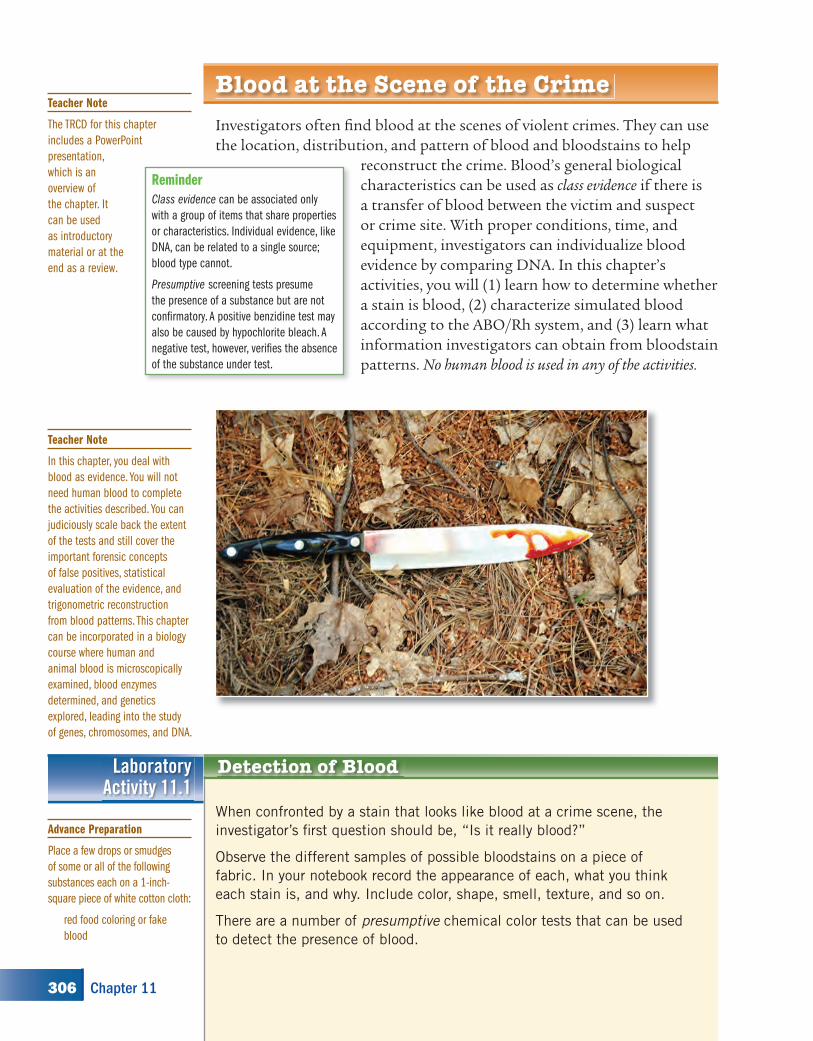
Investigators often fi nd blood at the scenes of violent crimes. They can use the location, distribution, and pattern of blood and bloodstains to help
reconstruct the crime. Blood’s general biological characteristics can be used as class evidence if there is a transfer of blood between the victim and suspect or crime site. With proper conditions, time, and equipment, investigators can individualize blood evidence by comparing DNA. In this chapter’s activities, you will (1) learn how to determine whether a stain is blood, (2) characterize simulated blood according to the ABO/Rh system, and (3) learn what information investigators can obtain from bloodstain patterns. No human blood is used in any of the activities.
Blood at the Scene of the Crime
Reminder
Class evidence can be associated only
with a group of items that share properties
or characteristics. Individual evidence, like
DNA, can be related to a single source;
blood type cannot.
Presumptive screening tests presume
the presence of a substance but are not
confi rmatory. A positive benzidine test may
also be caused by hypochlorite bleach. A
negative test, however, verifi es the absence
of the substance under test.
Detection of BloodLaboratoryActivity 11.1
When confronted by a stain that looks like blood at a crime scene, the
investigator’s fi rst question should be, “Is it really blood?”
Observe the different samples of possible bloodstains on a piece of
fabric. In your notebook record the appearance of each, what you think
each stain is, and why. Include color, shape, smell, texture, and so on.
There are a number of presumptive chemical color tests that can be used
to detect the presence of blood.
Teacher Note
The TRCD for this chapter
includes a PowerPoint
presentation,
which is an
overview of
the chapter. It
can be used
as introductory
material or at the
end as a review.
Teacher Note
In this chapter, you deal with
blood as evidence. You will not
need human blood to complete
the activities described. You can
judiciously scale back the extent
of the tests and still cover the
important forensic concepts
of false positives, statistical
evaluation of the evidence, and
trigonometric reconstruction
from blood patterns. This chapter
can be incorporated in a biology
course where human and
animal blood is microscopically
examined, blood enzymes
determined, and genetics
explored, leading into the study
of genes, chromosomes, and DNA.
Advance Preparation
Place a few drops or smudges
of some or all of the following
substances each on a 1-inch-
square piece of white cotton cloth:
red food coloring or fake
blood
306 Chapter 11
kh00116_CH11.indd Page 306 8/18/08 12:44:22 PM newuserkh00116_CH11.indd Page 306 8/18/08 12:44:22 PM newuser /Volumes/110/KHUS021/indd%0/Ch 11/Volumes/110/KHUS021/indd%0/Ch 11
Procedure
Do not write in your textbook. Take notes in your science notebook.1. Using a commercial blood testing reagent: The heme in hemoglobin
catalytically breaks down peroxides with the production of oxygen. Oxygen reacts with the benzidine product in the Hematest tablet or Hemastix test strip to turn it blue.
Press a piece of wet filter paper on the sample of stained cloth. Break a Hematest tablet into quarters. Put one portion of the tablet in the center of the transferred stain and add a drop of water onto the tablet. Make sure the water flows down the side of the tablet onto the stain. A blue-green ring spreading out on the filter paper from the tablet indicates that the spot is blood. You may be able to rinse and reuse the tablet for the next stain. A Hemastix strip can be rubbed on the wet stain; a green to blue color means that blood is present. Substances other than blood, such as dry bleach residues and some plastics, can cause similar results. Run a blank test on an unstained area of the filter paper. Make a data table for your results from the stained samples. Record your observations.
2. The Kastle-Meyer color test, like the Hematest above, is based on the catalytic breakdown of peroxides by hemoglobin. The contact of reduced phenolphthalein reagent and hydrogen peroxide with a bloodstain produces a deep pink color. If a pink color develops before adding hydrogen peroxide, you have a false positive—it is not blood. Unfortunately, there are a number of substances that give a false positive, such as potatoes and horseradish.
Press a piece of wet filter paper on a sample of stained cloth. Add a drop of K-M reagent to the paper where it touched the stain. Then add a drop of 3 percent hydrogen peroxide. Repeat this procedure for the other stains. You may also use a cotton swab instead of the filter paper. Repeat this test on an unstained area as a control. Record your results.
MaterialsFor each lab group:• suspected blood samples• Hematest tablets or Hemastix
strips• filter paper or cotton swabs
• Kastle-Meyer solution• luminol solution in a spray
bottle• hydrogen peroxide
SAFETY ALERT! CHEMICALS USEDAlways wear goggles and an apron when working in the labaratory
SAFETY NOTE Avoid inhalation, ingestion, and skin contact with
chemicals.!
Laboratory Activity 11.1, continued
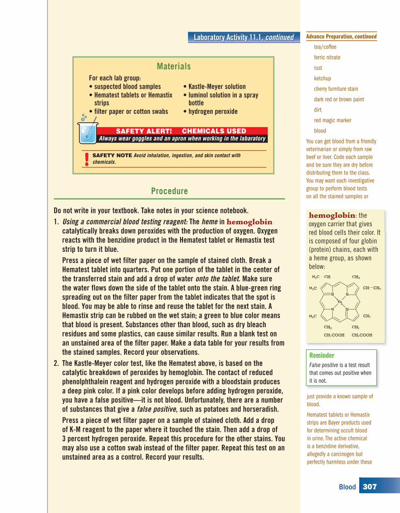
hemoglobin: the
oxygen carrier that gives
red blood cells their color. It
is composed of four globin
(protein) chains, each with
a heme group, as shown
below:
AA
OO
CH
AA
OO
AA
OO
AA
OO
PH2C
H3C
CH3
CHPCH2
CH2COOH
CH3
CH2A
CH2COOH
CH2A
H3C
N N
NN
Fe
Reminder
False positive is a test result
that comes out positive when
it is not.
tea/coffee
ferric nitrate
rust
ketchup
cherry furniture stain
dark red or brown paint
dirt
red magic marker
blood
You can get blood from a friendly
veterinarian or simply from raw
beef or liver. Code each sample
and be sure they are dry before
distributing them to the class.
You may want each investigative
group to perform blood tests
on all the stained samples or
just provide a known sample of
blood.
Hematest tablets or Hemastix
strips are Bayer products used
for determining occult blood
in urine. The active chemical
is a benzidine derivative,
allegedly a carcinogen but
perfectly harmless under these
Advance Preparation, continued
307Blood
kh00116_CH11.indd Page 307 8/18/08 12:44:27 PM newuserkh00116_CH11.indd Page 307 8/18/08 12:44:27 PM newuser /Volumes/110/KHUS021/indd%0/Ch 11/Volumes/110/KHUS021/indd%0/Ch 11
3. The luminol test is a very sensitive indicator for dried and even washed blood. You can quickly spray a suspected area and make old bloodstains glow (chemiluminesce). The area must be very dark and your eyes conditioned to the darkness to see the luminescence. Certain metals (Cu, Fe, Co), bleach, and sometimes even plaster walls can cause false positives. Describe what you observed.
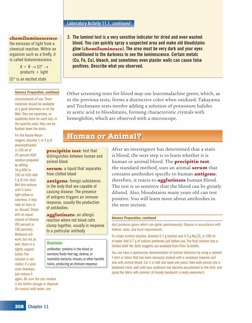
chemiluminescence:
the emission of light from a
chemical reaction. Within an
organism such as a fi refl y, it
is called bioluminescence.
A � B → [I]* → products � light
[I]* is an excited state
Laboratory Activity 11.1, continued
Human or Animal?
After an investigator has determined that a stain is blood, the next step is to learn whether it is human or animal blood. The precipitin test, the standard method, uses an animal serum that contains antibodies specifi c to human antigens; therefore, it reacts to agglutinate human blood. The test is so sensitive that the blood can be greatly diluted. Also, bloodstains many years old can test positive. You will learn more about antibodies in the next section.
precipitin test: test that
distinguishes between human and
animal blood
serum: a liquid that separates
from clotted blood
antigens: foreign substances
in the body that are capable of
causing disease. The presence
of antigens triggers an immune
response, usually the production
of antibodies.
agglutinate: an allergic
reaction where red blood cells
clump together, usually in response
to a particular antibody
Reminder
antibodies: proteins in the blood or
secretory fl uids that tag, destroy, or
neutralize bacteria, viruses, or other harmful
toxins, producing an immune response
Other screening tests for blood may use leucomalachite green, which, as in the previous tests, forms a distinctive color when oxidized. Takayama and Teichmann tests involve adding a solution of potassium halides in acetic acid to bloodstains, forming characteristic crystals with hemoglobin, which are observed with a microscope.
circumstances of use. These
materials should be available
at a good pharmacy or on the
Web. They are expensive, so
subdivide them for each test. In
the quantity used, they can be
fl ushed down the drain.
For the Kastle-Meyer
reagent, dissolve 1 to 2 g of
phenolphthalein
in 100 ml of
25 percent KOH
solution prepared
by adding
34 g KOH to
100 ml H2O. Add
1 g of zinc dust.
Boil this mixture
until it turns
light yellow or
colorless; it may
take an hour or
so. Decant. Dilute
with an equal
volume of ethanol
(95 percent or
100 percent).
Methanol will
work, but not as
well. Store in a
tightly capped
bottle. The
solution is not
stable; if a pink
color develops,
just reduce it
again. Be sure the zinc residue
is dry before storage or disposal.
On contact with water, zinc
Advance Preparation, continued
dust produces gases which can ignite spontaneously. Dispose in accordance with
federal, state, and local requirements.
To create luminol solution, dissolve 0.1 g luminol and 0.5 g Na2CO
3 in 100 ml
of water. Add 0.7 g of sodium perborate just before use. The fi nal solution has a
limited shelf life. Both reagents are available from Flinn Scientifi c.
You can have a spectacular demonstration of luminol detection by using a colored
T-shirt or fabric that has been obviously stained with a nonblood material and
also with animal blood. Cut it in half and wash one piece. Take both pieces into a
darkened room, wait until your audience has become accustomed to the dark, and
spray the fabric with luminol. (A bloody handprint is really awesome!)
Advance Preparation, continued
308 Chapter 11
kh00116_CH11.indd Page 308 8/18/08 12:44:28 PM newuserkh00116_CH11.indd Page 308 8/18/08 12:44:28 PM newuser /Volumes/110/KHUS021/indd%0/Ch 11/Volumes/110/KHUS021/indd%0/Ch 11
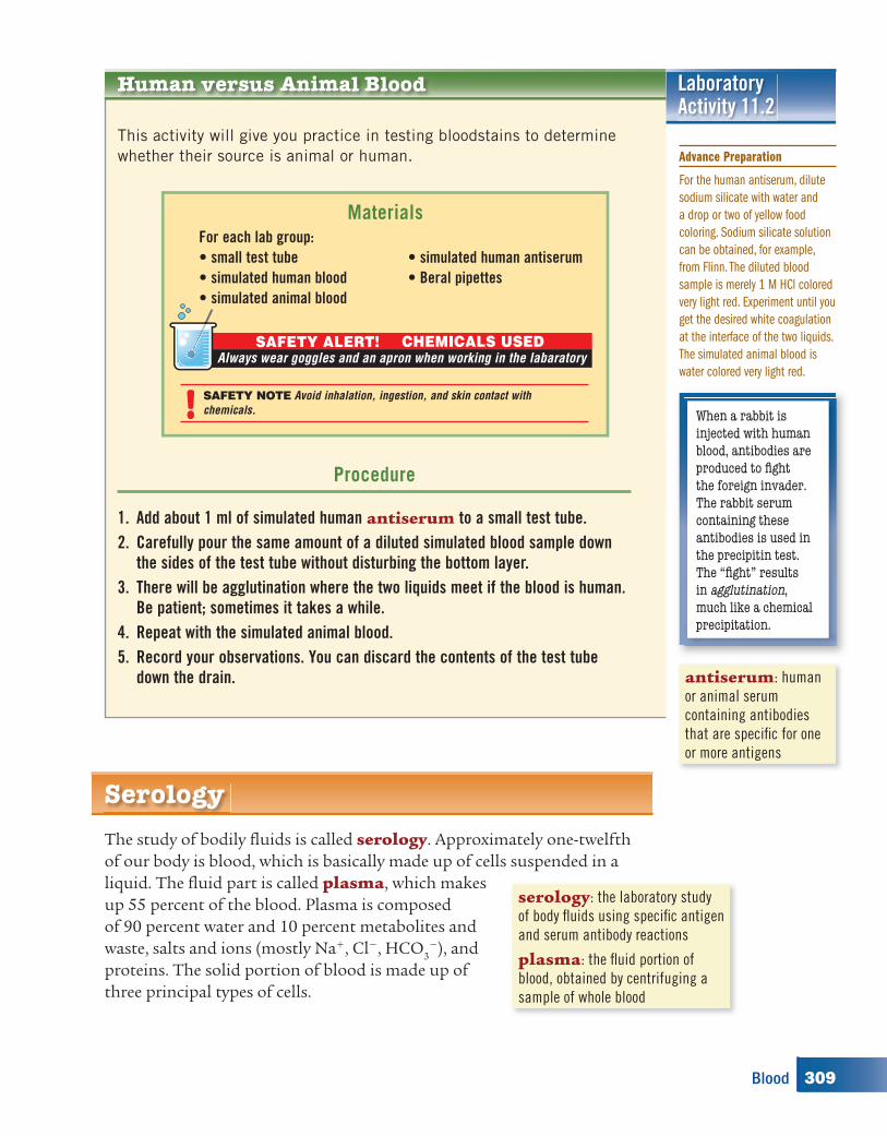
LaboratoryActivity 11.2
Human versus Animal Blood
This activity will give you practice in testing bloodstains to determine
whether their source is animal or human.
The study of bodily fl uids is called serology. Approximately one-twelfth of our body is blood, which is basically made up of cells suspended in a liquid. The fl uid part is called plasma, which makes up 55 percent of the blood. Plasma is composed of 90 percent water and 10 percent metabolites and waste, salts and ions (mostly Na�, Cl�, HCO3
�), and proteins. The solid portion of blood is made up of three principal types of cells.
Procedure
1. Add about 1 ml of simulated human antiserum to a small test tube.2. Carefully pour the same amount of a diluted simulated blood sample down
the sides of the test tube without disturbing the bottom layer.3. There will be agglutination where the two liquids meet if the blood is human.
Be patient; sometimes it takes a while.4. Repeat with the simulated animal blood.5. Record your observations. You can discard the contents of the test tube
down the drain.
For each lab group:• small test tube• simulated human blood• simulated animal blood
• simulated human antiserum• Beral pipettes
Materials
SAFETY ALERT! CHEMICALS USEDAlways wear goggles and an apron when working in the labaratory
SAFETY NOTE Avoid inhalation, ingestion, and skin contact with
chemicals.! When a rabbit is injected with human blood, antibodies are produced to fi ght the foreign invader. The rabbit serum containing these antibodies is used in the precipitin test. The “fi ght” results in agglutination, much like a chemical precipitation.
antiserum: human
or animal serum
containing antibodies
that are specifi c for one
or more antigens
Serology
serology: the laboratory study
of body fl uids using specifi c antigen
and serum antibody reactions
plasma: the fl uid portion of
blood, obtained by centrifuging a
sample of whole blood
Advance Preparation
For the human antiserum, dilute
sodium silicate with water and
a drop or two of yellow food
coloring. Sodium silicate solution
can be obtained, for example,
from Flinn. The diluted blood
sample is merely 1 M HCl colored
very light red. Experiment until you
get the desired white coagulation
at the interface of the two liquids.
The simulated animal blood is
water colored very light red.
309Blood
kh00116_CH11.indd Page 309 8/18/08 12:44:29 PM newuserkh00116_CH11.indd Page 309 8/18/08 12:44:29 PM newuser /Volumes/110/KHUS021/indd%0/Ch 11/Volumes/110/KHUS021/indd%0/Ch 11
Red cells, or erythrocytes, which contain hemoglobin. They transport oxygen from the lungs to the cells and then carry carbon dioxide back to the lungs, where it is exhaled.
White cells, or leukocytes, which are the primary cells of the immune system. They produce antibodies.
Platelets, which start the clotting process by initiating the formation of fi brin to form a clot. Removing the solid clotting material leaves a pale yellow, watery fl uid called serum.
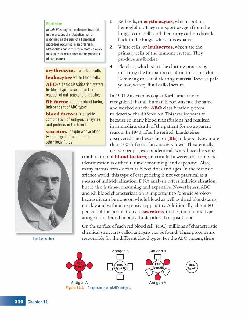
In 1901 Austrian biologist Karl Landsteiner recognized that all human blood was not the same and worked out the ABO classifi cation system to describe the differences. This was important because so many blood transfusions had resulted in immediate death of the patient for no apparent reason. In 1940, after he retired, Landsteiner discovered the rhesus factor (Rh) in blood. Now more than 100 different factors are known. Theoretically, no two people, except identical twins, have the same
combination of blood factors; practically, however, the complete identifi cation is diffi cult, time-consuming, and expensive. Also, many factors break down as blood dries and ages. In the forensic science world, this type of categorizing is not yet practical as a means of individualization. DNA analysis offers individualization, but it also is time-consuming and expensive. Nevertheless, ABO and Rh blood characterization is important to forensic serology because it can be done on whole blood as well as dried bloodstains, quickly and without expensive apparatus. Additionally, about 80 percent of the population are secretors; that is, their blood type antigens are found in body fl uids other than just blood.
On the surface of each red blood cell (RBC), millions of characteristic chemical structures called antigens can be found. These proteins are responsible for the different blood types. For the ABO system, there
1.
2.
3.erythrocytes: red blood cells
leukocytes: white blood cells
ABO: a basic classifi cation system
for blood types based upon the
reaction of antigens and antibodies
Rh factor: a basic blood factor,
independent of ABO types
blood factors: a specifi c
combination of antigens, enzymes,
and proteins in the blood
secretors: people whose blood
type antigens are also found in
other body fl uids
Antigen B
RBCType B
RBCType AB
Antigen B
Antigen A
RBCType A
Antigen A
RBCType O
Figure 11.1 A representation of ABO antigens
Karl Landsteiner
Reminder
metabolites: organic molecules involved
in the process of metabolism, which
is defi ned as the sum of all chemical
processes occurring in an organism.
Metabolites can either form more complex
molecules or result from the degradation
of compounds.
310 Chapter 11
kh00116_CH11.indd Page 310 8/18/08 12:44:30 PM newuserkh00116_CH11.indd Page 310 8/18/08 12:44:30 PM newuser /Volumes/110/KHUS021/indd%0/Ch 11/Volumes/110/KHUS021/indd%0/Ch 11
Collecting possible bloodstain
are two types of antigens, A and B. Type A blood cells have A antigens, type B blood cells have B antigens, type AB blood cells have both A and B antigens, and type O blood cells have neither antigen (see Figure 11.1).
Some white blood cells manufacture proteins called antibodies, which are found in the serum. These antibodies are produced to attack invaders that enter the bloodstream, that is, antigens that do not belong in your system (such as snake venom, bacteria, or someone else’s blood). For example, when viruses responsible for mumps enter the blood, the body recognizes them as foreign and begins making antibodies that combine only with the specifi c antigens on the virus. White blood cells destroy the antibody-coated viruses. If someone is exposed to mumps for a second time, the existing antibodies prevent him or her from getting the illness again. This is the basis of vaccines.
A person with type A blood has A antigens on his or her red blood cells. That person will produce specifi c antibodies, B antibodies, in his or her serum to attack and destroy type B blood cells as they are introduced into the body, including type AB blood cells.
CRUCIAL EVIDENCE IN MURDER CASE WITHHELD
Charges were made that evidence was suppressed in a 1981 murder trial that ended with a death sentence.According to a 1997 ruling by a federal district judge, “neither police nor the prosecution informed the defense team before trial that a Hemastix test had been done or that the chair back tested positive for the presence of blood.”
—excerpt from sfbg.com/news story dated November 24, 1999
Testing stain with a Hemastix
GO TO www.scilinks.org
TOPIC blood types
CODE forensics2E311
An allele is one member of
a pair of genes occupying
a specifi c spot on a
chromosome that controls
the same trait. For
example, a pair of alleles
may control the same
trait such as eye color:
One codes for blue eyes,
another for brown eyes.
311Blood
kh00116_CH11.indd Page 311 8/18/08 12:44:51 PM newuserkh00116_CH11.indd Page 311 8/18/08 12:44:51 PM newuser /Volumes/110/KHUS021/indd%0/Ch 11/Volumes/110/KHUS021/indd%0/Ch 11
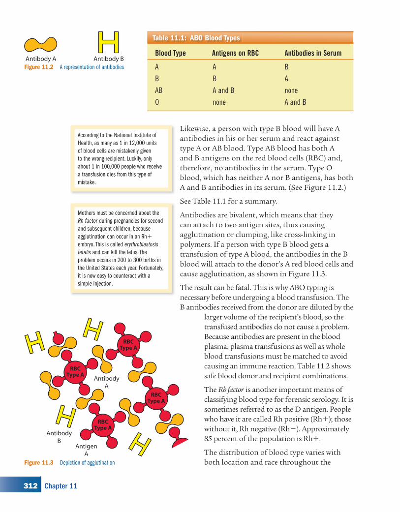
Likewise, a person with type B blood will have A antibodies in his or her serum and react against type A or AB blood. Type AB blood has both A and B antigens on the red blood cells (RBC) and, therefore, no antibodies in the serum. Type O blood, which has neither A nor B antigens, has both A and B antibodies in its serum. (See Figure 11.2.)
See Table 11.1 for a summary.
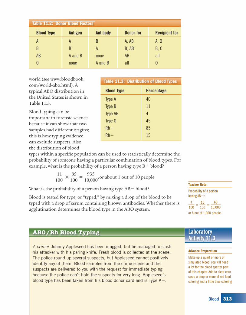
Antibodies are bivalent, which means that they can attach to two antigen sites, thus causing agglutination or clumping, like cross-linking in polymers. If a person with type B blood gets a transfusion of type A blood, the antibodies in the B blood will attach to the donor’s A red blood cells and cause agglutination, as shown in Figure 11.3.
The result can be fatal. This is why ABO typing is necessary before undergoing a blood transfusion. The B antibodies received from the donor are diluted by the
larger volume of the recipient’s blood, so the transfused antibodies do not cause a problem. Because antibodies are present in the blood plasma, plasma transfusions as well as whole blood transfusions must be matched to avoid causing an immune reaction. Table 11.2 shows safe blood donor and recipient combinations.
The Rh factor is another important means of classifying blood type for forensic serology. It is sometimes referred to as the D antigen. People who have it are called Rh positive (Rh�); those without it, Rh negative (Rh�). Approximately 85 percent of the population is Rh�.
The distribution of blood type varies with both location and race throughout the
Blood Type Antigens on RBC Antibodies in Serum
A A B
B B A
AB A and B none
O none A and B
Table 11.1: ABO Blood Types
Antibody A Antibody BFigure 11.2 A representation of antibodies
RBCType A
RBCType A
RBCType A
RBCType A
AntibodyA
AntibodyB
AntigenA
Figure 11.3 Depiction of agglutination
According to the National Institute of
Health, as many as 1 in 12,000 units
of blood cells are mistakenly given
to the wrong recipient. Luckily, only
about 1 in 100,000 people who receive
a transfusion dies from this type of
mistake.
Mothers must be concerned about the
Rh factor during pregnancies for second
and subsequent children, because
agglutination can occur in an Rh�
embryo. This is called erythroblastosis
fetalis and can kill the fetus. The
problem occurs in 200 to 300 births in
the United States each year. Fortunately,
it is now easy to counteract with a
simple injection.
312 Chapter 11
kh00116_CH11.indd Page 312 8/18/08 12:44:58 PM newuserkh00116_CH11.indd Page 312 8/18/08 12:44:58 PM newuser /Volumes/110/KHUS021/indd%0/Ch 11/Volumes/110/KHUS021/indd%0/Ch 11
Blood Type Percentage
Type A 40
Type B 11
Type AB 4
Type O 45
Rh� 85
Rh� 15
Table 11.3: Distribution of Blood Typesworld (see www.bloodbook.com/world-abo.html). A typical ABO distribution in the United States is shown in Table 11.3.
Blood typing can be important in forensic science because it can show that two samples had different origins; this is how typing evidence can exclude suspects. Also, the distribution of blood types within a specifi c population can be used to statistically determine the probability of someone having a particular combination of blood types. For example, what is the probability of a person having type B� blood?
11100
� 85
100 �
93510,000
, or about 1 out of 10 people
What is the probability of a person having type AB� blood?
Blood is tested for type, or “typed,” by mixing a drop of the blood to be typed with a drop of serum containing known antibodies. Whether there is agglutination determines the blood type in the ABO system.
ABO/Rh Blood Typing
A crime: Johnny Appleseed has been mugged, but he managed to slash
his attacker with his paring knife. Fresh blood is collected at the scene.
The police round up several suspects, but Appleseed cannot positively
identify any of them. Blood samples from the crime scene and the
suspects are delivered to you with the request for immediate typing
because the police can’t hold the suspects for very long. Appleseed’s
blood type has been taken from his blood donor card and is Type A�.
LaboratoryActivity 11.3
Blood Type Antigen Antibody Donor for Recipient for
A A B A, AB A, O
B B A B, AB B, O
AB A and B none AB all
O none A and B all O
Table 11.2: Donor Blood Factors
Advance Preparation
Make up a quart or more of
simulated blood; you will need
a lot for the blood spatter part
of this chapter. Add to clear corn
syrup a drop or more of red food
coloring and a little blue coloring
Teacher Note
Probability of a person
having AB�:
4
100 � 15
100 � 60
10,000
or 6 out of 1,000 people
313Blood
kh00116_CH11.indd Page 313 8/18/08 12:44:59 PM newuserkh00116_CH11.indd Page 313 8/18/08 12:44:59 PM newuser /Volumes/110/KHUS021/indd%0/Ch 11/Volumes/110/KHUS021/indd%0/Ch 11
Note: No blood or blood products will be used in this lab. This is merely
a simulation of the reactions that happen in the typing of blood, where
actual human blood and human antisera are used.
Laboratory Activity 11.3, continued
MaterialsFor each lab group:• stereomicroscope or
magnifying glass• glass slides• simulated blood from crime
scene• simulated blood from four
suspects
• simulated anti-A• simulated anti-B• simulated anti-Rh• pipettes• toothpicks or glass
stirring rods
SAFETY ALERT! CHEMICALS USEDAlways wear goggles and an apron when working in the labaratory
SAFETY NOTE Avoid inhalation, ingestion, and skin contact with
chemicals.!
Procedure
1. Place a slide under the stereomicroscope and focus.2. Add three drops of Suspect #1’s blood to the slide, side by side, making sure
that they do not touch each other.
The crime scene
to deepen the red, or use Congo
red. You won’t need much
simulated blood and sera for the
typecasting activity, but make up
enough for your ultimate crime
scene also.
To make simulated anti-A and
anti-Rh sera: Dissolve 16 g of
Ca(NO3)
2 in 100 ml of water
and color it yellow with food
dye. Divide in half for each
serum.
To make simulated anti-B
serum: Dissolve 5 g Na2CO
3
in 50 ml of water and color it
yellow with food dye.
To make simulated blood for
Suspect #1 (Type A�): Dissolve
5 g Na2CO
3 in 10 ml water,
add to 50 ml of the simulated
blood, and stir.
To make simulated blood for
Suspect #2 (Type B�): Dissolve
9 g Ca(NO3)
2 in 10 ml water,
add to 50 ml of the simulated
blood, and stir.
To make simulated blood
for Suspect #3 (Type AB�):
Dissolve 7 g Mg(SO4)
2 in 10 ml
water, add to 50 ml of the
simulated blood, and stir.
To make simulated blood for
Suspect #4 (Type O�): Add
10 ml water to 50 ml of the
simulated blood and stir.
To make simulated blood from
the crime scene: Pick one of
the above.
The chemicals can all be obtained
from any scientifi c supply house.
MgSO4 can be purchased there,
but it is a lot cheaper to buy it in
a supermarket as Epsom salts.
The MgCO3 reaction is more
of a coagulation with slight
cloudiness, whereas the
precipitates of Ca(CO3)
2
and CaSO4 show a distinct
cloudiness. Adding too much
serum dilutes the “blood drop”
and obscures the coagulation
and cloudiness. To be correct,
•
•
•
•
•
•
•
Advance Preparation, continued
314 Chapter 11
kh00116_CH11.indd Page 314 8/18/08 12:45:00 PM newuserkh00116_CH11.indd Page 314 8/18/08 12:45:00 PM newuser /Volumes/110/KHUS021/indd%0/Ch 11/Volumes/110/KHUS021/indd%0/Ch 11
Laboratory Activity 11.3, continued
3. Add one drop of serum with A antibodies (labeled Anti-A) to the first blood drop.
4. Add one drop of serum with B antibodies (Anti-B) to the second blood drop.5. Add one drop of the anti-Rh serum to the third blood drop.6. Mix the cells and the sera with a toothpick, watching to see whether
agglutination takes place. Be sure to clean the toothpick between drops to avoid contamination.
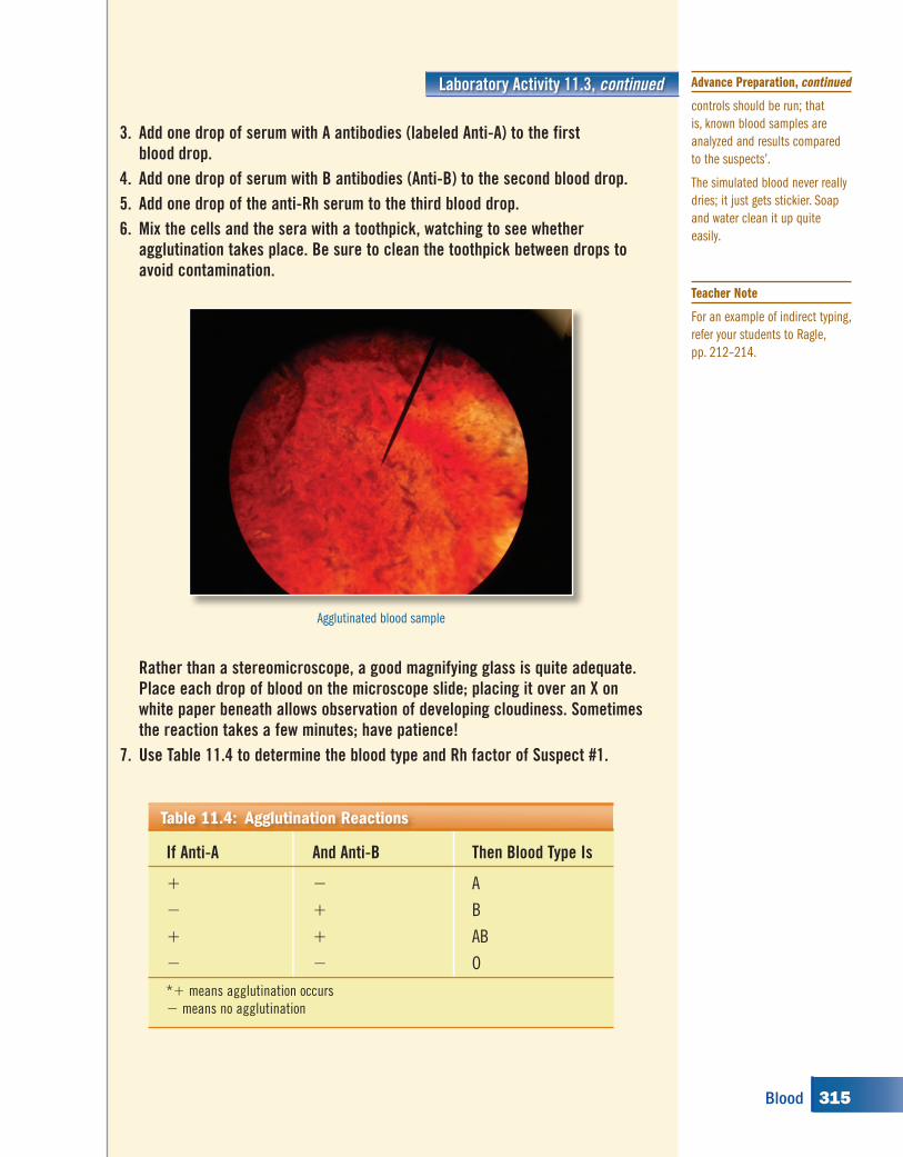
Rather than a stereomicroscope, a good magnifying glass is quite adequate. Place each drop of blood on the microscope slide; placing it over an X on white paper beneath allows observation of developing cloudiness. Sometimes the reaction takes a few minutes; have patience!
7. Use Table 11.4 to determine the blood type and Rh factor of Suspect #1.
Agglutinated blood sample
If Anti-A And Anti-B Then Blood Type Is
� � A
� � B
� � AB
� � O
*� means agglutination occurs
� means no agglutination
Table 11.4: Agglutination Reactions
controls should be run; that
is, known blood samples are
analyzed and results compared
to the suspects’.
The simulated blood never really
dries; it just gets stickier. Soap
and water clean it up quite
easily.
Advance Preparation, continued
Teacher Note
For an example of indirect typing,
refer your students to Ragle,
pp. 212–214.
315Blood
kh00116_CH11.indd Page 315 8/18/08 12:45:05 PM newuserkh00116_CH11.indd Page 315 8/18/08 12:45:05 PM newuser /Volumes/110/KHUS021/indd%0/Ch 11/Volumes/110/KHUS021/indd%0/Ch 11
Laboratory Activity 11.3, continued
8. Repeat the procedure for Suspects #2, #3, and #4 and the sample from the crime scene.
The Rh factor is either present or not present; agglutination indicates its presence, and no reaction indicates its absence.
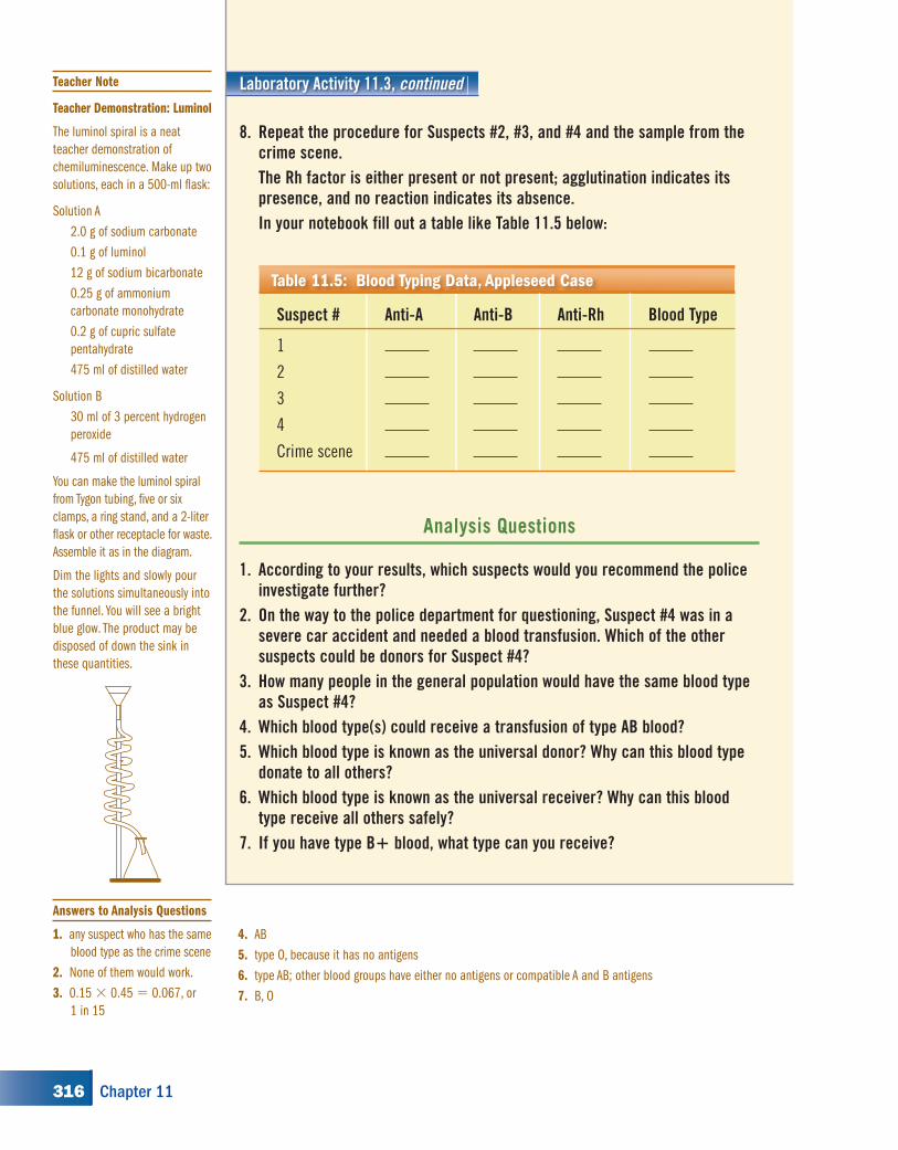
In your notebook fill out a table like Table 11.5 below:
Analysis Questions
1. According to your results, which suspects would you recommend the police investigate further?
2. On the way to the police department for questioning, Suspect #4 was in a severe car accident and needed a blood transfusion. Which of the other suspects could be donors for Suspect #4?
3. How many people in the general population would have the same blood type as Suspect #4?
4. Which blood type(s) could receive a transfusion of type AB blood?5. Which blood type is known as the universal donor? Why can this blood type
donate to all others?6. Which blood type is known as the universal receiver? Why can this blood
type receive all others safely?7. If you have type B� blood, what type can you receive?
Suspect # Anti-A Anti-B Anti-Rh Blood Type
1
2
3
4
Crime scene
Table 11.5: Blood Typing Data, Appleseed Case
4. AB
5. type O, because it has no antigens
6. type AB; other blood groups have either no antigens or compatible A and B antigens
7. B, O
Teacher Note
Teacher Demonstration: Luminol
The luminol spiral is a neat
teacher demonstration of
chemiluminescence. Make up two
solutions, each in a 500-ml fl ask:
Solution A
2.0 g of sodium carbonate
0.1 g of luminol
12 g of sodium bicarbonate
0.25 g of ammonium
carbonate monohydrate
0.2 g of cupric sulfate
pentahydrate
475 ml of distilled water
Solution B
30 ml of 3 percent hydrogen
peroxide
475 ml of distilled water
You can make the luminol spiral
from Tygon tubing, fi ve or six
clamps, a ring stand, and a 2-liter
fl ask or other receptacle for waste.
Assemble it as in the diagram.
Dim the lights and slowly pour
the solutions simultaneously into
the funnel. You will see a bright
blue glow. The product may be
disposed of down the sink in
these quantities.
Answers to Analysis Questions
1. any suspect who has the same
blood type as the crime scene
2. None of them would work.
3. 0.15 � 0.45 � 0.067, or
1 in 15
316 Chapter 11
kh00116_CH11.indd Page 316 8/20/08 7:12:53 PM newuserkh00116_CH11.indd Page 316 8/20/08 7:12:53 PM newuser /Volumes/110/KHUS021/indd%0/Ch 11/Volumes/110/KHUS021/indd%0/Ch 11
Real blood that has been dried can still be typed. The red blood cells have ruptured, but the antigens and antibodies are still there. Investigators use indirect methods. In crime scenes you may encounter later, the standard typing procedure can be used.
When packaging and storing blood evidence, do not block out air; a sealed container may trap any moisture present and cause mold and mildew to form. Paper bags or envelopes may be used.
Testing Dried Blood
Early on the morning of July 4, 1954, police received a call from
Dr. Sam Sheppard. He reported that his wife, Marilyn, was dead in
their bedroom. He explained to police that, the night before, Marilyn
had left him on the couch and gone to sleep in the twin bed next to
Sam’s. He fell asleep and awoke some time later,
believing he heard his wife calling his name.
Running upstairs, he saw a “form” struggling with
something or someone, and was suddenly struck from
behind. When he came to, he was lying on the fl oor.
His wife Marilyn was covered with blood.
He checked for her pulse and found none. Sheppard
heard a noise below, ran downstairs, and saw the back
door open and “a form progressing rapidly toward the
lake.” He chased the person across the lawn and down
the steps leading to the beach. He struggled with
a man, 6´3˝, middle-aged, with dark
bushy hair and a white shirt. Sheppard
was choked to unconsciousness.
Marilyn had 35 wounds to the head,
and blood drenched the walls, door,
and bed where she lay. Her face was
almost unrecognizable.
11.1: The Sam Sheppard Case
Sam and Marilyn Sheppard
317Blood
kh00116_CH11.indd Page 317 8/18/08 12:45:07 PM newuserkh00116_CH11.indd Page 317 8/18/08 12:45:07 PM newuser /Volumes/110/KHUS021/indd%0/Ch 11/Volumes/110/KHUS021/indd%0/Ch 11
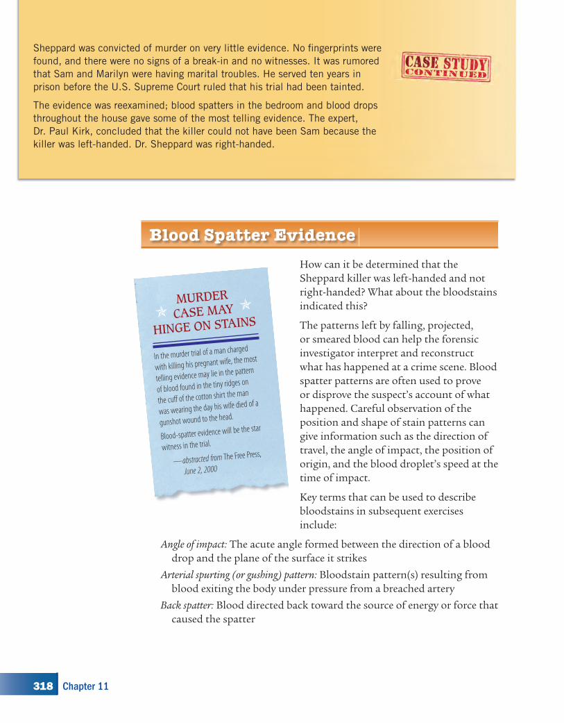
Sheppard was convicted of murder on very little evidence. No fi ngerprints were
found, and there were no signs of a break-in and no witnesses. It was rumored
that Sam and Marilyn were having marital troubles. He served ten years in
prison before the U.S. Supreme Court ruled that his trial had been tainted.
The evidence was reexamined; blood spatters in the bedroom and blood drops
throughout the house gave some of the most telling evidence. The expert,
Dr. Paul Kirk, concluded that the killer could not have been Sam because the
killer was left-handed. Dr. Sheppard was right-handed.
Blood Spatter Evidence
How can it be determined that the Sheppard killer was left-handed and not right-handed? What about the bloodstains indicated this?
The patterns left by falling, projected, or smeared blood can help the forensic investigator interpret and reconstruct what has happened at a crime scene. Blood spatter patterns are often used to prove or disprove the suspect’s account of what happened. Careful observation of the position and shape of stain patterns can give information such as the direction of travel, the angle of impact, the position of origin, and the blood droplet’s speed at the time of impact.
Key terms that can be used to describe bloodstains in subsequent exercises include:
Angle of impact: The acute angle formed between the direction of a blood drop and the plane of the surface it strikes
Arterial spurting (or gushing) pattern: Bloodstain pattern(s) resulting from blood exiting the body under pressure from a breached artery
Back spatter: Blood directed back toward the source of energy or force that caused the spatter
MURDER
CASE MAY
HINGE ON STAINS
In the murder trial of a man charged
with killing his pregnant wife, the most
telling evidence may lie in the pattern
of blood found in the tiny ridges on
the cuff of the cotton shirt the man
was wearing the day his wife died of a
gunshot wound to the head.
Blood-spatter evidence will be the star
witness in the trial.
—abstracted from The Free Press,
June 2, 2000
318 Chapter 11
kh00116_CH11.indd Page 318 8/18/08 12:45:12 PM newuserkh00116_CH11.indd Page 318 8/18/08 12:45:12 PM newuser /Volumes/110/KHUS021/indd%0/Ch 11/Volumes/110/KHUS021/indd%0/Ch 11
Blood spatter analysis: A fi eld of forensic science that deals with the physical properties of blood and the patterns produced under different conditions as a result of various forces applied to the source of blood
Bloodstain: Evidence that liquid blood has come into contact with a surface
Cast-off pattern: A bloodstain pattern created when blood is released or thrown from a moving blood-bearing object
Contact stain: Blood deposited from direct contact between two surfaces, at least one of which is bloody
Direction of fl ight: The trajectory of a blood drop, which can be established by its angle of impact and directionality angle
Directionality: The direction the blood was traveling when it hit the target surface; investigators can usually establish directionality of a blood drop’s fl ight from the geometric shape of its bloodstain
Directionality angle: The angle between the long axis of a bloodstain and a predetermined line on the plane of the target surface that represents 0 degrees
Draw-back effect: Blood in the barrel of a fi rearm that has been drawn backward into the muzzle
Drip pattern: A bloodstain pattern that results from blood dripping into blood
Expirated blood: Blood that is blown out of the nose, mouth, or a wound as a result of air pressure or air fl ow, which is the propelling force
Flight path: The path of the blood drop as it moves through space, from the impact site to the target
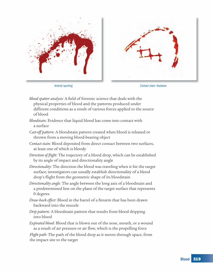
Arterial spurting Contact stain—footwear
319Blood
kh00116_CH11.indd Page 319 8/18/08 12:45:13 PM newuserkh00116_CH11.indd Page 319 8/18/08 12:45:13 PM newuser /Volumes/110/KHUS021/indd%0/Ch 11/Volumes/110/KHUS021/indd%0/Ch 11
Flow pattern: A change in the shape and direction of a bloodstain due to the infl uence of gravity or movement of the object
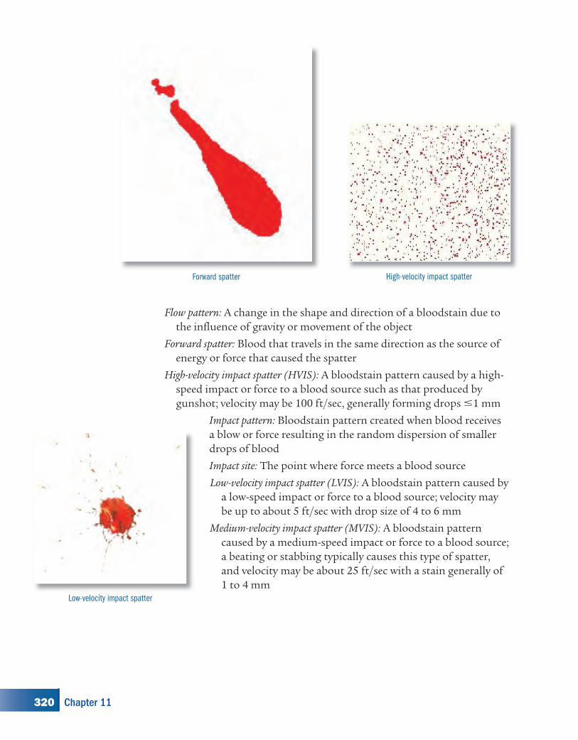
Forward spatter: Blood that travels in the same direction as the source of energy or force that caused the spatter
High-velocity impact spatter (HVIS): A bloodstain pattern caused by a high-speed impact or force to a blood source such as that produced by gunshot; velocity may be 100 ft/sec, generally forming drops �1 mm
Impact pattern: Bloodstain pattern created when blood receives a blow or force resulting in the random dispersion of smaller drops of blood
Impact site: The point where force meets a blood source
Low-velocity impact spatter (LVIS): A bloodstain pattern caused by a low-speed impact or force to a blood source; velocity may be up to about 5 ft/sec with drop size of 4 to 6 mm
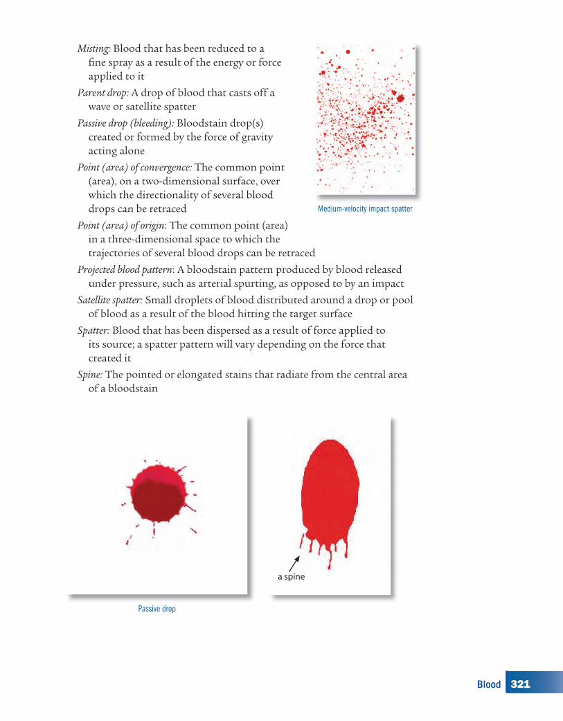
Medium-velocity impact spatter (MVIS): A bloodstain pattern caused by a medium-speed impact or force to a blood source; a beating or stabbing typically causes this type of spatter, and velocity may be about 25 ft/sec with a stain generally of 1 to 4 mm
High-velocity impact spatterForward spatter
Low-velocity impact spatter
320 Chapter 11
kh00116_CH11.indd Page 320 8/18/08 12:45:15 PM newuserkh00116_CH11.indd Page 320 8/18/08 12:45:15 PM newuser /Volumes/110/KHUS021/indd%0/Ch 11/Volumes/110/KHUS021/indd%0/Ch 11
Misting: Blood that has been reduced to a fi ne spray as a result of the energy or force applied to it
Parent drop: A drop of blood that casts off a wave or satellite spatter
Passive drop (bleeding): Bloodstain drop(s) created or formed by the force of gravity acting alone
Point (area) of convergence: The common point (area), on a two-dimensional surface, over which the directionality of several blood drops can be retraced
Point (area) of origin: The common point (area) in a three-dimensional space to which the trajectories of several blood drops can be retraced
Projected blood pattern: A bloodstain pattern produced by blood released under pressure, such as arterial spurting, as opposed to by an impact
Satellite spatter: Small droplets of blood distributed around a drop or pool of blood as a result of the blood hitting the target surface
Spatter: Blood that has been dispersed as a result of force applied to its source; a spatter pattern will vary depending on the force that created it
Spine: The pointed or elongated stains that radiate from the central area of a bloodstain
a spine
Passive drop
Medium-velocity impact spatter
321Blood
kh00116_CH11.indd Page 321 8/18/08 12:45:16 PM newuserkh00116_CH11.indd Page 321 8/18/08 12:45:16 PM newuser /Volumes/110/KHUS021/indd%0/Ch 11/Volumes/110/KHUS021/indd%0/Ch 11
Swipe pattern: The transfer of blood from a moving source onto an unstained surface; the direction of travel may be determined by the feathered edge
Target: The surface on which blood has been deposited
Transfer or contact pattern: A bloodstain pattern created when a wet, bloody surface comes in contact with a second surface; a recognizable image of all or a portion of the original surface may be observed in the pattern
Void: An absence of stains in an otherwise continuous bloodstain pattern, like a reverse shadow
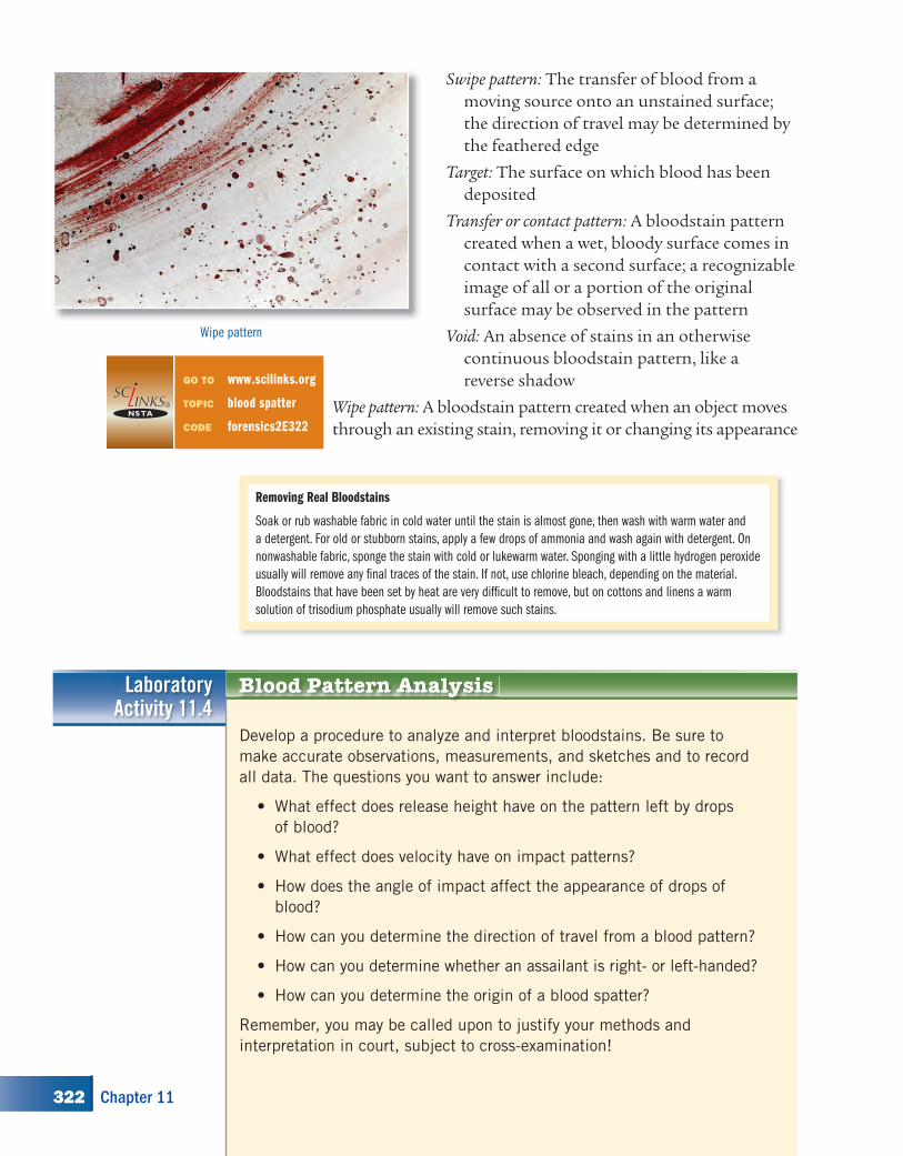
Wipe pattern: A bloodstain pattern created when an object moves through an existing stain, removing it or changing its appearance
Wipe pattern
Blood Pattern AnalysisLaboratoryActivity 11.4
Develop a procedure to analyze and interpret bloodstains. Be sure to
make accurate observations, measurements, and sketches and to record
all data. The questions you want to answer include:
What effect does release height have on the pattern left by drops
of blood?
What effect does velocity have on impact patterns?
How does the angle of impact affect the appearance of drops of
blood?
How can you determine the direction of travel from a blood pattern?
How can you determine whether an assailant is right- or left-handed?
How can you determine the origin of a blood spatter?
Remember, you may be called upon to justify your methods and
interpretation in court, subject to cross-examination!
•
•
•
•
•
•
Removing Real Bloodstains
Soak or rub washable fabric in cold water until the stain is almost gone, then wash with warm water and
a detergent. For old or stubborn stains, apply a few drops of ammonia and wash again with detergent. On
nonwashable fabric, sponge the stain with cold or lukewarm water. Sponging with a little hydrogen peroxide
usually will remove any fi nal traces of the stain. If not, use chlorine bleach, depending on the material.
Bloodstains that have been set by heat are very diffi cult to remove, but on cottons and linens a warm
solution of trisodium phosphate usually will remove such stains.
GO TO www.scilinks.org
TOPIC blood spatter
CODE forensics2E322
322 Chapter 11
kh00116_CH11.indd Page 322 8/18/08 12:45:18 PM newuserkh00116_CH11.indd Page 322 8/18/08 12:45:18 PM newuser /Volumes/110/KHUS021/indd%0/Ch 11/Volumes/110/KHUS021/indd%0/Ch 11
MaterialsFor each lab group:• wide roll of paper• simulated blood• pipettes• paper• plastic knife or tongue
depressor• protractor, ruler, meter stick• string and masking tape
• ring stand• syringe• spray aspirator• Beral pipette or eyedropper• sine table (see Table 11.6 on
page 327) or trig function calculator
• digital camera
SAFETY ALERT! CHEMICALS USEDAlways wear goggles and an apron when working in the labaratory
Caution: Also wear disposable laboratory gloves to protect yourself from
stains and spatter from the simulated blood.!
Laboratory Activity 11.4, continued
Procedure
1. Height: Work out a method to study the relationship of drop shape and size to the height of origin.
2. Velocity: The density of blood is 1.06 g/cc, and its viscosity is 6 times that of water. Its average drop size, because of surface tension and viscosity, is 0.05 ml for a free fall. As with all objects falling freely, a drop of blood accelerates because of gravity, 32 ft/sec2 (9.8 m/sec2). Any object will fall at this rate until it reaches its terminal velocity, which is a direct function of drop size. An average-sized drop reaches the terminal velocity of 25 ft/sec after a fall of 4 feet, so drop size should not change above that. The majority of high-velocity droplets, which tend to be less than 1 mm, usually travel no more than 46 inches in a horizontal direction. Check it out.
3. Angle of impact: How does the angle of impact of a drop of blood influence its shape?
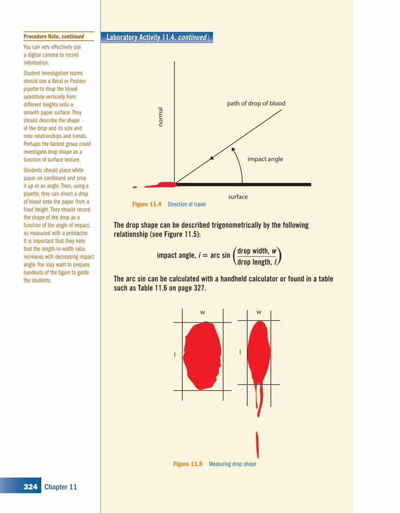
4. Direction of travel: By now, you should realize that the distorted end of the drop of blood points in the direction of travel. Verify this by flinging drops of blood off a stirring rod or your finger and note the shape of the stain relative to the direction of travel (see Figure 11.4 on page 324).
Advance Preparation
Make partial blood patterns
from different objects, such as
a hammer, screwdriver, crowbar,
tire iron, knife, letter opener, fork,
or brick, and ask students to
work out what the object was.
Procedure Note
This activity is inquiry-based,
so the student is given less
information than in some previous
activities. You can use the
teacher’s material to guide the
class through the steps required
to answer the questions posed.
You may want to lead a
discussion on the types of
information that can be gleaned
from the study of blood patterns,
for example, the type of weapon
used; how many gunshots, stabs,
or blows were infl icted; the
sequence of injuries; the position
of the victim and assailant;
whether they were moving during
the attack; whether the assailant
was right- or left-handed; the
degree of force used by the
perpetrator; whether there
might be blood on the assailant;
and whether the victim was
moved after the attack. This is
a good place to show at least
the bloodstain portions of
“The Killer’s Trail,” a 60-minute
Nova videotape about the Sam
Sheppard case.
Use a roll of freezer, butcher,
or kraft paper spread out and
taped to the fl oor and walls of
the working area. This will protect
those surfaces and allow samples
of the stains to be cut out.
Use the basic synthetic blood
substitute, corn syrup with food
coloring, but add 1 Tbsp (15 ml)
of dishwasher detergent to each
quart (or liter) to allow easy
cleanup. The drops will not dry
but will remain sticky.
This teacher image is
provided on the Teacher
Resource CD as Blackline
Master 11.1 for your
convenience.
323Blood
kh00116_CH11.indd Page 323 8/21/08 1:48:19 AM u-s082kh00116_CH11.indd Page 323 8/21/08 1:48:19 AM u-s082 /Volumes/110/KHUS021/indd%0/Ch 11/Volumes/110/KHUS021/indd%0/Ch 11
Laboratory Activity 11.4, continued
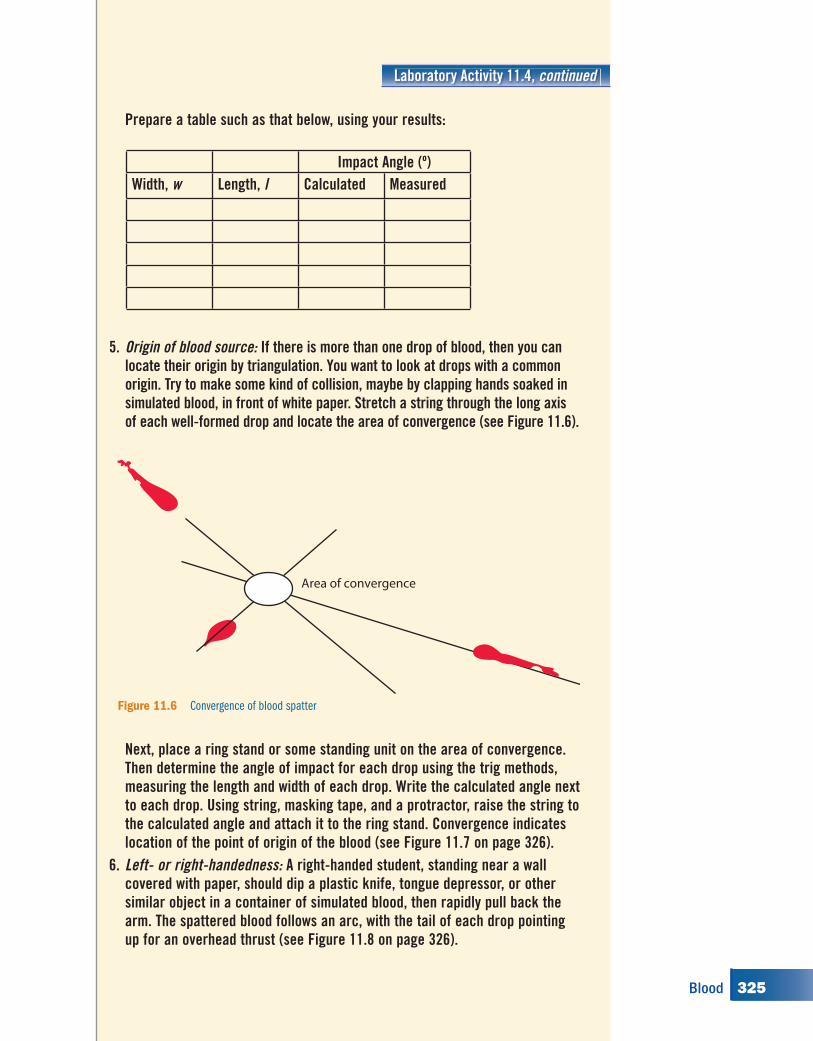
l
w
l
w
Figure 11.5 Measuring drop shape
surface
impact angle
path of drop of blood
no
rmal
Figure 11.4 Direction of travel
The drop shape can be described trigonometrically by the following relationship (see Figure 11.5):
impact angle, i � arc sin drop width, wdrop length, l� �
The arc sin can be calculated with a handheld calculator or found in a table such as Table 11.6 on page 327.
Procedure Note, continued
You can very effectively use
a digital camera to record
information.
Student investigation teams
should use a Beral or Pasteur
pipette to drop the blood
substitute vertically from
different heights onto a
smooth paper surface. They
should describe the shape
of the drop and its size and
note relationships and trends.
Perhaps the fastest group could
investigate drop shape as a
function of surface texture.
Students should place white
paper on cardboard and prop
it up at an angle. Then, using a
pipette, they can direct a drop
of blood onto the paper from a
fi xed height. They should record
the shape of the drop as a
function of the angle of impact,
as measured with a protractor.
It is important that they note
that the length-to-width ratio
increases with decreasing impact
angle. You may want to prepare
handouts of the fi gure to guide
the students.
324 Chapter 11
kh00116_CH11.indd Page 324 8/18/08 12:45:22 PM newuserkh00116_CH11.indd Page 324 8/18/08 12:45:22 PM newuser /Volumes/110/KHUS021/indd%0/Ch 11/Volumes/110/KHUS021/indd%0/Ch 11
Laboratory Activity 11.4, continued
Prepare a table such as that below, using your results:
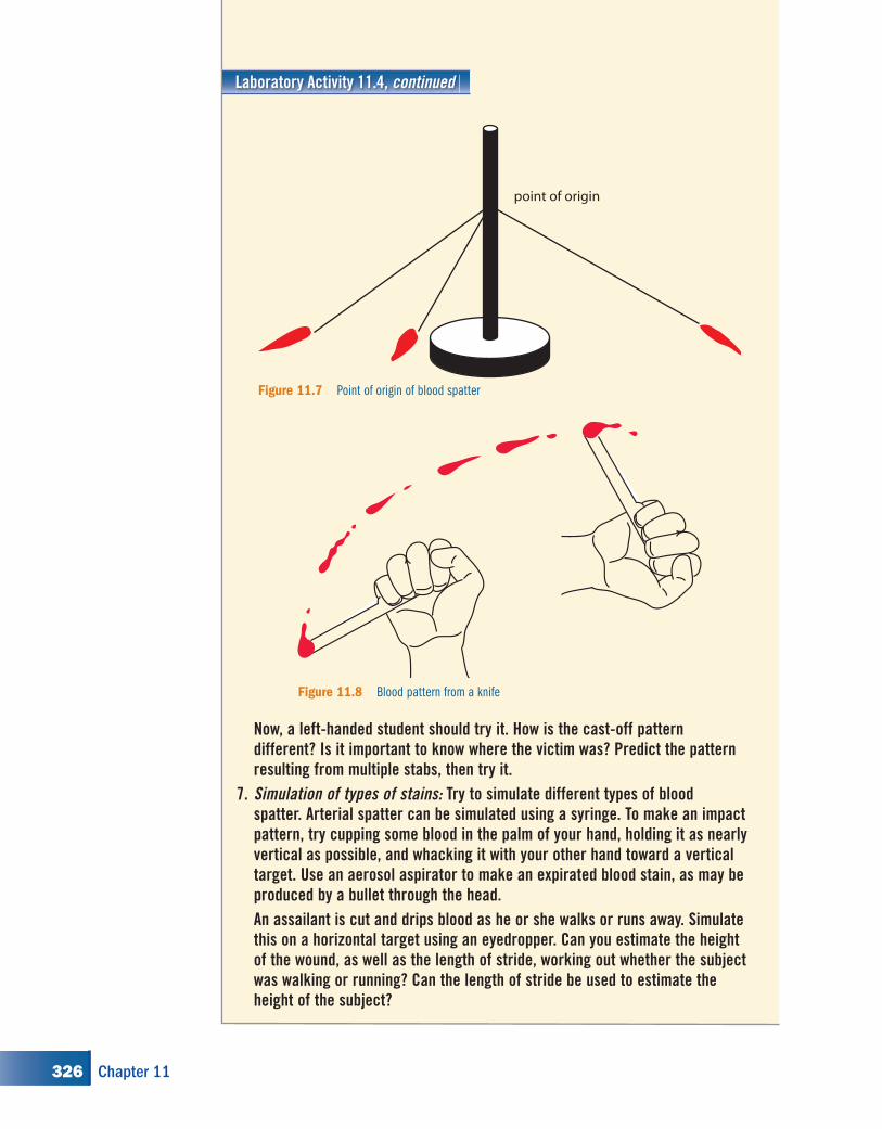
5. Origin of blood source: If there is more than one drop of blood, then you can locate their origin by triangulation. You want to look at drops with a common origin. Try to make some kind of collision, maybe by clapping hands soaked in simulated blood, in front of white paper. Stretch a string through the long axis of each well-formed drop and locate the area of convergence (see Figure 11.6).
Impact Angle (º)Width, w Length, l Calculated Measured
Area of convergence
Figure 11.6 Convergence of blood spatter
Next, place a ring stand or some standing unit on the area of convergence. Then determine the angle of impact for each drop using the trig methods, measuring the length and width of each drop. Write the calculated angle next to each drop. Using string, masking tape, and a protractor, raise the string to the calculated angle and attach it to the ring stand. Convergence indicates location of the point of origin of the blood (see Figure 11.7 on page 326).
6. Left- or right-handedness: A right-handed student, standing near a wall covered with paper, should dip a plastic knife, tongue depressor, or other similar object in a container of simulated blood, then rapidly pull back the arm. The spattered blood follows an arc, with the tail of each drop pointing up for an overhead thrust (see Figure 11.8 on page 326).
325Blood
kh00116_CH11.indd Page 325 8/18/08 12:45:22 PM newuserkh00116_CH11.indd Page 325 8/18/08 12:45:22 PM newuser /Volumes/110/KHUS021/indd%0/Ch 11/Volumes/110/KHUS021/indd%0/Ch 11
Laboratory Activity 11.4, continued
Now, a left-handed student should try it. How is the cast-off pattern different? Is it important to know where the victim was? Predict the pattern resulting from multiple stabs, then try it.
7. Simulation of types of stains: Try to simulate different types of blood spatter. Arterial spatter can be simulated using a syringe. To make an impact pattern, try cupping some blood in the palm of your hand, holding it as nearly vertical as possible, and whacking it with your other hand toward a vertical target. Use an aerosol aspirator to make an expirated blood stain, as may be produced by a bullet through the head.
An assailant is cut and drips blood as he or she walks or runs away. Simulate this on a horizontal target using an eyedropper. Can you estimate the height of the wound, as well as the length of stride, working out whether the subject was walking or running? Can the length of stride be used to estimate the height of the subject?
point of origin
Figure 11.7 Point of origin of blood spatter
Figure 11.8 Blood pattern from a knife
326 Chapter 11
kh00116_CH11.indd Page 326 8/18/08 12:45:23 PM newuserkh00116_CH11.indd Page 326 8/18/08 12:45:23 PM newuser /Volumes/110/KHUS021/indd%0/Ch 11/Volumes/110/KHUS021/indd%0/Ch 11
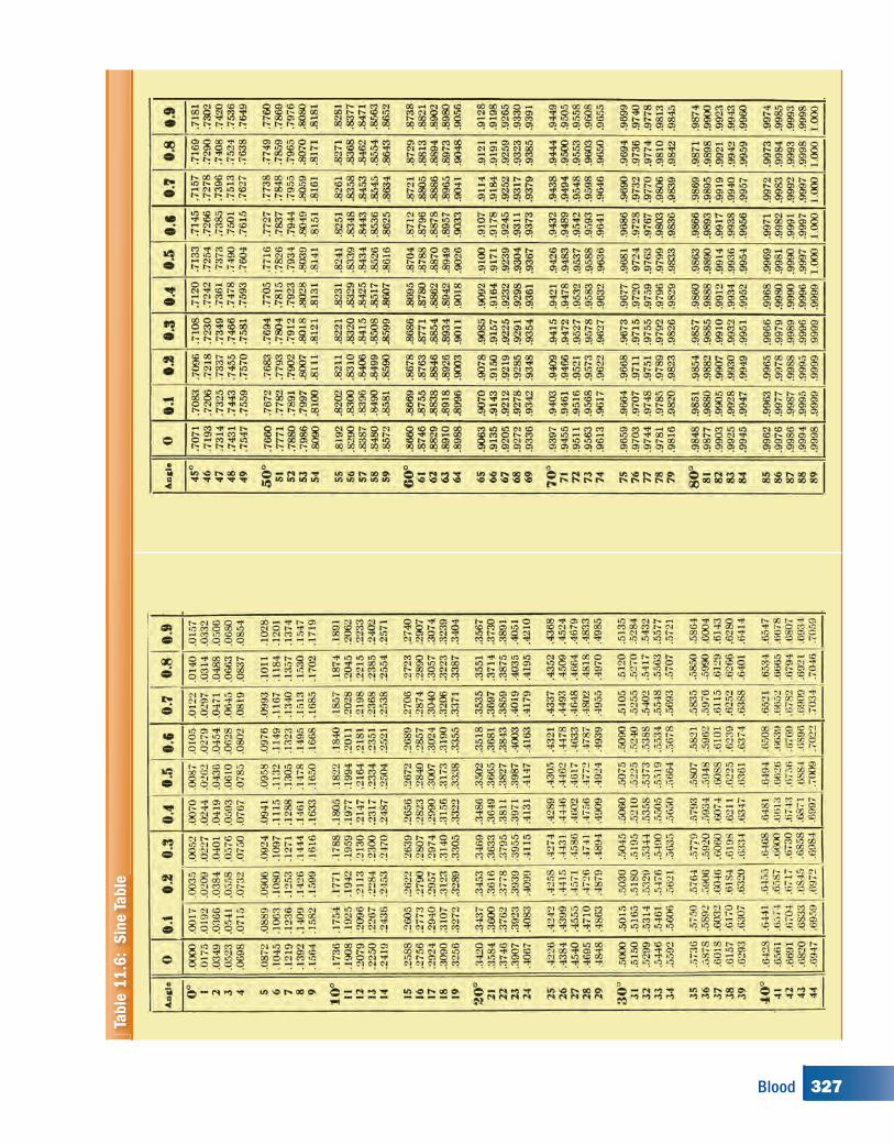
Table
11
.6:
Sin
e T
able
327Blood
kh00116_CH11.indd Page 327 8/18/08 12:45:23 PM newuserkh00116_CH11.indd Page 327 8/18/08 12:45:23 PM newuser /Volumes/110/KHUS021/indd%0/Ch 11/Volumes/110/KHUS021/indd%0/Ch 11
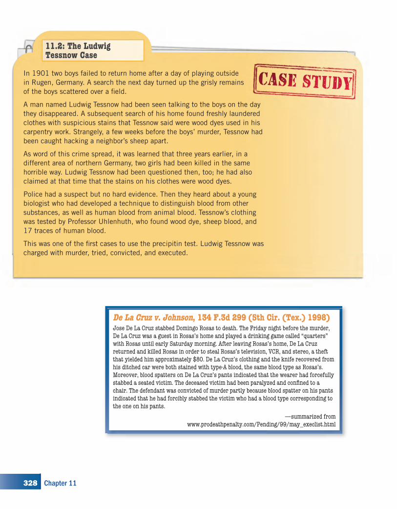
In 1901 two boys failed to return home after a day of playing outside
in Rugen, Germany. A search the next day turned up the grisly remains
of the boys scattered over a fi eld.
A man named Ludwig Tessnow had been seen talking to the boys on the day
they disappeared. A subsequent search of his home found freshly laundered
clothes with suspicious stains that Tessnow said were wood dyes used in his
carpentry work. Strangely, a few weeks before the boys’ murder, Tessnow had
been caught hacking a neighbor’s sheep apart.
As word of this crime spread, it was learned that three years earlier, in a
different area of northern Germany, two girls had been killed in the same
horrible way. Ludwig Tessnow had been questioned then, too; he had also
claimed at that time that the stains on his clothes were wood dyes.
Police had a suspect but no hard evidence. Then they heard about a young
biologist who had developed a technique to distinguish blood from other
substances, as well as human blood from animal blood. Tessnow’s clothing
was tested by Professor Uhlenhuth, who found wood dye, sheep blood, and
17 traces of human blood.
This was one of the fi rst cases to use the precipitin test. Ludwig Tessnow was
charged with murder, tried, convicted, and executed.
11.2: The Ludwig Tessnow Case
De La Cruz v. Johnson, 134 F.3d 299 (5th Cir. (Tex.) 1998)Jose De La Cruz stabbed Domingo Rosas to death. The Friday night before the murder, De La Cruz was a guest in Rosas’s home and played a drinking game called “quarters” with Rosas until early Saturday morning. After leaving Rosas’s home, De La Cruz returned and killed Rosas in order to steal Rosas’s television, VCR, and stereo, a theft that yielded him approximately $80. De La Cruz’s clothing and the knife recovered from his ditched car were both stained with type-A blood, the same blood type as Rosas’s. Moreover, blood spatters on De La Cruz’s pants indicated that the wearer had forcefully stabbed a seated victim. The deceased victim had been paralyzed and confi ned to a chair. The defendant was convicted of murder partly because blood spatter on his pants indicated that he had forcibly stabbed the victim who had a blood type corresponding to the one on his pants.
—summarized from www.prodeathpenalty.com/Pending/99/may_execlist.html
328 Chapter 11
kh00116_CH11.indd Page 328 8/18/08 12:45:34 PM newuserkh00116_CH11.indd Page 328 8/18/08 12:45:34 PM newuser /Volumes/110/KHUS021/indd%0/Ch 11/Volumes/110/KHUS021/indd%0/Ch 11
Checkpoint Questions
Answer the following questions. Keep the answers in your notebook, to be turned in to your teacher at the end of the unit.
1. What three questions should the investigator answer when examining an apparent dried bloodstain?
2. Defi ne a presumptive test.
3. What property of blood is used in most presumptive tests?
4. A forensic veterinarian may be asked to identify a specifi c animal’s blood. How would this be done?
5. What is a secretor?
6. Defi ne serology. How is it used in forensic investigations?
7. How much blood is there in the average adult human?
8. What test can determine whether blood is human or animal? What is the basis for this test? Why is the serum used in this test called human antiserum?
9. What are the four major blood types found in humans?
10. What happens if a person with type AB blood is given a transfusion of type A blood? Explain.
11. To whom can a person with type B blood donate blood and from whom can that person receive blood?
Answers
1. Is it blood? If so, animal or human? What is the
blood type?
2. a screening test that presumes the presence of a
substance
3. catalytic decomposition to oxygen
4. a precipitin test using serum with antibodies
specifi c to the test animal
5. A person whose blood antigens are found in other
body fl uids. About 80 percent of the population
are secretors.
6. The study of body fl uids using antigen–antibody
reactions. Serology can be used to exclude suspects.
7. about 5 quarts (students may have to look this up
or ask their biology teacher)
8. The precipitin test. Specifi c antibodies in an
animal serum react to human antigens.
9. A, B, AB, O
10. There will be no adverse reaction because there
are no antibodies to attack antigens.
11. He or she can donate to someone with B or AB
blood and receive blood from someone with
B or O.
329Blood
kh00116_CH11.indd Page 329 8/18/08 12:45:36 PM newuserkh00116_CH11.indd Page 329 8/18/08 12:45:36 PM newuser /Volumes/110/KHUS021/indd%0/Ch 11/Volumes/110/KHUS021/indd%0/Ch 11
12. Can a bloodstain be used for individualization?
13. What is the probability of an AB and Rh� blood type combination?
14. If a bloodstain found at the scene of a crime is found to be B, N, or Rh�, what is the probability that a suspect would have this combination of antigens? Is this good enough to convince a jury?
15. Calculate the angle of impact for the bloodstains below:
16. The precipitin test for human blood was developed in 1901 and is still in use today in forensic investigations, especially those involving the specifi c identifi cation of animal blood in cases involving poaching and possessing illegal wild game. Name two other forensic tests developed over 100 years ago that are still used in the same way today.
Blood typing can be applied to a host of enzymes and proteins that perform specifi c functions in the body. Their presence or absence varies within the population. More than 150 serum proteins and 250 cellular enzymes have been isolated. Therefore, investigators can use blood typing as individual evidence; however, it is not practical because of the time and techniques involved. Also, most factors degrade with time. Rather, ABO/Rh typing, and often MNS typing, are used as exclusionary tests in forensic science and paternity testing. The typical population in the United States shows an MNS distribution of M � 30 percent, N � 27 percent, S � 48 percent.
w
I
angle
D
w
I
angle
F
w
I
angle
E
w
I
angle
A
w
I
angle
B
w
I
angle
C
Answers, continued
12. Not with serology, but with DNA fi ngerprinting.
Many of the blood factors begin to degrade
immediately.
13. 0.04 � 0.15 � 0.0060, or 1 in 167
14. 0.11 � 0.27 � 0.15 � 0.0045, or 1 in 222
persons. Not suffi cient odds unless the fi eld
of suspects has been narrowed down by other
evidence.
15. A, 19º; B, 26º; C, 11º; D, 62º; E, 49º; F, 37º.
Refer to Blackline Masters 11.2 and 11.3 on the
TRCD for copies of illustrations.
16. fi ngerprints (1880); bite marks (1850); blood
(1863); bullet comparison (1835); blood
spatter (1514)
330 Chapter 11
kh00116_CH11.indd Page 330 8/20/08 7:12:56 PM newuserkh00116_CH11.indd Page 330 8/20/08 7:12:56 PM newuser /Volumes/110/KHUS021/indd%0/Ch 11/Volumes/110/KHUS021/indd%0/Ch 11
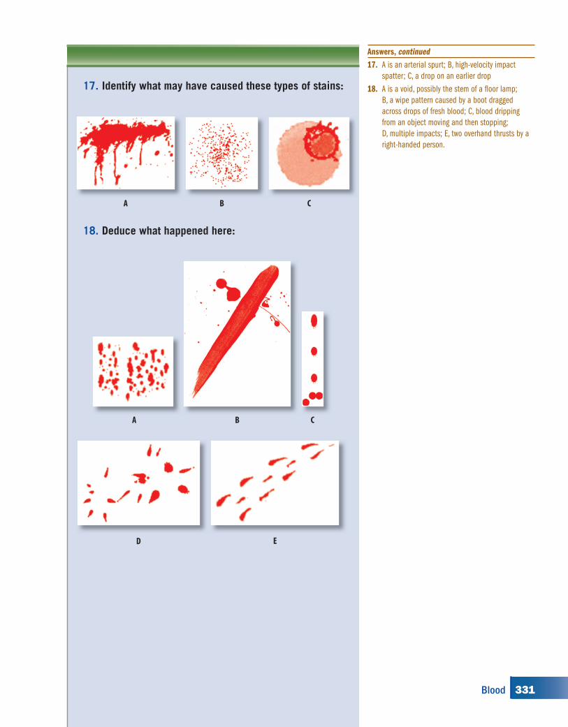
17. Identify what may have caused these types of stains:
18. Deduce what happened here:
A B C
A CB
D E
Answers, continued
17. A is an arterial spurt; B, high-velocity impact
spatter; C, a drop on an earlier drop
18. A is a void, possibly the stem of a fl oor lamp;
B, a wipe pattern caused by a boot dragged
across drops of fresh blood; C, blood dripping
from an object moving and then stopping;
D, multiple impacts; E, two overhand thrusts by a
right-handed person.
331Blood
kh00116_CH11.indd Page 331 8/20/08 7:12:57 PM newuserkh00116_CH11.indd Page 331 8/20/08 7:12:57 PM newuser /Volumes/110/KHUS021/indd%0/Ch 11/Volumes/110/KHUS021/indd%0/Ch 11
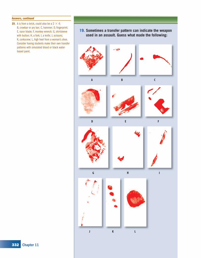
19. Sometimes a transfer pattern can indicate the weapon used in an assault. Guess what made the following:
CBA
ED F
LKJ
IHG
Answers, continued
19. A is from a brick, could also be a 2 � 4;
B, crowbar or pry bar; C, hammer; D, fi ngerprint;
E, razor blade; F, monkey wrench; G, shirtsleeve
with button; H, a fork; I, a knife; J, scissors;
K, corkscrew; L, high heel from a woman’s shoe.
Consider having students make their own transfer
patterns with simulated blood or black water-
based paint.
332 Chapter 11
kh00116_CH11.indd Page 332 8/18/08 12:45:40 PM newuserkh00116_CH11.indd Page 332 8/18/08 12:45:40 PM newuser /Volumes/110/KHUS021/indd%0/Ch 11/Volumes/110/KHUS021/indd%0/Ch 11
References
Books and ArticlesEvans, C. The Casebook of Forensic Detection. New
York: John Wiley, 1996. One hundred real cases.
Gottfried, S., and M. Sedotti, “Blood Markers,” from
Mystery Matters in Chem Matters, April 1992,
pp. 4–6.
Kurland, M. How to Solve a Murder. New York:
MacMillan, 1995.
Meloan, C. E., R. E. James, and J. R. Saferstein.
Criminalistics: An Introduction to Forensic
Science, Lab Manual (6th ed.). Upper Saddle
River, NJ: Prentice Hall, 1998.
Miller, L. S., and A. M. Brown. Criminal Evidence
Laboratory Manual: An Introduction to the Crime
Laboratory (2nd ed.). Cincinnati, OH: Anderson
Publishing Company, 1990.
Ragle, L. Chapter 6 in Crime Scene. New York: Avon
Books, 1995.
Films and Videos“The House That Roared,” Forensic Files, Court TV,
January 11, 2001. Available from Films for the
Humanities & Sciences, 24 min (www.films.
com). Use of luminol.
Nova, “The Killer’s Trail,” videotape on the Sam
Sheppard murder case with blood spatter
evidence. Available from http://main.wgbh.org/
wgbh/shop/products/wg2613.html.
A&E, “Dead Reckoning: Blood Spatter.” Videotape
includes the Sam Sheppard murder case with
blood spatter evidence, AAE73535. Available
from www.aetv.com.
Websiteswww.physics.carleton.ca/~carter; computerized blood
spatter analysis
http://anthro.palomar.edu/blood/default.htm; excellent
account of blood components, typing, and the like
www.crimelibrary.com; many good stories of crimes
and background material. See, for example, Sam
Sheppard, O. J. Simpson, and Jeffrey MacDonald.
www.bloodspatter.com/BPATutorial.htm; good tutorial
on bloodstains
Additional Projects
1. Investigate the Shroud of Turin from the aspect of whether
the imprints were actual blood. What scientific tests were
performed? Is there a controversy? What do you think?
(There are lots of websites for this topic; a good one is www.
shroudstory.com.)
2. How are blood types passed on to offspring? How does this
relate to paternity issues?
3. How could the sensitivity of luminol, Kastle-Meyer, and
Hemastix tests for blood be determined?
4. What is the effect of age, sunlight, freezing, and heat on
blood with respect to the standard presumptive tests?
Answers
1. See Web.
2. See Web; for example, http://www.biology.arizona.
edu/Human_Bio/problem_sets/blood_types/
markers.html.
3. progressive dilution
4. Web search
333Blood
kh00116_CH11.indd Page 333 8/20/08 7:12:58 PM newuserkh00116_CH11.indd Page 333 8/20/08 7:12:58 PM newuser /Volumes/110/KHUS021/indd%0/Ch 11/Volumes/110/KHUS021/indd%0/Ch 11