“kingdom” protista - bakersfield college 3a/bio 3a... · “kingdom” protista professor...
TRANSCRIPT

“KINGDOM” PROTISTA
Professor Andrea Garrison Biology 3A
Illustrations ©2014 Cengage Learning unless otherwise noted
Protista
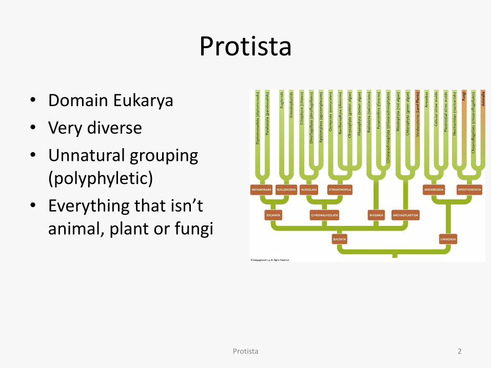
• Domain Eukarya
• Very diverse
• Unnatural grouping (polyphyletic)
• Everything that isn’t animal, plant or fungi
Protista 2

Protista
• Described by Leeuwenhoek in 1760s • Typically unicellular
– Complex cell structure – Some multicellular – Some colonial
• Autotrophic, heterotrophic or both – P/S forms often called algae
• Varied lifestyles: all require moisture – Seawater, freshwater, soil, decaying organisms, parasitic – Microscopic protists in lakes/oceans make up phytoplankton
• Food source for zooplankton and rest of food chain
• Some pathogenic – Amoebic dysentery, sleeping sickness, malaria
3 Protista

Protista
• Reproduction asexual or sexual or both – Asexual
• Binary fission
• Budding
• Colonies – May show division of labor
– Sexual • Meiosis, resulting gametes fuse
– Both • Elaborate life cycles specific to that group
Protista 4

Protista
• Cell structure – Cell wall, shell of mineral or organic matter, or
pellicle
– Most have mitochondria • Some with very reduced mitochondria
– P/S protists have plastids • Chloroplasts
– Chl a, and chl b or chl c
– Accessory pigments
• Other plastids contain pigments or store P/S products
Protista 5

Protistan Plastid Evolutionary Scenario
Protista 6

Protistan Plastid Evolutionary Scenario
Protista 7
Protista
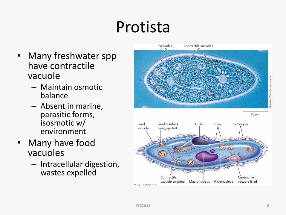
• Many freshwater spp have contractile vacuole – Maintain osmotic
balance – Absent in marine,
parasitic forms, isosmotic w/ environment
• Many have food vacuoles – Intracellular digestion,
wastes expelled
Protista 8
Protista
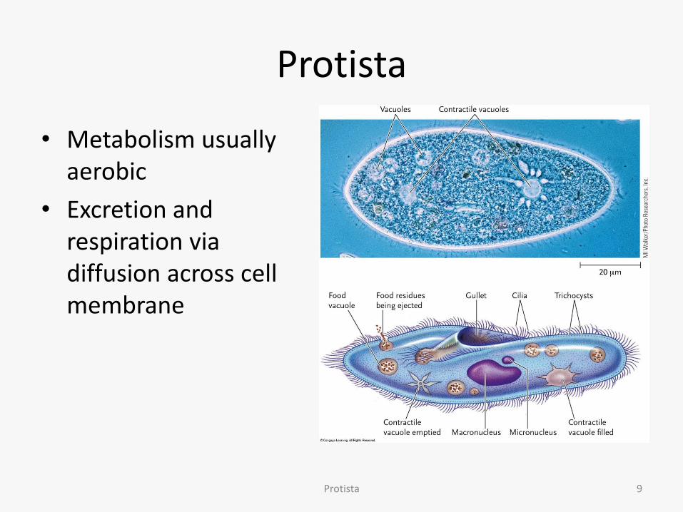
• Metabolism usually aerobic
• Excretion and respiration via diffusion across cell membrane
Protista 9
Protista
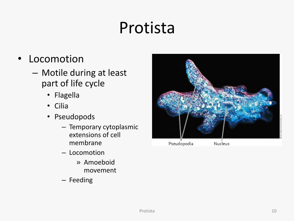
• Locomotion – Motile during at least
part of life cycle • Flagella
• Cilia
• Pseudopods – Temporary cytoplasmic
extensions of cell membrane
– Locomotion
» Amoeboid movement
– Feeding
Protista 10

Protistan Classification
Protista 11
SAR

Protistan Classification
• State of flux, not well understood (differs from text)
• Based on DNA data, structure
• 4 “supergroups” which also include the rest of Eukarya – Excavata
– SAR
– Archaeplastida
– Unikonta
Protista 12

BIKONTA
SAR
Arc
hae
pla
stid
a
Met
amo
nad
a
UNIKONTA
ARCHAEPLASTIDA EXCAVATA
Eugl
eno
zoa
Alv
eola
ta
Rh
izar
ia
Stra
men
op
ila
Am
oeb
ozo
a
Op
isth
oko
nta
p. 592
Protistan Classification

Protistan Classification
• 4 supergroups may eventually become kingdoms or suprakingdoms
• We will look at examples of each group
Protista 14
Excavata
• Parasitic, free-living species may be autotrophs or heterotrophs
• Feeding “groove” (excavate; ex=from, cavatum=cavity) in many, with flagellum in groove
• Reduced mitochondria in many – Mitochondrial genes incorporated into nuclei
• Locomotion via unique flagella – Crystalline or spiral rod inside
Protista 15

Excavata
• 3 clades
– Euglenozoa
– Diplomonada
– Parabasala
Protista 16

Excavata
• Clade Euglenozoa
– Highly motile
– Most P/S
– Some heterotrophic, or can be mixotrophic (both autotrophic and heterotrophic)
Protista 17

Excavata
• Subclade Euglenida – Free-living, most freshwater – 2 flagella
• 1 anterior flagellum – Can pull or push
• 1 very reduced
– Most P/S • Can also absorb nutrients across
cell membrane
– Contractile vacuole – Spiral-grooved pellicle replaces
cell wall • Protein strips under cell
membrane
– Photosensitive eyespot • Stay positioned in optimum light
levels
Protista 18
Excavata
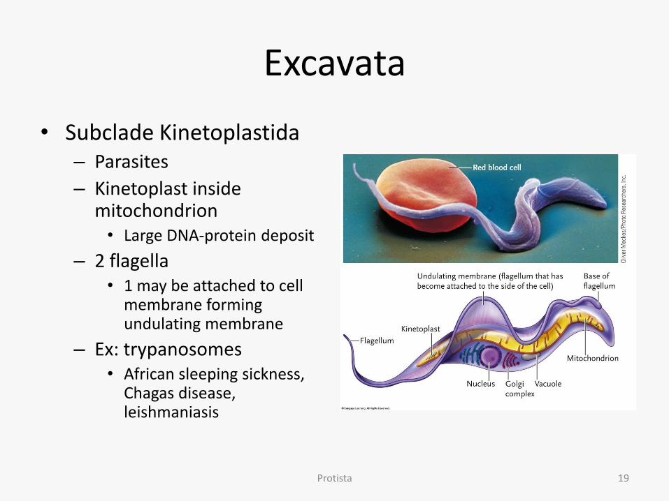
• Subclade Kinetoplastida – Parasites
– Kinetoplast inside mitochondrion • Large DNA-protein deposit
– 2 flagella • 1 may be attached to cell
membrane forming undulating membrane
– Ex: trypanosomes • African sleeping sickness,
Chagas disease, leishmaniasis
Protista 19

Excavata
• Clade Diplomonada
– Many parasitic (intestinal)
– Anaerobic
– 2 nuclei
– Multiple flagella
– Ex: Giardia sp. • Giardiasis
Protista; photo From Brooke, MM, Melvin, DM: Morphology of Diagnostic Stages of Intestinal Parasites of Man. Public Health Service Publication No. 1966, 1969, http://www.ncbi.nlm.nih.gov/books/NBK7889/figure/A4205/?report=objectonly
20
Giardia sp.
http://www.cdc.gov/parasites/giardia/images/giardia-banner.jpg

Excavata
• Clade Diplomonada
– Many parasitic (intestinal)
– Anaerobic
– 2 nuclei
– Multiple flagella
– Ex: Giardia sp. • Giardiasis
Protista; http://www.cdc.gov/parasites/giardia/pathogen.html 21

Excavata
• Clade Parabasala
– Many parasitic
– Anaerobic
– Undulating membrane attached to flagellum
– Trichomonas vaginalis • Trichomoniasis
Protista; http://www.cdc.gov/std/trichomonas/stdfact-trichomoniasis.htm 22
Trichomonas vaginalis

SAR
• Very diverse group, phylogeny controversial
• 3 clades
– Stramenopila
– Alveolata
– Rhizaria
Protista 23
SAR
• Clade Stramenopila
• P/S
• “Hairy” flagellum, usually a shorter smooth flagellum as well
• 3 subclades
– Bacillariophyta
– Chrysophyta
– Phaeophyta
Protista 24

SAR/Stramenopila
• Subclade Bacillariophyta
– Diatoms
– Marine and freshwater
– Primary P/S member of marine phytoplankton • Chlorophyll, fucoxanthin
and beta-carotene
– Covered by glassy silica shell • 2 halves; pill box
• Diatomaceous earth
Protista 25

SAR/Stramenopila
• Subclade Bacillariophyta
– Flagella in gametes only
– Mature forms move via secretion released through grooves in shell
Protista 26

SAR/Stramenopila
• Subclade Bacillariophyta
– Reproduction asexual and sexual
• Binary fission most frequent
Protista 27

SAR/Stramenopila
• Subclade Chrysophyta – Golden algae
– Freshwater or marine
– Mixotrophic • P/S
– Chlorophyll, carotenoids, fucoxanthin
• Heterotrophs when light too low
– Glassy silica plates or scales
– May “bloom” in spring or fall • Discolors water
Protista; photo: Frank Fox - http://www.mikro-foto.de; http://creativecommons.org/licenses/by-sa/3.0/de/legalcode
28
Dinobryon: sessile, colonial chrysophyte

SAR/Stramenopila
• Subclade Phaeophyta
– Brown algae (color from olive green to brown)
– All multicellular
• Range from few centimeters to over 30m+ (giant kelps)
– Autotrophs
– Chlorophyll and fucoxanthin
– Cell wall contain cellulose and alginic acid
• Algin harvested for thickener in ice cream, pudding, cosmetics
Protista 29

SAR/Stramenopila
• Subclade Phaeophyta – Large kelps look like plants
but different structure • Holdfast
– Looks like roots, but only anchor, no absorption
• Stipe – Looks like trunk, minimal
vascular tissue
• Blade – Looks like leaves
• Bladders – Hollow, gas-filled for
buoyancy
Protista—picture: Claire Fackler; NOAA Photo Library 30
SAR/Stramenopila
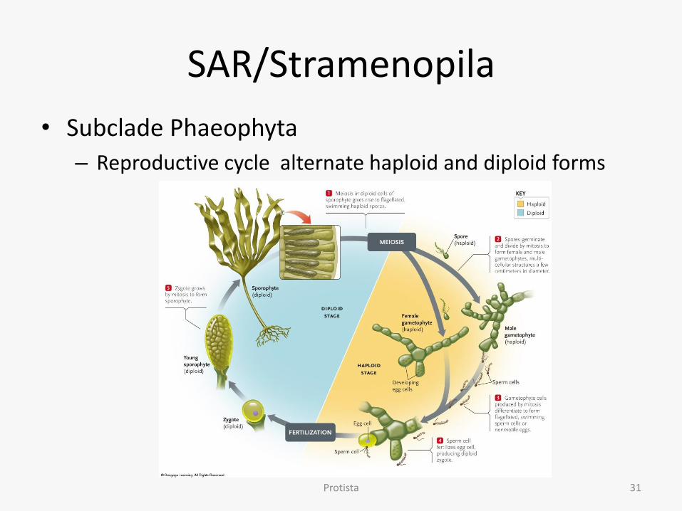
• Subclade Phaeophyta
– Reproductive cycle alternate haploid and diploid forms
Protista 31
SAR
• Clade Alveolata – Have alveoli (alvus = belly)
• Small flattened membrane vesicles just under cell membrane (=pellicle)
• Support membrane
– Tubular membranes inside mitochondria
– 3 subclades • Dinoflagellata
• Apicomplexa
• Ciliophora
Protista 32

SAR/Alveolata
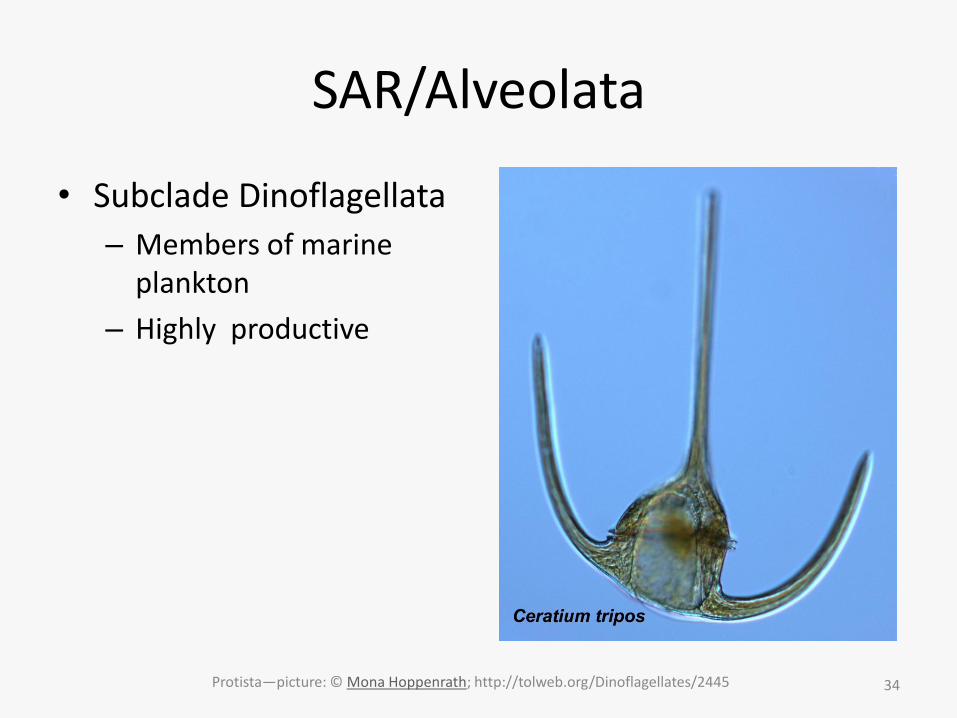
• Subclade Dinoflagellata
– Autotrophs or heterotrophs • Many be mixotrophic
• Some have algal symbionts
– Plates (cellulose) form “shell” under membrane
– Flagella beat within grooves between plates • Make cells spin
• Chloroplasts (P/S forms) have chlorophyll and carotenoids
Protista 33
SAR/Alveolata
• Subclade Dinoflagellata
– Members of marine plankton
– Highly productive
Protista—picture: © Mona Hoppenrath; http://tolweb.org/Dinoflagellates/2445 34
SAR/Alveolata
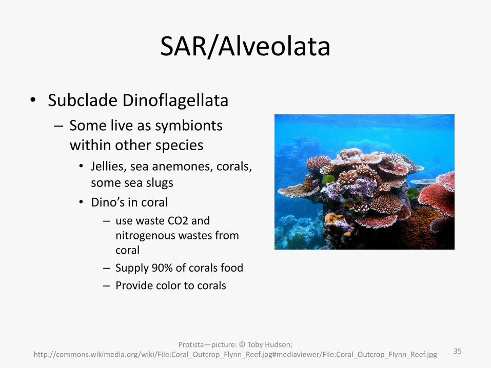
• Subclade Dinoflagellata
– Some live as symbionts within other species • Jellies, sea anemones, corals,
some sea slugs
• Dino’s in coral
– use waste CO2 and nitrogenous wastes from coral
– Supply 90% of corals food
– Provide color to corals
Protista—picture: © Toby Hudson; http://commons.wikimedia.org/wiki/File:Coral_Outcrop_Flynn_Reef.jpg#mediaviewer/File:Coral_Outcrop_Flynn_Reef.jpg 35
SAR/Alveolata
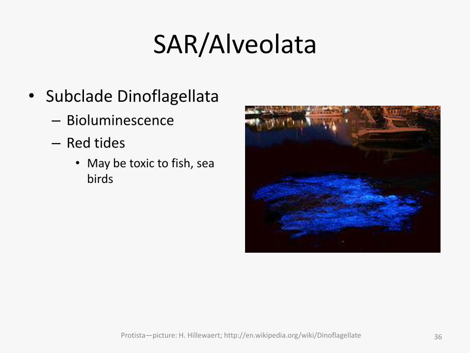
• Subclade Dinoflagellata
– Bioluminescence
– Red tides • May be toxic to fish, sea
birds
Protista—picture: H. Hillewaert; http://en.wikipedia.org/wiki/Dinoflagellate 36
SAR/Alveolata
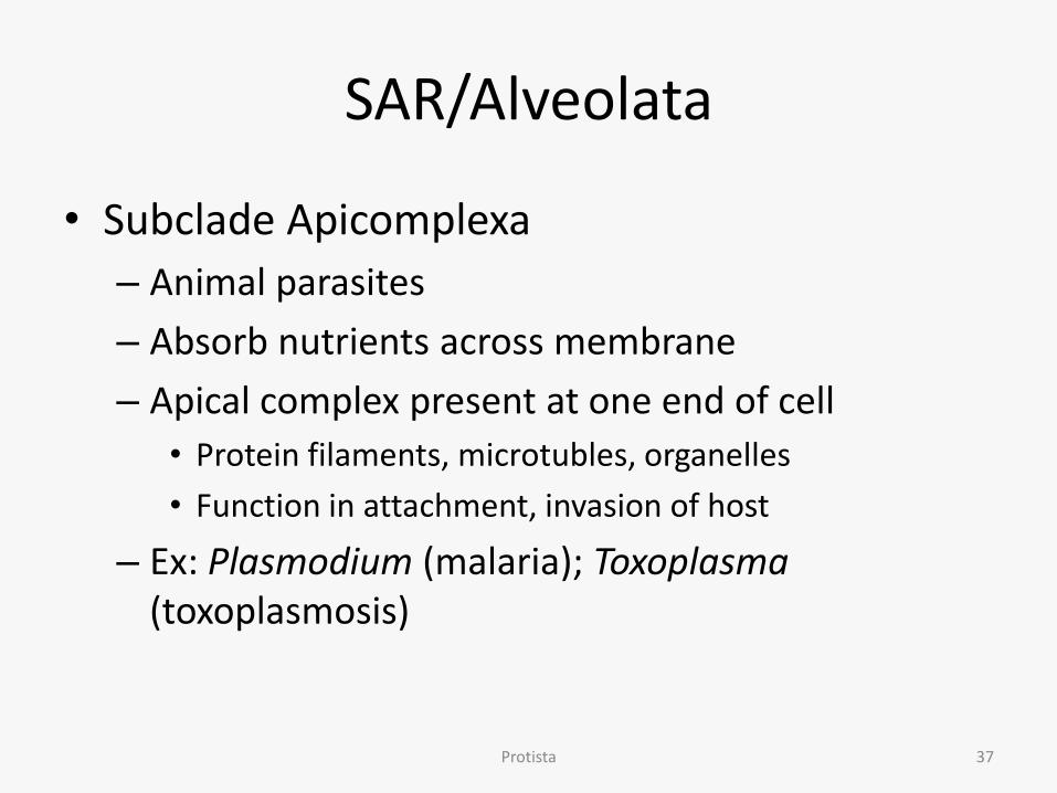
• Subclade Apicomplexa
– Animal parasites
– Absorb nutrients across membrane
– Apical complex present at one end of cell
• Protein filaments, microtubles, organelles
• Function in attachment, invasion of host
– Ex: Plasmodium (malaria); Toxoplasma (toxoplasmosis)
Protista 37

SAR/Alveolata
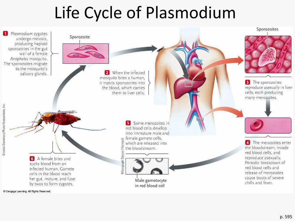
• Subclade Apicomplexa
– Complex life cycle
• Sexual and asexual components
Protista—picture from http://www.niaid.nih.gov/topics/Malaria/P
ages/lifecycle.aspx 38
p. 595
Life Cycle of Plasmodium

SAR/Alveolata
• Subclade Ciliophora – Marine, freshwater, soil – Cilia – Most heterotrophs
• Feed on bacteria and algae
Protista 40

SAR/Alveolata
• Subclade Ciliophora – Contractile vacuoles
– Gullet lined w/cilia for feeding • Food vacuoles • Waste vacuoles
– Trichocysts just under pellicle • Discharge threads when stressed
Protista 41

SAR/Alveolata
• Subclade Ciliophora – Two nuclei
• Macronucleus polyploid – Used in normal cell function
• Micronucleus diploid – Used in reproduction (sexual or asexual)
Protista 42

SAR/Alveolata
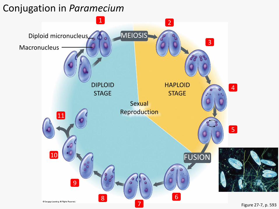
• Subclade Ciliophora – Asexual reproduction via binary fission
• Both nuclei replicated and divide via mitosis
– Sexual reproduction • Conjugation
– Cells pair up, form cytoplasmic bridge – Replicate and exchange micronuclei – 1 micronucleus forms macronucleus – Cells undergo binary fission
Protista 43
MEIOSIS
Sexual Reproduction
DIPLOID STAGE
HAPLOID STAGE
FUSION
Diploid micronucleus
Macronucleus
2 1
3
5
4
6 8 7
9
11
10
Figure 27-7, p. 593
Conjugation in Paramecium

SAR
• Clade Rhizaria
• Amoebas
• Stiff filamentous pseudopods
• Many have tests (hard shells)
• We’ll study 2 subclades
– Radiolaria
– Foraminifera
Protista 45
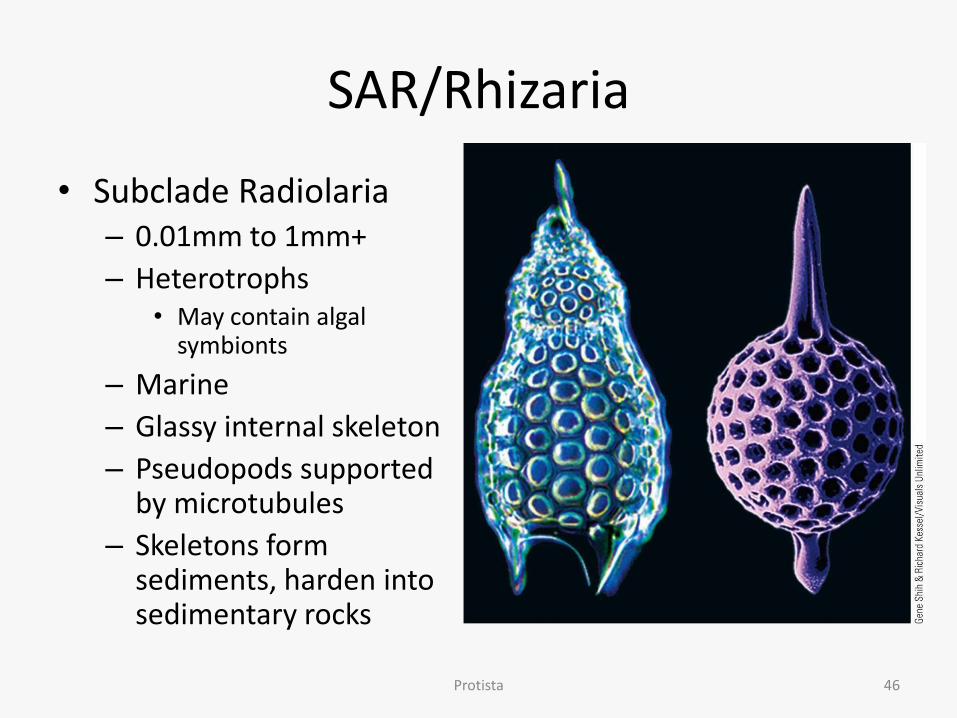
SAR/Rhizaria
• Subclade Radiolaria – 0.01mm to 1mm+
– Heterotrophs • May contain algal
symbionts
– Marine
– Glassy internal skeleton
– Pseudopods supported by microtubules
– Skeletons form sediments, harden into sedimentary rocks
Protista 46
SAR/Rhizaria
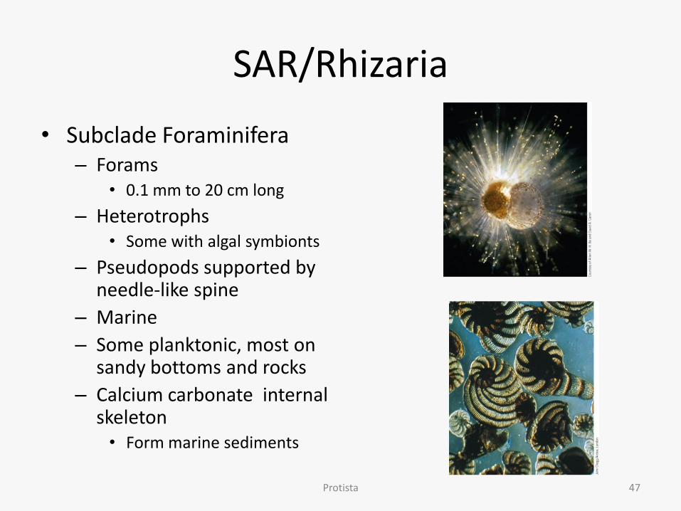
• Subclade Foraminifera – Forams
• 0.1 mm to 20 cm long
– Heterotrophs • Some with algal symbionts
– Pseudopods supported by needle-like spine
– Marine
– Some planktonic, most on sandy bottoms and rocks
– Calcium carbonate internal skeleton • Form marine sediments
Protista 47

Archaeplastida
• Closest relatives of land plants
• Plastids show no indication of secondary endosymbiosis
• 2 Clades
– Rhodophyta
– Chlorophyta
Protista 48

Archaeplastida
• Clade Rhodophyta – Red algae – Most marine, most free-living – Multicellular, most small (<1 ft)
• Filamentous or sheetlike forms • Some covered in calcium carbonate (coralline algae)
– Adhesive holdfast
Protista 49

Archaeplastida
• Clade Rhodophyta – Chlorophyll, phycobilins
• Phycobilins allow them to grow deeper than other algae (absorb blue light)
– Complex life cycles involving alternation of diploid sporophytes and haploid gametophytes
– Commercial use • Porphyra used for nori
• Gelidium used for agar
• Chondrus, Gigartina, Eucheuma used for carrageenan
Protista 50

Archaeplastida
Protista 51
• Clade Chlorophyta – Green algae
– Most freshwater; some symbionts • w/fungi (=lichen), sea
anemones, marine snails
– Most microscopic
– Very diverse species
Archaeplastida
• Clade Chlorophyta – Green algae – Most freshwater; some symbionts
• w/fungi (=lichen), sea anemones, marine snails
– Most microscopic – Very diverse species – Diverse life cycles
• Sexual, asexual, or alternate
– Chlorophyll, carotenoids – Nucleic acid sequences, pigments, chloroplasts, food
reserves most similar to land plants
Protista 52

Unikonta
• 2 Clades
– Amoebozoa
– Opisthokonts
• Closely related to Fungi and Animals
Protista 53

Unikonta/Amoebozoa
• Most of the amoebas and plasmodial slime molds
• We’ll study 2 subclades which aren’t plamodial slime molds
– Gymnamoeba
– Entamoeba
Protista 54

Unikonta/Amoebozoa
• Subclade Gymnamoeba
– Amoebas
– Marine and freshwater
– Most free-living
– Lobose pseudopods • unsupported by internal
structure
• Locomotion and feeding
• No fixed body shape
– Contractile vacuole
Protista 55

Unikonta/Amoebozoa
• Subclade Entamoeba – Parasitic
– Entamoeba histolytica • Amoebic dysentery
– Destroys cells lining intestine
– Ulcerations, cramps, bloody diarrhea, foul-smelling stools
– Cysts spread through contaminated food or water
– A leading cause of death in infants and small children in less developed nations
Protista; photo CDC.gov 56
Trophozoite with ingested RBCs
Unikonta/Opisthokonts
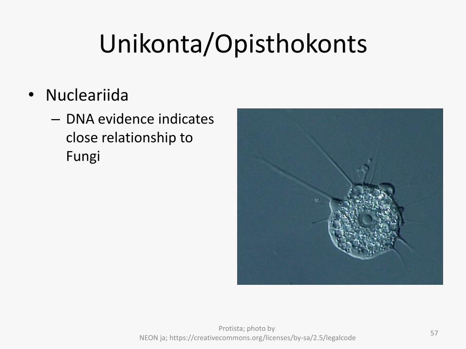
• Nucleariida
– DNA evidence indicates close relationship to Fungi
Protista; photo by NEON ja; https://creativecommons.org/licenses/by-sa/2.5/legalcode
57

Unikonta/Opisthokonts
• Choanoflagellida
– DNA evidence indicates close relationship to Animalia
Protista; photo lower left by DH Zanette;,public domain, https://en.wikipedia.org/wiki/Choanoflagellate#/media/File:Sphaeroeca-colony.jpg; photo upper right by Stephen
Fairclough, https://creativecommons.org/licenses/by-sa/2.5/legalcode; picture lower right by Wikipedia user Urutseg, https://creativecommons.org/licenses/by-sa/3.0/legalcode;
58