kinetics of metals in molluscan faecal pellets and ...users.uoa.gr/~anikol/papers pdf/26 nott and...
TRANSCRIPT

ELSEVIER Journal of Experimental Marine Biology and Ecology,
197 (1996) 203-218
JOURNAL OF EXPERIMENTAL MARINE BIOLOGY AND ECOLOGY
Kinetics of metals in molluscan faecal pellets and mineralized granules, incubated in marine sediments
James A. NottaX*, Artemis Nicolaidoub
“Plymouth Marine Laboratory, Citadel Hill, Plymouth, PLl 2PB, UK bDepartment of Biology, University of Athens, CR 15784, Athens, Greece
Received 23 March 1995; revised 1 August 1995; accepted 1 September 1995
Abstract
Marine snails excrete metals via the gut. The sediment-feeding, tower shell Cerithium
vu&turn, from a polluted environment, accumulates Cr and Ni to 3500 ppm dry weight in the faecal pellets. Pellets from C. vulgatum and the grazing, top shell Monodontu mutubilis were incubated in surface and deeper anoxic layers of clean and metal-polluted sediment. Pellets retained the original load of metals and in some cases gained Ti, Cr, Mn, Fe and Ni. Effects were modified by sediment properties. Pellets of C. vu&turn are durable, membrane-bound structures and they reduce metal bioavailability to food chains by compartmentalization. Intracellular, phosphate granules bind metals in digestive glands of snails and they are excreted via the gut and faecal pellets. These granules were extracted from digestive glands of both species of snail and incubated in the same sediments. Magnesium phosphate granules from M. mutubilis dissolved but calcium/metal phosphate granules from C. vulgutum remained; they had differentially retained or lost Mn, Fe, Co, Ni and Zn according to the type of sediment.
Keywords: Faecal pellets; Granules; Metals; Molluscs; Sediments
1. Introduction
In tissues of marine invertebrates, hard acid metals Na, Mg, K and Ca, transition metals Ti,V, Cr, Mn, Fe, Co and Ni, and post-transition Zn, can be detoxified by making them biologically unavailable as insoluble phosphate salts. These are produced intracel- lularly in the form of amorphous, mineral granules. In gastropod molluscs and decapod crustaceans these granules are spherical and concentrically structured. In the former, they occur in the basophil cells of the digestive gland (Simkiss and Mason, 1984; Taylor
*Corresponding author. Fax: (44) (1752) 63-3102.
00%0981/96/$15.00 0 1996 Elsevier Science B.V. All rights reserved SSDI 0022-0981(95)00155-7

204 J.A. NOR, A. Nicolaidou I .I. Exp. Mar. Riol. Ecol. 197 (1996) 203-218
and Simkiss, 1984) and, in the latter, in the R-cells of the hepatopancreas (Becker et al., 1974; Hopkin and Nott, 1979; Al-Mohanna and Nott, 1989; Icely and Nott, 1992). In bivalve molluscs they are irregularly shaped and occur in the epithelial tissue of the kidney (Carmichael et al., 1979; George et al., 1980). They have been the subject or a main feature of several literature reviews (Brown, 1982; Icely and Nott, 1980, Mason and Nott, 1981; Simkiss and Mason, 1984; Nott, 1993; Viarengo and Nott, 1993). When these tissues, complete with phosphate granules, are consumed by predators the granules are insoluble and they pass through the gut and are voided in the faeces, retaining a significant proportion of the original metal content (Nott and Nicolaidou, 1990, 1993, 1994). In these cases the system of detoxification is effective in both prey and predator and the result is bioreduction in the concentration of metals in tissues along food chains. The epithelia in the digestive gland and kidney have a single functional cycle, at the end of which the cells become necrotic and disintegrate into the lumen of the digestive gland or kidney. They are subsequently excreted, complete with granules, and become a component of the sediment. The next question is, what happens to the granules and in particular what happens to the metals they contain?
In the external environment, various constituents of granules will have different solubilities and these will be affected by the properties of sediments and seawater including pH, salinity and redox. These properties vary markedly on a regional basis and with depth and also on the clay and sand characteristics of sedimentary particles. Work on metals in food chains has been reviewed by Fowler (1982) and Suede1 et al. (1994) and a general conclusion is that the degree to which metals are transferred between organisms depends upon the bioavailability of each element.
The present work investigates elemental changes that occur to invertebrate faecal pellets and to biogenic phosphate granules when incubated within sediments which have contrasting characteristics.
2. Materials and methods
During March 1994, sediments were taken from two sites on mainland Greece. One is a polluted site adjacent to a nickel smelting plant at Larymna and the other is a cleaner site at Vravrona (Nicolaidou and Nott, 1990). Cores of sediment were taken with an adapted 50 ml syringe with the end cut off to create a single-diameter, open-ended tube. Sediment was sampled down to 7 cm and this core was sub-divided for investigations of the microbiological properties at several different depths. The microbiological results will be reported elsewhere. For the present investigation the surface ‘white‘ sediments and the 7 cm depth ‘black‘ sediments from both polluted and clean sites were used as ‘incubation media‘ for faecal pellets and phosphate granules.
Measurements of pH and redox were taken for white and black sediments at the time of sampling. A portable Russell RLlOO meter was used with a combination pH electrode and a combination platinum rod redox cell. Samples of all sediments were dried and stored for CHN and metal analysis.
The gastropods Cerithium vulgutum and Monodonta mutabilis were collected from
J.A. Nott, A. Nicolaidou I J. Exp. Mar. Biol. Ecol. 197 (1996) 203-218 205
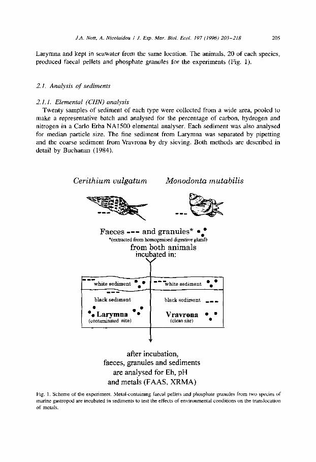
Larymna and kept in seawater from the same location. The animals, 20 of each species, produced faecal pellets and phosphate granules for the experiments (Fig. 1).
2. I. Analysis of sediments
2.1.1. Elemental (CHN) analysis
Twenty samples of sediment of each type were collected from a wide area, pooled to make a representative batch and analysed for the percentage of carbon, hydrogen and nitrogen in a Carlo Erba NA1500 elemental analyser. Each sediment was also analysed for median particle size. The fine sediment from Larymna was separated by pipetting and the coarse sediment from Vravrona by dry sieving. Both methods are described in detail by Buchanan (1984).
Cerithium vulgatum Monodonta mutabilis
Faeces -- - and granules* 0.’ *(extracted from homogenised digestive gland)
from both animals incubated in:
- 1-1 white sediment .a l __ -white sediment ‘. l
___
black sediment black sediment _ _ _
0
‘. Larymna l : (contaminated site)
Vravrona 0. l (clean site)
after incubation, faeces, granules and sediments
are analysed for Eh, pH and metals (FAAS, XRMA)
Fig. 1. Scheme of the experiment. Metal-containing faecal pellets and phosphate granules from two species of
marine gastropod are incubated in sediments to test the effects of environmental conditions on the translocation
of metals.

206 J.A. Nott, A. Nicolaidou I J. Exp. Mm. Biol. Ecol. 197 (1996) 203-218
2.1.2. Redox (E,, ) and pH E, measurements were taken with a combination, calomel/platinum rod, redox cell.
Five results were recorded for each sediment when the meter readings stabilized. The E, positive or negative millivolt measurements reflected the intensity of the oxidation- reduction condition of the interstitial water in the sediment. Measurements of pH were taken with a combination electrode. Both E, and pH readings were obtained with the same portable, Russell RLlOO meter.
2.1.3. Flame atomic absorption spectroscopy (FAAS)
The same mixed sediments were analysed for Cr, Mn, Co, Ni, Cu, Zn, Ag and Cd by FAAS in a Varian AA20 spectrophotometer. Values were produced for ‘total‘ metal concentrations extracted with 16 M HNO, and ‘bioavailable‘ concentrations extracted by 1 M HCl. Each analysis was continued until there was < 1% variation in absorbance. The performance of the spectrophotometer is checked with reference material once a month.
2.1.4. X-ray microanalysis (XRMA)
Mixed dry sediment of each type was spread thinly and evenly on a graphite stub coated with colloidal graphite and analysed by a Link XRMA system in a Jeol 35C scanning electron microscope (SEM). Each spectrum produced an integrated analysis from an area of 0.6 mm* on the stub.
2.2. Analyses of faecal pellets in sediments (Fig. 1)
Cerithium vulgatum and Monodonta mutabilis collected from Larymna were kept in separate aquaria in aerated seawater from Larymna. After 24 hours faecal pellets were collected from both species and added to three replicate 2 cm diameter glass vials containing 0.5 cm levels of wet, white or black sediments from Larymna and Vravrona. Vials containing black sediment plus pellets were flushed with nitrogen gas and sealed. Vials containing white sediment plus pellets were left open and kept wet by adding aerated seawater as required. Seawater was collected from the same site as the sediments.
For controls, fresh pellets were fixed on carbon SEM stubs with colloidal graphite to establish the metal content by XRMA prior to incubation. Additional vials were one third filled with wet, white and black sediments for E, and pH determinations. The black sediment vials were flushed with nitrogen and sealed.
After 5 days, 5 weeks and 15 weeks, the faecal pellets were recovered from the sediments and mounted on graphite specimen stubs for XRMA in the SEM. The SEM magnification was X 500 and the entire area scanned by the electron beam, namely 0.6 mm’, produced an integrated elemental analysis of a pellet.
Cerithium vulgatum produced sufficient faecal pellets for some to be bulked and analysed by FAAS.
J.A. Nott, A. Nicolaidou I J. Exp. Mar. Biol. Ecol. 197 (1996) 203-218 201
2.3. Analyses of intracellular granules in sediments (Fig. 1)
Digestive glands from Cerithium vulgatum and Monodonta mutabilis were diced into small pieces which were pulverised to a fluid. The fluid was mixed with seawater in a vial and allowed to stand for 30 mins. The top one third of the suspension which consisted of lipid and other buoyant material was removed and this left the mineral granules and dense cytological material in the lower layer. The granules were resuspended and added to the white and the black sediments from both Larymna and Vravrona. The amount of suspended granules was only sufficient to add to one vial of each type of sediment and also provide material for SEM (see below). The sediments were wet and filled the vials to a depth of 0.5 cm. Vials containing black sediments were flushed with nitrogen gas and sealed. White sediments were kept moist with additional, aerated seawater which was collected from the same site as the sediment. All vials were gently agitated in a slow-running rotator. After 4 days the vials were taken off the rotator and the sediments/granules were dried and stored for x-ray analysis in the SEM.
Fresh granules from the suspension of pulverised digestive gland were mounted on carbon SEM stubs for XRMA to establish the elemental composition prior to incubation and thus act as controls.
3. Results
3.1. Analysis of sediments
3.1.1. Elemental (CHN) analysis
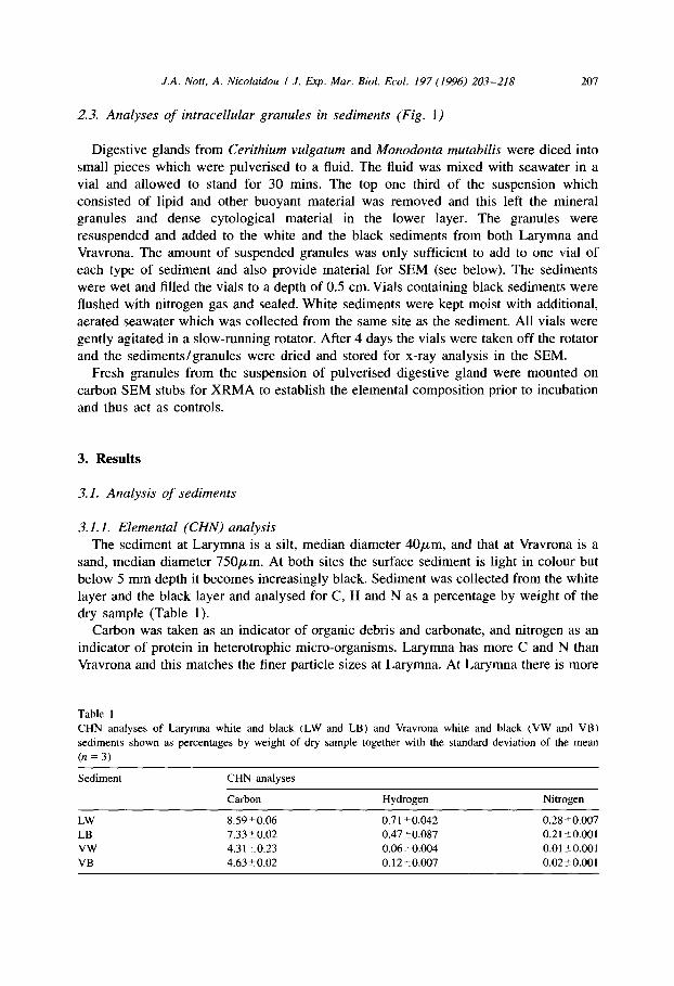
The sediment at Larymna is a silt, median diameter 40pm, and that at Vravrona is a sand, median diameter 750pm. At both sites the surface sediment is light in colour but below 5 mm depth it becomes increasingly black. Sediment was collected from the white layer and the black layer and analysed for C, H and N as a percentage by weight of the dry sample (Table 1).
Carbon was taken as an indicator of organic debris and carbonate, and nitrogen as an indicator of protein in heterotrophic micro-organisms. Larymna has more C and N than Vravrona and this matches the finer particle sizes at Larymna. At Larymna there is more
Table 1 CHN analyses of Larymna white and black (LW and LB) and Vravrona white and black (VW and VB)
sediments shown as percentages by weight of dry sample together with the standard deviation of the mean
(n=3)
Sediment CHN analyses
Carbon Hydrogen Nitrogen
LW 8.59?0.06 0.71?0.042 0.28-tO.007
LB 7.33kO.02 0.47fO.087 0.21 %O.OOl
VW 4.3120.23 0.06t0.004 0.01 ?O.OOl
VB 4.6320.02 0.12~0.007 0.02+0.001

208 J.A. Nort, A. Nicolaidou I J. Exp. Mar. Bid. Ed. 197 (1996) 20-T-218
Table 2
E, (mV) and pH measurements for Larymna white and black (LW and LB) and Vravrona white and black (VW
and VB) sediments
Sediment E, before experiment
LW + 46
LB - 331
VW f91
VB - 260
E, after experiment PH
+ 57 7.83
- 290 7.65
+ 110 7.78
~ 270 7.44
E, values are the means of five readings taken when the meter had stabilized.
organic matter in the white surface sediment than in the black material, whilst at Vravrona the C and N values are similar for both the surface and lower layers.
3.1.2. E,, und pH Redox measurements (Eh) for sediments (Table 2) were taken before and after the 5
day experiments. Sediments were used as an incubation medium for faecal pellets from Cerithium vulgatum and Monodonta mutubilis and mineral granules extracted from the digestive glands of these animals. White sediments were at the surface and black sediments were at a depth of 7 cm. Measurements of pH were taken before the experiments (Table 2).
3.1.3. Flame atomic absorption spectroscopy
In the spectrophotometer, samples were sprayed into the flame until there was less than 1% variation in absorbance (Table 3). For Co in Vravrona sediments the concentration of metal extracted by 1 M HCl to model bioavailability is higher than the concentration for ‘total‘ metal when extracted by 16 M HNO,. This observation has been made in connection with other investigations and it is assumed that Co is present as the sulphide in these sediments and as such it is not extracted efficiently by HNO,. This may explain a similar situation for Mn in Larymna white sediment.
Table 3
FAAS analyses for I M HCI extracts of Larymna white and black (LW and LB) and Vravrona white and black
(VW and VB) sediments and analyses for the ‘total’ metal concentrations” extracted with 16 M HNO,
Sediment Metal
Cd co Cr CU Mn Ni Zn
LW
LW”
LB
LB” VW VW“
VB
VB”
0.07 0.09 31.5 311.0 12.5 533.4 268.5 1.56.8 0.30 0.89 69.0 921.4 19.7 518.8 1266.2 221.8 0.1 I 0.16 21.7 523.2 11.9 346.4 268.7 200.8 0.40 0.92 67.5 1241.4 16.8 481.5 1347.9 238.8 0.03 0.47 12.4 4.5 2.5 185.3 4.4 IS.4 0.04 0.68 1.6 25.3 3.9 235.8 13.1 18.7 0.01 0.26 14.2 4.3 3.0 187.3 5.2 13.1 0.04 0.22 2.2 28.5 4.8 250.2 14.8 15.8
Figures in @g/g dry wt. Each analysis was continued until there was i 1% variation in absorbance.

J.A. Nott, A. Nicolaidou I J. Exp. Mar. Biol. Ecol. I97 (1996) 203-218 209
a I CaKa LW
FeKa
FeKP *
I...“....-
b CaRa LJ Si
5kV
c Si VW
Si \
5kkV
Fig. 2. X-ray microanalytical spectra of sediments: (a) Larymna white = LW, (b) Larymna black = LB; (c)
Vravrona white =VW, (d) Vravrona black =VB. Area scanned by the electron beam = 0.6 mm2. Full vertical
scale = 2000 x-ray counts. Horizontal scale = x-ray energy.
3.1.4. X-ray microanalysis In sediments from Larymna, spectra generated by the white sample (Fig. 2a) have
smaller S, Cr and Fe peaks relative to Si and Ca than occur in spectra from the black sample (Fig. 2b).
In sediments from Vravrona the white sample (Fig. 2c) does not have the S peak that occurs in the black sample (Fig. 2d).
There are differences between Larymna and Vravrona spectra. In spectra from Vravrona the Si peak is higher than the Ca peak and the Fe peak is markedly lower, relative to Si and Ca, than it is from Larymna. Absence of a K peak at Vravrona matches the lower CHN values (Table 1) for organic material.
Spectra have major peaks for Si and Ca which indicates that they are predominantly sand, and shell and limestone particles. Minor peaks for Al indicate that there is a small amount of clay (hydrated aluminium silicate) in the sediments.
3.2. Analysis of faecal pellets in sediments
Before the experiments Cerithium vulgatum produced sufficient pellets, albeit only 12.98 mg, for some to be analysed by FAAS and XRMA whereas Monodonta mutabilis only produced sufficient for XRMA. In all instances, after pellets were incubated in the different sediments, there were insufficient quantities of pellets retrieved for FAAS. Individual pellets and their component parts were analysed by XRMA in the SEM.
Pellets from each treatment gave a distinctive spectrum which could be characterised by the presence or absence of different elements and the relative peak heights. The
210 J.A. Nott, A. Nicolaidou I J. Exp. Mar. Biol. Ecoi. 197 (1996) 203-218
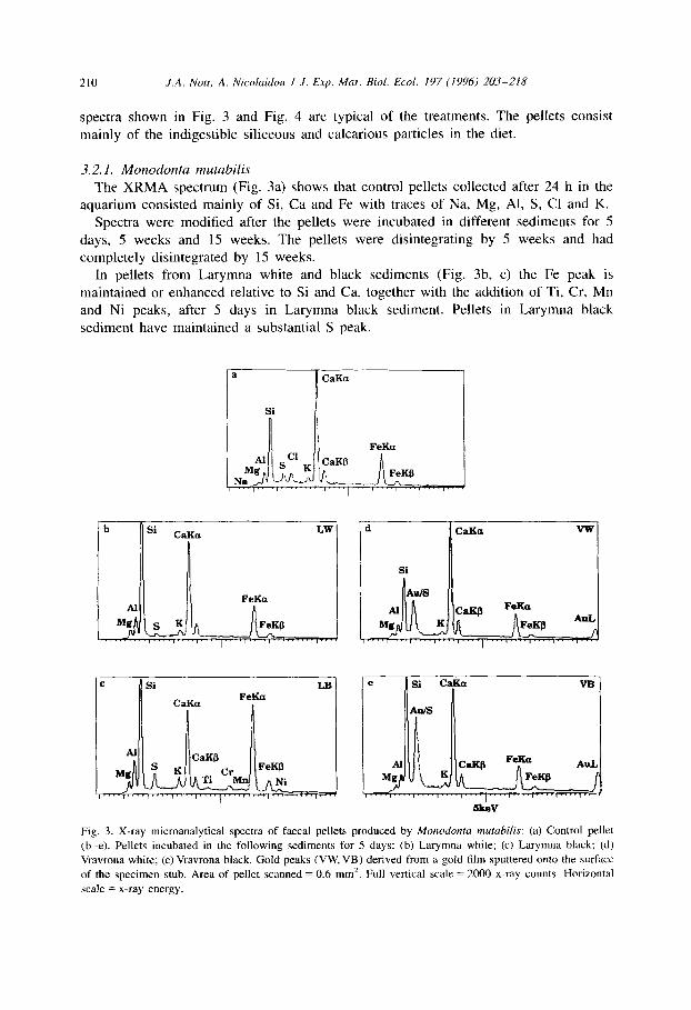
spectra shown in Fig. 3 and Fig. 4 are typical of the treatments. The pellets consist mainly of the indigestible siliceous and calcarious particles in the diet.
3.2.1. Monodonta mutabilis
The XRMA spectrum (Fig. 3a) shows that control pellets collected after 24 h in the aquarium consisted mainly of Si, Ca and Fe with traces of Na, Mg, Al, S, Cl and K.
Spectra were modified after the pellets were incubated in different sediments for 5 days, 5 weeks and 15 weeks. The pellets were disintegrating by 5 weeks and had completely disintegrated by 15 weeks.
In pellets from Larymna white and black sediments (Fig. 3b, c) the Fe peak is maintained or enhanced relative to Si and Ca, together with the addition of Ti, Cr, Mn and Ni peaks, after 5 days in Larymna black sediment. Pellets in Larymna black sediment have maintained a substantial S peak.
a CaKa
Si
FeKP n
b Si CaKa LW
I
FeKa Al
FeKP
1 I ,
c Si LB
CaKa FeKa
I I
Al CaKP FeKP
Ni ,““,““I’~“,““,-’ ““r--l--.,~~..,--J
VW
e Si C&k2 VB
Au/S
Fig. 3. X-ray microanalytical spectra of faecal pellets produced by Monodonta m&bilk: (a) Control pellet
(b-e). Pellets incubated in the following sediments for 5 days: (b) Larymna white; (c) Larymna black; (d) Vravrona white; (e) Vravrona black. Gold peaks (VW, VB) derived from a gold film sputtered onto the surface
of the specimen stub. Area of pellet scanned = 0.6 mm’. Full vertical scale = 2000 x-ray counts. Horizontal
scale = x-ray energy.

J.A. Nott, A. Nicolaidou I J. Exp. Mar. Biol. Ecol. 197 (1996) 203-218 211
a Si
CaKa FeKa
b Si LW
CaKa
___ c Si LB
CaKa FeKa
d Si VW
e
! Si VB
CaKa
Fig. 4. X-ray microanalytical spectra of faecal pellets produced by Cerithium vulgatum. (a) Control pellet
(b-e). Pellets incubated in the following sediments for 5 days: (b) Larymna white; (c) Larymna black; (d)
Vravrona white; (e) Vravrona black. Area of pellet scanned = 0.6 mm’. Full vertical scale = 2000 x-ray counts.
Horizontal scale = x-ray energy.
Pellets in Vravrona sediments are not modified (Fig. 3d, e).
3.2.2. Cerithium vulgatum
The XRMA spectrum (Fig. 4a) shows that control pellets collected after 24 h consisted of Si, Ca and Fe with traces of Na, Mg, Al, S, Cl, K, Ti, Cr and Ni. The limit of detection is 0.1% of the mass of material irradiated by the probe, which translated into units used by FAAS is equivalent to 1000 ppm. Cerithium vulgatum produced sufficient pellets for analysis by FAAS (Table 4) which gave values for Ag, Cd, Cu, Zn, and Co which were not detectable by XRMA.
After incubation in Larymna white and black sediments for 5 days (Fig. 4b, c), 5 weeks and 15 weeks, pellets remained substantially unchanged, neither gaining nor losing peak heights relative to Si.

212 J.A. Nott, A. Nicolaidou I J. Exp. Mar. Biol. Ecol. 197 (1996) 203-218
Table 4
FAAS analyses of Cerithium vulgatum faecal pellets
Ag Cd co Cr Cu Mn Ni Zn
3.5 34.1 229 3555 61 2114 3485 426
FAAS analyses for the ‘total‘ metal concentrations extracted with 16 M HNO,. Figures in pg/g dry wt. Each
analysis was continued until there was < 1% variation in absorbance
In Vravrona sediments the relative peak heights of pellets remained substantially unchanged (Fig. 4d, e). S was a minor constituent in all Cerithium vulgatum pellets.
3.3. Analysis of intra-cellular granules in sediments
The samples of pellets did not provide sufficient material for FAAS. In the SEM individual granules were identified and analysed. As with the pellets, the granules from the different gastropods and the different treatments gave distinctive XRMA spectra; examples of these are shown in Fig. 5 and Fig. 6.
3.3.1. Monodonta mutabilis
Some granules collected from M. mutabilis digestive gland were smeared directly on a graphite specimen stub to act as controls. They consisted of Mg phosphate associated with S, Cl, K and Ca (Fig. 5). The counting efficiency of the x-ray detector decreases at lower energies to the extent that the sensitivity to Mg at 1.25 keV is 25% of the sensitivity to Ca at 3.69 keV. Thus, the larger Mg peak indicates that the mineral is mainly Mg phosphate.
Additional granules from Monodonta mutabilis were mixed with white and black sediments from Larymna and Vravrona, and incubated for 4 days. At the end of the experiment, samples were smeared on graphite specimen stubs and searched in the SEM. Granules were not found in any of the sediments.
Fig. S. X-ray microanalytical spectrum of a single, spherical phosphate granule produced intracellularly in the
digestive gland of Monodonta mutahiks. Granule in tissue smeared on a graphite stub. Granule probed with a
stationary, focused electron beam. Full vertical scale = 2000 x-ray counts. Horizontal scale = x-ray energy.

J.A. Nott, A. Nicolaidou / J. Exp. Mar. Biol. Ecol. I97 (1996) 203-218 213
b P LW
T
Fig. 6. X-ray microanalytical spectra of single, spherical, phosphate granules produced by Cerifhium vu~gatum:
(a) Control granule in digestive gland smeared on a graphite stub. (b-e) Granules incubated in the following
sediments for 4 days: (b) Larymna white; (c) Larymna black; (d) Vravrona white; (e) Vravrona black. Granule
in (c) was situated on a grain of silica. Individual granules probed with a stationary, focused electron beam.
Full vertical scale = 2000 x-ray counts. Horizontal scale = x-ray energy.
3.32 Cerithium vulgatum Granules collected from Cerithium vulgatum digestive gland were smeared directly on
a graphite stub. They contained P together with Na, Mg, S, Cl, K, Ca, Mn, Fe, Co, Ni and Zn (Fig. 6a). Unlike Monodonta mutabilis these granules were not Mg phosphate; they were a mixture of Ca and other metal phosphates. These specimens were used as
controls (see below). Granules were incubated for 4 days in white and black sediments from Larymna and
Vravrona, and then samples were smeared on graphite stubs. In the SEM, granules were found amongst the sediment particles, and probed with a focused, stationary electron
beam for XRMA

214 J.A. Nat, A. Nicolaidou I .I. Exp. Mar. Biol. Ecol. 197 (1996) 203-218
Granules in Larynna white sediment (Fig. 6b) When compared with controls, XRMA peaks for Ca and Zn were maintained or
enhanced whereas peaks for K, Mn, Fe, Co, and Ni were reduced relative to P.
Granules in L.urynna black sediment (Fig. SC)
Peaks for Ca, Mn, Fe and Zn were maintained or enhanced but Co and Ni were lost.
Granules in Vruvrona white sediment (Fig. 6d) Peaks for all metals were reduced except for Ca and Zn which were maintained or
enhanced.
Granules in Vravrona black sediment (Fig. 6e)
Relative to P, peaks for Ca, Mn and Fe were maintained but Co, Ni, and Zn were either reduced or lost.
Granules in all treatments generated S peaks of similar size.
4. Discussion
Larymna sediment contains more organic matter (Table l), is more anoxic and marginally more alkaline (Table 2) than Vravrona sediment. Larymna sediment contains high concentrations of metals, notably transition elements Ni and Cr which exceed concentrations at Vravrona by factors of X 90 and X 40, respectively (Table 3). These are a direct consequence of operations of the nickel smelting plant at Larymna (Nicolaidou and Nott, 1989). Estuaries act as efficient traps for heavy metals which are transferred from water to biota and sediment where they are accumulated (Turekian, 1977). However, both in biota and sediments the relative proportions of different metals can differ substantially from those of the original inputs. Thus, at Larymna three species of gastropod Cerithium vulgatum, Monodonta mutabilis and Murex trunculus each had markedly different accumulations of metals in the digestive gland (Nott and Nicolaidou, 1989) and these were different from the relative concentrations in the sediment (Nicolaidou and Nott, 1989). All these analyses differ from those of laterite ore used in the smelter and slag produced by the extraction process. Since 1990 (Nicolaidou and Nott, 1990) some metals in the sediments, including Ni and Cr have maintained or increased their high concentrations, but others including Cu, Zn and Co have remained at the same low concentrations. Low concentrations of some metals can reflect low input levels and/or greater solubility in seawater which permits dispersal away from the smelter. Cadmium is particularly soluble and mobile, and unlike many other metals it is readily transferred along food chains (Nott and Nicolaidou, 1994).
Concentrations of most metals in marine invertebrates represent the net balance of continual uptake and loss. Cadmium is an exception in that it can be accumulated with a negligible degree of loss (Nott et al., 1993; Bebianno et al., 1992; Langston and Zhou, 1987; Rainbow and White, 1989; Rainbow, 1985). Also, unlike other crustaceans, barnacles accumulate Cu and Zn as well as Cd, with no significant excretion (Rainbow
J.A. Nott, A. Nicolaidou I J. Exp. Mar. Biol. Ecol. 197 (1996) 203-218 215
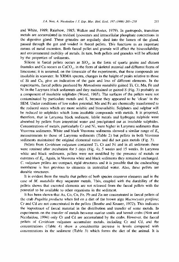
and White, 1989; Rainbow, 1985; Walker and Foster, 1979). In gastropods, transition metals are accumulated in residual lysosomes and intracellular phosphate concretions in the digestive gland. These granules are regularly shed into the lumen of the gland, passed through the gut and voided in faecal pellets. This functions as an important means of metal excretion. Both faecal pellet and granule will affect the bioavailability and environmental mobility of metals. In turn, both pellets and granules will be affected by the properties of sediments.
Silicon in faecal pellets occurs as SiO,, in the form of quartz grains and diatom frustules and Ca occurs as CaCO,, in the form of skeletal material and different forms of limestone; it is assumed, on the timescale of the experiments, that these compounds are insoluble in seawater. In XRMA spectra, changes in the height of peaks relative to those of Si and Ca, give an indication of the gain and loss of different elements. In the experiments, faecal pellets produced by Monodonta mutabilis gained Ti, Cr, Mn, Fe and Ni in the Larymna black sediments and they maintained or gained S (Fig. 3) probably as a component of insoluble sulphides (Wood, 1965). The surfaces of the pellets were not contaminated by particulate metals and S, because they appeared to be ‘clean‘ in the SEM. Under conditions of low redox potential, Mn and Fe are chemically transformed to the reduced states which are more soluble and bioavailable. Sulphates and sulphur will be reduced to sulphides which form insoluble compounds with metals. It is proposed, therefore, that in Larymna black sediment, labile metals and hydrogen sulphide were absorbed by pellets from interstitial water and precipitated out as insoluble sulphides. Concentrations of metals, particularly Cr and Ni, were higher in Larymna sediments than Vravrona sediments. White and black Vravrona sediments showed a similar range of E,, measurements to those of Larymna sediments (Table 2) but pellets in both Vravrona sediments maintained the original elemental ratios and did not gain metals (Fig. 3).
Pellets from Cerithium vulgatum contained Ti, Cr and Ni and in all sediments these were retained after incubation for 5 days (Fig. 4), 5 weeks and 15 weeks. In Larymna white and black sediments, pellets were not modified by the presence of metals or extremes of E,,. Again, in Vravrona white and black sediments they remained unchanged. C. vu&turn pellets are compact, rigid structures and it is possible that the ensheathing membrane is less pervious to elements in interstitial water. Also, these pellets are durable structures.
It is evident from the results that pellets of both species conserve elements and in the case of M. mutubilis they sequester metals. This, coupled with the durability of the pellets shows that excreted elements are not released from the faecal pellets with the potential to be available to other organisms in the sediment.
It has been shown that As, Co, Cu, Fe, Pb and Zn are concentrated in faecal pellets of the crab Pugeltia producta when fed on a diet of the brown alga Macrocystis pyrifera; Cr and Cd are not concentrated in the pellets (Boothe and Knauer, 1972). This indicates the importance of faecal material in the distribution and transfer of some metals. In experiments on the transfer of metals between marine snails and hermit crabs (Nott and Nicolaidou, 1994) only Cr and Cd are accumulated by the crabs. However, the faecal pellets of Cerithium vulgatum accumulate metals, including Cr and Cd, and the concentrations (Table 4) show a considerable increase in levels compared with concentrations in the sediment (Table 3) which forms the diet of the animal. It is
216 J.A. Nrm, A. Nicolaidou I J. Exp. Mar. Bid. Ed. 197 (1996) 20-KZIR
proposed that the animals ingest organic matter preferentially from the sediment and incidentally take-up associated metals.
There must be two strategies for the excretion route into pellets. Either the metals are not assimilated when the organic matter is digested and they pass straight through the gut or, when they are assimilated, they are rendered insoluble within gut epithelial cells in granules and residual lysosomes and returned to the lumen of the gut during cell break-down at the end of a digestive cycle.
In the case of Cerithium vulgutum, high levels of metals, if bioavailable, might make the pellets toxic to consumers. However, the pellets are essentially packages of sediment and therefore much larger than the individual particles; this could prevent consumption by sediment feeders. Also, pellets are structurally durable and can remove metals from food chain cycles for as long as they remain intact.
Proportions of metals in the granules do not match proportions in the pellets. In granules, Mn and Zn are dominant (Fig. 6), whilst in pellets Cr, Mn and Ni are dominant (Table 4). This indicates that Cr and Ni in faecal pellets are not in phosphate granules and must, presumably, be associated with organic material and/or take the form of mineral particles from the smelter.
Phosphate granules produced in the molluscan digestive gland contain detoxified metals. These granules pass through the gut when the digestive cells breakdown and also they pass straight through the gut of a predatory mollusc when it eats the digestive gland tissue of its prey (Nott and Nicolaidou, 1990). Furthermore, when they pass through the gut they retain a substantial proportion of the original heavy metal content (Nott and Nicolaidou, 1993). In some entirely different experiments (Davies and Simkiss, personal communication), it was found that phosphate granules extracted from the crab Curcinus maenas absorb zinc from solution in seawater.
There are, however, differences in the elemental composition of granules in different species of mollusc. In Monodontu mutahilis, granules are mainly Mg phosphate (Fig. 5) whereas in Littorina littoreu they are a mixture of Mg and Ca phosphates (Nott and Nicolaidou, 1990) and in Cerithium vulgutum they are a mixture of Ca and other metal phosphates (Fig. 6a). Phosphorus is indirectly affected by change of redox potential through changes in the reactivities of organic matter and inorganic sediment constituents including metals.
In the experiment, granules from Monodonta mutubilis disappeared from all sediments after 4 days incubation. It is suggested, therefore, that as Mg-phosphate they dissolve in interstitial seawater under all conditions. However, they did not contain transition metals so none were returned to the sediment by this route.
Calcium phosphate granules from Cerithium vulgatum were found after incubation in sediments and the composition varied according to sediment type. The basic ratio of P/Ca remained stable but transition metals Mn, Fe, Co and Ni were reduced, particularly in the white sediments. Mn was maintained in the black sediments. Zinc was maintained or accumulated at Larymna but was removed in Vravrona black sediment.
Thus, granules in sediments are affected differentially according to changes in redox, pH and C, H and N content. In general, transition metals are removed, but it is not known whether they are precipitated as insoluble salts, bound to organic material or remain in solution.
J.A. Nott, A. Nicolaidou I J. Exp. Mar. Biol. Ecol. 197 (1996) 203-218 217
Acknowledgments
We thank Mr. G. Burt and Mr. R.N. Head for FAAS and CHN analyses and Mrs L.J. Mavin for general assistance. Funds for operational costs and exchange visits by both authors to carry out this work were provided by the University of Athens and Grant ATH/882/2/EXH from the British Council in Athens under a Joint Research Pro- gramme.
References
Al-Mohanna, S.Y. and J.A. Non, 1989. Functional cytology of the hepatopancreas of Penaeus semisulcutus
(Crustacea: Decapoda) during the moult cycle. Mar. Biol., Vol. 101, pp. 535-544.
Bebianno, M.J., W.J. Langston and K. Simkiss, 1992. Metallothionein induction in Littorina littorea (Mollusca:
Prosobranchia) on exposure to cadmium. J. Mar. Biol. Ass. U.K., Vol. 72, pp. 329-342.
Becker, G.L., C.-H. Chen, J.W. Greenawalt and A.L. Lehninger, 1974. Calcium phosphate granules in the
hepatopancreas of the blue crab Callinectes sapidus. J. Cell. Biol., Vol. 61, pp. 316-326.
Boothe, P.N. and G.A. Knauer, 1972. The possible importance of fecal material in the biological amplification
of trace and heavy metals. Limnol. Oceanogr., Vol. 17, pp. 270-274.
Brown, B.E., 1982. The form and function of metal-containing ‘granules‘ in invertebrate tissues. Biol. Rev.,
Vol. 57, pp. 621-667.
Buchanan, J.B., 1984. Sediment analysis. In, Methods for the study of marine benthos, edited by N.A. Holme
and A.D. McIntyre, IBP Handbook 16, 2nd ed., Blackwell Scientific Publications, Oxford, pp. 41-65.
Carmichael, N.G., KS. Squibb and B.A. Fowler, 1979. Metals in the molluscan kidney: a comparison of two
closely related bivalve species (Argopecten), using X-ray microanalysis and atomic absorption spec-
troscopy. J. Fish. Res. Bd Can., Vol. 36, pp. 1149- 1155.
Fowler, S.W., 1982. Biological transfer and transport processes. In, Pollutant Transfer and Transport in the
Sea, edited by G. Kullenberg, Vol. 11 CRC Press Inc., Boca Raton, Florida, pp. l-65.
George, S.G., B.J.S. Pirie and T.L. Coombs, 1980. Isolation and elemental analysis of metal-rich granules from
the kidney of the scallop, Pecten maximus (L.). J. Exp. Mar. Biol. Ecol., Vol. 42, pp. 143-156.
Hopkin, S.P and J.A. Nott, 1979. Some observations on concentrically structured, intracellular granules in the
hepatopancreas of the shore crab Carcinus maenas (L.). J. Mar. Biol. Ass. U.K., Vol. 59, pp. 867-877.
Rely, J.D. and J.A. Nott, 1980. Accumulation of copper within the ‘hepatopacreatic’ caeca of Corophium
volutator (Crustacea: Amphipopda). Mar. Biol., Vol. 57, pp. l93- 199.
Icely, J.D. and J.A. Nott, 1992. Digestion and Absorption: Digestive system and associated organs. In,
Microscopic Anatomy of Invertebrates, edited by F.W. Harrison and A.G. Humes. Decapod Crustucea, Vol.
10, pp. 147-201.
Langston, W.J. and M. Zhou, 1987. Cadmium accumulation, distribution and metabolism in the gastropod
Littorina littorea: the role of metal-binding proteins. J. Mar. Biol. Ass. U.K., Vol. 67, pp. 585-601.
Mason, A.Z. and J.A. Nott, 1981. The role of intracellular biomineralized granules in the regulation and
detoxification of metals in gastropods with special reference to the marine prosobranch Littorina littowa.
Aquat. Toxic., Vol. I., pp. 239-256.
Nicolaidou, A. and J.A. Nott, 1989. Heavy metal pollution induced by a ferro-nickel smelting plant in Greece.
Sci. Tot. Etwir., Vol. 84, pp. 1 13-l 17.
Nicolaidou, A. and J.A. Nott, 1990. Mediterranean pollution from a ferro-nickel smelter: differential uptake of
metals by some gastropods. Mar. Poll. Bull., Vol. 21, pp. 137-143. Nott, J.A., 1993. Cytology of pollutant metals in marine invertebrates: A review of microanalytical
applications. In, X-ray microprobe analysis of chemical elements in biology: from origins to current
practice, edited by B.L. Gupta and G.M. Roomans, Scanning Microscopy International, AMF O’Hare,
Chicago IL, pp. 283-298.

218 J.A. Nott, A. Nicoluidou I .I. Exp. Mar. Biol. Ecol. 197 (1996) 203-218
Non, J.A. and A. Nicolaidou, 1989. The cytology of heavy metal accumulations in the digestive glands of
three marine gastropods. Proc. R. Sot. Lond. B., Vol. 237, pp. 347-362.
Nott, J.A. and A. Nicolaidou, 1990. Transfer of metal detoxification along marine food chains. J. Mar. Biol.
Ass. C/K., Vol. 70, pp. 905-912. Nott, J.A. and A. Nicolaidou, 1993. Bioreduction of zinc and manganese along a molluscan food chain. Camp.
Biochem. Physiol., Vol. 104A, pp. 235-238.
Nott, J.A. and A. Nicolaidou, 1994. Variable transfer of detoxified metals from snails to hermit crabs in marine
food chains. Mar. Biol., Vol. 120, pp. 3699377.
Nott, J.A., K.P. Ryan and L.J. Mavin, 1993. Chemical and cryo-fixation for cadmium retention in a marine
snail. Cell Biol. Int., Vol. 17, pp. 71 I-722.
Rainbow, P.S., 1985. Accumulation of Zn. Cu and Cd by crabs and barnacles. Est. Coast. Shelf S-i., Vol. 21,
pp. 6699686.
Rainbow, P.S. and S.L. White, 1989. Comparative strategies of heavy metal accumulation by crustaceans: zinc,
copper and cadmium in a decapod, an amphipod and a barnacle. Hydrohiol., Vol. 174, pp. 245-262.
Simkiss, K. and AZ. Mason, 1984. Cellular responses of molluscan tissues to environmental metals. Mur.
Envir. Res., Vol. 14, pp. 103-I 18.
Suede], B.C., J.A. Boraczek, R.K. Peddicord, P.A. Clifford and T.M. Dillon, 1994. Trophic transfer and
biomagnification potential of contaminants in aquatic ecosystems. Rev. Environ. Contam. Toxico/.,Vol. 136,
pp. 21-89.
Taylor, M.G. and K. Simkiss, 1984. Inorganic deposits in invertebrate ttasues. Environ. Chew., Vol. 3, pp.
1022138.
Turekian, K.K., 1977. The fate of metals in the oceans. Geochim. et Cosmochim. Acta. Vol. 4 I, pp. 1 139% I 144.
Viarengo, A. and J.A. Nott, 1993. Mechanisms of heavy metal cation homeostasis in marine invertebrates. Camp. Biochrm. Physiol., Vol. 104C, pp. 355-372.
Walker, G. and P. Foster, 1979. Seasonal variation of zinc in the barnacle, Balmus halanoidrs (L.) maintained
on a raft in the Menai Strait. Mar. Envir. Res., Vol. 2, pp. 2099221.
Wood, E.J.F., 1965. Marine Microbial Ecology. Chapman and Hall, London, 243 pp.