kibion — iris® and 13c-breath tests for the assessment of...
TRANSCRIPT

www.kibion.com
IRIS® and 13C-Breath Tests for the Assessment of Specific Enzymatic and Metabolic Functions in vivo

NOTEThe information in this brochure is based on literature references, which are believed to be correct. The possibility of mistakes or errors cannot be excluded completely. Therefore Kibion AB does not accept any legal or other liability with respect to incorrect details and their consequences.
DISCLAIMERPlease note that all substrates described in this brochure, except 13C-urea – Diabact® UBT 50 mg (registered pharmaceutical), are laboratory chemicals (also called extempore or special medicine preparations –human use). Please contact your local pharmacy for more information about how to order substrates in your region.
It is also advisable to contact the relevant medical product agency (MPA) for more precise information about the use of substrates and its accompanying responsibilities.

ContentsIntroduction ...................................................................................................... 4
13C-Urea Breath Test – Diabact® UBT ....................................................................... 6
13C-Aminopyrine Breath Test .................................................................................... 8
13C-Methacetin Breath Test ..................................................................................... 10
13C-L-Methionine Breath Test ................................................................................ 12
13C-Sodium-Acetate Breath Test ............................................................................. 14
13C-Sodium-Octanoate and 13C-Octanoic Acid Breath Test ................................... 16
13C-Mixed Triglyceride Breath Test ......................................................................... 18
GRAPHIC DESIGN: MATADOR KOMMUNIKATION AB.FRONT COVER: JULIAN WINSLOW/CORBIS. PRINT: ARK-TRYCKAREN, 2014. © KIBION AB, 2014

Kibion is a dynamic, world-leading supplier of simple and reliable breath tests for diagnosing the stomach ulcer bacterium Helicobacter pylori.
A subsidiary of the Swedish pharmaceutical company
Orexo AB, Kibion was founded in 2005 to create a
dedicated platform for commercializing breakthrough
discoveries in the diagnosis of Helicobacter pylori.
Kibion together with its subsidiary in Bremen,
Germany, is the present day provider of complete
solutions of both diagnostic breath tests and
instruments, and has attained a leading position in
the testing of H. pylori. The tests and instruments
are cost effective, reliable and easy to use in settings
including the hospital, laboratory and doctor’s office.
Quality Kibion provides customers with high quality products
and services.
The quality of our processes, products and services
are continuously optimized and improved to meet
customers’ demands and needs.
Kibion AB is certified based on EN ISO 13485 – Medical
Devices – Quality Management Systems – Requirements
for regulatory purposes. The scope of the certificate
includes development, production and distribution of
IVD medical devices. The Certification was carried out
by TÜV SÜD Product Service GmbH which is a globally
recognized Certification Body.
The EN ISO 13485 certification allows Kibion AB
to further strengthen and develop its leading
position as provider of breath tests for detection of
Helicobacter pylori worldwide.
Metabolic breath testsNon-invasive breath tests can serve as valuable diagnostic
tools in medicine as they can determine particular
enzymatic and metabolic functions in vivo. This has wide
applications in the fields of gastroenterology, oncology,
hepatology and nutrition control. A 13CO2 breath test
measures increased levels of 13CO2 in exhaled breath
after ingestion of a stable 13C isotope labelled substance
and its subsequent metabolism with a specific function
or enzyme as a rate limiting step. Breath samples are
collected and measured, for example, with an IRIS®
instrument, measuring the stage between ingestion by
the patient of the labeled substance and its appearance
in the exhaled breath.
This brochure describes the principles and general test
procedures based on information in published literature
for a number of tests, which are the most common in
today’s clinical research.
Introduction
04

IRISIRIS® is a foremost instrument for quantitative
diagnosis of breath tests. IRIS® employs detectors
of non-radioactive 13C-labelled stable isotope
based on infra-red technology.
The IRIS® Infra Red Isotope analyzer measures the 13CO2 and 12CO2 concentrations from sequences of
breath samples and relates their ratios to the PDB-13C stable isotope standard. The reproducibility
is in optimal conditions better than 0.2 δ ‰
(IRIS-Doc: 0.4 δ ‰) over a wide range of 13C/12C
stable isotope ratios, and over a wide range of
CO2 concentrations in breath.
Measurements are made on breath samples as they
come from the breath sample bags or tubes. No
separation of water or isolation of CO2 is required prior
to analysis. Standard breath bags have a volume of
120 ml breath gas, which allows for two measurements
per sample.
The IRIS® instrument is available in two different models,
IRIS®-3 and IRIS®-Doc and can be connected to the IRIS®-
Multisampler for high throughput testing.
13C-Urea
13C-Urea Breath Test – Diabact® UBT
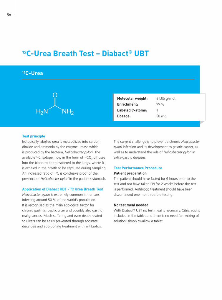
Test principleIsotopically labelled urea is metabolized into carbon
dioxide and ammonia by the enzyme urease which
is produced by the bacteria, Helicobacter pylori. The
available 13C isotope, now in the form of 13CO2 diffuses
into the blood to be transported to the lungs, where it
is exhaled in the breath to be captured during sampling.
An increased ratio of 13C is conclusive proof of the
presence of Helicobacter pylori in the patient’s stomach.
Application of Diabact UBT -13C Urea Breath TestHelicobacter pylori is extremely common in humans,
infecting around 50 % of the world’s population.
It is recognised as the main etiological factor for
chronic gastritis, peptic ulcer and possibly also gastric
malignancies. Much suffering and even death related
to ulcers can be easily prevented through accurate
diagnosis and appropriate treatment with antibiotics.
The current challenge is to prevent a chronic Helicobacter
pylori infection and its development to gastric cancer, as
well as to understand the role of Helicobacter pylori in
extra-gastric diseases.
Test Performance ProcedurePatient preparationThe patient should have fasted for 6 hours prior to the
test and not have taken PPI for 2 weeks before the test
is performed. Antibiotic treatment should have been
discontinued one month before testing.
No test meal neededWith Diabact® UBT no test meal is necessary. Citric acid is
included in the tablet and there is no need for mixing of
solution; simply swallow a tablet.
Molecular weight: 61.05 g/mol
Enrichment: 99 %
Labeled C-atoms: 1
Dosage: 50 mg
06

References1. Uemura N, Okamoto S, Yamamoto S, Matsumura N, Yamaguchi S, Yamakido M, et al. Helicobacter pylori infection and the development of gastric cancer.
N Engl J Med 2001;345:784–9.2. Correa P. Bacterial infections as a cause of cancer. J Natl Cancer Inst 2003;95:E3.3. Eidt S, Stolte M, Fischer R. Helicobacter pylori gastritis and primary gastric non-Hodgkin’s lymphomas. J Clin Pathol 1994;47:436–9.4. Talley NJ, Vakil N. Guidelines for the management of dyspepsia. Am J Gastroenterol 2005;100:2324–375. Delaney BC, Moayyedi P, Forman D. Initial management strategies for dyspepsia. Cochrane Database Syst Rev 2005;(4):CD001961.6. Malfertheiner P, Megraud F, O’Morain C, et al. Management of Helicobacter pylori infection - the Maastricht IV / Florence Consensus Report. Gut 2012;61:646-664.7. Wong et al. A rapid release 50 mg tablet-based 13C-urea breath test for the diagnosis of Helicobacter pylori infection. Aliment Pharmacol Ther 2003: 17:253-257.8. Gatta et al. A rapid, low-dose 13C-urea breath test for the detection of Helicobacter pylori infection before and after treatment. Aliment Pharmacol Ther
2003:17:793-798.9. Spiegel BM, Vakil NB, Ofman JJ. Dyspepsia management in primary care: a decision analysis of competing strategies. Gastroenterology 2002;122:1270–85.10. Jarbol DE, Kragstrup J, Stovring H, Havelund T, Schaffalitzky de Muckadell OB. Proton pump inhibitor or testing for Helicobacter pylori as the first step for
patients presenting with dyspepsia? A clusterrandomized trial. Am J Gastroenterol 2006;101:1200–8.
Test procedure1. Patient exhales into basal sample tubes (0-tubes).
2. Patient swallows a Diabact® UBT tablet with a glass
of water.
3. After a 10-minute wait, patient exhales into sample
tubes.
4. Samples are analysed with IRIS®-Doc or IRIS®-3.
Results and InterpretationDiabact® UBT for diagnosis of Helicobacter pylori is
a qualitative test. The result will show if the patient is
infected or not infected.
The established cut-off using mass spectrometry is
<1.5 ‰ δ value = Negative H.pylori status
>1.5 ‰ δ value = Positive H.pylori status
The cut-off when using IRIS-3 is 1.5 ‰ ± 0.2.
The cut-off when using IRIS-Doc is 1.5‰ ± 0.4.
3. After a 10-minute wait, breathe into sample tubes
4. Send the tubes for analysis.
1. Breath into base line tubes 2. Swallow Diabact® UBT tablet
07

13C-Aminopyrine
13C-Aminopyrine Breath Test
Metabolic principle 13C-Aminopyrine undergoes a two-step N-demethylation
by cytochrome P-450 monooxygenases including
CYP2C19, CYP1A2 and CYP3A4, yielding formaldehyde
and amino-antipyrine1. The formaldehyde is further
oxidized to bicarbonate and exhaled as 13CO2, or
deposited in the bicarbonate pool2. As N-demethylation
occurs exclusively in the liver with a low extraction
rate, this para meter is an overall reflection of the
efficiency of amino pyrine metabolism3. It is therefore
a good measure of hepatic metabolic capacity, i.e. the
“functional hepatic mass”.
Applications of 13C-Aminopyrine Breath TestThe 13C-Aminopyrine Breath Test is very useful for
quantitative assessment of liver function in conditions
such as established chronic hepatitis and cirrhosis4,5. For
example, it can be used to quantify progression of the
disease in Hepatitis C patients6.
The patient should have fasted for 8 hours prior to
the test. Smoking should also be avoided at least one
hour prior to the test7. The patient should not drink
carbonated water or soft drinks prior to the test since
that might interfere with the results. In addition, oxygen
supplementation should be avoided because increased
oxygen content in exhaled breath can influence 13CO2
measurement by NDIRS8.
Test Performance Procedure (see IRIS® Operating Manual for additional information).1. Collect zero (basal) breath sample as described in
manual.
2. Patient takes 13C-Aminopyrine (75 mg) dissolved in
warm water (100 ml).
3. Collect additional breath samples as shown below
(Table 1).
4. Analyze all 10 breath samples with IRIS®-3.
#1 Bag
#2Bag
#3 Bag
#4 Bag
#5 Bag
#6 Bag
#7 Bag
#8 Bag
#9 Bag
#10Bag
0 min 10 min 20 min 30 min 40 min 50 min 60 min 80 min 100 min 120 min
Table 1: 13C-Aminopyrine Breath Test Sample Collection
Molecular weight: 233.29 g/mol
Enrichment: 99 %
Labeled C-atoms: 2
Dosage: 75 mg
08

Fig. 3,4: 13C-Aminopyrine Breath Test, Dose/h curve and % Cum Dose curve, subject with liver disease11
Fig. 1,2: 13C-Aminopyrine Breath Test, Dose/h curve and % Cum Dose curve, healthy (normal) subject11
References1. Armuzzi, A. et al. Review article: breath testing for human liver function assessment. Aliment. Pharmacol. Ther. 16, 1977–1996 (2002).2. Perri, F., Pastore, M., Annese, V. & Andriulli, A. The aminopyrine breath test. Ital J Gastroenterol 26, 306–317 (1994).3. Nista, E. C. et al. 13C-breath tests in the study of microsomal liver function. Eur Rev Med Pharmacol Sci 8, 33–46 (2004).4. Morelli, A., Narducci, F., Pelli, M. A., Farroni, F. & Vedovelli, A. The relationship between aminopyrine breath test and severity of liver disease in cirrhosis. Am. J.
Gastroenterol. 76, 110–113 (1981).5. Giannini, E. et al. 13C-aminopyrine breath test to evaluate severity of disease in patients with chronic hepatitis C virus infection. Aliment. Pharmacol. Ther.
16, 717–725 (2002).6. Rocco, A. et al. 13C-aminopyrine breath test accurately predicts long-term outcome of chronic hepatitis C. J. Hepatol. 56, 782–787 (2012).7. Kasicka-Jonderko, A., Loska, D., Jonderko, K., Kaminska, M. & Błonska-Fajfrowska, B. Interference of acute cigarette smoking with [13C]methacetin
breath test. Isotopes Environ Health Stud 47, 34–41 (2011).8. Riecke, B., Neuhaus, P. & Stockmann, M. Major influence of oxygen supply on 13CO2:
12CO2 ratio measurement by nondispersive isotope-selective infrared spectroscopy. Helicobacter 10, 620–622 (2005).
9. Merkel, C. et al. Aminopyrine breath test in the prognostic evaluation of patients with cirrhosis. Gut 33, 836–842 (1992).10. Urbain, D., Muls, V., Thys, O. & Ham, H. R. Aminopyrine breath test improves long-term prognostic evaluation in patients with alcoholic cirrhosis in
Child classes A and B. J.Hepatol. 22, 179–183 (1995).11. Merz, B. Evaluierung des Aminopyrin-Atemtests bei chronischer Hepatitis C. Hepatologie und Infektiologie, (2005).
Results and interpretationTypical results for the 13C-Aminopyrine Breath Test are
presented in Figures 1 to 4. The 13C-Aminopyrine test is
very sensitive and precise, as can be seen from the very
narrow “normal” range. This makes it even possible to
detect patients with early stage liver disease6,9,10.
Condition dose/hr (‰) at 30 min % cum. dose at 120 min
Fibrosis stages 0/1/2 6.62 - 7.10 ± 2.9 9.21 - 10.06 ± 3.8
Fibrosis stages 3 / 4 2.48 - 3.13 ± 1.2 3.62 - 4.56 ± 2.0
Cirrhosis, not established 6.77 ± 2.7 9.63 ± 3.6
Cirrhosis, established 2.48 ± 1.2 3.68 ± 1.9
For the 13C-Aminopyrine Breath Test, cut-off values
have been established in a study with 135 patients11
(see table below).
0 2 4 6 8
10 12
0 20 40 60 80 100 120
Dos
e /h
[%]
Time [min]
Normal Min Normal Max Patient
0 2 4 6 8
10 12 14 16 18
0 20 40 60 80 100 120
Cum
ulat
ive
Dos
e [%
]
Time [min]
Normal Min Normal Max Patient
0 2 4 6 8
10 12
0 20 40 60 80 100 120
Dos
e /h
[%]
Time [min]
Normal Min Normal Max Patient
0 2 4 6 8
10 12 14 16 18
0 20 40 60 80 100 120 C
umul
ativ
e D
ose
[%]
Time [min]
Normal Min Normal Max Patient
Table 2: Cut-off values for 13C-Aminopyrine Breath Test 11
09

13C-Methacetin
13C-Methacetin Breath Test
Metabolic principleMethacetin is metabolized rapidly in normal subjects,
being highly extracted by the liver1, implying that the
metabolism of methacetin is mainly dependent on
hepatic blood flow, the latter being generally decreased
in cirrhotic patients2. Methacetin undergoes dealkylation
by hepatic CYP1A2 to acetaminophen3 with the
methoxy group being eliminated as 13CO2.
Published data of previous studies suggest that the
Methacetin Breath Test is a rapid and precise quantitative
liver function test without any evidence of toxicities due to
the small doses used, in contrast to other substrates4–7.
Applications of 13C-Methacetin Breath TestThe liver status of patients who have been diagnosed
with liver disease can be assessed or monitored non-
invasively using the 13C-Methacetin Breath Test:
The patient should have fasted for 8 hours prior to the test.
Smoking should also be avoided at least one hour prior to
the test13. The patient should not drink carbonated water
or soft drinks prior to the test since this might interfere with
the results. In addition, oxygen supplementation should
be avoided because increased oxygen content in exhaled
breath can influence 13CO2 measurement by NDIRS14.
Test Performance Procedure (see IRIS® Operating Manual for additional information).1. Collect zero (basal) breath sample as described in
the manual.
2. Patient takes 13C-Methacetin (75 mg) dissolved in
water (100 ml).
3. Collect additional breath samples as shown below
(Table 2).
4. Analyze all 10 breath samples with IRIS®-3 or IRIS®-Doc.
Molecular weight: 166.19 g/mol
Enrichment: 99 %
Labeled C-atoms: 1
Dosage: 75 mg
#1 Bag
#2Bag
#3 Bag
#4 Bag
#5 Bag
#6 Bag
#7 Bag
#8 Bag
#9 Bag
#10Bag
0 min 10 min 20 min 30 min 40 min 50 min 60 min 80 min 100 min 120 min
Table 2: 13C-Methacetin Breath Test Sample Collection
Condition Assessment
Non-alcoholic steatohepatitis (NASH) or alcoholic steatohepatitis (ASH), Fibrosis or Cirrhosis
State of evolution (correlation with Child-Pugh Score) 8,9
Fibrosis or Cirrhosis State of evolution (correlation with Child-Pugh Score) 8,9
Liver tumor Hepatic reserve
Hepatitis B or C Hepatic reserve 10
Long-term medication e.g. anticonvulsants Monitor hepatotoxicity
Liver transplant Liver status of both donor and recipient 11,12
Table 1: Liver diseases assessed by 13C-Methacetin Breath Test
10

% Cumulative Dose, 120 min Indication/ Correlation
31.0 (25.9 – 38.7) Normal
13.6 (5.7 – 22.3) Cirrhosis, Child-Pugh Class A
3.1 (1.1 – 16.5) Cirrhosis, Child-Pugh Class B
0.6 (-1.1 – 3.5) Cirrhosis, Child-Pugh Class C
Table 4: Correlation of 13C-Methacetin Breath Test (% cum dose) with stage of liver disease8
Results and interpretationIn healthy subjects a peak in the exhaled Dose/h of
labeled CO2 is to be expected after 10 to 20 minutes
(see Figure 1). About 30% of the administered dose is
recovered as 13CO2 after 120 minutes (see Figure 2). In
general, the more severe the liver disease, the lower the
% cum dose after 120 minutes.8,10,15
The value of the maximum metabolic rate (dose/h) has
been shown to be a good quantitative predictor of
cirrhosis and fibrosis in chronic hepatitis C (Table 3).
The % cumulative dose at 120 minutes has been shown
to correlate with different stages of liver disease (Table 4).
Cut-off Sensitivity Specificity
Liver Cirrhosis 13C-Methacetin Breath Test < 14.6 % 92.6 % 84.1 %
Fibroindex > 1.82 70.4 % 91.3 %
Advanced Fibrosis 13C-Methacetin Breath Test < 21 ‰ 75.4 % 79.5 %
Fibroindex > 1.35 66.7 % 84.6 %
Table 3: Comparison of 13C-Methacetin Breath Test and FibroIndex as predictors of cirrhosis and fibrosis. (Adapted from Dinesen et al.17)
Fig. 1-2: 13C-Methacetin Breath Test, Dose/h curve and % Cum Dose, healthy (normal) subject16
0
10
20
30
40
50
60
0 20 40 60 80 100 120
Dos
e /h
[%]
Time [min]
Normal Min Normal Max
Healthy Liver disease
0
10
20
30
40
0 20 40 60 80 100 120
Cum
ulat
ive
Dos
e [%
]
Time [min]
Normal Min Normal Max
Healthy Liver disease
References1. Armuzzi, A. et al. Review article: breath testing for human liver function assessment. Aliment. Pharmacol. Ther. 16, 1977–1996 (2002).2. Moreno, A. H. et al. Portal blood flow in cirrhosis of the liver. J. Clin. Invest. 46, 436–445 (1967).3. Kasicka-Jonderko, A., Nita, A., Jonderko, K., Kamińska, M. & Błońska-Fajfrowska, B. C-methacetin breath test reproducibility study reveals persistent CYP1A2
stimulation on repeat examinations. World J. Gastroenterol. 17, 4979–4986 (2011).4. Matsumoto, K. et al. [13C]methacetin breath test for evaluation of liver damage. Dig. Dis. Sci. 32, 344–348 (1987).5. Festi, D. et al. Measurement of hepatic functional mass by means of 13C-methacetin and 13C-phenylalanine breath tests in chronic liver disease: comparison with
Child-Pugh score and serum bile acid levels. World J. Gastroenterol. 11, 142–148 (2005).6. Candelli, M. et al. 13C-methionine breath tests for mitochondrial liver function assessment. Eur Rev Med Pharmacol Sci 12, 245–249 (2008).7. Nista, E. C. et al. 13C-breath tests in the study of microsomal liver function. Eur Rev Med Pharmacol Sci 8, 33–46 (2004).8. Pfaffenbach, B., Götze, O., Szymanski, C., Hagemann, D. & Adamek, R. J. [The 13C-methacetin breath test for quantitative noninvasive liver function analysis
with an isotope-specific nondispersive infrared spectrometer in liver cirrhosis]. Dtsch. Med. Wochenschr. 123, 1467–1471 (1998).9. Klatt, S., Taut, C., Mayer, D., Adler, G. & Beckh, K. Evaluation of the 13C-methacetin breath test for quantitative liver function testing. Z Gastroenterol 35,
609–614 (1997).10. Goetze, O. et al. 13C-methacetin breath test as a quantitative liver function test in patients with chronic hepatitis C infection: continuous automatic molecular
correlation spectroscopy compared to isotopic ratio mass spectrometry. Aliment. Pharmacol. Ther. 26, 305–311 (2007).11. Lock, J. F. et al. Initial liver graft function is a reliable predictor of tacrolimus trough levels during the first post-transplant week. Clin Transplant 25, 436–443 (2011).12. Stockmann, M. et al. How to define initial poor graft function after liver transplantation? - a new functional definition by the LiMAx test. Transpl. Int. 23,
1023–1032 (2010).13. Kasicka-Jonderko, A., Loska, D., Jonderko, K., Kaminska, M. & Błonska-Fajfrowska, B. Interference of acute cigarette smoking with [13C]methacetin breath test.
Isotopes Environ Health Stud 47, 34–41 (2011).14. Riecke, B., Neuhaus, P. & Stockmann, M. Major influence of oxygen supply on 13CO2:
12CO2 ratio measurement by nondispersive isotope-selective infrared spectroscopy. Helicobacter 10, 620–622 (2005).
15. Lane, E. A. & Parashos, I. Drug pharmacokinetics and the carbon dioxide breath test. J Pharmacokinet Biopharm 14, 29–49 (1986).16. Paul, M. Reference Values - Internal Data, Done at University of Erlangen. (1998).17. Dinesen, L. et al. 13C-methacetin-breath test compared to also noninvasive biochemical blood tests in predicting hepatic fibrosis and cirrhosis in chronic hepatitis
C. Dig Liver Dis 40, 743–748 (2008).
11

13C-L-Methionine Breath Test
Metabolic principleMethionine is an essential amino acid, metabolized in the
liver through two major pathways: transamination and
transmethylation. Transmethylation is the predominating
metabolic pathway by which methionine is normally
converted to S-adenosyl-L-methionine (SAM) and
which is used as a cofactor by methyltransferases
to transfer the 13C-methyl group to different target
molecules (methylation). However, the major pathway
to remove excess methionine and for the transfer of its
methyl group is via sarcosine production, which in this
instance generates 13C-sarcosine. The labeled sarcosine
is oxidized by sarcosine dehydrogenase to produce 13C-formaldehyde in the mitochondria which is further
oxidized to 13CO2 and expired. Since the oxidation
of sarcosine occurs in the mitochondria of the liver1, 13C-methionine can be used to evaluate the oxidative
capacity of the liver2. This test is therefore a good
measure of the hepatic metabolic capacity.3–5
Applications of 13C-L-Methionine Breath TestThe 13C-L-Methionine Breath Test is a non-invasive
diagnostic test to assess in vivo hepatic mitochondrial
function. Dysfunction of hepatic mitochondria is
associated with several chronic liver diseases and the
test can be applied to investigate drug-related acute
liver toxicity6,7, ethanol-induced liver oxidative stress8,
impaired hepatic mitochondrial oxidation in liver
steatosis such as non-alcoholic fatty liver disease (NAFLD)
or cirrhosis4,9.
The patient should have fasted for 8 hours prior to
the test. Smoking should also be avoided at least one
hour prior to the test10. The patient should not drink
carbonated water or soft drinks prior to the test since
that might interfere with the results. In addition, oxygen
supplementation should be avoided because increased
oxygen content in exhaled breath can influence 13CO2
measurement by NDIRS11.
Test Performance Procedure (see IRIS® Operating Manual for additional information)1. Collect zero (basal) breath sample as described in
the manual.
2. Patient takes 13C-L-Methionine (75 mg) dissolved in
water (100 ml).
3. Collect additional breath samples as shown below
(Table 2).
4. Analyze all 10 breath samples with IRIS®-3 or IRIS®-Doc.
13C-L-methionine
Molecular weight: 150.2 g/mol
Enrichment: 99 %
Labeled C-atoms: 1
Dosage: 75 mg
#1 Bag
#2Bag
#3 Bag
#4 Bag
#5 Bag
#6 Bag
#7 Bag
#8 Bag
#9 Bag
#10Bag
0 min 10 min 15 min 20 min 25 min 30 min 40 min 60 min 90 min 120 min
Table 1: 13C-L-Methionine Breath Test Sample Collection
12

References1. Frisell, W. R., Cronin, J. R. & Mackenzie, C. G. Coupled flavoenzymes in mitochondrial oxidation of N-methyl groups. J. Biol. Chem. 237, 2975–2980 (1962).2. Candelli, M. et al. 13C-breath tests in the study of mitochondrial liver function. Eur Rev Med Pharmacol Sci 8, 23–31 (2004).3. Milazzo, L. et al. [13C]Methionine breath test: a novel method to detect antiretroviral drug-related mitochondrial toxicity. J. Antimicrob. Chemother. 55, 84–89 (2005).4. Banasch, M., Ellrichmann, M., Tannapfel, A., Schmidt, W. E. & Goetze, O. The non-invasive (13)C-methionine breath test detects hepatic mitochondrial
dysfunction as a marker of disease activity in non-alcoholic steatohepatitis. Eur. J. Med. Res. 16, 258–264 (2011).5. Candelli, M. et al. 13C-methionine breath tests for mitochondrial liver function assessment. Eur Rev Med Pharmacol Sci 12, 245–249 (2008).6. Spahr, L. et al. Acute valproate-associated microvesicular steatosis: could the [13C]methionine breath test be useful to assess liver mitochondrial function? Dig.
Dis. Sci. 46, 2758–2761 (2001).7. Banasch, M. et al. Impact of antiretroviral treatment on (13) C-methionine metabolism as a marker of hepatic mitochondrial function: a longitudinal study. HIV
Med. 12, 40–45 (2011).8. Armuzzi, A. et al. Non-Invasive assessment of human hepatic mitochondrial function through the 13C-methionine breath test. Scand. J. Gastroenterol. 35,
650–653 (2000).9. Spahr, L. et al. Impaired hepatic mitochondrial oxidation using the 13C-methionine breath test in patients with macrovesicular steatosis and patients with
cirrhosis. Med. Sci. Monit. 9, CR6–11 (2003).10. Kasicka-Jonderko, A., Loska, D., Jonderko, K., Kaminska, M. & Błonska-Fajfrowska, B. Interference of acute cigarette smoking with [13C]methacetin breath
test. Isotopes Environ Health Stud 47, 34–41 (2011).11. Riecke, B., Neuhaus, P. & Stockmann, M. Major influence of oxygen supply on 13CO2:12CO2 ratio measurement by nondispersive isotope-selective infrared
spectroscopy. Helicobacter 10, 620–622 (2005).12. Stüwe, S. H. et al. Hepatic mitochondrial dysfunction in manifest and premanifest Huntington disease. Neurology 80, 743–746 (2013).
Results and interpretationIn healthy subjects, a peak in the exhaled Dose/h of
labeled CO2 is to be expected after 30 to 60 minutes
(see Figure 1). According to published values by
Armuzzi et al.’ the cumulative dose in healthy
controls after 120 minutes reaches 6.07±0.46%8
whereas control groups in the following studies also
showed slightly increased values (e.g. cumulative
dose after 90 minutes: 7.16% ± 1.91%; see Stüwe
et al., 201312). In general, the more severe the liver
disease, the lower the % cumulative dose after 90
or 120 minutes.4,7,8
In another study by Banasch et al. specific cut-off values
for the cumulative dose at 90 minutes to assess non-
alcoholic steatohepatitis and fibrosis stage 0-1 versus
fibrosis stage 2-3 in a NAFLD cohort have been calculated.
0
1
2
3
4
5
6
0 20 40 60 80 100 120
Dos
e/h
[%]
Time [min]
Healthy control
Ethanol-induced oxidative stress
0
1
2
3
4
5
6
7
0 20 40 60 80 100 120
Cum
ulat
ive
Dos
e [%
]
Time [min]
Healthy control Ethanol-induced oxidative stress
Cut-off
non-alcoholic steatohepatitis (NASH) vs. non-NASH < 4.20 %
Fibrosis stage 0-1 vs. Fibrosis stage 2-3 (within
NAFLD cohort)
< 3.65 %
Table 3: Cut-off values for non-alcoholic steatohepatitis (NASH) and mild vs. severe fibrosis in a NAFLD cohort according to Banasch et al., 20114
Fig. 1,2: Example of 13C-Methionine Breath Test, Dose/h curve and % Cum Dose, (Armuzzi et al., 20008)
13

13C-Sodium-Acetate
13C-Sodium-Acetate Breath Test
Metabolic principle13C-Sodium-Acetate is administered together with a
liquid or semi-solid test meal. After passing through the
stomach, where it is not absorbable, it is absorbed in
the small intestine and metabolized in the liver1. Whilst
some of the labeled carbon is incorporated in different
metabolic pathways, about 50 % enters the body´s
bicarbonate pool and is exhaled2. As the rate-limiting step
in this process is the stomach-emptying rate, this test is a
reliable application to assess liquid gastric emptying3,4.
Applications of 13C-Sodium-Acetate Breath TestThe 13C-Sodium-Acetate Breath Test is very useful for
the investigation of functional dyspepsia and autonomic
diabetic neuropathy5. Gastroparesis has also been shown
to be associated with functional gastrointestinal6,7 and
inflammatory disorders of the gastrointestinal tract8.
The patient should have fasted for 10 hours prior to the
test. The patient should not drink carbonated water or
soft drinks prior to the test since that might interfere with
the results. In addition, oxygen supplementation should
be avoided because increased oxygen content in exhaled
breath can influence 13CO2 measurement by NDIRS9.
Test Performance Procedure (see IRIS® Operating Manual for additional information)1. Collect zero (basal) breath sample as described in
manual.
2. Enter patient height and weight into the IRIS®-3 or
IRIS®-Doc Software.
3. Patient takes 13C-Sodium-Acetate (75 mg) dissolved
in a liquid or semi-solid test-meal with about 250
kcal (e.g. 200 ml Fresubin®, Fresenius Kabi AG,
Switzerland)
4. Collect breath samples as shown below (Table 1).
5. Analyze all 13 breath samples with IRIS®-3 or IRIS®-Doc.
Molecular weight: 145.21 g/mol
Enrichment: 99 %
Labeled C-atoms: 1
Dosage: 75 mg
#1 Bag
#2Bag
#3 Bag
#4 Bag
#5 Bag
#6 Bag
#7 Bag
#8 Bag
#9 Bag
#10Bag
#11Bag
#12Bag
#13Bag
0 min
15 min
30 min
45 min
60 min
75 min
90 min
105 min
120 min
150 min
180 min
210 min
240 min
Table 1: 13C-Sodium-Acetate Test Sample Collection
14

Fig. 1: Example of 13C-Sodium-Acetate gastric emptying breath test, Dose/h curve
References1. Goetze, O. et al. Effects of postgastric 13C-acetate processing on measurement of gastric emptying: a systematic investigation in health.
Neurogastroenterol. Motil. 21, 1047–e85 (2009).2. Sanaka, M. & Nakada, K. Stable isotope breath tests for assessing gastric emptying: A comprehensive review. J Smooth Muscle Res 46, 267–280 (2010).3. Braden, B. et al. The [13C]acetate breath test accurately reflects gastric emptying of liquids in both liquid and semisolid test meals. Gastroenterology
108, 1048–1055 (1995).4. Mossi, S. et al. Gastric emptying of liquid meals measured noninvasively in humans with [13C]acetate breath test. Dig. Dis. Sci. 39, 107S–109S (1994).5. Braden, B., Lembcke, B., Kuker, W. & Caspary, W. F. 13C-breath tests: current state of the art and future directions. Dig Liver Dis 39, 795–805 (2007).6. Caballero-Plasencia, A. M., Valenzuela-Barranco, M., Herrerías-Gutiérrez, J. M. & Esteban-Carretero, J. M. Altered gastric emptying in patients with
irritable bowel syndrome. Eur J Nucl Med 26, 404–409 (1999).7. Evans, P. R., Bak, Y. T., Shuter, B., Hoschl, R. & Kellow, J. E. Gastroparesis and small bowel dysmotility in irritable bowel syndrome. Dig. Dis. Sci. 42,
2087–2093 (1997).8. Keller, J., Beglinger, C., Holst, J. J., Andresen, V. & Layer, P. Mechanisms of gastric emptying disturbances in chronic and acute inflammation of the
distal gastrointestinal tract. Am. J. Physiol. Gastrointest. Liver Physiol. 297, G861–868 (2009).9. Riecke, B., Neuhaus, P. & Stockmann, M. Major influence of oxygen supply on 13CO2:
12CO2 ratio measurement by nondispersive isotope-selective infrared spectroscopy. Helicobacter 10, 620–622 (2005).
10. Ghoos, Y. F. et al. Measurement of gastric emptying rate of solids by means of a carbon-labeled octanoic acid breath test. Gastroenterology 104, 1640–1647 (1993).
11. Braden, B. et al. Measuring gastric emptying of semisolids in children using the 13C-acetate breath test: a validation study. Dig Liver Dis 36, 260–264 (2004).12. Hauser, B. et al. Variability of the 13C-acetate breath test for gastric emptying of liquids in healthy children. J. Pediatr. Gastroenterol. Nutr. 42, 392–397 (2006).
Results and interpretationGastric emptying parameters are assessed by calculation
of the half-emptying time (T1/2B), the lag phase (TlagB)
and the gastric emptying coefficient (GEC), which have
been introduced and validated against scintigraphy by
Ghoos et al10. This method is still the most frequently
applied method, although different analytical methods
are currently under validation. These parameters are
estimated by non-linear regression analysis directly
with the IRIS®-3 or IRIS®-Doc Software (please refer to
the manual).
As the results are dependent on the test meal, it is
strongly recommended that each laboratory establishes
its own reference values. For semi-solid test meals,
Braden et al. found cut-off values of 106 minutes (mean
+ 2 SD) for the half-emptying time and 55 minutes
(mean + 2 SD) for the peak excretion in 20 healthy
patients3. Another study by Braden et al. resulted in
half-emptying times of 90 minutes as cut-off value in
children11. In 2006, Hauser et al. found median values
of 81 minutes for T1/2B and 47 minutes for TlagB with a
liquid test meal in children12.
0
10
20
30
40
50
60
0 30 60 90 120 150 180 210 240
Dos
e /h
[%]
Time [min]
15

13C-Sodium-Octanoate and 13C-Octanoic Acid Breath Test
Metabolic principle13C-Sodium-octanoate or 13C-Octanoic acid is
administered together with solid test meals to assess
the gastric emptying. Labeled octanoic acid is most
commonly administered in egg yolk, into which it can
be injected before baking1,2. After passing through
the stomach, it is absorbed in the small intestine and
catabolized in the liver3. Whilst some of the labeled
carbon is incorporated into different metabolic
pathways, about 50 % enters the body´s bicarbonate
pool and is exhaled4. As the rate-limiting step in this
process is the stomach-emptying rate, this test is a
reliable application to assess solid gastric emptying5–7.
Whether 13C-sodium-octanoate or 13C-octanoic acid is
used is a matter of feasibility.
Applications of 13C-Sodium-Octanoate Breath TestThe 13C-Sodium-Octanoate Breath Test is very useful for
the investigation of functional dyspepsia and autonomic
diabetic neuropathy8. Gastroparesis has also been shown
to be related to irritable bowel syndrome (IBS)9,10 and
inflammation of the distal gastrointestinal tract11.
The patient should have fasted for 10 hours prior to the
test. The patient should not drink carbonated water or
soft drinks prior to the test since that might interfere with
the results. In addition, oxygen supplementation should
be avoided because increased oxygen content in exhaled
breath can influence 13CO2 measurement by NDIRS12.
Test Performance Procedure (see IRIS® Operating Manual for additional information)1. Mix an egg with 100 mg of 13C-sodium-octanoate or
inject 91 mg of 13C-octanoic acid into an egg yolk, mix
it with egg white and bake. Serve it with 60 g of white
bread, 5 g of margarine and 150ml of water (14 g of
protein, 26 g of carbohydrate and 9 g of fat, 250 kcal)13.
2. Collect zero (basal) breath sample as described in
manual.
3. Enter patient height and weight into the IRIS®-3 or
IRIS®-Doc Software.
4. Allow patient to eat the prepared egg meal.
5. Collect breath samples as shown below (Table 1).
6. Analyze all 13 breath samples with IRIS®-3 or IRIS®-Doc.
13C-Sodium-Octanoate
13C-Octanoic Acid
Molecular weight: 167.2 g/mol
Enrichment: 99 %
Labeled C-atoms: 1
Dosage: 100 mg
#1 Bag
#2Bag
#3 Bag
#4 Bag
#5 Bag
#6 Bag
#7 Bag
#8 Bag
#9 Bag
#10Bag
#11Bag
#12Bag
#13Bag
0 min
15 min
30 min
45 min
60 min
75 min
90 min
105 min
120 min
150 min
180 min
210 min
240 min
Table 1: 13C-Sodium-Octanoate Test Sample Collection
Molecular weight: 145.21 g/mol
Enrichment: 99 %
Labeled C-atoms: 1
Dosage: 91 mg
16

Fig. 1: Example of 13C-Sodium-Octanoate gastric emptying breath test, Dose/h curve
0
10
20
0 30 60 90 120 150 180 210 240
Dos
e /h
[%]
Time [min]
References1. Maes, B. D. et al. [*C]octanoic acid breath test to measure gastric emptying rate of solids. Dig. Dis. Sci. 39, 104S-106S (1994)2. Maes, B. D., Geypens, B. J., Ghoos, Y. F., Hiele, M. I. & Rutgeerts, P. J. 13C-Octanoic acid breath test for gastric emptying rate of solids. Gastroenterology 114,
856-859 (1998)3. Parkman, H. P. et al. Gastroparesis and functional dyspepsia: excerpts from the AGA/ANMS meeting. Neurogastroenterol. Motil. 22, 113-133 (2010).4. Sanaka, M. & Nakada, K. Stable isotope breath tests for assessing gastric emptying: A comprehensive review. J Smooth Muscle Res 46, 267-280 (2010) 5. Keller, J., Andresen, V., Wolter, J., Layer, P. & Camilleri, M. Influence of clinical parameters on the results of 13C-octanoic acid breath tests: examination of
different mathematical models in a large patient cohort. Neurogastroenterol. Motil. 21, 1039-e83 (2009)6. Delbende, B. et al. 13C-octanoic acid breath test for gastric emptying measurement. Eur J Gastroenterol Hepatol 12, 8591 (2000). 7. Lee, J. S. et al. Toward office-based measurement of gastric emptying in symptomatic diabetics using [13C]octanoic acid breath test. Am. J. Gastroenterol. 95,
2751-2761 (2000)8. Braden, B., Lembcke, B., Kuker, W. & Caspary, W. F. 13C-breath tests: current state of the art and future directions. Dig Liver Dis 39, 795-805 (2007)9. Caballero-Plasencia, A. M., Valenzuela-Barranco, M., HerrerHer-GutirHerrer, HerrerBarrancoCarretero, J. M. Altered gastric emptying in patients with irritable
bowel syndrome. Eur J Nucl Med 26, 404-409 (1999)10. Evans, P. R., Bak, Y. T., Shuter, B., Hoschl, R. & Kellow, J. E. Gastroparesis and small bowel dysmotility in irritable bowel syndrome. Dig. Dis. Sci. 42, 2087-2093
(1997).11. Keller, J., Beglinger, C., Holst, J. J., Andresen, V. & Layer, P. Mechanisms of gastric emptying disturbances in chronic and acute inflammation of the distal
gastrointestinal tract. Am. J. Physiol. Gastrointest. Liver Physiol. 297, G861-868 (2009). 12. Riecke, B., Neuhaus, P. & Stockmann, M. Major influence of oxygen supply on 13CO2:
12CO2 ratio measurement by nondispersive isotope-selective infrared spectroscopy. Helicobacter 10, 620-622 (2005).
13. Ghoos, Y. F. et al. Measurement of gastric emptying rate of solids by means of a carbon-labeled octanoic acid breath test. Gastroenterology 104, 1640-1647 (1993).
14. Keller, J., Franke, A., Storr, M., Wiedbrauck, F. & Schirra, J. [Clinically relevant breath tests in gastroenterological diagnostics--recommendations of the German Society for Neurogastroenterology and Motility as well as the German Society for Digestive and Metabolic Diseases]. Z Gastroenterol 43, 1071-1090 (2005).
Results and interpretationGastric emptying parameters are assessed by calculation
of the half-emptying time (T1/2B), the lag phase (TlagB)
and the gastric emptying coefficient (GEC), which have
been introduced and validated against scintigraphy by
Ghoos et al13. This method is still the most frequently
applied method, although different analytical methods are
currently under validation. These parameters are estimated
by non-linear regression analysis directly with the IRIS®-3
or IRIS®-Doc Software (please refer to the manual).
As the results are dependent on the test set-up –
especially the calories of the provided meal - and the
population, it is strongly recommended that each
laboratory establishes its own reference values. For solid
test meals, Delbende et al. found a cut-off value for T1/2B
of 124 minutes compared to scintigraphy for diagnosis
of delayed gastric emptying6. Normal values calculated
and corrected with scintigraphy by Ghoos et al. are for
T1/2B = 72 ± 22 minutes and TlagB = 32 ± 20 minutes for a
test meal of 250 kcal.13. Delbende and Ghoos adjusted
to the scintigraphy by subtraction of 67 minutes and
66 minutes, respectively. Recommended cut-off values
for the breath test result are 130 minutes for TlagB and
200 minutes for T1/2B14.
17

13C-Mixed Triglyceride
13C-Mixed Triglyceride Breath Test
Metabolic principle1,3-distearyl-2-{carboxyl-13C}octanoylglycerol, the so-
called 13C-Mixed Triglyceride passes through the stomach
and is digested by lipase activity in the duodenum1.
The two distearyl groups have to be hydrolyzed by
pancreatic lipase before absorption and metabolism of
the 13C-octanoyl monoglyceride2. Thus, the oxidation to 13CO2 is dependent on the rate-limiting step of hydrolysis
of the fatty acids in positions 1 and 33.
Applications of 13C-Mixed Triglyceride Breath TestThe 13C-Mixed Triglyceride Breath Test assesses duodenal
pancreatic lipase activity. It is therefore useful for the
investigation of severe exocrine pancreatic insufficiency4,5.
If applied under strict conditions even mild to moderate
forms can be assessed with high sensitivity and specificity6.
The patient should have fasted for 10 hours prior to the
test. The patient must not drink carbonated water or soft
drinks prior to the test since that might interfere with
the results. In addition oxygen supplementation should
be avoided because increased oxygen content in exhaled
breath can influence 13CO2 measurement by NDIRS7.
Test Performance Procedure (see IRIS® Operating Manual for additional information)1. Mix 150 mg of 13C-Mixed Triglyceride with 0.25 g of
butter per kg body weight and prepare it with 100 g
of bread.
2. Collect zero (basal) breath sample as described in
manual.
3. Enter patient height and weight into the IRIS®-3 or
IRIS®-Doc Software.
4. Allow the patient to eat the prepared bread.
5. Collect breath samples as shown below (Table 1).
6. Analyze all 13 breath samples with IRIS®-3 or IRIS®-
Doc.
Molecular weight: 752.0 g/mol
Enrichment: 99 %
Labeled C-atoms: 1
Dosage: 150 mg
#1 Bag
#2Bag
#3 Bag
#4 Bag
#5 Bag
#6 Bag
#7 Bag
#8 Bag
#9 Bag
#10Bag
#11Bag
#12Bag
#13Bag
0 min
30 min
60 min
90 min
120 min
150 min
180 min
210 min
240 min
270 min
300 min
330 min
360 min
Table 1: 13C-Mixed Triglyceride Test Sample Collection
13C-Mixed Triglyceride consists of a Triglyceride containing two Stearic Acid molecules and one Octanoic Acid molecule. The Octanoic Acid molecule is labeled with 13C at the carboxyl carbon.
18

References1. Swart, G. R. et al. Evaluation studies of the 13C-mixed triglyceride breath test in healthy controls and adult cystic fibrosis patients with exocrine pancreatic
insufficiency. Digestion 58, 415–420 (1997).2. Van Dijk-van Aalst, K. et al. 13C mixed triglyceride breath test: a noninvasive method to assess lipase activity in children. J. Pediatr. Gastroenterol. Nutr. 32,
579–585 (2001).3. Ghoos, Y. F., Vantrappen, G. R., Rutgeerts, P. J. & Schurmans, P. C. A mixed-triglyceride breath test for intraluminal fat digestive activity. Digestion 22, 239–247 (1981).4. Vantrappen, G. R., Rutgeerts, P. J., Ghoos, Y. F. & Hiele, M. I. Mixed triglyceride breath test: a noninvasive test of pancreatic lipase activity in the duodenum.
Gastroenterology 96, 1126–1134 (1989).5. Löser, C., Brauer, C., Aygen, S., Hennemann, O. & Fölsch, U. R. Comparative clinical evaluation of the 13C-mixed triglyceride breath test as an indirect pancreatic
function test. Scand. J. Gastroenterol. 33, 327–334 (1998).6. Keller, J., Brückel, S., Jahr, C. & Layer, P. A modified 13C-mixed triglyceride breath test detects moderate pancreatic exocrine insufficiency. Pancreas 40,
1201–1205 (2011).7. Riecke, B., Neuhaus, P. & Stockmann, M. Major influence of oxygen supply on 13CO2:
12CO2 ratio measurement by nondispersive isotope-selective infrared spectroscopy. Helicobacter 10, 620–622 (2005).
Results and interpretationPancreatic function is assessed by the 6 hour cumulative 13CO2 excretion. This can be calculated by the IRIS®-
Software if the correct values for height and weight are
entered. Vantrappen et al. found normal values to be at
35.6 % ± 2.8 %4. Another study by Swart et al. resulted
in a normal value of 33.6 % ± 4.6 %1. For detection
of disease-diminished lipase output Vantrappen et al.
suggested a cut-off value of 22 % cumulative CO2 after
6 hours (sensitivity 0.89, specificity 0.81)4.
The two figures above show examples of curves for a
5-hour test set-up, taken from Löser et al.5.
As the results are dependent on the test set-up and
the population, it is strongly recommended that each
laboratory establishes its own reference values.
Fig. 1: Example of 13C-Mixed Triglyceride breath test, Dose/h (%) curve (see Löser et al.5)
0
1
2
3
4
5
6
7
8
0 30 60 90 120 150 180 210 240 270 300
Dos
e /h
[%]
Time [min]
Dose/h (%) healthy Dose/h (%) severe pancreatic insufficiency
Fig. 2: Example of 13C-Mixed Triglyceride breath test, cum.dose (%) curve (see Löser et al.5)
0
5
10
15
20
25
0 30 60 90 120 150 180 210 240 270 300
Dos
e /h
[%]
Time [min]
cum.dose (%) healthy cum.dose (%) severe pancreatic insufficiency
19

Kibion ABPhone: +46 18 780 88 00 · Fax: +46 18 780 88 88P.O. Box 303, SE-751 05 Uppsala, [email protected] · www.kibion.com
Ref
.no
Br-
121-
01/J
an 2
014
©K
ibio
n A
B