jurnal usg
DESCRIPTION
manfaat USG Pada nodul tiroidTRANSCRIPT

Korean J Radiol 12(1), Jan/Feb 2011www.kjronline.org 1
previous studies have demonstrated that thyroid nodules are found in 4-8% of the general population with the use of palpation, in 19-67% of patients with the use of US and in 50% of autopsy specimens (1-4). Malignancies have been found in 9-15% of the nodules that were evaluated with fi ne-needle aspiration (FNA) biopsy (1-5). The same as in other countries, the incidence of thyroid cancer is rapidly increasing in Korea and it is becoming the most common cancer in Korean women, followed by breast cancer, according to the recent report (6).
Thyroid nodules are especially more common in elderly patients, female patients, patients with iodine defi ciency and patients with a history of neck irradiation. Uncommonly, a thyroid nodule can cause local compression or hyperthyroidism and so it should be treated accordingly.
INTRODUCTION
Thyroid nodules are a common clinical problem and the incidence of thyroid nodules has increased with the recently increased use of thyroid ultrasonography (US). Several
Ultrasonography and the Ultrasound-Based Management of Thyroid Nodules: Consensus Statement and RecommendationsWon-Jin Moon, MD1, Jung Hwan Baek, MD2, So Lyung Jung, MD3, Dong Wook Kim, MD4, Eun Kyung Kim, MD5, Ji Young Kim, MD3, Jin Young Kwak, MD5, Jeong Hyun Lee, MD2, Joon Hyung Lee, MD6, Young Hen Lee, MD7, Dong Gyu Na, MD8, 9, Jeong Seon Park, MD10, Sun Won Park, MD11; for Korean Society of Thyroid Radiology (KSThR), Korean Society of Radiology1Department of Radiology, Konkuk University Medical Center, Konkuk University School of Medicine, Seoul 143-914, Korea; 2Department of Radiology and Research Institute of Radiology, Asan Medical Center, University of Ulsan College of Medicine, Seoul 138-736, Korea; 3Department of Radiology, Seoul St. Mary’s Hospital, The Catholic University of Korea, Seoul 137-701, Korea; 4Department of Radiology, Busan Paik Hospital, Inje University College of Medicine, Busan 614-735, Korea; 5Department of Radiology, Research Institute of Radiological Science, Yonsei University College of Medicine, Seoul 120-752, Korea; 6Department of Radiology, Dong-A University Medical Center, Dong-A University College of Medicine, Busan 602-715, Korea; 7Department of Radiology, Ansan Hospital, Korea University School of Medicine, Gyeonggi-do 425-707, Korea; 8Human Medical Imaging & Intervention Center, Seoul 137-902, Korea; 9Healthcare System Gangnam Center, Seoul National University Hospital, Seoul 135-984, Korea; 10Department of Radiology, Hanyang University Hospital, Hanyang University College of Medicine, Seoul 133-792, Korea; 11Department of Radiology, SMG-SNU Boramae Medical Center, Seoul National University College of Medicine, Seoul 156-707, Korea
The detection of thyroid nodules has become more common with the widespread use of ultrasonography (US). US is the mainstay for detecting and making the differential diagnosis of thyroid nodules as well as for providing guidance for a biopsy. The Task Force on Thyroid Nodules of the Korean Society of Thyroid Radiology has developed recommendations for the US diagnosis and US-based management of thyroid nodules. The review and recommendations in this report have been based on a comprehensive analysis of the current literature, the results of multicenter studies and from the consensus of experts. Index terms: Thyroid, US; Thyroid, neoplasms; Thyroid, aspiration biopsy
Received June 10, 2010; accepted after revision September 16, 2010.Corresponding author: Won-Jin Moon, MD, Department of Radiology, Konkuk University Hospital, 4-12 Hwayang-dong, Gwangjin-gu, Seoul 143-914, Korea.• Tel: (822) 2030-5544 • Fax: (822) 2030-5549 • E-mail: [email protected], [email protected] This is an Open Access article distributed under the terms of the Creative Commons Attribution Non-Commercial License (http://creativecommons.org/licenses/by-nc/3.0) which permits unrestricted non-commercial use, distribution, and reproduction in any medium, provided the original work is properly cited.
Review Article
DOI: 10.3348/kjr.2011.12.1.1pISSN 1229-6929 · eISSN 2005-8330Korean J Radiol 2011;12(1):1-14

Korean J Radiol 12(1), Jan/Feb 2011 www.kjronline.org2
Woon-Jin Moon et al.
Yet the clinical importance of thyroid nodules lies in the detection of malignancy, and malignancy comprises approximately 5% of all thyroid nodules irrespective of the size (7). The risk factors associated with an increased likelihood of a malignancy in thyroid nodules include a previous history of irradiation, a family history of medullary thyroid carcinoma or multiple endocrine neoplasia (MEN) type II, patients who are younger than 20 years or older than 60 years, male patients, rapid growth of a nodule, a nodule with a fi rm and hard consistency, an inconspicuous margin of the nodule on palpation, the presence of enlarged cervical lymph nodes and the presence of a fi xed nodule (4, 7, 8).
Among the modern imaging modalities, high-resolution US is the most sensitive diagnostic modality for the detection of the thyroid nodules and it is necessary to perform US for the nodules found after palpation (8). In addition, US can evaluate the size and characteristic of nonpalpable nodules, it can guide FNA for thyroid nodules and it can diagnose lymph node metastasis. Although thyroid US has been regarded as the mainstay for the management of the thyroid nodules, there has been no clear consensus on the US-based management such as follow-up for thyroid US and the selection of a nodule for FNA biopsies, as well as the standardized terminology for thyroid US. There are many different guidelines and recommendations for the management of thyroid nodules detected on US, and these recommendations and guidelines have been described by different organizations (4, 7, 8).
The thyroid study group of the Korean Society of Radiology (TSGKSR) organized a task force group in 2005 and the task force members undertook a complete literature review in 2006 and 2009. The relevant articles from 1985 to 2009 were collected by searching MEDLINE using the following search terms: thyroid nodule, thyroid malignancy, thyroid carcinoma, US, aspiration biopsy, biopsy and follow-up.
Since the TSGKSR fi rst organized the taskforce team to provide recommendations for thyroid US and to undertake a multicenter study (9), the thyroid study group published its recommendations for the US management of thyroid nodules in 2006 (10) and the group revised the recommendations in 2009. Meanwhile, the TSGKSR has been transformed into the Korean Society of Thyroid Radiology (KSThR). By the inclusion of new references (up to May 2010), we provide here this article for any radiologists who perform thyroid US. We have reviewed the standardized terminology for thyroid US as proposed by our task force, the US fi ndings of
thyroid nodules and the strategy for US follow-up and US-FNA biopsies. We also discuss the current issues for the role of US screening for thyroid nodules.
ANALYSIS OF THE US FINDINGS OF THYROID
NODULES
THE NODULE SIZE
The size of a thyroid nodule is not helpful for distinguishing a malignant nodule from a benign nodule. The nodule size should be precisely documented for the purpose of follow-up. Although malignancy is believed to grow more prominently than benignancy, even benign nodules can grow with time and about 90% of benign nodules have demonstrated an increase in volume by 15% over a 5-year follow-up period (11, 12). Cystic nodule showed slower growth than did solid nodule (13). The rapid growth of thyroid nodules can be seen for anaplastic thyroid carcinoma, lymphoma, sarcoma and rarely for high-grade carcinoma (14).
Although in principle the size of thyroid nodules should be measured in all three dimensions, only the maximal diameter of the nodule can be measured and documented. When measuring the nodule size, it is advisable to locate the calipers at the outer margin of the halo of the nodule (4).
There has been no clear consensus on the defi nition of nodule growth. According to the American Thyroid Association (ATA) guideline, a reasonable defi nition of growth is a 20% increase in the nodule diameter with a minimum increase in two or more dimensions of at least 2 mm, which is roughly a 50% increase in volume (8). Some groups prefer a 15% increase in the nodule volume as a defi nition of nodule growth (13, 15). Yet substantial interobserver bias has previously been observed, and especially for less than a 50% volume increase in small nodules (16).
Accordingly, we recommend the defi nition of nodule growth as a 20% increase in the nodule diameter or a 50% increase in the nodule volume.
INTERNAL CONTENT
Although a mainly cystic nodule is rare in thyroid carcinoma, a cystic component is found in 13-26% of all thyroid carcinomas (17, 18). Approximately 5% of all partially cystic nodules have been reported to be malignant in a recent study (19). In this case, the presence of a solid
Korean J Radiol 12(1), Jan/Feb 2011www.kjronline.org 3
Consensus Statement and Recommendations for US-Based Thyroid Nodule Management
component with vascularity, an eccentric location of the solid portion or microcalcifi cation may suggest malignant nodule and especially papillary thyroid carcinoma (17, 19, 20). A nodule with multiple microcystic spaces separated by thin septae or intervening isoechoic parenchyma (a ‘spongiform’ appearance) is regarded as a benign nodule with a specifi city of 99.7-100% (9, 21, 22).
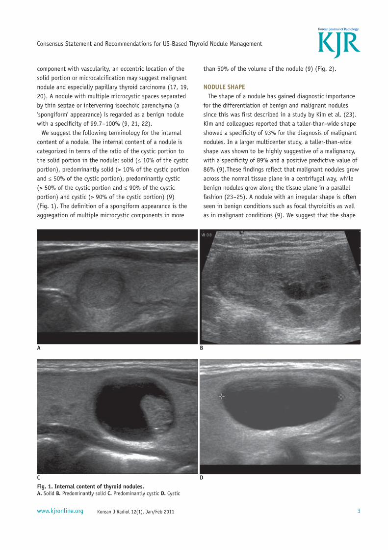
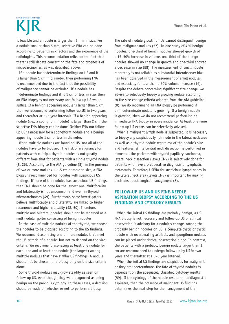
We suggest the following terminology for the internal content of a nodule. The internal content of a nodule is categorized in terms of the ratio of the cystic portion to the solid portion in the nodule: solid (≤ 10% of the cystic portion), predominantly solid (> 10% of the cystic portion and ≤ 50% of the cystic portion), predominantly cystic (> 50% of the cystic portion and ≤ 90% of the cystic portion) and cystic (> 90% of the cystic portion) (9) (Fig. 1). The defi nition of a spongiform appearance is the aggregation of multiple microcystic components in more
than 50% of the volume of the nodule (9) (Fig. 2).
NODULE SHAPE
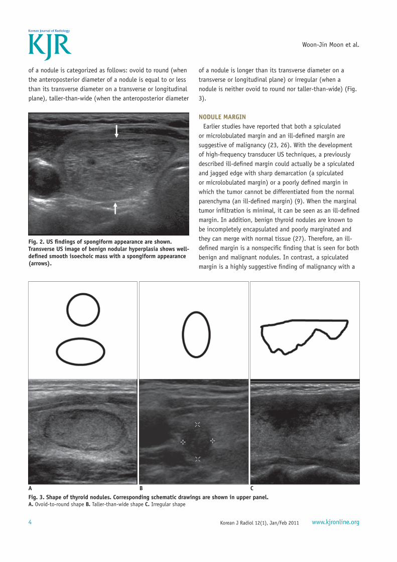
The shape of a nodule has gained diagnostic importance for the differentiation of benign and malignant nodules since this was fi rst described in a study by Kim et al. (23). Kim and colleagues reported that a taller-than-wide shape showed a specifi city of 93% for the diagnosis of malignant nodules. In a larger multicenter study, a taller-than-wide shape was shown to be highly suggestive of a malignancy, with a specifi city of 89% and a positive predictive value of 86% (9).These fi ndings refl ect that malignant nodules grow across the normal tissue plane in a centrifugal way, while benign nodules grow along the tissue plane in a parallel fashion (23-25). A nodule with an irregular shape is often seen in benign conditions such as focal thyroiditis as well as in malignant conditions (9). We suggest that the shape
A B
C D
Fig. 1. Internal content of thyroid nodules.
A. Solid B. Predominantly solid C. Predominantly cystic D. Cystic
Korean J Radiol 12(1), Jan/Feb 2011 www.kjronline.org4
Woon-Jin Moon et al.
of a nodule is categorized as follows: ovoid to round (when the anteroposterior diameter of a nodule is equal to or less than its transverse diameter on a transverse or longitudinal plane), taller-than-wide (when the anteroposterior diameter
of a nodule is longer than its transverse diameter on a transverse or longitudinal plane) or irregular (when a nodule is neither ovoid to round nor taller-than-wide) (Fig. 3).
NODULE MARGIN
Earlier studies have reported that both a spiculated or microlobulated margin and an ill-defi ned margin are suggestive of malignancy (23, 26). With the development of high-frequency transducer US techniques, a previously described ill-defi ned margin could actually be a spiculated and jagged edge with sharp demarcation (a spiculated or microlobulated margin) or a poorly defi ned margin in which the tumor cannot be differentiated from the normal parenchyma (an ill-defi ned margin) (9). When the marginal tumor infi ltration is minimal, it can be seen as an ill-defi ned margin. In addition, benign thyroid nodules are known to be incompletely encapsulated and poorly marginated and they can merge with normal tissue (27). Therefore, an ill-defi ned margin is a nonspecifi c fi nding that is seen for both benign and malignant nodules. In contrast, a spiculated margin is a highly suggestive fi nding of malignancy with a
B CA
Fig. 3. Shape of thyroid nodules. Corresponding schematic drawings are shown in upper panel.
A. Ovoid-to-round shape B. Taller-than-wide shape C. Irregular shape
Fig. 2. US fi ndings of spongiform appearance are shown.
Transverse US image of benign nodular hyperplasia shows well-
defi ned smooth isoechoic mass with a spongiform appearance
(arrows).

Korean J Radiol 12(1), Jan/Feb 2011www.kjronline.org 5
Consensus Statement and Recommendations for US-Based Thyroid Nodule Management
specifi city of 92% and a positive predictive value of 81% (9). Accordingly, we suggest that the margin of a nodule is
categorized as follows: smooth, spiculated/microlobulated or ill-defi ned (Fig. 4).
ECHOGENICITY
In terms of echogenicity, a solid component must be considered. When a solid component is heterogeneous, the nodular echogenicity is defi ned by that of the majority of the nodule. Marked hypoechogenicity is highly specifi c for malignant nodule with a specifi city of 92-94% (9, 23). Although the parenchymal echogenicity of a thyroid gland can vary among individuals, it is used as a reference for nodule echogenicity. Another reference to defi ne nodule echogenicity is the strap muscles with low-echogenicity such as the sternothyroid muscle, the sternothyroid muscle and the sternocleidomastoid muscles (9, 23).
We suggest that nodule echogenicity is categorized according to the relative echogenicity compared to that of
a reference as follows. Nodule echogenicity includes marked hypoechoic (when a nodule is hypoechoic relative to the adjacent strap muscle), hypoechoic (when a nodule is hypoechoic relative to the thyroid parenchyma), isoechoic (when a nodule has the same echogenicity as that of the thyroid parenchyma) and hyperechoic (when a nodule is echogenic relative to the thyroid parenchyma) (Fig. 5).
CALCIFICATION
Calcifi cations can be seen in both benign and malignant nodules. Calcifi cations may be microcalcifi cation, coarse or macrocalcifi cation or peripheral or rim calcifi cations in thyroid nodules. Pathologically, microcalcifi cation is a psammoma body that is comprised of 10-100 μm round, laminar, crystalline, calcifi c deposits, which is very specifi c for thyroid carcinoma, and especially for papillary thyroid carcinoma.
Microcalcifi cations on US are fi ndings that are highly suggestive for malignancy with a specifi city of 86-95%
A B
C
Fig. 4. Margin of thyroid nodules.
A. Smooth margin B. Spiculated or microlobulated margin (arrow) C. Ill-defi ned margin (arrows)

Korean J Radiol 12(1), Jan/Feb 2011 www.kjronline.org6
Woon-Jin Moon et al.
and a positive predictive value of 42-94% (9, 23, 26, 28, 29). Large and irregular shaped dystrophic calcifi cations may develop secondarily due to tissue necrosis and these calcifi cations can be seen in both benign and malignant nodules. A solid nodule with macrocalcifi cation larger than 1 mm suggests the presence of a malignancy rather than a benign nodule (9). The meaning of peripheral, eggshell or rim calcifi cation is still being debated for making the differentiation between benign and malignant nodules. Recent reports have found that when a nodule has eggshell or rim calcifi cations, a hypoechoic halo and/or disruption of eggshell calcifi cations, these are fi ndings that suggest malignancy (9, 30-32).
On US, a calcifi cation is defi ned as a prominent echogenic focus with or without posterior shadowing. The absence of posterior shadowing does not rule out the possibility
of calcifi cation since some calcifi cations are too small to produce posterior shadowing. When punctuate echogenic foci are accompanied by reverberation artifacts, they should be due to colloid materials and they can be easily differentiated from calcifi cation on real-time US.
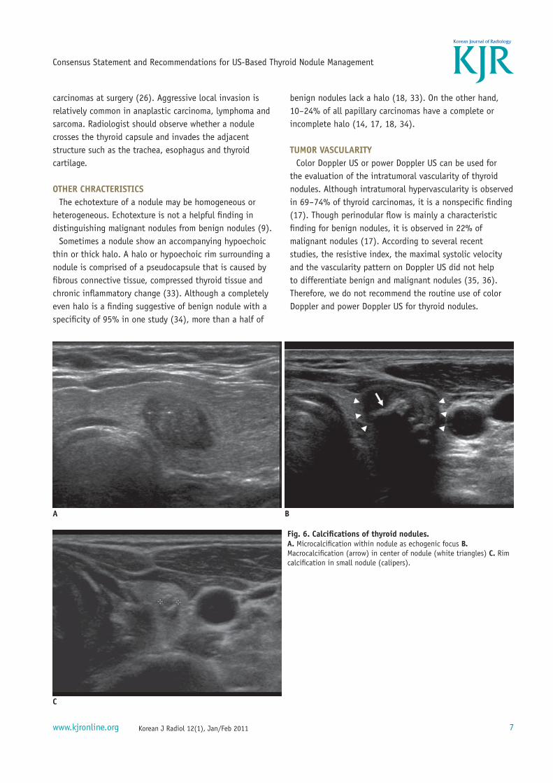
We suggest that calcifi cation is categorized with respect to its size as follows. Calcifi cations include microcalcifi cations (when there are tiny, punctuate echogenic foci of 1 mm or less either with or without posterior shadowing), macrocalcifi cations (when punctuate echogenic foci are larger than 1 mm in size) and rim calcifi cations (when a nodule has peripheral curvilinear or eggshell calcifi cation) (Fig. 6).
EXTRACAPSULAR INVASION
Extracapsular extension is observed in 36% of all thyroid
A B
C D
Fig. 5. Echogenicity of thyroid nodules.
A. Marked hypoechogenicity of nodule is shown. Note more hypoechoic nature of nodule (arrow) as compared to that of strap muscles (asterisk). B. Hypoechogenicity of nodule (arrows) C. Isoechogenicity of nodule (arrows) D. Hyperechogenicity of nodule (arrow)
Korean J Radiol 12(1), Jan/Feb 2011www.kjronline.org 7
Consensus Statement and Recommendations for US-Based Thyroid Nodule Management
carcinomas at surgery (26). Aggressive local invasion is relatively common in anaplastic carcinoma, lymphoma and sarcoma. Radiologist should observe whether a nodule crosses the thyroid capsule and invades the adjacent structure such as the trachea, esophagus and thyroid cartilage.
OTHER CHRACTERISTICS
The echotexture of a nodule may be homogeneous or heterogeneous. Echotexture is not a helpful fi nding in distinguishing malignant nodules from benign nodules (9).
Sometimes a nodule show an accompanying hypoechoic thin or thick halo. A halo or hypoechoic rim surrounding a nodule is comprised of a pseudocapsule that is caused by fi brous connective tissue, compressed thyroid tissue and chronic infl ammatory change (33). Although a completely even halo is a fi nding suggestive of benign nodule with a specifi city of 95% in one study (34), more than a half of
benign nodules lack a halo (18, 33). On the other hand, 10-24% of all papillary carcinomas have a complete or incomplete halo (14, 17, 18, 34).
TUMOR VASCULARITY
Color Doppler US or power Doppler US can be used for the evaluation of the intratumoral vascularity of thyroid nodules. Although intratumoral hypervascularity is observed in 69-74% of thyroid carcinomas, it is a nonspecifi c fi nding (17). Though perinodular fl ow is mainly a characteristic fi nding for benign nodules, it is observed in 22% of malignant nodules (17). According to several recent studies, the resistive index, the maximal systolic velocity and the vascularity pattern on Doppler US did not help to differentiate benign and malignant nodules (35, 36). Therefore, we do not recommend the routine use of color Doppler and power Doppler US for thyroid nodules.
A B
C
Fig. 6. Calcifi cations of thyroid nodules.
A. Microcalcifi cation within nodule as echogenic focus B. Macrocalcifi cation (arrow) in center of nodule (white triangles) C. Rim calcifi cation in small nodule (calipers).
Korean J Radiol 12(1), Jan/Feb 2011 www.kjronline.org8
Woon-Jin Moon et al.
US ELASTOGRAPHY
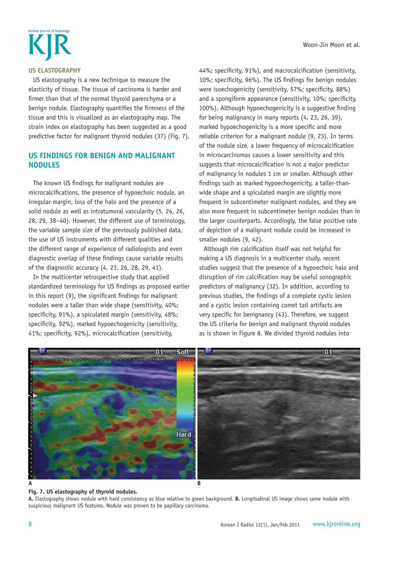
US elastography is a new technique to measure the elasticity of tissue. The tissue of carcinoma is harder and fi rmer than that of the normal thyroid parenchyma or a benign nodule. Elastography quantifi es the fi rmness of the tissue and this is visualized as an elastography map. The strain index on elastography has been suggested as a good predictive factor for malignant thyroid nodules (37) (Fig. 7).
US FINDINGS FOR BENIGN AND MALIGNANT
NODULES
The known US fi ndings for malignant nodules are microcalcifi cations, the presence of hypoechoic nodule, an irregular margin, loss of the halo and the presence of a solid nodule as well as intratumoral vascularity (5, 24, 26, 28, 29, 38-40). However, the different use of terminology, the variable sample size of the previously published data, the use of US instruments with different qualities and the different range of experience of radiologists and even diagnostic overlap of these fi ndings cause variable results of the diagnostic accuracy (4, 23, 26, 28, 29, 41).
In the multicenter retrospective study that applied standardized terminology for US fi ndings as proposed earlier in this report (9), the signifi cant fi ndings for malignant nodules were a taller than wide shape (sensitivity, 40%; specifi city, 91%), a spiculated margin (sensitivity, 48%; specifi city, 92%), marked hypoechogenicity (sensitivity, 41%; specifi city, 92%), microcalcifi cation (sensitivity,
44%; specifi city, 91%), and macrocalcifi cation (sensitivity, 10%; specifi city, 96%). The US fi ndings for benign nodules were isoechogenicity (sensitivity, 57%; specifi city, 88%) and a spongiform appearance (sensitivity, 10%; specifi city, 100%). Although hypoechogenicity is a suggestive fi nding for being malignancy in many reports (4, 23, 26, 39), marked hypoechogenicity is a more specifi c and more reliable criterion for a malignant nodule (9, 23). In terms of the nodule size, a lower frequency of microcalcifi cation in microcarcinomas causes a lower sensitivity and this suggests that microcalcifi cation is not a major predictor of malignancy in nodules 1 cm or smaller. Although other fi ndings such as marked hypoechogenicity, a taller-than-wide shape and a spiculated margin are slightly more frequent in subcentimeter malignant nodules, and they are also more frequent in subcentimeter benign nodules than in the larger counterparts. Accordingly, the false positive rate of depiction of a malignant nodule could be increased in smaller nodules (9, 42).
Although rim calcifi cation itself was not helpful for making a US diagnosis in a multicenter study, recent studies suggest that the presence of a hypoechoic halo and disruption of rim calcifi cation may be useful sonographic predictors of malignancy (32). In addition, according to previous studies, the fi ndings of a complete cystic lesion and a cystic lesion containing comet tail artifacts are very specifi c for benignancy (43). Therefore, we suggest the US criteria for benign and malignant thyroid nodules as is shown in Figure 8. We divided thyroid nodules into
A B
Fig. 7. US elastography of thyroid nodules.
A. Elastography shows nodule with hard consistency as blue relative to green background. B. Longitudinal US image shows same nodule with suspicious malignant US features. Nodule was proven to be papillary carcinoma.
Korean J Radiol 12(1), Jan/Feb 2011www.kjronline.org 9
Consensus Statement and Recommendations for US-Based Thyroid Nodule Management
three categories: suspicious malignant nodules, probably benign nodules and indeterminate nodules. A taller-than-wide shape, a spiculated margin, marked hypoechogenicity, microcalcifi cations and macrocalcifi cations are suggestive fi ndings for malignancy. The presence of at least one of the fi ndings for malignancy defi nes a nodule as a suspicious malignant nodule. In contrast, a simple cyst, a predominantly cystic or cystic nodule with reverberating artifacts and a nodule with a spongiform appearance (especially with intervening isoechoic parenchyma) are defi ned as probably benign nodules. Although a cystic nodule with more than a 90% cystic component is very rare for a thyroid malignancy, a mural solid component within the cystic nodule may be papillary thyroid carcinoma (43). Therefore, a small solid component in a predominantly cystic or cystic nodule should be carefully examined and it should be aspirated in the case with the presence of a suspicious malignant feature.
Indeterminate nodules include nodules having US fi ndings with neither malignant nor benign features. The US fi ndings for an indeterminate nodule include isoechogenicity, hypoechogenicity, and hyperechogenicity, an ovoid-to-round or irregular shape, a smooth or ill-defi ned margin and a rim calcifi cation. Although, isoechogenicity is more suggestive of benignancy, 14% of the isoechoic nodules were malignant in one study (9). Although rim calcifi cation is classifi ed as an indeterminate factor for malignancy, the presence of a
hypoechoic halo and rim disruption is more suggestive of malignancy (32). The indeterminate category is currently a term for all nodules that are without clear evidence of being benign and malignant. The criteria for suspicious malignant nodules are identical to the US fi ndings for papillary carcinoma, which comprises most thyroid carcinomas. The US fi ndings for medullary carcinoma are almost the same as those for papillary carcinoma (44). Yet these criteria have limited value in diagnosing thyroid carcinomas other than a subtype of papillary carcinoma. Relatively uncommon FTC and other histologic types of thyroid carcinoma can be eliminated by the use of these criteria (45, 46). Along with an accumulation of evidence from further studies on thyroid US, the indeterminate category can be subclassifi ed into truly benign nodule or adenoma or malignant nodule.
INDICATIONS FOR US-GUIDED FINE-NEEDLE
ASPIRATION BIOPSY ACCORDING TO THE US
CRITERIA
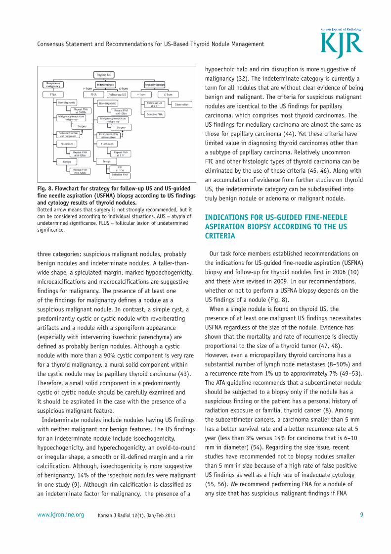
Our task force members established recommendations on the indications for US-guided fi ne-needle aspiration (USFNA) biopsy and follow-up for thyroid nodules fi rst in 2006 (10) and these were revised in 2009. In our recommendations, whether or not to perform a USFNA biopsy depends on the US fi ndings of a nodule (Fig. 8).
When a single nodule is found on thyroid US, the presence of at least one malignant US fi ndings necessitates USFNA regardless of the size of the nodule. Evidence has shown that the mortality and rate of recurrence is directly proportional to the size of a thyroid tumor (47, 48). However, even a micropapillary thyroid carcinoma has a substantial number of lymph node metastases (8-50%) and a recurrence rate from 1% up to approximately 7% (49-53). The ATA guideline recommends that a subcentimeter nodule should be subjected to a biopsy only if the nodule has a suspicious fi nding or the patient has a personal history of radiation exposure or familial thyroid cancer (8). Among the subcentimeter cancers, a carcinoma smaller than 5 mm has a better survival rate and a better recurrence rate at 5 year (less than 3% versus 14% for carcinoma that is 6-10 mm in diameter) (54). Regarding the size issue, recent studies have recommended not to biopsy nodules smaller than 5 mm in size because of a high rate of false positive US fi ndings as well as a high rate of inadequate cytology (55, 56). We recommend performing FNA for a nodule of any size that has suspicious malignant fi ndings if FNA
Fig. 8. Flowchart for strategy for follow-up US and US-guided
fi ne needle aspiration (USFNA) biopsy according to US fi ndings
and cytology results of thyroid nodules.
Dotted arrow means that surgery is not strongly recommended, but it can be considered according to individual situations. AUS = atypia of undetermined signifi cance, FLUS = follicular lesion of undetermined signifi cance.
Korean J Radiol 12(1), Jan/Feb 2011 www.kjronline.org10
Woon-Jin Moon et al.
is feasible and a nodule is larger than 5 mm in size. For a nodule smaller than 5 mm, selective FNA can be done according to patient’s risk factors and the experience of the radiologists. This recommendation relies on the fact that there is still debate concerning the fate and prognosis of microcarcinomas, as was described above.
If a nodule has indeterminate fi ndings on US and it is larger than 1 cm in diameter, then performing FNA is recommended due to the fact that the possibility of malignancy cannot be excluded. If a nodule has indeterminate fi ndings and it is 1 cm or less in size, then an FNA biopsy is not necessary and follow-up US would suffi ce. If a benign appearing nodule is larger than 1 cm, then we recommend performing follow-up US in two years and thereafter at 3-5 year intervals. If a benign appearing nodule (i.e., a spongiform nodule) is larger than 2 cm, then selective FNA biopsy can be done. Neither FNA nor follow up US is necessary for a spongiform nodule and a benign appearing nodule 1 cm or less in diameter.
When multiple nodules are found on US, not all of the nodules have to be biopsied. The risk of malignancy for patients with multiple thyroid nodules is not greatly different from that for patients with a single thyroid nodule (8, 26). According to the ATA guideline (8), in the presence of two or more nodules 1-1.5 cm or more in size, a FNA biopsy is recommended for nodules with suspicious US fi ndings. If none of the nodules has suspicious US fi ndings, then FNA should be done for the largest one. Multifocality and bilaterality is not uncommon and even in thyroid microcarcinomas (49). Furthermore, some investigators believe multifocality and bilaterality are linked to higher recurrence and higher mortality (48, 50). Therefore, multiple and bilateral nodules should not be regarded as a multinodular goiter consisting of benign nodules.
In the case of multiple nodules of the thyroid, we choose the nodules to be biopsied according to the US fi ndings. We recommend aspirating one or more nodules that meet the US criteria of a nodule, but not to depend on the size criteria. We recommend aspirating at least one nodule for each lobe and at least one nodule (the largest) among multiple nodules that have similar US fi ndings. A nodule should not be chosen for a biopsy only on the size criteria alone.
Some thyroid nodules may grow steadily as seen on follow-up US, even though they were diagnosed as being benign on the previous cytology. In these cases, a decision should be made on whether or not to perform a biopsy.
The rate of nodule growth on US cannot distinguish benign from malignant nodules (57). In one study of 420 benign nodules, one-third of benign nodules showed growth of a 15-30% increase in volume, one-third of the benign nodules showed no change in growth and one-third showed a decrease in size (58). The measurement of small nodule reportedly is not reliable as substantial interobserver bias has been observed in the measurement of small nodules, and especially for less than a 50% volume increase (16). Despite the debate concerning signifi cant size change, we advise to selectively biopsy a growing nodule according to the size change criteria adopted from the ATA guideline (8). We do recommend an FNA biopsy be performed if an indeterminate nodule is growing. If a benign nodule is growing, then we do not recommend performing an immediate FNA biopsy in every incidence. At least one more follow-up US exams can be selectively advised.
When a malignant lymph node is suspected, it is necessary to biopsy any suspicious lymph node in the lateral neck area as well as a thyroid nodule regardless of the nodule’s size and features. While central neck dissection is performed in almost all the patients with thyroid papillary carcinoma, lateral neck dissection (levels II-V) is selectively done for patients who have a preoperative diagnosis of lymphatic metastasis. Therefore, USFNA for suspicious lymph nodes in the lateral neck area (levels II-V) is important for making decisions about surgical management (8).
FOLLOW-UP US AND US FINE-NEEDLE
ASPIRATION BIOPSY ACCORDING TO THE US
FINDINGS AND CYTOLOGY RESULTS
When the initial US fi ndings are probably benign, a US-FNA biopsy is not necessary and follow-up US or clinical observation is advisory for a nodule’s change. Among the probably benign nodules on US, a complete cystic or cystic nodule with reverberating artifacts and spongiform nodules can be placed under clinical observation alone. In contrast, the patients with a probably benign nodule larger than 1 cm are recommended to undergo follow-up by US in two years and thereafter at a 3-5 year interval.
When the initial US fi ndings are suspicious for malignant or they are indeterminate, the fate of thyroid nodules is dependent on the adequately classifi ed cytology results (59). If the cytology of the nodule results in nondiagnostic aspirates, then the presence of malignant US fi ndings determines the next step for the management of the
Korean J Radiol 12(1), Jan/Feb 2011www.kjronline.org 11
Consensus Statement and Recommendations for US-Based Thyroid Nodule Management
nodule. Non-diagnostic cytology results occur in 5-10% of cases under US guidance and these non-diagnostic cases are due to the operator’s inexperience, aspiration of cystic fl uid and the presence of a bloody aspirate (38). Five percent of nodules with nondiagnostic cytology after an initial biopsy are eventually diagnosed as malignant and approximately 18% of malignant nodules are diagnosed by at least two aspiration cytology examinations (60, 61). Therefore, thyroid nodules with non-diagnostic cytology and malignant US fi ndings should be followed by US-FNA biopsy at a 3-6 month interval. When a nodule does not have any malignant US fi ndings, but it has non-diagnostic cytology, it is recommended that the nodule should be followed up by US-FNA biopsy in 6-12 months.
When the cytology result for a nodule is malignant, the patient should undergo surgery and follow-up by US (7, 8). When the cytology results of thyroid nodules are indeterminate (suspicious for a papillary carcinoma, atypical cells, follicular lesion or follicular neoplasm), the subtype of the indeterminate cytology and the presence of malignant US fi ndings determines the next step for the nodules’ management. An indeterminate cytology fi nding, although the cytology results can vary as determined at different institutions, is responsible for approximately 15-30% of all the fi ne needle aspiration cytologies. The cytology results of a suspicious for papillary carcinoma or Hurthle cell neoplasm necessitates surgery (lobectomy or total thyroidectomy) followed by US. When the cytology of a nodule is indicative of a follicular neoplasm, it is recommended to consider surgery fi rst although repeated US-FNA biopsies are preferable in certain situations. When the cytology of a nodule is a follicular lesion of undetermined signifi cance or atypia of undetermined signifi cance (59), a nodule 1 cm or more is recommended to undergo a repeated US-FNA biopsy in 6-12 months in the case of malignant US fi ndings, while a nodule without malignant US fi ndings can be subjected to a repeated US-FNA in 1-1.5 years.
When the cytology of a nodule is indicative of being benign, the follow-up strategy is as follows and according to the US fi ndings. A nodule with malignant US fi ndings is recommended to undergo repeat US-FNA in 6-12 months, while a nodule without malignant fi ndings is recommended to undergo repeat US in one year or to repeat US-FNA selectively. Since the false-negative rate of USFNA is low but not negligible, it is reasonable to repeat US-FNA in certain conditions, and especially for thyroid nodules with malignant US fi ndings (62). A nodule with a benign
cytology and that has been subjected to at least two US-FNA biopsies is regarded as a benign nodule and it can be followed up in 3-5 years.
ROLE OF THYROID US AS A SCREENING TEST
The role of a screening test for thyroid nodules is limited. Because of the very high prevalence of thyroid nodules and the very good prognosis and survival rate, the current consensus is that a screening test for thyroid malignancy cannot be justifi ed (7). As smaller malignant nodules can be detected on thyroid US, the survival rate and prognosis may improve regardless of the actual effect of the treatments, and even with an increasing prevalence of disease (54, 63). Thyroid cancer detected by the use of an early screening test may tend to progress less rapidly than clinically detected disease. There may be cases that would regress, remain stable or progress too slowly to become clinically apparent during the lifetime of the patient (63).
However, a screening test can be justifi ed in high-risk groups such as patients with a history of familial thyroid carcinoma, a history of MEN or a history of childhood irradiation of the head and neck area.
CONCLUSION
US for thyroid nodules is the most sensitive diagnostic modality for making the diagnosis of thyroid carcinoma and this modality provides valuable guidance to perform an aspiration biopsy and follow-up. On the US of thyroid nodule, the size of the nodule, the internal texture, the shape, the echogenicity, the margin, the presence of calcifi cation and the presence of adjacent structures should be carefully scrutinized. The fi ndings for a suspicious malignant nodule include a taller-than-wide shape, a spiculated or microlobulated margin, marked hypoechogenicity, microcalcifi cations and macrocalcifi cations. Presence of at least one of the malignant US fi ndings suggests the presence of a malignancy. According to these fi ndings and the resultant category of a nodule, the nodule should be aspirated or followed-up with US, or it should remain under clinical observation.
Acknowledgment :
KSThR Taskforce on Thyroid Nodule members are as following in alphabetical order: Jung Hwan Baek, MD
Korean J Radiol 12(1), Jan/Feb 2011 www.kjronline.org12
Woon-Jin Moon et al.
(University of Ulsan College of Medicine), So Lyung Jung, MD (College of Medicine, The Catholic University of Korea), Dong Wook Kim, MD (Inje University College of Medicine), Eun Kyung Kim, MD (Yonsei University College of Medicine), Ji Young Kim, MD (College of Medicine, The Catholic University of Korea), Ji Hoon Kim, MD (Seoul National University College of Medicine), Jin Young Kwak, MD (Yonsei University College of Medicine), Jeong Hyun Lee, MD (University of Ulsan College of Medicine), Joon Hyung Lee, MD (Dong-A University College of Medicine), Young Hen Lee, MD (Korea University School of Medicine), Won-Jin Moon, MD (Konkuk University School of Medicine), Dong Gyu Na, MD (Human Medical Imaging & Intervention Center), Jeong Seon Park, MD (Hanyang University College of Medicine), Sun Won Park, MD (Seoul National University College of Medicine), Jung Hee Shin, MD (Sungkyunkwan University School of Medicine).
The authors sincerely thank Ji Hoon Kim, MD and Jung Hee Shin, MD for their most valuable advice and support in developing recommendations. In addition, the authors sincerely appreciate Jinna Kim, MD for her contribution in organizing the former Thyroid Study Group of Korean Society of Radiology (TSGKSR), which was a predecessor of KSThR.
REFERENCES
1. Harach HR, Franssila KO, Wasenius VM. Occult papillary carcinoma of the thyroid. A “normal” fi nding in Finland. A systematic autopsy study. Cancer 1985;56:531-538
2. Brander A, Viikinkoski P, Nickels J, Kivisaari L. Thyroid gland: US screening in a random adult population. Radiology 1991;181:683-687
3. Tan GH, Gharib H. Thyroid incidentalomas: management approaches to nonpalpable nodules discovered incidentally on thyroid imaging. Ann Intern Med 1997;126:226-231
4. Frates MC, Benson CB, Charboneau JW, Cibas ES, Clark OH, Coleman BG, et al. Management of thyroid nodules detected at US: Society of Radiologists in Ultrasound consensus conference statement. Radiology 2005;237:794-800
5. Nam-Goong IS, Kim HY, Gong G, Lee HK, Hong SJ, Kim WB, et al. Ultrasonography-guided fi ne-needle aspiration of thyroid incidentaloma: correlation with pathological fi ndings. Clin Endocrinol (Oxf) 2004;60:21-28
6. National Cancer Information Center K. 2005 annual report of the Korea central cancer registry [www document]. Available at : http://www.cancer.go.kr last accessed; Oct 2008
7. Gharib H, Papini E, Valcavi R, Baskin HJ, Crescenzi A, Dottorini ME, et al. American Association of Clinical Endocrinologists and Associazione Medici Endocrinologi medical guidelines for clinical practice for the diagnosis and
management of thyroid nodules. Endocr Pract 2006;12:63-102 8. American Thyroid Association (ATA) Guidelines Taskforce on
Thyroid Nodules and Differentiated Thyroid Cancer, Cooper DS, Doherty GM, Haugen BR, Kloos RT, Lee SL, et al. Revised American Thyroid Association management guidelines for patients with thyroid nodules and differentiated thyroid cancer. Thyroid 2009;19:1167-1214
9. Moon WJ, Jung SL, Lee JH, Na DG, Baek JH, Lee YH, et al. Benign and malignant thyroid nodules: US differentiation--multicenter retrospective study. Radiology 2008;247:762-770
10. Moon WJ, Na DG, Jung SL, Lee JH, Kim J, Kim HS, et al. Recommendations for ultrasound-based management of thyroid nodules. In: 62nd Scientifi c Assembly of the Korean Radiological Society. Seoul: The Korean Radiological Society, 2006
11. Brander AE, Viikinkoski VP, Nickels JI, Kivisaari LM. Importance of thyroid abnormalities detected at US screening: a 5-year follow-up. Radiology 2000;215:801-806
12. Kuma K, Matsuzuka F, Yokozawa T, Miyauchi A, Sugawara M. Fate of untreated benign thyroid nodules: results of long-term follow-up. World J Surg 1994;18:495-498
13. Alexander EK, Hurwitz S, Heering JP, Benson CB, Frates MC, Doubilet PM, et al. Natural history of benign solid and cystic thyroid nodules. Ann Intern Med 2003;138:315-318
14. Hoang JK, Lee WK, Lee M, Johnson D, Farrell S. US features of thyroid malignancy: pearls and pitfalls. Radiographics 2007;27:847-860
15. Papini E, Petrucci L, Guglielmi R, Panunzi C, Rinaldi R, Bacci V, et al. Long-term changes in nodular goiter: a 5-year prospective randomized trial of levothyroxine suppressive therapy for benign cold thyroid nodules. J Clin Endocrinol Metab 1998;83:780-783
16. Brauer VF, Eder P, Miehle K, Wiesner TD, Hasenclever H, Paschke R. Interobserver variation for ultrasound determination of thyroid nodule volumes. Thyroid 2005;15:1169-1175
17. Chan BK, Desser TS, McDougall IR, Weigel RJ, Jeffrey RB Jr. Common and uncommon sonographic features of papillary thyroid carcinoma. J Ultrasound Med 2003;22:1083-1090
18. Watters DA, Ahuja AT, Evans RM, Chick W, King WW, Metreweli C, et al. Role of ultrasound in the management of thyroid nodules. Am J Surg 1992;164:654-657
19. Lee MJ, Kim EK, Kwak JY, Kim MJ. Partially cystic thyroid nodules on ultrasound: probability of malignancy and sonographic differentiation. Thyroid 2009;19:341-346
20. Hatabu H, Kasagi K, Yamamoto K, Iida Y, Misaki T, Hidaka A, et al. Cystic papillary carcinoma of the thyroid gland: a new sonographic sign. Clin Radiol 1991;43:121-124
21. Bonavita JA, Mayo J, Babb J, Bennett G, Oweity T, Macari M, et al. Pattern recognition of benign nodules at ultrasound of the thyroid: which nodules can be left alone? AJR Am J Roentgenol 2009;193:207-213
22. Moon WJ, Kwag HJ, Na DG. Are there any specifi c ultrasound fi ndings of nodular hyperplasia (“leave me alone” lesion) to differentiate it from follicular adenoma? Acta Radiol
Korean J Radiol 12(1), Jan/Feb 2011www.kjronline.org 13
Consensus Statement and Recommendations for US-Based Thyroid Nodule Management
2009;50:383-38823. Kim EK, Park CS, Chung WY, Oh KK, Kim DI, Lee JT, et al. New
sonographic criteria for recommending fi ne-needle aspiration biopsy of nonpalpable solid nodules of the thyroid. AJR Am J Roentgenol 2002;178:687-691
24. Alexander EK, Marqusee E, Orcutt J, Benson CB, Frates MC, Doubilet PM, et al. Thyroid nodule shape and prediction of malignancy. Thyroid 2004;14:953-958
25. Stavros AT, Thickman D, Rapp CL, Dennis MA, Parker SH, Sisney GA. Solid breast nodules: use of sonography to distinguish between benign and malignant lesions. Radiology 1995;196:123-134
26. Papini E, Guglielmi R, Bianchini A, Crescenzi A, Taccogna S, Nardi F, et al. Risk of malignancy in nonpalpable thyroid nodules: predictive value of ultrasound and color-Doppler features. J Clin Endocrinol Metab 2002;87:1941-1946
27. Reading CC, Charboneau JW, Hay ID, Sebo TJ. Sonography of thyroid nodules: a “classic pattern” diagnostic approach. Ultrasound Q 2005;21:157-165
28. Khoo ML, Asa SL, Witterick IJ, Freeman JL. Thyroid calcifi cation and its association with thyroid carcinoma. Head Neck 2002;24:651-655
29. Peccin S, de Castsro JA, Furlanetto TW, Furtado AP, Brasil BA, Czepielewski MA. Ultrasonography: is it useful in the diagnosis of cancer in thyroid nodules? J Endocrinol Invest 2002;25:39-43
30. Kwak MS, Baek JH, Kim YS, Jeong HJ. Patterns and signifi cance of peripheral calcifi cations of thyroid tumors seen on ultrasound. J Korean Radiol Soc 2005;53:401-405
31. Yoon DY, Lee JW, Chang SK, Choi CS, Yun EJ, Seo YL, et al. Peripheral calcifi cation in thyroid nodules: ultrasonographic features and prediction of malignancy. J Ultrasound Med 2007;26:1349-1355
32. Kim BM, Kim MJ, Kim EK, Kwak JY, Hong SW, Son EJ, et al. Sonographic differentiation of thyroid nodules with eggshell calcifi cations. J Ultrasound Med 2008;27:1425-1430
33. Propper RA, Skolnick ML, Weinstein BJ, Dekker A. The nonspecifi city of the thyroid halo sign. J Clin Ultrasound 1980;8:129-132
34. Lu C, Chang TC, Hsiao YL, Kuo MS. Ultrasonographic fi ndings of papillary thyroid carcinoma and their relation to pathologic changes. J Formos Med Assoc 1994;93:933-938
35. Tamsel S, Demirpolat G, Erdogan M, Nart D, Karadeniz M, Uluer H, et al. Power Doppler US patterns of vascularity and spectral Doppler US parameters in predicting malignancy in thyroid nodules. Clin Radiol 2007;62:245-251
36. Moon HJ, Kwak JY, Kim MJ, Son EJ, Kim EK. Can vascularity at power Doppler US help predict thyroid malignancy? Radiology 2010;255:260-269
37. Lyshchik A, Higashi T, Asato R, Tanaka S, Ito J, Mai JJ, et al. Thyroid gland tumor diagnosis at US elastography. Radiology 2005;237:202-211
38. Kim SJ, Kim EK, Park CS, Chung WY, Oh KK, Yoo HS. Ultrasound-guided fi ne-needle aspiration biopsy in nonpalpable thyroid nodules: is it useful in infracentimetric
nodules? Yonsei Med J 2003;44:635-64039. Wienke JR, Chong WK, Fielding JR, Zou KH, Mittelstaedt CA.
Sonographic features of benign thyroid nodules: interobserver reliability and overlap with malignancy. J Ultrasound Med 2003;22:1027-1031
40. Iannuccilli JD, Cronan JJ, Monchik JM. Risk for malignancy of thyroid nodules as assessed by sonographic criteria: the need for biopsy. J Ultrasound Med 2004;23:1455-1464
41. Frates MC, Benson CB, Doubilet PM, Cibas ES, Marqusee E. Can color Doppler sonography aid in the prediction of malignancy of thyroid nodules? J Ultrasound Med 2003;22:127-131
42. Popowicz B, Klencki M, Lewi ’nski A, Słowi ’nska-Klencka D. The usefulness of sonographic features in selection of thyroid nodules for biopsy in relation to the nodule’s size. Eur J Endocrinol 2009;161:103-111
43. Ahuja A, Chick W, King W, Metreweli C. Clinical signifi cance of the comet-tail artifact in thyroid ultrasound. J Clin Ultrasound 1996;24:129-133
44. Kim SH, Kim BS, Jung SL, Lee JW, Yang PS, Kang BJ, et al. Ultrasonographic fi ndings of medullary thyroid carcinoma: a comparison with papillary thyroid carcinoma. Korean J Radiol 2009;10:101-105
45. Jeh SK, Jung SL, Kim BS, Lee YS. Evaluating the degree of conformity of papillary carcinoma and follicular carcinoma to the reported ultrasonographic fi ndings of malignant thyroid tumor. Korean J Radiol 2007;8:192-197
46. Kim DS, Kim JH, Na DG, Park SH, Kim E, Chang KH, et al. Sonographic features of follicular variant papillary thyroid carcinomas in comparison with conventional papillary thyroid carcinomas. J Ultrasound Med 2009;28:1685-1692
47. Mazzaferri EL. Management of a solitary thyroid nodule. N Engl J Med 1993;328:553-559
48. Mazzaferri EL, Jhiang SM. Long-term impact of initial surgical and medical therapy on papillary and follicular thyroid cancer. Am J Med 1994;97:418-428
49. Pazaitou-Panayiotou K, Capezzone M, Pacini F. Clinical features and therapeutic implication of papillary thyroid microcarcinoma. Thyroid 2007;17:1085-1092
50. Baudin E, Travagli JP, Ropers J, Mancusi F, Bruno-Bossio G, Caillou B, et al. Microcarcinoma of the thyroid gland: the Gustave-Roussy Institute experience. Cancer 1998;83:553-559
51. Chow SM, Law SC, Au SK, Mang O, Yau S, Yuen KT, et al. Changes in clinical presentation, management and outcome in 1348 patients with differentiated thyroid carcinoma: experience in a single institute in Hong Kong, 1960-2000. Clin Oncol (R Coll Radiol) 2003;15:329-336
52. Ito Y, Uruno T, Nakano K, Takamura Y, Miya A, Kobayashi K, et al. An observation trial without surgical treatment in patients with papillary microcarcinoma of the thyroid. Thyroid 2003;13:381-387
53. Noguchi S, Yamashita H, Murakami N, Nakayama I, Toda M, Kawamoto H. Small carcinomas of the thyroid. A long-term follow-up of 867 patients. Arch Surg 1996;131:187-191
54. Noguchi S, Yamashita H, Uchino S, Watanabe S. Papillary microcarcinoma. World J Surg 2008;32:747-753
Korean J Radiol 12(1), Jan/Feb 2011 www.kjronline.org14
Woon-Jin Moon et al.
55. Kim DW, Lee EJ, Kim SH, Kim TH, Lee SH, Kim DH, et al. Ultrasound-guided fi ne-needle aspiration biopsy of thyroid nodules: comparison in effi cacy according to nodule size. Thyroid 2009;19:27-31
56. Mazzaferri EL, Sipos J. Should all patients with subcentimeter thyroid nodules undergo fi ne-needle aspiration biopsy and preoperative neck ultrasonography to defi ne the extent of tumor invasion? Thyroid 2008;18:597-602
57. Asanuma K, Kobayashi S, Shingu K, Hama Y, Yokoyama S, Fujimori M, et al. The rate of tumour growth does not distinguish between malignant and benign thyroid nodules. Eur J Surg 2001;167:102-105
58. Erdogan MF, Gursoy A, Erdogan G. Natural course of benign thyroid nodules in a moderately iodine-defi cient area. Clin Endocrinol (Oxf) 2006;65:767-771
59. Cibas ES, Ali SZ. The Bethesda System for Reporting Thyroid
Cytopathology. Thyroid 2009;19:1159-116560. Alexander EK, Heering JP, Benson CB, Frates MC, Doubilet
PM, Cibas ES, et al. Assessment of nondiagnostic ultrasound-guided fi ne needle aspirations of thyroid nodules. J Clin Endocrinol Metab 2002;87:4924-4927
61. Ogawa Y, Kato Y, Ikeda K, Aya M, Ogisawa K, Kitani K, et al. The value of ultrasound-guided fi ne-needle aspiration cytology for thyroid nodules: an assessment of its diagnostic potential and pitfalls. Surg Today 2001;31:97-101
62. Kwak JY, Koo H, Youk JH, Kim MJ, Moon HJ, Son EJ, et al. Value of US correlation of a thyroid nodule with initially benign cytologic results. Radiology 2010;254:292-300
63. Black WC, Welch HG. Advances in diagnostic imaging and overestimations of disease prevalence and the benefi ts of therapy. N Engl J Med 1993;328:1237-1243