journal.pone.0049346(1) lipid irfa
DESCRIPTION
percobaan tikus demostika pada metabolisme lipidTRANSCRIPT

Exploration of Lipid Metabolism in Relation with PlasmaMembrane Properties of Duchenne Muscular DystrophyCells: Influence of L-CarnitineFrancoise Le Borgne1, Stephane Guyot2, Morgan Logerot1, Laurent Beney2, Patrick Gervais2,
Jean Demarquoy1*
1 Laboratoire Bio-PeroxIL, Biochimie du Peroxysome, Inflammation et Metabolisme Lipidique, Universite de Bourgogne - Faculte des Sciences Gabriel, Dijon, France,
2 UMR A 02.102 Procedes Alimentaires et Microbiologiques, Equipe Procedes Microbiologiques et Biotechnologiques, AgroSup Dijon/Universite de Bourgogne, bat
Erasme, Dijon, France
Abstract
Duchenne muscular dystrophy (DMD) arises as a consequence of mutations in the dystrophin gene. Dystrophin is amembrane-spanning protein that connects the cytoskeleton and the basal lamina. The most distinctive features of DMD area progressive muscular dystrophy, a myofiber degeneration with fibrosis and metabolic alterations such as fatty infiltration,however, little is known on lipid metabolism changes arising in Duchenne patient cells. Our goal was to identify metabolicchanges occurring in Duchenne patient cells especially in terms of L-carnitine homeostasis, fatty acid metabolism both atthe mitochondrial and peroxisomal level and the consequences on the membrane structure and function. In this paper, wecompared the structural and functional characteristics of DMD patient and control cells. Using radiolabeled L-carnitine, wefound, in patient muscle cells, a marked decrease in the uptake and the intracellular level of L-carnitine. Associated with thischange, a decrease in the mitochondrial metabolism can be seen from the analysis of mRNA encoding for mitochondrialproteins. Probably, associated with these changes in fatty acid metabolism, alterations in the lipid composition of the cellswere identified: with an increase in poly unsaturated fatty acids and a decrease in medium chain fatty acids, monounsaturated fatty acids and in cholesterol contents. Functionally, the membrane of cells lacking dystrophin appeared to beless fluid, as determined at 37uC by fluorescence anisotropy. These changes may, at least in part, be responsible for changesin the phospholipids and cholesterol profile in cell membranes and ultimately may reduce the fluidity of the membrane. Asupplementation with L-carnitine partly restored the fatty acid profile by increasing saturated fatty acid content anddecreasing the amounts of MUFA, PUFA, VLCFA. L-carnitine supplementation also restored muscle membrane fluidity. Thissuggests that regulating lipid metabolism in DMD cells may improve the function of cells lacking dystrophin.
Citation: Le Borgne F, Guyot S, Logerot M, Beney L, Gervais P, et al. (2012) Exploration of Lipid Metabolism in Relation with Plasma Membrane Properties ofDuchenne Muscular Dystrophy Cells: Influence of L-Carnitine. PLoS ONE 7(11): e49346. doi:10.1371/journal.pone.0049346
Editor: Petras Dzeja, Mayo Clinic, United States of America
Received June 15, 2012; Accepted October 10, 2012; Published November 27, 2012
Copyright: � 2012 Le Borgne et al. This is an open-access article distributed under the terms of the Creative Commons Attribution License, which permitsunrestricted use, distribution, and reproduction in any medium, provided the original author and source are credited.
Funding: Funding came from Association Francaise contre les Myophaties. The funders had no role in study design, data collection and analysis, decision topublish, or preparation of the manuscript.
Competing Interests: The authors have declared that no competing interests exist.
* E-mail: [email protected]
Introduction
In Duchenne muscular dystrophy patient, muscle cells are
lacking dystrophin. Dystrophin functions as part of a large protein
complex that includes dystroglycans, sarcoglycans, dystrobrevins,
syntrophins, and sarcospan [1]. The absence of dystrophin has a
dramatic impact on cell membrane stability and structure as this
dystrophin–glycoprotein complex mechanically stabilizes the
sarcolemma against shear stresses imposed during muscle activity
[2]. In the early stages of the disease, muscle undergoes active
regeneration [3,4,5,6], but as the disease progresses, the regener-
ation process is not efficient enough and muscle degeneration
exceeds the regeneration process leading to muscle loss.
To cure DMD, gene therapy is likely to be the only efficient
approach, but its application will still need many years before
being routinely used in patients [7,8]. In the interval, the
development of palliative treatments appears very useful [9].
L-carnitine is a small molecule derived from lysine and
methionine. It is implicated in the fatty acid metabolism both at
the mitochondrial and peroxisomal levels [10] and as a cofactor in
several other cellular functions such as the acetylation of proteins.
L-carnitine present in human organisms comes from the food
supply and from an endogenous synthesis occurring in the liver
and the kidney [11]. Once synthesized, L-carnitine is distributed to
tissues and organs whose metabolism is dependent on fatty acid
metabolism. Muscles concentrate most (up to 95%) of all the L-
carnitine present in human organism. Alteration in L-carnitine
homeostasis leads to a decrease in muscle function and a
deterioration in neuronal functions. This effect is due to a
decrease in oxidative pathways, an increase in free radical
production and likely to an impairment in other functions
depending on L-carnitine. L-carnitine can be regarded as a
regulatory nutrient able to control the metabolic flux and that can
improve muscle energy production and muscle function.
Previous studies described alterations in L-carnitine metabolism
in DMD patients. Berthillier was the first, in 1982, to report
muscle carnitine deficiency in 12 children affected with Duchenne
PLOS ONE | www.plosone.org 1 November 2012 | Volume 7 | Issue 11 | e49346

muscular dystrophy (DMD) [12]. These results were later
confirmed by two other studies [13,14]. The molecular bases of
this decrease in L-carnitine level are not established yet. Several
hypotheses may be drawn. Altered structure of the muscle cell,
decreased activity in patient muscle, alteration in the exchange
through cell membrane, are among the hypotheses that may
explain this decrease in L-carnitine content in patient cells. But,
whatever the origin is, DMD patients muscles have, clearly, a
major defect in L-carnitine homeostasis.
Besides L-carnitine alteration, lipid content and lipid metabo-
lism have been reported altered in Duchenne patient cells coming
from different tissues. In erythrocytes harvested from DMD
patients, a decrease in the concentration of unsaturated fatty acids
(oleic, linoleic and arachidonic acids) and conversely, an increase
in saturated fatty acid amounts were observed [15]. Carroll et al.
(1983) reported an increase of long chain acyl CoA in muscle from
patients with DMD while free and short chain acylcarnitine were
reduced, suggesting a disruption of fatty acid oxidation [16]. More
recently, a case of Duchenne muscular dystrophy and severe
mental retardation was described in a very young boy with
chromosomal anomaly, medium chain fatty acid level was found
to high in the cerebrospinal fluid [17].
In mdx mice, alteration in phospholipids composition has also
been reported [18,19]. Even et al. (1994) also reported an
impaired fatty acid metabolism in mdx mice and interestingly
notice that several major symptoms observed in the muscles of
mdx mice seems similar to those observed in muscles of patients or
animals suffering severe food restriction [20]. The relation
between the fatty acid composition and the severity of the disease
was studied in dystrophic muscle. Phospholipids extracted from
mdx mice contained less docosahexaenoic acid (C22:6 n–3) and
more linoleic acid (C18:2 n–6) and some correlation can be drawn
between phospholipid composition and muscle strength [21].
The lipid tails of the phospholipids composing the plasma
membranes can affect mechanical properties, including its
resistance to stretching and bending. Alterations in the lipid
metabolism of heart from mdx mice have also been reported [22].
Mdx heart perfusions with stable isotopes revealed a marked shift
in substrate fuel selection from fatty acids to carbohydrates,
suggesting, again, alterations in fatty acid metabolism. However,
none of these studies investigate in depth the biochemical and
molecular bases of these changes in DMD patients.
Surprisingly, identifying lipid composition alterations in human
muscle cells has never been done. Plasma membranes of DMD
patient cells seem to undergo several rearrangements in terms of
lipid composition. The fatty acid composition of the phospholipids
present in the membranes is a consequence of both the food supply
and the lipid metabolism.
Membrane structure has been studied years before the discovery
of dystrophin and very little since. In the early 809s Rowland [23]
reported that many changes occurred in DMD muscle cell
membrane, like membrane weakness and alterations in calcium
metabolism and regarding the origins of such changes this author
wrote ‘‘The evidence is by no means conclusive, however, and
some of it is contradictory’’.
In general, the content of long chain fatty acids and
polyunsaturated fatty acids in membrane phospholipids influences
membrane rigidity and very likely membrane fragility [24,25]. If
fatty acid metabolism is altered, membrane composition and
structure are likely to be changed.
Few studies aimed at looking at membrane fluidity in DMD
muscle cells were conducted. In 1986, Chabanel et al. reported
alteration in membrane elasticity in DMD patient erythrocytes
[26]. Membrane fluidity was also studied on intact fibroblasts,
erythrocyte ghosts, and intact lymphocytes from DMD patients
[27]. These authors found alterations in membrane fluidity but
their conclusions were about the implication of a toxic factor
which attacks lymphocyte membranes and possibly muscle
membranes at the same time. Several other studies showed
fluidity changes in DMD cells, most of them on erythrocytes and
all of them before 1985. Studying muscle membrane fluidity and
integrity with recent approaches could give rise to new informa-
tion.
The aim of this project was to characterize metabolic alterations
occurring in DMD patient muscle cells and the consequences of
such changes on membrane composition and the physiological
function of these membranes. An eventual protective role of L-
carnitine was also estimated. Our objectives were to (i) determine
the alterations in lipid composition of human muscle cell
membrane in Duchenne patients, (ii) identify the origins of these
changes by identifying the metabolic pathways that are altered by
the absence of dystrophin, (iii) characterize the physiological
consequences of these alterations in terms of membrane structure
and fluidity, and (iv) determine if a supplementation in L-carnitine
is able to counteract some of the deleterious effects of Duchenne
disease on these metabolic parameters.
Materials and Methods
1 – Chemicals, antibodiesAll chemicals were purchased from Sigma (St Quentin Fallavier,
France). Culture medium, fetal bovine serum and other cell
culture ingredients were purchased from Lonza (Levallois, France).
2 – Cells and cultureAll these experiments were carried out on human cells provided
by Myosix and the AFM. Cells were cultured as recommended
and differentiated before use. Our experiments were carried out
on patient cells MX00709MBS and unaffected MX01809MBS
cells. MX00709MBS cells derived from a 14 year-old male DMD
patient, they were CD56 positive at 92%. MX01809MBS cells
were derived from an unaffected 13 year-old boy and were found
to be CD56 positive at 94%. Cells were cultured in HAM-F10
supplemented with 20% FCS, 1% PS, 480 ng/mL of Dexameth-
asone, and 10 ng/mL of beta-FGF. Cells (80% confluence) were
differentiated 72 hours before use by replacing the culture
medium by a differentiation medium composed of DMEM, 1%
Penicilin/Streptomycin, 1% glutamine, and 2% Horse serum. L-
carnitine was prepared as a stock solution (100 mM), filtrated and
added to the culture medium at a final concentration of 500 mM.
3 – Carnitine determination, carnitine transport activityThe amount of L-carnitine present in the cells was estimated as
previously described [28]. L-carnitine transport activity was
determined using tritiated L-carnitine (L-[methyl-3H]carnitine,
specific activity 80 Ci/mmol from Amersham Pharmacia Biotech
(Saclay, France) as previously described [29]. Briefly, L-carnitine
uptake studies were carried out at 37uC, in 12-well plates with cells
at the density of 256104 per cm2. The medium contained
12.5 nM of radiolabeled L-carnitine and unlabeled L-carnitine
(100 mM). After a 30-min incubation, the medium was removed
and the cells were washed, scraped in 1 mL of phosphate buffer
saline and the radioactivity measured.
4 – Fatty acid profile determinationFatty acid profile and cholesterol content of DMD and healthy
cells were determined using Gas Chromatography-Mass Spec-
trometry (GC/MS). Cells were harvested in PBS, pelleted by
Lipid Metabolism in DMD Cells
PLOS ONE | www.plosone.org 2 November 2012 | Volume 7 | Issue 11 | e49346
centrifugation, membranes were prepared by differential centrifu-
gation [30]. Lipids were extracted from the membranes according
to [31] and the lipid composition in phospholipids was analyzed.
Fatty acid were derived with PFBBr (pentafluorobenzylbromide)
and DIPEA (diisopropylethylamine) and analyzed on HP5MS
column (Agilent, 30 m60.25 mm) and a mass detector (Agilent,
MSD 5973). The gas used as a vector was Helium.
Sterols were analyzed after lipid extraction (see above) and
separated on a GC/MS apparatus with a HP5MS column and a
mass detector (both from Agilent).
5 – mRNA profileThis study was conducted by RT-Q-PCR. Total RNA was
isolated from differentiated muscle cells using the Trizol reagent
according to the manufacturer’s protocol (Invitrogen, Carlsbad,
CA, U.S.A.) and RNA were treated with DNAse I before reverse
transcription. Reverse transcription was performed using random
primers and the M-MLV reverse transcriptase (Promega, France).
Amplification was done for RNA involved in fatty acids,
cholesterol, carnitine, energy metabolism and in inflammation.
Primers were designed using Primer-blast and Primer Premier
(sequences in supplementary data). PCR reactions were conducted
with the Mesagreen QPCR reagents (Eurogentec, Belgium) and
reactions were conducted in a StepOne apparatus (Applied
biosciences). mRNA levels were compared between untreated
and L-carnitine treated cells using the REST algorithm [32] using
the actin, 18S and RPLP0 as standards.
6 – Membrane fluidityPlasma membrane fluidity of DMD cells cultured in the
presence or in the absence of L-carnitine was estimated by
fluorescence anisotropy (r) measurement using the hydrophobic
fluorescent probe 1,6 diphenyl 1,3,5 hexatriene (DPH, Sigma) to
label plasma membrane. r was measured using the vertically
polarized excitation with the horizontal and vertical emission
components as shown by Equation 1, where I is the intensity, the
first subscript is the position of the excitation polarizer and the
second the position of the emission one (H: horizontal; V: vertical)
and G is the grating factor defined by Equation 2.
r~IVV {G:IVH
IVV z2:G:IVH
ð1Þ
G~IHV
IHH
ð2Þ
Cell pellets were harvested after centrifugation at 10006g for
5 min at 4uC and then resuspended in Opti-MEMH Reduced
Serum Medium (Life Technologies, Saint Aubin, France) at a
concentration of 106 cells per mL. Prepared samples were
subsequently introduced into a 1 cm path length spectroscopic
quartz cuvette (VWR International, Limonest, France) placed in a
stirred and thermostatically controlled chamber in a FluorologH-3
spectrofluorimeter with a T configuration (Jobin-Yvon, Horriba
Group, Edison, NJ, USA). After 6 min maintenance at 37uC,
plasma membrane was labeled by introducing 2 mL of a stock
solution of the fluorescent probe DPH into 2 mL samples. The
signal-to-noise ratio which was at least 8 was appreciated through
Ivv measurement. The concentration of the stock solution was 1
mM in tetrahydrofuran (Sigma) and it was stored at 220uC in
absence of light. r was measured after 23.3 min maintenance at
37uC in the presence of DPH then, cell suspensions were chilled to
4uC.
7 – Statistical analysisStatistical analyses were done with a Mann-Whitney test.
Significance was assumed at P,0.05. In the tables and figures, two
identical letters placed after the values indicated a significant
difference between the two samples.
Results and Discussion
1 – Alteration in L-carnitine metabolism in DMD cellsAs shown in figure 1, a 34% decrease in L-carnitine content was
found in differentiated patient cells. This reduced level in L-
carnitine was associated with a decrease in the L-carnitine uptake.
In patient cells, L-carnitine transport was found to be 23% less
than in control cells. In muscle cells, L-carnitine uptake is carried
out by OCTN2 [33], the level of OCTN2 mRNA level was
estimated by RT-Q-PCR and a 28% decrease in OCTN2 mRNA
was found.
Decrease in L-carnitine level is one of the features of Duchenne
disease. It has been described in the early 80 s [34] but still remain
incompletely understood. Decrease in L-carnitine content and L-
carnitine related enzymatic activities observed in Duchenne
patients may be the result of a decrease in energy needs in muscle
cells, it may also be due to the alteration in membrane structure
that may lead to a slower process of fatty acids and carnitine
transport through the muscle cell membranes. In the present
model, none of the cells are submitted to contraction and the
difference in L-carnitine content may then look associated with
altered structure of plasma membranes. It is possible to
hypothesize that the changes in membrane structure associated
with the absence of dystrophin may alter carnitine uptake. Other
compounds appear to be abnormally transported into DMD cells:
calcium uptake, for instance, has been shown to be dramatically
increased in DMD cells [35]. Furthermore, the distribution of
several proteins involved in energy production has been shown to
be altered, as, for instance for inositol 1,4,5-trisphosphate
receptors [36]. OCTN2 is a transmembrane protein whose
primary structure is not altered in DMD patient cells but whose
function is likely to be altered by the lack of dystrophin and the
subsequent membrane rearrangements.
Adding L-carnitine into the culture medium allowed to increase
L-carnitine levels in both control and patient cells. In control cells,
the increase was +26% and in patient cells a 56% increase in L-
carnitine content was observed. The level of L-carnitine in L-
carnitine treated patient cells was in the same range than control.
Adding L-carnitine allowed to increase L-carnitine uptake by 16%
in control cells and by 28% in DMD patient cells. L-carnitine had
no significant effect on L-carnitine transport or on OCTN2
mRNA level (figure 1). L-carnitine supplementation allowed to
restore L-carnitine levels in DMD cells. This increase in L-
carnitine level is not associated with an increase in OCTN2
activity or mRNA levels. L-carnitine supplementation does not
modify OCTN2 gene expression but allows for more L-carnitine
bioavailable, permitting an increase of intracellular carnitine.
2 – Alterations in the lipid composition of thephospholipids of muscle membranes in DMD patients
Lipid (cholesterol and fatty acid) profile was determined in
membranes extracted from human cells derived from patients and
healthy controls by GC/MS and our data showed that the relative
proportion of fatty acids changed between control and patient cells
Lipid Metabolism in DMD Cells
PLOS ONE | www.plosone.org 3 November 2012 | Volume 7 | Issue 11 | e49346

Figure 1. L-carnitine related parameters in muscle cells. L-carnitine content, transport and OCTN2 mRNA levels were determined incontrol and DMD patient cell treated (or not) with 500 mM of L-carnitine. Results are presented as histograms. Each histogram representedthe means +/2 sem of 7 independent determinations. Control cells were represented by white histogram, control cells treated with L-carnitine bylight grey histogram, DMD cells by dark grey histogram and L-carnitine treated DMD cells by a black histogram. Statistical differences betweensamples are indicated by letters on top of the histograms. Two identical letters placed indicated a significant difference between the two samples(p,0.05). (A) L-carnitine content was determined in cultured muscle cells and L-carnitine content was expressed in nmol per mg of protein. (B) L-carnitine uptake was determined in cultured cells and expressed in fmol of L-carnitine transported per hour and per mg of protein. (C) OCTN2 mRNAlevels were determined by RT-q-PCR. The amount was normalized and expressed relatively to control cells.doi:10.1371/journal.pone.0049346.g001
Table 1. Cholesterol content and Fatty acid composition of phospholipids in control, treated and patient muscle cells.
Control Control + LC DMD DMD + LC
Medium chain fatty acids 4.760.6 (a, b) 2.960.4 2.860.4 (a) 3.360.3 (b)
Saturated fatty acids 46.165.9 (c) 42.666.2 (d) 44.062.1 (e) 66.963.4 (c,d,e)
Mono-unsaturated fatty acids 47.464.6 (f, g) 51.062.6 (h, i, j) 41.363.3 (f, i, k) 25.664.9 (g, j, k)
Poly unsaturated fatty acids 6.560.8 (l) 6.360.7 (m) 14.661.5 (l, m, n) 7.561.8 (n)
Very long chain fatty acids (.20) 4.861.0 (o) 4.960.6 (p) 10.261.9 (o, p, q) 4.660.8 (q)
Cholesterol 7.8960.9 (r, s) 8.0261.1 (t, u) 5.4660.4 (r, t) 4.9560.7 (s, u)
Membrane fatty acid profile was determined using GC/MS. For each fatty acid, the relative amount (amount for each FA/total FA amount) was calculated and fatty acidswere set in several classes: medium chain fatty acid (from C10 to C14), saturated fatty acid (from C10 to C26), mono- and polyunsaturated fatty acids and finally verylong chain fatty acids (C.20). Control cells were cultured under regular conditions (Control) or in the presence of L-carnitine (500 mM), DMD patient cells were alsocultured either in the absence (DMD) or the presence of L-carnitine (DMD + LC 500 mM). In the table, each value represents the percentage of the FA family concerned.As many fatty acids can be present in several columns (eg a saturated very long chain fatty acid is going to be present in both the saturated and the VLCFA columns)the total is likely to be different of 100. Cholesterol level is expressed in mg per million cells. Each value is the average + sem of 7 experiments. (letters indicate significantdifference P,0.05).doi:10.1371/journal.pone.0049346.t001
Lipid Metabolism in DMD Cells
PLOS ONE | www.plosone.org 4 November 2012 | Volume 7 | Issue 11 | e49346

and following L-carnitine treatment (Table 1 and supplementary
data).
In DMD differentiated muscle cells, some major changes were
recorded: the relative amount of VLCFA and PUFA was doubled
(x2.1 and x2.2, respectively). The amount of mono unsaturated
fatty acids was diminished by 13% in patient cells and the amount
of medium chain fatty acids was reduced by 38%. In DMD cells,
the amount of cholesterol was diminished by 36%. The relative
amount of saturated fatty acids remained the same between
control and patient cells. A similar pattern was described in
muscles of patients [16].
The concomitant reduction of medium chain fatty acids and the
increase in long chain fatty acids has been described as a
consequence of a disrupted fatty acid oxidation [37] or, at least, of
an altered fatty acid metabolism. Alteration in fatty acid
metabolism has been described a long time ago, in DMD patients
[38]. In patient, it is always difficult to estimate the major cause of
these changes as they may result from the structural and metabolic
changes occurring in muscle cells or from the reduced muscle
activity due to the effect of the disease. In our cell model, fatty acid
profile is not influenced by physical activity and the observed
changes are clearly related to alteration in fatty acid metabolism
and principally to a decrease in mitochondrial metabolism.
L-carnitine supplementation has very limited effect on control
cells (a 38% decrease in MCFA) but major effects on DMD cells.
This is not an unexpected result, as in many cases, the
supplementation of normal cells has very limited effect [39]. On
the other hand, on patient cells, L-carnitine supplementation
induces a decrease of 38% of the relative amount of MUFA, a
52% increase in saturated fatty acids, a 49% decrease in PUFA
and a 61% decrease in VLCFA. L-carnitine seems to be able to
increase mitochondrial activity and partially restore mitochondrial
fatty acid oxidation. There is a large amount of publications that
describe a regulatory role of L-carnitine on altered fatty acid
metabolism (reviewed in [40]).
Cholesterol content was markedly changed in DMD patient
cells. Cholesterol levels were reduced by 35% in DMD cells.
Cholesterol metabolism has not been extensively studied in DMD
patients. In the past, it has been reported changes in cholesterol
content in DMD patient cells, in 1983, Fischbeck et al. reported
changes in the repartition of cholesterol in patient cells with an
overall increase in cholesterol content [41], more recently
Tahallah et al. also reported a redistribution of cholesterol in
cells lacking dystrophin [42]. Cholesterol (as well as fatty acids,
proteins and many other compounds) is an important factor in
stabilizing the muscle cell membrane, its content but also its
repartition are critical for the stability of the structure of the
membrane [43]. The lack of dystrophin has an effect on
membrane structure and likely induces a defect in cholesterol
inclusion in cell membrane. L-carnitine supplementation had no
effect on cholesterol content in muscle membrane neither in
patient nor in control cells. This suggests that the defects in
cholesterol and fatty acids contents in DMD cells are not directly
linked. Restoring L-carnitine level allowed for restoring fatty acid
oxidation but remained ineffective for restoring cholesterol content
in membranes.
3 – Molecular bases of the alterations of the lipid profilein DMD patients cells
Plasma membrane composition as well as intracellular content
in lipids is strictly dependent on fatty acid metabolism that was
estimated by quantifying mRNAs of key enzymes of these
metabolic pathways (Table 2).
Analysis of mitochondrial enzymes showed a marked decrease
in the amount of mRNA encoding enzymes involved in the
mitochondrial beta oxidation. mRNA levels for CPT1, CPT2, and
CACT the three protein of the carnitine shuttle, ACOT2 the
mitochondrial thioesterase and the cytosolic enzyme ACSL1, the
enzyme involved in the activation of long chain fatty acid into
acyl-CoA were significantly reduced (decrease ranging from 71 to
38%). OCTN1 is an L-carnitine transporter that has been
described in mitochondrial membranes [44], it does not seem to
be involved in the L-carnitine dependent transport of fatty acids
across mitochondrial membranes but likely in other mitochondrial
functions for L-carnitine. OCTN1 mRNA level was also
significantly decreased but to a less extend (218%). This also
strengthened the hypothesis of a reduced mitochondrial metabo-
lism of fatty acid in the mitochondria. Similar data were recently
presented in dog lacking dystrophin [45]. In these dogs, an overall
decrease in the expression of enzymes involved in energy
production was described.
Our data also showed that the levels of mRNA for peroxisomal
enzymes remained unchanged in DMD patient cells (Table 2).
Peroxisome is not implicated in energy production [46] but
primarily in the synthesis of complex fatty acids. The supplemen-
tation with L-carnitine did not modify the expression of neither the
mitochondrial nor the peroxisomal mRNAs. While L-carnitine has
a major impact on fatty acids composition in the cells, this is not
associated with changes in mRNA levels and, more likely, L-
carnitine supplementation might improve enzymatic and/or
transport activity in and across the mitochondria.
All together, these data suggest that dystrophin-lacking cells
exhibit a decrease in L-carnitine content associated with changes
Table 2. mRNA expression for mitochondrial andperoxisomal metabolisms of fatty acids.
Control Control + LC DMD DMD + LC
Mitochondrial metabolism
CPT 1 100 (a, b) 102611 (c, d) 3764 (a, c) 4265 (b, d)
CPT2 100 (e, f) 109610 (g, h) 2967 (e, g) 3367 (f, h)
CACT 100 (i, j) 9666 (k, l) 3365 (i, k) 4067 (j, l)
OCTN1 100 (m, n) 100611 8768 (m) 8567 (n)
ACOT2 100 (o, p) 10368 (q, r) 4366 (o, q) 5165 (p, r)
ACSL1 100 (s, t) 10368 (u, v) 6268 (s, u) 6267 (t, v)
Peroxisomal metabolism
ACOX1 100 (w, x) 8167 7567 (w) 7367 (x)
EHHADH 100 115610 8768 118610
Thiolase 100 106610 8766 9169
SCP 100 (y, z) 12368 7769 (y) 7966 (z)
mRNA levels were determined on cells after extraction of mRNA and reversetranscription. Results are presented in relative expression of mRNA compared tocontrol cells. Each value is the average + sem of 6 experiments. (Letters indicatesignificant difference P,0.05). Control cells were cultured under regularconditions (Control) or in the presence of L-carnitine (500 mM), DMD patientcells were also cultured either in the absence (DMD) or the presence of L-carnitine (DMD + LC (500 mM)). Enzymes of the mitochondrial metabolism areCPT1 (Carnitine palmitoyl transferase 1), CPT2 (Carnitine palmitoyl transferase2), CACT (Carnitine acylcarnitine translocase), OCTN1 (Organic cationtransporter new 1), ACOT2 (Acyl-CoA thioesterase 2) and ACSL1 (acyl-CoAsynthetase long-chain family member 1, a cytosolic enzyme required for FAactivation). Studied peroxisomal enzymes were ACOX 1 (acyl-CoA oxidase 1),EHHADH (enoyl-CoA, hydratase/3-hydroxyacyl CoA dehydrogenase), thiolase (3-Ketoacyl-CoA thiolases) and SCPx (Propanoyl-CoA C-acyltransferase).doi:10.1371/journal.pone.0049346.t002
Lipid Metabolism in DMD Cells
PLOS ONE | www.plosone.org 5 November 2012 | Volume 7 | Issue 11 | e49346
in the lipid composition and the mitochondrial lipid metabolism.
These biochemical changes have impact on the structure of the
membrane of the cell as they are associated with changes in the
composition of phospholipids and the cholesterol distribution in
membrane. L-carnitine supplementation allowed to restore L-
carnitine level in muscle cells but it does not modify mRNA levels
for mitochondrial enzymes of the beta oxidation.
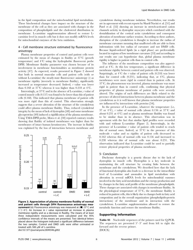
4 - Cell membrane structure estimated by fluorescenceanisotropy
Plasma membrane properties of control and patient cells were
estimated by the mean of changes in fluidity at 37uC (growth
temperature) and 4uC using the hydrophobic fluorescent probe
DPH. Membrane fluidity parameter was chosen because of its
involvement in membrane functionalities as membrane protein
activity [47]. As expected, results presented in Figure 2 showed
that both in normal muscular cells and patient cells (with or
without L-carnitine) the steady-state fluorescence anisotropy (r) as
membrane rigidity (inversely to membrane fluidity), significantly
increased as temperature decreased. Indeed, r value was lower
than 0.168 at 37uC whereas it was higher than 0.218 at 4uC.
Interestingly, at 37uC and in the absence of L-carnitine, r value of
control muscle cells (0.157) was found to be lower than this of patient
cells (0.168). This indicated that plasma membrane of patient cells
was more rigid than this of control. This observation strongly
suggests that a severe alteration of the structure of the cytoskeleton
could affect plasma membrane fluidity. Thus, reduced cytoskeletal
integrity by the mean of lack of dystrophin [48] and some membrane
glycoproteins [49] induced a rigidification of the plasma membrane.
Using TMA-DPH probe, Mora et al. [50] reported contrary results
showing that fluidity of isolated membranes was higher than for
membranes of intact human keratinocytes. The increase in fluidity
was explained by the loss of interactions between membrane and
cytoskeleton during membrane isolation. Nevertheless, our results
are in agreement with recent reports by Sharif-Naeini et al. [51] and
Patel et al. [52] showing an increase in stretch-activated cation
channels activity in dystrophic myocytes which correlated with the
destabilization of the cortical actin cytoskeleton and consequent
alteration of membrane surface tension. According to these authors,
disruption of the cytoskeleton is thought to increase the radius of
membrane curvature meaning that unaffected cells are able to form
indentations with low radius of curvature and not DMD cells.
Because liquid-ordered lipids (in a rigid phase) are preferentially
located in regions of low membrane curvature [53] and according to
our results one can hypothesize that, global plasma membrane
rigidity is higher in patient cells than in control cells.
The influence of the membrane composition was also appreci-
ated at 4uC. At this low temperature, a significant difference in
membrane rigidity between control and patient cells was observed.
Surprisingly, at 4uC the r value of patient cells (0.218) was lower
than for control cells (0.231), indicating that, at 4uC, plasma
membranes were more rigid in control than in patient cells. At
37uC, the phenomenon was inverted, plasma membrane was more
rigid in patient than in control cells, confirming that physical
properties of plasma membrane of patient cells were severely
altered. The origins of such differences are likely related to the
presence of altered interactions between plasma membrane and the
cytoskeleton as it is well known that plasma membrane properties
are influenced by interactions with proteins [54].
In the presence of L-carnitine, whatever the temperature (i.e,
37 or 4uC), r value of normal cells (about 0.156 at 37uC and
about 0.231 at 4uC) and so plasma membrane fluidity was found
to be similar than in its absence. This observation was in
agreement with the fact that similar lipid profiles were recorded
with and without L-carnitine (Figure 2). Interestingly, in the
presence of L-carnitine r value of patient cells tended to reach
this of normal ones. Indeed, at 37uC in the presence of this
molecule r value and so rigidity of patient cells decreased to
0.162 whereas this of normal cells was 0.156 and increased to
0.238 whereas this of normal cells was about 0.231. This
observation indicated that L-carnitine could be used to partially
restore physical properties of plasma membrane.
5- ConclusionDuchenne dystrophy is a genetic disease due to the lack of
dystrophin in muscle cells. Dystrophin is a key molecule in
maintaining the cell structure by creating links between the
membrane and the cytoskeleton. We described here that the lack
of functional dystrophin also leads to a decrease in the intracellular
level of L-carnitine and anomalies in lipid metabolism with
alteration in several mRNA levels for mitochondrial enzymes
involved in the fatty acid metabolism and the energy production and
by changes in the fatty acid profile of the membrane phospholipids.
These changes are associated with changes in membrane fluidity. At
the physiological temperature of 37uC, the membrane fluidity is
reduced in patient cells, this is likely due to changes both in fatty acid
composition and in the overall organization (phospholipids-protein
interactions) of the membrane and its interaction with the
cytoskeleton. L-carnitine supplementation allowed to restore the
metabolic function and restore the membrane fluidity.
Supporting Information
Table S1 Nucleotide sequences of the primers used for Q-PCR.
The sequences are presented 59–39 and from left to right the
forward and the reverse primer.
(DOCX)
Figure 2. Appreciation of plasma membrane fluidity of normaland patient cells through DPH fluorescence anisotropy mea-surement (r). Fluorescence anisotropy was measured at (&) 37uC and(%) 4uC. An Increase in r value represented an increase in plasmamembrane rigidity and so a decrease in fluidity. The means of at leastthree independent measurements were calculated and the 95%confidence intervals of the means are presented. Two identical lettersplaced above the histogram indicated a significant difference betweenthe two samples. Control and DMD cells were either untreated ortreated with 500 mM of L-carnitine.doi:10.1371/journal.pone.0049346.g002
Lipid Metabolism in DMD Cells
PLOS ONE | www.plosone.org 6 November 2012 | Volume 7 | Issue 11 | e49346

Table S2 Fatty acid profile of the PL extracted from membranes
of control and patient cells. The content for each fatty acid was
determined and expressed in percent of the total amount of fatty
acids. Those values represent the raw data used for making
Table 1. Each number is the average of 7 independent
experiments 6 sem.
(DOCX)
Acknowledgments
The authors wish to thank the Plateau Technique Imagerie/Spectroscopie
which is a part of DimaCell platform (Universite de Bourgogne, Dijon,
France) and the Plateau Technique Lipidomique (IFR100 Sante-STIC).
Author Contributions
Conceived and designed the experiments: FLB SG ML LB PG JD.
Performed the experiments: FLB SG ML LB PG JD. Analyzed the data:
FLB SG ML LB PG JD. Wrote the paper: FLB SG ML LB PG JD.
References
1. Blake DJ, Weir A, Newey SE, Davies KE (2002) Function and genetics ofdystrophin and dystrophin-related proteins in muscle. Physiol Rev 82: 291–329.
2. Ervasti JM (2003) Costameres: the Achilles’ heel of Herculean muscle. J Biol
Chem 278: 13591–13594.
3. Deconinck N, Dan B (2007) Pathophysiology of duchenne muscular dystrophy:
current hypotheses. Pediatr Neurol 36: 1–7.
4. Mouly V, Aamiri A, Perie S, Mamchaoui K, Barani A, et al. (2005) Myoblast
transfer therapy: is there any light at the end of the tunnel? Acta Myol 24: 128–133.
5. Negroni E, Butler-Browne GS, Mouly V (2006) Myogenic stem cells:regeneration and cell therapy in human skeletal muscle. Pathol Biol (Paris) 54:
100–108.
6. Shi X, Garry DJ (2006) Muscle stem cells in development, regeneration, and
disease. Genes Dev 20: 1692–1708.
7. Muir LA, Chamberlain JS (2009) Emerging strategies for cell and gene therapy
of the muscular dystrophies. Expert Rev Mol Med 11: e18.
8. Chamberlain JS (2002) Gene therapy of muscular dystrophy. Hum Mol Genet
11: 2355–2362.
9. Kapsa R, Kornberg AJ, Byrne E (2003) Novel therapies for Duchenne muscular
dystrophy. Lancet Neurol 2: 299–310.
10. Demarquoy J, Rigault C, Le Borgne F (2010) ‘‘L-carnitine’’. Handbook of
Analysis of Active Compounds in Functional Foods (CRCPress - Francis &Taylor Group USA): In press.
11. Rigault C, Le Borgne F, Demarquoy J (2006) Genomic structure, alternative
maturation and tissue expression of the human BBOX1 gene. Biochim Biophys
Acta 1761: 1469–1481.
12. Berthillier G, Eichenberger D, Carrier HN, Guibaud P, Got R (1982) Carnitine
metabolism in early stages of Duchenne muscular dystrophy. Clin Chim Acta122: 369–375.
13. Camina F, Novo-Rodriguez MI, Rodriguez-Segade S, Castro-Gago M (1995)Purine and carnitine metabolism in muscle of patients with Duchenne muscular
dystrophy. Clin Chim Acta 243: 151–164.
14. Sharma U, Atri S, Sharma MC, Sarkar C, Jagannathan NR (2003) Skeletal
muscle metabolism in Duchenne muscular dystrophy (DMD): an in-vitro protonNMR spectroscopy study. Magn Reson Imaging 21: 145–153.
15. Piperi C, Papapanagiotou A, Kalofoutis C, Zisaki K, Michalaki V, et al. (2004)Altered long chain fatty acids composition in Duchenne muscular dystrophy
erythrocytes. In Vivo 18: 799–802.
16. Carroll JE, Villadiego A, Brooke MH (1983) Increased long chain acyl CoA in
Duchenne muscular dystrophy. Neurology 33: 1507–1510.
17. Kawashima H, Watanabe K, Morishima Y, Ioi H, Kashiwagi Y, et al. (2012)
High concentration of middle chain fatty acid in a case of Duchenne musculardystrophy with severe mental retardation. Pediatr Int 54: 137–140.
18. Benabdellah F, Yu H, Brunelle A, Laprevote O, De La Porte S (2009) MALDI
reveals membrane lipid profile reversion in MDX mice. Neurobiol Dis 36: 252–
258.
19. Touboul D, Piednoel H, Voisin V, De La Porte S, Brunelle A, et al. (2004)
Changes of phospholipid composition within the dystrophic muscle by matrix-assisted laser desorption/ionization mass spectrometry and mass spectrometry
imaging. Eur J Mass Spectrom (Chichester, Eng) 10: 657–664.
20. Even PC, Decrouy A, Chinet A (1994) Defective regulation of energy
metabolism in mdx-mouse skeletal muscles. Biochem J 304 (Pt 2): 649–654.
21. Tuazon MA, Henderson GC (2012) Fatty acid profile of skeletal muscle
phospholipid is altered in mdx mice and is predictive of disease markers.Metabolism 61: 801–811.
22. Khairallah M, Khairallah R, Young ME, Dyck JR, Petrof BJ, et al. (2007)
Metabolic and signaling alterations in dystrophin-deficient hearts precede overt
cardiomyopathy. J Mol Cell Cardiol 43: 119–129.
23. Rowland LP (1980) Biochemistry of muscle membranes in Duchenne muscular
dystrophy. Muscle Nerve 3: 3–20.
24. Spector AA, Yorek MA (1985) Membrane lipid composition and cellularfunction. J Lipid Res 26: 1015–1035.
25. Stubbs CD, Smith AD (1984) The modification of mammalian membranepolyunsaturated fatty acid composition in relation to membrane fluidity and
function. Biochim Biophys Acta 779: 89–137.
26. Chabanel A, Spiro A, Schachter D, Chien S (1986) Some biophysical properties
of the erythrocyte membrane in Duchenne muscular dystrophy. J Neurol Sci 76:131–142.
27. Hubner C, Kohlschutter A, Gartner J (1987) Membrane fluidity of nonmusclecells in Duchenne muscular dystrophy: effect on lymphocyte membranes of
incubation in patient and control sera. Pediatr Res 22: 488–492.
28. Demarquoy J, Georges B, Rigault C, Royer M, Clairet A, et al. (2004)
Radioisotopic determination of -carnitine content in foods commonly eaten in
Western countries. Food Chemistry 86: 137–142.
29. Georges B, Le Borgne F, Galland S, Isoir M, Ecosse D, et al. (2000) Carnitine
transport into muscular cells. Inhibition of transport and cell growth bymildronate. Biochem Pharmacol 59: 1357–1363.
30. Le Borgne F, Ben Mohamed A, Logerot M, Garnier E, Demarquoy J (2011)Changes in carnitine octanoyltransferase activity induce alteration in fatty acid
metabolism. Biochem Biophys Res Commun 409: 699–704.
31. Folch J, Lees M, Sloane Stanley GH (1957) A simple method for the isolationand purification of total lipides from animal tissues. J Biol Chem 226: 497–509.
32. Pfaffl MW, Horgan GW, Dempfle L (2002) Relative expression software tool(REST) for group-wise comparison and statistical analysis of relative expression
results in real-time PCR. Nucleic Acids Res 30: e36.
33. Tamai I (2012) Pharmacological and pathophysiological roles of carnitine/
organic cation transporters (OCTNs: SLC22A4, SLC22A5 and Slc22a21).
Biopharm Drug Dispos.
34. Shumate JB, Carroll JE, Brooke MH, Choksi RM (1982) Palmitate oxidation in
human muscle: comparison to CPT and carnitine. Muscle Nerve 5: 226–231.
35. Imbert N, Cognard C, Duport G, Guillou C, Raymond G (1995) Abnormal
calcium homeostasis in Duchenne muscular dystrophy myotubes contracting in
vitro. Cell Calcium 18: 177–186.
36. Cardenas C, Juretic N, Bevilacqua JA, Garcia IE, Figueroa R, et al. (2010)
Abnormal distribution of inositol 1,4,5-trisphosphate receptors in human musclecan be related to altered calcium signals and gene expression in Duchenne
dystrophy-derived cells. FASEB J 24: 3210–3221.
37. Spiekerkoetter U, Wood PA (2010) Mitochondrial fatty acid oxidation disorders:
pathophysiological studies in mouse models. J Inherit Metab Dis 33: 539–546.
38. Nishio H, Wada H, Matsuo T, Horikawa H, Takahashi K, et al. (1990) Glucose,free fatty acid and ketone body metabolism in Duchenne muscular dystrophy.
Brain Dev 12: 390–402.
39. Broad EM, Maughan RJ, Galloway SD (2011) Effects of exercise intensity and
altered substrate availability on cardiovascular and metabolic responses toexercise after oral carnitine supplementation in athletes. Int J Sport Nutr Exerc
Metab 21: 385–397.
40. Reuter SE, Evans AM (2012) Carnitine and acylcarnitines: pharmacokinetic,pharmacological and clinical aspects. Clin Pharmacokinet 51: 553–572.
41. Fischbeck KH, Bonilla E, Schotland DL (1983) Freeze-fracture analysis ofplasma membrane cholesterol in Duchenne muscle. Ann Neurol 13: 532–535.
42. Tahallah N, Brunelle A, De La Porte S, Laprevote O (2008) Lipid mapping in
human dystrophic muscle by cluster-time-of-flight secondary ion massspectrometry imaging. J Lipid Res 49: 438–454.
43. Bastiaanse EM, Hold KM, Van der Laarse A (1997) The effect of membranecholesterol content on ion transport processes in plasma membranes. Cardiovasc
Res 33: 272–283.
44. Lamhonwah AM, Tein I (2006) Novel localization of OCTN1, an organic
cation/carnitine transporter, to mammalian mitochondria. Biochem Biophys
Res Commun 345: 1315–1325.
45. Guevel L, Lavoie JR, Perez-Iratxeta C, Rouger K, Dubreil L, et al. (2011)
Quantitative proteomic analysis of dystrophic dog muscle. J Proteome Res 10:2465–2478.
46. Poulos A (1995) Very long chain fatty acids in higher animals–a review. Lipids30: 1–14.
47. Beney L, Gervais P (2001) Influence of the fluidity of the membrane on the
response of microorganisms to environmental stresses. Applied Microbiologyand Biotechnology 57: 34–42.
48. Koenig M, Monaco AP, Kunkel LM (1988) The complete sequence ofdystrophin predicts a rod-shaped cytoskeletal protein. Cell 53: 219–228.
49. Ervasti JM, Ohlendieck K, Kahl SD, Gaver MG, Campbell KP (1990)Deficiency of a glycoprotein component of the dystrophin complex in dystrophic
muscle. Nature 345: 315–319.
50. Mora MP, Tourne-Peteilh C, Charveron M, Fabre B, Milon A, et al. (1999)Optimisation of plant sterols incorporation in human keratinocyte plasma
membrane and modulation of membrane fluidity. Chemistry and Physics ofLipids 101: 255–265.
Lipid Metabolism in DMD Cells
PLOS ONE | www.plosone.org 7 November 2012 | Volume 7 | Issue 11 | e49346

51. Sharif-Naeini R, Folgering JH, Bichet D, Duprat F, Lauritzen I, et al. (2009)
Polycystin-1 and -2 dosage regulates pressure sensing. Cell 139: 587–596.52. Patel A, Sharif-Naeini R, Folgering JR, Bichet D, Duprat F, et al. (2010)
Canonical TRP channels and mechanotransduction: from physiology to disease
states. Pflugers Archiv - European Journal of Physiology 460: 571–581.
53. Parthasarathy R, Yu CH, Groves JT (2006) Curvature-modulated phase
separation in lipid bilayer membranes. Langmuir 22: 5095–5099.54. Lee DC, Chapman D (1987) The effects of temperature on biological
membranes and their models. Symposia of the Society for Experimental Biology
41: 35–52.
Lipid Metabolism in DMD Cells
PLOS ONE | www.plosone.org 8 November 2012 | Volume 7 | Issue 11 | e49346