journal of the egyptian society of cardio-thoracic …4-2005.pdfjournal of the egyptian society of...
TRANSCRIPT

Journal of the
Egyptian Society of
Cardio-thoracic SurgeryEDITOR-IN-CHIEF
Ezzeldin A. Mostafa, MD
PAST EDITORSHassouna M. El-sabea, FRCS (1995-1996)
Mohamed S. El-fiky, MD (1997-2004)
CO-EDITORYasser M. Hegazy, FRCS
STATISTICS EDITOR Ahmed A. Hassouna, MD
ETHICS EDITORM. Anwar Balbaa, MD
ASSOCIATE (SECTION) EDITORSAhmed M. Deebis, MD
Ibrahim M. Abdel Meguid, MDMohamed A. Nasser, MD
Samir A. Hassan, MDSamir A. Keshk, MD
Website & Managing EditorMohamed A. Othman, MS
Submit Manuscripts: Editorial office Journal of the Egyptian Society of Cardio-Thoracic Surgery
330 El Sudan Street, Embaba , EgyptTel. (+ 202) 303 8054
Website: www.arabmedics.com/jescts.htmlEmail : [email protected]


A3The Journal of Egyptian Society of Cardiothoracic Surgery ● Volum 13, Number (3-4)
Abdel Rahman A Fahmy , Cairo , EgyptAbdel Fattah A. Abid ,Tunis , TunisiaAmal Ayoub, Cairo, EgyptAhmed M. Amin, Cairo, EgyptAhmed M. Ali, Banha, EgyptAhmed R. Nasr, Cairo, EgyptA. Samir El-Kosheiry , Cairo , EgyptAli S. Maklad , Cairo , EgyptM. Ayman A Soieb, Cairo, EgyptMamdoud A. Sharawi,Zagazig,EgyptAhmed El-Kerdani, Cairo, EgyptAlradi Kamal, Zagazig, EgyptBabulal Sethia, London, EnglandBertrand M. Goudot, Paris, FranceB Ben-Ismail , Tunis , TunisiaB M Fabri , Liverpool , EnglandBryn T Williams, Weybridge, EnglandDaniel G. Guilmet, Paris, FranceDavid J. Wheatley, Glasgow, EnglandEl Nouri Ahmed , Cairo , EgyptEl Hussieiny Gamil , Cairo , EgyptFawzi Estefanos , Cleveland , USAFouad Z Abdalla , Cairo , EgyptGerard Block, Paris, FranceGamal O. Abou Senna , Cairo , EgyptGraham E. Venn, London, EnglandHasan Alzahrani, Mekka, Saudi ArabiaHussein A. Gaafar, Cairo, EgyptHamdy M. El-Sayed, Cairo , EgyptHassan Ezzeldin Attia, Cairo , EgyptHamed M. Al Akshar , Tanta , EgyptHisham A. Sawki, Cairo , EgyptIsmail A. Sallam , Cairo , EgyptIbrahim Haggag, Cairo , EgyptJames J. Pollock, Glasgow, England
Jean E. Bachet, Paris, FranceJean-Paul F. Bessou, Rouen, FranceJohn R. Pepper , London , EnglandLotfi Eissa, Cairo , EgyptMohamed A. Hamed, Cairo , EgyptMohamed Abou El-Ezz, Cairo , EgyptMostafa Agha, Alexandria, EgyptMohamed F. Bassiouni , Cairo , EgyptMarc de Leval , London , EnglandM El-Fakih , Riadh , Saudi ArabiaMamdouh Gamal , Einthoven, HollandM. Ezzeldin Abdel Raouf ,Cairo,EgyptMaher Fourati, Tunis, TunisiaMagdi Gomaa , Cairo , EgyptMohamed S El-Fiky, Cairo, EgyptMarco Pozzi, Liverpool, EnglandM S Ammar, Tunis, TunisiaMaher Shoier, Cairo, EgyptMogazy A. Tantawy, Cairo, EgyptMedhat A. El-Gamal, Cairo , EgyptMostafa M. Radwan , Cairo , EgyptNahed Attia , Assiout , EgyptPierre Michel Roux, Metz, FranceRobert M. Soyer, Rouen, FranceSherif Abdel Hady , Cairo , EgyptShaaban Abu Elelaa , Mansoura , EgyptSamieh A Amer , Cairo , EgyptSami S. Kabbani , Damascus , SyriaSamir Mahmoudi , Cairo , EgyptSteven Tsui , Cambridge , EnglandTarek Z. Shallaby Cairo , EgyptWadih R. Dimitri, Birmingham, EnglandWahid Osman , Cairo , EgyptZohair Al-Halees, Riyadh, Saudi ArabiaZohni M. Farrag , London , England
EDITORIAL BOARD
Production EditorHesham O. Saied
Managing EditorMohamad A. Othman
Journal Secretary A A Kalifa


A5The Journal of Egyptian Society of Cardiothoracic Surgery ● Volum 13, Number (3-4)
The Society Board of Directors2004-2006
THE EGYPTIAN SOCIETY OF
CARDIO-THORACIC SURGERYPresident
Samieh A. Amer, MD Vice President
Samir A. Keshk, MD
General SecretaryM. Magdy Mostafa Ali , MD
TreasurerLotfi M. Eissa , MD
Immediate Past PresidentMohamed F. Bassiouni , MD
BoardAhmed B. Elkerdani, MDAhmed M. Deebis, MD
Ezzeldin A. Mostafa, MDM. Ezzeldin Abdel Raouf, MDM. Mamhouh A. Sharawi, MD
M. Mostafa A. Agha, MDMedhat A. El Gamal, MDMohamed A. Nasser, MDMostafa M. Radwan, MD
Samir A. Hasan, MDYasser M. Hegazy, MD


A7The Journal of Egyptian Society of Cardiothoracic Surgery ● Volum 13, Number (3-4)
CONTENTS
ANNOUCEMENT A8 Guidelines for authorsA14 Condition for publication form A16 Guidelines for reviewersA18 Events of interests
EDITORIAL1 Editorial Letter Ezzeldin A. Mostafa, MD5 Obituary Mohamed Nasr, MD
STATISTICS
6 Statistics for Clinicians: (2) The Normal Distribution and the Intervals of
Confidence. Ahmed A. Hassouna, MD
CARDIOVASCULAR11 Does retrograde crystalloid cardioplegia offer
additional protection against Ischemia and Oxi-dative Stress in Coronary Bypass Surgeries?Magued A. Zikri, MD, Saed Abdel Aziz, MD, Amr M. Roushdi, MD, Walid Abusenna, MD, Sameh S. Marzouk, MD *, Ahmed S. Ahmed, MD *
20 Systemic Normothermic Versus Hypothermic Cardiopulmonary Bypass in Mitral Valve Re-placement Hossam F. El Shahawy, MD, Mohamed Attia, MD, Hassan Moftah, MD, Hany Abd El Maboud, MD, Mohamed M. El-Fiky, MD , M. Ayman Shoeb, MD
26 Harvesting of the Radial Artery for Coronary Artery Bypass Grafting: Comparison of Ultra-sonic Harmonic Scalpel Dissector with the Con-ventional Technique.Hosam F. Fawzy, MD
31 Valve Sparing Operations For Type A Aortic Dis-section: Initial Experience And Early ResultsAmr Mohamed Rushdi, MD Tarek Hussein El-Taweel, MD Mohamed Helmi, MD , Saed Abdelaziz Badr, MD , Ahmed Helmi, MD
Journal of The Egyptian Society of Cardio-Thoracic Surgery
Volume 13 March-June 2005 Number 1 ISSN 1110-578X
37 Impact of Single Clamp Technique :an Important Adjunct to Myocardial and Cerebral Protection in Coronary OperationHany A. Maboud, MD
42 Pathological Changes by the Effect of Ultrasonic and Electrocautery Harvesting Procedures on the Internal Thoracic Artery EndotheliumHossam F El Shahawy, MD, Ahmed Badawy, MB-BCH, MS Hisham Abd El Rahman, MD, Abdel Salam El Henawi , MD, Sherif Azab, MD, Ezzeldin A. Mostafa, MD
49 Ascending Aortic Surgery : Multi-centre Study In EgyptWael AbdelAziz AbdelHameed MD Gamal Sami, MD, Reda Ahmed AbulMaaty, MD, Bahaa Badry, AbdelHakam M.Sc Magdy Mammdouh, MD, Sameh Ibrahim Sersar, MD
THORACIC56 Empyema Thoracis. Outcome of 181 Patients
Ahmed El Nouri, MD, Hatem Yazid, MD, Tarek Ab-del Aziz, MD, Mohamed Atia, MD, Mohamed Abdel Fattah. MD, Mostafa Abdel Azeem, MD
62 Surgical Treatment of Hydatid Disease of the LungHossam El Okda1, MD, Ahmed El Nori1, MD, Mohammed Attia1, MD, Nashwa I. Ramadan2 and Heba E. Abdel Aaty2, MD,
THE WAY I DO IT70 An Easy Way to Band and to to Deband the Pul-
monary ArteryEzzeldin A. Mostafa, MD
CTS NOTES 72
CTS QUIZ73
READERS’ CORNER74


A9The Journal of Egyptian Society of Cardiothoracic Surgery ● Volum 13, Number (3-4)
Editorial Office Please address all correspondence to:Ezzeldin A. Mostafa, MD, Editor, In-chiefJournal of the Egyptian Society of Cardio-thoracic Surgery 330 El-Sudan St., Imbaba, Cairo, Egypt.Telephone: (+202) 303 6634Fax: (+202) 303 8054E-Mail: [email protected]
The Journal of the Egyptian Society of Cardio-Thoracic Surgery [ISSN 1110-578 X] is the official publication of the Egyptian Society of Cardio-thoracic Surgery. The journal is published every three months .
General Instructions
Every submission must include: Cover letter, indicating the category of article , the Complete manuscript, including title page, abstract, text, tables, ac-knowledgments ,references and illustrations .
Required Disclosures;
A. Conditions for Publication Form which includes dis-closures regarding freedom of investigation and conflicts of interest, signed by all authors. In single Author publication an additional Senior Consultant Signature is required.B. Written permission from the publisher (copyright holder) is required to reproduce any previously published table(s), illustration(s) or photograph(s) in both print and electronic media. C. Written permission from unmasked patients appearing in photographs is also required.
Revised_Manuscripts:Revised manuscripts must be submitted in three parts as Microsoft word-processing files : (1) cover letter with responses to reviewers’ comments (2) revised, marked manuscript showing additions and deletions; (3) revised, un-marked manuscript.
General Information Three copies of the Manuscripts should be sent preferably
prepared in Microsoft Word , typed double-spaced throughout (including title page, abstract, text, references, tables and legends) with one (1) inch (2.5 cm) margins all around. Place Author name and page number in the upper right corner of each page. Manuscripts written in 12 point Arial or Times New Roman fonts are preferred (Note: Do not submit your manuscript in PDF format it causes problems in processing your submis-sion.)Arrange manuscript as follows: (1) title page, (2) abstract, (3) text, (4) acknowledgments, (5) disclosures if required, (6) references, (7) tables and (8) legends. Number pages consecu-tively, beginning with the title page as page 1 and ending with the legend page.If your manuscript contains illustrations, in addition to submit-ting them online, you must send two sets of original illustra-tions to the editorial office labeled with manuscript number, first author, and figure number on back. Tables and figures should be provided separate from the text while there position in the text should be marked on the manu-script.
Word Limits by Category of Manuscript
Original articles should not exceed 4500 words including title page, abstract of 150-200 words, text, figure legends and refer-ences. The combined total of illustrations and tables should not exceed 10 and the number of references should not exceed 40.
Case reports and “The way I do it” articles are limited to a total of 1500 words including title page, abstract, text, refer-ences and figure legends. For each illustration subtract 100 words and for each table subtract 300 words from the word limit. References are limited to eight. A “how to do it” article should be a description of a useful surgical technique and con-tain descriptive, illustrative material.
Images in cardiothoracic surgery are limited to 350 words including title and text and to two, possibly three figures. The entire contribution must fit on one printed page .
Review articles are limited to 6500 words including title page, abstract, text, figure legends and all references. The total number of references should not exceed 80. Subtract 100
Guidelines for Authors
Journal of The Egyptian Society of Cardio-Thoracic Surgery (J. Egypt. Soc. Cardiothorac. Surg.)
A10 The Journal of Egyptian Society of Cardiothoracic Surgery ● Sep-Dec 2005 A11The Journal of Egyptian Society of Cardiothoracic Surgery ● Volum 13, Number (3-4)
words for each illustration and 300 words for each table.
Our surgical heritage articles are limited to 2500 words in-cluding title page, abstract, text, figure legends and references. Subtract 100 words for each illustration and 300 words for each table.
Correspondence (Letters to the Editor) and commentaries are limited to 500 words. Subtract 100 words for each illustration and 300 words for each table.
Editorials are limited to 2500 words including references. Subtract 100 words for each illustration and 300 words for each table.
Manuscript Preparation
Title Page (first page)
The title is limited to 100 characters and spaces for original manuscripts and to 80 characters and spaces for all other cat-egories of manuscripts. The title may not contain acronyms or abbreviations. All submissions, must have a title.
Running Head. Supply a short title of 40 characters and spaces.
Authors. List all authors by first name, all initials, family name and highest academic degree using “MD, PhD” for hold-ers of both degrees ( if more then 7 Authors justifie).
Institution and Affiliations. List the name and full address of all institutions where the work was done. List departmental affiliations of each author affiliated with that institution after each institutional address.
Meeting Presentation. If the paper has been or is to be pre-sented at the annual meeting of The Society, provide the name, location and dates of the meeting.
Keywords. Provide up to 5 keywords selected from the ap-pended list to describe the manuscript. Do not use any key-words that are not on the list.
Word Count. Provide the electronic total word count of the entire manuscript including title page, abstract,text,figure leg-ends and entire reference list.
Corresponding Author. Provide the name, exact postal ad-dress with postal code, telephone number, fax number and e-mail address of the author to whom communications, proofs and requests for reprints should be sent.
Abstract Page (Second page)
Original articlesProvide a structured Abstract, no longer than 250 words, di-vided into four sections: Background or Objective, Methods, Results, Conclusions. Avoid abbreviations and acronyms. In-
dicate the abstract word count below the abstract.
Case reports, “the way i do it” articles, review articles and our surgical heritage articles. Provide an unstructured abstract of 100 words.
Images, correspondence, commentaries, editorials and up-dates. No abstract is required.
Text Text should be organized as follows: Introduction, Mate-rial (or Patients) and Methods, Results, and Comment.Cite references,illustrations and tables in numeric order by order of mention in the text.
Avoid abbreviations. Consult the American Medical Associa-tion Manual of Style, 9th edition, for recommended abbrevia-tions. Define abbreviations at first appearance in the text. If 8 or more abbreviations or acronyms are used, provide a separate table of abbreviations and acronyms.
Measurements and weights should be given in standard metric units.Statistical nomenclature and data analysis. Fol-low the “Guidelines for Data Reporting and Nomenclature” published in The Annals of Thoracic Surgery (1988;46:260-1). Footnotes. Type footnotes at the bottom of the manuscript page on which they are cited. Suppliers of drugs, equipment and other brand mentioned in the article within parentheses , giving company name, city and country .
AcknowledgmentsGrants, financial support and technical or other assistance must be acknowledged at the end of the text before the references.
ReferencesIdentify references in the text using Arabic numerals in brack-ets on the line.Type references double-spaced after text or acknowl-edgments beginning on a separate sheet. Number con-secutively in the order in which they appear in the text.Journal references should provide inclusive page num-bers; book references should cite specific page numbers.Journal abbreviations should conform to those used in Index Medicus. follow the formats outlined below:
Journal ArticleJones DR, Stiles BM, Denlinger CE, Antie P . Pulmonary segmentectomy: results and complications. Ann Thorac Surg 2000;76:343-9.(List all authors if 6 or fewer; otherwise list first 3 and add “et al.”)
Chapter in Book12. Vinten-Johansen J, Zhao Z-Q, Guyton RA. Cardiac surgi-cal physiology. In: Cohn LH, Edmunds LH Jr, eds. Cardiac Surgery in the Adult. 2nd ed. New York, NY: McGraw-Hill; 2003:53-84.

A10 The Journal of Egyptian Society of Cardiothoracic Surgery ● Sep-Dec 2005 A11The Journal of Egyptian Society of Cardiothoracic Surgery ● Volum 13, Number (3-4)
Internet Address3. 1996 NRC Guide for the Care and Use of Laboratory Ani-mals. Available at: http://www.nap.edu/readingroom/books/labrats/contents.html. Accessed October 20, 2003.
Tables :Tables should be typewritten double-spaced on separate sheets (one to each page). Do not use vertical lines. Each table should be numbered (Arabic) and have a title above. Legends and explanatory notes should be placed below the table. Abbrevia-tions used in the table follow the legend in alphabetic order. Lower case letter superscripts beginning with “a” and follow-ing in alphabetic order are used for notations of within-group and between-group statistical probabilities.
FigureLegends :Figure Legends should be numbered (Arabic) and typed double-spaced in order of appearance beginning on a sepa-rate sheet. Identify (in alphabetical order) all abbreviations appearing in the illustrations at the end of each legend. Cite the source of previously published material in the legend and indicate permission has been obtained. Proof of permis-sion must be surface mailed or faxed to the editor .
Illustrations :You must send two sets of original illustrations to the editorial office labeled with manuscript number, first author, and figure number on back.
Images or figures are submitted online as one or more separate files that may contain one or more images. Within each file containing images, use the figure number (eg, Figure 1A) as the image filename. The system accepts Powerpoint (.ppt) files Most illustrations will be reproduced at a width of one column (8.25 cm; 3 1/4 inches). Black, white and widely crosshatched bars are preferable; do not use stippling, gray fill or thin lines.
Instructions :Identify print proofs of figures on the back with figure number and name of the first author; when necessary, indicate the top with an up arrow For figures submitted in electronic format, all images should be at least 5 inches wide. Graphics software such as Photoshop and Illustrator, should be used to create art. Color images need to be at least 300 dpi.Gray scale images should be at least 300 dpi .Line art should be at least 1200 DPI .
Cover letter :Include with the manuscript a cover letter that provides 1) the category of manuscript (e.g., original research, Brief Commu-nication, Letter to the Editor); 2) statement that the material
has not been previously published or submitted elsewhere for publication; 3) information about any personal conflicts of interest of any of the authors; and 4) names of sources of out-side support for research, including funding, equipment, and drugs .You may also submit the name of one reviewer of your choice. You should include that individual’s mailing address, telephone, fax and e-mail address. Editorial Policies Scientific Responsibility StatementBefore publication of an accepted manuscript, each author is required to certify by signing the Conditions for Publication Form that he or she has participated sufficiently in the work and approved the final version of the manuscript to be pub-lished. Exclusive Publication StatementEach author must certify that none of the material in this manuscript has been published previously in either print or electronic form, and that none of this material is currently under consideration for publication elsewhere. This includes symposia and preliminary publications of any kind except an abstract of 400 words or fewer.
Conflict of Interest :Authors should disclose any conflict of interests. Authors who have a financial relationship with one or more companies whose products are featured in an article will disclose the ex-istence of this relationship in a box at the bottom of the first page of the published article.
Consultant Statistician and Statistical Methods : All manuscripts with statistical analysis are required to undergo biostatistical review .The most appropriate way is to involve a biostatistician consultant or coauthor from the investigators’ home institution . Manuscripts may undergo further biostatistical review by the Journal after submission. Additional information on statistical methods can be found in “Uniform Requirements for Manuscripts Submitted to Biomedical Journals”(www.acponline.org/journals/resource/unifreqr.htm).
Copyright :Authors of articles submitted to The J. Egypt. Soc. Cardiotho-rac. Surg. must transfer copyright to The Egyptian Society of Cardio-Thoracic Surgery by signing the “Conditions for Publi-cation Form.” This transfer becomes binding upon acceptance of the article for publication. No part of the published material may be reproduced elsewhere without written permission.Date of Receipt: The “received for publication” date is the date when the editorial office receives the manuscript, the cover let-ter, and the Copyright Transfer and Author Declaration State-ment, signed by all authors. For Date of acceptance : letter is provided from the editor.
A12 The Journal of Egyptian Society of Cardiothoracic Surgery ● Sep-Dec 2005
Checklist
A] Cover Letter □ Letter to the Editor □ Manuscript category designation .□ Single-journal submission affirmation .□ Conflict of interest statement (if appropriate). □ Sources of outside funding. □ Signed Statistical Collaboration .
B] Complete Manuscript□ Title page .□ Title of article□ Full name(s), academic degrees, and affiliation(s) of authors.□ Corresponding author .□ Telephones, fax, and e-mail address□ Abstract (250 words; double-spaced) .□ Ultramini-abstract (50 words) .□ Text (double-spaced). □ References (double-spaced; separate pages). □ Tables (double-spaced; separate pages). □ Figures (separate files; on hardcopy; properly identified), □ Figure legends (double-spaced; separate pages) .□ Word count.
C] Required Disclosures □ Conditions for Publication Form signed by all authors. Which transfers copyright to The
Egyptian Society of Cardio-Thoracic Surgery□ Written permission from the publisher to reproduce any previously published material .□ Written permission from unmasked patients .
A13The Journal of Egyptian Society of Cardiothoracic Surgery ● Volum 13, Number (3-4)
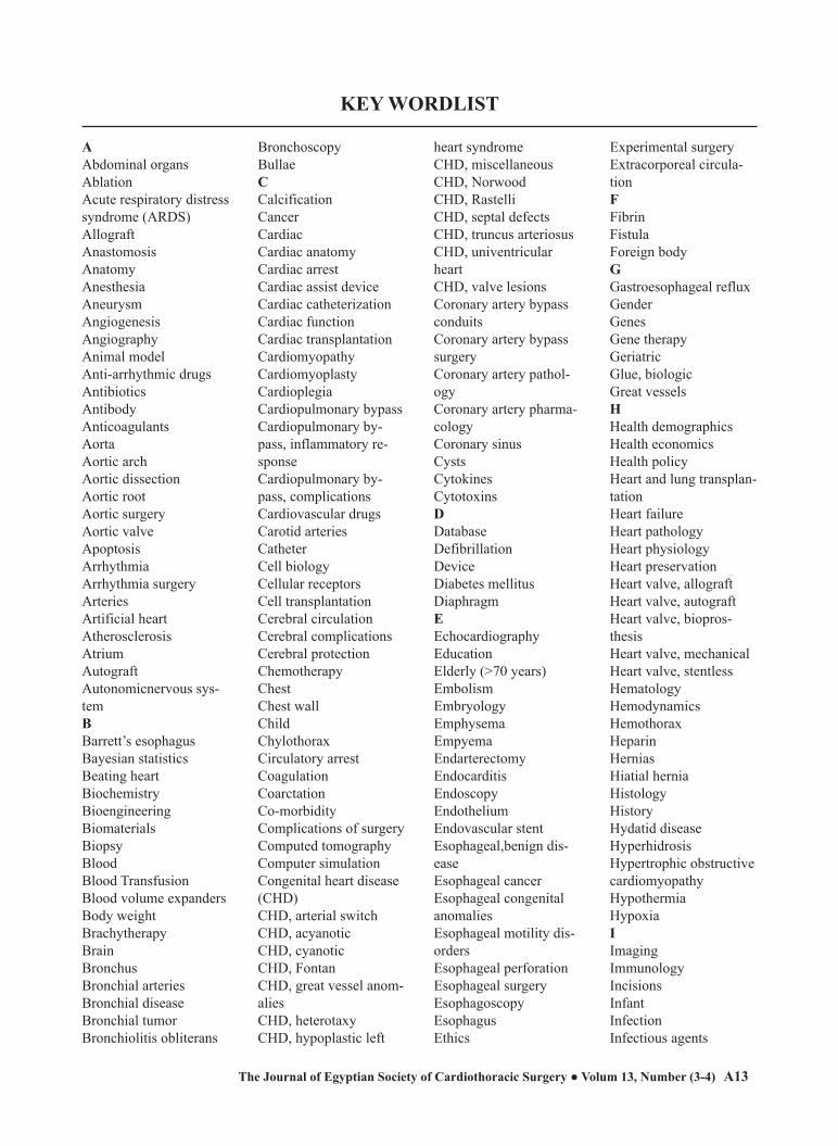
AAbdominal organs Ablation Acute respiratory distress syndrome (ARDS) Allograft Anastomosis Anatomy Anesthesia Aneurysm Angiogenesis Angiography Animal model Anti-arrhythmic drugs Antibiotics Antibody Anticoagulants Aorta Aortic arch Aortic dissection Aortic root Aortic surgery Aortic valve Apoptosis Arrhythmia Arrhythmia surgery Arteries Artificial heart Atherosclerosis Atrium Autograft Autonomicnervous sys-tem BBarrett’s esophagus Bayesian statistics Beating heart Biochemistry Bioengineering Biomaterials Biopsy Blood Blood Transfusion Blood volume expanders Body weight Brachytherapy Brain Bronchus Bronchial arteries Bronchial disease Bronchial tumor Bronchiolitis obliterans
KEY WORDLIST
Bronchoscopy Bullae CCalcification Cancer Cardiac Cardiac anatomy Cardiac arrest Cardiac assist device Cardiac catheterization Cardiac function Cardiac transplantation Cardiomyopathy Cardiomyoplasty Cardioplegia Cardiopulmonary bypass Cardiopulmonary by-pass, inflammatory re-sponse Cardiopulmonary by-pass, complications Cardiovascular drugs Carotid arteries Catheter Cell biology Cellular receptors Cell transplantation Cerebral circulation Cerebral complications Cerebral protection Chemotherapy Chest Chest wall Child Chylothorax Circulatory arrest Coagulation Coarctation Co-morbidity Complications of surgery Computed tomography Computer simulation Congenital heart disease (CHD) CHD, arterial switch CHD, acyanotic CHD, cyanotic CHD, Fontan CHD, great vessel anom-alies CHD, heterotaxy CHD, hypoplastic left
heart syndrome CHD, miscellaneous CHD, Norwood CHD, Rastelli CHD, septal defects CHD, truncus arteriosus CHD, univentricular heart CHD, valve lesions Coronary artery bypass conduits Coronary artery bypass surgery Coronary artery pathol-ogy Coronary artery pharma-cology Coronary sinus Cysts Cytokines Cytotoxins DDatabase Defibrillation Device Diabetes mellitus Diaphragm EEchocardiography Education Elderly (>70 years) Embolism Embryology Emphysema Empyema Endarterectomy Endocarditis Endoscopy Endothelium Endovascular stent Esophageal,benign dis-ease Esophageal cancer Esophageal congenital anomalies Esophageal motility dis-orders Esophageal perforation Esophageal surgery Esophagoscopy Esophagus Ethics
Experimental surgery Extracorporeal circula-tion FFibrin Fistula Foreign body GGastroesophageal reflux Gender Genes Gene therapy Geriatric Glue, biologic Great vessels HHealth demographics Health economics Health policy Heart and lung transplan-tation Heart failure Heart pathology Heart physiology Heart preservation Heart valve, allograft Heart valve, autograft Heart valve, biopros-thesis Heart valve, mechanical Heart valve, stentless Hematology Hemodynamics Hemothorax Heparin Hernias Hiatial hernia Histology History Hydatid disease Hyperhidrosis Hypertrophic obstructive cardiomyopathy Hypothermia Hypoxia IImaging Immunology Incisions Infant Infection Infectious agents

A14 The Journal of Egyptian Society of Cardiothoracic Surgery ● Sep-Dec 2005
Intraoperative care Intubation Ischemia Ischemia/reperfusion Ischemic heart disease Ischemic mitral regurgi-tation KKidney LLarynx Lasers Left ventricular assist device Less invasive surgery Leukocytes Lobectomy Lung Lung cancer Lung cancer, biology Lung cancer, diagnosis and staging Lung cancer, neuroen-docrine Lung cancer surgery Lung, congenital lesions Lung, decortication Lung infection Lung pathology Lung physiology Lung preservation Lung transplantation Lung volume reduction Lymph nodes MMagnetic resonance an-giography Magnetic resonance im-aging Mediastinal disease Mediastinal lymph nodes Mediastinal tumor Mediastinitis Mediastinoscopy Mediastinum Mesothelioma Metabolism Metastasectomy Mitral valve Mitral valve repair Mitral valve replacement Molecular biology
Morbidity Mortality Multiple Valve Surgery Myasthenia gravis Myocardial infarction Myocardial injury Myocardial mechanics Myocardial metabolism Myocardial remodeling Myocardium Myocyte Myxoma NNeonate Neurocognitive deficits Neuroendocrine tumor Neurogenic tumor Neurologic injury Nitric oxide OOff-pump On-pump Outcomes Oxygen PPacemaker Pathology Pathophysiology Pediatric Perfusion Pericardium Peripheral vascular dis-ease Pharmacology Phrenic nerve Physiology Platelets Pleura Pleural effusion Pleural space Pneumothorax Polymerase chain reac-tion (PCR) Positron emission to-mography (PET) Postinfarction cardiac complications Postoperative care Preconditioning Pregnancy Preoperative care Professional affairs
Prognosis Prophylaxis Prostaglandins Prosthesis Pulmonary arteries Pulmonary embolism Pulmonary function Pulmonary valve Pulmonary vascular re-sistance QQuality of life RRadiation therapy Radiofrequency Radiology Regression analysis Regurgitation Rejection Remodeling Reoperation Reperfusion Research Restenosis Resuscitation Retrograde perfusion Revascularization Right ventricle Risk analysis Risk models Robotics Ross operation Rupture SSaphenous vein Sarcoma Shock Shunts Smoking Spinal cord Statistics Stenosis Stents Sternum Stroke Surgery Surgical instruments Survival analysis Suture Sympathectomy TTetralogy of Fallot
Thoracic duct Thoracic outlet Thoracoplasty Thoracoscopy Thoracotomy Thrombosis Thymectomy Thymoma Thymus Tissue engineering Tomography Trachea Tracheal injury Tracheal stenosis Tracheal surgery Tracheal tumor Trauma Trauma, blunt Trauma, penetrating Tricuspid valve Tuberculosis Tumor, benign Tumor, malignant UUltrasound VVagus nerve Valve disease Vascular disease Vascular tone and reac-tivity Video-assisted thoracic surgery (VATS) Veins Venous disease Ventilation Ventricle WWound closure Wound dehiscence Wound healing Wound infection XXenograft X-ray

A15The Journal of Egyptian Society of Cardiothoracic Surgery ● Volum 13, Number (3-4)
This form MUST be completed, signed by ALL authors, and returned to the Editorial Office before your manuscript can be accepted for publication.
Scientific Responsibility Statement:Each author must sign this form to certify that he or she
has participated sufficiently in the work to take responsibility for a meaningful share of the content of the manuscript, and that this participation included: (a) conception or design of the experiment(s), or collection and analysis or interpretation of data; (b) drafting the manuscript or revising its intellectual content; and (c) approval of the final version of the manuscript to be published. In addition, each author must indicate whether or not he or she has had full freedom of investigation; defined as freedom from outside interests in controlling the design of the study, collection, analysis, and interpretation of data, and having freedom to full disclose all results.
Exclusive Publication Statement:Each author must sign this form to certify that none of the
material in this manuscript has been published previously in either print or electronic form, and that none of this material is currently under consideration for publication elsewhere. This includes symposia, transactions, books, articles published by invitation and preliminary publications of any kind except an abstract of 400 words or fewer.
Copyright Transfer Agreement:Each author must sign this form to certify that, if the manu-
script is accepted for publication in the Journal of the Egyptian Society of Cardio-Thoracic Surgery ( JESCTS), copyright (including the right to obtain copyright registration, whether separately or as part of a journal issue .) in and to the above article transfers throughout the world and for the full term and all extensions and renewals thereof to: THE EGYPTIAN SO-CIETY OF CARDIO-THORACIC SURGERY
This transfer includes the right to adapt the article for use in conjunction with computer systems and programs, includ-ing reproductions or publication and incorporation in retrieval systems.
Rights of authors:The ESCTS hereby licenses the following rights back to
the author(s): A. Patent and trademark rights to any process or procedure
described in the article. B. The right to photocopy or make single electronic copies of
the article for their own personal use, including for their
Conditions for Publication Form
own classroom use, or for the personal use of colleagues, provided the copies are not offered for sale .
C.The right, subsequent to publication, to use the article or any part thereof free of charge in a printed compilation of works of their own, such as collected writings or lecture notes.
Note: All copies, paper or electronic, or other use of the informa-
tion must include an indication of The ESCTS copyright and a full citation of the journal source.
Authorship: If copyright is held by the employer, the employer or an
authorized representative of the employer must sign in addi-tion to the author(s).
Warranties: The author(s) warrant that the article is the author’s origi-
nal work and has not been published before. The author(s) war-rant that the article does not infringe on the rights of others. If excerpts from copyrighted works are included, the author(s) has (have) obtained written permission from the copyright owners and will credit the sources in the article.
Preprints: The author(s) warrant(s) that if a prior version of this work
(normally a preprint) has been posted to an electronic server, such version was accessible to only a small group of individu-als and the author(s) will cause its prompt removal from such server.
Conflict of Interest Disclosure Statements: Each author must indicate below that either (a) no financial
conflict of interest exists with any commercial entity whose products are described, reviewed, evaluated or compared in the manuscript, except for that disclosed under “Acknowledgements” or (b) a potential conflict of interest exists with one or more commercial entities whose products are described, reviewed, evaluated or compared in the manuscript through the existence of one or more of the following relationships: the author is a full or part-time employee of a company; has an existing or optional equity interest in a company; owns or partly owns patents licensed to a company; has an ongoing retainer relationship (consultantship, speaker, etc.) with a company for which he/she receives financial remuneration; or has received financial compensation for this publication. If Yes is checked, a box on the first page of the published article will read: ?Dr. X discloses that he/she has a financial relationship with company Y.?

A16 The Journal of Egyptian Society of Cardiothoracic Surgery ● Sep-Dec 2005
If there are additional authors on the article, please photocopy this form and attach additional sheets as need be with appropri-ate information and signatures affixed .
Author: Manuscript Title:
I agree with the preceding conditions and provide the appropriate signatures and information below accordingly:
Author’s Name:_____________________________________________________________________________________Signature:______________________________________________ Date:_______________________________________Author’s employer’s signature, if appropriate: ___________________________________________________________Conflict of interest: Yes ___ No ___ If yes, with which entity: _______________________________________________Did you have freedom of investigation in all aspects of this work?: Yes ___ No ___
Author’s Name:_____________________________________________________________________________________Signature:______________________________________________ Date:_______________________________________Author’s employer’s signature, if appropriate: ___________________________________________________________Conflict of interest: Yes ___ No ___ If yes, with which entity: _______________________________________________Did you have freedom of investigation in all aspects of this work?: Yes ___ No ___
Author’s Name:_____________________________________________________________________________________Signature:______________________________________________ Date:_______________________________________Author’s employer’s signature, if appropriate: ___________________________________________________________Conflict of interest: Yes ___ No ___ If yes, with which entity: _______________________________________________Did you have freedom of investigation in all aspects of this work?: Yes ___ No ___
Author’s Name:_____________________________________________________________________________________Signature:______________________________________________ Date:_______________________________________Author’s employer’s signature, if appropriate: ___________________________________________________________Conflict of interest: Yes ___ No ___ If yes, with which entity: _______________________________________________Did you have freedom of investigation in all aspects of this work?: Yes ___ No ___
Author’s Name:_____________________________________________________________________________________Signature:______________________________________________ Date:_______________________________________Author’s employer’s signature, if appropriate: ___________________________________________________________Conflict of interest: Yes ___ No ___ If yes, with which entity: _______________________________________________Did you have freedom of investigation in all aspects of this work?: Yes ___ No ___
Author’s Name:_____________________________________________________________________________________Signature:______________________________________________ Date:_______________________________________Author’s employer’s signature, if appropriate: ___________________________________________________________Conflict of interest: Yes ___ No ___ If yes, with which entity: _______________________________________________Did you have freedom of investigation in all aspects of this work?: Yes ___ No ___
Author’s Name:_____________________________________________________________________________________Signature:______________________________________________ Date:_______________________________________Author’s employer’s signature, if appropriate: ___________________________________________________________Conflict of interest: Yes ___ No ___ If yes, with which entity: _______________________________________________Did you have freedom of investigation in all aspects of this work?: Yes ___ No ___
A17The Journal of Egyptian Society of Cardiothoracic Surgery ● Volum 13, Number (3-4)
Purpose of Peer ReviewOne is to evaluate objectively the science of the submitted
paper and the other is to provide a constructive critique indicat-ing how the paper could be or could have been improved by the authors. Reviewers should avoid unpleasant comments.
Acceptance of a Manuscript for ReviewReviewers should accept assignments to review manu-
scripts that are within their sphere of expertise, which they plan to review within the 21 day deadline. Reviewers should decline assignments for which a conflict exists between the reviewer and authors or between the reviewer and commercial products that are integral to the content of the article.
Category of the Manuscript The broad categories of papers for which peer review is
undertaken are original scientific articles; new technology papers; case reports, the way i do it articles , images; and re-view articles. The editor and/or associate editors review corre-spondence, invited commentaries, editorials, surgical heritage , ethical and statistical papers.
General Requirements for PublicationThe paper should conform to the format and restrictions
for the category to which it belongs and be written in good, readable English. The paper should address an important or in-teresting subject and provide new and original information. Il-lustrative material should be well chosen and of good quality.
Original Scientific ArticleThe reviewer should assess the articles’ interest to readers;
strengths and weaknesses; originality; clarity of text, tables, illustrations and figure legends; presentation; analysis of re-sults; credibility of results; importance of the findings; depth of scholarship and relationship of the results to the existing lit-erature Ethical issues, such as prior publication of all or part of the data; plagiarism; transgression of human or animal rights; or dishonesty should be noted, if detected.
The following topics are offered to help guide the review-
er’s assessment of an original scientific article. • ‘Title’ should reflect the content of the article and be concise
and clear• ‘Abstract’ should indicate the purpose of the study, subjects
and methods used, most important results and the main con-clusions supported by results.
• ‘Introduction’ should indicate the rationale and focus of the study and state the purpose or hypothesis.
• ‘Methods’ should present the design of the study, fully de-scribe the number and subjects and exclusion and inclusion criteria; whether subjects were enrolled consecutively; meth-ods used to gather data, including follow-up data; methods
Guidelines for Reviewersby which control and experimental groups were assembled; the primary outcome variable; secondary outcome variables; how outcome measurements were made and validated; the statistical design of the study; and the statistical methods used to analyze the study.
• ‘Results’ should concisely present the most important find-ings in text . Data should be reported as means or medians with appropriate indicators of variance and exact p values in tables and text. Figures should be well selected to high-light important findings . Survival and event curves should indicate specified confidence limits or subjects at risk. Re-gression diagrams should include the regression equations, regression coefficient and exact p value in the figure legend. Figure legends should adequately and clearly describe the important information illustrated.
• ‘Comment’ should not repeat results, but should point out the significance and conclusions of the new data, integrate the authors’ new data with that in the prior literature, draw inferences and conclusions regarding the question or purpose addressed by the study and point out the limitations of the study. The ‘Comment’ section should not be a review of the literature.
• References should be properly cited, reasonably current, ac-curateand in proper format.
New TechnologyArticles describing new technology are necessarily de-
scriptive and do not pose or test a hypothesis. These articles evaluate new devices, systems, monitors, implantable mate-rial and similar technology designed for improving patient care and outcomes. The reviewer is asked to evaluate the efficacy, safety and indications of the new technology .
The reviewer needs to inspect the ‘Disclosure statement’ after the text, before References. This statement should dis-close the source of funds used for the evaluation study and whether or not the product was purchased, borrowed or do-nated by the manufacturer or inventor. Conflicts of interest statements for authors are managed by the editorial staff.
Case Reports, The Way I Do It, ImagesCase reports describe interesting presentations of disease
and innovative management of the patient’s or patients’ problem. How to Do It articles emphasize innovations in the operative management of technical challenges and new ways of doing things. Images, which must fit on one printed page, are graphics of interesting presentations of disease within the chest.
Reviewers should evaluate the clarity and completeness of the case or procedure descriptions and the selection and quality of the illustrative material. Reviewers should also note whether or not the paper adheres to the format restrictions enumerated in “Information for Authors”. The reference list

A18 The Journal of Egyptian Society of Cardiothoracic Surgery ● Sep-Dec 2005
should be selective rather than inclusive.
Review ArticleReviewers should assess the importance of the subject
matter, need for the review and probable interest to readers. Reviews of very rare and unusual diseases are discouraged . Reviewers should note if authors have respected the format and restrictions of this category as stated in “Information for Authors”.
The ‘Introduction’ should provide the rationale for re-viewing the subject matter and provide the outlines of what is included and not included in the review. In the ‘Methods’ section reviewers should assess the methods used to search for articles, including search words and databases probed. The body of the review should be well organized with well chosen topical headings arranged in logical order. Within each topi-
cal heading the material should be presented in an integrated, comprehensive, objective manner. Statements should be refer-enced accurately. Reviewers should look for a “summing up” of the topical content .
The review should provide a general overview of the subject matter assessing progress, pointing out deficiencies in present management and indicating opportunities and di-rections of future work. The reviewer should also assess the selection of references .
FootnoteThe reviewer remains anonymous . The reviewer should
direct his or her critique to the authors in the style and format that suits them best. The recommendation to the editor is made separately with or without additio

A19The Journal of Egyptian Society of Cardiothoracic Surgery ● Volum 13, Number (3-4)
30 January - 1 February 2006 New Orleans, LA United States 42nd Annual Meeting of The Society of Thoracic Surgeons633 N. Saint Clair Street, Suite 2320Chicago, Illinois 60611-3658Phone: 312-202-5800Fax: 312-202-5801Email: [email protected] - 3 February 2006 Applied Basic Science for Cardiothoracic Surgical TraineesRoyal College of Surgeons of England Phone: 0131 668 9209Email: [email protected] - 25 March 2006 Prague Czech Republic Applied Science for Cardio-Thoracic Surgeons organised by EACTSIKEM (Institute for Clinical and Experimental Medicine) Congress CenterFor information, contact:EACTS Executive Secretariat3 Park Street, Windsor, Berkshire SL4 1LU, UK.Phone: +44 1753 832166Fax: +44 1753 620407Email: [email protected] information: http://www.ctsnet.org/doc/103493 - 8 April 2006 Bergamo Italy European School for Cardio-Thoracic Surgery, Thoracic Course level AVilla EliosFor information, contact:EACTS Executive Secretariat3 Park Street, Windsor, Berkshire SL4 1LUPhone: +44 1753 832166Fax: +44 1753 620407Email: [email protected] information: http://www.eacts.org5 - 6 April 2006 London United Kingdom Applied Basic Science for Cardiothoracic Surgical TraineesRoyal College of Surgeons of EnglandFor information, contact:Lorraine JudgePhone: 0131 668 9209Email: [email protected] April 2006 Madrid Spain International Society For Heart and Lung Transplantation
Events of Interest
(ISHLT) Annual Meeting and Scientific SessionsAuditorium HotelAbstract submission deadline: 3 October 2005For information, contact:International Society For Heart and Lung Transplantation14673 Midway Road, Suite 200, Addison, Texas 75001Phone: 1 972 490-9495Fax: 1 972 490-9499Email: [email protected] information: http://www.ishlt.org6 - 8 April 2006 Brescia Italy 5th Course for Medical Writing and Congress Presentation, approved by EACTSFor information, contact:Dr. Roberto Lorusso, Email: [email protected] or Course Secretariat HEADCOHEADCO, Via dei Mille, 45, Brescia, 25128 ItalyPhone: 39 030 3099291Email: [email protected] - 11 April 2006 London United Kingdom Charing Cross 28th International Symposium - More Vascular & Endovascular Controversies - Incorporating The Global Endovascular ForumSherfield Building at Imperial CollegeFor information, contact:Chris Timmins, Richard Steele or Mary KennedyBIBA Medical Ltd., 87 Greyhound Road, London, W6 8NJ UKPhone: +44 (0) 20 7381 1333Fax: +44 (0) 20 7381 8838Email: [email protected] information: http://www.cxsymposium.com/24 - 25 April 2006 New York, NY United States ACTS 2006: Advanced Cardiac Techniques in Surgery: The Fifth in the SeriesThe Equitable Center & The Sheraton New York Hotel and TowersFor information, contact:Promedica International CME, a California Corporation2333 State Street, Suite 203, Carlsbad, CA 92008Phone: 1 760 720-2263Fax: 1 760 720-6263Email: [email protected] information: http://www.promedicacme.com/ 27 - 28 April 2006 New York, NY United States
13th Egyptian Society of Cardio-Thoracic Surgery Annual MeetingOrganized by the Department of Cardio-Thoracic Surgery Kasr ElEini Hospital Cairo University.
Timing : .........................................................................14-17 March 2006Location: ........................................................................Cairo – EgyptEmail : .........................................................................rumnrtvl@link.net

A20 The Journal of Egyptian Society of Cardiothoracic Surgery ● Sep-Dec 2005
Aortic Surgery Symposium XThe Sheraton New York Hotel and TowersAbstract submission deadline: 16 December 2005For information, contact:Promedica International CME, a California Corporation2333 State Street, Suite 203, Carlsbad, CA 92008Phone: 1 760 720-2263Fax: 1 760 720-6263Email: [email protected] information: http://www.promedicacme.com/ 27 - 28 April 2006 San Francisco, CA United States The 3rd Annual Symposium on New Interventions In Tran-scatheter Valve TechniquesFairmont HotelAbstract submission deadline: 29 January 2006For information, contact:Conference secretariat, CCI Ltd,CCI Ltd is at Compass House, Vision Park, Chivers Way His-ton, Cambridge CB4 9AD, UKPhone: +44 1223 257 727Fax: +44 1223 257 827Email: [email protected] information: http://www.tvsymposium.com 27 - 29 April 2006 Washington, DC United States Cardiothoracic Surgical Critical Care 2006: An International CME Conference - Innovative Concepts and Technology to Increase Precision, Effectiveness, Safety, and Patient ComfortOmni Shoreham HotelAbstract submission deadline: 1 March 2006For information, contact:Alexander T. Taft, III, Conference Coordinator616 E Street NW, #316, Washington, DC 20004Phone: 1 202 536-4822Fax: 1 202 715-4413Email: [email protected] information: http://www.ctscriticalcare.ws 28 April 2006 Philadelphia, PA United States Oral Review Course - Cardiovascular and Thoracic Surgery Oral Board Review CourseCourtyard by Marriott DowntownFor information, contact:LDS Hospital, Department of SurgeryPhone: 1 800-262-5374, Ext.1085Additional information: http://www.corereview.org 29 April - 3 May 2006 Philadelphia, PA United States 86th Annual Meeting - American Association for Thoracic SurgeryPennsylvania Convention CenterFor information, contact:Amercian Association for Thoracic Surgery900 Cummings Center, Suite-U, Beverly, MA 01915Phone: 1 978 927-8330Fax: 1 978 524-8890Email: [email protected] information: http://www.aats.org/annualmeeting 11 - 14 May 2006 St. Petersburg Russian Federation 55th International Congress of The European Society for Car-diovascular Surgery
Abstract submission deadline: 15 December 2005For information, contact:Professor Claudio Muneretto, Secretary GeneralEuropean Society for Cardiovascular Surgery, UDA Cardio-chirurgia – Spedali Civili, P.le Spedali Civili 1, 25123 Brescia (Italy)Phone: + 39 030 399 6401Fax: + 39 030 399 6096Email: [email protected] information: http://www.escvsannualcongress.org 12 - 13 May 2006 Cluj Napoca Romania 3rd Spring Meeting Of The European Society of Thoracic SurgeonsAbstract submission deadline: 1 March 2006For information, contact:Sue HesfordEuropean Society of Thoracic Surgeons, PO Box 159, Exeter, EX2 5SHPhone: +44 1392 430671Fax: +44 1392 430671Email: [email protected] information: http://www.ests.org/ 14 - 16 May 2006 Moscow Russian Federation 10th Annual Session Of The Bakoulev Scientific Center For Cardiovascular Surgery, Russian Association Of Cardiovascu-lar Surgeons (RAMS), With All-Russian Conference Of The Young ScientistsV.I. Bourakovsky Institute for Cardiac SurgeryAbstract submission deadline: 10 February 2006For information, contact:Mrs. Natalya Griniova, Mrs. Ida Livshitz-Ozerskaya, Organ-izing Committee121552 Russia, Moscow, Rublevskoye shosse, 135 Bakoulev Scientific Center for Cardiovascular Surgery, (RAMS)Phone: (095) 141 77 34Fax: (095) 414 76 68Email: [email protected] information: http://www.bakulev.ru/ 14 - 16 May 2006 Las Vegas, NV United States Valvular Heart Disease: New Strategies for Evaluation and Management - Non-invasive & Surgical ApproachesBellagio ResortFor information, contact:Phone: 1 800 283-6296Email: [email protected] Additional information: http://www.heartvalvesocietyofamerica.org/anmeeting.html 15 - 20 May 2006 Bergamo Italy European School for Cardio-Thoracic Surgery, Cardiac Course level AVilla EliosFor information, contact:EACTS Executive Secretariat3 Park Street, Windsor, Berkshire SL4 1LU, UKPhone: +44 1753 832166Fax: +44 1753 620407Email: [email protected] information: http://www.eacts.org

Editorial
Editorial
1The Journal of Egyptian Society of Cardiothoracic Surgery ● Volum 13, Number (3-4)
Invasive coronary procedures such as coronary artery bypass grafting (CABG) and percutane-ous transluminal coronary angioplasty (PTCA) have changed the face of cardiac care, providing significant improvements in survival and quality
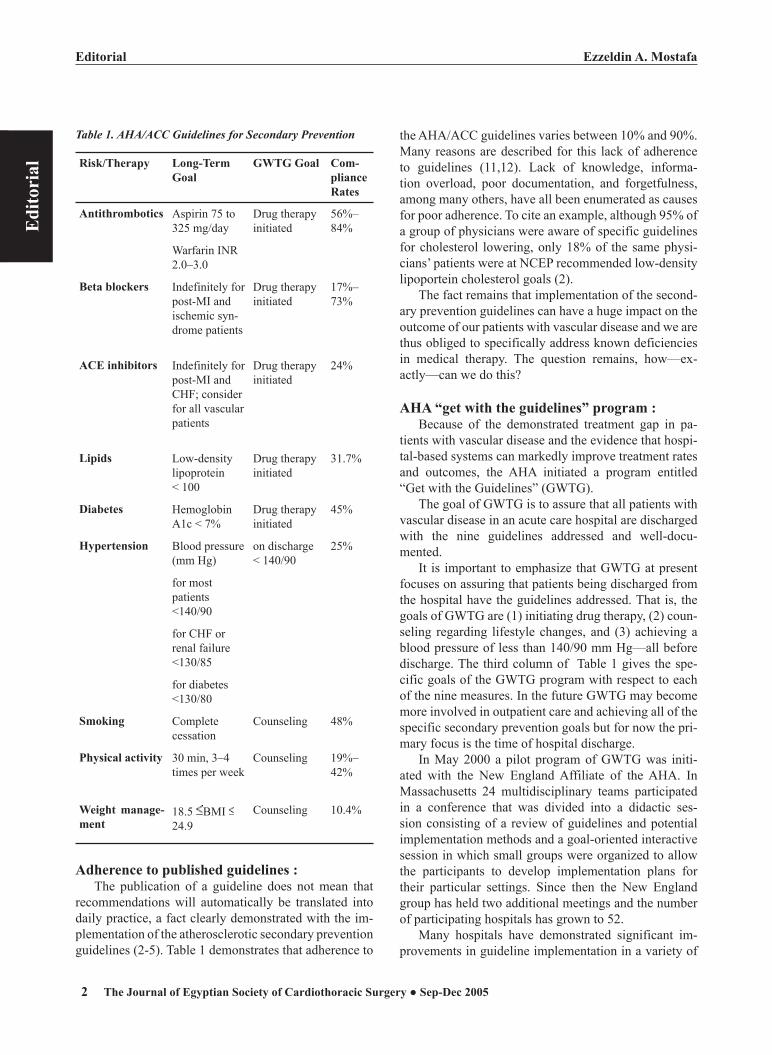
of life for patients with coronary artery disease (CAD). In 1999 there were almost 1 million invasive procedures performed in the United States (1) and their clinical benefit has been demonstrated in a multitude of inves-tigations. The importance of these procedures is clear but they do not exist in isolation—their foundation lies in the medical therapy that should be optimized in all patients with CAD. Although the importance of optimal medical therapy is self-evident, a large body of literature demonstrates its underutilization in patients with vascular disease (2-5). This treatment gap indicates we are not provid-ing medical therapy for patients who need it most. In this review we discuss medical therapies known to alter the atherosclerotic process based on the secondary pre-vention guidelines of the American Heart Association (AHA) and the American College of Cardiology (ACC). We also introduce a nationwide program from the AHA called “Get with the Guidelines,”GWTG” the goal of which is to assure that all patients with known vascular disease are discharged from the hospital with the sec-ondary prevention guidelines addressed.
Secondary prevention guidelines :The AHA and ACC have published detailed second-
ary prevention guidelines for medical therapy in patients with vascular disease (Table 1) that include specific drug recommendations (antithrombotics, beta blockers, angi-
Therapy or Secondary Prevention after Coronary Bypass Surgery: “ Postoperative Drug Get with the Guidelines” “GWTG” Program of AHA
otension-converting enzyme [ACE] inhibitors, and lipid agents), disease management (diabetes, hypertension), and lifestyle changes (exercise, smoking cessation, weight management).
The most recent guideline iteration addresses new data and recommendations from other national organi-zations (6). Changes from previous guidelines include 1 considering ACE inhibitors for all patients with atherosclerotic disease, 2 considering diabetic patients as “vascular disease equivalents” for the purposes of li-pid therapy, 3 establishing a new goal for blood pressure in diabetic patients, 130/80 mm Hg, 4 recommending a more conservative body mass index (lower limit 18.5 kg/m2), and 5 removing estrogen recommendations.
Further, the guidelines now strongly support the concept that these medical therapies should be started in the hospital during a patient’s acute coronary event or vascular procedure.
These recommendations are based on compelling data indicating that in-hospital initiation of medical therapy can improve patient compliance and outcomes (7 – 10).
The first column is the risk factor or therapy to be ad-dressed, the second column is the specific recommended goal, and the third column is the Get With the Guidelines (GWTG) goal prior to hospital discharge. Column four is the rate of compliance from various studies in the medical literature.
ACE = angiotensin-converting enzyme; AHA/ACC = American Heart Association/American College of Cardiology; BMI = body mass index; CHF = congestive heart failure; INR = international normalized ratio; MI = myocardial infarction.
Ezzeldin A. MostafaEditorial
Edi
tori
al
2 The Journal of Egyptian Society of Cardiothoracic Surgery ● Sep-Dec 2005
Ezzeldin A. Mostafa Editorial
Edi
tori
al
3The Journal of Egyptian Society of Cardiothoracic Surgery ● Volum 13, Number (3-4)
Table 1. AHA/ACC Guidelines for Secondary Prevention
Risk/Therapy Long-Term Goal
GWTG Goal Com-pliance Rates
Antithrombotics Aspirin 75 to 325 mg/day
Drug therapy initiated
56%–84%
Warfarin INR 2.0–3.0
Beta blockers Indefinitely for post-MI and ischemic syn-drome patients
Drug therapy initiated
17%–73%
ACE inhibitors Indefinitely for post-MI and CHF; consider for all vascular patients
Drug therapy initiated
24%
Lipids Low-density lipoprotein < 100
Drug therapy initiated
31.7%
Diabetes Hemoglobin A1c < 7%
Drug therapy initiated
45%
Hypertension Blood pressure (mm Hg)
on discharge < 140/90
25%
for most patients <140/90
for CHF or renal failure <130/85
for diabetes <130/80
Smoking Complete cessation
Counseling 48%
Physical activity 30 min, 3–4 times per week
Counseling 19%–42%
Weight manage-ment
18.5 BMI 24.9
Counseling 10.4%
Adherence to published guidelines :The publication of a guideline does not mean that
recommendations will automatically be translated into daily practice, a fact clearly demonstrated with the im-plementation of the atherosclerotic secondary prevention guidelines (2-5). Table 1 demonstrates that adherence to
the AHA/ACC guidelines varies between 10% and 90%. Many reasons are described for this lack of adherence to guidelines (11,12). Lack of knowledge, informa-tion overload, poor documentation, and forgetfulness, among many others, have all been enumerated as causes for poor adherence. To cite an example, although 95% of a group of physicians were aware of specific guidelines for cholesterol lowering, only 18% of the same physi-cians’ patients were at NCEP recommended low-density lipoportein cholesterol goals (2).
The fact remains that implementation of the second-ary prevention guidelines can have a huge impact on the outcome of our patients with vascular disease and we are thus obliged to specifically address known deficiencies in medical therapy. The question remains, how—ex-actly—can we do this?
AHA “get with the guidelines” program :Because of the demonstrated treatment gap in pa-
tients with vascular disease and the evidence that hospi-tal-based systems can markedly improve treatment rates and outcomes, the AHA initiated a program entitled “Get with the Guidelines” (GWTG).
The goal of GWTG is to assure that all patients with vascular disease in an acute care hospital are discharged with the nine guidelines addressed and well-docu-mented.
It is important to emphasize that GWTG at present focuses on assuring that patients being discharged from the hospital have the guidelines addressed. That is, the goals of GWTG are (1) initiating drug therapy, (2) coun-seling regarding lifestyle changes, and (3) achieving a blood pressure of less than 140/90 mm Hg—all before discharge. The third column of Table 1 gives the spe-cific goals of the GWTG program with respect to each of the nine measures. In the future GWTG may become more involved in outpatient care and achieving all of the specific secondary prevention goals but for now the pri-mary focus is the time of hospital discharge.
In May 2000 a pilot program of GWTG was initi-ated with the New England Affiliate of the AHA. In Massachusetts 24 multidisciplinary teams participated in a conference that was divided into a didactic ses-sion consisting of a review of guidelines and potential implementation methods and a goal-oriented interactive session in which small groups were organized to allow the participants to develop implementation plans for their particular settings. Since then the New England group has held two additional meetings and the number of participating hospitals has grown to 52.
Many hospitals have demonstrated significant im-provements in guideline implementation in a variety of

Ezzeldin A. MostafaEditorial
Edi
tori
al
2 The Journal of Egyptian Society of Cardiothoracic Surgery ● Sep-Dec 2005
Ezzeldin A. Mostafa Editorial
Edi
tori
al
3The Journal of Egyptian Society of Cardiothoracic Surgery ● Volum 13, Number (3-4)
areas of cardiovascular care.As an example one rural Massachusetts teaching
hospital attained a 100% success rate in applying all of the nine guidelines to its patients with coronary artery disease. Because of the success of the New England pilot program the AHA national organization approved GWTG to be rolled out across the United States and is now being initiated in all regions of the country.
Implementation in cardiac surgery Cardiovascular surgical programs are ideal locations
for GWTG. Post-CABG patients (or any vascular sur-gery patient) are in a controlled environment in which patient and family education is easier and both patient and family are motivated to make changes in their lives given the procedure that they have just undergone. Most post-CABG patients also have a “standard” postopera-tive course that is easily modifiable by a series of clini-cian reminders, standard orders, and other systems that assure all patients with vascular disease are discharged with the nine guidelines addressed.
To cite a specific example the Division of Cardi-othoracic Surgery at Cedars-Sinai Medical Center has been successful in achieving significant improvements in medical therapy after CABG. Through educational programs (physicians, physician assistants, nurses, residents, and cardiology fellows), reminders, changes in standard orders, and a computerized discharge system they have been able to increase their appropriate treat-ment rate to exceed 90% (Fig 1).
Clearly some of the deficiencies were poor docu-mentation but GWTG addresses these issues. We believe that this type of progress is possible in all cardiovascular surgery programs of all sizes. The future :
Implementation of optimal medical care in vascular disease patients can provide significant survival and quality of life benefits, and through GWTG the AHA is attempting to mobilize medical communities throughout the country to join the effort. A variety of national, re-gional, and local organizations have joined the GWTG program to achieve these goals. Lipid organizations, governmental public health divisions, state medical organizations, and many others are participating in the GWTG program. We believe that the cardiovascular sur-gery community in general and the Society of Thoracic Surgeons (STS) in particular would be a formidable ad-dition to GWTG.
Participation might occur at various levels. First, the STS might encourage all members to participate directly in the regional and national GWTG efforts. That would include STS participation in the national and regional
meetings in addition to serving as local experts on op-timizing medical care. Second, the STS as an organiza-tion might consider modifying the national database to include the nine guidelines as measures of in-hospital quality of care—to be tracked and reported, just like mortality and morbidity. Furthermore all cardiovascular surgeons—irrespective of their direct involvement in the GWTG program—could provide even more patient benefit by assuring that when a patient leaves their care, the patient has received every beneficial therapy, both surgical and medical.
Historically cardiovascular surgeons have always been at the forefront of care—in developing new tech-nology, in moving that technology to the bedside, and in proving that a new technology can provide significant benefit. The cardiovascular surgical community would be a major addition to the GWTG effort locally, region-ally, nationally, and on the individual patient level. Please join us.
References :1. Popvic JR. 1999 National hospital discharge survey: annual
summary with detailed diagnosis and procedure data. Vital Health Stat 2001;13
2. Pearson T.A., Laurora I., Chu H., Kafonek S. The lipid treatment assessment project (L-TAP): a multicenter sur-vey to evaluate the percentages of dyslipidemic patients receiving lipid-lowering therapy and achieving low-density lipoprotein cholesterol goals. Arch Intern Med 2000;160:459-467.
3. Pearson TA, Peters TD. The treatment gap in coronary ar-tery disease and heart failure: community standards and the post-discharge patient. Am J Cardiol 1997;80:45H–52H
4. Abookire S.A., Karson A.S., Fiskio J., et al. Use and monitoring of “statin” lipid-lowering drugs compared with guidelines. Arch Intern Med 2001;161:53-58.
5. Muhlestein J.B., Horne B.D., Bair T.L., et al. Usefulness of in-hospital prescription of statin agents after angiographic diagnosis of coronary artery disease in improving compli-ance and reduced mortality. Am J Cardiol 2001;87:256-261.
6. Smith S.C., Blair S.N., Bonow R.O., et al. AHA/ACC guidelines for preventing heart attack and death in patients with atherosclerotic cardiovascular disease: 2001 update. Circulation 2001;104:1577-1579.
7. Grundy S.M., Balady G.J., Criqui M.H., et al. When to start cholesterol-lowering therapy in patients with coronary heart disease. A statement for healthcare professionals from the American Heart Association task force on risk reduc-tion. Circulation 1997;95:1683-1685.
8. Fonarow G.C., Gawlinski A., Moughrabi S., Tillisch J.H. Improved treatment of coronary heart disease by imple-mentation of a cardiac hospitalization atherosclerosis management program (CHAMP). Am J Cardiol 2001;87:819-822.

Ezzeldin A. MostafaEditorial
Edi
tori
al
4 The Journal of Egyptian Society of Cardiothoracic Surgery ● Sep-Dec 2005
9. Roberts C.S. Postoperative drug therapy to extend survival after coronary artery bypass grafting. Ann Thoracic Surg 2000;69:1315-1316.
10.Schwartz G.G., Olsson A.G., Ezekowitz M.D., et al. Myo-cardial Ischemia Reduction with Aggressive Cholesterol Lowering (MIRACL) Study Investigators. Effects of atorv-astatin on early recurrent ischemic events in acute coronary syndromes. The MIRACL study: a randomized controlled trial. JAMA 2001;285:1711-1718.
11. Larme A.C., Pugh J.A. Attitudes of primary care provid-ers toward diabetes: barriers to guideline implementation. Diabetes Care 1998;21:1391-1396.
12.Smith WR. Evidence for the effectiveness of techniques to change physician behavior. Chest 2000;118:8S–17S
Ezzeldin A. Mostafa, MD Editor-in-chief

Edi
tori
al
5The Journal of Egyptian Society of Cardiothoracic Surgery ● Volum 13, Number (3-4)
The Egyptian Society of Cardio-Thoracic Surgery lost in 2005 one of its founders Professor Hassouna SABAA a pioneer of of Cardio-Thoracic surgery . he was born in Mansoura in may 1926. He did his medical stud-ies in Kasr El Aini university hospital of Cairo University and graduated in 1949 with many other pioneers in dif-ferent medical fields such Professors Ibrahim BADRAN Professor of surgery and ex Minister of Health , Hashem Fouad ex dean of Cairo University Medical School and Late Egyptian Prime Minister Fouad Moheildine.
Immediately after ending his medical studies , he was assigned resident of general surgery . He ended his speciality in 1952 and was assigned as surgeon in Port Said . He was a successful general surgeon with a wide clientele in this wealthy region . He then decided to travel to the United Kingdom to get more training and to take the British Fellowship of Surgery . In The United King-dom , he specialized in thoracic surgery . He came back to Egypt in 1963 after having passed his Fellowship in Surgery .
The Ministry of Health confined to him the mission of creating an Institute of cardio-thoracic surgery . He was given a small building related to Imbaba General hospital to start in it his institution . Another surgeon joined him , late professor Fouad GAMALI . Both of them started the department of thoracic surgery in the first Egyptian In-stitute of Cardio-Thoracic surgery. It was also necessary to create a department of Cardiology and an intensive care unit. Late Professor Moustafa EL NAHHAS created the department of cardiology before dying suddenly in his own intensive care in 1977. The Department of cardi-ology continued by the following pioneers , Professors Youssef Ryad, Awad Ibrahim and Fayez Fayek . They were then followed by Professors Diaa Abou Shokka , Adel Imama , Nabil Gobran and Nabil El Malaty.
All members of the team started to train on dogs to assure safety and stability of the extracorporeal bypass circulation . The first open heart procedure was done in 1966 . It was an ASD. Then the program of cardiac
Professor Hassouna Sabaa 1926 -2005A Pioneer of Cardio-Thoracic Surgery
surgery was stopped because of the war from 1967 to 1973.
In 1974 , the program of cardiac surgery started again Professor Hassouna SABAA asked help from many in-ternational surgical teams . Mr Donald ROSS ; Professor Magdy Yacoub from The United Kingdom , Professors Dubost , Neveux , Logeais , Michaud , Chassignol from France , Dr Ffloyd LOOP from the United States all were used to come regularly for short visit to perform surgery and to train the local personnel. Many surgeons and phy-sicians have been sent to get advanced training in the west to come back to develop the Egyptian Institute that became in late 80s the Egyptian National Heart Institute.
Cardiac Surgery Could not progress without develop-ment of a Department of Anesthesia headed by a pioneer Professor Samia ABDEL FATTAH . She had a great share in the progress and development of cardiac surgery in The National Heart Institute
Early in the 80s , the surgical team of the National heart Institute was used to perform 3 open heart proce-dures per week . In late 90s, the same team was perform-ing around 3000 open heart procedures per year.
Professor Hassouna SABAA retired in 1986 . He remained practicing until in 1995 when he developed a massive brain hemorrhage after performing a cardiac surgery a Friday morning . He was operated and survived but remained on a wheel chair for 10 years . Two years before his death in 2005 his only daughter Mrs. Mona SABAA died suddenly but her children remained sur-rounding him and enriching his life . During the last ten years of his life his wife Mrs. Sana Barakat was beside him and did not leave him even for a second .
Professor Hassouna SABA died after leaving behind him an important medical institution and a large number of disciples in cardiology , cardiac surgery , anesthesia and intensive care.
Mohamed Ahmed-nasr
Statistics
The Journal of Egyptian Society of Cardiothoracic Surgery ● Sep-Dec 20056
Ahmed A. Hassouna
The Journal of Egyptian Society of Cardiothoracic Surgery ● Volum 13, Number (3-4)
Statistics
Stat
istic
s
7
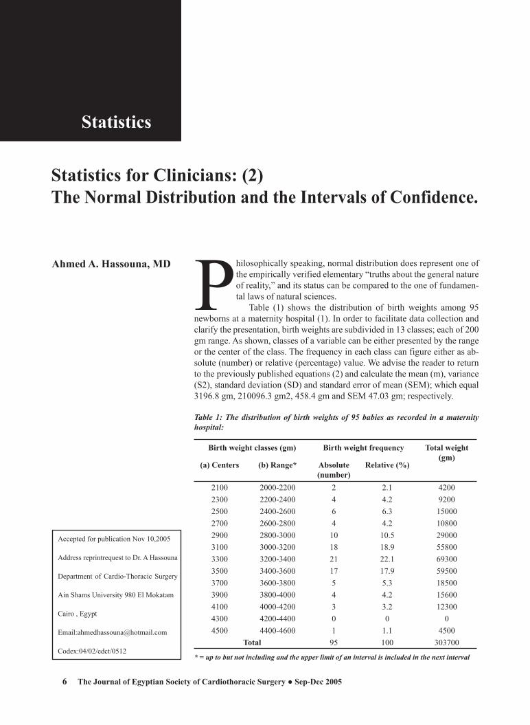
Ahmed A. Hassouna, MD Philosophically speaking, normal distribution does represent one of the empirically verified elementary “truths about the general nature of reality,” and its status can be compared to the one of fundamen-tal laws of natural sciences.
Table (1) shows the distribution of birth weights among 95 newborns at a maternity hospital (1). In order to facilitate data collection and clarify the presentation, birth weights are subdivided in 13 classes; each of 200 gm range. As shown, classes of a variable can be either presented by the range or the center of the class. The frequency in each class can figure either as ab-solute (number) or relative (percentage) value. We advise the reader to return to the previously published equations (2) and calculate the mean (m), variance (S2), standard deviation (SD) and standard error of mean (SEM); which equal 3196.8 gm, 210096.3 gm2, 458.4 gm and SEM 47.03 gm; respectively.
Table 1: The distribution of birth weights of 95 babies as recorded in a maternity hospital:
Birth weight classes (gm) Birth weight frequency Total weight (gm)
(a) Centers (b) Range* Absolute(number)
Relative (%)
2100 2000-2200 2 2.1 42002300 2200-2400 4 4.2 92002500 2400-2600 6 6.3 150002700 2600-2800 4 4.2 108002900 2800-3000 10 10.5 290003100 3000-3200 18 18.9 558003300 3200-3400 21 22.1 693003500 3400-3600 17 17.9 595003700 3600-3800 5 5.3 185003900 3800-4000 4 4.2 156004100 4000-4200 3 3.2 123004300 4200-4400 0 0 04500 4400-4600 1 1.1 4500
Total 95 100 303700
* = up to but not including and the upper limit of an interval is included in the next interval
Statistics for Clinicians: (2)The Normal Distribution and the Intervals of Confidence.
Accepted for publication Nov 10,2005
Address reprintrequest to Dr. A Hassouna
Department of Cardio-Thoracic Surgery
Ain Shams University 980 El Mokatam
Cairo , Egypt
Email:[email protected]
Codex:04/02/edct/0512

Statistics
The Journal of Egyptian Society of Cardiothoracic Surgery ● Sep-Dec 20056
Ahmed A. Hassouna
The Journal of Egyptian Society of Cardiothoracic Surgery ● Volum 13, Number (3-4)
Statistics
Stat
istic
s
7
Figure 1 is a relative frequency histogram of data presented in Table 1, with a vertical arrow (ab) passing through the mean birth weight (3196.8 gm).
Figure 1: Relative frequency histogram of data presented in Table1 with normal distribution curve
-40 -
The vertical arrow (ab) passes through the mean birth weight (3196.8 gm).
The vertical arrow (ab) passes through the mean birth weight (3196.8 gm).
Our histogram has several characters: firstly, birth weights are centered on their mean value and the num-bers of births (and corresponding relative frequencies) decrease as we get further away of the mean. Secondly, there are specific relations between the mean and the SD. An interval sandwiching the mean by 1 SD on each side (the interval formed between 3196.8 - 458.4 = 2738.4 gm and 3196.8 + 458.4 = 3655.2 gm) comprises about 2/3 of values (66 births or 69.5% of the total 95 births).
The interval formed by the mean + 2 SD (3196.8 + 916.8 = 2280 and 4113.6 gm) comprises about 95% of values (92 of the 95 births or 96.8% of the study sample) and nearly all births are comprised within the interval formed by the mean + 3 SD (3195.8 + 1375.2 gm).
Thirdly, a curved line joining the center of the class-es creates an inverted bell-shaped curve which summit overlays the mean birth weight.
Such characteristics put our data within the limits of what is known as a “Normal distribution”; which is typi-cally presented in figure 2.
As a reference, statisticians have created a perfect (standard) Normal distribution with a mean value of 0 and a SD of 1. Returning to our example, it appears that our data (vide-supra) are not far from the figures of the model, where “exactly” 68.3%, 95.5% and 99.7% of observations lie within a distance of 1, 2 and 3 SD from either sides of the mean; respectively (Figure 2).
Figure 2: The standard normal distribution curve: zLaplace- Gauss
Hence, our role is to try to fit (not to force) our data in this model, in order to ensure Normality (which is another term that can be used) however, some rules have to be drawn here (1, 3):1- Normal is just a name and does not mean that other
distributions are abnormal. 2- By Normality we mean: Normal distribution of the
studied variable in “the population of concern”, and not necessarily in the studied “sample”. As an exam-ple, if we are studying serum albumin in a group of patients - and even if serum albumin can not be dem-onstrated to be normally distributed in this particular sample or group- normality can be assumed because we already know that serum albumin is normally distributed among the “population” from which our sample was drawn.
3- On the other hand, if the distribution of the studied var-iable in the population is either unknown or known to be other than normal, the inclusion of > 30 patients per studied group is sufficient to consider a practical near normal distribution. This is based upon the central lim-it theorem, where the means of random samples from any distribution (Normal or other) will themselves have a Normal distribution. In consequence, the more we include patients, the more the variability is diluted and the more we approach a Normal distribution.
4- There are tests for checking normality and the simpler of which, even if not totally reliable, is to plot a his-togram of the data as shown. If the distribution of the recorded values (x) is far from Normality, a simple change of value like (1/x), log, and log-10 may be all what is needed. In fact, a “perfect” Normal distribu-tion is rare and a “near” Normal distribution is usually sufficient.
5- Lastly, what is the big deal about the variable being Normally distributed? Normality is a plus but not a

Ahmed A. Hassouna
The Journal of Egyptian Society of Cardiothoracic Surgery ● Sep-Dec 2005
StatisticsSt
atis
tics
8
Ahmed A. Hassouna
The Journal of Egyptian Society of Cardiothoracic Surgery ● Volum 13, Number (3-4)
Statistics
Stat
istic
s
9
necessity: Most of the commonly used statistical tests (Student’s test, ANOVA, correlation, regression, etc…) do necessitate the presence of certain parameters for being applied (that is why those tests are called para-metric statistical tests); the most important of which is the Normal distribution of the studied variable. Even though other tests that are known as distribution-free tests are as effective and do not necessitate parameters for application; normality included.
Figure 3: The Standard (Z) Normal distribution and Table.
0 Z
A confidence interval can be thought of as the set of true
0.00 0.01 0.02 0.03 0.04 0.05 0.06 0.07 0.08 0.09 0.0 0.0000 0.0040 0.0080 0.0120 0.0160 0.0199 0.0239 0.0279 0.0319 0.03590.1 0.0398 0.0438 0.0478 0.0517 0.0557 0.0596 0.0636 0.0675 0.0714 0.07530.2 0.0793 0.0832 0.0871 0.0910 0.0948 0.0987 0.1026 0.1064 0.1103 0.11410.3 0.1179 0.1217 0.1255 0.1293 0.1331 0.1368 0.1406 0.1443 0.1480 0.15170.4 0.1554 0.1591 0.1628 0.1664 0.1700 0.1736 0.1772 0.1808 0.1844 0.18790.5 0.1915 0.1950 0.1985 0.2019 0.2054 0.2088 0.2123 0.2157 0.2190 0.22240.6 0.2257 0.2291 0.2324 0.2357 0.2389 0.2422 0.2454 0.2486 0.2517 0.25490.7 0.2580 0.2611 0.2642 0.2673 0.2704 0.2734 0.2764 0.2794 0.2823 0.28520.8 0.2881 0.2910 0.2939 0.2967 0.2995 0.3023 0.3051 0.3078 0.3106 0.31330.9 0.3159 0.3186 0.3212 0.3238 0.3264 0.3289 0.3315 0.3340 0.3365 0.33891.0 0.3413 0.3438 0.3461 0.3485 0.3508 0.3531 0.3554 0.3577 0.3599 0.36211.1 0.3643 0.3665 0.3686 0.3708 0.3729 0.3749 0.3770 0.3790 0.3810 0.38301.2 0.3849 0.3869 0.3888 0.3907 0.3925 0.3944 0.3962 0.3980 0.3997 0.40151.3 0.4032 0.4049 0.4066 0.4082 0.4099 0.4115 0.4131 0.4147 0.4162 0.41771.4 0.4192 0.4207 0.4222 0.4236 0.4251 0.4265 0.4279 0.4292 0.4306 0.43191.5 0.4332 0.4345 0.4357 0.4370 0.4382 0.4394 0.4406 0.4418 0.4429 0.44411.6 0.4452 0.4463 0.4474 0.4484 0.4495 0.4505 0.4515 0.4525 0.4535 0.45451.7 0.4554 0.4564 0.4573 0.4582 0.4591 0.4599 0.4608 0.4616 0.4625 0.46331.8 0.4641 0.4649 0.4656 0.4664 0.4671 0.4678 0.4686 0.4693 0.4699 0.47061.9 0.4713 0.4719 0.4726 0.4732 0.4738 0.4744 0.4750 0.4756 0.4761 0.47672.0 0.4772 0.4778 0.4783 0.4788 0.4793 0.4798 0.4803 0.4808 0.4812 0.48172.1 0.4821 0.4826 0.4830 0.4834 0.4838 0.4842 0.4846 0.4850 0.4854 0.48572.2 0.4861 0.4864 0.4868 0.4871 0.4875 0.4878 0.4881 0.4884 0.4887 0.48902.3 0.4893 0.4896 0.4898 0.4901 0.4904 0.4906 0.4909 0.4911 0.4913 0.49162.4 0.4918 0.4920 0.4922 0.4925 0.4927 0.4929 0.4931 0.4932 0.4934 0.49362.5 0.4938 0.4940 0.4941 0.4943 0.4945 0.4946 0.4948 0.4949 0.4951 0.49522.6 0.4953 0.4955 0.4956 0.4957 0.4959 0.4960 0.4961 0.4962 0.4963 0.49642.7 0.4965 0.4966 0.4967 0.4968 0.4969 0.4970 0.4971 0.4972 0.4973 0.49742.8 0.4974 0.4975 0.4976 0.4977 0.4977 0.4978 0.4979 0.4979 0.4980 0.49812.9 0.4981 0.4982 0.4982 0.4983 0.4984 0.4984 0.4985 0.4985 0.4986 0.49863.0 0.4987 0.4987 0.4987 0.4988 0.4988 0.4989 0.4989 0.4989 0.4990 0.4990
The values inside the given table represent the areas under the standard normal curve for values between 0 and the relative z-score. For exam-ple, to determine the area under the curve between 0 and 2.36, look in the intersecting cell for the row labeled 2.30 and the column labeled 0.06. The area under the curve is .4909. To determine the area between 0 and a negative value, look in the intersecting cell of the row and column which sums to the absolute value of the number in question. For example, the area under the curve between -1.3 and 0 is equal to the area under the curve between 1.3 and 0, so look at the cell on the 1.3 row and the 0.00 column (the area is 0.4032).

Ahmed A. Hassouna
The Journal of Egyptian Society of Cardiothoracic Surgery ● Sep-Dec 2005
Statistics
Stat
istic
s
8
Ahmed A. Hassouna
The Journal of Egyptian Society of Cardiothoracic Surgery ● Volum 13, Number (3-4)
Statistics
Stat
istic
s
9
(but unknown) differences that are statistically compat-ible with the observed difference (4).
Returning to the Standard Normal distribution (Figure 2), 95.5% of observations are included in the interval formed by the mean + 2 SD. Looking the other way round; we can state that the probability for an observation to be included in the interval formed by the mean + 2SD is 95.5%. Statisticians have calculated this probability by measuring the surface area under the curve that is included between the mean and the number of SD in question (2 in our case), on either side of the mean. Then, this area was referred to the total surface area under the curve; which in our case was found to be 0.955 of that area. In fact, statisticians had the courtesy to calculate all expected probabilities (relative surface areas under the curve) not for 1, 2 and 3 SD; but for all possible positions as one moves on the horizontal scale, further away from the mean. They did so, for whole as well as fractions of SD (0.01 to 3 SD) and presented the “Z table” (table 4). However, the units on the horizon-tal scale are no more the units and fractions of SD of the standard Normal curve, but are those of any other Normal distribution, that are first standardized so as to fit the perfect model before reading from the table the expected probability.
Those units are called the “relative Z values” and are calculated by the following simple equation: for any given value (x) of any studied Normal distribution with a mean value (m) and a SD (σ);
Z = (x-m)/ σ.The probability of a Z value of 2 is the probability
for a value (x) to be included in an interval formed by 2 σ (2SD), on either of the mean (m). As shown in the figure above Table (4), probabilities are given for Z val-ues on 1 side of the mean or the absolute Z values; the other side being a mirror image of the one figuring in the table. In other words, the probability for an observation to be included in the interval formed by the mean + 2SD = that figuring in the table for a Z value of 2 multiplied by 2 = 0.4772x2= 0.95.4 = 95.4%. Checking Table 4 at the intersection of line 1.9 and column 0.06 which sums a Z value of 1.96 shows that a variable chosen at random from the population of which our studied sample was drawn has a 95% chance (0.475x2) to lie in the interval formed by 1.96 SD, on either sides of the mean value of our sample.
Applying this to our example: there is a 95% chance that the birth weight of (a coming baby) -from the population from which our sample was drawn- will be included in the interval formed by the mean of our sam-ple (3196.8 gm) + 1.96 SD (1.96x 458.4=898.5 gm) or
between 2298.3 and 4095.3 gm. This is the interval of confidence of a subject at 95%:
IC of a subject at 95% = mean + 1.96 SDBy checking Table 4, one can calculate other intervals
of confidence with more or less precisions as indicated; e.g. the interval of confidence at 99% = mean + 2.6 SD, and the interval of confidence at 75% = mean + 0.385. The more we include patients in our study, the more SD will decrease and the more the interval of confidence narrows. Those upper and lower limits of the interval of confidence figure for normal values on our laboratory sheets (e.g. blood sugar levels, renal functions, hepatic functions, red blood cell count, white cell count, etc...).
To end, our work permits us to calculate another important interval of confidence; which is the interval of confidence of the mean:
IC of mean at 95% = m + 1.96 SEMTo explain the interval of confidence of mean, let’s
imagine that the previous study was repeated, whether by the same researcher or by others, on another com-parable group of patients. No two studies will ever calculate the same mean, SD and SEM values and one should ask himself: what is the true mean birth weight (M) of all newborns of comparable mothers? For sure no one can have the definite answer, but one can always calculate a range (i.e. an interval of confidence) which would embrace (M).
In other words, one should expect that the calculated mean birth weight in other comparable studies will have a 95% chance to be within the range of m + 1.96 SEM = 3196.8 + 92.1 = between 3104.7 and 3288.9 gm. Also the more I include cases in my study; the lower would be my SEM and the narrower and more approaching to the real values, would be my calculated interval of con-fidence. Remember that unlike that of a subject, the IC of the mean does not necessitate the normal distribution of the studied variable (vide-supra).
A confidence interval is typically reported in the following way: “The mean birth weight was 3196.8 gm (95% CI, 3104.7 to 3288.9 gm)”
This means that even though the observed birth weight was 3196.8 gm, the data are statistically com-patible with a true birth weight difference as small as 3104.7 gm or as large as 3288.9 gm. True differences that lie outside the 95% confidence interval are not impossible; they merely have less statistical evidence supporting them than values within it. The choice of 95% as the standard convention is somewhat arbitrary and corresponds to the use of a threshold of P < 0.05 for statistical significance.

Ahmed A. Hassouna
The Journal of Egyptian Society of Cardiothoracic Surgery ● Sep-Dec 2005
StatisticsSt
atis
tics
10
References:1- Schwartz D. Les fluctuations d’échantillonnage d’une moy-
enne. In: Schwartz D, ed. Méthodes statistiques a l’usage des médecins et des biologistes. 3rd edition, Paris: Mé-decines-Sciences Flammarion; 1969: 103-116.
2- Ahmed HASSOUNA: Statistics for clinicians: the 4 basic indices. J Egypt Soc Cardiothorac Surg 2005; 13 (1): 14-6.
3- Altman DG and Bland JM. Statistics notes. The normal distribution. BMJ 1995; 310: 298.
4- Goodman SN and Berlin JA. The use of predicted confi-dence intervals when planning experiments and the misuse of power when interpreting results. Ann Intern Med 1994; 121 (3): 200-6.
5- Schwartz D. Les fluctuations d’échantillonnage d’une moy-
enne. In: Schwartz D, ed. Méthodes statistiques a l’usage des médecins et des biologistes. 3rd edition, Paris: Mé-decines-Sciences Flammarion; 1969: 103-116.
6- Ahmed HASSOUNA: Statistics for clinicians: the 4 basic indices. J Egypt Soc Cardiothorac Surg 2005; 13 (1): 14-6.
7- Altman DG and Bland JM. Statistics notes. The normal distribution. BMJ 1995; 310: 298.
8- Goodman SN and Berlin JA. The use of predicted confi-dence intervals when planning experiments and the misuse of power when interpreting results. Ann Intern Med 1994; 121 (3): 200-6.
Editorial
CARDIOVASCULAR
The Journal of Egyptian Society of Cardiothoracic Surgery ● Volum 13, Number (3-4) 11
Magued A. Zikri, MDSaed Abdel Aziz, MDAmr M. Roushdi, MDWalid Abusenna, MDSameh S. Marzouk, MD *Ahmed S. Ahmed, MD *
Background: Both short and long term results of c oronary bypass surgery are partly dependent on adequacy of myocardial protection especially during the cardioplegic arrest portion of the operation. The attraction of using the venous end of the capillary bed for cardioplegia delivery is well founded based on the only too frequent incidence of extensive obstruc-tive pathology in the conventional antegrade delivery route in patients presenting in the present era for CABG. Objective analysis of adequacy of myocardial protection includes changes in serum concentrations of dif-ferent cardiac enzymes such as creatine kinase and its myocardial brain isoenzyme (CK-MB), Troponin-I (TnI). Other indirect indices of tissue malperfusion and oxidative stress are also valuable including blood lac-tate level, and two anti oxidant enzymes, Superoxide desmutase (SOD) and glutathione peroxidase (GSH-Px).Methods: Forty patients scheduled for conventional CABG using car-diopulmonary bypass (CPB) were divided into two equal groups based on the extent of the pathology of there coronary arteries. Group I had a moderate form of coronary artery disease while Group II had severe and extensive pathology. Subsequently , each group was randomly divided into two equal subgroups according to the route of cardioplegia delivery : a- Antegrade cardioplegia and b- combined antegrade-retrograde cardio-plegia. Blood samples were taken from each patient for analysis of (Tn-I), CK-MB, lactic acid, (SOD), and (GSH-Px). Results and Conclusion: As a general outlook, combining retrograde with anterograde cardioplegia was associated with statistically significant lower values of the indices of ischemia and oxidative stress including (Tn-I), CK-MB, lactic acid, (SOD), and (GSH-Px) relative to that of the antegrade cardioplegia only. Keywords: CABG, retrograde cardioplegia, oxidative stress, ischemia
Improved results of coronary artery surgeries (CABG) with the tradi-tional use of cardiopulmonary bypass (CBP) entail adequate intraop-erative myocardial protection(1). Effective intraoperative myocardial protection requires adequate distribution of cardioplegic solution to all myocardial segments in a safe, simple, and rapid fashion(2). Though
Does Retrograde Crystalloid Cardioplegia Offer Addi-tional Protection Against Ischemia and Oxidative Stress in Coronary Bypass Surgeries?
Accepted for publication oct20,2005
Address reprintrequest to Dr M A Zekri
Departments of Cardiothoracic Surgery
Faculty of Medicine,Cairo University
168 Tahrir Street .
Cairo Egypt.
Email:[email protected]
Codex:04/13/cord/0512

Magued A. Zikri et alCardiovascular
Car
diov
ascu
lar
The Journal of Egyptian Society of Cardiothoracic Surgery ● Sep-Dec 200512
Magued A. Zikri et al Cardiovascular
Car
diov
ascu
lar
The Journal of Egyptian Society of Cardiothoracic Surgery ● Volum 13, Number (3-4) 13
antegrade cardioplegia is an advantageous route, yet, it is associated with a number of actual and theoreti-cal limitations(3,4). Among these drawbacks the non-homogenous distribution of antegrade cardioplegia in severe critical proximal coronary artery stenosis and in evolving myocardial infarction(5), coronary ostial inju-ry during and after aortic valve surgery(6), poor distribu-tion in patients with aortic regurgitation unless the aorta is opened and the coronary ostia are perfused directly(7), the need to interrupt the continuity of mitral valve proce-dures in order to remove the retractors and avoid aortic distortion during cardioplegic replenishment(8), and it may not be technically possible in patients with type A aortic dissection (9).To obviate these limitations, retro-grade coronary sinus perfusion has been proposed as an alternative method of providing myocardial protection that offers an excellent protection of the left ventricle in cases of severe coronary artery stenosis and when internal mammary artery grafts are used (10). However, recent studies have documented that retrograde cardio-plegia does not adequately perfuse the right ventricle. The possibility of delayed cardiac arrest due to the low flow rate used for retrograde cardioplegia has also been noted (11).
After testing in our center the efficacy and safety of retrograde cardioplegia delivery in a cohort of patients with extensive coronary artery disease, we thought of designing a prospective randomized clinical study to as-sess and compare combined alternate use of antegrade-retrograde cardioplegia versus antegrade cardioplegia in providing adequate myocardial preservation during coronary artery bypass graft surgery.
A combination of clinical, hemodynamic, electrocar-diographic, and biochemical parameters of ischemia and oxidative stress were used to monitor potential differ-ences between the two methods.
Methodology:Forty patients (34 males and 6 females) scheduled
for conventional CABG surgery using CPB, were oper-ated upon in new Kasr El-Aini teaching hospital, Facul-ty of Medicine, Cairo University, in the period between December 2002 and November 2004.
Patients were categorized according to the severity of their coronary artery disease pathology into two groups of equal number , group I and group II. Subsequently, patients within a given group were randomly assigned to either receive a conventional antegrade cardioplegia protocol or a combined antegrade / retrograde cardio-plegia protocol.
Criteria for extensive coronary artery lesion includ-ing in group II included :
1- Left main coronary artery involvement.2- Severe occlusion of the left coronary artery > 80% by
coronary catheter study.3- Severe occlusion of the right coronary artery > 80%
by catheter study.
These patients were randomly and equally distrib-uted to two subgroups :Group IIa and IIb.
The other twenty patients selected with moderately extensive coronary artery disease were similarly distrib-uted to two subgroups : Group Ia and Ib.
Groups Ia, IIa: were scheduled for antegrade cold crystalloid cardioplegia.
Group Ib, IIb: were scheduled for alternate ante-grade-retrograde cold crystalloid cardioplegia .
The mode of cardioplegia delivery was the only vari-able between the two groups, the
composition , timing and frequency of delivery of cardioplegia being identical. All cases in a given group of cardioplegia delivery protocol were operated upon by the same surgeon.
The criteria of exclusion of patients from the study included single vessel coronary artery disease, re opera-tion, combined or emergency procedures.
All patients had a history of chest pain. Routine labo-ratory investigations were carried out as well as 12 leads electrocardiogram (ECG) one day before surgery. All patients were diagnosed to have coronary artery disease and the extent of the disease was evaluated based on selective coronary angiography.
Biochemical Studies and Sampling Protocol: Blood samples were taken from a special channel of
the C.V.P. catheter. Serum levels of creatine Kinase, MB isoenzymes
(CK-MB), Troponin I, Lactic acid, Superoxide des-mutase, and Glutathione peroxidase were taken with the following time schedule:• Baseline reading: on arrival to the O.R.• Post-induction: 15 minutes following induction of
general anesthesia.• Post-declamping: 15 minutes following declamping
of the aorta.• Post-recovery: Two hours after arrival to the postop-
erative ICU.
Anaesthetic Management:For each patient in the four groups, standard an-
esthetic protocol was followed in each patient. An-esthesia was induced using thiopentone sodium (3–4 mg/kg), fentanyl (5–7 µg/kg), and vecuronium bromide (0.08–0.1 mg/kg) to facilitate tracheal intubation. The
Magued A. Zikri et alCardiovascular
Car
diov
ascu
lar
The Journal of Egyptian Society of Cardiothoracic Surgery ● Sep-Dec 200512
Magued A. Zikri et al Cardiovascular
Car
diov
ascu
lar
The Journal of Egyptian Society of Cardiothoracic Surgery ● Volum 13, Number (3-4) 13
rest of the monitoring aids were applied: capnography and temperature monitors. Maintenance of anesthesia was carried out using isoflurane 0.8–1% was used as inhalation anaesthetic according to the haemodynamic parameters. Fentanyl was stepped to 8–12 µg/kg before sternotomy and a top-up dose of the muscle relaxant was given whenever needed.
Cannulation for cardioplegia deliveryA Retrograde Cannula was inserted in 20 patients
transatrially guided by feeling the tip of the catheter negotiating the entrance of the coronary sinus from the diaphragmatic surface of the right ventricle before go-ing on bypass with the right side adequately filled. This step of the operation was performed successfully after a single attempt in 18 patients and multiple attempts in two cases. In all situations, its insertion was safe with no injury to the coronary sinus. In all cases a 14 fr. retro-grade cardioplegia cannula with self-inflatable medium balloon (18 mm) and rigid insertion stylet, CHASE, Medical inc., Richardson. Texas was used. This cannula has a side arm for pressure monitoring in the coronary sinus.An Antegrade Cannula was routinely inserted in the aortic root in all 40 patients. We used a cannula provided with two side ports, one for venting and the other for pressure monitoring (Research Medical, Inc., Midvale, UT, USA.
Conduct of operation and protocol of Cardioplegia delivery:
Patients in the Antegrade cardioplegia only sub-groups, groups Ia and IIa, were operated upon using the conventional routine of distal anastomosis performed with the cross clamp on while the proximal anastomosis were performed after aortic declamping using a side bit-ting clamp on the ascending aorta. The pneumatic pump is inflated to 300 mm Hg and a St. Thomas’ Hospital solution no. 1 chilled to 4°C was infused in a dose of 15 mL per kg body weight initially and repeated every 15 minutes in a dose of 200 mL.
In group Ib and IIb patients, in whom a combined antegrade / retrograde protocol was used, all distal and proximal anastomosis were performed with one aortic clamp application. Half of the initial dose was given an-tegradely with a high aortic root pressure and the other half was given retrogradely with coronary sinus pres-sure ranging between 30 and 50 mm Hg. All subsequent doses were given retrogradely through the coronary sinus as described before Magued et Al . The two routes of infusion were never used simultaneously.
Parameters of Intraoperative evaluation:Apart from the biochemical data already mentioned,
the following data had been estimated:• Total bypass time (min.).• Total ischemic time (min.).• Ease of weaning from CPB.• Incidence of use of defibrillator.• Average dose of inotrops (epinephrine μgm/kg/min).• Incidence of post-bypass ventricular dysrhythmias.• Incidence of post-bypass persistent myocardial
ischemia or infarction.• Incidence of postoperative mortality.
Statistical analysis:Statistical analysis was done using SPSS version
7.0 for Windows© and Microsoft® Excel© 2000. All data are presented as means and standard deviations. Inter-group comparisons were made using unpaired t-test. Intra-group comparisons were made using one-way ANOVA. A P value of less than 0.05 was considered statistically significant.
Results:The four groups of the study were similar in
number, age range, average weight, and preoperative morbidity with no statistical significance in these re-spects (Table: 1).
The preoperative hemodynamics was almost similar in the subgroups of the same main group. However, groups IIa and IIb of severely extensive coronary lesion have got a significantly lower ejection fraction, and a higher incidence of unstable angina compared to groups Ia and Ib (Table: 1). The quantitative analysis of serum Troponin-I (Tp-I) revealed no statistical difference in the basal level between the four groups of the study (Table: 2). In each group of the study, it showed a significant increase in the post-declamping and recovery readings compared to their initial basal readings. However, there was a statistically significant lower value in combined A-C groups compared to the Antegrade cardioplegia groups (Table: 2).
Serum levels of Superoxide Desmutase, Glutath-ione peroxidase, and lactic acid had a similar pattern of change in this study (Table: 2). Within each group, they exhibited an initial, but statistically significant rise in the post-induction readings, that become more significant in the post-declamping readings, followed by a mild but still significant decrease in the serum levels in the post-recovery readings. However, the combined A-R cardio-plegia groups showed a significantly lower value than the Antegrade groups of the same main group.
Because of performing proximal and distal anasto-mosis with cross clamp on in combined cardioplegia

Magued A. Zikri et alCardiovascular
Car
diov
ascu
lar
The Journal of Egyptian Society of Cardiothoracic Surgery ● Sep-Dec 200514
Magued A. Zikri et al Cardiovascular
Car
diov
ascu
lar
The Journal of Egyptian Society of Cardiothoracic Surgery ● Volum 13, Number (3-4) 15
Moderately Extensive (Group I) Severely Extensive (Group II)
For AC (a) For AC+RC (b) For AC (a) For AC+RC (b)
Number (n) 10 10 10 10
Age (Years) 56.8 (6.4) 55.2(4.8) 57.5(2.6) 58.2(3.4)
Weight (Kg) 88.4(3.6) 86.2(4.0) 89.0(2.3) 87.6(2.6)
Male: Female ratio 4:1 9:1 4:1 9:1
Preoperative Morbidity:
Hypertension (%)60 % 50 % 60 % 60 %
Diabetes (%) 50 % 40 % 40 % 50 %
Hypercholesterolemia (%) 60 % 60 % 70 % 50 %
Ex- Smoking (%): 80 % 80 % 80 % 80 %
History of recent MI (within 6 months) (%) 10 % 10 % 20 % 20 %
Preoperative Hemodynamics EF (%) 40.2 (2.34) 39.2(3.4) 34.3(3.0) 33.7(2.6)
SBP (mmHg) 160.2(8.6) 155.7(9.4) 152.5(7.7) 158(9.0)
DBP (mmHg) 88.2 (4.1) 89.4(6.4) 82.4(7.2) 85.4(5.9)
MBP (mmHg) 113.2(8.4) 111.2(7.6) 104.5(6.6) 108(5.9)
Heart Rate (Beat/min) 83.4(11.2) 82(12.4) 84(11.7) 85(11.5)
ST Segment (mm) - 1.4 (0.35) - 1.0 (0.22) - 1.5 (0.24) - 1.3 (0.31)
Unstable angina (%) 30 % 30 % 50 % 50 %
Extent of Coronary artery disease (%):
•Single vessel
•Two vessels
•Three vessels
•More th
0 %
100 %
0 %
0 %
0 %
100 %
0 %
0 %
0 %
0 %
30 %
70 %
0 %
0 %
20 %
80%
SD: Standard Deviation of the mean AC: Ante grade CardioplegiaRC: Retrograde Cardioplegia MI: Myocardial InfarctionEF: Ejection Fraction SBP: Systolic Blood PressureDBP: Diastolic Blood Pressure MBP: Mean Blood Pressure
Table: (1) Demographic, Clinical and Preoperative hemodynamic data of patients of the different groups of the study. Values are expressed as mean (SD) or as percentage of the patients
groups, both cross clamp and bypass durations time were significantly longer than in the antegrade only groups. However, combined groups showed a statisti-cally significant decrease in inotropic dose, incidence of post-bypass dysrhythmias and post bypass ischemia
This was also reflected on the ease of separation from cardio pulmonary bypass machine observed in the combined cardioplegia group and was even more pronounced in the severely affected coronary lesions subgroup (Table: 3).
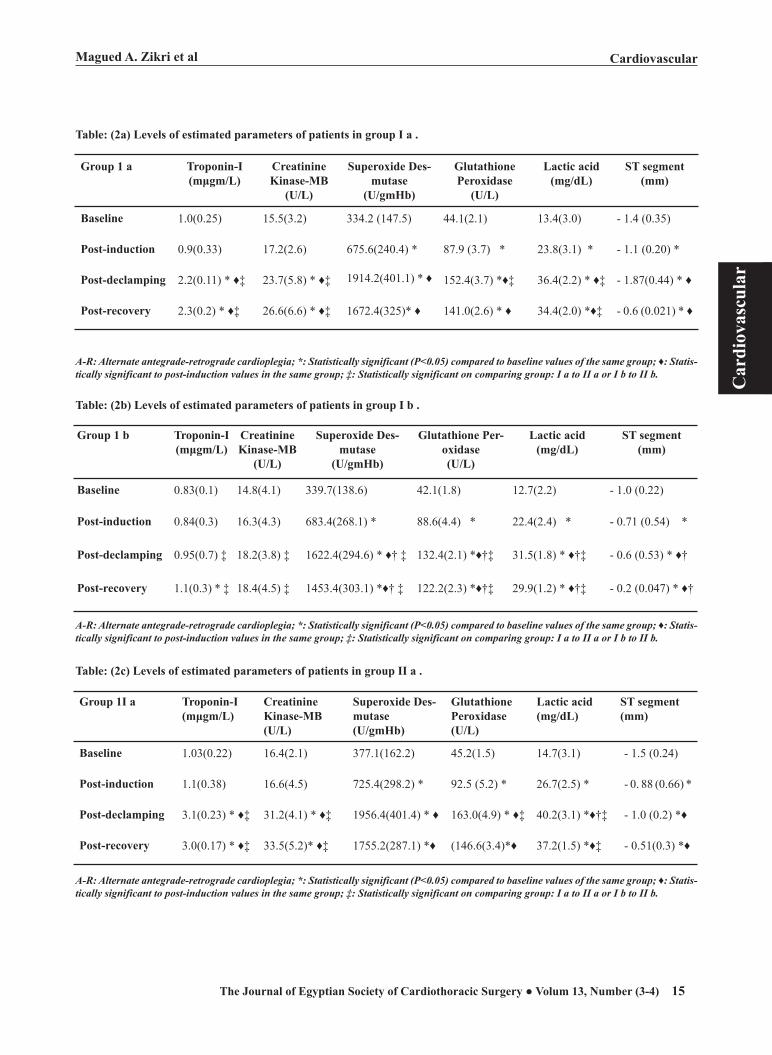
Magued A. Zikri et alCardiovascular
Car
diov
ascu
lar
The Journal of Egyptian Society of Cardiothoracic Surgery ● Sep-Dec 200514
Magued A. Zikri et al Cardiovascular
Car
diov
ascu
lar
The Journal of Egyptian Society of Cardiothoracic Surgery ● Volum 13, Number (3-4) 15
Group 1 a Troponin-I(mμgm/L)
Creatinine Kinase-MB
(U/L)
Superoxide Des-mutase
(U/gmHb)
Glutathione Peroxidase
(U/L)
Lactic acid(mg/dL)
ST segment(mm)
Baseline 1.0(0.25) 15.5(3.2) 334.2 (147.5) 44.1(2.1) 13.4(3.0) - 1.4 (0.35)
Post-induction 0.9(0.33) 17.2(2.6) 675.6(240.4) * 87.9 (3.7) * 23.8(3.1) * - 1.1 (0.20) *
Post-declamping 2.2(0.11) * ♦‡ 23.7(5.8) * ♦‡ 1914.2(401.1) * ♦ 152.4(3.7) *♦‡ 36.4(2.2) * ♦‡ - 1.87(0.44) * ♦
Post-recovery 2.3(0.2) * ♦‡ 26.6(6.6) * ♦‡ 1672.4(325)* ♦ 141.0(2.6) * ♦ 34.4(2.0) *♦‡ - 0.6 (0.021) * ♦
Table: (2a) Levels of estimated parameters of patients in group I a .
A-R: Alternate antegrade-retrograde cardioplegia; *: Statistically significant (P<0.05) compared to baseline values of the same group; ♦: Statis-tically significant to post-induction values in the same group; ‡: Statistically significant on comparing group: I a to II a or I b to II b.
Table: (2b) Levels of estimated parameters of patients in group I b .
Group 1 b Troponin-I(mμgm/L)
Creatinine Kinase-MB
(U/L)
Superoxide Des-mutase
(U/gmHb)
Glutathione Per-oxidase(U/L)
Lactic acid(mg/dL)
ST segment(mm)
Baseline 0.83(0.1) 14.8(4.1) 339.7(138.6) 42.1(1.8) 12.7(2.2) - 1.0 (0.22)
Post-induction 0.84(0.3) 16.3(4.3) 683.4(268.1) * 88.6(4.4) * 22.4(2.4) * - 0.71 (0.54) *
Post-declamping 0.95(0.7) ‡ 18.2(3.8) ‡ 1622.4(294.6) * ♦† ‡ 132.4(2.1) *♦†‡ 31.5(1.8) * ♦†‡ - 0.6 (0.53) * ♦†
Post-recovery 1.1(0.3) * ‡ 18.4(4.5) ‡ 1453.4(303.1) *♦† ‡ 122.2(2.3) *♦†‡ 29.9(1.2) * ♦†‡ - 0.2 (0.047) * ♦†
A-R: Alternate antegrade-retrograde cardioplegia; *: Statistically significant (P<0.05) compared to baseline values of the same group; ♦: Statis-tically significant to post-induction values in the same group; ‡: Statistically significant on comparing group: I a to II a or I b to II b.
Table: (2c) Levels of estimated parameters of patients in group II a .
A-R: Alternate antegrade-retrograde cardioplegia; *: Statistically significant (P<0.05) compared to baseline values of the same group; ♦: Statis-tically significant to post-induction values in the same group; ‡: Statistically significant on comparing group: I a to II a or I b to II b.
Group 1I a Troponin-I(mμgm/L)
Creatinine Kinase-MB(U/L)
Superoxide Des-mutase(U/gmHb)
Glutathione Peroxidase(U/L)
Lactic acid(mg/dL)
ST segment(mm)
Baseline 1.03(0.22) 16.4(2.1) 377.1(162.2) 45.2(1.5) 14.7(3.1) - 1.5 (0.24)
Post-induction 1.1(0.38) 16.6(4.5) 725.4(298.2) * 92.5 (5.2) * 26.7(2.5) * - 0. 88 (0.66) *
Post-declamping 3.1(0.23) * ♦‡ 31.2(4.1) * ♦‡ 1956.4(401.4) * ♦ 163.0(4.9) * ♦‡ 40.2(3.1) *♦†‡ - 1.0 (0.2) *♦
Post-recovery 3.0(0.17) * ♦‡ 33.5(5.2)* ♦‡ 1755.2(287.1) *♦ (146.6(3.4)*♦ 37.2(1.5) *♦‡ - 0.51(0.3) *♦
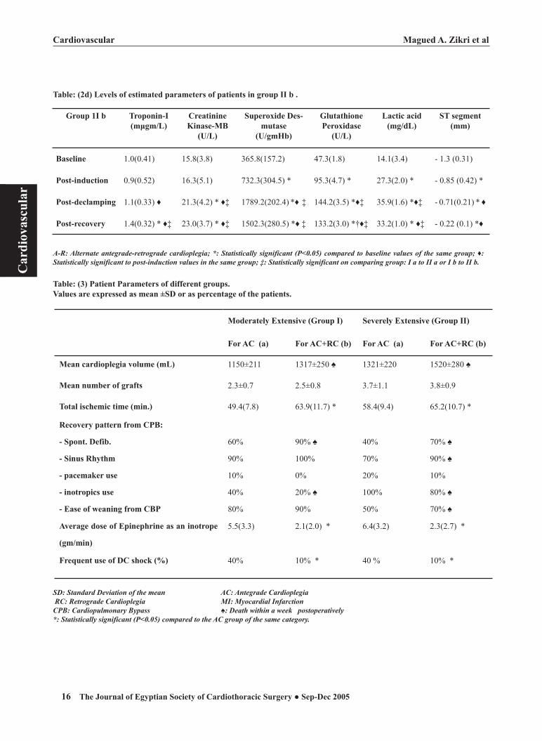
Magued A. Zikri et alCardiovascular
Car
diov
ascu
lar
The Journal of Egyptian Society of Cardiothoracic Surgery ● Sep-Dec 200516
Magued A. Zikri et al Cardiovascular
Car
diov
ascu
lar
The Journal of Egyptian Society of Cardiothoracic Surgery ● Volum 13, Number (3-4) 17
Table: (2d) Levels of estimated parameters of patients in group II b .
A-R: Alternate antegrade-retrograde cardioplegia; *: Statistically significant (P<0.05) compared to baseline values of the same group; ♦: Statistically significant to post-induction values in the same group; ‡: Statistically significant on comparing group: I a to II a or I b to II b.
Group 1I b Troponin-I(mμgm/L)
Creatinine Kinase-MB
(U/L)
Superoxide Des-mutase
(U/gmHb)
Glutathione Peroxidase
(U/L)
Lactic acid(mg/dL)
ST segment(mm)
Baseline 1.0(0.41) 15.8(3.8) 365.8(157.2) 47.3(1.8) 14.1(3.4) - 1.3 (0.31)
Post-induction 0.9(0.52) 16.3(5.1) 732.3(304.5) * 95.3(4.7) * 27.3(2.0) * - 0.85 (0.42) *
Post-declamping 1.1(0.33) ♦ 21.3(4.2) * ♦‡ 1789.2(202.4) *♦ ‡ 144.2(3.5) *♦‡ 35.9(1.6) *♦‡ - 0.71(0.21) * ♦
Post-recovery 1.4(0.32) * ♦‡ 23.0(3.7) * ♦‡ 1502.3(280.5) *♦ ‡ 133.2(3.0) *†♦‡ 33.2(1.0) * ♦‡ - 0.22 (0.1) *♦
Table: (3) Patient Parameters of different groups. Values are expressed as mean ±SD or as percentage of the patients.
SD: Standard Deviation of the mean AC: Antegrade Cardioplegia RC: Retrograde Cardioplegia MI: Myocardial InfarctionCPB: Cardiopulmonary Bypass ♠: Death within a week postoperatively*: Statistically significant (P<0.05) compared to the AC group of the same category.
Moderately Extensive (Group I) Severely Extensive (Group II)
For AC (a) For AC+RC (b) For AC (a) For AC+RC (b)
Mean cardioplegia volume (mL) 1150±211 1317±250 ♠ 1321±220 1520±280 ♠
Mean number of grafts 2.3±0.7 2.5±0.8 3.7±1.1 3.8±0.9
Total ischemic time (min.) 49.4(7.8) 63.9(11.7) * 58.4(9.4) 65.2(10.7) *
Recovery pattern from CPB:
- Spont. Defib.
- Sinus Rhythm
- pacemaker use
- inotropics use
- Ease of weaning from CBP
60%
90%
10%
40%
80%
90% ♠
100%
0%
20% ♠
90%
40%
70%
20%
100%
50%
70% ♠
90% ♠
10%
80% ♠
70% ♠
Average dose of Epinephrine as an inotrope
(gm/min)
5.5(3.3) 2.1(2.0) * 6.4(3.2) 2.3(2.7) *
Frequent use of DC shock (%) 40% 10% * 40 % 10% *

Magued A. Zikri et alCardiovascular
Car
diov
ascu
lar
The Journal of Egyptian Society of Cardiothoracic Surgery ● Sep-Dec 200516
Magued A. Zikri et al Cardiovascular
Car
diov
ascu
lar
The Journal of Egyptian Society of Cardiothoracic Surgery ● Volum 13, Number (3-4) 17
Discussion:The optimal route of delivery of cardioplegia is still
in debate in patients with ischemic heart disease. Cardi-othoracic surgeons and anesthesiologists exhibited great concern about the comparison between different routes and composition of the cardioplegic solutions in coro-nary patients in particular since myocardial protection is the cobblestone of improvement of the early and delayed surgical outcome of such surgical procedures (12).
Since the introduction of the retrograde route for the delivery of cardioplegia through the coronary sinus by Lillchei and colleagues in 1956 (13), many studies have been postulated to investigate its effectiveness in myocardial preservation in surgeries of cardiac valve replacement, until late in 1970s, when interest emerged in retrograde coronary perfusion in coronary surgery(14). Some studies raised concerns that the sole use of retrograde perfusion created inadequate preser-vation of the right ventricle in particular (15). Other studies even concluded that the combined use of ante-grade and retrograde routes offered the same degree of myocardial preservation induced by the antegrade route alone, by this, they ignoring the role of the retrograde perfusion(16). Other studies reported significant differ-ences favoring the retrograde route over the traditional antegrade route with respect to homogeneous distribu-tion of the cardioplegia(17), myocardial recovery time (18), ease of weaning from CPB, and Swan-Ganz hemo-dynamic measurement on emergence from CPB. The ra-tionale for combined antegrade and retrograde routes of cardioplegic delivery is based on anatomic and experi-mental arguments (19, 20). The coronary venous system is composed of two interrelated systems: the epicardial or superficial (greater) system and the endocardial or deep (lesser) system. The greater system includes the coronary sinus and its tributaries, the small cardiac vein, and the anterior cardiac vein that drain into the coronary sinus. The lesser system comprises the vessels that drain directly into the cardiac chambers. There is widespread anastomosis at all levels of the cardiac venous circula-tion. Thus, the myocardium may be adequately perfused retrogradely by this large venous network free of athero-sclerotic changes (21). The study of the biochemical differences between both techniques opened the way to a new field of evaluation(22).
The troponin complex is the regulatory element of the myofilament, which mediates the calcium depend-ence of muscle contraction in both cardiac and skeletal muscle. Its three components, troponin I (TnI), troponin C (TnC), and the skeletal troponin T (TnT), interact with each other and other thin filament proteins ( actin and tropomyosin) through both calcium-dependent and
independent associations (23). Hence, cardiac troponin-I is a new marker for disruption of cardiac myocytes with the potential for detection of minor differences in myo-cardial ischemia (24,25,26). In the present study, serum Troponin-I didn’t change significantly after induction of anesthesia in the four groups of the study. However, it significantly increased in both post-declamping and post-recovery reading compared each to its post-induc-tion reading. Within each category, the subgroup of combined antegrade and retrograde cardioplegia showed statistically significant lower readings compared to the antegrade subgroups, indicating superior myocardial protection from combined antegrade-retrograde car-dioplegia that was more significant in severe coronary lesions.
CK-MB is not a specific parameter for myocardial damage or cardiac ischemic changes(27). However, in this work, it showed a significant rise in the post-dec-lamping measurement relative to the pre-operative one within the same group, and this rise was significantly lower in the combined antegrade-retrograde cardiople-gia groups relative to the antegrade groups. This reflects a better myocardial preservation when sequentially combining the two techniques rather than applying the antegrade technique alone.
In the present study, lactic acid levels were sig-nificantly increased after induction in the study groups. However, the post-declamping and post-recovery lactic acid levels were significantly higher in the antegrade groups compared to the combined antegrade – retro-grade cardioplegia groups.
Lactate is an end product of anaerobic metabolism. It is one of the most common forms of metabolic ac-ids resulting from either tissue hypoperfusion and/or hypoxemia. Measuring arterial lactate concentration is a prompt, easy and relatively non-invasive way to esti-mate tissue oxygen metabolism. Initial increases in the lactate level following induction of anaesthesia in the present study may be related to the stress of induction, intubation and artificial ventilation(28). Lactate levels increased significantly during post-CPB measurements in a majority of the patients. The increases in lactate levels are affected by the changes in inter-organ blood flow, blood glucose levels and/or blood pH, in addi-tion to the effects of the CPB-priming lactated Ringer’s solution(29). Studies of the lactate levels in cardiac surgeries with extracorporeal circulation (CPB) suggest that high lactate levels are indicative of inadequate oxy-gen delivery (DO2), or a defect in the oxidative utilisa-tion despite adequate DO2 (30).
Tissue perfusion was found to be at risk during cardiac surgery with CPB and in the immediate post-

Magued A. Zikri et alCardiovascular
Car
diov
ascu
lar
The Journal of Egyptian Society of Cardiothoracic Surgery ● Sep-Dec 200518
Magued A. Zikri et al Cardiovascular
Car
diov
ascu
lar
The Journal of Egyptian Society of Cardiothoracic Surgery ● Volum 13, Number (3-4) 19
operative period. The association of low blood flows with metabolic acidosis and accumulation of lactate perioperatively has been well established. With the improvements in cardiopulmonary bypass and overall haemodynamic management, severe peri- and post-operative hypoperfusion has become rare. Despite the rarity of severe postoperative complications, several lines of evidence suggest that episodes of less severe hypoperfusion and borderline tissue oxygenation are relatively common. Measurement of blood lactate levels is widely used to assess the adequacy of tissue perfusion that apparently looked to be superior when applying the two techniques of cardioplegia rather than the antegrade technique only. However, the interpretation of elevated blood lactate levels is limited by several confounding variables including acute changes in acid-base balance, inter-organ substrate flux, peripheral and visceral tissue perfusion, and hepatic lactate uptake will all influence blood lactate levels and may occur during and after car-diac surgery with CPB (31).
Superoxide desmutase (SOD) and glutathione per-oxidase (GSH-Px) are enzymes acting as free radical scavengers. Following induction of general anesthesia, levels exhibited a significant increase compared to the pre-induction levels but insignificant changes between the four groups. The readings referred as post-declamp-ing and post-recovery levels exhibited a significant increase compared to the post-induction levels, and a significant increase of these levels in the antegrade groups compared to the combined antegrade-retrograde groups.
Evidences are accumulating that most of the degen-erative diseases that affect humans have their origin in deleterious free radical reactions. The increase of the post-induction levels of all of SOD, and GSH-Px re-flected the oxidative stress of induction of anaesthesia as well as endotracheal intubation that was mild and comparable in the four groups of the study.
CPB induced a significant oxidative stress. This may be attributed to the ischemic reperfusion injury caused by aortic cross-clamping, inflammatory response, cardiac neutrophil accumulation and trans-coronary neutrophil activation during clinical cardiopulmonary bypass(32) and the significant activation of antithrombotic protein C pathway during cardiopulmonary bypass, mainly during the minutes after aortic unclamping in the ischemic vas-cular beds. All these factors could result into an oxida-tive damage that may impair the post-ischemic recovery of human heart and circulation(33). The lower readings of (SOD) and (GSH-Px) in the antegrade-retrograde cardioplegia groups compared to the antegrade groups indicate less oxidative stress and better scavenging of
the free radicals. These laboratory data potentiate our previous study that analysed a number of favourable clinical end points as an evidence of adequacy of retro-grade cardioplegia delivery(34).
In conclusion, coronary artery surgeries, particularly those done for severely extensive lesions exhibited bet-ter preservation of the myocardial tissue from ischemic/reperfusion insults indicated by the estimated levels of (Tn I) and (CK-MB), better tissue perfusion estimated by the level of serum lactate, and better protection from the oxidative stress by (SOD) and (GSH- Px) levels when done by a combined sequential antegrade-retro-grade technique for the cardioplegia than those done with antegrade cardioplegia alone.
References :1- Buckberg GD. Antegrade-retrograde blood cardioplegia to
ensure cardioplegic distribution. Operative techniques and objectives. J Card Surg. 1989; 4:216–38.
2- Masuda M, Yonenaga K, Shiki K, Moris S, Kohno H, Toku-naga K. Myocardial protection in coronary occlusion by retrograde cardioplegic infusion via the coronary sinus in dogs. J Thorac Cardiovasc Surg 1986;92:255–63.
3- Franke U, Wahlers T, Cohnert TU, Koenig J, Rath NF, Wirs-ing M, Haverich A. Retrograde versus antegrade crystalloid cardioplegia in coronary surgery: value of troponin-I meas-urement. Ann Thorac Surg 2001;71(1):249-53
4- Jasinski MJ, Wos S, Kadziola Z, Wenzel-Jasinska IA, Spyt TJ. Does simultaneous antegrade and retrograde cardiople-gia improve functional recovery and myocardial homeosta-sis? J Card Surg 2000;15(5):354-61.
5- Carrier M, Pelletier LC, Searle NR. Does retrograde ad-ministration of blood cardioplegia improve myocardial protection during first operation for coronary artery bypass grafting? Ann Thorac Surg 1997;64(5):1256-61.
6- Menasché P, Kural S, Fauchet M, et al. Retrograde coronary sinus perfusion: a safe alternative for ensuring cardioplegic delivery in aortic valve surgery. Ann Thorac Surg 1982;34: 647–58.
7- Bhayana JN, Kalambach T, Booth FVMcL, Mentzer RM Jr, Schimert G. Combined antegrade/retrograde cardioplegia for myocardial protection: a clinical trial. J Thorac Cardiovasc Surg1989; 98:956–60.
8- Buckberg GD. Update on current techniques of myocardial protection. Ann Thorac Surg 1995;60:805–14.
9- Van der Salm TJ, Okike ON, Cutler BS, Parasakos JA, Ferulo J, Daggette M. Improved myocardial preservation by improved distribution of cardioplegic solutions. J Tho-rac Cardiovasc Surg 1982;83:767–71.
10- Gundry SR, Kirsh MM. A comparison of retrograde car-dioplegia versus antegrade cardioplegia in the presence of coronary artery obstruction. Ann Thorac Surg 1984; 38:124–7.
11- Allen BS, Winkelman JW, Hanafy H, et al. Retrograde car-dioplegia does not adequately perfuse the right ventricle. JThorac Cardiovasc Surg 1995;109:1116–26.

Magued A. Zikri et alCardiovascular
Car
diov
ascu
lar
The Journal of Egyptian Society of Cardiothoracic Surgery ● Sep-Dec 200518
Magued A. Zikri et al Cardiovascular
Car
diov
ascu
lar
The Journal of Egyptian Society of Cardiothoracic Surgery ● Volum 13, Number (3-4) 19
12- Neumann F, Mohl W, Griesmacher A, Simon P, Zweytick B, Kupilik N, Stix G, Moidl R, Wolner E. Perioperative myocardial injury with different modes of antegrade and retrograde cardioplegic delivery. Eur J Cardiothorac Surg 1996;10(3):185-93.
13- Lillehei CW, Dewall RA, Gott VAL, Varco RL. The direct vision correction of calcific aortic stenosis by means of pump oxygenator and retrograde coronary sinus perfusion. Dis Chest 1956;30:123–7.
14- Shiki K, Masuda M, Yonenaga K, Asou T, Tokunaga K. Myocardial distribution of retrograde flow through the coronary sinus of the excised normal canine heart. Ann Thorac Surg 1986;41:265–71.
15- Stirling MC, McClanahan TB, Schott RJ, et al. Distribu-tion of cardioplegic solution infused antegradely and retro-gradely in normal canine hearts. J Thorac Cardiovasc Surg 1989;98:1066–76.
16- Partington MT, Acar C, Buckberg GD, Julia P, Kofsky ER, Bugyi HI. Studies of retrograde cardioplegia. I. Capillary blood flow distribution to myocardium supplied by open and occluded arteries. J Thorac Cardiovasc Surg 1989; 97:605–12.
17- Partington MT, Acar C, Buckberg GD, Julia P, Kofsky ER, Bugyi HI. Studies of retrograde cardioplegia. II. Advan-tages of retrograde/antegrade cardioplegia to optimize dis-tribution in jeopardized myocardium. J Thorac Cardiovasc Surg 1989;97:613–22.
18- Hilton CJ, Teubl W, Acker M, et al. Inadequate cardiople-gic protection with obstructed coronary arteries. Ann Tho-rac Surg 1979;28:323–8.
19-Hochberg MS, Austen WG. Selective retrograde coronary venous perfusion. Ann Thorac Surg 1980;29:578–88.
20- Bates RJ, Toscano M, Balderman SC, Anagnostopoulos CE. The cardiac veins and retrograde coronary venous perfusion. Ann Thorac Surg 1977;23:83–8.
21- Pakalaska E, Kolff WJ. Anatomical basis for retrograde coronary vein perfusion. Minnesota Med 1980;63:795.
22- Katus HA, Remppis A, Looser S, Hallermeier K, Shef-fold T, Kubler W. Enzyme-linked immunoassay of cardiac troponin T for the detection of acute myocardial infarction. J Mol Cell Cardiol 1989;21:1349–53.
23- Shlomo Matetzky, ; Tali Sharir, ; Michelle Domingo,; Marko Noc; Kuang-Yuh Chyu, ; Sanjay Kaul; Neal Ei-
gler; Prediman K. Shah;Bojan Cercek. Elevated Troponin I Level on Admission Is Associated With Adverse Outcome of Primary Angioplasty in Acute Myocardial Infarction. Circulation. 2000;102:1611.
24- Jason L. McDonough, D. Kent Arrell, Jennifer E. Van Eyk Troponin I Degradation and Covalent Complex Formation Accompanies Myocardial Ischemia/Reperfusion Injury. Circulation Research. 1999;84:9-20.
25- Michael S. Lüscher; Kristian Thygesen; Jan Ravkilde; Lene Heickendorff. Applicability of Cardiac Troponin T and I for Early Risk Stratification in Unstable Coronary Artery Disease. Circulation. 1997;96:2578-2585.
26- Kenneth W. Mahaffey, Joseph S. Alpert,Durham, NC, and Tucson, Ariz. Cardiac enzyme elevations after cardiac surgery: The cardiologist’s perspective. Am Heart J. 2001 •141 • 113-121.
27- Lene Holmvang; Birgit Jurlander; Christian Rasmussen; Jens J. Thiis; Peer Grande and Peter Clemmensen. Use of biochemical markers of infarction for diagnosing periop-erative myocardial infarction and early graft occlusion after coronary artery bypass surgery. Chest. 2002;121:103-111.
28-Adams HA, Muller H, Borner U, Hempelmann G. Effect of acebutolol on plasma catecholamines and perioperative endocrine stress parameters. Anaesthesist 1988; 37:77–83.
29- Shime N, Kageyama K, Ashida H, et al. Perioperative as-sessment of blood lactate levels in pediatric heart surgery. Masui 2001; 50:752–757.
30- Landow L. Splanchnic lactate production in cardiac sur-gery patients. Crit Care Med 1993; 21(2 Suppl): S84–S91.
31- Takala J, Uusaro A, Parviainen I, Ruokonen E. Lactate metabolism and regional lactate exchange after cardiac surgery. New Horiz 1996; 4:483–492.
32- Ciolino HP, Levine RL. Modification of proteins in en-dothelial cell death during oxidative stress. Free Radic Biol Med 1997; 22:1277–1282.
33- Spolarics Z, Siddiqi M, Siegel JH, et al. Increased inci-dence of sepsis and altered monocyte functions in severely injured type A- glucose-6-phosphate dehydrogenase-de-ficient African American trauma patients. Crit Care Med 2001; 29:728–736.
34- Zikri M, Retrograde Cardiolpegia in CABG for extensive coronary artery disease. J of Egypt. Society of Cardiotho-rac. Surg. 1997, 5:29-36.

Cardiovascular Hossam F. El Shahawy et al
Car
diov
ascu
lar
The Journal of Egyptian Society of Cardiothoracic Surgery ● Sep-Dec 200520
Hossam F. El Shahawy et al Cardiovascular
Car
diov
ascu
lar
The Journal of Egyptian Society of Cardiothoracic Surgery ● Volum 13, Number (3-4) 21
Systemic Normothermic Versus Hypothermic Cardiopulmonary Bypass in Mitral Valve Replacement
Objective & Background: The optimal systemic temperature that should be used during cardiopulmonary bypass has been debated since CPB was first introduced. The aim of this study was to compare the overall effects of systemic normothermic versus hypothermic CPB during elective mitral valve replacement (MVR) . Methods: Forty patients with mitral valve disease who had MVR at Zaga-zig University Hospital between June 2001 to February 2004 were studied. They were divided into 2 groups; namely, Group A: 20 patients underwent MVR with routinely used systemic hypothermic perfusion and Group B: 20 patients underwent MVR with systemic normothermic perfusion. Both groups received cold crystalloid cardioplegic solution with topical cool-ing of the myocardium. Preoperatively, the 2 groups were comparable as regard clinical diagnosis, age, sex, body surface area, echocardiographic data and laboratory data. Results: Intraoperatively, the reperfusion period and total bypass time were significantly reduced in normothermic group (P<0.001). Also, nor-mothermic group had a high incidence of spontaneous resumption of cardiac rhythm and less need for inotropics and vasodilators. Postopera-tively, the study compared the normothermic and hypothermic strategies and found some differences between the two methods. Indeed, an analysis of data seems to favor the normothermic group in regard to pulmonary status as the mechanical ventilation time till extubation was significantly reduced in normothermic group (P=0.011), need for blood transfusion was highly significantly reduced in normothermic group (P<0.001) as the post-operative blood loss in hypothermic group was significantly higher than normothermic group (P<0.01). By analysis of variance the hypothermic group had a significant decrease in postoperative platelet number and he-moglobin percentage (P<0.001) when compared to normothermic group. The length of postoperative ICU and hospital stay were significantly re-duced in normothermic group (P<0.001) which seems to slightly favor the use of normothermic strategy resulting in reduction of cost and resource utilization. Conclusion: The technique of cold heart and warm body proved to be safe and effective in simplifying surgical procedures, protection of systemic organ functions and facilitating postoperative managements. Key words: normothermia, hypothermia, open heart surgery, cardiopul-monary bypass.
Accepted for publication Dec 20 . 2005
Address reprint request to Dr. H. El
Shahawy
The Departmen of Cardiothoracic Sur-
gery, Ain Shams University Hospital
Email : [email protected]
Codex : 04/ 16 /cbpr /0612
Hossam F. El Shahawy, MDMohamed Attia, MDHassan Moftah, MDHany Abd El Maboud, MDMohamed M. El-Fiky, MD M. Ayman Shoeb, MD

Cardiovascular Hossam F. El Shahawy et al
Car
diov
ascu
lar
The Journal of Egyptian Society of Cardiothoracic Surgery ● Sep-Dec 200520
Hossam F. El Shahawy et al Cardiovascular
Car
diov
ascu
lar
The Journal of Egyptian Society of Cardiothoracic Surgery ● Volum 13, Number (3-4) 21
Systemic normothermia during cardiopul-monary bypass (CPB) has some advantage in recent studies in place of routinely used hypothermia(1).
Methods:Forty patients were selected for this study because of
mitral valve disease that was surgically treated by elec-tive mitral valve replacement (MVR) at Zagazig univer-sity hospital between June 2001 to February 2004.
The patients were divided into two groups of equal number (No=20 patients).Group (A): Underwent MVR under systemic normothermia (34-37oC).Group (B): underwent MVR under moderate systemic hypothermia (25-28oC).
All patients had transthoracic echocardiography studies before operation and within 1-2 weeks and late within 3-6 months after operation.
Intraoperatively, cold crystalloid “Antegrade” cardioplegia (about 1,500 ml) was given into aortic root using a separate cardioplegia cannula. Also topi-cal irrigation of myocardium with cold saline and ice slush. Systemic temperature is the point of research in the study where group A: it was near normothermic temperature (34o – 37oC). Group B: systemic cooling is induced to moderate hypothermia (25o – 28oC). Postop-eratively, early in ICU all parameters were observed as hemodynamics, pharmacological support, postoperative arrhythmia, coagulation status with chest tube drain-age and coagulation profile were done and the need for blood or blood products transfusion.Late postoperative studies were done especially echocardiography. Statisti-cal analysis was done using “t” test, paired “t” and Chi-squared test whenever appropriate.
Results:Group A: included 20 patients, with 10 males and 10 females. Their age ranged from 23-42 years with a mean of 28.6 + 5.89 years, with body weight that ranged from 41-67 kg with a mean of 52.33 + 6.35 kg. Eight patients were in normal sinus rhythm and 12 patients were in atrial fibrillation with controlled ventricular rhythm.Group B: included 20 patients, 14 males and 6 females. Their age ranged from 19-46 years with a mean of 28.6 + 5.89 years, with body weight that ranged from 39-65 kg with a mean of 51.33 + 6.35 kg. Fourteen patients were in normal sinus rhythm and 6 were in atrial fibrillation. Intraoperatively, MVR was done for all patients through median sternotomy and left atriotomy. Bypass
was sqine as regards the oxygenator (Maxima plus membrane oxygenator).Priming was the same (Ringer solution, Mannitol 0.5 mg/kg, 5000 u heparin, NaHCO3 lmEq/kg, antibiotics, Dexamethasoue 8 mg). Priming in normothermic group was actively warmed before institution of bypass to 37oC. Anesthesia and operative techniques were identical in both groups. The following results were observed:Table (1): Cardiopulmonary bypass variables.
DataGroup A X + SD (range)
Group BX + SD (range)
t p
Bypass time
(minutes)
66.5 + 4.7 (58 – 75)
59.2 + 4.9
(49 – 73)
3.62 <0.001 H.S
XCLTime (min).
48.5 + 13.5
(42 – 66)
46.8 + 8.2
(35 – 65)
1.57 <0.11 N.S
Reperfu-sion period
(min).
17 + 3.5 (12 – 24)
12.3 + 2.1
(10 – 16)
5.18 <0.001 H.S
XCL = Cross clamp.
Table (2): Myocardial performance after removal of cross clamp and during weaning from bypass (Fig. 1).
Data Group AHypothermic
Group BNormothermic X2 P
Spontaneous defibrillation
N=4 20%
N=18 90%
19.8 <0.001 H.S
D.C. shock N=16 80%
N=2 10%
15.0 <0.001 H.S
Need forinotropic support V.P
vasodilatorV.D
N=2 10% N=18 90%
N=2 10% N=4 20%
0.0 19.8
1.0 <0.001
N.S H.S
V.P= vasopressor V.D= vasodilator
Table (3): Duration and type of pharmacological support after weaning from bypass.
Data Group AHypothermic
Group BNormothermic
Test of sign. p
Duration of support/hour
10.9 + 2.8 (8 – 15)
7 + 1.1 (6 – 8)
T=2.35 0.028 S
V.P V.D
N=14 (70%) N= 18(90%)
N=4 (20%) N=4 (20%)
X2
10.019.8
0.001 0.001
H.S H.S
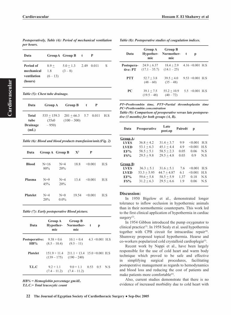
Cardiovascular Hossam F. El Shahawy et al
Car
diov
ascu
lar
The Journal of Egyptian Society of Cardiothoracic Surgery ● Sep-Dec 200522
Hossam F. El Shahawy et al Cardiovascular
Car
diov
ascu
lar
The Journal of Egyptian Society of Cardiothoracic Surgery ● Volum 13, Number (3-4) 23
Postoperatively, Table (4): Period of mechanical ventilation per hours.
Data Group A Group B t P
Period of mechanical ventilation (hours)
8.9 + 1.8 (6 – 13)
5.0 + 1.3 (3 – 8)
2.49 0.011 S
Table (5): Chest tube drainage.
Data Group A Group B t P
Total tube
Drainage (mL)
535 + 159.3(35s0 – 950)
201 + 66.3(100 – 300)
5.7 0.011 H.S
Table (6): Blood and blood products transfusion/unit.(Fig. 2)
Data Group A Group B X2 P
Blood N=16 80%
N=4 20%
18.8 <0.001 H.S
Plasma N=9 45%
N=4 20%
13.4 <0.001 H.S
Platelet N=4 20%
N=0 0.0%
19.54 <0.001 H.S
Table (7): Early postoperative Blood picture.
DataGroup A
Hypother-mic
Group BNormother-
mict p
Postoperative:HB%
9.38 + 0.6(8.5 – 10.4)
10.1 + 0.4(9.5 – 11)
4.3 <0.001 H.S
Platelet 151.9 + 11.4(139 – 175)
211.1 + 13.4(190 – 240)
15.0 <0.001 H.S
T.L.C 9.2 + 1.1(7.4 – 11.2)
9.0 + 1.1(7.4 – 11.2)
0.53 0.5 N.S
HB% = Hemoglobin percentage gm/dL.T.L.C.= Total leucocytic count
Table (8): Postoperative studies of coagulation indices.
DataGroup A
Hypother-mic
Group BNormother-
mict p
Postopera-tive: PT
24.9 + 6.37(17.1 – 35.7)
18.4 + 2.9(14.1 – 25)
4.16 <0.001 H.S
PTT 52.7 + 3.8(48 – 60)
39.5 + 4.0(35 – 48)
9.53 <0.001 H.S
PC 39.1 + 7.5(19.5 – 48)
55.2 + 10.9(40 – 72)
5.5 <0.001 H.S
PT=Prothrombin time, PTT=Partial thromboplastin timePC=Prothrombin concentrationTable (9): Comparison of preoperative versus late postopera-tive (3 months) for both groups (A, B).
Data Preoperative Late post.op Paired t p
Group A:LVESLVEDEF%FS%
36.8 + 4.253.1 + 6.358.5 + 5.129.5 + 9.8
31.4 + 3.745.1 + 4.458.5 + 2.329.5 + 4.8
9.94.90.050.03
<0.001 <0.001
0.060.9
H.S H.SN.SN.S
Group B:LVESLVEDEF%FS%
36.3 + 5.153.3 + 5.9559.6 + 5.831.2 + 6.3
31.6 + 5.144.7 + 4.8758.5 + 5.929.5 + 6.6
7.66.11.371.9
<0.001 <0.001
0.180.06
H.S H.SN.SN.S
Discussion:In 1950 Bigelow et al., demonstrated longer
tolerance to inflow occlusion in hypothermic animals than in their normothermic counterparts. This work led to the first clinical application of hypothermia in cardiac surgery(2).
In 1954 Gibbon introduced the pump oxygenator to clinical practice(3). In 1958 Sealy et al. used hypothermia together with CPB circuit for intracardiac repair(4). Shumway proposed topical hypothermia. Hearse and co-workers popularized cold crystalloid cardioplegia(5).
Recent work by Nappi et al., have been largely responsible for the use of cold heart and warm body technique which proved to be safe and effective in simplifying surgical procedures, facilitating postoperative management as regards to hemodynamics and blood loss and reducing the cost of patients and make patients more comfortable(6).
Also, current studies demonstrate that there is no evidence of increased morbidity due to cold heart with

Cardiovascular Hossam F. El Shahawy et al
Car
diov
ascu
lar
The Journal of Egyptian Society of Cardiothoracic Surgery ● Sep-Dec 200522
Hossam F. El Shahawy et al Cardiovascular
Car
diov
ascu
lar
The Journal of Egyptian Society of Cardiothoracic Surgery ● Volum 13, Number (3-4) 23
systemic normothermic CPB in valve replacement(7).In the present study 40 patients with mitral valve
disease were studied. Twenty patients underwent MVR under conventional systemic hypothermic CPB (Group A), the other 20 underwent MVR under systemic normothermic CPB (Group B) to evaluate the validity of systemic normothermic perfusion. All patients in both groups were subjected to cold crystalloid cardioplegic arrest with topical hypothermia. Both groups had similar anesthetic and operative management.
Evaluating the validity of normothermic CPB associated with topical hypothermia and cold cardioplegic technique proved to be safe, effective in simplefying surgical procedures and reduction of total bypass time(6).
In our study total bypass time was significantly re-duced in normothermic group (59.2 + 4.9 min) versus (66.5 + 4.7 min) in hypothermic group (P<0.001). This result correlates well the result of Tonz and coworkers, who studied 100 consecutive patients who underwent MVR to evaluate the effect of normothermic CPB and increased morbidity in valve surgery. Fifty eight patients were done under systemic normothermic bypass (venous temperature > 35oC), 42 were done under systemic hypo-thermic bypass (<28oC). The CPB time was significantly reduced in normothermic group versus hypothermic one (54+6.3 vs. 68 + 6 min, P= 0.02, mean + 1 standard error of the mean). The 2 groups were similar in terms of age, sex, preoperative functional class, pathology and preop-erative echocardiographic data(7).
In view of similarity of cross clamp time in both groups in our study and shorter reperfusion period in normothermic group (12.3 + 2.1 vs. 17 + 3.5 min, P<0.001), one might have expected that CPB time to be shorter in normothermic group than in hypothermic one. This result agrees with that of Moriyama and his colleagues who evaluate the influence of body temperature during CPB on postoperative systemic embolism. Their study included 32 patients divided into 2 groups; hypothermic (n=16) and normothermic (n=16). Analysis of their varience showed that CPB and operation time were significantly shorter in normothermic group than in hypothermic one (P<0.001). They attributed this finding to the reduction in reperfusion period which was 11.01+1.2 in normothermic group versus 14 + 4 min in hypothermic group(8).
The potasium requirement of the arrested heart is increasing as for the temperature goes up from hypothermia toward normothermia. This is more exaggerated when continuous or multidose techniques of cardioplegia delivery over cross clamp were used(9).
In our study rate of resumption of cardiac rhythm
was significantly higher in normothermic group compared to hypothermic one (90% vs. 20%) (Fig. 1). The need for DC shock was significantly reduced in normothermic group (10%) versus hypothermic one (80%) (P<0.001). This could be explained by higher threshold of ventricular fibrilation with systemic hypothermia which need more DC shock for resumption of rhythm(10). Samuel and co-workers obtained the same results with significantly higher rate of spontaneous defibrillation in systemic normothermic group (95%) as compared to only (16%) with systemic hypothermic group (P<0.001) (11).
In our study, reperfusion period was significantly shorter in normothermic group than in hypothermic group (12.3 + 2.1 vs 17 + 3.5 min). The reperfusion interval needed for successful weaning from bypass is an indicator for degree of postischemic contractile recovery of myocardium, so shorter reperfusion period was an argument in favour of better myocardial preservation with systemic normothermia(12).
In our study, during weaning of bypass the need for inotropic support (VP) in both groups was similar (10% in both groups, P= 1.0) but the use of vasodilator was significantly higher in hypothermic group than in normothermic group (90% versus 20%, P<0.001). After weaning from bypass, use of inotropic (VP) and vasodilators were significant in hypothermic patients (VP 70%, VD 90%) versus (VP 20% and VD 20%) in normothermic group and duration of use of these drugs was longer in hypothermic patients.
Our results correlate with those of Nappi and co-workers who conducted a randomized study on 100 patients, divided into 2 groups, hypothermic group A (n=50) and normothermic group B(50 patients). They found that in order to disconnect patients from CPB, VD drugs were infused in 96% of patients of group A and in 40% of group B. Also immediately after bypass and early postoperative period, positive inotropic agents and VD were used in 67% of patients in group A and in 22% of patients of group B (P=0.0003) (6).Higher rate of spontaneous resumption of rhythm, shorter reperfu-sion time and less need for inotropic agents suggested better myocardial and systemic vascular behaviour and performance after cross clamp period reflecting superior myocardial and systemic vascular regulation offered by systemic normothermic perfusion.
In our present study no neurological complications were detected in both groups which reflects the equal-at least- neuroprotective action of systemic normothermic perfusion compared to hypothermic one, indicating no further need to systemic hypothermia with its adverse effects on systemic body metabolic activity and organ

Cardiovascular Hossam F. El Shahawy et al
Car
diov
ascu
lar
The Journal of Egyptian Society of Cardiothoracic Surgery ● Sep-Dec 200524
Hossam F. El Shahawy et al Cardiovascular
Car
diov
ascu
lar
The Journal of Egyptian Society of Cardiothoracic Surgery ● Volum 13, Number (3-4) 25
functions. Our study was carried out at 34oC to 35oC in normothermic group and at 28oC in hypothermic group.
Singh and his colleagues compared a large series of patients undergoing CPB at 35oC versus 28oC. In their study the stroke incidence was 1% and 1.3% in the warm and cold groups respectively(13).
Hypothermia reduces platelet aggregation and endothelial-associated coagulation with subsequent increases in postoperative bleeding and requirements for blood products. Decreased bleeding in postoperative period associated with shorter intubation time and a lower requirement for inotropic support and so, shortens ICU stay(14).
Other reports had demonstrated better coagulation profile during normothermic bypass in comparison to cold bypass so; one can expect that rising systemic temperature will result in improvement in coagulation profiles(15). This suggestion was proved clinically in our study where the normothermic group had a highly significant reduction in postoperative blood loss with a mean of (212+66.3 ml) in comparison to that of cold group (535 + 159.3) (P=0.001).
These results are compatible with Birdi and coworkers who reported that total chest drainage was significantly reduced in patients undergoing normothermic CPB (34oC to 35oC), they also required less transfusions of blood (P<0.05 vs 28oC) and platelets (P=0.001 vs 28oC) in postoperative period which necessitate more transfusion in cold group(10).
These previous reports suggest a better coagulation profile with normothermic bypass in comparison to cold one and this clearly reflects the reduction for the need of blood transfusion with its known hazards and cost, decrease incidence of reexploration due to bleeding and reducing over all ICU and hospital stay.
LVEDD and LVESD usually decrease within one week after surgical relief of mitral valve pathology especially regurgitant lesion(16). This result correlates well with our results in the present study in both groups where the LVED and LVES were decreased in early and late postoperatively when compared with the preopera-tive measures. Waldermar and coworkers reported that warm body “normothermic CPB” during valve surgery is as equal as if not superior to techniques of hypother-mic CPB with significant improvement of postoperative systemic organs functions, but showed no difference in left ventricular dimensions between normothermic and hypothermic group of patients(1).
One of the most common features of hypothermic bypass is a rapid and early decrease in platelet number that exceeds the decrease explained by hemodilution of bypass alone(17). In their study they report a significant
reduction in platelet number in hypothermic group versus normothermic group (P<0.05).
The previous results correlate with our results where there was a marked significant reduction in platelet number in hypothermic group (P=0.001).
These results correlate with Parodi and coworkers who reported that hypothermic bypass patients usually have a decreased hemoglobin level and platelet number immediately and early postoperatively versus normothermic group (P<0.001 for platelet and P<0.05 for hemoglobin)(10).
Conclusion:The technique of cold heart and warm body proved to
be safe and effective in simplifying surgical procedures, protection of systemic organ functions and facilitating postoperative managements.
Our results provide reassurance that systemic normothermic bypass with cold cardioplegic arrest provides excellent myocardial and total body protection during valvular surgery and is particularly suitable for high-risk patients.
In conclusion, normothermic CPB is favorable because it can reduce costs, improve the management of a cardiac surgery unit and is more comfortable to the patients.
The warm body and cold heart strategy is not necessarily recommended for routine cases of mitral valve surgery. Its use is recommended in cases at high risk with border-line pulmonary function, patients with rare blood groups to decrease the need for tranfusions and those in whom a long cross clamp time is anticipated.
References:1. Waldermar G, Wojeiech K and Grazyna D (2000): The
central nervous system during CABG in hypothermia or normothermia. Evaluation based on serum level of S-100 protein. Polish Heart Journal; III-8.
2. Bigelow WG, Calleghan JC and Hopps JA (1950): General hypothermia for experimental intracardiac surgery. Ann Surg; 132: 531-539.
3. Gibbon JH (1954): Application of mechanical heart and lung apparatus to cardiac surgery. Minn Med; 37: 171-180.
4. Sealy WC, Brown Jr and Young Jr (1958): A report on the use of both extracorporeal circulation and hypothermia for open heart surgery. Ann Surg; 147: 603-613.
5. Hearse DJ, Stewart DA and Brainbridge MV (1996): Cellular protection during myocardial ischemia. The development and characterization of a procedure for the induction of reversible ischemic arrest. Circulation; 54: 193-202.
6. Nappi G, Torella M and Romano G (2002): Clinical evalu-ation of normothermic cardiopulmonary bypass and cold cardioplegia. J Cardiovasc Surg (Torino); 43(1): 31-36.
Cardiovascular Hossam F. El Shahawy et al
Car
diov
ascu
lar
The Journal of Egyptian Society of Cardiothoracic Surgery ● Sep-Dec 200524
Hossam F. El Shahawy et al Cardiovascular
Car
diov
ascu
lar
The Journal of Egyptian Society of Cardiothoracic Surgery ● Volum 13, Number (3-4) 25
7. Tonz M, Mihaljevic T, Pasic M, Von-Segeer LK, et al. (1998): Is normothermic cardiopulmonary bypass associ-ated with increased morbidity ? Helv Chir Acta; 60(3): 387-391.
8. Moriyama S, Utoh J, Okamoto K, et al. (1998): Clinical benefits of normothermic cardiopulmonary bypass on postoperative systemic metabolism. Jpn J Thorac Cardiovasc Surg; 46(2): 164-169.
9. Buckburg GD (1999): Update on current techniques of myocardial protection. Ann Thorac Surg; 60: 805-814.
10.Birdi I, Regragui I, Izzat MB, et al. (1997): Influence of normothermic systemic perfusion during coronary artery bypass operations: a randomized prospective study. J Tho-rac Cardiovasc Surg; 114(3): 475-481.
11. Samuel VL, Fremes SE, Abdel JG, et al. (2001): Technical aspects of warm heart surgery. Lancet; 1: 443-454.
12.Parodi E, Lijol A, Scarano F, et al. (2000): Normothermic versus hypothermic perfusion during cardiopulmonary bypass. Minerva Cardioangiol; 48(12): 435-440.
13.Singh AK, Bert AA, Feng WC, et al. (1997): Stroke during coronary artery bypass grafting using hypothermic versus normothermic perfusion. Ann Thorac Surg; 60: 84-89.
14. Timothy J and Raymond GR (2000): Should patients be normothermic in the immediate postoperative period. The Ann Thorac Surg; 1: 196-199.
15. Tonz M, Mihaljevic T, Von Segesser LK, et al. (1995): Normothermia versus hypothermia during cardiopulmonary bypass: a randomized, controlled trial. Ann Thorac Surg; 59(1): 137-143.
16.Crawford JM, Soucheck T, Oprain C, Miller S, Ratintolla K and Hammermeister K (1990): Determinants of survival and left ventricular performance after mitral valve replacement. Circulation; 81: 1173.
17.Rinder HM, Tracey JB, Techt M, et al. (1998): Differences in platelet granule release between normal and immune thromocytopenic patients. Thromb Haemost; 80: 457-462.

Hosam F. FawzyCardiovascular
Car
diov
ascu
lar
The Journal of Egyptian Society of Cardiothoracic Surgery ● Sep-Dec 200526
Hosam F. Fawzy Cardiovascular
Car
diov
ascu
lar
The Journal of Egyptian Society of Cardiothoracic Surgery ● Volum 13, Number (3-4) 27
Hosam F. Fawzy, MD Objectives:Use of the radial artery for coronary artery bypass grafting is getting more popular. We started routine use of the ultrasonic dissecting scalpel in harvesting radial arteries aiming to minimize harvesting time, improve graft quality and reduce wound complications. Methods: Radial artery harvesting technique using Harmonic Scalpel (43 patients) was compared with the conventional technique (Haemostatic clips and scissors) (53 patients). To avoid spasm, the radial artery was not skeletonized and papaverine was used to irrigate radial artery routinely in all patients.Results:Radial artery dissection with the Harmonic Scalpel had a sig-nificantly shorter harvesting time and required a significantly smaller number of haemostatic clips compared with the conventional technique. There was no forearm wound infection in Harmonic Scalpel group. There was no graft failure, reoperation for bleeding or hand ischemia with the use of either technique. Conclusion: Harvesting the radial artery using the Harmonic Scalpel is less time consuming and decreased the use of haemostatic clips rather atraumatic with good quality graft.
Carpentier (1) was the first surgeon to propose using the radial artery (RA) as a conduit for revascularization during coronary artery bypass grafting (CABG) procedures. However, because of RA spasm, which induced a 35% incidence of vessel occlu-sion, he later abandoned this procedure (Carpentier) (2). The
vasospasm was thought to be a consequence of the trauma of the harvesting procedure and thermal injury resulting from electrocautery (3&4). Graft occlu-sion resulting from subintimal hyperplasia was another problem that evoked against the use of RA(5) .
Since these early attempts, the use of new pharmacologic antispasmodic agents (e.g., Calcium channel blockers) and improved methods for harvesting replacement vessels have resulted in enhanced arterial patency and a resur-gence in the use of RA grafts for CABG(6). Comparative studies showed that the use of the radial artery improved the results of CABG surgery (7), perhaps because the radial artery is of good size and easier to anastomose sequentially than several of the other arterial conduits.
The use of the radial artery is a significant aspect of the “all arterial con-duits” approach that is currently favored and which is associated with im-proved long-term results.The ultracision Harmonic Scalpel (HS) (Ethicon Endo-Surgery, Inc., Cincinnati, OH) is used as an adjunct to or substitute for
Harvesting of the Radial Artery for Coronary Artery Bypass Grafting: Comparison of Ultrasonic Harmonic Scalpel Dissector with the Conventional Technique.
Accepted for publication Nov 10 , 2005
Address reprint request to Dr.
Hosam F. Fawzy
Cardiac Services Department,
Section of Cardiac Surgery,North
West Armed Forces Hospital,
Tabuk, Saudi Arabia.
Email : [email protected]
Codex : 04/12/cord/0512

Hosam F. FawzyCardiovascular
Car
diov
ascu
lar
The Journal of Egyptian Society of Cardiothoracic Surgery ● Sep-Dec 200526
Hosam F. Fawzy Cardiovascular
Car
diov
ascu
lar
The Journal of Egyptian Society of Cardiothoracic Surgery ● Volum 13, Number (3-4) 27
electrosurgery, lasers, and steel scalpels.The quality of radial arteries for CABG procedures
has improved as a result of using the HS rather than blunt and sharp dissection procedures. Ultrasonic dis-section of the RA with the HS has resulted in less spasm and a marked decrease in the number of clips required to control bleeding (8). Because this technique obviates the use of electrocautery, the possibility of thermal dam-age is reduced. Furthermore, while the surgeon is able to identify nerve structures, the use of the HS minimizes trauma to these structures, which could result in vessel spasm (9).
This study was designed to compare the HS and the conventional technique (clips and scissors) for harvest-ing of radial arteries for subsequent CABG procedures. Relevant information was collected on pre- and post har-vest artery quality, harvesting time, the number of clips used, wound and neurological complications.
Harmonic Scalpel Device: The HS is an ultrasonic surgical instrument for cut-
ting and coagulating tissues. It consists of a generator, hand piece and a disposable blade.
The electrical energy provided by the microproces-sor controlled-generator is converted into mechanical energy by the hand piece through a piezoelectric crystal system. The blade or tip of the instrument being used vibrates axially with a constant frequency of 55.5 KHz.
This form of mechanical energy produces less heat and less smoke during tissue dissection than does regular electrocautery. While electrocautery forms the coagulum by heating tissues to denature protein, the HS denatures the protein by the transfer of mechanical energy to the tissue, which is sufficient to break tertiary hydrogen bonds, and by the generation of the heat from internal cellular friction, which results from the high-frequency vibration of the tissue (10).
Material and Methods :Between August 2003 and December 2004, we
started to use the harmonic scalpel in our unit in har-vesting the radial arteries in all our patients for CABG procedure. The total number of patients was 43 patients. Most of them (91%) were male patients.
The mean age was 57.1 years (range 39-75 years). We retrospectively compared this group with a similar group of patients (53 patients), who had CABG done with the use of the radial artery harvested by the con-ventional technique (scissors and hemostatic clips) at our institution. Patients’ characteristics for both groups are shown in table No. (1).Table No. 1: Preoperative Clinical Characteristics of Patients
Undergoing CABG Using Radial Artery Harvested with Har-monic Scalpel Versus the Conventional Technique:
Characteristics HS(n = 43)
Conventional(n = 53)
P-value
Age > 65 years 10 (23%) 9 (17%) NSAge < 65 years 33 (77%) 44 (83%) NSMale Patients 39 (91%) 48 (91%) NSDiabetes Mellitus 20 (47%) 25 (47%) NSHypertension 22 (51%) 24 (45%) NSSmoking 15 (35%) 22 (42%) NSLVEF < 30% 4 (9%) 5 (9%) NSRecent MI 17 (40%) 17 (32%) NS Left Main Stenosis 2 (5%) 5 (9%) NSPrevious CABG 1 (2%) 1 (2%) NSUnstable Angina 6 (14%) 9 (17%) NSElective Operation 34 (79%) 45 (85%) NS
Preoperative preparation:The radial artery was usually harvested from the
non-dominant arm after a negative modified Allen test. Both the radial and ulnar arteries were occluded and the patient asked to tightly close his/her hand “make a fist” for 30 seconds.
The patient then relaxed the tight fist, the ulnar artery was released (with the RA still occluded) and the time for complete capillary refilling of the palmer surface of the hand was measured (in seconds). Refilling within 12 seconds was accep table for harvesting.
Technique of radial artery harvesting:A standard curvilinear incision over the radial artery
was used to facilitate harvesting the radial artery from its origin in the proximal forearm to the wrist crease.
Only the skin incision was performed with a surgi-cal blade. Low-grade current electrocautry used for the subcutaneous tissue and deep fascia.
A self-retaining retractor was placed between the brchioradialis and flexor carpi radialis muscles. Careful retraction of these muscles reveals the entire course of the radial artery in the forearm. Initial dissection was started from the medial side of the artery. The two satel-lite veins and the surrounding adipose tissue were left attached to the radial artery to preserve its blood supply as much as possible and prevent spasm.
The subsequent dissection differed for the two groups. In the conventional harvest group, mobiliza-tion of the RA pedicle was done using a combination of blunt and sharp dissection with control of vessel branches using two clips and division of the branches
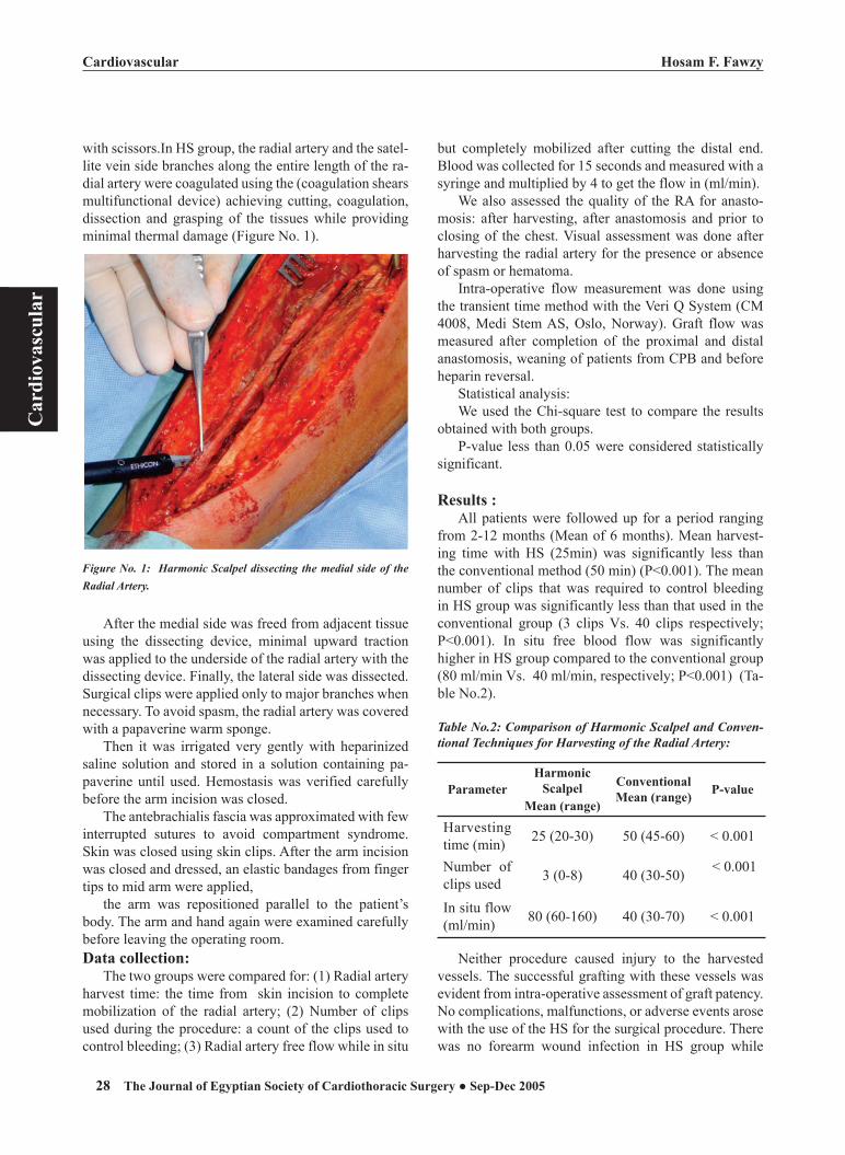
Hosam F. FawzyCardiovascular
Car
diov
ascu
lar
The Journal of Egyptian Society of Cardiothoracic Surgery ● Sep-Dec 200528
Hosam F. Fawzy Cardiovascular
Car
diov
ascu
lar
The Journal of Egyptian Society of Cardiothoracic Surgery ● Volum 13, Number (3-4) 29
with scissors.In HS group, the radial artery and the satel-lite vein side branches along the entire length of the ra-dial artery were coagulated using the (coagulation shears multifunctional device) achieving cutting, coagulation, dissection and grasping of the tissues while providing minimal thermal damage (Figure No. 1).
Figure No. 1: Harmonic Scalpel dissecting the medial side of the Radial Artery.
After the medial side was freed from adjacent tissue
using the dissecting device, minimal upward traction was applied to the underside of the radial artery with the dissecting device. Finally, the lateral side was dissected. Surgical clips were applied only to major branches when necessary. To avoid spasm, the radial artery was covered with a papaverine warm sponge.
Then it was irrigated very gently with heparinized saline solution and stored in a solution containing pa-paverine until used. Hemostasis was verified carefully before the arm incision was closed.
The antebrachialis fascia was approximated with few interrupted sutures to avoid compartment syndrome. Skin was closed using skin clips. After the arm incision was closed and dressed, an elastic bandages from finger tips to mid arm were applied,
the arm was repositioned parallel to the patient’s body. The arm and hand again were examined carefully before leaving the operating room.Data collection:
The two groups were compared for: (1) Radial artery harvest time: the time from skin incision to complete mobilization of the radial artery; (2) Number of clips used during the procedure: a count of the clips used to control bleeding; (3) Radial artery free flow while in situ
but completely mobilized after cutting the distal end. Blood was collected for 15 seconds and measured with a syringe and multiplied by 4 to get the flow in (ml/min).
We also assessed the quality of the RA for anasto-mosis: after harvesting, after anastomosis and prior to closing of the chest. Visual assessment was done after harvesting the radial artery for the presence or absence of spasm or hematoma.
Intra-operative flow measurement was done using the transient time method with the Veri Q System (CM 4008, Medi Stem AS, Oslo, Norway). Graft flow was measured after completion of the proximal and distal anastomosis, weaning of patients from CPB and before heparin reversal.
Statistical analysis:We used the Chi-square test to compare the results
obtained with both groups. P-value less than 0.05 were considered statistically
significant.
Results :All patients were followed up for a period ranging
from 2-12 months (Mean of 6 months). Mean harvest-ing time with HS (25min) was significantly less than the conventional method (50 min) (P<0.001). The mean number of clips that was required to control bleeding in HS group was significantly less than that used in the conventional group (3 clips Vs. 40 clips respectively; P<0.001). In situ free blood flow was significantly higher in HS group compared to the conventional group (80 ml/min Vs. 40 ml/min, respectively; P<0.001) (Ta-ble No.2).
Table No.2: Comparison of Harmonic Scalpel and Conven-tional Techniques for Harvesting of the Radial Artery:
ParameterHarmonic
ScalpelMean (range)
ConventionalMean (range) P-value
Harvesting time (min) 25 (20-30) 50 (45-60) < 0.001
Number of clips used 3 (0-8) 40 (30-50) < 0.001
In situ flow (ml/min) 80 (60-160) 40 (30-70) < 0.001
Neither procedure caused injury to the harvested vessels. The successful grafting with these vessels was evident from intra-operative assessment of graft patency. No complications, malfunctions, or adverse events arose with the use of the HS for the surgical procedure. There was no forearm wound infection in HS group while
Hosam F. FawzyCardiovascular
Car
diov
ascu
lar
The Journal of Egyptian Society of Cardiothoracic Surgery ● Sep-Dec 200528
Hosam F. Fawzy Cardiovascular
Car
diov
ascu
lar
The Journal of Egyptian Society of Cardiothoracic Surgery ● Volum 13, Number (3-4) 29
three patients developed superficial wound infection in the conventional group with no statistical significance. Three patients in HS and five patients in conventional group developed transient dysthesia of the thumb with no functional abnormalities.
In both groups, there were no cases of radial artery graft failure requiring reoperation. No re-exploration for bleeding from forearm wound was done in both groups. There was no clinical evidence of lack of collat-eral blood flow in the hand or ischemia in the arm from which the radial artery was harvested in both groups. All patients were alive and free of symptoms during the period of the study.
Discussion :Spasm occurs with harvesting of all arterial and ve-
nous conduits and appears to be related to mechanical and thermal trauma. Although ultrasonic dissection is associated with transformation of mechanical to thermal energy and protein denaturation, it generates less heat than electrocautery. Recent reports have suggested that use of the harmonic scalpel during radial artery harvest-ing may be less traumatic compared with conventional techniques, thereby decreasing spasm of this delicate conduit (8, 11, 12).
In our series, harvesting time was significantly less in HS compared with the conventional technique. While Ronan et al., (8) reported a comparable harvesting time in both groups (48.4 min in the standard group vs. 43.6 min in HS group, with insignificant P-value).
The HS provided better control of bleeding, which was accomplished without the potential for thermal in-jury associated with the use of electrocautery. Wright et al., (9) reported that the median number of clips required to control bleeding when the procedure was performed with HS was 0 (range of 0-28), which was significantly less (P<0.001) than the median of 69 clips (range of 25-102) used when the procedure was performed with the conventional technique. We also had a significantly less clips used in HS group than in the conventional group.
In an earlier report from Marc Moon et al., (13) it was noted that in situ flow through the radial artery was higher after harmonic versus conventional harvesting (53ml/min versus 17 ml/min; P<0.001). Ronan et al., (8) similarly reported a significantly greater flow in their HS group. These come in agreement with our results.Complications:
We only had three patients (7%) in HS group and five patients (9%) in the conventional group, who developed post-operative thumb dysthesia that resolved during the first two weeks post-operatively. Meharwal and Trehan (14) reported a 28% incidence of hand numbness or par-
athesia in their patients immediately after radial artery harvesting. Denton and associates (11) similarly report-ed a 30% incidence of hand complaints in their patients immediately postoperatively with most patients reported resolution by 9 months. Budillon and colleagues (15) re-ported sensory complaints in 9% at the time of hospital discharge, which decrease to only 4% at 8 weeks.
However, the impact of the harmonic scalpel on neu-rological injury in the immediate postoperative period remains unknown, as does the long –term incidence of neurological complications with either technique (13).
In the current report, forearm numbness was not specifically evaluated, but Connolly and coworkers (16) reported absence of forearm symptoms in the series. While others like Marc Moon et al., (13) reported fore-arm sensory loss in 10% of their patients.
As we have not used hydrostatic dilation because intraluminal pressure is difficult to quantitate, and if excessive may lead to endothelial or mural injury. This modality was used together with probe dilation in the early experience with the RA and may have contributed to conduit failure (17). A recent study has suggested that the HS may be the device of choice for harvesting radial arteries (8). The present study confirms these earlier findings that the HS can be used successfully to harvest radial arteries for CABG procedures.
Conclusion :Use of the Harmonic Scalpel for harvesting the ra-
dial artery for CABG, is less time consuming, decreases the use of hemostatic clips and offers some benefit in regards to flow dynamics immediately after harvesting. Therefore, the Harmonic Scalpel becomes our standard technique for harvesting the radial arteries in our CABG patients.
References :1- Carpentier A, Geurmonprez JL, Deloche A, et al. The aorta-
to-coronary, radial artery bypass graft: a technique avoiding pathological changes in grafts. Ann Thorac Surg 1973; 16: 111-21.
2-Carpentier A, Geha AS, Krone RJ, et al. Discussion of selec-tion of coronary, bypass anatomy, physiological, and angi-ographic considerations of vein and mammary artery grafts. J Thorac Cardiovasc Surg 1975; 70: 414-31.
3-Acar C, Jebara VA, Portoghese M, et al. Revival of the radial artery for coronary artery bypass grafting. Ann Thorac Surg 1992; 54:652-60.
4-Dietl C, Benoit C. Radial artery graft for coronary revascu-larization: technical considerations. Ann Thorac Surg 1995; 60:102-10.
5-Curtis JJ, Stoney WS, Alford WC Jr, et al. Intimal hyper-plasia: a cause of radial artery aortocoronary bypass graft

Hosam F. FawzyCardiovascular
Car
diov
ascu
lar
The Journal of Egyptian Society of Cardiothoracic Surgery ● Sep-Dec 200530
failure. Ann Thorac Surg 1975; 20: 628-35.6-Hayes EC, Lecuyer KM. A standard of care for radial artery
grafting. Am J Crit Care 1998; 7:429-35.7-Acar C, Jebara VA, Portoghese M, et al. Comparative
anatomy and histology of the radial artery and the internal thoracic artery: implication for coronary artery bypass. Ra-diol Anat 1991; 13: 283-88.
8-Ronan JW, Perry LA, Barner HB, Sundt TM. Radial artery harvest: comparison of ultrasonic dissection with standard technique. Ann Thorac Surg 2000; 69:113-4.
9-Wright CB, Barner HB, Gao A, Obial R, Bandy B, Perry L, Ronan, Kelly CR.The advantages of the harmonic scalpel for the harvesting of radial arteries forcoronary artery by-pass. The Heart surgery Forum. 2001; 3244: 226-230.
10-Posacioglu H, Atay Y, Cetindag B, Saribulbul O, Buket S, Hamulu A. Easy,harvesting of radial artery with ultrasoni-cally activated scalpel. Ann Thorac Surg 1998; 65:984-5.
11-Denton TA, Trento L, Cohen M, et al. Radial artery harvest-ing for coronary bypass operations: neurologic complica-tions and their potential mechanisms. J Thorac Cardiovasc Surg 2001; 121:951-6.
12-Galajda Z, Szentkiralyi I, Peterffy A. Neurologic complica-tions after radial artery harvesting. J Thorac Cardiovasc Surg 2000; 123:194-5.
13-Moon MR, Barner HB, Baily MS, Lawton JS, Moazami N, Pasque MK, Damiano RJ. Long –term neurological hand complications after radial artery harvesting using conven-tional cold and harmonic scalpel technique.Ann Thorac Surg 2004; 78:535-8.
14-Meharwal ZS, Trehan N. Functional status of the hand after radial artery,harvesting: results in 3,977 cases. Ann Thorac Surg 2001; 72:1557-61.
15-Budillon AM, Nicolini F, Agostinelli A, et al. Complica-tions after radial artery harvesting for coronary artery by-pass grafting: our experience.Surgery 2003; 133:283-7.
16-Connolly MW, Torrillo LD, Stauder MJ, et al. Endoscopic radial artery,harvesting: results of first 300 patients. Ann Thorac Surg 2002; 74:502-6.
17-Van Son JAM, Tavilla G, Noyez L. Detrimental squelae on the wall of the,internal mammary artery caused by hydro-static dilation with diluted ,papaverine solution. J Thorac Crdiovasc Surg 1992; 104: 972-6.
Amr Mohamed Rushdi et al Cardiovascular
Car
diov
ascu
lar
The Journal of Egyptian Society of Cardiothoracic Surgery ● Volum 13, Number (3-4) 31
Valve Sparing Operations for Type a Aortic Dissection: Initial Experience and Early Results
Background: Valve-sparing operations for type A aortic dissection are increasingly accepted, but techniques and results are still in evolution. Wesummarized our experience to determine early outcomes.Methods : From October 2003 to May 2005, we studied prospectively 25 patients with type A aortic dissection. 14 patients had Supra commissural ascending aortic replacement (Group A). The remodeling technique was performed in 5 patients (Group B), while aortic root replacement with a valved conduit was done in 6 patients (Group C). Mean age of patients was 53.7±8.7 years In Group “A”, 51.8±9.9 years in group “B”, and 59.8±11 years in group “C”. 30.8% of patients in group “A”, 40% of patients in group “B”, and 50% of patients in group “C” suffered from Marfan syndrome.Results : operative mortality was 14.3% in group A, and late mortality was 16.6% in group C. There was no mortality in group B. 21.4% of patients in group “A”, 20% of patients in group “B” , and 16.6%1of patients in group “C” had a delayed recovery of conscious level with complete remission postoperatively. In group” A” two patients (14.3%)had a Coronary artery bypass grafting and one patient (7.1%) had mitral valve repair with commissural stitches. Two patients (14.3%) in group “A” had a complete arch replacement. 20% of patients in group “B” and 33.3%of patients in group “C” had a hemiarch replacement. Bypass time was 165 ±30 minutes in group “A”, 180±45 minutes in group B and 152±8 minutes in group C. Patients were followed up clinically and echocardiographically for 10.9 ± 7.4 months (group A), 12.6 ±8.6 months (group B) and 12 ± 7.8 months (group C). There were no aortic regurge in 21.4%of patients in group” A” and 40 of patients in group “B”. Grade I-II aortic regurge was found in 57.1% of patients in group “A”, and 40% of patients in of group “B”. 14.2% of patients in group “A” and 20 % of patients in group “B” had reoperation for aortic valve incompetence.Conclusions: Valve sparing operations in type A dissection can be performed with adequate short term results. Favorable durability of valve function encourages wider use of this technique.
Severe aortic regurge represents one of the major complications of the aneurysmatic aorta despite the structural integrity of aortic cusps. Ectasia of the aortic root may cause aortic valve insuffi-ciency by dilation of the sinotubular junction or aortic annulus or both1. Moreover, in acute dissection of the ascending aorta, usually
the entry is located in the ascending aorta just above the valve commissures2. In majority of patients the aortic root is also involved by retrograde dissection leading to acute aortic insufficiency when the commissural area is detached
Accepted for publication Dec 20,05
Address reprint request to Dr A.M.
Rushdi
Department of Cardio-thoracic Surgery
University of Cairo Medical school
Adress: 9, Galal El din El hahamsy st,
Agouza,Giza
E-mail: [email protected]
Codex :04/15/arvs /0512
Amr Mohamed Rushdi, MD Tarek Hussein El-Taweel, MD Mohamed Helmi, MD Saed Abdelaziz Badr, MD Ahmed Helmi, MD

Cardiovascular Amr Mohamed Rushdi et al
Car
diov
ascu
lar
The Journal of Egyptian Society of Cardiothoracic Surgery ● Sep-Dec 200532
Amr Mohamed Rushdi et al Cardiovascular
Car
diov
ascu
lar
The Journal of Egyptian Society of Cardiothoracic Surgery ● Volum 13, Number (3-4) 33
and prolapsing. However the dissecting process does not affect the aortic annulus or the aortic leaflets 3.
Replacement of the ascending aorta and aortic valve with a valved conduit is the standard surgical therapy for patients who have hemodynamically relevant aortic incompetence secondary to enlarged ascending aorta. Reported results demonstrate that this technique achieved excellent results4, 5. However, valve sparing aortic root reconstruction has the potential for avoiding prosthetic valve disease including thromboembolism, hemodynamic mismatch, hydraulic dysfunction, and bacterial endocarditis6. It offers the advantage of a near physiologic native valve function 7.
Within the past decade, valve-sparing aortic root replacement has evolved into an increasingly accepted treatment modality for patients with proximal aortic disease and valve regurgitation with good long-term results8,9,10,11,12. Despite these results, concern remains over the risk of recurrent aortic insufficiency13. Principally three different surgical techniques are used aiming at the preservation of the aortic valve leaflets: firstly the conventional supracommissural replacement of the ascending aorta after reconstruction of the dissected sinuses including resuspension of the detached commissures14; secondly the remodeling technique15, and thirdly the reimplantation technique 7. This study summarizes our experience over the past 2-years, with supracommissural aortic root replacement and remodeling valve sparing techniques.
Patients and Methods From October 2003 to May 2005, 25 patients with
Stanford type A aortic dissection underwent surgery performed by one surgeon in kaser El- Aini hospitals. Most patients of acute dissection were operated upon within 24 hours after the onset of symptoms (6 hours to 40 hours). All patients had thin normal aortic valve cusps and aortic regurge as proved by preoperative echocardiography and by actual intraoperative observation. If the diameter at the level of the sinotubular junction did not exceed 30 mm, supracommissural ascending aortic replacement with valve resuspension was performed in a standard fashion (group A, n=14). In patients with moderate dilatation of the aortic root (sinutubular diameter less than 50 mm, aortoventricular diameter less than 30 mm), remodeling of the aortic root was performed (group B, n=5). In Patients with severe retraction or calcification of the aortic valve cusps, and with severe root dilatation (sinutubular diameter more than 50 mm, aortoventricular junction more than 30
mm), replacement of the ascending aorta with a valved conduit was performed (group C, n=6).Patient’s characteristics are listed in table 1.
Table 1: Patients characteristics
Group A(n-14)
Group B (n=5)
Group C(n=6)
Age (years) 53.7±8.7 51.8±9.9 59.8±11
Male gender 12(85.7%) 4(80%) 5(83%)
Marfan syndrome
4(30.8%) 2(40%) 3(50%)
Acute dissection 12(85.7%) 5(100%) 3(50%)
Chronic dissection
2(14.3%) 0 3(50%)
LVEF>60% 2(14.3%) 1(20%) 1(16.7%
LVEF40–59% 8(57%) 3(60%) 4(66.7%)
LVEF20–39% 4(30.8%) 1(20%) 1(16.7%)
Coronary artery disease
2(14.3%) 0 0
Mitral regurgitation
1(7.7%) 0 0
Transverse arch aneurysm
2(14.3%) 1(20%) 2(33.3%)
AI (grade I-II) 11(78.6% 3(60%) 2(33.3%)
AI (grade III-IV)
3(21.4%) 2(40%) 4(66.7%)
LVEF=Left ventricular ejection fraction, AI=Aortic insufficiencyPercentages are shown in parentheses.
Operative Technique Standard median sternotomy and extracorporeal
circulation techniques with a membrane oxygenator were used in all patients. Arterial inflow of cardiopulmonary bypass was held through non dissected right axillary arteries in 10 patients of all groups. Non dissected femoral arteries were used in the rest.
An 8 mm Dacron graft was anastomosed end to side to the artery using running 6/0 polypropylene suture to which the arterial line is connected. Venous return was done via bicaval cannulae.
Systemic hypothermia of 28°C was used when arrest of the circulation was not required, and moderate hypothermia of 24°C to 22°C nasopharyngeal temperature was used when circulatory arrest was planned. Hypothermic circulatory arrest was used in all patients except only 3 patients of group” C “to perform an open distal anastomosis at the origin of the aortic arch

Cardiovascular Amr Mohamed Rushdi et al
Car
diov
ascu
lar
The Journal of Egyptian Society of Cardiothoracic Surgery ● Sep-Dec 200532
Amr Mohamed Rushdi et al Cardiovascular
Car
diov
ascu
lar
The Journal of Egyptian Society of Cardiothoracic Surgery ● Volum 13, Number (3-4) 33
or to partially or completely replace the arch. Retrograde cerebral perfusion was done in all patients except 4 patients of group” A” who had an antegrade cerebral perfusion. The later was done via the axillary artery while clamping the inominate artery on the right side, and via direct cannulation of left common carotid artery.The final decision regarding the type of surgery was made after aortotomy and aortic valve and root inspection. Diameters of sinutubular and aortoventricular junction were measured within the true lumen. Valve-sparing was done only in patients with macroscopically intact valve with mild to moderate extension of the dissection into the sinuses. Intermittent cold blood or crystalloid cardioplegia was administered directly into the coronary ostia in for myocardial protection all patients.
The distal aorta was completely transected at the site of the anastomosis. The distal anastomosis of graft to transected aorta was made with continuous 3-0 or 4-0 polypropylene suture (Prolene, Ethicon Inc) usually buttressed with a Teflon felt strip on the outside of the aorta.
Annulus stabilization In Marfan patients, an aortic annuloplasty was done
in both techniques to prevent future annular dilatation. ( except the first patient of group “B”). A band of Dacron fabric is sutured on the outside of the ventricular outflow tract. Multiple interrupted horizontal mattress sutures were placed through a single horizontal plane on the fibrous components of the left ventricular outflow tract and through the Dacron band beneath the commissures of the noncoronary cusps where Most of the dilation of the aortic annulus occurs.
Supra commissural replacement techniqueThe ascending aorta was transected 5mm above
the commissures. The latter were resuspended with pledgeted 3-0 polypropylene mattress suture. Depending on the friability of the tissues, the dissected components were readapted and strengthened either by multiple rows of an interrupted mattress sutures with small felt pledgets, or by an interposed Teflon felt fixed with gelatin-resocrine-formol glue ([GRF] Cardial, Saint E’tienne, France).
The proximal anastomosis of the graft to the supra-commissural area was made by continuous 3-0 or 4-0 polypropylene suture. The 3 commissures were spaced equidistantly in the graft.
Remodeling technique Diseased aortic sinuses were excised up to a
remnant of 2 to 3 mm. Coronary arteries were detached from the aortic wall, preserving buttons of aorta surrounding the ostia and the main coronary arteries were gently mobilized from surrounding tissues. The three commissures were stretched in a vertical direction by the use of three horizontal mattress sutures placed just above the top of each commissure. Ideal valve coaptation was considered to occur when 30% to 50% of the cusp area was involved. The distance between the commissures in their new position and the diameter of the left ventricular outflow tract determine the size of the Dacron tube. The end of the Dacron graft was trimmed to produce three separate tongue- shaped extensions in proportion to the size of each cusp. The width of each extension is measured to be equal to one third of the circumference. The initial length of the tongue-shaped process is made to equal at least one and
Figure 1. (a) Intraoperative view of an acute type A dissection. One can clearly see the site of the entry tear in the intima 1.5 cm above the aortic valve (A). (b) Diagram showing the remodeling technique. The three tongues of the Dacron graft were sutured to the base of the sinuses, while the commissural areas were resuspended.

Cardiovascular Amr Mohamed Rushdi et al
Car
diov
ascu
lar
The Journal of Egyptian Society of Cardiothoracic Surgery ● Sep-Dec 200534
Amr Mohamed Rushdi et al Cardiovascular
Car
diov
ascu
lar
The Journal of Egyptian Society of Cardiothoracic Surgery ● Volum 13, Number (3-4) 35
a half times the depth of the patients’ own aortic sinuses as measured from the top of the commissures to the bottom of the cusp. This allowed replacing the sinuses with a special technique to allow them to bow outward. The position of the commissures in the tube graft were secured with sutures first while the depth of the sinuses were determined later (Fig 1- B). In 2 patients, the left coronary sinus was found relatively intact, so we did not replace it. Thereafter proximal anastomosis to the aortic annulus was done with three 4-0 polypropylene sutures using the running technique. To complete the aortic root reconstruction, the coronary buttons were attached directly to openings made in the aortic tube graft with 6-0 polypropylene suture. In cases with more extensive ascending aortic or arch replacements, a second prosthesis was used.
Follow up and data collection Data were collected prospectively from patients
files and gathered using an office 2003 excel® software. Routine long-term management following the valve sparing operations included aspirin for anticoagulation and β-blockers to minimize hemodynamic stress. In patients with prosthetic valves, Warfarin was used for anticoagulation. Antibiotic prophylaxis for endocarditis was also recommended. All survivors were followed clinically with transthoracic color Doppler echocardiography after 3, 6, and 12 months and at yearly intervals thereafter. Valve morphology and systolic and diastolic function were assessed. Aortic regurgitation was assessed semiquantitatively as follows: 0, none; I, minimal; II, mild; III, moderate; and IV, severe. Infectious, thromboembolic, and bleeding complications were recorded as required by the guidelines of the American Association for Thoracic Surgery/Society of Thoracic Surgeons16.
Results Perioperative Mortality
There was no operative mortality in groups “B and C”. Two patients (14.3%) died in group” A”, one after 4 days due to multiorgan failure, and one patient after 5 days due to brain death.
Neurological complicationsThree patients (21.4%) in group “A”, one patient in
group “B” (20%) , and 1 patient in group “C” (16.6%) had a delayed recovery of the conscious level (>24 hours) with complete remission postoperatively. One patient in group “A” (7.1%) had a massive stroke on top of a pre-existing non-haemorrhagic stroke. The later was secondary to preoperative great vessels dissection.
Perioperative outcomeOperative and perioperative variables are listed
in table 2. In group” A” two patients (14.3%)had a Coronary artery bypass grafting and one patient (7.1%) had mitral valve repair with commissural stitches. Two patients (14.3%) in group “A” had a complete arch replacement. One patient in group “B” (20%) and two patients in group “C” (33.3%) had a hemiarch replacement.
Table 2: Operative and peri-operative data
Group A(n=14 )
Group B(n=5 )
Group C(n=6)
Axillary artery cannulation
6(42.8%) 2(40%) 2(33%)
Circulatory arrest time(min)
35.6±5.6 34.5±7.8 39.3±5
Bypass time(min) 165.8±30 180±45 152.8±18
Cross clamp time (min)
117.1±28.3 152.4±37.2 128.3±18.7
Graft diameter( range 26-30)
27.6±1.2 28±1.4 28.7±1
Re-exploration for bleeding
1(7.1%) 0 1(16.6%)
ICU stay(days) 3.65±1.1 3.4±1.1 3.3±1
Hospital stay(days) 11.8±2.3 11.2±2.4 10.8±2.5
Follow-upPatients were followed up to 10.9±7.4 months in
group A, 12.6±8.6 months in group “B” , and 12±7.8 months in group “C”.( range between 3-24 months). There were no aortic regurge in three patients (21.4%) of group”A” and two patients of group “B”. Grade I-II aortic regurge was found in 8 patients (57.1%) of group “A”, and two patients (40%) of group “B”. 2 patients (14.2%) of group “A” required reoperation for grade III-IV aortic regurge one after 2 months and the other after 8 months. One patient (20%) of group “B” had reoperation for aortic valve failure after 10 months. One patient (16.6%) had a reoperation for an early prosthetic valve endocarditis 40 days postoperatively. This patient died 3 days after the second operation due to septic shock.
Discussion :This study summarizes our early experience of
valve- sparing aortic root replacement. Potential recurrence of aortic insufficiency has been a proposed as a limitation of this technique. Progressive dilatation of the aortic annulus in Marfan patients 10, accelerated
Cardiovascular Amr Mohamed Rushdi et al
Car
diov
ascu
lar
The Journal of Egyptian Society of Cardiothoracic Surgery ● Sep-Dec 200534
Amr Mohamed Rushdi et al Cardiovascular
Car
diov
ascu
lar
The Journal of Egyptian Society of Cardiothoracic Surgery ● Volum 13, Number (3-4) 35
leaflet degeneration as a consequence of systolic contact between leaflet and the aortic wall due to lack of expansion of the sinuses17 , and sagging of the leaflet coaptation below the annulus due to inadequate re-suspension of the commissures18 have been attributed as causes of failure of valve sparing techniques.
To avoid those potential causes of failure we measured the true internal diameter of the aortic annulus with a Hegar dilator while the commissures were pulled superiorly to an apparent appropriate sinotubular junction diameter. The coaptation of the leaflets was tested with a small amount of saline. If the semi-lunar valve cusp coaptation areas appear to be less than optimal, then the next size down graft is usually selected. Furthermore, we buttressed the base of the aortic root with Dacron strip secured with sutures placed from beneath the valve leaflet base to prevent ‘annulus’ late dilatation in patients with connective tissue disorders.
There remains some controversy regarding the optimal surgical techniques used for proximal repair at initial operation. Supracommissural tube graft replacement with aortic valve resuspension represents the most often adopted technique (14/19 patients) as it shortens the operation time. On the other hand, this conservative treatment exposes the patient to long-term changes of both aortic valve and aortic root. Clinically important aortic valve regurgitation was found in two Marfan patients of group” A” (14.2 %). Mazzucotelli, Graeter and their assocaites reported an incidence of 20% to 45 % of aortic regurge in patients after supracommissural tube graft replacement 19, 20.
The advantage of the remodeling technique is that it is easier technically than the reimplantation method and provides a simpler method for accurate re-suspension of the pillars. Remodeling methods leave the dynamic expansion of the aortic root complex functionally intact, which should be advantageous in optimizing leaflet performance and durability and reducing the opposition to ejection, making left ventricular work more efficient. This also affects the position and degree of tethering of the base of the cusps 21. Additionally the outward bowing of the neo-sinuses favors the development of Da Vinci eddy currents behind the leaflets, contributing to a smoother closure of aortic valve, and reducing leaflet fatigue 22.
We reported a single patient (20%) in the remodeling group who had a reoperation for severe aortic regurge 10 months postoperatively. He was the only Marfan patient who did not receive an aortic annuloplasty. In a report by Yacoub and associates, who used the remodeling technique without annuloplasty in 158 patients, the freedom from aortic root replacement was
89% at 10 years, and moderate aortic incompetence was documented in one third of the patient’s postoperatively9.
Furthermore, avoidance of the long-term use of anticoagulation is believed to favor thrombosis of the false channel, thus preventing subsequent dilatation of the aorta23. Distal surgical repair fails to achieve this objective in most cases (up to 78%) 24. There were no endocarditis or new thromboembolic events among patients of groups “A” and “B”, supporting the rationale of using valve sparing techniques. The single patient of group “C” who had an event of endocarditis was found to have a preoperative dental caries.
Conclusion : Valve-sparing root replacement definitely appears
as a reasonable option for those patients who develop dissection because of preexistent root pathology. However, for many patients supracommissural root replacement still remains a relatively simple and reproducible therapeutic approach. The remodeling approach has been more complex, as reflected by longer cross-clamp times. Finally, we conclude that the valve-sparing approach can be used in the setting of type A dissection with good results. However, Long-term observations will be necessary to assess whether these valve-preserving operations will minimize the need for proximal reoperation comparable to that seen in composite replacement.
References :1- David TE, Feindel CM, Bos J. Repair of the aortic valve in
patients with aortic insufficiency and aortic root aneurysm. J Thorac Cardiovasc Surg 1995;109:345–52.
2-Ehrlich MP, Ergin MA, McCullough JN, et al. Results of immediate surgical treatment of all acute type A dissections. Circulation 2000;102(Suppl 3):248–52.
3- Yacoub M. Valve-conserving operations for aortic root aneurysm or dissection. In: Cox JL, Sundt TM, eds. Operative techniques in cardiac and thoracic surgery: a comparative atlas. Philadelphia: WB Saunders, 1996:57–67.
4- Kouchoukas NT, Wareing TH, Murphy SF, Perillo JB. Sixteen-year experience with aortic root replacement : results of 172 operations. Ann Surg.1991;214:308-320
5- Ehrlich MP, Ergin MA, McCullough JN, et al. Favorable outcome after composite valve-graft replacement in patients older than 65 years. Ann Thorac Surg 2000;71:1454–1459.
6- Cabrol C, Pavie A, Mesnildrey P, et al. Long-term results with total replacement of the ascending aorta and reimplantation of the coronary arteries. J Thorac Cardiovasc Surg.1986;91:17–25.
7- David TE, Feindel CM. An aortic valve-sparing operation
Cardiovascular Amr Mohamed Rushdi et al
Car
diov
ascu
lar
The Journal of Egyptian Society of Cardiothoracic Surgery ● Sep-Dec 200536
for patients with aortic incompetence and aneurysm of the ascending aorta. J Thorac Cardiovasc Surg 1992;103:617–621.
8-Erasmi AW, Stierle U, Matthias Bechtel JF, et al. Up to 7 Years’ Experience With Valve-Sparing Aortic Root Remodeling/Reimplantation for Acute Type A Dissection Ann Thorac Surg 2003;76:99 –104.
9- Yacoub MH, Gehle P, Chandrasekaran V, et al. Late results of a valve-preserving operation in patients with aneurysms of the ascending aorta and root. J Thorac Cardiovasc Surg 1998;115:1080–90.
10- David T.E., Ivanov J., Armstrong S., et al .Aortic Valve-Sparing Operations in Patients With Aneurysms of the Aortic Root or Ascending Aorta. Ann Thorac Surg 2002;74:S1758–1761.
11- Bethea B.T., Fitton T.P., Alejo D.E., et al .Results of Aortic Valve-Sparing Operations:Experience With Remodeling and Reimplantation Procedures in 65 Patients. Ann Thorac Surg 2004;78:767–72.
12- Settepani F., Ornaghi D., Barbone A. , et al. Aortic valve-sparing operations in patients with aneurysms of the aortic root or ascending aorta: preliminary results. Interactive CardioVascular and Thoracic Surgery 2005;4: 137–139
13-. Leyh R G, Fischer S, Kallenbach K, et al.High Failure Rate After Valve-sparing Aortic Root Replacement Using the “Remodeling Technique” in Acute Type A Aortic Dissection. Circulation 2002; 106[suppl I]: I-229-I-233.
14. Weinschelbaum EE, Schamun C, Caramutti V, et al . Surgical treatment of acute type A dissecting aneurysm, with preservation of the native aortic valve and use of biologic glue. J Thorac Cardiovasc Surg. 1992;103:369–374.
15- Sarsam MAI, Yacoub M. Remodeling of the aortic valve annulus. J Thorac Cardiovasc Surg 1993;105:435–438.
16-Edmunds LH Jr, Clark RE, Cohn LH, et al. Guidelines for reporting morbidity and mortality after cardiac valvular operations. Ann Thorac Surg. 1996; 62:932–935.
17-Casselman FP, Tan ES, Vermeulen FE, et al. Durability of aortic valve preservation and root reconstruction in acute type A aortic dissection. Ann Thorac Surg. 2000; 70:1227-33.
18-Kallenbach K, Hagl C, Walles T, et al. Harringer W. Results of valve-sparing aortic root reconstruction in 158 consecutive patients. Ann Thorac Surg 2002;74:2026–33.
19. Mazzucotelli JP, Deleuze PH, Baufreton C, et al. Preservation of the aortic valve in acute aortic dissection: long-term Echocardiographic assessment and clinical outcome. Ann Thorac Surg. 1993;55: 1513–1517.
20 Graeter TP, Langer F, Nikoloudakis N, et al. Valve-preserving operation in acute aortic dissection type A. Ann Thorac Surg 2000;70:1460–5.
21-Lansac E, Lim HS, Shomura Y, et al. A four-dimensional study of the aortic root dynamics. Eur J Cardiothoracic Surg 2002;22:497–503.
22- Underwood MJ, EL Khory G, Glineur D, Dion R. The aortic root: structure, function, and surgical reconstruction. Heart 2000;83:376-380
23-Ergin MA, Philips RA, Galla JD, et al. Significance of distal false lumen after type A dissection repair. Ann Thorac Surg 1994;57:820–824.
24- Kirsch M, Soustelle C, Houël R, et al. Risk factor analysis for proximal and distal reoperations after surgery for acute type A aortic dissection. J Thorac Cardiovasc Surg 2002;123:318-25

Hany A. Maboud Cardiovascular
Car
diov
ascu
lar
The Journal of Egyptian Society of Cardiothoracic Surgery ● Volum 13, Number (3-4) 37
Hany A. Maboud, MD background ; Atherosclerotic disease of the aorta has been identified as a risk factor for neurologic complication following coronary artery by-pass grafting (CABG). Neurologic impairment after CABG is associated with cerebral embolization . An important cause of embolism is aortic manipulation . Constructing both distal and proximal anastomoses dur-ing a single period of aortic cross-clamping avoids this source of embolism and may reduce neurologic injury after CABG . Methods: To determine the myocardial and cerebral protective properties of the single cross-clamp (group I; n=100) versus the partial occluding clamp (group II; n 90) technique for construction of the proximal anasto-moses ,patients undergoing isolated ABG by a single surgeon were identi-fied as having double clamp technique (DCT)(aortic cross-clamp+ side-biting clamp)or single clamp technique(SCT) (aortic cross-clamp only) . Results: 100 patients had SCT and 90 patients had DCT performed . In-traoperatively ,patients with SCT had shorter bypass times (76minutes vs 80minutes, p=0.09),longer aortic cross clamp time (58 minutes vs 44 minutes, p=0.0001) fewer coronary grafts (2.4 vs 3.1,p =0.001 ).and had a higher mean arterial blood pressure on cardiopulmonary bypass (70 mmHg vs 65 mmHg ,p=0.001) Postoperatively, the SCT group had fewer stroke (1 vs 3.3%, NS) ,and neurologic injuries (3.1%vs 8.9%,p= 0.007). By multivariate analysis, factors that were related to neurologic injury were DCT (p=0.04), age (p= 0.001),and number of coronary grafts (p=0.04). Conclusion : The results of this study suggest improved cerebral protection is associated with the single aortic cross-clamp technique for CABG with no increase in myocardial damage .
The benefits of coronary artery bypass grafting (CABG) over medical therapy and catheter-based interventional procedures are limited by the complications of the operation .Although the overall incidence of complications after CABG has decreased ,the incidence of postoperative stroke remains unchanged and is re-
ported in 0.8 to 6% of patients after CABG.(1) There have been many attempts to identify patients at higher risk, not just increasing age .In two recent studies (2 , 3) similar preexisting medical variables were found to be risk factors in the identification of high –risk patients , ie, past stroke ,hypertension, increased age , carotid artery disease, aortoiliac arterial disease, a recent myocardial infrac-tion and prior history of cerebrovascular event .Such factors are also related to the development of atherosclerotic disease . These ,as well as intra-operative
Impact of Single Clamp Technique :an Important Ad-junct to Myocardial and Cerebral Protection in Coro-nary Operation
Accepted for publication Nov 10,2005
Address reprint request to Dr A Maboud
Department of Cardio-Thoracic Surgery
Ain Shams University
Cairo , Egypt
Email : [email protected]
Codex : 04/14/cord/0512

Hany A. MaboudCardiovascular
Car
diov
ascu
lar
The Journal of Egyptian Society of Cardiothoracic Surgery ● Sep-Dec 200538
Hany A. Maboud Cardiovascular
Car
diov
ascu
lar
The Journal of Egyptian Society of Cardiothoracic Surgery ● Volum 13, Number (3-4) 39
factors ,have been implicated in contributing to stroke risk ,including hypoperfusion and embolic phenomena .For example ,atherosclerotic disease involving the as-cending aorta has been found contribute to the release of multible emboli during aortic manipulation and clamp-ing during cardiac surgery operations (4, 5) . Among the strategies developed to minimize aortic clamping/aortic trauma ,and therapy to prevent neurologic complica-tions, is the use of a single clamp technique (SCT) rather than a double clamp technique (DCT) requiring use of both an aortic cross clamp and a side-biting clamp. In this study we examined and compared the use of SCT versus DCT to determine the impact of the technique used to construct the proximal anastomoses on adverse outcome rates related to myocardial and cerebral protec-tion by analyzing the clinical end points of perioperative death, the myocardial infarction/low cardiac out put state, and stroke rates .
Material and methods :From January 2003 to December 2004, 190 consecu-
tive patients underwent a primary CABG procedure per-formed by the same surgeon at Ain Shams University and specialized Hospitals .They ranged in age from 45 to 70 years, with a median age of 65 years ; 25%were 70 years and older ;34% were female ; 40%were dia-betics . Patients were divided into two groups. In group 1,a single period of aortic cross-clamping was used to construct distal and proximal anastomoses . In group 2, the cross-clamp was removed after construction of the distal anastomoses and the proximal anastomoses were each constructed with a partial occluding aortic clamp .The decision regarding the clamping technique to be used was not randomized, but a selection bais did exist in that elderly and sicker patients were more likely to be in group 1 . Patients with history of cerebrovascular disease, carotid bruit , aortic calcification, atrial fibril-lation, or any patients who had a combined CABG and valvular or congenital heart procedures were excluded . Anesthetic ,cardiopulmonary bypass ,and myocardial protection techniques were standardized . The bypass circuit used a hollow-fiber membrane oxygenator, nonpulsatile flow ,40 µm arterial line filter .Flow was 2.4 L.min/m² at37ºC falling to 1.8 L at 32ºC . Arterial pressure was maintained between 50 and 70 mm Hg ,hematocrit between 0.20 and 0.25, and alpha stat blood gas management was used .Cold blood cardioplegia was given antegrade every 20 minutes . For patients in the SCT group ,the aortic cross clamp remained in place for both distal and proximal coronary anastomoses. For patients in the DCT group ,the aortic cross clamp was applied and distal anastomoses were made . The aortic
cross clamp was then released and the sidebiting clamp was applied once or twice ,after which the proximal cor-onary anastomoses was made. The clinical diagnosis of stoke made by the neurologist independent of confirma-tion by brain imaging. Fifty percent of the stroke were confirmed by imaging in this series. A major stroke was defined as the appearance of a permanent neurologic deficit upon recovery from anesthesia that was likely to be related to atheroembolism caused by manipulation of the ascending aorta A myocardial infarction was defined by the presence of a new Q wave on the electrocardio-gram ,or an elevated creatine kinase MB isoenzzyme re-lease of 40 IU/L or greater in the presence of a new seg-mental wall motion abnormality on echocardiograms, or both of these findings .The low cardiac output state was defined as the need for more than one inotropic agent or as the need for an intraaortic balloon pump ,or both. Adverse outcome was defined as the occurrence of any of the just described events.
Statistical analysis:Univariate analyses were done with χ² or Fisher´s
exact test and the Wilcoxon rank sum test. Multivariate stepwise logistic regression analyses were used to iden-tify independent variables that predict a perioperative adverse outcome.
Results :
Table (1 ) the preoperative and intraoperative variables in group (1& 2 )
Characteristic Group 1(100)
Group 2 (90)
P Value
Age (y)Patients ≤70female patientsdiabetic patientsHypertensive patientsNYHA functional class 1VNo. having preop IABPEmergency operationNo. of graftsLIMAEndarterectomySequential graftCross-clamp time(min)Bypass time (min)
69 (5.596)40 40%)34 (34%)50 (50%)80 (80%)20 (20%)15 (15%)12 (12%)2.5(0.503)87 (87%)10 (10%)50 (50%)58 (16.82)76 (15.98)
62 (9.847)52 (57.78%)
36 (40%)42 (46.6%)75 (83.3%)6 (6.66%)6 (6.66%)4 (4.4%)
3.1 (0.606)75 (83.3%)6 (6.66%)56 (62.2%)44 (12.76)80 (16.35)
<0.00010.02130.48050.74680.69040.01390.10980.1044
<0.00010.60780.57120.1225
<0.00010.09
IABP = intraaortic balloon pump, NYHA= New York Heart Associa-tion, LIMA= left internal mammary .
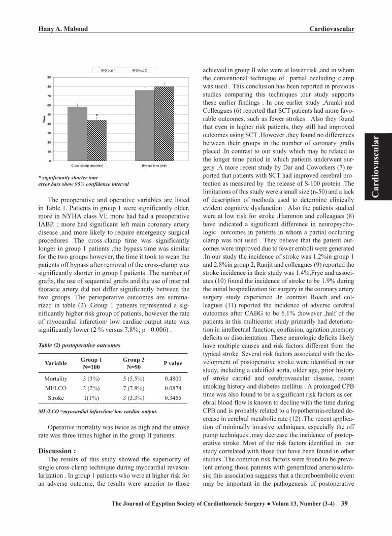
Hany A. MaboudCardiovascular
Car
diov
ascu
lar
The Journal of Egyptian Society of Cardiothoracic Surgery ● Sep-Dec 200538
Hany A. Maboud Cardiovascular
Car
diov
ascu
lar
The Journal of Egyptian Society of Cardiothoracic Surgery ● Volum 13, Number (3-4) 39
�
Age (y) Patients �70female patients diabetic patients Hypertensive patients NYHA functional class 1VNo. having preop IABP Emergency operation No. of grafts LIMAEndarterectomy Sequential graft Cross-clamp time(min) Bypass time (min)
69 (5.596) 40 (40%) 34 (34%) 50 (50%) 80 (80%) 20 (20%)
15 (15%) 12 (12%) 2.5 (0.503) 87 (87%) 10 (10%) 50 (50%) 58 (16.82) 76 (15.98)
62 (9.847) 52 (57.78%) 36 (40%) 42 (46.6%) 75 (83.3%) 6 (6.66%)
6 (6.66%) 4 (4.4%) 3.1 (0.606) 75 (83.3%) 6 (6.66%) 56 (62.2%) 44 (12.76) 80 (16.35)
<0.00010.02130.48050.74680.69040.0139
0.10980.1044<0.00010.60780.57120.1225<0.00010.09
IABP = intraaortic balloon pump, NYHA= New York Heart Association, LIMA= left internal mammary .
0
10
20
30
40
50
60
70
80
90
Cross-clamp time(min) Bypass time (min)
Tim
e
Group 1 Group 2
*
* significantly shorter time error bars show 95% confidence interval
The preoperative and operative variables are listed in Table 1. Patients in group 1 were significantly older, more in NYHA class VI; more had had a preoperative IABP. ; more had significant left main coronary artery
* significantly shorter timeerror bars show 95% confidence interval
The preoperative and operative variables are listed in Table 1. Patients in group 1 were significantly older, more in NYHA class VI; more had had a preoperative IABP. ; more had significant left main coronary artery disease ,and more likely to require emergency surgical procedures .The cross-clamp time was significantly longer in group 1 patients ,the bypass time was similar for the two groups however, the time it took to wean the patients off bypass after removal of the cross-clamp was significantly shorter in group I patients .The number of grafts, the use of sequential grafts and the use of internal thoracic artery did not differ significantly between the two groups .The perioperative outcomes are summa-rized in table (2) .Group 1 patients represented a sig-nificantly higher risk group of patients, however the rate of myocardial infarction/ low cardiac output state was significantly lower (2 % versus 7.8%; p= 0.006) .
Table (2) postoperative outcomes
Variable Group 1N=100
Group 2N=90 P value
MortalityMI/LCOStroke
3 (3%)2 (2%)1(1%)
5 (5.5%)7 (7.8%)3 (3.3%)
0.48000.08740.3465
MI /LCO =myocardial infarction/ low cardiac output.
Operative mortality was twice as high and the stroke rate was three times higher in the group II patients.
Discussion :The results of this study showed the superiority of
single cross-clamp technique during myocardial revascu-larization . In group 1 patients who were at higher risk for an adverse outcome, the results were superior to those
achieved in group II who were at lower risk ,and in whom the conventional technique of partial occluding clamp was used . This conclusion has been reported in previous studies comparing this techniques ;our study supports these earlier findings . In one earlier study ,Aranki and Colleagues (6) reported that SCT patients had more favo-rable outcomes, such as fewer strokes . Also they found that even in higher risk patients, they still had improved outcomes using SCT .However ,they found no differences between their groups in the number of coronary grafts placed .In contrast to our study which may be related to the longer time period in which patients underwent sur-gery .A more recent study by Dar and Coworkers (7) re-ported that patients with SCT had improved cerebral pro-tection as measured by the release of S-100 protein .The limitations of this study were a small size (n-50) and a lack of description of methods used to determine clinically evident cognitive dysfunction . Also the patients studied were at low risk for stroke .Hammon and colleagues (8) have indicated a significant difference in neuropsycho-logic outcomes in patients in whom a partial occluding clamp was not used . They believe that the patient out-comes were improved due to fewer emboli were generated .In our study the incidence of stroke was 1.2%in group 1 and 2.8%in group 2, Ranjit and colleagues (9) reported the stroke incidence in their study was 1.4%,Frye and associ-ates (10) found the incidence of stroke to be 1.9% during the initial hospitalization for surgery in the coronary artery surgery study experience .In contrast Roach and col-leagues (11) reported the incidence of adverse cerebral outcomes after CABG to be 6.1% ;however ,half of the patients in this multicenter study primarily had deteriora-tion in intellectual function, confusion, agitation ,memory deficits or disorientation .These neurologic deficits likely have multiple causes and risk factors different from the typical stroke .Several risk factors associated with the de-velopment of postoperative stroke were identified in our study, including a calcified aorta, older age, prior history of stroke carotid and cerebrovascular disease, recent smoking history and diabetes mellitus . A prolonged CPB time was also found to be a significant risk factors as cer-ebral blood flow is known to decline with the time during CPB and is probably related to a hypothermia-related de-crease in cerebral metabolic rate (12) .The recent applica-tion of minimally invasive techniques, especially the off pump techniques ,may decrease the incidence of postop-erative stroke .Most of the risk factors identified in our study correlated with those that have been found in other studies .The common risk factors were found to be preva-lent among those patients with generalized arteriosclero-sis; this association suggests that a thromboembolic event may be important in the pathogenesis of postoperative

Hany A. MaboudCardiovascular
Car
diov
ascu
lar
The Journal of Egyptian Society of Cardiothoracic Surgery ● Sep-Dec 200540
Hany A. Maboud Cardiovascular
Car
diov
ascu
lar
The Journal of Egyptian Society of Cardiothoracic Surgery ● Volum 13, Number (3-4) 41
strokes .An initial suggestion of this was noted in our study ,that the absence of anticoagulation therapy during the preoperative period was more frequent among the stroke patients . Salerno (13) repoted on 87 consecutive patients in whom the SCT was used . He observed a low mortality and morbidity in these patients, thereby estab-lishing the safety of the procedure in the clinical setting . Weisel and associates (14) conducted a randomized clini-cal trial in which SCT was used in 46 patients and the conventional technique was used in 45 patients ,with similar risk factors for both groups .Despite a significantly increased cross-clamp time ,lower myocardial tempera-ture , lower creatine kinase release and earlier return of myocardial lactate extraction was observed for the pa-tients treated with SCT .The same group reported similar results for a smaller number of patients who were not ran-domized (15) . The advantages of this technique appear to be related to the more homogenous distribution of cardio-plegia and the more homogenous cooling that occurs when distal and proximal grafts are sequentially con-structed .This allows for a reduction in the metabolic de-mand during ischemia and for a continuous washout of lactic acid on repeated infusion of cardioplegia through constructed grafts .On release of the cross-clamp ,there is a maximal reactive hyperemia and an immediate maximal coronary reperfusion that results in complete washout of the lactic acid accumulation and speedy replenishment of the substrates needed for aerobic metabolism (16) . In contrast, when the DCT is used ,there is inadequate coro-nary hyperemia and reperfusion, accompanied by a dis-cordant increase in myocardial temperature . This results in a reperfusion myocardial injury stemming from the continued ischemia ,the increased production and accu-mulation of lactic acid (17) and generation of oxygen free radicals (18) This is supported by the findings of Khoury and colleagues (19) who observed a significant decline in the myocardial pH, along with an increase in the myocar-dial temperature, after removal of the cross-clamp during the period when the proximal anastomoses would be con-structed .This phenomenon occurred regardless of whether crystalloid or blood cardioplegia was used . Controversy exist regarding the efficacy of SCT . Musumeci and cow-orkers (20) reported that the SCT was not as effective as intermittent ischemic arrest in the prevention of myocar-dial ischemia and neurologic problems . Kim and col-leagues (21) concluded that use of a SCT does not prevent stroke, but their study also did not have an adequate sam-ple size to detect a significant difference in stroke occur-rence. We emphasize that our analysis also was not statis-tically powered to detect a difference in stroke rate .How-ever ,in addition to an improved neurologic injury out-come with SCT, a trend toward improvement in stroke rate
was observed . The assessment of neurologic injury (other than stroke) following cardiac surgery has not been exten-sively studied in the past,. Patients with a neurologic in-jury in this analysis have an increased mortality and hos-pital length of stay . Rolfson and coworkers (22) reported an incidence of 32% of patients having delirium postop-eratively in a sample of elderly CABG patients .This is a clinically important outcome that requires further exami-nation and the development of methods for prevention .In a study by the McSPI group (23) ,the incidence of this neurologic injury was 7.3%,with proximal aortic arterio-sclerosis being a risk factor . This is critical because by the75 to80 years old the incidence of severe aortic arterio-sclerosis in patients is 10% . Studies where epiaortic ultra-sound was used to examine the aorta for the presence of atherosclerotic disease (24) have demonstrated that aortic disease is correlated with stroke outcome both within 30 days and up to 5 years postoperatively (15) .Our study has indicated a correlation with age and the higher incidence of neurologic injury .In this study a major stroke occurred in 1.2 % of the patients in group 1 (SCT) whose median age was 70 years ,compared with 2.8%of the patients of group 2 (DCT) whose median age was 65 years .Although this difference was not statistically significant ,it repre-sents remarkably low stroke rate for a significantly older and higher-risk group of patients . Because the use of a partial occluding clamp was the only difference in the operative technique between the two groups ,it is highly implicated as a major contributor to the occurrence of atheroembolic events. Loop and colleagues (26) reported a stroke rate of 0.7% for patients undergoing CABG procedures(n=691) in whom the SCT was used. This was significantly lower than the stroke rate in the group of patients (n=2,214) in whom DCT was used .The authors speculated that the reduced manipulation of and trauma to the ascending aorta achieved by single application of the cross-clamp may have led to this significant reduction in neurologic complication .Application of a partial occlud-ing clamp is probably the most traumatic manipulation of the ascending aorta ,with the total force of the clamp being distributed on a small area ,which may increase the likeli-hood of an intimal tear and the dislodgment of a plaque or atheromatous material that could embolize upon removal of the clamp .In contrast ,the force of the total occluding clamp is equally distributed along the whole circumfer-ence of the aorta . At Wake Forest Medical University ,screening for embolic events using transcarotid echogra-phy revealed that the application and removal of the par-tial occluding clamp was associated with 11%and 17% of the total embolic events ,respectively .the corresponding figures for the total occluding clamp were 0.1%and 9%,respectively (26) .
Hany A. MaboudCardiovascular
Car
diov
ascu
lar
The Journal of Egyptian Society of Cardiothoracic Surgery ● Sep-Dec 200540
Hany A. Maboud Cardiovascular
Car
diov
ascu
lar
The Journal of Egyptian Society of Cardiothoracic Surgery ● Volum 13, Number (3-4) 41
In conclusion ,we believe that identifying and un-derstanding preoperative risk factors and comorbidities for neurologic complication is important This study has identified clamp technique as an important operative risk factor and supports the significant benefits of the single clamp technique in the neurologic outcomes for patients undergoing coronary artery bypass surgery .In addition ,use of partial occluding clamp was a significant predic-tor for an adverse outcome . the advantages of the single cross-clamp in terms of myocardial protection may be related the more homogeneous cardioplegia delivery and myocardial cooling associated with its use ,and to the synchronized rewarming and maximal reperfusion that take place upon removal of the clamp .This reduce the incidence of reperfusion injury and allows for better myocardial recovery . Eliminating the need for a partial occluding clamp avoids additional manipulation, trauma ,and atheroembolic events, and this may confer better cerebral protection during CABG procedures .Therefore ,despite a significant increase in the total ischemia time ,the single cross-clamp method is a simple and safe tech-nique, and appears to have better myocardial and cerebral protective properties than does the conventional technique of partial aortic occlusion .
References :1.John R, Choudhri Af, Weinber AD, et al., Multicenter Review
of Pre operative risk factors for stroke after coronary Artery Bypass grafting. Ann Thorac surg. 2000; 69 : 30 – 67.
2.Stamou S.C, Hill PC, Dangas G et al., Stroke after Coronary artery bypass : incidence, predictors, and clinical outcome, Stroke 2001; 32 : 1508 – 13.
3.Van der Linden I, Casimir-Ahn H. When do cerebral emboli appear during open heart operation ? A transcranial Doppler study. Ann Thorac surg. 1991; 51 : 23 7 – 41.
4.Barbut D, Hinton RB, Szatrowski TP : Cerebral emboli detected during bypass surgery are associated with clamp removal. Stroke 1994, 25 : 2398 – 402.
5.Mckham GM, Gold Brough MA, Borowicz LM, et al., pre-dictors of stroke risk in coronary artery bypass patients. Ann Thorac surg. 1997, 63 : 516 – 21.
6.Aranki Sf, Rizzo RJ, Adams DH, et al., single clamp tech-nique adjunct to myocardial and cerebral protection in cor-onary operation. Ann Thorac surg. 1994; 58 : 296 – 302.
7.Dar MI, Gillot T, Giullif, Cooper GJ. Single aortic cross-clamp technique reduces S-100 release after coronary artery surgery. Ann Thorac surg 2001; 71 : 794 – 6.
8.Hammon JW, Stump OA, Kon ND et al., the development of neurobehavioral changes after coronary artery bypass graft-ing. Ann Thorac surg 1997; 63: 1613-8.
9.Maura A. Grega, MSM, Louis, Metal, Impact of single clamp versus double clamp technique on neurologic out-come . Ann Thorac surg 2003 97: 1387-91.
10.Frye RL, Kronomal R, Achaff HV, Myers WO, Gweah BJ. Stroke in coronary artery bypass, graft surgery an analysis
of the CASS experience. The participants in the coronary artery surgery study. INI J. Cardiol 1992; 36 : 213 – 21.
11.Roach GW, Kanchuger M, Mangano CM, et al., Adverse cerebral outcomes after coronary surgery. N Engl J. Med, 1996; 335:1857 – 63.
12.Roov, Christokis GT, Weisel RD, et al., Risk factors for stroke following coronary bypass surgery. J. Cad. Surg. 1995; (suppl) : 468-74.
13.Salerno TA. Single aortic cross-claming for distal and proxi-mal anastomoses in coronary surgery : an alternative to con-ventional techniques Ann Thorac surg 1982; 33 : 518 – 20.
14.Weisel RD, Hoy FB, Baird RJ, et al., Improved myocardial protection during a pro longed cross-clamp period. Ann Thorac surg 1983; 36 : 664 – 74.
15.Loop FD, Higgins TT., Panda R., Pearce G , et al. Myo-cardial protection during cardiac operation : decreased morbidity and lower coast with blood cordioplegia and coronary rinus perfusion. J Thorac CardioVasc surg. 1992; 104 : 608 – 18.
16.LiChetnstein SV, Abel JG, Panos A, et al., Warm heart Sur-gery : experience with long cross-clamp. Ann Thorac surg. 1991; 51: 418-23.
17.Buckberg GD. A proposed solution to the codioplegic con-troversy J.Thorac cardio vasc. Surg. 1979; 77 : 803 – 15.
18.buckberg GD. Myocardial temperature management during aortic clamping for cardiac surgery. J. Thorac cardiovasc Surg. 1991; 102: 895 – 23.
19.Kihuri SF, Warner KG, Josa N, et al., The superiority of continuous cold blood cardioplegia in the metabolic protec-tion of the hypertrophied human heart. J. Thorac cardiovasc Surg. 1988; 95:442- 54.
20.Musumeci F, Feccia M. Macarthy PA, et al., prospective randomized trial of single clamp technique versus intermit-tent ischemic arrest : myocardial and neurological outcome. Eur J. cardio Thorac surg. 1998 ; 13 : 703 – 9.
21.Kim RW, Mariconda DC, Tellides G, et al., single-clamp technique does not protect against cerebr vascular accident in coronary artery bypass grafting. Eur J. cardio vasc. Surg. 2001 ; 20 : 127 – 32.
22.Rolfson DB, MC El Haney JE, Rockwood K, et al., In-cidence and risk factors for delirium and other adverse outcomes in older adults after coronary artery bypass graft surgery. Can. J. Cordial 1999 ; 15:771 – 32.
23.Wolman RJ, Nussmeier N, Aggarial A. et sl., cerebral injury after cardiac surgery, identification of a group at extraor-dinary risk. Multicenter study of preoperative ischemia re-search group (MCSPI) and the ischemia research education formation (IREF) investigators. Stroke 1990; 30 : 514 – 27.
24.Goto T., Baba T., Yoshi take A., et al., Cranio cervical and aortic atherosclerosis as neurologic risk factors in coronary surgery. Ann Thorac surg 200 ; 69 : 438 – 40.
25.Hammon TW, Stump DA, Butter worth J. Moody DM. Ap-proaches to reduce neurologic complications during cardiac surgery. Sem Thorac cardiovasc Surg. 2001 ; 13 : 184 – 91.
26.Davila – Roman VG, Pillips KJ, Daily BB, Davial RM, Kouchoukos NT, Ultrasound for assessment of atheros-derosis of the thoracic aorta. J.AM. collcardial 1996 ; 28 : 942 – 7.
Cardiovascular Hossam F El Shahawy et al
Car
diov
ascu
lar
The Journal of Egyptian Society of Cardiothoracic Surgery ● Sep-Dec 200542
Hossam F El Shahawy et al Cardiovascular
Car
diov
ascu
lar
The Journal of Egyptian Society of Cardiothoracic Surgery ● Volum 13, Number (3-4) 43
Pathological Changes by the Effect of Ultrasonic andElectrocautery Harvesting Procedures on the InternalThoracic Artery Endothelium
Objective: The aim of the study is to evaluate the extent of damage done to the endothelium when the internal thoracic artery (ITA) is taken down with the harmonic scalpel (HS) as compared with the standard technique by using high frequency electrocautery (EC).Patients & Methods: Fifty patients were non randomly classified into two groups. Group I, comprising 25 patients for whom LITAs were harvested by HS and group II, comprising 25 patients for whom LITAs were taken down by EC. Wide pedicled grafts were done for the first five patients within each group -control for the study - and skeletonized ones for the rest of the patients in both groups. Evaluation of endothelial integrity was performed by scanning electron microscopy according to the score system for description of the endothelial damage.Results: Scanned fields of wide pedicled LITAs in each group showed nonsignificant (P value > 0.05) changes completely confluent endothelium (grade I integrity) in 95% and partially confluent endothelium (grade II) in 5%. Scanned fields of skeletonized LITAs in group I showed completely confluent endothelium (grade I) in 62.5%, partially confluent endothe-lium (grade II) in 23.8%, islands of endothelium (grade IV) in 12.5% and no endothelium (grade V) in 1.3%. Scanned fields of skeletonized LITAs of group II showed highly significant (P value < 0.01) changes grade I in 0%, grade II in 7.5%, grade IV in 31.3% and grade V in 61.3%. Free flow of skeletonized LITAs showed highly significant (P value < 0.01) changes 101.29 ± 9.40 mL/min while that of wide pedicled LITAs was 69.50 + 6.85 mL/min.The mean length of skeletonized LITAs showed highly significant (P value < 0.01) changes 19.31 ± 0.81 cm while that of wide pedicled LITAs was 17.10 ± 0.57 cm. Harvesting spasm showed significant (P value < 0.05) changes in 47.5% of skeletonized ITAs and in 10% of wide pedicled ITAs . The harvesting duration was significantly shorter for wide pedicled ITAs than for skeletonized ITAs regardless of the harvesting machine. Electro-cautery was significantly faster than the harmonic scalpel regardless of the width of the ITA pedicle. Other intraoperative and postoperative data showed no significant difference among both groups under study.Conclusion: For LITA skeletonization HS is more preservative to the en-dothelium than EC. Flow and length of skeletonized LITAs are greater than those of wide pedicled LITAs. EC is faster than HS in LITA taking down.
Accepted for publication Dec 20 . 2005
Address reprint request to Dr. H. El
Shahawy
The Departmen of Cardiothoracic Sur-
gery, Ain Shams University Hospital
Email : [email protected]
Codex : 04 / 18 /cord /0512
Hossam F El Shahawy, MDAhmed Badawy, MBBCH, MS Hisham Abd El Rahman, MDAbdel Salam El Henawi , MDSherif Azab, MDEzzeldin A. Mostafa, MD
Cardiovascular Hossam F El Shahawy et al
Car
diov
ascu
lar
The Journal of Egyptian Society of Cardiothoracic Surgery ● Sep-Dec 200542
Hossam F El Shahawy et al Cardiovascular
Car
diov
ascu
lar
The Journal of Egyptian Society of Cardiothoracic Surgery ● Volum 13, Number (3-4) 43
The internal thoracic artery is the best con-duit currently available for CABG, with a patency rate of 85-95% after 10 years . The use of this graft is the single most important factor in improved survival and freedom
from angina. Its long term patency is significantly supe-rior to vein grafts (1).
The aim of the study is to evaluate the extent of damage done to the endothelium when the internal mammary artery (IMA) is taken down with the harmonic scalpel as compared with the standard technique by using high frequency electrocautery.(1)
The effects on the endothelium of the mammary arteries-when the harvest was done either by the harmonic scalpel (HS) or the high frequency electro-cautery (EC) - were compared. The HS has a positive effect on endothelial preservation, especially when the preparation is done closely to the internal thoracic artery(2). The harmonic scalpel consists of a generator, and a foot pedal. The blade vibrates longitudinally with a frequency of 55,000Hz. Different from the high frequency cautery, there is no conduction of electricity. The effect (the denaturation of proteins) is done completely mechanically. The depth of penetration of the scalpel is regulated by the pressure applied to the scalpel (2).
The blade of the ultrasonically activated scalpel vibrates longitudinally at 55.500 Hz. The blade moves 50-100 microns, depending on the power setting of the generator. The moving blade couples with the tissue, resulting in vibration of the tissue. The transfer of this mechanical energy from the blade to the protein in the tissue breaks the Hydrogen bonds in the tissue. This results in breakdown of the protein into sticky coagulum. This coagulum seals blood vessels. The friction and shear from the vibration generate heat up to 80-100 degrees c (much less than laser or electrosurgery) (3).
Methods:This is a prospective non randomized comparative
study that was conducted in the period from August 2001 – May 2003. It was carried out on 50 patients undergoing GABG.
Inclusion criteria
• Male patients with single or multivessel disease < 65 years.
• Patients with normal or moderately impaired LV systolic function EF ³≥ 40%.
• Patients with left main disease.
Routine preoperative investigations; ECG, chest x-ray, blood chemistry, echocardiography and coronary angiography were performed to all patients. Stress ECG and myocardial viability study were done in some patients.
The patients were classified into two groups according to the device used for LITA harvesting. Group I (25 patients) and group II (25 patients). The mean age of patients in group I was 49.84 ± 7.57 years while that of patients of group II was 48.28 ± 9.08, p value > 0.05 (NS). Diabetic patients were 40% and 60%, hypertensive were 48% and 60%, smokers were 76% and 80%, obese patients were 36% and 60% and those with positive family history for ischemic heart disease were 52% and 56% in group I and II respectively (p values > 0.05, NS).
Patients belonging to, NYHA class I were 4% and 0%, class II were 48% and 44%, class III were 32% and 40% and class IV were 16% and 16% in group I and II respectively with p value > 0.05 (NS).
For patients of group I, LITAs were harvsted by the harmonic scalpel, dissecting hook type, ethicon endosurgery, Cincinnati, OH while for group II patients, LITAs were takendown by electrocautery, valleylab Inc Boulder, USA. For the first five patients within each group, LITAs were takendown as wide pedicled grafts and these 10 patients were the control for the study. For the rest of patients within each group LITAs were taken as narrow pedicled or skeletonized grafts.
LITA harvesting as wide pedicled grafts (n=10) was done by separating or peeling the pleura from the chest wall along the length of the LITA. Then the endothoracic fascia is cut along the left border of the sternum.
The LITA is freed from the chest wall by cutting its branches with electrocautery adjusted at 25 watt for patients in group II and with the harmonic scalpel adjusted at level II for patients in group I.
Then the endothoracic fascia lateral to LITA was cut along the length of LITA leaving it in a pedicle of 1.5-2 cm width. Then it was cut about 1-2 cm distal to its bifurcation after heparinization (300IU/kg) and its distal ends were clipped with hemoclips. Then it is wrapped in warm 0.2% papaverine soaked sponge and left till use. Skeletonization of LITA (N=40) was done by peeling the pleura from the chest wall as mentioned previously. (The endothoracic fascia was cut just medial to the medial vena comitantes along the LITA length).
The last l-2cm was cut, opened longitudinally and then was immediately washed gently with a physiologic saline solution, and immersed in 2.5% glutraldehyde for 24 hours. All samples were washed in cacodylate buf-fer, postfixed in 1% osmium tetroxide, and thereafter
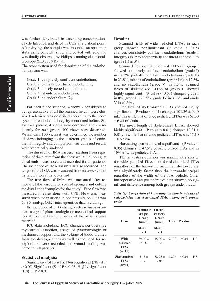
Cardiovascular Hossam F El Shahawy et al
Car
diov
ascu
lar
The Journal of Egyptian Society of Cardiothoracic Surgery ● Sep-Dec 200544
Hossam F El Shahawy et al Cardiovascular
Car
diov
ascu
lar
The Journal of Egyptian Society of Cardiothoracic Surgery ● Volum 13, Number (3-4) 45
was further dehydrated in ascending concentrations of ethylalcohol, and dried in CO2 at a critical point. After drying, the sample was mounted on specimen stubs using colloidal silver and coated with gold and was finally observed by Philips scanning electronmi-croscope XL3 at 30 Kv (4).The score system used for description of the endothe-lial damage was:
Grade 1, completely confluent endothelium;Grade 2, partially confluent endothelium;Grade 3, loosely netted endothelium;Grade 4, islands of endothelium;Grade 5, no endothelium (2).
For each piece scanned, 4 views - considered to be representative of all the scanned fields - were cho-sen. Each view was described according to the score system of endothelial integrity mentioned before. So, for each patient, 4 views were described and conse-quently for each group, 100 views were described. Within each 100 views it was determined the number of views belonging to the different grades of endo-thelial integrity and comparison was done and results were statistically analysed.
The duration of IMA harvest - starting from sepa-ration of the pleura from the chest wall till clipping its distal ends - was noted and recorded for all patients. The incidence of IMA spasm was also recorded. The length of the IMA was measured from its upper end to its bifurcation at its lower end.
The free flow of IMAs was measured after re-moval of the vasodilator soaked sponges and cutting the distal ends “samples for the study”. Free flow was measured in cases done with CPB. Flow was mea-sured when mean arterial blood pressure on CPB was 70-80 mmHg. Other intra operative data including;
the incidence of ECG changes after revasculariza-tion, usage of pharmacologic or mechanical support to stabilize the haemodynamics of the patients were recorded.
ICU data including; ECG changes, perioperative myocardial infarction, usage of pharmacologic or mechanical support and the volume of blood drained from the drainage tubes as well as the need for re-exploration were recorded and wound healing was noted for all patients.
Statistical analysis:Significance of Results: Non significant (NS) if P
> 0.05, Significant (S) if P < 0.05, Highly significant (HS) if P < 0.01
Results:Scanned fields of wide pedicled LITAs in each
group showed nonsignificant (P value > 0.05) changes completely confluent endothelium (grade I integrity) in 95% and partially confluent endothelium (grade II) in 5%.
Scanned fields of skeletonized LITAs in group I showed completely confluent endothelium (grade I) in 62.5%, partially confluent endothelium (grade II) in 23.8%, islands of endothelium (grade IV) in 12.5% and no endothelium (grade V) in 1.3%. Scanned fields of skeletonized LITAs of group II showed highly significant (P value < 0.01) changes grade I in 0%, grade II in 7.5%, grade IV in 31.3% and grade V in 61.3% .
Free flow of skeletonized LITAs showed highly significant (P value < 0.01) changes 101.29 ± 9.40 mL/min while that of wide pedicled LITAs was 69.50 + 6.85 mL/min .
The mean length of skeletonized LITAs showed highly significant (P value < 0.01) changes 19.31 ± 0.81 cm while that of wide pedicled LITAs was 17.10 ± 0.57 cm .
Harvesting spasm showed significant (P value < 0.05) changes in 47.5% of skeletonized ITAs and in 10% of wide pedicled ITAs .
The harvesting duration was significantly shorter for wide pedicled ITAs than for skeletonized ITAs regardless of the harvesting machine. Electrocautery was significantly faster than the harmonic scalpel regardless of the width of the ITA pedicle. Other intraoperative and postoperative data showed no sig-nificant difference among both groups under study.
Table (1): Comparison of harvesting duration in minutes of wide-pedicled and skeletonized ITAs, among both groups under
Item
Harmonic scalpelGroup (n=25)
Electro-cauteryGroup (n=25) T test P value
Mean ± SD
Mean ± SD
Wide pedicled
ITAs(n=15)
39.00 ± 4.18
15.00 ± 3.54
9.798 <0.01 HS
Skeletonized ITAs
(n=20)
51.5 ± 9.33
38.75 ± 7.05
4.876 <0.01 HS

Cardiovascular Hossam F El Shahawy et al
Car
diov
ascu
lar
The Journal of Egyptian Society of Cardiothoracic Surgery ● Sep-Dec 200544
Hossam F El Shahawy et al Cardiovascular
Car
diov
ascu
lar
The Journal of Egyptian Society of Cardiothoracic Surgery ● Volum 13, Number (3-4) 45
Table (2): Comparison of harvesting duration of wide pedi-cled versus skeletonized ITAs in both groups under study.
Item
Wide pedicled
ITAs (n=5)
SkeletonizedITAs (n=20) T test P value
Mean ± SD Mean ± SD
Harmonic scalpelGroup
39.00 ± 4.18 51.5 ± 9.33 2.887 <0.01 HS
Electro-cautery group
15.00 ± 3.54 38.75 ± 7.05 7.226 <0.01 HS
Table (3): Comparison of free flow and length of wide-pedicled ITAs vs skeletonized ITAs.
Item
Wide pedicled
ITAs (n=10)
SkeletonizedITAs (n=40) T test P value
Mean ± SD Mean ± SD
Free flow (ml/
min.)
69.50 ± 6.85 101.29 ± 9.40 9.851 <0.01 HS
Length (Cm.)
17.10 ± 0.57 19.31 ± 0.81 8.158 <0.01 HS
Table (4): Comparison of incidence of harvesting spasm according to studied groups.
Item Harmonic scalpelGroup (n=25)
Eletro-cauteryGroup (n=25)
Chi2 P value
No. % No. %
Harvesting spasm among skeletonized ITA (n=40)
Yes 9 45.0 10 50.0No 11 55.0 10 50.0 0.10 >0.05 NS
Harvesting spasmWide
pedicled(n=10)
Skeletonized(n=40)
No. % No. %Yes 1 10.0 19 47.5No 9 90.0 21 52.5 4.69 <0.05 S
Table (5): Comparison of endothelial integrity as – deter-mined by SEM – in wide pedicled ITAs & skeletonized ITAs of both groups under study.
Item Harmonic scalpel
Group (n=25)
Eletro-cauteryGroup (n=25)
Chi2 P value
No. % No. %
Endothelial integrity grade in wide pedicled ITAs (n=10
specimens i.e 40 views)
Grade 1 19 95.0 19 95.0Grade 2 1 5.0 1 5.0 0 >0.05 NSEndothelial integrity grade in skeletonized ITAs (n=40
specimens i.e 160 views)Grade 1 50 62.5 0 0Grade 2 19 23.8 6 7.5Grade 4 10 12.5 25 31.3Grade 5 1 1.3 49 61.3 109.27 <0.01 HS
Table (6): Comparison of perioperative events, among both groups under study.
Item
Harmonic scalpelGroup (n=25)
Eletro-cauteryGroup (n=25) Chi2 P value
No. % No. %
Myocardial
infarction
5 20.0 5 20.0 0 >0.05 NS
Pharmacologic
support
15 60.0 18 72.0 0.88 >0.05 NS
Mechanical
support
3 12.0 3 12.0 0.0 >0.05 NS
Table (7): Comparison of mediastinal drainage, among both groups under study.
Item Harmonic scalpelGroup (n=25)
Electro-cauteryGroup (n=25)
T test P value
Mean ± SD Mean ± SD
Volume of
Mediastinal
drainage
728.17 ±
392.02
833.33 ±
446.89
0.858 >0.05 NS

Cardiovascular Hossam F El Shahawy et al
Car
diov
ascu
lar
The Journal of Egyptian Society of Cardiothoracic Surgery ● Sep-Dec 200546
Hossam F El Shahawy et al Cardiovascular
Car
diov
ascu
lar
The Journal of Egyptian Society of Cardiothoracic Surgery ● Volum 13, Number (3-4) 47
Table (8): Comparison of postoperative events, among both groups under study.
Item Harmonic scalpelGroup (n=25)
Eletro-cauteryGroup (n=25)
Chi2 P value
No. % No. %
Exploration for bleeding
2 8.0 3 12.0 0.22 >0.05 NS
Wound healing
Good healing 20 83.3 19 79.2
Little serous discharge
1 4.2 2 8.3
Superficial infection
2 8.3 2 8.3
Deep infection 1 4.2 1 4.2 0.36 >0.05 NS
Intrahospital mortality
2 8.0 1 4.0 0.35 >0.05 NS
DISCUSSION:In view of the clear advantages of the internal
thoracic artery versus the saphenous vein for left anterior descending coronary artery bypass, expanded use of arterial grafts has long been advocated (5).
Paracrine endothelial function is considered one of the crucial factors responsible for the observed differences in the fate of arterial and venous coronary artery bypass grafts. The level of nitric oxide production by the endothelium of ITA is very high when compared with the low and variable amount produced by the saphenous vein(6) .
Skeletonization of ITA is postulated to improve graft length, early blood flow, stemal blood supply and postoperative respiratory function (6). Also skeletonization facilitates construction of sequential anastomoses(7) hence, it broadens the spectrum of the target vessels that could be bypassed by ITAs.
Unlike wide pedicled ITA harvesting, skeletonized ITA is more prone to be traumatized either mechanically due to rough manipulation or thermally depending on the instrument used for harvesting. Therefore harmonic scalpel skeletonization being associated with less temperature release, has the advantage of being more preservative to the endothelial lining of internal thoracic artery.
In this study all patients were males because it is
usually easier to deal with their larger ITAs. Regarding the risk factors for IHD and preoperative data of the patients, there were no differences of statistical significance.
The time required for ITA harvesting as a wide pedicled graft was significantly shorter than that needed for its skeletonization. Using the harmonic scalpel for ITA harvesting - as a wide pedicled or skeletonized -was significantly slower than using electrocautery. This was consistent with Brose et al., 2002. They compared the harmonic scalpel, argon beam and electrocautery as regards ITA harvesting as a wide pedicled graft and they found that electrocautery was significantly faster than the harmonic scalpel(9).
The lengths of ITAs prepared as skeletonized pedicles were found to be significantly greater than that prepared as wide pedicled grafts (about 2cm difference). Higami et al., 2001 also found that ultrasonic complete skeletonization increases the effective length of ITAs than conventional wide pedicled harvesting (9). Also Deja et al., 1999 concluded that skeletonization of ITAs is associated with greater ITA length (6).
It has been suggested that absence of the broad flap of fascial and fatty tissue makes the skeletonized ITA more pliable and lying more medially in its proximal course and therefore easily reaching the distal LAD or even distal branches of circumflex artery (6).
Indeed discarded segments of skeletonized ITAs are significantly longer than those discarded from wide pedicled ones. This may be of value as the most distal muscular segments of ITAs are no longer used and the more proximal, elastic and bigger in diameter arteries are used for anastomosis construction (6).
Free blood flow from ITAs was measured after cutting the specimen piece while the patient on cardiopulmonary bypass with mean arterial blood pressure between 70-80mmHg. It was found that flow of skeletonized ITA was significantly greater than that of wide pedicled ITA i.e. the difference was related to the width of ITA pedicle rather than the machine used for dissection. Wilmo et al., 2000 found that the free flows of wide pedicled ITAs - whether harvested by the harmonic scalpel or electrocautery - were not significantly different(10). Also Takami and Ina, 2002 who studied the effects of skeletonization on intraoperative flow, found that skeletonization was associated with significantly greater blood flow(11).
However Choi and Lee, 1996 found that free flows from skeletonized ITAs were similar to those obtained from wide pedicled ITAs prepared by intraluminal papaverin injection and flow of both skeletonized and wide pedicled ITAs (prepared by intraluminal papaverin)

Cardiovascular Hossam F El Shahawy et al
Car
diov
ascu
lar
The Journal of Egyptian Society of Cardiothoracic Surgery ● Sep-Dec 200546
Hossam F El Shahawy et al Cardiovascular
Car
diov
ascu
lar
The Journal of Egyptian Society of Cardiothoracic Surgery ● Volum 13, Number (3-4) 47
was significantly greater than that of wide pedicled ITAs wrapped in sponges soaked in dilute papaverin(12). In our study we did not inject papaverin solution intraluminally to avoid the possibility of endothelial injury, instead we wrapped the ITAs -wide-pedicled or skeletonized in sponges soaked - in vasodilator solution. Better free flow of skeletonized ITAs than wide pedicled ones was recorded also by other investigators(13,7,6,9). Local sympathectomy which occurs with skeletonization of ITAs is a possible explanation for greater blood flow of skeletonized ITAs (6).
The incidence of spasm immediately after ITA taking down was significantly lower among the wide pedicled ITAs than skeletonized ones. However no significant difference exist when we compared ITAs skeletonized by electrocautery with those skeletonized by the harmonic scalpel. Choi and Lee, 1996 stated that arterial spasm can be avoided by skeletonization(12). Most probably this controversy resulted because in our study it was necessary to continue ITA dissection for a considerable distance beyond the bifurcation to allow for taking a good piece for the study without compromising the graft length. So we were obliged to deal with the terminal muscular branches of ITAs. So more direct manipulation that was associated with skeletonization resulted in higher incidence of spasm - mainly of the distal end - among the skeletonized ITAs. Given the information that we measured free flow after cutting the distal segment - piece for study -explains the greater free flow of skeletonized ITAs in spite of greater incidence of harvesting spasm.
Using scanning electron microscopy for evaluation of endothelial integrity revealed that harvesting the internal thoracic artery as a wide pedicled graft either by the harmonic scalpel (group 1) or by electrocautery (group 2) was associated with good preservation of endothelial integrity. Ninety five percent of scanned fields - in each group - showed totally confluent endothelium (grade 1) and only five percent of scanned field - in each group - showed partially confluent endothelium (grade 2).
The difference was apparent with skeletonized ITAs. In group 1 -harmonic scalpel group - 62.5% of scanned field showed totally confluent endothelium (grade 1), 23.8% of the fields showed partially confluent endothelium (grade 2), 12.5% showed islands of endothelium (grade 4) and 1.3% showed totally damaged or no endothelium (grade 5). While in electrocautery group - group 2 - none of the fields showed totally confluent endothelium (grade 1), 7.5% of fields showed partially confluent endothelium (grade 2), 31.3% showed islands of endothelium (grade 4) and 61.3% showed totally damaged endothelium (grade
5). P value was < 0.012 indicating a highly significant difference existing between both groups under study.
Lamm et al., 2000 who performed a similar study on 24 patients found that, if a distance of more than 0.5cm between the IMA and the edges of the IMA pedicle was kept - during harvesting either by harmonic scalpel or electrocautery - no difference would exist as regards the degree of endothelial preservation and all samples in both groups showed completely confluent endothelium. While if the IMA pedicle width was <0.5cm, endothelial morphology was hardly altered - i.e. totally confluent-with harmonic scalpel dissection in contrast to considerable endothelial cell losses with electrocautery dissection(2).
Yoshikai et al., 2004 who studied the endothelial integrity of ultrasonically skeletonized ITA by SEM, found completely confluent endothelium in all specimens of ITAs skeletonized by ultrasonic scalpel(4).
Yoshida et al., 1995 comparing the effects of monopolar and bipolar cauterization on canine skeletonized ITAs - found that using monopolar cautery was associated with almost complete loss of endothelial cells as determined by SEM. While bipolar cauterization was associated with partial loss of endothelial cells (14) .
Lethola et al., 1989 - found that when ITA mobilization was performed as a wide pedicled grafts the flow surface was well preserved regardless the instrument used (either electrocautery or sharp dissection). However if a contact of electrocautery blade with the wall of IMA or with a metallic clip parallel to the wall was allowed, a clearly visible zone of endothelial damage sometimes associated with mural thrombus formation was observed (15).
Wilmo et al., 2000 also found no intimal damage when ITAs were taken down as wide pedicled grafts regardless of machine used for dissection- electrocautery or harmonic scalpel(10). Also similar result was recorded by Brose et al., 2002(8).
In the present study we only investigated the terminal portion of the ITAs by the SEM study and did not examine the ITA over its full length. In general, the terminal portion of the ITAs has a thinner vessel wall than the proximal portion, so the results of this study can be extrapolated to the full length of the ITA. Also, we evaluated the endothelial cell integrity of the ITA only in a morphological study using SEM. The morphological intact endothelium seems to have normal endothelial cell function, however, further examinations concerning the endothelial cell function are called for.
There was no significant difference among patients of both groups regarding, the incidence of perioperative

Cardiovascular Hossam F El Shahawy et al
Car
diov
ascu
lar
The Journal of Egyptian Society of Cardiothoracic Surgery ● Sep-Dec 200548
myocardial infarction, the need for pharmacologic or mechanical support, the volume of blood drained from the chest tubes or the need for re-exploration for surgi-cal bleeding. Wound healing was good among most of the patients of both groups. Deep wound infection with sternal dehiscence occurred only in one patient in each group. Also the incidence of intrahospital mortality did not show significant difference.
References:1. Fawzy HF: Radial artery for coronary artery bypass
grafting. J of Egypt Society of Cardiothorac Surg 2004; 12:45.
2. Lamm P, Juchem G, Weyrich P, Schutz A and Reichart B: The harmonic scalpel: Optimizing the quality of mammary artery bypass grafts. Ann Thorac Surg2000; 69:1833.
3. Fowler DL: Uses of ultrasonically activated scalpel and shears in endoscopic surgery. Presented At Third International Congress On New Technology And Advanced Techniques In Surgery, Luxembourg, June 1995.
4. Yoshikai M, Ito T, Kamohara K and Yunoki J: Endothelial integrity of ultrasonically skeletonized internal thoracic artery: Morphological analysis with scanning electron microscopy. Eur J Cardiothorac Surg 2004; 25(2):208.
5. Amer SA, El Boraey WS, Gaafar AH and Rushdi AM: Arterial versus saphenous vein grafts in coronary artery surgery. J of Egypt Society of Cardiothorac Surg 2003; 11:69.
6. Deja MA, Wos S, Golba KS, Zurek P, Domaradzki W, Bachowski R and Spyt TJ: Intraoperative and laboratory evaluation of skeletonized versus pedicled internal thoracic artery. Ann Thorac Surg 1999; 68:2164.
7.Wendler 0, Tscholl D, Huang Q and Schafers HJ: Free flow
capacity of skeletonized versus pedicled internal thoracic artery grafts-coronary . artery bypass grafts. Eur J Cardio-thoracic Surg 1999; 15(3):247.
8. Brose S, Fabricius AM, Falk V, Autschbach R, Weidenbach H and Mohr FW: Comparison of ultrasonic scalpel versus argon-beam and conventional electrocautery for internal thoracic artery dissection. Thorac Cardiovasc Surg 2002; 50(2):71.
9. Higami T, Yamashita T, Nohara H, Iwaliashi K, Shida T and Ogawa K: Early results of coronary grafting using ultrasonically skeletonized internal thoracic arteries. Ann Thorac Surg 2001; 71(4):1224.
10. Wilmo OC, Aquiles VB, Vincent DJ and Haroutune MA: Internal mammary artery harvesting using the harmonic scalpel. ASAIO J 2000; 46(1):99.
11. Takami Y and Ina H: Effects of skeletonization on intraop-erative flow and anastomosis diameter of internal thoracic arteries in coronary artery bypass grafting. Ann Thorac Surg 2002; 73(5): 1441.
12.Choi JB and Lee SY: Skeletonized and pedicled internal thoracic artery grafts: Effect on free flow during bypass. Ann Thorac Surg 1996; 61:909.
13.Huang Q, Wendler O, Langer F, Tscholl D and Schafer HJ: Effects of skeletonized versus pedicled internal thoracic artery grafts on free flow capacity during bypass. J Tongji Med Univ 2000; 20(4):308,
14. Yoshida H, Wu MH, Kouchi Y, Onuki Y, Shi Q and Sauvage LR: Comparison of the effects of monopolar and bipolar cauterization on skeletonized, dissected internal thoracic arteries. J Thorac Cardiovasc Surg 1995; 110:504.
15.Lethola A, Verkkala K and Jarvinen A: Is electrocautery safe for internal mammary artery (IMA) mobilization? A study using scanning electron microscopy. Thorac Cardio-vasc Surg 1989; 37(1):55.

Wael AbdelAziz et al Cardiovascular
Car
diov
ascu
lar
The Journal of Egyptian Society of Cardiothoracic Surgery ● Volum 13, Number (3-4) 49
Ascending Aortic Surgery : Multi-centre Study in Egypt
Abstract: The aim of this prospective retrospective study is to evaluate the ascending aortic surgery results in Egypt.Methods:Between January 2000 and December 2005, 35 patients underwent ascending aortic surgery. Mean age was 46 ± 11.3 years, ranging from 32 to 77 years. The patients were divided into 3 groups. Group (a) for whom bental procedure was done :25 patients, group (b) for whom Supracoronary replacement was done :7 patients and the arch was replaced in 3 cases(group (c). Results: The overall early hospital mortality was 5 (14.2%) patients. The most frequently found complication resulted was postoperative bleeding and respiratory complications(10) patients and low cardiac output in 5 patients.Conclusion: We conclude that the Ascending aorta replacement with a valved conduit offers an acceptable early and long-term outcome.
Galen is credited as the first to describe arterial aneurysms. This was based on his observation of false aneurysms in gladiators injured during battle in the 2nd century A.D. Antyllus, during the same time period, made the distinction between traumatic aneurysms and those of a degenerative etiology. Antyllus was
also the first to attempt surgical treatment of aneurysms with proximal and distal ligation. The earliest surgical treatment of aneurysms consisted of interruption of arterial flow via either ligation or stimulation of thrombosis. Innovative measures used to cause thrombosis of aneurysms included the insertion of long segments of wire with the application of an electric current, and wrapping of aneurysms with cellophane or other irritating materials. In 1888, Rudolph Matas introduced a very different approach called obliterative endoaneurysmorraphy, then he developed restorative or reconstructive endoaneurysmorraphy. (1).
The incidence of thoracic aortic aneurysms is estimated to be 5.9 cases per 100,000 person-years and replacement of the ascending aorta accounts for the majority of thoracic aortic procedures. The mean age at the time of diagnosis ranges from 15 to 69 year. Men are typically diagnosed at a younger age and there is a 2:1 to 4:1 male predominance. (2)
Traditional risk factors include smoking, hypertension, atherosclerosis, and well-defined genetic disorders such as Marfan syndrome and Ehlers-Danlos syndrome. Subtler forms of inherited metabolic disorders are being elucidated, and perhaps play a role in more instances than was previously suspected. Syphilis, at one time the predominant etiology of ascending aortic aneurysms, has become very uncommon with the development of effective antibiotics. Bicuspid and unicuspid aortic valves are associated with ascending aortic aneurysms and dissections beyond that which can be attributed to simple
Accepted for publication Dec10,05
Please address reprint request to Dr
W Abdel Aziz
Mansoura University Cardiothoracic
Surgery Department
Email :[email protected]
Codex :05/02/othr/0512
Wael AbdelAziz , MDGamal Sami, MDReda Ahmed AbulMaaty, MDBahaa AbdelHakam M.Sc Magdy Mammdouh, MDSameh Ibrahim Sersar, MD

Cardiovascular Wael AbdelAziz et al
Car
diov
ascu
lar
The Journal of Egyptian Society of Cardiothoracic Surgery ● Sep-Dec 200550
Wael AbdelAziz et al Cardiovascular
Car
diov
ascu
lar
The Journal of Egyptian Society of Cardiothoracic Surgery ● Volum 13, Number (3-4) 51
hemodynamic disturbance, suggesting an underlying abnormality of the aortic wall. (3)
Cystic medial degeneration or cystic medial necrosis is considered as the main aetiologic factor. Marfan syndrome is an autosomal dominant connective tissue disorder, with potentially life-threatening cardiovascular manifestations. This morphology, referred to as annuloaortic ectasia, is the classic presentation of Marfan syndrome, but can occur in the absence of a known connective tissue disorder. Because of the frequent aortic root involvement, aortic insufficiency is common. One third of patients with Marfan syndrome also have mitral regurgitation. (4)
Ehlers-Danlos syndrome is an inherited disorder of connective tissue with multiple subtypes. Type IV Ehlers-Danlos may be associated with life-threatening cardiovascular manifestations. (5)
Certain families, without phenotypic expression of Marfan syndrome, exhibit strong histories of ascending aortic aneurysm formation and dissection transmitted in an autosomal dominant fashion. Atherosclerosis is less commonly seen in ascending aortic aneurysms than in descending thoracic or abdominal aortic aneurysms. It has long been theorized that the development of invasive atheromas results in destruction of elastic fibers and smooth muscle cells in the media, resulting in weakening and dilation. (6)
Bicuspid and unicuspid aortic valves are associated with ascending aortic aneurysm formation. Although initially thought to be secondary to poststenotic dilation, a primary structural abnormality of the aortic wall appears contributory. Aortic enlargement occurs at an accelerated pace in congenitally stenosed valves compared to trileaflet valves with equivalent degrees of stenosis. (7)
True primary bacterial infection of the ascending aortic wall resulting in aneurysm formation is rare. This
is believed to occur either after an episode of bacterial endocarditis or from an aortic jet lesion causing endothelial trauma. (6)
Syphilitic aortitis, caused by the spirochete Treponema pallidum, was once the most common cause of ascending aortic aneurysms. Takayasu's arteritis most commonly involves the aortic arch and its major branches, but may involve any or all segments of the aorta. Chronic traumatic aneurysms of the ascending aorta are rare. Although the ascending aorta is the site of rupture in 5% of blunt aortic injuries, survival beyond the initial injury is unusual, with the patient usually succumbing to acute cardiac tamponad. (8)
The wall of a pseudoaneurysm is composed primarily of adventitia, thrombus, and surrounding structures.Postoperative pseudoaneurysms may occur at an aortic suture line or at the site of aortic cannulation. Causes include technical error, acute dissection, native tissue degeneration, or deterioration of the graft or suture material. The use of modern monofilament suture and low-porosity collagen- or gelatin-impregnated Dacron grafts, as well as the abandonment of the inclusion cylinder technique, have lessened the incidence of this complication. Less commonly, pseudoaneurysms of the ascending aorta occur after trauma or infection. Many ascending aortic aneurysms are asymptomatic when diagnosed, being incidentally noted on chest x-ray or other imaging study. Echocardiographic evaluation of aortic insufficiency is also a frequent mode of diagnosis. Between 25% and 75% of patients, however, present with chest pain that results in the diagnosis of an aneurysm. Pain from the ascending aorta is usually localized to the anterior chest. The pain may be acute in onset signifying impending rupture, or a chronic gnawing pain from compression of the overlying sternum. Occasionally signs of superior vena caval or airway compression are

Cardiovascular Wael AbdelAziz et al
Car
diov
ascu
lar
The Journal of Egyptian Society of Cardiothoracic Surgery ● Sep-Dec 200550
Wael AbdelAziz et al Cardiovascular
Car
diov
ascu
lar
The Journal of Egyptian Society of Cardiothoracic Surgery ● Volum 13, Number (3-4) 51
present. Less commonly, aneurysms of the ascending aorta or aortic root can rupture into the right atrium or the superior vena cava, presenting with high output cardiac failure or into the lungs with ensuing hemoptysis. Hoarseness resulting from stretch injury of the left recurrent laryngeal nerve suggests involvement of the distal aortic arch or proximal descending thoracic aorta. In contrast, dissection of the ascending aorta presents with severe "tearing" pain in 75% of patients. (9)
Emergent operation is indicated in the setting of acute ascending aortic dissection or rupture. Symptomatic aortic insufficiency or stenosis may be the primary indication for operation. It is recommended that aortic diameters of 4 to 5 cm be dealt with at the time of aortic valve surgery. Further incentive for earlier surgery is the improved possibility of native valve preservation. Because the diameter of an aneurysm strongly correlates with the risk of rupture or dissection, size has long been used as the criteria for elective surgical intervention. As opposed to absolute size criteria, some surgeons prefer the use of ratios of measured to expected size. The expected size is based on the body surface area and age of the patient.
The ratio indicating intervention is adjusted based on the underlying etiology. Ergin et al advocate a ratio of 1.5 for the average patient with an asymptomatic incidentally discovered ascending aortic aneurysm. This leads to intervention at a size of only 4.8 to 5.0 cm in an adult less than 40 years of age with a body surface area of 2 m2. Because the ascending aorta normally increases in size with age, the diameter for intervention would be higher in a patient more than 40 years old. The rate of expansion is also an important consideration. Reported mean growth rates of thoracic aneurysms vary from 0.10 to 0.42 cm per year. (10)
Patients with Marfan syndrome or with familial aneurysms, particularly when there is a history of early
dissection or rupture, should undergo earlier interven-tion. Gott et al recommend intervention in patients with Marfan syndrome at an ascending aortic diameter of 5.0 to 6.0 cm. Coady et al recommend intervention at 5.0 cm. Ergin et al recommend a measured to expected size ratio of 1.3. Patients with chronic dissection should be considered to have similar intervention criteria as those with Marfan syndrome. Patients with bicuspid and uni-cuspid aortic valves are probably at intermediate risk, and Ergin et al recommend intervention at a ratio of 1.4 in these patients. Pseudoaneurysms are at a high risk of rupture and should be treated when discovered.(11).
Aim Of The Work:This prospective study done in retrospect aims at
reviewing our experience in Egypt in ascending aortic with or without aortic arch surgery .
Patients and methods:This is a prospective study done in retrospect in-
cluding 35 patients for whom ascending aortic with or without aortic arch surgery was done in Mansoura Uni-versity Hospitals, Dar Alfouad Hospital, Nasser institute and Mansoura International Hospital Cardiothoracic surgery Departments. Between January 2000 and De-cember 2005. The mean age was 46 ± 11.3 years, rang-ing from 32 to 77. Annuloaortic ectasia was the most frequent cause of aortic disease in this series, followed by atherosclerotic aneurysm followed by type A acute aortic dissection. Duration of follow-up ranged from 3 to 60 months.
Anaesthesia and CPB :Anaesthesia consisted in propofol (3 mg/kg/h) com-
bined with remifentanyl (0.5 to 1 g/kg/min). Femoral arterial cannulation was performed in patients with aortic dissection, ascending aorta aneurysm extending to aortic arch, and emergency status. In other patients the CPB was instituted using ascending aortic cannu-lation and a two-stage venous cannulation in the right atrium. Intermittent antegrade and retrograde cold blood cardioplegia and moderate hypothermia were applied in all patients.
Surgical technique All patients underwent longitudinal median
sternotomy. After clamping the ascending aorta, a longitudinal aortotomy was made and extended into the non-coronary sinus of Valsalva, away from the right coronary ostium. The valved conduit was sewn to the aortic annulus with a series of pledgeted matters sutures with 2-0 Ticron. We employed the classic Bentall's

Cardiovascular Wael AbdelAziz et al
Car
diov
ascu
lar
The Journal of Egyptian Society of Cardiothoracic Surgery ● Sep-Dec 200552
Wael AbdelAziz et al Cardiovascular
Car
diov
ascu
lar
The Journal of Egyptian Society of Cardiothoracic Surgery ● Volum 13, Number (3-4) 53
technique (side-to-side anastomosis, without excision of the coronaries from the aortic wall) in 10 patients The ‘Button technique’ for coronary reimplantation, consisting in end-to-side anastomosis of the coronary arteries to the valved conduit was employed in 15 patients.
The decision to undergo each of these two surgical alternatives for coronary reimplantation was mainly based on surgeons’ preference, however, when the coronary ostium was found to be low in the aorta, we preferred the ‘Button technique’. Suturing of the left coronary artery was carried out first, using a running 5/0 Prolene suture. The anastomosis of the right coronary ostium to the valved conduit was carried out in a similar fashion.
Then, the distal ascending aorta was transected and anastomosed to the valved conduit with continuous 3/0-4/0 Prolene suture and in some cases reinforced by a strip of teflon felt placed outside of the aorta. When deep hypothermic circulatory arrest was needed, we applied a retrograde selective cerebral perfusion, via the superior vena cava, or antegrade, via the brachiocephalic trunk and left carotid artery, for brain protection.
Three patients necessitating proximal aortic arch reconstruction or total aortic arch replacement underwent deep hypothermic arrest.
The anterograde selective cerebral perfusion was employed in one patient, instead in 2 other patients was employed the retrograde selective cerebral perfusion. Open technique was performed for distal anastomoses under hypothermia at 16–18°C. In patients undergoing arch replacement, a long tongue of the valved conduit was cut to allow tangential replacement of the aortic arch along its lesser curvature.
Follow-up :Survival status was determined by contacting
the patients by telephone. Clinical follow-up data were obtained by means of direct contact with the patients, family, their referring cardiologist and family physicians. Duration of follow-up ranged from 3 to 60 months, mean 29±11 months.
The first control visit was performed within 1 year after surgery unless there is an indication . Most of the contacted survivors underwent exercise tolerance test, transthoracic ecocardiography and/or computed tomography within 3 months after surgery. Fisher's exact test was used for the non-continuous variables.
The relationship between preoperative and postop-erative variables within the same group was assessed by the McNemar test. Significance between data was considered achieved when P<0.05.
Results: The overall early hospital mortality was 5 (14.2%)
patients. The most frequently found complication resulted was postoperative bleeding and respiratory complications(10) patients and low cardiac output in 5 patients. The incidence of perioperative myocardial infarction, neurological complications, respiratory complications, renal failure and coagulopathy incidence were significantly higher in patients with longer cardiopulmonary bypass (CPB) time, longer ischaemic times and total aortic arch replacement. The multivariate analysis revealed the aortic dissection, age >65 years, associated coronary artery disease, NYHA functional class > 2 , LVEF <35% and total arch reconstruction as strong predictors for poor overall survival in patients undergoing ascending aortic surgery.
Table 1. Demographic and preoperative patient parameters
Parameter Bentall(25) Supracoronary replacement(7)
With arch(3)
Age (years) 43.5±10.4 53.5±12.1 54.5±12.4
Men 20 (80%) 6 (85.71%) (3)100
Interval 113.2±6.8 h 81.7±6.1 h 99.7±6.1 h
Hemodynamic
instability
6(24%) 3(42.85%) 2(66.66%)
Neurologic
disorder
2 (10%) 2 (28.5%) 1(33.33%)
Pericardial
tamponade
2 (10%) 2 (28.5%) 1(33.33%)
Renal failure 0 1 (14.28%) 0
Coronary
artery disease
2 (8%) 2 (28.5%) 2(66.66%)
Table 2. Aetiology:
Parameter Bentall(25) Supracoronary replacement(7)
With arch(3)
Hypertensive 20 5 1
Marfan 13 4 3
Aneurysm 5 3 1
Dissection 1 1 0
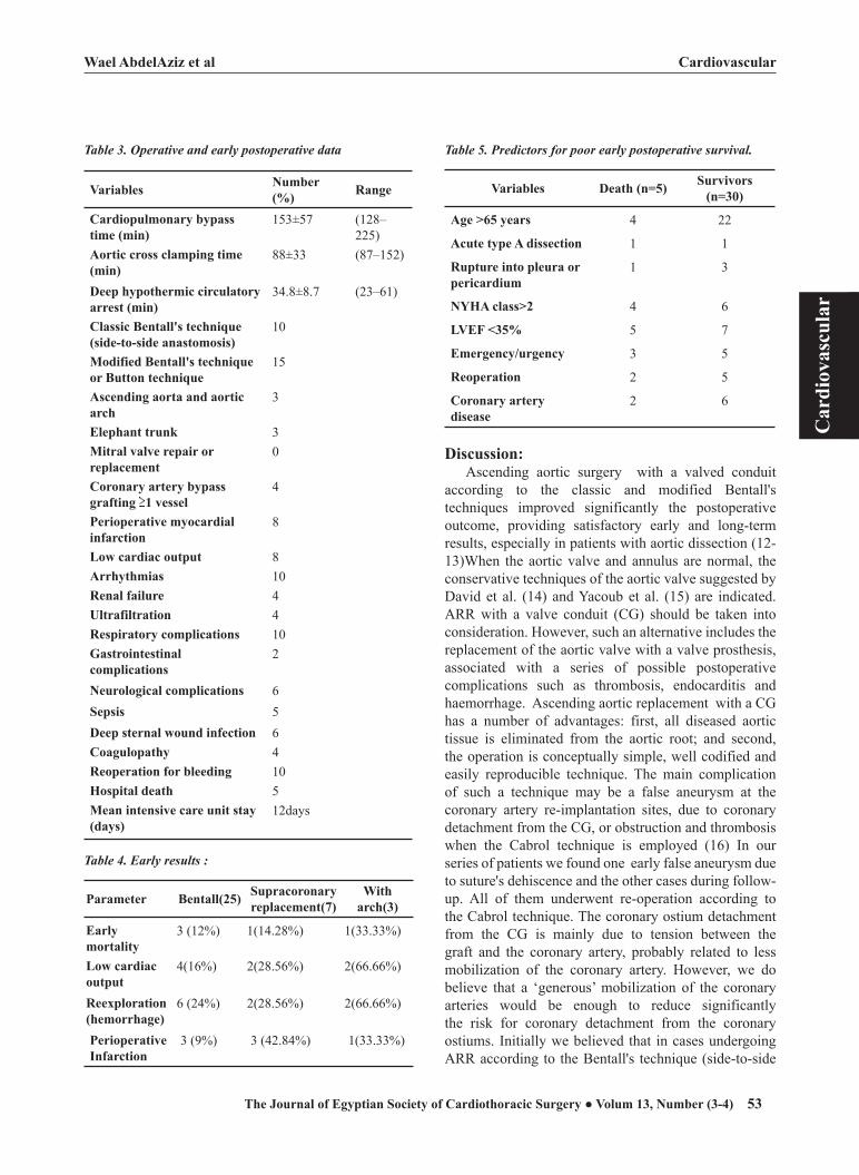
Cardiovascular Wael AbdelAziz et al
Car
diov
ascu
lar
The Journal of Egyptian Society of Cardiothoracic Surgery ● Sep-Dec 200552
Wael AbdelAziz et al Cardiovascular
Car
diov
ascu
lar
The Journal of Egyptian Society of Cardiothoracic Surgery ● Volum 13, Number (3-4) 53
Table 3. Operative and early postoperative data
Variables Number (%) Range
Cardiopulmonary bypass time (min)
153±57 (128–225)
Aortic cross clamping time (min)
88±33 (87–152)
Deep hypothermic circulatory arrest (min)
34.8±8.7 (23–61)
Classic Bentall's technique (side-to-side anastomosis)
10
Modified Bentall's technique or Button technique
15
Ascending aorta and aortic arch
3
Elephant trunk 3Mitral valve repair or replacement
0
Coronary artery bypass grafting 1 vessel
4
Perioperative myocardial infarction
8
Low cardiac output 8Arrhythmias 10Renal failure 4Ultrafiltration 4Respiratory complications 10Gastrointestinal complications
2
Neurological complications 6Sepsis 5Deep sternal wound infection 6Coagulopathy 4Reoperation for bleeding 10Hospital death 5Mean intensive care unit stay (days)
12days
Table 4. Early results :
Parameter Bentall(25) Supracoronary replacement(7)
With arch(3)
Early mortality
3 (12%) 1(14.28%) 1(33.33%)
Low cardiac output
4(16%) 2(28.56%) 2(66.66%)
Reexploration (hemorrhage)
6 (24%) 2(28.56%) 2(66.66%)
Perioperative Infarction
3 (9%) 3 (42.84%) 1(33.33%)
Table 5. Predictors for poor early postoperative survival.
Variables Death (n=5) Survivors (n=30)
Age >65 years 4 22
Acute type A dissection 1 1
Rupture into pleura or pericardium
1 3
NYHA class>2 4 6
LVEF <35% 5 7
Emergency/urgency 3 5
Reoperation 2 5
Coronary artery disease
2 6
Discussion:Ascending aortic surgery with a valved conduit
according to the classic and modified Bentall's techniques improved significantly the postoperative outcome, providing satisfactory early and long-term results, especially in patients with aortic dissection (12-13)When the aortic valve and annulus are normal, the conservative techniques of the aortic valve suggested by David et al. (14) and Yacoub et al. (15) are indicated. ARR with a valve conduit (CG) should be taken into consideration. However, such an alternative includes the replacement of the aortic valve with a valve prosthesis, associated with a series of possible postoperative complications such as thrombosis, endocarditis and haemorrhage. Ascending aortic replacement with a CG has a number of advantages: first, all diseased aortic tissue is eliminated from the aortic root; and second, the operation is conceptually simple, well codified and easily reproducible technique. The main complication of such a technique may be a false aneurysm at the coronary artery re-implantation sites, due to coronary detachment from the CG, or obstruction and thrombosis when the Cabrol technique is employed (16) In our series of patients we found one early false aneurysm due to suture's dehiscence and the other cases during follow-up. All of them underwent re-operation according to the Cabrol technique. The coronary ostium detachment from the CG is mainly due to tension between the graft and the coronary artery, probably related to less mobilization of the coronary artery. However, we do believe that a ‘generous’ mobilization of the coronary arteries would be enough to reduce significantly the risk for coronary detachment from the coronary ostiums. Initially we believed that in cases undergoing ARR according to the Bentall's technique (side-to-side

Cardiovascular Wael AbdelAziz et al
Car
diov
ascu
lar
The Journal of Egyptian Society of Cardiothoracic Surgery ● Sep-Dec 200554
Wael AbdelAziz et al Cardiovascular
Car
diov
ascu
lar
The Journal of Egyptian Society of Cardiothoracic Surgery ● Volum 13, Number (3-4) 55
anastomosis), wrapping the aneurysmal sack around the prosthesis (inclusion technique) may reduce further the tension between the graft and coronary artery and also may prevent the catastrophic consequences secondary to coronary detachment. Overall hospital mortality was 14.2%, greater than Bachet J et.al.,1996.(17) and near to those of AbdelAziz S 2005 which was 15.62% as his study was done also in Egypt with nearly the same type of patients and circumstances(18). Hospital mortality rate for patients undergoing operation in elective status was less than those done on an emergent basis. These results are gratifying given the high mortality and morbidity attending untreated ascending aortic aneurysm, dissection and root infection. The early postoperative morbidity resulted to be high in this series of patients, due to a high presence of starting experience in 4 centres in Egypt,late presentation, associated CAD and patients with depressed left ventricular function demonstrated by a low preoperative LVEF. The authors observed that the postoperative morbidity was strongly correlated with a long CPB time, circulatory arrest and extended surgical procedure to the aortic arch. There is a strong correlation between the postoperative renal failure and circulatory arrest time and aortic arch replacement is probably related with longer CPB time that these procedures require.
In our series, old age >65 years, aortic dissection, redo surgery, aneurysm rupture into pleura or pericardium, emergency status, NYHA functional class >2 and LVEF <35% were strong predictors of early death. Such predictors seems to be similar to the findings of Gott et al., At 1 year follow-up, all survivors demonstrated a significant improvement of NYHA functional class. We have the policy of indicating coronary angiography for all patients >40 years old, undergoing elective ascending aortic surgery. This strategy will decrease significantly the perioperative mortality due to myocardial infarction and low cardiac output in this group of patients. The late morbidity and mortality is closely related to thromboembolism or haemorrhage risk; The rate of thromboembolic events in our series was 4%; while , Gott et al. 1996 (19)reported an incidence of 0.42 events per 100 patient years; they attributed this lower incidence to the absence of pledgets and suture material in the bloodstream, and to a smaller amount of exposed sewing ring. Despite the employment of Ticron sutures with pledgets in our series, the incidence of thromboembolic events remained low. In the series of Gott 1995,(20) prosthetic endocarditis is the most common late complication of valved replacement and occurs in 4–5% of patients followed for a period of 14–17 years; our results showed
that, this complication occurred only in 8.57% of the followed patients, significantly higher than the previous reported results.
Conclusion:We may conclude that Ascending aorta with or
without arch replacement with or without a valved conduit offers an acceptable early and long-term outcome. The long-term outcome demonstrates a low related morbidity. The predictors for poor overall survival in those patients seem to be preoperative aortic dissection extended into the aortic arch, old age, depressed left ventricular function and associated CAD.
References:1. Anderson CA, Rizzo RJ, Cohn LH. Ascending Aortic
Aneurysms. In: Cohn LH, Edmunds LH Jr, eds. Cardiac Surgery in the Adult. New York: McGraw-Hill, 2003:11231148.
2. Bickerstaff LK, Pairolero PC, Hollier LH, et al: Thoracic aortic aneurysms: a population-based study. Surgery 1982; 92:1103.
3. Pressler V, McNamara JJ: Thoracic aortic aneurysm: natural history and treatment. J Thorac Cardiovasc Surg 1980; 79:489.
4. Marsalese DL, Moodie DS, Vacante M, et al: Marfan's syndrome: natural history and long-term follow-up of cardiovascular involvement. J Am Coll Cardiol 1989; 14:422.
5. Cikrit DF, Miles JH, Silver D: Spontaneous arterial perfora-tion: the Ehlers-Danlos specter. J Vasc Surg 1987; 5:248.
6. Guo D, Hasham S, Kuang SQ, et al: Familial thoracic aor-tic aneurysms and dissections: genetic heterogeneity with a major locus mapping to 5q13-14. Circulation 2001; 103:2461.
7. Pachulski RT, Weinberg AL, Chan KL: Aortic aneurysm in patients with functionally normal or minimally stenotic bicuspid aortic valve. Am J Cardiol 1991; 67:781.
8. Schoen F: Blood vessels, in Cotran R, Kanor V, Collins C (eds): Robbins Pathologic Basis of Disease. Philadelphia, WB Saunders, 1999.
9. Stowe CL, Baertlein MA, Wierman MD, et al: Surgical management of ascending and aortic arch disease: refined techniques with improved results. Ann Thorac Surg 1998; 66:388.
10.Ergin MA, Spielvogel D, Apaydin A, et al: Surgical treat-ment of the dilated ascending aorta: when and how? Ann Thorac Surg 1999; 67:1834.
11.Crawford ES, Svensson LG, Coselli JS, Safi HJ, Hess KR: Surgical treatment of aneurysm and/or dissection of the ascending aorta, transverse aortic arch, and ascending aorta and transverse aortic arch: factors influencing survival in 717 patients. J Thorac Cardiovasc Surg 1989; 98:659.
12.Niederhauser U, Kunzli A, Genoni M, Vogt P, Lachat M, Turina M. Composite graft replacement of the aortic root: long-term results, incidence of reoperations. Thorac
Cardiovascular Wael AbdelAziz et al
Car
diov
ascu
lar
The Journal of Egyptian Society of Cardiothoracic Surgery ● Sep-Dec 200554
Wael AbdelAziz et al Cardiovascular
Car
diov
ascu
lar
The Journal of Egyptian Society of Cardiothoracic Surgery ● Volum 13, Number (3-4) 55
Cardiovasc Surg 1999;47(5):317 321, Niederhauser U, Ru-diger H, Vogt P, Kunzli A, Zund G, Turina M., Composite graft replacement of the aortic root in acute dissection. Eur J Cardiothoracic Surg 1998;13(2):144–150.
14-David TE, Feindel CM. An aortic valve sparing operation for patients with aortic incompetence and aneurysm of the ascending aorta. J Thorac Cardiovasc Surg 1992;103:617–622.
15-Yacoub MH, Gehle P, Chandrasekaran V, Birks EJ, Child A, Radley- Smith R. Late results of valve-preservation in patients with aneurysms in the ascending aorta and root. J Thorac Cardiovasc Surg 1998;115:1080–1090.
16-Svensson LG, Crawford ES, Hess KR, Coselli JS, Safi HJ. Composite valve graft replacement of the proximal aorta: comparison of techniques in 348 patients. Ann Thorac Surg 1992;54:427–439.
17- Bachet J., Termignon J.-L., Goudot B., Dreyfus G., Pi-quois A., Brodaty D., Dubois C., Delentdecker P., Guilmet
D. Aortic root replacement with a composite graft. Factors influencing immediate and long-term results. Eur J Cardio-thorac Surg 1996;10:207-213.
18-AbdelAziz S. Total Circulatory Arrest and retrograde cerebral perfusion During Ascending aorta and arch surgery.Journal of the Egyptian society of cardiothoracic surgery :13(1-2); 39-43.
19-Gott V.L., Greene P.S., Alejo D.E., Cameron D.E., Naftel D.C., Miller D.C., Gillinov A.M., Laschinger J.C., Pyeritz R.E. Replacement of the aortic root in patients with Marfan's syndrome. New Engl J Med 1999;340(17):1307-1313.
20-Gott V.L., Gillinov A.M., Pyeritz R.E., Cameron D.E., Reitz B.A., Greene P.S., Stone C.D., Ferris R.L., Alejo D.E., McKusick V.A. Aortic root replacement. Risk factor analysis of a seventeen-years experience with 270 patients. J Thorac Cardiovasc Surg 1995;109(3):536-545.
Thoracic
The Journal of Egyptian Society of Cardiothoracic Surgery ● Sep-Dec 200556
Ahmed El Nouri et al Thoracic
Tho
raci
c
The Journal of Egyptian Society of Cardiothoracic Surgery ● Volum 13, Number (3-4) 57
Ahmed El Nouri, MDHatem Yazid, MDTarek Abdel Aziz, MDMohamed Atia, MDMohamed Abdel Fattah. MDMostafa Abdel Azeem, MD
objective: To review our experience in treatment of patients with pleural empyema due to different causes. Methods: A total of 181 patients (109 males and 72 females); their mean age, 34 years; range, 2 to 73 years underwent procedures for empyema thoracis between January 1998 and December 2004. 127 patients were post pneumonic empyema, 13 patients were post resection, 23 patients were due to minor surgical procedures, 18 patients were post traumatic, and 8 patients were due to other causes. 7 patients had thoracocentesis as the initial procedure, 117 patients had intercostal tube as initial proce-dure, and 3 patients had decortication as the initial surgical procedure.Results&Conclusion: 1 patient improved with the thoracocentesis, 43 pa-tients improved with intercostal tube insertion, 6 patients improved with video assisted thoracoscopic debridement, and 77 patients underwent open decortication, two of them needed thoracoplasty.
Pleural infection was first described by Hippocrates in 500BC. Open thoracic drainage was the only treatment for this disorder until the 19th century when closed chest tube drainage was first described but not adopted1. This technique became widely practised during an influenza epidemic in 1917–19 when open surgical drainage
was associated with a mortality rate of up to 70%2. A military commission in-vestigated this high mortality rate and produced recommendations that remain the basis for treatment today. They advocated adequate pus drainage with a closed chest tube, avoidance of early open drainage, obliteration of the pleural space, and proper nutritional support. These changes reduced the mortality rate to 3.4% during the later stages of the epidemic
Methods:During the period from January 1998 till December 2004, 181 patients
were referred to the cardiothoracic surgical units at Ain Shams University Hospitals with pleural infection. There were 109 males and 72 females. Their mean age was 34 +/- 22 years (range from 2 to 73 years).
All these patients had pleural effusion with elevated white blood cell count or had bacterial organisms demonstrated on Gram stain or culture. Patients
Empyema Thoracis: Outcome of 181 Patients
Accepted for publication dec, 10,2005
Address reprint request to Dr A. El Nouri
Department of Cardio-Thoracic Surgery
Ain Shams University
Cairo , Egypt
Email: [email protected]
Codex :05/03/trbr/0512
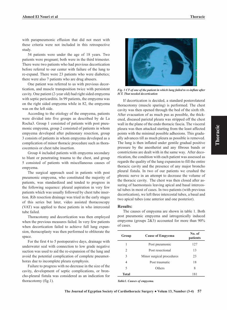
Thoracic
The Journal of Egyptian Society of Cardiothoracic Surgery ● Sep-Dec 200556
Ahmed El Nouri et al Thoracic
Tho
raci
c
The Journal of Egyptian Society of Cardiothoracic Surgery ● Volum 13, Number (3-4) 57
with parapneumonic effusion that did not meet with these criteria were not included in this retrospective study.
54 patients were under the age of 18 years. Two patients were pregnant; both were in the third trimester. There were two patients who had previous decortication before referral to our center with failure of the lung to re-expand. There were 23 patients who were diabetics; there were also 7 patients who are drug abusers.
One patient was referred to us with previous decor-tication, and muscle transposition twice with persistent cavity. One patient (2-year old) had right sided empyema with septic pericarditis. In 99 patients, the empyema was on the right sided empyema while in 82, the empyema was on the left side.
According to the etiology of the empyema, patients were divided into five groups as described by de La Rocha3. Group 1 consisted of patients with post pneu-monic empyema, group 2 consisted of patients in whom empyema developed after pulmonary resection, group 3 consists of patients in whom empyema developed as a complication of minor thoracic procedure such as thora-cocentesis or chest tube insertion.
Group 4 included patients with empyema secondary to blunt or penetrating trauma to the chest, and group 5 consisted of patients with miscellaneous causes of empyema.
The surgical approach used in patients with post pneumonic empyema, who constituted the majority of patients, was standardized and tended to progress in the following sequence: pleural aspiration in very few patients which was usually followed by chest tube inser-tion. Rib resection drainage was tried in the early stages of this series but later, video assisted thoracoscopy (VAT) was applied to these patients in who intercostal tube failed.
Thoracotomy and decortication was then employed when the previous measures failed. In very few patients when decortication failed to achieve full lung expan-sion, thoracoplasty was then performed to obliterate the space.
For the first 4 to 5 postoperative days, drainage with underwater seal with connection to low grade negative suction was used to aid the re-expansion of the lung and avoid the potential complication of complete pneumot-horax due to incomplete pleura symphysis.
Failure to progress with no decrease in the size of the cavity, development of septic complications, or bron-cho-pleural fistula was considered as an indication for thoracotomy (fig.1).
Fig. 1 CT of one of the patient in which lung failed to re-inflate after ICT. That needed decortication
If decortication is decided, a standard posterolateral thoracotomy (muscle sparing) is performed. The chest cavity was then opened through the bed of the sixth rib. After evacuation of as much pus as possible, the thick-ened, diseased parietal pleura was stripped off the chest wall in the plane of the endo thoracic fascia. The visceral pleura was then attacked starting from the least affected points with the minimal possible adhesions. This gradu-ally advances till as much pleura as possible is removed. The lung is then inflated under gentile gradual positive pressure by the anesthetist and any fibrous bands or constrictions are dealt with in the same way. After deco-rtication, the condition with each patient was assessed as regards the quality of the lung expansion to fill the entire thoracic cavity and the presence of any major broncho pleural fistula. In two of our patients we crushed the phrenic nerve in an attempt to decrease the volume of the thoracic cavity. The chest was then closed after as-suring of haemostasis leaving apical and basal intercos-tal tubes in most of cases. In two patients (with previous decortication), we left three intercostal tubes, a basal and two apical tubes (one anterior and one posterior).
Results:The causes of empyema are shown in table 1. Both
post pneumonic empyema and iatrogenically induced empyema (groups 2&3) accounted for more than 90% of cases.
Group Cause of Empyema No. of patients
1 Post pneumonic 127
2 Post resectional 13
3 Minor surgical procedures 23
4 Post traumatic 18
5 Others 8
Total 181
Table1. Causes of empyema.
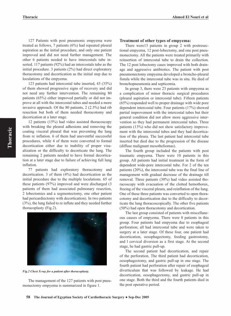
Ahmed El Nouri et alThoracic
Tho
raci
c
The Journal of Egyptian Society of Cardiothoracic Surgery ● Sep-Dec 200558
Ahmed El Nouri et al Thoracic
Tho
raci
c
The Journal of Egyptian Society of Cardiothoracic Surgery ● Volum 13, Number (3-4) 59
127 Patients with post pneumonic empyema were treated as follows, 7 patients (6%) had repeated pleural aspiration as the initial procedure, and only one patient improved and did not need further management. The other 6 patients needed to have intercostals tube in-serted. 117 patients (92%) had an intercostals tube as the initial procedure. 3 patients (2%) had direct exploratory thoracotomy and decortication as the initial step due to loculations of the empyema.
123 patients had intercostal tube inserted, 43 (35%) of them showed progressive signs of recovery and did not need any further intervention. The remaining 80 patients (65%) either improved partially or did not im-prove at all with the intercostal tubes and needed a more invasive approach. Of the 80 patients, 2 (2.5%) had rib resection but both of them needed thoracotomy and decorication at a later stage.
12 patients (15%) had video assisted thoracoscopy with breaking the pleural adhesions and removing the coating visceral pleural that was preventing the lung from re inflation. 6 of them had uneventful successful procedures, while 4 of them were converted to formal decortication either due to inability of proper visu-alization or the difficulty to decorticate the lung. The remaining 2 patients needed to have formal decortica-tion at a later stage due to failure of achieving full lung inflation.
77 patients had exploratory thoracotomy and decortication. 3 of them (4%) had decortication as the initial procedure due to the multiple loculations. 65 of these patients (97%) improved and were discharged (3 patients of them had associated pulmonary resection, 2 lobectomies and a segmentectomy, one other patient had pericardiectomy with decortication). In two patients (3%), the lung failed to re inflate and they needed further thoracoplasty (Fig.2).
Fig.2 Chest X-ray for a patient after thoracoplasty.
The management of the 127 patients with post pneu-monectomy empyema is summarized in figure 1.
Treatment of other types of empyema:There were13 patients in group 2 with postresec-
tional empyema, 12 post-lobectomy, and one post pneu-monectomy. All the patients were treated primarily with reinsertion of intercostal tube to drain the collection. The 12 post lobectomy cases improved with both drain-age and aggressive antibiotics. The patient with post pneumonectomy empyema developed a broncho-pleural fistula while the intercostal tube was in situ. He died of bronchopneumonia and septicemia.
In group 3, there were 23 patients with empyema as a complication of minor thoracic surgical procedures (pleural aspiration or intercostal tube). Fifteen patients (65%) responded well to proper drainage with wide pore dependent intercostal tube. Four patients (17%) showed partial improvement with the intercostal tubes but their general condition did not allow more aggressive inter-vention so they had permanent intercostal tubes. Three patients (13%) who did not show satisfactory improve-ment with the intercostal tubes and they had decortica-tion of the pleura. The last patient had intercostal tube inserted but died due to the progression of the disease (diffuse malignant mesotheliomas).
The fourth group included the patients with post traumatic empyema. There were 10 patients in this group. All patients had initial treatment in the form of dependent wide-pore intercostal tube. For 2 of the ten patients (20%), the intercostal tube was the final line of management with gradual decrease of the drainage till removal. Three patients (30%) had video assisted tho-racoscopy with evacuation of the clotted hemothorax, freeing of the visceral pleura, and reinflation of the lung. One of these three patients was converted to open thora-cotomy and decortication due to the difficulty to decor-ticate the lung thoracoscopically. The other five patients (50%) had open thoracotomy and decortication.
The last group consisted of patients with miscellane-ous causes of empyema. There were 8 patients in this group. Four patients had empyema due to esophageal perforation; all had intercostal tube and were taken to surgery at a later stage. Of these four, one patient had decortication, oesophagectomy, feeding gastrostomy, and l cervical diversion as a first stage. At the second stage, he had gastric pull-up.
The second patient had decortication, and repair of the perforation. The third patient had decortication, oesophagectomy, and gastric pull-up in one stage. The fourth patient had perforation after repair of esophageal diverticulum that was followed by leakage. He had decortication, oesophagectomy, and gastric pull-up in one stage. Both the third and the fourth patients died in the post operative period.

Ahmed El Nouri et alThoracic
Tho
raci
c
The Journal of Egyptian Society of Cardiothoracic Surgery ● Sep-Dec 200558
Ahmed El Nouri et al Thoracic
Tho
raci
c
The Journal of Egyptian Society of Cardiothoracic Surgery ● Volum 13, Number (3-4) 59
Two patients had empyema due to extension of a subphrenic collection. Both treated with intercostal tubes and improved.
One patient developed empyema after rupture of a lung cyst and pneumothorax. He had decortication and resection of the pulmonary cyst. The last patient was referred to us after multiple operations; he had deco-rtication at one stage which was followed by muscle transposition on two successive stages. He had residual pocket with discharging sinuses. No operative interven-
tion could be offered to him. Discussion:
The objectives of the surgical management of em-pyema thoracis are, to evacuate all the infected materi-als and to re-expand the lung. Re-expansion is of great importance, as residual space will almost certainly lead to re-infection. Appropriate long-term anti biotic cover-age is also required to control the local and systemic infection.
Post pneumonic empyema constituted the leading
3 Patients Decortication as initial
procedure6ICT
117 Patients had ICT as initial
procedure
123With ICT
2 patients rib resection
12 Patients VATS
66 Patients Decortication
6 Patients improved
6 patients Failed
127 patients with post pneumonic empyema
7 Patients Pleural Aspiration
1Improved
43 patientsImproved & discharged
77 patients had decortication
2 patients had thoracoplasty
75 patients improved (3 had associated
pulmonary, resection 1 had pericardiectomy)
summary of the 127 patients with post pneumonectomy empyema.

Ahmed El Nouri et alThoracic
Tho
raci
c
The Journal of Egyptian Society of Cardiothoracic Surgery ● Sep-Dec 200560
Ahmed El Nouri et al Thoracic
Tho
raci
c
The Journal of Egyptian Society of Cardiothoracic Surgery ● Volum 13, Number (3-4) 61
cause of empyema in this series of patients. When used as the initial mode of drainage, repeat thoracentesis was successful in only 1 of 7 patients (14%). This ratio was considerably lower than other published series (36%) (4), and (25%) (5) and (90%) (6). This is probably at-tributed to delay in seeking medical opinion and delayed referral to surgical intervention(7) in our patients. This is supported by the low number of our patients who had repeat thoracentesis (4%) when compared with others (25%) (5) and (11%) (6).
The drainage of the empyema with chest tube was our second line of management. It achieved a cure rate 35%. Some series reported similar success rate to ours (35%) (4), while others have higher rates (62%) (6). The rate of achieving cure with intercostal tube depends mainly on the stage of empyema. Usually the rate of suc-cess is much higher in stage II, while it is much lower in patients in stage III.
Only 2 of our patients had rib resection drainage. These 2 patients were managed before the introduction of the thoracoscopy to our unit. There is some contro-versy about the best methods of drainage of the chest. Some are advocates of rib resection drainage and usually perform it as a routine if no progress is achieved with the intercostal tube, while others are rarely perform rib resection drainage. Lemmer and colleagues found that closed tube thoracostomy, as initial treatment, was suc-cessful in only 35%, while rib resection provided cure or control in 91% when employed as the first treatment method4.Others do don’t consider rib resection when dealing with empyema especially after the introduction of video assisted thoracoscopy(8).
Thoracoscopy was rediscovered in thoracic in the early 90s. It was introduced to our unit in the late 90s. We had 12 patients with post pneumonic empyema who were treated with video assisted thoracoscopy when the drainage with intercostal tube was found unsatisfactory. 6 patients (50%) had successful procedures. In 4 patients (33%) the thoracoscopy was not satisfactory and the decision was made for conversion to open decortication. The last 2 patients (17%) had video assisted thoracos-copy and decortication. In the post operative period the lung inflation was not satisfactory and both had open decortication (7), and 9 days post thoracoscope.
Our total number of thoracoscopic management in post pneumonic empyema was (9%) is relatively low in comparison with the other series (8&9).This is due to our early experience which made us very selective as regards choosing patients for the thoracoscopy. Our suc-cess rate in dealing with such a problem thoracoscopi-cally was 50%. Roberts9 reported a success rate of endo-scopic decortication of 38%, Lardinois7 reported 56%,
and Luh8 reported 86%. We believe that the very wide variation in the results of the different series is mainly due to the experience of the centre and the stage of the empyema at the time of presentation.
In our patients with post pneumonic empyema, a to-tal of 77 patients (60%) had open decortication which, we believe, still remains the standard treatment for the chronic empyema. In many other series, decortication was the treatment of choice in patients with chronic empyema (10, 11). 75 patients (97%) improved two of them had left lower lobectomy, one patient had segmen-tectomy, and one patient had pericardiectomy with the decortication. All these patients were approached from the lateral thoracotomy except one patient who had septic pericarditis with right sided empyema who was approached from the median sternotomy (fig.3).
In two patients (3%) we failed to achieve lung expansion which led to gradual progressive lung col-lapse that necessitated thoracoplasty. Thoracoplasty was usually reserved for those patients in whom all other interventions failed(12, 13).13 patients had post resec-tion empyema.
Fig.3 CT of the patient with right sided empyema (drained with ICT) and septic pericarditis (drained with pig-tail catheter) before having decortication and pericardiectomy.
In 12 patients the surgical procedure was lobectomy and in one patient the procedure was pneumonectomy. All the 12 patients with post lobectomy empyema responded well to drainage with intercostal tube and systemic antibiotics. All the 12 patients were immedi-ate after surgery. We did not have any late cases of post resection empyema. The last patient who pad post pneu-monectomy empyema had an intercostal tube and irriga-tion with antibiotics. He developed a broncho-pleural fistula while on irrigation. The decision was made to re-explore his to close the broncho-pleural fistula but was postponed more than one time due to the poor general condition of the patient. The patient died due to

Ahmed El Nouri et alThoracic
Tho
raci
c
The Journal of Egyptian Society of Cardiothoracic Surgery ● Sep-Dec 200560
Ahmed El Nouri et al Thoracic
Tho
raci
c
The Journal of Egyptian Society of Cardiothoracic Surgery ● Volum 13, Number (3-4) 61
bronchopneumonia of the other lung before having the chance to close the fistula.
23 patients developed empyema following minor surgical procedures like pleural aspiration on intercostal tube. Deneuville reported the development of pleural empyema 2% of 128 patients who required intercostal tubes (14).
10 patients developed empyema thoracis after trauma. Two patients had penetrating trauma and 8 had blunt trauma. All of them had intercostal tube but only two of them (20%) improved and did not need any fur-ther management. 3 patients had VAT decortication, two improved and the third was converted to open decorti-cation. All the other 5 patients had open decortication. All the procedures were uneventful. The evacuation of post traumatic collection with or without infection was observed by Heniford and his colleagues and they found that the rate of conversion to open procedure is mostly due the time period between the trauma and interven-tion. They reported a higher rate of conversion if this gap is more than 7 days(15).
There was a small group (7 patients) with miscel-laneous causes of empyema, two patients from a sub-phrenic collection. Both were managed with intercostal tubes and systemic antibiotics and both resolved.
In patients with perforation of the oesophagus the treatment and outcome are largely determined by the time to presentation. Our mortality rate was high (50%) in patients with empyema due to esophageal perforation. This was mainly due to late presentation. The number of this subset is too small for any comparison.
References:1. Gotthard Bulau and closed water-seal drainage for e pyema,
1875–1891. Meyer JA. Ann Thorac Surg 1989; 48:597–9. 2. Empyema thoracis: historical perspective. Peters RM.Ann
Thorac Surg 1989; 48:306–83. Empyema thoracis. Surg.De La RocheGynecol. Obstet.
1982; 155:839-54.(Quoted from Idris Ali & Helmut Unruh. Management of empyema thoracis Ann Thorac Surg 1990; 50:355–9).
4. Modern management of adult thoracic empyemaJH Lemmer, MJ Botham, and MB Orringer J. Thorac. Car-diovasc. Surg., Jul 1985; 90: 849 - 855.
5. Treatment of spontaneous bacterial empyema thora cis AK Mandal and H Thadepalli . J. Thorac. Cardiovasc. Surg., Jul 1987; 94: 414 - 8.
6. Outcome of primary empyema thoracis: therapeutic and microbiologic aspects AK. Mandal, H. Thadepalli, A K. Mandal, and U. Chettipally. Ann Thorac Surg 1998;66:1782-1786
7. Delayed Referral and Gram-Negative Organisms Increase the Conversion Thoracotomy Rate in Patients Undergo-ing Video-Assisted Thoracoscopic Surgery for EmpyemaDidier Lardinois, Michael Gock, Edgardo Pezzetta, Christian Buchli, Valentin Rousson, Markus Furrer, and Hans-Beat RisAnn. Thorac. Surg., Jun 2005; 79: 1851 - 1856.
8. Video-Assisted Thoracoscopic Surgery in the Treatment of Complicated Parapneumonic Effusions or Empyemas.
Outcome of 234 Patients Shi-Ping Luh, MD; Ming-Chih Chou, MD; Liang-Shun Wang, MD; Jia-Yuh Chen, MD and Tsong-Po Tsai, MD Chest, April 1, 2005; 127(4): 1427 - 1432.
9. Minimally invasive surgery in the treatment of empyema: intra-operative decision making John R. Roberts, MD, Ann Thorac Surg 2003;76:225-230
10. Management of Parapneumonic Empyema in Children Ahmet Çekirdekçi, MD, Oguz Köksel, MD, Tugrul Göncü, MD, Oktay Burma, MD, Ali Rahman, MD, Ihsan Sami Uyar, MD, Erhan Ayan, MD, Ayhan Uysal, MDAsian Car-diovasc Thorac Ann 2000;8:137-140.
11. Is aggressive surgery in pleural empyema justified? H. Renner, S. Gabor, H. Pinter, A. Maier, G. Friehs, F.M. Smolle-Juettner Eur J Cardiothorac Surg 1998;14:117-122
12.Postpneumonectomy Bronchopleural Fistula Formation and Surgical Management Ünal Açikel, MD, Erdem Silis-treli, MD, Nilgün Özelsancak, MD, Özalp Karabay, MD, Eyüp Sabri Uçan, MD,1, Eyüp Hazan, MD, Öztekin Oto, MD Asian Cardiovasc Thorac Ann 1999;7:49-51
13.Evaluation of surgical treatment of pyothorax with spe-cial reference to the usefulness of the omental pedicle flap method J Shimizu, M Oda, Y Hayashi, S Murakami, K Kobayashi, Y Arano and Y Watanabe Eur J of Cardiothorac Surg 1993; 7: 543-547.
14.Morbidity of percutaneous tube thoracostomy in trauma patients M. Deneuville, Eur J Cardiothorac Surg 2002;22:673-678
15. The Role of Thoracoscopy in the Management of Retained Thoracic Collections After Trauma B. Todd Heniford, MD, Eddy H. Carrillo, MD, David A. Spain, MD, Jorge L. Sosa, MD, Robert L. Fulton, MD, J. David Richardson, MD ,Ann Thorac Surg 1997;63:940-943

Hossam El Okda et al
Tho
raci
cThoracic
The Journal of Egyptian Society of Cardiothoracic Surgery ● Sep-Dec 200562
Hossam El Okda et al
Tho
raci
c
Thoracic
The Journal of Egyptian Society of Cardiothoracic Surgery ● Volum 13, Number (3-4) 63
Surgical Treatment of Hydatid Disease of the Lung
Background:Lung is the second most common site for hydatid disease after the liver. The aim of this study is to present the clinical symptoma-tology, diagnostic evaluation including immunological monitoring and surgical techniques for the treatment of lung hydatid disease (cystic echi-nococcosis, CE).Methods: In the period from November 1998 till April 2004, 55 patients with lung hydatid cysts were treated surgically in Ain Shams University Hospitals. The cysts were located in the right lung in 22 patients (40%), in the left lung in 33 patients (60%). Diagnosis was established preoperatively in all cases; chest radiography and computed tomography were most helpful for diagnosis. Three different methods of immunological monitoring were used in diagnosis as well as follow up of patients, before and after surgery: Latex agglutination test (LAT), Indirect hemagglutination test (IHAT) and Enzyme-linked immumosorbent assays (ELISA). The 3 different modalities were compared as regards sensitivity and specificity. All cases were managed surgically, with several types of radical (2 cases) or conservative (53 cases) procedures. Radical procedures were in the form of lobectomy. Cystectomy with capitonnage was the most commonly performed conservative procedure (22 cases); other procedures included cystectomy with closure of bronchial openings (18 cases) and peri-cystectomy with capitonnage (11 cases). Decortication was done in 2 cases .Results:Postoperative morbidity occurred in 7 cases (12.7%) and the 30-day mortality rate was 0%. The median hospital stay for uncomplicated cases was 9 days and for complicated cases 19 days. Conclusion;In conclusion, a lung-conserving operation is the treatment of choice for lung hydatid disease and offers a good surgical outcome with a minimal recurrence rate. LAT offers many advantages for its easy use as a diagnostic and prognostic test in the pre- and post-surgical evaluation of cases of CE. Moreover, circulating antibodies detection of specific IgG subclasses in patients having CE may provide a more sensitive index of disease activity than total IgG antibodies. The broad dynamic range of serum IgG4 antibody concentrations and its correlation with treatment response and with relapse suggests its usefulness as an index of post-treat-ment disease activity.
Hydatid disease; caused by Echinococcus granulosis, is endemic in some countries, particularly were sheep and cattle are raised, such as Australia, New Zealand, the Mediterranean countries, the Middle East , and South America. After the liver, the lung is the second most common site for hydatid cysts in adults
(1, 2,3). This study aimed to present the clinical symptomatology, diagnostic evaluation especially the different methods of immunological monitoring and various surgical techniques used for the treatment of lung hydatid disease.
Accepted for publication Sep 10,2005
Address reprint request to Dr H El
Okda
Department of Cardio-Thoracic Surgery
Ain Shams University
Cairo , Egypt
Email:[email protected]
Codex :05/02/trbr/0512
Hossam El Okda 1, MDAhmed El Nori 1, MDMohammed Attia 1, MDNashwa I. Ramadan 2 Heba E. Abdel Aaty 2, MDDepartments of Cardiothoracic Surgery 1 Medical Parasitology 2. Faculty of Medicine, Ain Shams University

Hossam El Okda et al
Tho
raci
c
Thoracic
The Journal of Egyptian Society of Cardiothoracic Surgery ● Sep-Dec 200562
Hossam El Okda et al
Tho
raci
c
Thoracic
The Journal of Egyptian Society of Cardiothoracic Surgery ● Volum 13, Number (3-4) 63
Methods :Fifty five patients who had undergone operations
for hydatid disease of the lung in Ain Shams University Hospitals in the period from November 1998 and April 2004 were reviewed. The group consisted of 30 male and 25 female patients with a mean age of 28.7 + 9.7 years, ranging from 10 to 59 years.
Radiological data in the form of plain and lateral chest X-rays, CT of thorax and abdomen, and ultrasonography for abdomen were collected. The location of cysts in the different patients is shown by table 1
UpperLobe
MiddleLobe
LowerLobe
Total %
Right lung 7 5 10 22 40
Left lung 15 - 18 33 60
Total 22 5 28 55 100
Table 1. Location of the hydatid cysts in 55 patients
Laboratory tests :Patients as well as a control group of 20 normal
subjects were separated into 4 groups:Grou1: Patients with active newly diagnosed hydatidosis. This group comprised 7 patients who had non-complicated, or calcified cysts as detected clinically and by different imaging techniques and confirmed by serology. All the 7 patients were later surgically managed.Group 2: Patients who were followed up within 2 years of surgical treatment. This group compromised 34 patients with a past history of hydatidosis, who received surgical treatment and admitted for follow up at various times within 2 years of surgery.Group 3: Patients who were followed up after 2 years of surgical treatment. This group compromised 21 patients with a past history of hydatidosis, who received surgical treatment and admitted for follow up at various times after 2 years of surgery.Group 4: Control group of 20 normal subjects.
The following serological tests were performed to the 3 groups: 1- Latex agglutination test for detection of circulating
hydatid antigen in serum;Human hydatid cyst fluid (HHCF) was aspirated from intact unilocular lung cysts removed from the patients during surgery and was processed according to the method described by Parija and Rao (1986) (4). Hydatid fluid was aseptically aspirated, checked for the presence
of protoscolices and hooklets, then aliquoted and stored at -20°C until used.
Hyperimmune hydatid antiserum was raised in adult parasite free rabbits (3-4kg) as the procedure described by Parija et al. (1997) (5). Sterile HHCF was emulsified with an equal volume of Freund’s complete and incomplete adjuvants (Sigma Chemical Co., USA). Rabbits were injected with this emulsion intramuscularly at separate settings 6 weeks interval. Blood was collected when the titer of the antibodies to HHCF was 1:1024 monitored by an indirect hemagglutination assay (IHA) using a kit (Fumouze Laboratories Diagnostics, France). Hyperimmune antiserum was then purified and dialyzed as the method described by Gottstein in 1984 (6). A 2 ml cold serum-saline mixture (pH 7) was added to 2 ml cold saturated ammonium sulfate, pH 7, with stirring for 30 minutes on ice and then centrifuged at 3,000 x g for 15 minutes at 0°C. The supernatant was then discarded and the precipitate was suspended in 2 ml saline and the procedure was repeated until the supernatant was colorless. The final precipitate was suspended in 1 ml saline and dialyzed against phosphate-buffered saline (PBS), pH 7.2, to remove all the residual ammonium sulfate. The titer of the purified antiserum was 1:2048 by the IHA.
Detection of hydatid antigen in serum: It was performed as described by Tilton in 1987 (7). A 1% standardized polysterene latex suspension (0.81 µm; Sigma Chemical Co, USA) was prepared by mixing 0.1 ml of latex suspension with 9.9 ml of glycine-buffered saline (GBS), pH 8.4. This was stored at 4°C until used. One milliliter of 1% latex suspension was mixed with 1 ml of the purified hyperimmune antisera raised against HHCF in rabbits. The mixture was incubated at 37°C for two hours in a water bath. Antibody-sensitized latex particles were then washed two times with GBS, pH 8.4, and centrifuged at 3,000 x g for five minutes. The pellet of antibody-sensitized latex particles was emulsified with GBS, pH 8.4, and 1% bovine serum albumin to make a suspension of 2%. The particles were stored at 4°C until used. Latex particles coated with normal rabbit serum were used as control. A drop of test serum was placed on each half of a glass slide. An equal volume of sensitized latex reagent was added to the serum on one half. The same volume of a control latex suspension was added to the serum on the other half as a control. The slide was then manually rotated for two minutes and inspected. Agglutination with sensitized latex reagent and not with the control latex reagent was considered a positive result. Controls were examined in parallel in each test. The circulating antigen titer in the serum was estimated by performing a quantitative test. It was
Hossam El Okda et al
Tho
raci
cThoracic
The Journal of Egyptian Society of Cardiothoracic Surgery ● Sep-Dec 200564
Hossam El Okda et al
Tho
raci
c
Thoracic
The Journal of Egyptian Society of Cardiothoracic Surgery ● Volum 13, Number (3-4) 65
performed by testing serum at several dilutions. The highest dilution of the serum showing agglutination was the LAT titer.2- Indirect hemagglutination test for detection of
antibodies in serum: It was performed using the laboratories Fumouze commercial kit according to the manufacture’s instructions (Fumouze Laboratories Diagnostics, France). Results were recorded after incubation at room temperature for 2 h and titres ≥ 1/ 320 were considered positive.
3- Enzyme-linked immumosorbent assay for detection of total IgG and IgG subclasses ( IgG 1, 2, 3 & 4):Antigen preparation: Hydatid cyst fluid was
aseptically aspirated from unilocular hydatid cysts in camel’s lungs slaughtered at local abattoirs in Cairo, Egypt. HCF was centrifuged at 30000g for 10 min. at 4°C. The supernatant was collected and dialyzed using cellulose membrane with molecular weight cut off of 3.5 kDa (Spectrum Medical Industries, USA), against three changes of deionized water per 24 hrs over 3 successive days. The dialyzate was centrifuged again at 10000g for 30 min. at 4°C and the supernatant was collected. Protein content of the crude antigen was determined using a Bio-Rad assay (Bio-Rad, USA). The Ag was then aliquoted and stored at -70°C. The purification and concentration of the antigen was made as described by Dottorini et al. (1981) (8)
ELISA assay: was done according to Ramzy et al. (1999) (9). 50 µl HCF antigen diluted in 0.06 m carbonate buffer, pH 9.6, to a final concentration of 5 µg/ml, was added to 96-well polyvinyl microtitre plates (Dynatech Laboratories, Alexandria, VA). Plates were incubated at 37 °C for 3 hrs and left overnight at 4 °C. Wells were washed once with phosphate buffer saline (PBS), and then blocked with 0.01 M PBS pH 7.4 containing 0.05% Tween 20 (Sigma Chemical Co, USA) and 5% fetal calf serum (PBS-T/FCS) for 1 h at 37 °C. Sera diluted 1:100 in PBS-T/FCS were incubated in duplicate wells for 2 h at 37 °C. Antibody binding was detected with a peroxidase-conjugated goat antihuman total IgG or IgG1-4 (Hybridoma Reagent Laboratory, Baltimore, MD), as appropriate, diluted to 1:20 000 in PBS-T/FCS. A substrate solution of 4 mg/ml of o-phenylenediamine dihydro-chloride (Sigma Chemical Co, USA) plus 0.005% hydrogen peroxide was added and incubated at room temperature for 15–20 minutes. Color development was stopped with 4M sulfuric acid and absorbance values (ODs) were measured versus a PBS blank at 490 nm with an ELISA plate reader (MR 4000 Dynatech Laboratories, Alexandria, VA).
Interpretation of results: ELISAs were all optimized by checkerboard titration using defined positive and
negative serum pools tested in duplicate on each ELISA plate. The optical density (OD) of the negative serum pool was defined as zero and the difference in net OD obtained with the positive and negative serum pools was defined to correspond to 100 antibody units. Test sera with net OD cut-off values greater than the mean + 3 SD (Standard Deviation) of those of negative control sera, were considered positive in this test.
Operative techniques:All procedures were performed under general
anesthesia. A posterolateral thoracotomy through the fifth or sixth intercostal space was accomplished in lateral decubitus position. When the hydatid cyst was identified, the surgical wound and adjacent tissue were covered with packed gausses soaked in 1% povidone-iodine so that only the area of the lung containing the cyst was exposed. In patients with perforated and infected complicated cysts, after removal of remnants of germinative membranes and laminated membranes, the residual cavity was carefully cleaned and re-examined for spillage of daughter vesicles. The cystic cavity was cleaned by suction irrigated with 1% povidone-iodine in all patients. Cystectomy plus capitonnage were performed in these patients, however recently cystectomy plus closure of bronchial openings technique are more used. Bronchial openings were detected using saline solution, and closed with a 3-0 Ethibond. Decortication was performed in 2 patients who developed infection with pleural complications.
With application of positive endopulmonary pressure, air escaping through any bronchial openings is visualized by the formation of bubbles. This maneuver was repeated until all air leaks were sealed. There were 2 patients who underwent resections (1 left lower lobectomy, and 1 right lower lobectomy) due to a destroyed lobe. In all patients, either a 32 F or 28F chest tube was positioned posteriorly and anteriorly, in a respective order. All of the patients were transferred to the intensive care unit following operation. During the postoperative period, chest tube were placed on 20-25 cm H2O suction and were removed when no air leak was evident and when drainage was less than 150 ml in 24 hours.
Twenty one patients were given albendazol in a dosage of 10 mg/Kg as a postoperative prophylactic measure to prevent recurrence. Treatment was given as 3 sequential 28-day courses, with 14-day intervals between courses and continued for two years.
Statistical analysis Data were statistically analysed using the microcomputer program for Windows; Statistical Package for Social Science (SPSS), version
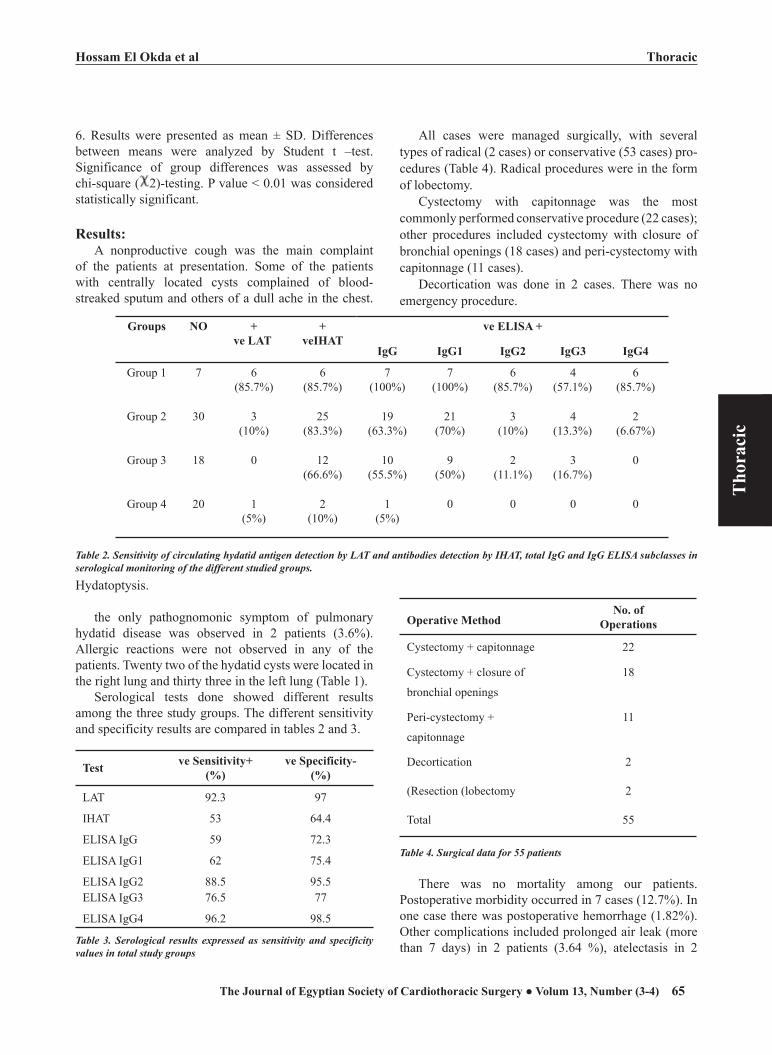
Hossam El Okda et al
Tho
raci
c
Thoracic
The Journal of Egyptian Society of Cardiothoracic Surgery ● Sep-Dec 200564
Hossam El Okda et al
Tho
raci
c
Thoracic
The Journal of Egyptian Society of Cardiothoracic Surgery ● Volum 13, Number (3-4) 65
6. Results were presented as mean ± SD. Differences between means were analyzed by Student t –test. Significance of group differences was assessed by chi-square ( 2)-testing. P value < 0.01 was considered statistically significant.
Results: A nonproductive cough was the main complaint
of the patients at presentation. Some of the patients with centrally located cysts complained of blood-streaked sputum and others of a dull ache in the chest.
All cases were managed surgically, with several types of radical (2 cases) or conservative (53 cases) pro-cedures (Table 4). Radical procedures were in the form of lobectomy.
Cystectomy with capitonnage was the most commonly performed conservative procedure (22 cases); other procedures included cystectomy with closure of bronchial openings (18 cases) and peri-cystectomy with capitonnage (11 cases).
Decortication was done in 2 cases. There was no emergency procedure.
Groups NO +ve LAT
+veIHAT
ve ELISA +
IgG IgG1 IgG2 IgG3 IgG4
Group 1 7 6(85.7%)
6(85.7%)
7(100%)
7(100%)
6(85.7%)
4(57.1%)
6(85.7%)
Group 2 30 3(10%)
25(83.3%)
19(63.3%)
21(70%)
3(10%)
4(13.3%)
2(6.67%)
Group 3 18 0 12(66.6%)
10(55.5%)
9(50%)
2(11.1%)
3(16.7%)
0
Group 4 20 1(5%)
2(10%)
1(5%)
0 0 0 0
Hydatoptysis.
the only pathognomonic symptom of pulmonary hydatid disease was observed in 2 patients (3.6%). Allergic reactions were not observed in any of the patients. Twenty two of the hydatid cysts were located in the right lung and thirty three in the left lung (Table 1).
Serological tests done showed different results among the three study groups. The different sensitivity and specificity results are compared in tables 2 and 3.
Test ve Sensitivity+(%)
ve Specificity-(%)
LAT 92.3 97
IHAT 53 64.4
ELISA IgG 59 72.3
ELISA IgG1 62 75.4
ELISA IgG2 88.5 95.5ELISA IgG3 76.5 77
ELISA IgG4 96.2 98.5
Table 3. Serological results expressed as sensitivity and specificity values in total study groups
Operative MethodNo. of
Operations
Cystectomy + capitonnage 22
Cystectomy + closure of
bronchial openings
18
Peri-cystectomy +
capitonnage
11
Decortication 2
(Resection (lobectomy 2
Total 55
Table 4. Surgical data for 55 patients
There was no mortality among our patients. Postoperative morbidity occurred in 7 cases (12.7%). In one case there was postoperative hemorrhage (1.82%). Other complications included prolonged air leak (more than 7 days) in 2 patients (3.64 %), atelectasis in 2
Table 2. Sensitivity of circulating hydatid antigen detection by LAT and antibodies detection by IHAT, total IgG and IgG ELISA subclasses in serological monitoring of the different studied groups.

Hossam El Okda et al
Tho
raci
cThoracic
The Journal of Egyptian Society of Cardiothoracic Surgery ● Sep-Dec 200566
Hossam El Okda et al
Tho
raci
c
Thoracic
The Journal of Egyptian Society of Cardiothoracic Surgery ● Volum 13, Number (3-4) 67
patients (3.64%) and recurrence in 2 patients (3.64%) (Table 5).
Complications No. of patients % Operative
technique
Hemorrhage 1 1.82 Pericystectomy +
Capitonnage
Prolonged air
leaks ( > 7 days)
2 3.64 Cystectomy +
Capitonnage
Atelectasis 2 3.64 Cystectomy +
Capitonnage
Recurrence 2 3.64 Cystectomy +
Capitonnage
Table 5. Postoperative complications and operative techniques
The patient who had hemorrhage was re-operated. Prolonged parenchymal air leak (>7 days) was managed by continuous negative aspiration and chest physiotherapy until the lung was fully expanded. The 2 cases with active post-surgical relapsing hydatidosis were both in group 2 and both of them received no chemotherapy after their primary operation. One of them was reoperated and received cystectomy and capitonnage. The other case had multiple hydatidosis in the lung as well as the liver and received only medical treatment. In both cases, IgG1 and IgG4 ELISA gave the highest sensitivity (100%), whereas all other tests showed only 50%. There was no correlation between postoperative complications and the technique used (Table 5). The median hospital stay for uncomplicated cases was 9 days and for complicated cases 19 days
Discussion:This study was based on a retrospective review of
surgical treatment of 55 patients with hydatid cysts of the lung, where our experience with respect to the clinical characteristics of these patients, immunological modalities of diagnosis as well as follow up, different treatment modes and treatment outcomes is presented.
Diagnosis of pulmonary hydatid cysts is generally based on clinical radiological and immunological findings. Pulmonary hydatid disease presents a wide range of radiological findings (10).
Uncomplicated cysts are seen as round opaque lesions on chest radiography. Infection and perforation may change the radiographic appearances of hydatid cyst, which may lead to an incorrect diagnosis and
delayed treatment. Although infected cysts are usually associated with perforation, this is not generally true and some ruptured cysts may remain uninfected (11, 12). Computed tomography (CT) may be helpful in establishing the diagnosis in complicated cysts , but the routine use of CT is not recommended unless a complicated hydatid cyst is suspected. CT is also helpful to detect malignant lesions in elderly patients and a useful guide for surgeons, allowing to perform a single operation for bilateral pulmonary and concomitant liver cysts (13).
The “gold standard” immunological test would be the one which could detect early complete resolution of the disease after medical or surgical treatment. Detection of serum antibodies, circulating antigen, and circulating immune complexes has been reported to be of potential use for support of clinical diagnosis in monitoring suspected cases of cystic echinococcosis after surgical/chemotherapeutic treatments for detection of relapses (14).
In this study, changes in concentrations of serum CAg and CAb were correlated with the clinical and radiological outcome of patients. CAb detection by IgG4 & IgG2 ELISA and CAg detection by LAT provided the highest sensitivity (96.2%, 88.5% and 92.3%) and specificity (98.5%, 95.5% and 97%) in total studied groups with a significant difference (p<0.01) when compared to IgG3 ELISA (76.5% sensitivity &77% specificity) and a highly significant difference (p<0.001) with IgG1 & IgG ELISA and IHAT (62%, 59% & 53% sensitivity & 75.4%. 72.3 & 64.6% specificity) (Table 3). Babba et al. (1994) reported that immunologic methods are more reliable as screening tests because they are technologically simpler, but they lack sufficient sensitivity for the detection of extrahepatic cysts (15). However, Gadea et al., (1999) concluded that the results of sensitivity and specificity are based on the cyst location in the patients. They found that antibody detection by LAT was negative in only 33.3% of treated and cured patients and was positive in 73.9% of patients with active disease. They reported that IHA could detect all active cysts because only active hydatid cysts from the liver and one disseminated relapse were included; on the other hand, it remained positive in 89% of patients treated and cured (16). Ramzy et al. (1999) in an ELISA to measure total E. granulosus-specific IgG antibodies and IgG subclasses, found no correlation between antibody levels for total IgG or all IgG subclasses and the organ affected with hydatid cysts (9). D’amelio et al. (1983) observed negative results in LAT in five surgically confirmed cases of CE and attributed this to various reasons (17). One possibility is that the intact

Hossam El Okda et al
Tho
raci
c
Thoracic
The Journal of Egyptian Society of Cardiothoracic Surgery ● Sep-Dec 200566
Hossam El Okda et al
Tho
raci
c
Thoracic
The Journal of Egyptian Society of Cardiothoracic Surgery ● Volum 13, Number (3-4) 67
cysts of E. granulosus may release only small amounts of antigens or no antigen into the circulation. This view was supported by the fact that macromolecules such as host albumin and immunoglobulin penetrate only 20% of the cysts under in vitro conditions. These macromolecules may pass into the parasite only after fissuring of the cyst wall. Another possibility is that the antigen released from the parasite form immune complexes with the antibodies in serum. Finally, the strain type, anatomic location of the cyst, cyst wall structure, and the speed and type of growth are other factors that may influence the level of circulating antigen (18). On the other hand, Ravinder et al. (1997) reported that LAT for detection of hydatid antigen in serum can be used as a prognostic test in the pre-surgical and post-surgical/chemotherapeutic evaluation of cases (19).
In active newly diagnosed hydatidosis patients in the present work, IgG and IgG1 ELISA provided 100% sensitivity, IgG4 ELISA, IgG2 ELISA, LAT and IHAT provided 85.7% with a significant difference (p<0.001) when compared to IgG3 ELISA which provided the least sensitivity of 57.1%. This is comparable with the results of Ramzy et al. (1999) who detected a diagnostic value of IgG1 of 97.7%, which was superior to the use of total IgG (65.1%) and IgG2 to IgG4 (77.8, 57.9, and 39.6%) (9). Similarly, cross-sectional studies of patients with CE using an ELISA incorporating partially purified hydatid cyst fluid antigen 5 (Ag5) and antigen B (AgB) have found a predominance of IgG1 and IgG4 antibodies expression in serum and concentrations of IgG4 were greater in symptomatic cases compared with those asymptomatic (20).
Concerning the 2 cases with active post-surgical relapsing hydatidosis, IgG1 and IgG4 ELISA gave the highest sensitivity (100%), whereas all other tests showed only 50%. However, the number of patients (two) is too small to get any statistical significance. Rigano et al. (1995) reported that patients with relapsing hydatid disease maintain high IgG4 titers in ELISA suggesting that the IgG4 subclass antibody is a good marker for hydatidosis follow-up (21). In a study on alveolar echinococcosis (AE) patients. McVie et al., (1997) correlated serum concentrations of IgG subclasses antibody responses in patients with symptomatic CE and indicated that IgG4 antibodies might correlate well with disease activity (22).
Looking for inactive cases within 2 years of post-surgical successful cure, the positivity of IgG4 ELISA (6.67%) was the least, followed by LAT and IgG2 ELISA (10%) and IgG3 ELISA (13.3%) indicating cure, although other tests remained high with insignificant false positive results (83.3% for IHAT, 63.3% for
IgG ELISA and 70% for IgG1 ELISA) with a highly significant difference (p<0.00001). In inactive cases after 2 years of post-surgical successful cure, the LAT and IgG4 ELISA were negative indicating complete cure, followed by IgG2 (11.1%) and IgG3 ELISA (16.7%). The other tests started to decline but were still significantly high (p<0.00001) (66.6% for IHAT, 55.5% for IgG ELISA and 50% for IgG1 ELISA). These results are comparable with those of Ravinder et al, (1997) who detected CAg pre-operatively in patients’ sera (10 out of 11) by the coagglutination-A (Co-A) and counter-immunoelectrophoresis (CIEP) tests and their levels showed a gradual decline by the seventh day post-operatively and were completely absent in serum 1 month after surgery and 6 months after chemotherapy (19). Almost all post-operative serum samples collected 2 years after surgical removal of the cyst, in cases of old hydatid disease, were negative for CAg by both the CIEP and Co-A tests. Unlikely, they denoted that no marked differences were observed between the CAb profile of the pre- and post-treatment sera, as shown by the IHA test, even 1 year after surgery or chemotherapy, and with a marginal decrease which is still detectable in all serum samples even 2 years after surgical removal of the cyst. Also, Guerri et al. (2000) found that IgG4 subclass became soon negative after surgery in patients from whom cysts had been removed successfully and if there is a good clinical response by the patient. However, it turns to be positive when there is a disease recrudescence, and keeps positive when residual cysts are left (23).
As mentioned before, it has to be mentioned that the results of the present study obtained in relapsing hydatidosis cases are not reliable because of the few number of patients that was available at the time of such preliminary study. Therefore to obtain better assessment, a prospective study should be conducted for better serological evaluation. Furthermore, the lack of available standardized assays in serological studies on hydatidosis, makes it difficult to compare between different laboratories.
Although a recent report has suggested medical therapy with albendazole in patients with hydatid cyst disease (24), surgery continues to be the treatment of choice in pulmonary hydatid disease. Chemotherapy alone is not reliable in controlling this disease. Alben-dazole is used routinely only in the treatment of patients with thoracoabdominal multiple hydatid disease, those with complicated hydatid cysts of the lung, in patients with recurrent cysts and in inoperable cystic hydatidosis. Various surgical procedures have been described in the literature, namely, excision of the entire cyst by enucle-
Hossam El Okda et al
Tho
raci
cThoracic
The Journal of Egyptian Society of Cardiothoracic Surgery ● Sep-Dec 200568
Hossam El Okda et al
Tho
raci
c
Thoracic
The Journal of Egyptian Society of Cardiothoracic Surgery ● Volum 13, Number (3-4) 69
ation (Barret technique), excision of pericyst ( Perez Fontana), cystectomy plus capitonnage, wedge resec-tion, segmentectomy, and lobectomy (2). The choice of surgical technique depends on the conditions encoun-tered during surgery. As a rule, the lung parenchyma should be preserved as much as possible in patients with pulmonary hydatid disease and radical procedures must be avoided (25). If, however, bronchiectasis or severe inflammation is present, the affected lung should be ex-cised. Lung resection was only carried out in 2 (3.6%) of our patients with infected cysts. In general, we tried to avoid lung resection in treatment of the infected cysts as much as possible and we believe this policy is important to the successful outcome of the surgical treatment of this disease. Decortication was performed in 2 patients (3.6%), because of the pleural thickening. Conservative surgical techniques, such as cystectomy plus capiton-nage constituted our routine surgical approach. How-ever recently, more cystectomy plus closure of bronchial openings technique is used. While conservative surgical technique such as capitonnage is widely performed for the management of the residual cystic space, it can cause atelectasis by obliterating the bronchus surrounding the cyst or the residual cavity may not be obliterated com-pletely by this procedure. Especially in patients with perforated cysts, postoperative complication rates can be lowered by the application of the cystectomy plus closure of bronchial openings technique (25).
In conclusion, circulating hydatid antigen detection by the latex agglutination test was shown to be a simple, rapid and specific test that could be available in diagnostic parasitology laboratory. The test is very easy to perform on a microscopic glass slide with successful results and without training or specific skills or any special equipments or reagents. Therefore, together with its promising results detected in this study, the LAT offers many advantages for its easy use as a diagnostic and prognostic test in the pre- and post-surgical evaluation of cases of CE. Moreover, CAb detection of specific IgG subclasses in patients having CE may provide a more sensitive index of disease activity than total IgG antibodies. Combined quantitative and qualitative standardization of IgG1 and IgG4 antibodies detection assays would be potentially useful for the serological monitoring and follow-up of human CE. The broad dynamic range of serum IgG4 antibody concentrations and its correlation with treatment response and with relapse suggests its usefulness as an index of post-treatment disease activity.
It is also concluded that conservative surgical procedures should be used as first choice line in treatment of pulmonary hydatid disease. An appropriate
surgical approach results in low complication and recurrence rates.
References :1. Aletras H, Symbas PN. Hydatid disease of the lung
. In : Shields TW, eds. General Thoracic Surgery (3ed). Philadelphia, PA, Lea & Febiger, 2000; 1113-22
2. Aytac A, Yurdkul Y, Ikizler C, et al. Pulmonary hydatid disease: Report of 100 patients. Ann Thorac Surg. 1977; 23: 145-51
3. Kalyoncu AF, Selcuk ZT, Emri AS, et al. Echinococcosis in the Middle East and Turkey. Reviews of Infectious Disease. 1991; 13: 1028-29
4. Parija SC, Rao RS. Enhancement of sensitivity of the haemagglutination test for echinococcosis by use of Staphylococcus aureus protein. A J Med Microbiol. 1986; 22: 241-144
5. Parija SC, Ravinder PT, Rao RS. Detection of hydatid antigen in urine by countercurrent-immunoelectrophoresis. J Clin Microbiol. 1997; 35: 1571-1574
6. Gottstein B. An immunoassay for the detection of circulating antigens in human echinococcosis. Am J Trop Med Hyg. 1984; 33: 1185–1190
7. Tilton RC Immunology in the clinical microbiology laboratory. In Clinical and Pathogenic Microbiology, edited by Howard BJ, Klaas J, Rubin SJ, Weissfeld AS, Tilton RC, C.V. Mosby Company, St. Louis, Washington, D.C., U.S.A. 1987
8. Dottorini S, Tassi C, Baldelli F. ELISA (Enzyme-Linked Immuno Assay) for diagnosis of human hydatid disease. Bull. Ist Sieroter Milanese. 1981; 60: 137-143
9. Ramzy RM, Helmy H, El Zayyat EA, et al. An enzyme-linked immunosorbent assay for detection of IgG1 antibodies specific to human cystic echinococcosis in Egypt. Trop Med Int Health. 1999; 4: 616-620
10.Gouliamos AD , Kaloidouris A, Papailoy J, et al. CT appearance of pulmonary hydatid disease. Chest. 1991; 100: 1578-81
11. Dogan R, Yuksel M, Cetin G. Surgical treatment hydatid cysts of the lung: report on 1055 patients. Thorax. 1989; 44: 192-99
12.Celik M, Senol C, Keles M, et al. Surgical treatment of pul-monary hydatid disease in children: Report of 122 cases. J Pediatr Surg. 2000; 35: 1710-3
13.Saksouk FA, Fahl LF, Rizk GK. Computed tomography of pulmonary hydatid disease. J Comput Assist Tomogr 1986; 10: 226-32
14.Poretti D, Felleisen E, Grimm F, et al. Differential immunodiagnosis between cystic hydatid disease and other cross reactive pathologies. Am. J Trop Med Hyg. 1999; 60: 193-202
15.Babba H, Messedi AS, Masmoudi M, et al. Diagnosis of human hydatidosis: Comparison between imagery and six serologic techniques. Am J Trop Med Hyg. 1994; 50: 64-68
16.Gadea I, Ayala G, Diago MT, et al. Immunological Diagnosis of Human Cystic Echinococcosis: Utility of

Hossam El Okda et al
Tho
raci
c
Thoracic
The Journal of Egyptian Society of Cardiothoracic Surgery ● Sep-Dec 200568
Hossam El Okda et al
Tho
raci
c
Thoracic
The Journal of Egyptian Society of Cardiothoracic Surgery ● Volum 13, Number (3-4) 69
Discriminant Analysis Applied to the Enzyme-Linked Im-munoelectrotransfer Blot. Clin Diag Lab Immunol. 1999; 6: 504-508
17.D’Amelio R, Pontesilli O, Palmisano L, et al. Detection and partial characterization of circulating immune complexes in hydatid disease. J Clin Microbiol. 1983; 18: 1021–1026
18.Shariff GM and Parija SC Countercurrent immunoelectrophoresis for the serodiagnosis of hydatid disease by detection of circulating hydatid antigen. J Microbiol Methods. 1991; 14: 71–76
19.Ravinder PT, Parija SC, Subba Rao KS. Evaluation of hu-man hydatid disease before and after surgery and chemo-therapy by demonstration of hydatid antigen and antibodies in serum. J Med Microbiol. 1997; 46: 859–864
20.Daeki AO, Craig PS, Shambesh MK. IgG-subclass anti-body responses and the natural history of hepatic cystic echinococcosis in asymptomatic patients. Ann Trop Med Parasitol. 2000; 94: 319-328
21.Rigano R, Profumo E, Ioppolo S, et al. Immunological
markers indicating the effectiveness of pharmacological treatment in human hydatid disease. Clin Exp Immunol. 1995; 102: 281-285
22.McVie A, Ersfeld K, Rogan MT et al. Expression and im-munological characterisation of Echinococcus granulosus recombinant antigen B for IgG4 subclass detection in hu-man cystic echinococcosis. Acta Trop. 1997; 67: 19–35
23. Guerri, ML, Davila M, Rodriguez M, et al. Utility of IgG subclasses in the diagnosis and follow up of hydatidosis. Enferm Infecc Microbiol Clin. 2000; 18: 262-266
24.Gil-Grande LA, Rodriguez-Caberio F, Prieto JG, et al. Randomized controlled trial of efficacy of albendazole in intraabdominal hydatid disease. Lancet 1993; 342: 1269-72
25.Halezeroglu S, Celik M, Uysal A, et al. Giant hydatid cysts of the lung. J Thorac Cardiovasc Surg. 1997; 113: 712-17

The Way I do it
The Journal of Egyptian Society of Cardiothoracic Surgery ● Sep-Dec 200570
Ezzeldin A. Mostafa The way I do it
The
way
I do
it
The Journal of Egyptian Society of Cardiothoracic Surgery ● Volum 13, Number (3-4) 71
An Easy Way to Band and to to Deband the Pulmonary Artery
Muller and Dammann (1) introduced pulmonary artery banding(PAB) in clinical practice in 1951. Since then, this operation has been used as a palliative procedure for small infants with congenital heart defects, to be followed by definitive repair at an older age. The technical difficulty
involved in primary repair and concerns regarding the use of cardiopulmonary bypass in small infants led many institutions to favor this staged approach. However, the perioperative morbidity and mortality of early primary repair has significantly improved in recent years.
Many neonates and infants are now treated by primary repair with good results, and PA banding is selected as the initial operative procedure in fewer patients (2-6). However, this operation still remains the procedure of choice for a diagnostic subset of patients (7).
The most common general indication for banding is congestive heart failure in infancy with anticipated delayed repair. Single ventricle is the most common lesion requiring banding, as this protects the pulmonary bed for future Fontan conversion. (9,10)
·Unbalanced AV canal ·Multiple VSD’s ·VSD and coarctation ·Single ventricle with increased pulmonary blood flow ·Contraindications to CPB (intracranial bleeding, thrombocytopenia) ·Late presentation of TGA (with shunt)
Surgical procedureThe pulmonary artery is approached through a median sternotomy. Some
precautions have been used to aid in the reoperation. Firstly, preservation (no resection) of the thymus, as usual, but trying to divide the two lobes. Secondly, a small limited pericardial incision (about 3cm) to preserve the lower part of the pericardium in the second stage operation.
Trusler’s rule for circumference of band is used: a) Simple defect (tetral-ogy, large VSD) = 20 mm + wt (kg) and b) Mixing defect (single ventricle, transposition) = 24 mm + wt (kg). This is marked with propylene 5/0 at the two ends. This formula is a modification of that suggested by Trusler and Mustard
Ezzeldin A. Mostafa, MD
Accepted for publication Sep 2005
Address for reprints: Ezzeldin A.
Mostafa, MD.
Department of Thoracic & Cardiovas-
cular Surgery, Ain-Shams University
Hospital 26 El-Sergany Str.,Abbassia
11381, Cairo,Egypt.
E-mail: [email protected]
Codex :09/01/epar/0506
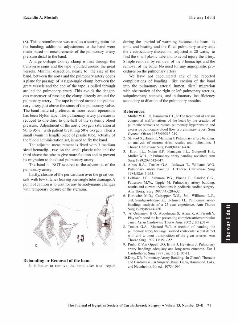
The Way I do it
The Journal of Egyptian Society of Cardiothoracic Surgery ● Sep-Dec 200570
Ezzeldin A. Mostafa The way I do it
The
way
I do
it
The Journal of Egyptian Society of Cardiothoracic Surgery ● Volum 13, Number (3-4) 71
(8). This circumference was used as a starting point for the banding; additional adjustments to the band were made based on measurements of the pulmonary artery pressure distal to the band.
A large c-shape Cooley clamp is first through the transverse sinus and the tape is pulled around the great vessels. Minimal dissection, nearly to the size of the band, between the aorta and the pulmonary artery opens a plane for passage of a right-angle clamp between the great vessels and the end of the tape is pulled through around the pulmonary artery. This avoids the danger-ous maneuver of passing the clamp directly around the pulmonary artery. The tape is placed around the pulmo-nary artery just above the sinus of the pulmonary valve. The band material preferred in more recent operations has been Nylon tape. The pulmonary artery pressure is reduced to one-third to one-half of the systemic blood pressure. Adjustment of the aortic oxygen saturation at 90 to 95% , with patient breathing 50% oxygen. Then a small (4mm in length) piece of plastic tube, actually of the blood administration set, is used to fix the band.
The adjusted measurement is fixed with 3 medium sized hemaclip , two on the small plastic tube and the third above the tube to give more fixation and to prevent its migration to the distal pulmonary artery.
The band is NOT secured to the adventitia of the pulmonary artery.
Lastly, closure of the pericardium over the great ves-sels with few stitches leaving one single tube drainage. A point of caution is to wait for any hemodynamic changes with temporary closure of the sternum.
Debanding or Removal of the bandIt is better to remove the band after total repair
during the period of warming because the heart is tonic and beating and the filled pulmonary artery aids the electrocautary dissection, adjusted at 20 watts, to find the small plastic tube and to avoid injury the artery. Simple removal by removal of the 3 hemaclips and the removal of the band. No need for any angioplastic pro-cedures on the pulmonary artery
We have not encountered any of the reported complications of banding like erosion of the band into the pulmonary arterial lumen, distal migration with obstruction of the right or left pulmonary arteries, subpulmonary stenosis, and pulmonary insufficiency secondary to dilation of the pulmonary annulus.
References:1. Muller W.H., Jr, Dammann F.J., Jr The treatment of certain
congenital malformations of the heart by the creation of pulmonic stenosis to reduce pulmonary hypertension and excessive pulmonary blood flow: a preliminary report. Surg Gynecol Obstet 1952;95:213-219.
2. Stewart S., Harris P., Manning J. Pulmonary artery banding: an analysis of current risks, results, and indications. J Thorac Cardiovasc Surg 1980;80:431-436.
3. Kron I.L., Nolan S.P., Flanagan T.L., Gutgesell H.P., Muller W.H., Jr Pulmonary artery banding revisited. Ann Surg 1989;209:642-647.
4. Albus R.A., Trusler G.A., Izukawa T., Williams W.G. Pulmonary artery banding. J Thorac Cardiovasc Surg 1984;88:645-653.
5. LeBlanc J.G., Ashmore P.G., Pineda E., Sandor G.G., Patterson M.W., Tipple M. Pulmonary artery banding: results and current indications in pediatric cardiac surgery. Ann Thorac Surg 1987;44:628-632..
6. Horowitz M.D., Culpepper W.S., 3rd, Williams L.C., 3rd, Sundgaard-Riise K., Ochsner J.L. Pulmonary artery banding: analysis of a 25-year experience. Ann Thorac Surg 1989;48:444-450.
7. Al Qethamy, H O, Aboelnazar S, Aizaz K, Al Faraidi Y. Play safe: band the late presenting complete atrioventricular canal. Asian Cardiovasc Thorac Ann. 2002 ;10(1):31-4.
8. Trusler G.A., Mustard W.T. A method of banding the pulmonary artery for large isolated ventricular septal defect with and without transposition of the great arteries. Ann Thorac Surg 1972;13:351-355.
9. Pinho P, Von Oppell UO, Brink J, Hewitson J. Pulmonary artery banding: adequacy and long-term outcome. Eur J Cardiothorac Surg 1997 Jan;11(1):105-11.
10.Doty, DB. Pulmonary Artery Banding. In Glenn’s Thoracic and Cardiovascular Surgery (Baue, Geha, Hammond, Laks, and Naunheim), 6th ed., 1073-1094.
CTS Notes & Quiz
The Journal of Egyptian Society of Cardiothoracic Surgery ● Sep-Dec 200572
Notes
Pulmonary Metastases
Pathogenesis · The lung is the first capillary bed draining most pri-
mary sites, with tumor cells usually depositing in the periphery
· 10-20% of patients with pulmonary metastases have disease confined to the lungs (especially with sarco-mas)
Diagnosis· CT scanning is sensitive but not specific, and may un-
derestimate the number of malignant nodules· Needle biopsy rarely adds additional information· Sarcomas and melanomas are the most likely to cause
a solitary metastasis
Patient Selection· There are four criteria which should be met prior to
resection of pulmonary metastases:1. Resection should only be performed if removal of all
disease is possible2. The patient must have adequate pulmonary reserve to
tolerate resection3. Local control of the primary tumor4. Absence of metastases elsewhere in the patient
Prognostic Factors· Histologic cell type affects the pattern of metastasis as
well as outcome· Tumors with longer doubling time have better survival· The number of metastases, the disease-free interval,
and unilateral vs. bilateral disease are not prognosti-cally significant
· Complete resectability is the most important indicator of improved survival
Operative Technique· Wedge resections should be performed wherever pos-
sible to preserve parenchymal tissue· Manual exploration is preferred to thoracoscopic ex-
amination to identify all nodules· Bilateral disease may be treated either by staged bilat-
eral thoracotomy or median sternotomy for a single operation
Results· Outcomes vary according to primary tumor type Tumor type 5-year survival
Soft tissue sarcoma 25%Osteogenic sarcoma 20-40%Colon/rectal carcinoma 8-37%Renal cell carcinoma 13-50%Breast carcinoma 14-49%Head/neck carcinoma 40-50%Melanoma 25%
Selected ArticlesMcCormack PM, Bains MS, Begg CB, Burt ME,
Downey RJ, Panicek DM, Rusch VW, Zakowski M, Ginsberg RJ. Role of video-assisted thoracic surgery in the treatment of pulmonary metastases: Results of a prospective trial. Annals of Thoracic Surgery 1996 62(1):213-7.
McAfee MK, Allen MS, Trastek VF, Ilstrup DM, Des-champs C, Pairolero PC. Colorectal lung metastases: Results of surgical excision. Annals of Thoracic Sur-gery 1992 53(5):780-5.
Staren ED, Salerno C, Rongione A, Witt TR, Faber LP. Pulmonary resection for metastatic breast cancer. Ar-chives of Surgery 1992 127(11):1282-4.

CTS Notes & Quiz
Not
es &
Qui
z
The Journal of Egyptian Society of Cardiothoracic Surgery ● Volum 13, Number (3-4) 73
Pulmonary Metastases
Question 1: Which of the following statements is true regarding the pathogenesis and diagnosis of pulmonary metastastes? A. Less than 10% of patients at autopsy have metastatic
disease confined to the lungs. B. Endobronchial lesions are always primary lung
tumors. C. CT scan can underestimate the number of malignant
nodules by up to 75%. D. A solitary lesion is more likely to be metastatic if the
primary tumor is breast carcinoma. E. Percutaneous biopsy should be performed in order to
plan operative management.
Question 2: Which of the following statements is true regarding results after resection of pulmonary metastases? A. Partial resection of extensive pulmonary metastatic
disease can improve survival. B. Disease-free interval has not been shown to have
prognostic value for survival. C. Patients with a greater number of metastatic nodules
have poorer survival.. D. CEA levels do not affect 5-year survival after
resection of metastatic colon carcinoma. E. Metastatic head and neck cancers have poor outcome
after pulmonary resection.
Question 3: Which of the following statements is true regarding the clincal manifestations of lung cancer? A. Following regional lymph nodes, the most common
metastatic location is the contralateral lung. B. Constant chest pain associated with lung cancer may
indicate nerve and bone invasion. C. Hoarseness represents cervical lymph node metastasis
and imparts a poor prognosis. D. Hypertrophic pulmonary osteoarthropathy
(“clubbing”)occurs most commonly with small-cell carcinomas.
E. The most common hormonal manifestation is inappropriate secretion of antidiuretic hormone.
Question 4: Which of the following statements is true regarding the operative management of lung cancer? A. A bloody pleural effusion is not a contraindication to
surgery for peripherally located carcinomas. B. Preoperative CT scanning can accurately predict the
number of tumor nodules. C. Wedge resection is adequate therapy for most
peripherally located carcinomas. D. Advanced age is no longer considered a risk factor for
surgical treatment of lung cancer. E. Only 25% of all patients with lung cancer will be
operative candidates.
Question 5: Which of the following statements is true regarding the results for treatment of lung cancer? A. 5-year survival in the absence of hilar node metastasis
following resection approaches 50%. B. Cell type has little prognostic value on long-term
survival. C. Women have improved survival when compared to
men. D. Pancoast tumors are associated with poorer long-term
survival. E. Malignant solitary pulmonary nodules occur most
frequently in patients under the age of 60. Answer Key:
Quiz
1. C
2. B
3. B
4. E
5. A

Readers’ Corner
The Journal of Egyptian Society of Cardiothoracic Surgery ● Sep-Dec 200574
The JOURNAL reserves the right to edit letters submitted to “Readers’ Corner” for brevity and clarity. Readers are advised to limit their letters to a maximum of 300 words.
Dear SirsA big problema 61 Y male patient with chronic renal failure had right cephalo -brachial shunt in his right arm and within 48 hours his hand swollen tremendously , a good case of right subclavian vein stenosis which was confirmed by duplex and venousn angiogram ,I have booked him for right subclavian vein stenting which i have used 12mm by 4cm precise control stent by CORDIS , (it seems small size but we do not have anything bigger) you can see in the attched picture the venous stent ,then i have noticed that the stent moves proximal 1 mm
each heart beat , i thought it will stopa plane x ray after 24 hours should the stent in the right ventricle as confirmed by echocardiography , patient is asympotomatic and his swollen arm is much betterthe more i read about embolized venous stent , the more i get depressedknow what to do ?1-leave it alone2refer to cardiothroacic surgeon3Endovascular retrieval , how ? with what ? what is the dangerappreciate your urgent advise and experience in this regaredMr. Mohamed Omar El-FarokThe JOURNAL reserves the right to edit letters submitted to “Readers’ Corner” for brevity and clarity. Readers are advised to limit their letters to a maximum of 300 words.

Readers’ Corner
The Journal of Egyptian Society of Cardiothoracic Surgery ● Sep-Dec 200574
