journal of biochemistry and molecular biology vol. 40, no
TRANSCRIPT

Journal of Biochemistry and Molecular Biology, Vol. 40, No. 5, September 2007, pp. 625-635
Molecular Cloning and Characterization of the Yew Gene EncodingSqualene Synthase from Taxus cuspidata
Zhuoshi Huang1, Keji Jiang1, Yan Pi1, Rong Hou1, Zhihua Liao1,3, Ying Cao1, Xu Han1,
Qian Wang1, Xiaofen Sun1 and Kexuan Tang1,2,*1State Key Laboratory of Genetic Engineering, School of Life Sciences, Fudan-SJTU-Nottingham Plant Biotechnology R&D Center,
Morgan-Tan International Center for Life Sciences, Fudan University, Shanghai 200433, People’s Republic of China2Plant Biotechnology Research Center, Fudan-SJTU-Nottingham Plant Biotechnology R&D Center,
School of Agriculture and Biology, Shanghai Jiao Tong University, Shanghai 200030, People’s Republic of China3Laboratory of Natural Products and Metabolic Engineering, Institute of Biotechnology, School of Life Sciences,
Southwest China University, Chongqing 400715, People’s Republic of China
Received 18 September 2006, Accepted 28 March 2007
The enzyme squalene synthase (EC 2.5.1.21) catalyzes a
reductive dimerization of two farnesyl diphosphate (FPP)
molecules into squalene, a key precursor for the sterol and
triterpene biosynthesis. A full-length cDNA encoding
squalene synthase (designated as TcSqS) was isolated from
Taxus cuspidata, a kind of important medicinal plants
producing potent anti-cancer drug, taxol. The full-length
cDNA of TcSqS was 1765 bp and contained a 1230 bp open
reading frame (ORF) encoding a polypeptide of 409 amino
acids. Bioinformatic analysis revealed that the deduced
TcSqS protein had high similarity with other plant
squalene synthases and a predicted crystal structure
similar to other class I isoprenoid biosynthetic enzymes.
Southern blot analysis revealed that there was one copy of
TcSqS gene in the genome of T. cuspidata. Semi-
quantitative RT-PCR analysis and northern blotting
analysis showed that TcSqS expressed constitutively in all
tested tissues, with the highest expression in roots. The
promoter region of TcSqS was also isolated by genomic
walking and analysis showed that several cis-acting
elements were present in the promoter region. The results
of treatment experiments by different signaling components
including methyl-jasmonate, salicylic acid and gibberellin
revealed that the TcSqS expression level of treated cells had
a prominent diversity to that of control, which was
consistent with the prediction results of TcSqS promoter
region in the PlantCARE database.
Keywords: RACE, RT-PCR, Squalene synthase, Taxus
cuspidata, TcSqS
Introduction
Isoprenoids, also named as terpenoids, play important roles in
plant metabolism. The plant isoprenoids can be divided into
two classes, primary and secondary metabolites. The best-
known primary metabolites of isoprenoids include sterols,
carotenoids and gibberellins while the secondary metabolites
include various kinds of terpenes, such as mono-, sesqui-, di-
and triterpenes (Chappell, 1995). In plants, the sterols, also
called as phytosterols, not only act as essential molecules for
the primary functioning for eukaryotic organisms (Bloch,
1992), but also exhibit important pharmacological activities
(Jones et al., 1997). The triterpenes in plants also exhibit a
wide range of structural diversity and biological activity, and
their glycosides saponins are considered to be natural
medicines with economical importance (Osbourn, 2003;
Connolly and Hill, 2005). Despite classified into different
classes, the sterols and triterpenes biosynthetic pathways share
Database Accession No: DQ836053 for TcSqS mRNA and
DQ860509 for TcSqS Promoter Region
Abbreviations: CTAB, Cetyl trimethyl ammonium bromide; FPP,
farnesyl diphosphate; GA3, gibberellic acid; GGPP, geranylgeranyl
pyrophosphate; HMGR, 3-hydroxy-3-methylglutaryl coenzyme A
reductase; IPP, isopentenyl diphosphate; MeJA, methyl-jasmonate,
ORF, open reading frame; PCR, Polymerase chain reaction; RACE,
rapid amplification of cDNA ends; RT-PCR, reverse transcriptase-
polymerase chain reaction; SA, salicylic acid, SqS, Squalene syn-
thase.
*To whom correspondence should be addressed.
Tel: 86-21-65642772; Fax: 86-21-65643552
E-mail: [email protected] or [email protected]

626 Zhuoshi Huang et al.
a committed precursor - squalene - which is synthesized from
two molecules of farnesyl diphosphate (FPP) by a two-step
reductive dimerization reaction catalyzed by the enzyme
squalene synthase (SqS) (Abe et al., 1993).
SqS is an endoplasmic reticulum associated enzyme with
carboxy terminus anchored in the ER membrane and the
amino terminus including active site exposed in the cytosol
(Stamellos et al., 1993). The soluble, bioactive SqS enzymes
have been isolated from rat (Shechter et al., 1992) and
tobacco (Hanley and Chappell, 1992) by methods which
overcome the clag of the hydrophobic ER association for the
purification. cDNA clones for squalene synthase have been
isolated and characterized from various species, such as rice,
maize, soybean (Hata et al., 1997), tobacco (Hanley et al.,
1996; Devarenne et al., 1998), Arabidopsis thaliana
(Nakashima et al., 1995; Kribii et al., 1997) and other plants,
as well as mammals (Robinson et al., 1993; Inoue et al.,
1995) and yeast (Jennings et al., 1991; Robinson et al., 1993;
Merkulov et al., 2000). By the method of the cDNA
expression in E. coli, more soluble squalene synthase proteins
with enzymatically activity have been isolated and characterized,
such as those of human (Thompson et al., 1998), yeast
(Jennings et al.,1991), mouse (Inoue et al., 1995), tobacco
(Hanley et al., 1996; Devarenne et al., 1998), and Arabidopsis
(Nakashima et al., 1995). Since catalyzing the first committed
step in the formation of cholesterols, bile acids and steroid
hormones, human SqS is identified as an attractive target for
therapeutic intervention and draws great attention. Using the
method of x-ray diffraction, the proper crystal structure for
human squalene synthase which is similar to other class I
isoprenoid biosynthetic enzymes has been determined (Pandit
et al., 2000). The interest in the investigation of the squalene
synthase regulation in plants increases steadily in recent years.
The addition of fungal elicitors into tobacco cell cultures
induces the decline in sterol biosynthesis which correlates
with a suppression of SqS activity (Devarenne et al., 1998)
and the overexpression of squalene synthase in Panax ginseng
roots results in a remarkable increase of phytosterols and
triterpene saponins (Lee et al., 2004).
Squalene synthase is commonly described as an incipient
and crucial branch point enzyme away from the main
isoprenoids biosynthetic pathway and a potential regulatory
point that controls carbon flux into sterols and triterpenes
biosynthesis. Lots of evidence support that inhibition of the
squalene synthase enzyme is a potential means of redirecting
FPP away from the sterol biosynthesis, towards the synthesis
of other commercially interesting isoprenoids. The disruption
of sterol biosynthesis at squalene synthase leads a remarkable
accumulation of FPP in an erg9 mutant strain of Saccharomyces
cerevisiae (Song, 2003). Besides FPP, the increase of IPP and
GGPP was also observed when the rat liver cells were treated
with zaragozic acid A, a potent inhibitor of squalene synthase
(Keller, 1996) and similar effects have been observed in plants
(Fulton et al., 1995). A previous study shows that the
inhibition of SqS in tobacco results in up-regulation of mRNA
level and enzyme activity of the HMGR (Wentzinger et al.,
2002), the major limiting-step enzyme in MVA pathway. The
disruption at squalene synthase leads the depletion of squalene
derived products, and the induced compensatory responses
mediated by HMGR contribute to the biosynthesis of other
commercially interesting isoprenoids. The disruption of squalene
synthase as well as the overexpression of downstream
enzymes results in a 7-fold increase in the production of the
carotenoid lycopene in yeast (Shimada et al., 1998).
Taxol, a diterpenoid alkaloid produced by yew (Taxus)
species (Baloglu and Kingston, 1999), is one of the most
efficient anticancer drugs approved by American Food and
Drug Administration (Kohler and Goldspiel, 1994). The
biosynthesis of taxol and squalene derived products share the
same isoprenoids biosynthetic pathway upon the precursor
FPP. As decreasing the flux through competitive pathways is
considered to be one of the trends for the plant secondary
metabolism (Oksman-Caldentey and Inze, 2004), the regulation
of squalene synthase will draw more attentions in the
secondary metabolism of Taxus cuspidata. Here, we report the
molecular cloning and characterization of the squalene
synthase gene from Taxus cuspidata, as the first step for the
regulation of TcSqS in the isoprenoids biosynthetic pathway.
Materials and Methods
Plant materials. T. cuspidata plants were grown in greenhouse at
Fudan University, China. T. cuspidata cultured callus lines, initiated
from hypocotyl of yew seeds, were maintained in improved B5
solid medium (Sigma, USA) supplemented with 1 mg/L naphthalene-
acetic acid (NAA), 0.5 mg/L thidiazuron (TDZ), 1 mg/L 2,4-
dichlorophenoxy-acetic acid (2, 4-D) and 5 basic amino acids:
0.8 g/L tyrosine, 0.4 g/L alanine, 0.4 g/L praline, 0.4 glutamic acid
and 0.4 g/L lysine.
RNA and DNA isolation. Total RNAs were extracted from
different tissues including roots, stems and leaves of 3-year-old T.
cuspidata plants and from calluses using the special method for
Taxus species (Liao et al., 2004) and then stored at −80oC prior to
cDNA cloning and semi-quantitative RT-PCR analysis. Genomic
DNA was isolated using a CTAB-based method (Rechards et al.,
1995) for the walking library construction and Southern blot
analysis. The quality and concentration of RNA and DNA samples
were examined by EB-stained agarose gel electrophoresis and
spectrophotometer analysis.
Isolation of the TcSqS full-length cDNA. According to the
manufacturer’s protocol (PowerScriptTM Reverse Transcriptase),
single-strand cDNA library was constructed by reverse
transcription from total RNA isolated from T. cuspidata cultured
cell lines. After RNaseH treatment, the cDNA library was used as
templates for PCR amplification of the conserved region of SqS
from T. Cuspidata. A pair of degenerate oligonucleotide primers,
TcSQSF and TcSQSR, designed according to the conserved
sequences of other plant and fungal SqS genes, was used for the

Squalene synthase Gene from Taxus cuspidata 627
amplification of the core cDNA fragment of TcSqS by standard
gradient PCR amplification. All primers used in the study were
listed in Table 1. The PCR products were analyzed in 0.8% agarose/
EB gel, along with DL2000 DNA size markers (TaKaRa) and the
result showed a single major product sized about 0.4 kb was
amplified via PCR. The amplified PCR product was purified and
cloned into pMD18-T vector (TaKaRa, Japan) followed by
sequencing. BLAST analysis showed that the 450 bp fragment
sequenced above had high homology to other plant SqS genes. This
fragment was subsequently used for designing gene specific
primers for the cloning of 5' end of the cDNA of TcSqS by RACE.
The 5'-RACE-Ready first-strand cDNA library was synthesized
following the protocol provided by the manufacturer (SMARTTM
RACE). The 5'-end of TcSqS cDNA was amplified using two 5'-end
gene specific primers (GSP) and the universal primers provided by
the kit. For the first PCR amplification, TcSQS5-1 and UPM
(Universal Primer A Mix, Table 1) were used as the primers and 5'-
RACE-Ready cDNA library as the template. For the nested PCR
amplification, TcSQS5-2 and NUP (Nested Universal Primer A,
Table 1) were used as the nested PCR primers, while the products
of the first PCR amplification were used as templates. Both the first
and the nested PCR amplification procedures were carried out at
the following condition: 3 min at 94oC, 28 cycles (35 s at 94oC, 35 s
at 60oC, 2 min at 72oC) and 8 min at 72oC. The nested amplification
products were purified and subcloned into pMD18-T vector
followed by sequencing. The sequences of the 5'-end and the core
fragment were aligned and assembled perfectly on Contig Express
(Vector NTI 9.0), and an 800 bp-length cDNA sequence including
the whole 5'-UTR and part of the CDs of the TcSqS gene was
deduced. This fragment was subsequently used for designing gene
specific primers for the cloning of the full-length cDNA of TcSqS
by RT-PCR.
To amplify the full-length cDNA of TcSqS, two gene specific
primers, TcSQSFull-1 and TcSQSFull-2, were synthesized following
the 5'-UTR sequence. The RT-PCR amplification was carried out
following the protocol of One Step RNA PCR Kit (TaKaRa) in
which the AMV RTase XL was used to reversely transcribe mRNA
to cDNA. TcSQSFull-1 and adapter primer (AP) with a poly (T) tail
were used as RT-PCR primers and 1 µg total RNA as template.
PCR amplification procedure was carried out at the following
condition: 30 min at 50oC for reverse transcription, 2 min at 94oC
for RTase denaturation followed by amplification of 30 cycles
(94oC for 30 s, 65oC for 30 s, and 72oC for 3 min). The products of
the RT-PCR were diluted and used as the template for the nested
amplification PCR. The nested PCR used TcSQSF-2 and AP as
primers, and the PCR amplification procedure was carried out as
following: 3 min at 94oC followed by 30 cycles of amplification
(94oC for 30 s, 65oC for 30 s, 72oC for 3 min) and by 72oC for
8 min. The 1.8 kb-length nested amplification products were
purified and subcloned into pMD18-T vector followed by
sequencing.
Isolation of the TcSqS promoter. GenomeWalker DNA libraries
were constructed following the user manual provided by the
manufacturer (Universal GenomeWalkerTM Kit). The T. cuspidata
genomic DNA isolated from fresh leaves was completely digested
separately with six different blunt-end restriction enzymes (EcoRV,
HpaI, NaeI, NruI, SnaBI and StuI). After purification, each of the
blunt-end digestions was ligated to the GenomeWalker adaptor by
T4 DNA ligase. The adaptor-ligated genomic DNA fragments
referred as GenomeWalker libraries were used as the template in
the following PCR-based DNA walking.
The amplification of TcSqS promoter region consisted of two
walking amplifications. The primary PCR used the outer adaptor
primer AP1 provided in the kit and the gene specific primer
TcSqSW-1, and the amplification was performed in the process of 7
cycles (94oC for 30 s; 72oC for 3 min), followed by 32 cycles (94oC
for 25 s; 67oC for 3 min) and by 67oC for 8 min. The primary PCR
mixture was then diluted 50-fold and used as the template for
nested PCR with the nested adaptor primer AP2 provided in the kit
and a nested gene-specific primer TcSqSW-2. The amplification
was performed in the process of 5 cycles (94oC for 30 s, 72oC for
3 min), followed by 32 cycles (94oC for 30 s, 67oC for 3 min) and
by 67oC for 8 min. The PCR products were analyzed by
Table 1. The PCR primers used in the study
Primer Direction Sequence
TcSQSF + 5'-GT(C/T)GA GGA(C/T)G A(C/T)AC(A/T/C/G) AG(C/T)AT (A/T)(C/G)C-3'
TcSQSR - 5'-(T/G)(G/A)T(T/C)T TCTG(A/C/G) AGAAA (C/T)A(A/G)(G/A)C CCAT-3'
TcSQS5-1 - 5'-CAGTT GACAC GTGAT GAAAT TGGTC-3'
TcSQS5-2 - 5'-AAGAG AAGTG CCACG AAGGA TCATA-3'
TcSQSFull-F1 + 5'-GCCCT AACTT CTGAA TCACA CAGTT C-3'
TcSQSFull-F2 + 5'-GGCTG GGACG GGCTC AATTT TGATC AAT-3'
TcSQSW-1 - 5'-CTGTG TGATT CAGAA GTTAG GGCGC TAGAA-3'
TcSQSW-2 - 5'-GCCGT GCGCT TATAA ATTGT TTGAC ATCA-3'
UPM* +Long: 5'-CTAAT ACGAC TCACT ATAGG GCAAG CAGTG GTATC AACGC AGAGT-3'Short: 5'-CTAAT ACGAC TCACT ATAGG GC-3'
NUP* + 5'-AAGCA GTGGT ATCAA CGCAG AGT-3'
AP* - 5'-GGCCA CGCGT CGACT AGTAC TTTTT TTTTT TTTTT TT-3'
Walker-AP1* + 5'-GTAAT ACGAC TCACT ATAGG GC-3'
Walker-AP2* + 5'-ACTAT AGGGC ACGCG TGGT-3'
The primers provided by commercial kits were marked with *.
628 Zhuoshi Huang et al.
electrophoresis, purified from agarose gel, and then cloned into
pMD18-T vector followed by sequencing.
Semi-quantitative RT-PCR analysis. Semi-quantitative RT-PCR
was carried out to investigate the mRNA expression profiles of
TcSqS in different tissues of T. Cuspidata and under different
treatments. All RNA templates were digested with DNaseI and then
purified. RT-PCR was performed under the following condition:
50oC for 30 min for reverse transcription, 94oC for 2 min for RTase
denaturation followed by the amplifications (94oC for 30 s; 58oC for
30 s and 72oC for 50 s) with the cycles ranged from 20-30,
according to the protocol of One Step RNA PCR Kit (TaKaRa).
The consistency of each example was measured by Ultraviolet-
Visible Spectrophotometer to make sure that fixed quantificational
RNA of each example (500 ng per reaction) was used for RT-PCR
analysis. Two gene-specific primers, TcSQS5-1 and TcSQSFull-F1,
were used to amplify a 500 bp length fragment in the semi-
quantitative RT-PCR as the consults of TcSqS expression levels. At
the same time, the RT-PCR reaction for the house-keeping gene
(18S gene) using specific primers 18SF and 18SR designed
according to the conserved regions of plant 18S genes was
performed to estimate if equal amounts of RNA among samples
were used in RT-PCR as an internal control. Amplifications were
performed under the following conditions: 50oC for 30 min, 94oC
for 2 min followed by 20 cycles of amplification (94oC for 30 s,
58oC for 30 s and 72oC for 50 s). The amplified products were
separated on 1% agarose gel and analyzed with Gene analysis
software package (Gene Company).
Southern blot analysis. Total genomic DNA was isolated from
fresh T. cuspidata leaves following CTAB based method. Aliquots
of DNA (30 µg/sample) were digested respectively at 37oC with
EcoRI, HindIII and XbaI, which did not cut within the probe
region. Fully digested samples were fractionated by 0.85% agarose
gel electrophoresis, and transferred to a positively charged Hybond-
N+ nylon membrane (Amersham Pharmacia). The TcSqS 5'-end
cDNA fragment amplified with the primers TcSqSFullF-1 and
TcSqS5-1 was used as probe labeling. Probe labeling (biotin),
hybridization and signal detection were carried out using Gene
images random prime labeling module and CDP-Star detection
module following the manufacturer’s instructions (Amersham
Pharmacia). The filter was washed under high stringency condition
at the temperature of 60oC and the hybridized signals were
visualized by exposure to Fuji X-ray film at room temperature for
0.5 h.
Northern blotting analysis. To investigate the TcSqS expression
pattern in different tissues of T. cuspidata, high quality total RNAs
were extracted from root, stem, and leaf of three-year-old T.
cuspidata plants. Equivalent of each RNA sample (20 µg/sample)
was denatured and separated on 1% agarose gel containing 0.66M
formaldehyde, and blotted onto a positively charged Hybond-N+
nylon membrane (Amersham Pharmacia). The Probe labeling
(biotin), hybridization and signal detection were carried out with
the same protocol and same probe in Southern analysis. The
membrane was washed under high stringency condition at the
temperature of 65oC and the hybridized signals were visualized by
exposure to Fuji X-ray film at room temperature for 1 h.
Results and Discussion
Amplification of the TcSqS conserved region. Agarose gel
analysis revealed that gradient PCR amplification with
primers TcSqS-F and TcSqS-R generated a single major
product of 390 bp in length with the annealing temperature of
54oC. BLAST-n analysis indicated that it had wide similarity
to known squalene synthases from A. thaliana, Glycine max
and Oryza sativa, implying that it was probably a part of the
squalene synthase gene.
Rapid amplification of TcSqS 5'-cDNA end. Electrophoresis
analysis of primary PCR product of 5' RACE showed a
specific band of about 600 bp in length. Nested amplification
was carried out using primary PCR product as template and a
specific DNA band of about 550 bp was amplified. After
cutting away the adapter sequences, the final 5' cDNA end of
TcSqS turned out to be 540 bp. An overlap of 94 bp was found
between the 5' and the conserved fragments. BLAST-n analysis
of this sequence resulted in a similar result as that of the
conserved region. Based on the sequence of the 5' UTR region
of TcSqS, two pairs of primers were designed: TcSqSFull-F1
and TcSqSFull-F2 for the full-length cDNA amplification,
TcSqSW-1 and TcSqSW-2 for the genomic walking.
Isolation and analysis of TcSqS full-length cDNA. Agarose
gel analysis revealed that amplification with primers TcSqSFull-
F1 and poly(T) primer AP resulted in a DNA band of about
1.9 kb accompanied by some faint bands and smearing.
Nested amplification resulted in a bright band of about 1.8 kb
as anticipated. Sequencing result of this sequence coincided
with the 5'-end of TcSqS cDNA sequence amplified before.
After cutting away the adapter sequences, the 1765 bp cDNA
of TcSqS possessed a 1530-bp open reading frame (ORF)
from 60 bp to 1289 bp of the sequence besides a 59-bp 5'
UTR and a 476-bp 3' UTR including a PolyA tail. Nucleotide-
nucleotide BLAST of the TcSqS full-length cDNA sequence
on NCBI website indicated that its conserved segments had
similarity to the known SqS of other plant species such as G.
max, Lotus japonicus, and Panax ginseng.
Characterization of the TcSqS protein. The open reading
frame of TcSqS was obtained by the ORF Finder on the NCBI
Web (http://www.ncbi.nlm.nih.gov/gorf/gorf.html). The predicted
TcSqS protein precursor was 409 aa in length with a theoretical
molecular weight of 46.46 kDa and pI value of 6.45.
According to NCBI conserved domain search, a conserved
domain “Trans_IPPS_HH” (cd00683) was found to penetrate
through the core of predicted TcSqS protein.
The NCBI protein-protein BLAST showed that the deduced
TcSqS amino acid sequence had 72% (282/387) identities and
84% (327/387) positives in local alignments to G. max SqS. It
also shared very broad and high local identities and positives
to other plant species of broad classes such as Panax
notoginseng, P. ginseng, Solanum tuberosum, L. japonicus,
Nicotiana benthamiana and A. thaliana. The deduced TcSqS

Squalene synthase Gene from Taxus cuspidata 629
protein shared a less identity to the squalene synthases of
mammalian (human and rat) and fungi (yeast). Multi-
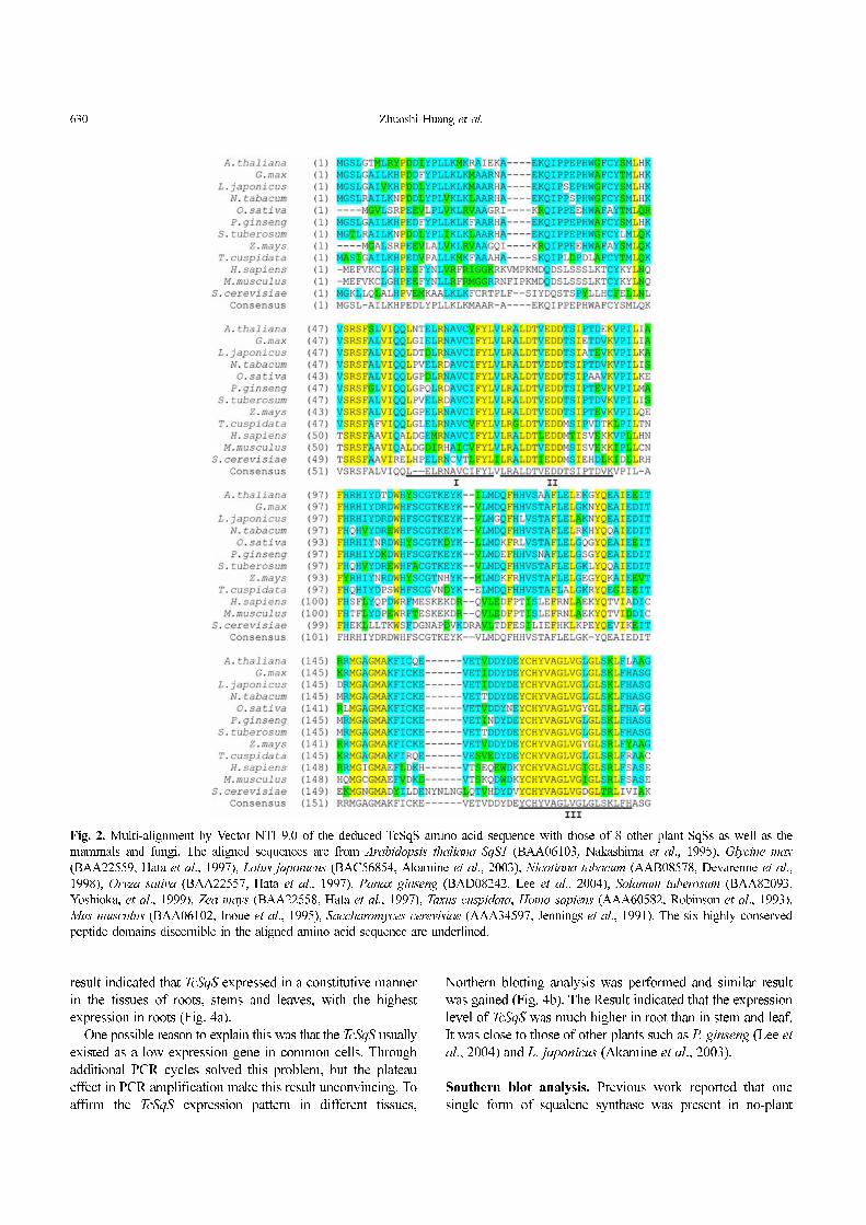
alignment of TcSqS with other SqS was conducted (Fig. 2).
Six highly conserved peptide domains of 1423 amino acids
which might be important for catalytic/functional activity
(Robinson et al., 1993) were discernible in the TcSqS amino
acid sequence (Fig. 2). Domains III, IV, and V showed a
highly conserved consensus sequence with other SqS enzymes,
while domains I and II were much less conserved. Domain II
was highly conserved with an aspartate-rich-motif among the
other SqS proteins (Devarenne et al., 1998). Similar motifs
had been noted in other isoprenoid biosynthetic enzymes,
such as FPP synthase (Song and Poulter, 1994) and
trichodiene synthase (Cane et al., 1995). These aspartate-rich
motifs were proposed to coordinate/facilitate FPP binding
through a magnesium ion requirement. Despite of a low level
of sequence identity in the aligned SqS enzymes, the domain
IV in the carboxyl terminus of proteins exhibited very
hydrophobic characteristics and might be able to perform a
putatively similar function (Gu et al., 1998). The TMpred
analysis (Hofmann and Stoffel, 1993) for the deduced TcSqS
protein predicted a transmembrane region in the carboxyl
terminus. The carboxyl-terminal deletion of residues resulted
in functionally soluble SqS enzyme activity and SqS protein
accumulation in tobacco (Devarenne et al., 1998), capsicum
(Lee et al., 2002) and yeast (LoGrasso et al., 1993). Therefore,
the domain VI was implicated as a membrane targeting and
anchoring signal for the SqS enzyme.
The 3D model of the predicted TcSqS protein was built
with ESyPred3D Web Server 1.0 (http://www.fundp.ac.be/
sciences/biologie/urbm/bioinfo/esypred) (Lambert et al., 2002),
using the 3D structure of human SqS (PDB accession code
1EZF) as template. The template shared 40.8% identities with
the TcSqS sequence (using the ALIGN program). The
predicted structure of TcSqS is entirely á-helices, with the
axes of all the helices somewhat aligned and arranged in three
layers (Fig. 3). The protein is folded as a single domain, with
a large channel running through the center, surrounded by
helices C, F, G, H, and K. On the basis of reaction
mechanisms, the enzymes involved in isoprenoid biosynthesis
were subdivided into two classes. Class I enzymes generate a
comparatively stable allylic carbocation species by the release
of a pyrophosphate group. Class II enzymes generate the
carbocation species by protonating a C-C double bond or the
corresponding epoxide (Wendt and Schulz, 1998). The TcSqS
core structure is similar to that of the several class I isoprenoid
biosynthetic enzymes whose crystal structures are known and
the conserved feature in all the structures is a α-helical core
surrounding a central active site cavity (Pandit et al., 2000).
The structural homology of TcSqS with other class I enzymes
is consistent with conclusions that most class I isoprenoid
biosynthetic enzymes have developed similar structures through
the divergence of evolution regardless of the homologous
degree at amino acid level (Akamine et al., 2003).
Expression profile of TcSqS in different tissues. To
investigate the TcSqS expression pattern in different tissues of
T. cuspidata, total RNAs extracted from roots, stems, and
leaves were used as the template in semi-quantitative RT-PCR
analysis. With the amplification of twenty-five cycles, the
difference of expression levels among the tissues was too
ambiguous to be defined through agarose/EtBr gel
electrophoresis. After five additional cycles, the electrophoresis
Fig. 1. Nucleotide and predicted amino acid sequences of TcSqS
gene from T. cuspidata. Nucleotides are numbered starting at the
first nucleotide of the PCR Primer TcSqsFull5-2 (shown by
arrows). The ATG start codon and TAA terminate codon are
underlined. An open reading frame of 1227 bp from 60 to 1289
encoding 409 amino acids is shown below the nucleotide
sequence. Genbank Accession DQ836053.
630 Zhuoshi Huang et al.
result indicated that TcSqS expressed in a constitutive manner
in the tissues of roots, stems and leaves, with the highest
expression in roots (Fig. 4a).
One possible reason to explain this was that the TcSqS usually
existed as a low expression gene in common cells. Through
additional PCR cycles solved this problem, but the plateau
effect in PCR amplification make this result unconvincing. To
affirm the TcSqS expression pattern in different tissues,
Northern blotting analysis was performed and similar result
was gained (Fig. 4b). The Result indicated that the expression
level of TcSqS was much higher in root than in stem and leaf.
It was close to those of other plants such as P. ginseng (Lee et
al., 2004) and L. japonicus (Akamine et al., 2003).
Southern blot analysis. Previous work reported that one
single form of squalene synthase was present in no-plant
Fig. 2. Multi-alignment by Vector NTI 9.0 of the deduced TcSqS amino acid sequence with those of 8 other plant SqSs as well as the
mammals and fungi. The aligned sequences are from Arabidopsis thaliana SqS1 (BAA06103, Nakashima et al., 1995), Glycine max
(BAA22559, Hata et al., 1997), Lotus japonicus (BAC56854, Akamine et al., 2003), Nicotiana tabacum (AAB08578, Devarenne et al.,
1998), Oryza sativa (BAA22557, Hata et al., 1997), Panax ginseng (BAD08242, Lee et al., 2004), Solanum tuberosum (BAA82093,
Yoshioka, et al., 1999), Zea mays (BAA22558, Hata et al., 1997), Taxus cuspidata, Homo sapiens (AAA60582, Robinson et al., 1993),
Mus musculus (BAA06102, Inoue et al., 1995), Saccharomyces cerevisiae (AAA34597, Jennings et al., 1991). The six highly conserved
peptide domains discernible in the aligned amino acid sequence are underlined.

Squalene synthase Gene from Taxus cuspidata 631
species, such as human and yeast (Robinson et al., 1993).
Rice (Hata et al., 1997) and green microalga (Okada et al.,
2000) genomes also seemed to contain a single form of SqS
gene. However, cDNA for a second form of squalene synthase
was isolated from several plants, such as from G. glabra
(Hayashi et al., 1999) and A. thaliana (Kribii et al., 1997). To
investigate how many copies of TcSqS presented in T. cuspidata
genome, southern blot analysis was performed by digesting T.
cuspidata genomic DNA with restriction endonucleases
EcoRI, HindIII and XbaI respectively. A 488-bp 5’-end cDNA
Fig. 2. Continued.

632 Zhuoshi Huang et al.
fragment amplified with primers TcSqSFull-F1 and TcSqS5-1
was used as probe. The result showed that only one hybridization
band was present in each lane (Fig. 5), indicating that TcSqS is
a single copy gene in the genome of T. cuspidata.
Isolation and characterization of TcSqS promoter region.
Agarose gel electrophoresis analysis showed the nested PCR
using the product of the primary PCR of HpaI library as
template generated a band of DNA sized about 1.2 kb. After
cutting away the genomic walker adapter sequence, the 1132
bp genomic sequence upstream TcSqS (Fig. 6) was assembled
with the 5'-end sequence of TcSqS cDNA acquired in the 5'-
RACE by the software Vector NTI 9.0. A 63 bp overlap
fragment was found and the transcriptional start site (TSS) of
the TcSqS mRNA was confirmed at the site “A” of the
putative initiator sequence “TTCATTCG”.
The upstream region of TcSqS possessed a typically high
A + T content of 65.9%, which was commonly found in other
plant promoters. The sequence was analyzed in the PlantCARE
database (http://bioinformatics.psb.ugent.be/webtools/plantcare/
html/) (Lescot et al., 2002) for cis-acting regulatory elements.
A short fragment “TAATATATAT” which was consisted
completely of A and T was found around the −30 bp position
upstream from the TSS and was predicted to be the most
probable sequence including the TATA Box. Eighteen putative
CAAT sequences were predicted upstream from the start site
of transcription. These sequences might correspond to the
CAAT box, which was sometimes important for the efficiency
of eukaryotic transcription (Kazok, 1987).
Several important cis-acting elements for gene regulation
were predicted within TcSqS promoter region, which include:
1) 1 site of CGTCA-motif and 1 site of TGACG-motif, both
of which were cis-acting regulatory element involved in the
MeJA-responsiveness; 2) 2 sites of TCA-element, a cis-acting
Fig. 3. The 3D model of the predicted TcSqS protein built with
ESyPred3D Web Server, using the human SqS 3D structure
(PDB accession code 1EZF) as template. The main α-helices are
numbered with A-M from the N-terminal to the C-terminal.
Fig. 4. Expression profile of TcSqS in different tissues of T.
cuspidata including root, stem and leaf. (A) Semi-quantitative
RT-PCR analysis (30 cycles). The 18S rRNA is used as internal
control (20 cycles). (B) Northern blotting analysis. Aliquots of
20 µg/sample total RNA isolated from root, stem and leaf tissues
of Taxus cuspidata was hybridized with the biotin-labeled TcSqS
5' end cDNA fragment (upper panel). Electrophoresis bands of
18S and 28S rRNAs were used as internal control (lower panel).
Fig. 5. Southern blot analysis. Total genomic DNA isolated from
fresh leaves of Taxus cuspidata was digested EcoRI, HindIII and
XbaI respectively and hybridized with the biotin-labeled TcSqS
5’-end cDNA fragment.

Squalene synthase Gene from Taxus cuspidata 633
element involved in salicylic acid responsiveness, with conform
sequence “CCATCTTTTT”; 3) 2 sites of P-box, gibberellin-
responsive element, with conform sequence “CCTTTTG”; 4)
1 site of HSE with conform sequence “AAAAAATTTC”, a
kind of cis-acting element involved in heat stress responsiveness.
Expression of TcSqS under MeJA, SA and GA3 treatments.
To validate the predicted cis-acting elements, the expression
of TcSqS under MeJA, SA and GA3 treatments was analyzed
by semi-quantitative RT-PCR. Total RNA isolated from
calluses of T. cuspidata treated respectively with SA, MeJA
and GA3 for different durations, including 30 min, 1 h, 2 h,
4 h, 8 h and 16 h, was used in analysis. MeJA was identified
earlier as a signal of altered gene expression in various plant
responses to biotic and abiotic stresses as well as of distinct
stages of plant development (Creelman and Mullet, 1997;
Wasternack and Parthier, 1997). Previous work indicated that
the transcript levels of P. ginseng SqS increased remarkably
under the treatment of MeJA (Lee et al., 2004). Subjected to
treatment by 0.1 mM MeJA, the expression of TcSqS in T.
cuspidata was up-regulated markedly compared to the untreated
control (Fig. 7a). However, the up-regulated expression level
was not influenced by the treating duration. The potential
reason to explain this was that the TcSqS gene induced a rapid,
steady and long-lasting respondence to the treatment of
MeJA. Similar results were also observed when T. cuspidata
callus was treated with 0.1 mM SA, an important component
of signal transduction cascades activating plants’ defense
response against pathogen attack (Durner et al., 1997), though
the increasing ranges of transcript levels were not so
remarkable compared to MeJA under the same concentration
(Fig. 7b). Like MeJA, gibberellic acid (GA3) was known as a
signaling molecule inducing defense- or stress-related genes
in plants. Like the results from MeJA and SA treatments,
under GA3 (250 mg/L) treatment, TcSqS transcript levels were
up-regulated compared to the untreated control (Fig. 7c). All
Fig. 6. Genomic nucleotide sequence of the promoter region for the TcSqS gene amplified from the HpaI genomic walking library. The
digested HpaI site is found at the 5’ end of the sequence. Nucleotides are numbered starting at the transcriptional start site “A”. The
putative initiator, TATA-box, and other important cis-acting elements for gene regulation predicted in the PlantCARE are marked out.
Genbank Accession DQ860509.

634 Zhuoshi Huang et al.
these results reflect well with the TcSqS promoter analysis results
and indicate that TcSqS is signaling molecules-responsive gene,
which may be involved in the defense responses and regulation of
secondary metabolites in T. cuspidata.
Squalene synthase catalyzes the first enzymatic step
committing carbon away from the backbone pathway of
isoprenoid toward sterols and triterpenes biosynthesis.
Regulation of squalene synthase gene in plants presents as a
key of the regulation of isoprenoid biosynthetic pathways.
The cloning and characterization of TcSqS as well as its
promoter region from T. cuspidata not only constitutes an
important step unraveling the key committed step of the
sterols and triterpenes biosynthetic pathway, but also reveals
an advance in understanding of the regulation of the
isoprenoid pathway genes in this medicinal plant.
Acknowledgments This research was funded by China
National “863” High-Tech Program, China Ministry of
Education and Shanghai Science and Technology Committee.
References
Abe, I., Rohmer, M. and Prestwish, G. D. (1993) Enzymatic
cyclization of squalene and oxidosqualene to sterols and
triterpenes. Chem. Rev. 93, 2189-2206.
Akamine, S., Nakamori, K., Chechetka, S. A., Banba, M.,
Umehara, Y., Kouchi, H., Izui, K. and Hata, S. (2003) cDNA
cloning, mRNA expression, and mutational analysis of the
squalene synthase gene of Lotus japonicus. Biochim. Biophys.
Acta 1626, 97-101.
Baloglu, E. and Kingston, D. G. I. (1999) The taxane diterpenoids.
J. Nat. Prod. 62, 1448-1472.
Bloch, K. (1992) Sterol molecule: structure, biosynthesis, and
function. Steroids 57, 378-383.
Cane, D. E., Shim, J. H., Xue, Q., Fitzsimons, B. C. and Hohn, T.
M. (1995) Trichodiene synthase. Identification of active site
residues by site-directed mutagenesis. Biochemistry 34, 2480-
2488.
Chappell, J. (1995) The biochemistry and molecular biology of
isoprenoid metabolism. Plant Physiol. 107, 1-6.
Connolly, J. D. and Hill, R. A. (2005) Triterpenoids. Nat. Prod.
Rep. 22, 487-503.
Creelman, R. A. and Mullet, J. E. (1997) Biosynthesis and action
of jasmonates in plants. Annu. Rev. Plant Physiol. Plant Mol.
Biol. 48, 355-381.
Devarenne, T. P., Shin, D. H., Back, K., Yin, S. and Chappell, J.
(1998) Molecular characterization of tobacco squalene synthase
and regulation in response to fungal elicitor. Arch. Biochem.
Biophys. 349, 205-215.
Durner, J., Shah, J. and Kessig, D. F. (1997) Salicylic acid and
disease resistance in plants. Trends Plant Sci. 2, 266-274.
Fulton, D. C., Tait, M. and Threlfall, D. R. (1995) Comparative
study of the inhibition of rat and tobacco squalene synthase by
squalestatins. Phytochemistry 38, 1137-1141.
Gu, P., Ishii, Y., Spencer, T. A. and Shechter, I. (1998) Function-
structure studies and identification of three enzyme domains
involved in the catalytic activity in rat hepatic squalene
synthase. J. Biol. Chem. 273, 12515-12525.
Hanley, K. and Chappell, J. (1992) Solubilization, Partial
Purification, and Immunodetection of Squalene Synthetase from
Tobacco Cell Suspension Cultures. Plant Physiol. 98, 215-220.
Hanley, K. M., Nicolas, O., Donaldson, T. B., Smith-Monroy, C.,
Robinson, G. W. and Hellmann, G. M. (1996) Molecular
cloning, in vitro expression and characterization of a plant
squalene synthetase cDNA. Plant Mol. Biol. 30, 1139-1151.
Hata, S., Sanmiya, K., Kouchi, H., Matsuoka, M., Yamamoto, N.
and Izui, K. (1997) cDNA cloning of squalene synthase genes
from mono- and dicotyledonous plants, and expression of the
gene in rice. Plant Cell Physiol. 38, 1409-1413.
Hayashi, H., Hirota, A., Hiraoka, N. and Ikeshiro, Y. (1999)
Molecular cloning and characterization of two cDNAs for
Glycyrrhiza glabra squalene synthase. Biol. Pharm. Bull. 22,
947-950.
Hofmann, K. and Stoffel, W. (1993) TMbase - A database of
membrane spanning proteins segments. Biol. Chem. Hoppe-
Seyler 374, 166.
Inoue, T., Osumi, T. and Hata, S. (1995) Molecular cloning and
functional expression of a cDNA for mouse squalene synthase.
Biochim. Biophys. Acta 1260, 49-54.
Jennings, S. M., Tsay, Y. H., Fisch, T. M. and Robinson, G. W.
(1991) Molecular cloning and characterization of the yeast
gene for squalene synthetase. Proc. Natl. Acad. Sci. USA 88,
6038-6042.
Jones, P. J. H., MacDougall, D. E., Ntanios, F. and Vanstone, C.
Fig. 7. Expression profile of TcSqS under MeJA, SA and GA3
treatments. Total RNA is isolated from the 20-day-old Taxus
cuspidata calluses under different treatments of MeJA (a), SA
(b), GA3 (c) for various durations and analyzed by semi-
quantitative RT-PCR analysis (20 cycles). The 18S rRNA is used
as internal control (20 cycles).

Squalene synthase Gene from Taxus cuspidata 635
A. (1997) Dietary phytosterols as cholesterol-lowering agents
in humans. Can. J. Physiol. Pharmacol. 75, 217-227.
Kazok, M. (1987) An analysis of 5'-noncoding sequence from 699
vertebrate messenger RNAs. Nucleic Acids Res. 15, 8125-8148.
Keller, R. K. (1996). Squalene synthase inhibition alters
metabolism of nonsterols in rat liver. Biochim. Biophys. Acta
1303, 169-179.
Kohler, J. and Goldspiel, B. R. (1994) Evaluation of new drug
Paclitaxel (Taxol). Pharmacotherapy 14, 3-34.
Kribii, R., Arro, M., Del Arco, A., Gonzalez, V., Balcells, L.,
Delourme, D., Ferrer, A., Karst, F. and Boronat, A. (1997)
Cloning and characterization of the Arabidopsis thaliana SQS1
gene encoding squalene synthase-involvement of the C-terminal
region of the enzyme in the channeling of squalene through the
sterol pathway. Eur. J. Biochem. 24, 61- 69.
Lambert, C., Leonard, N., De Bolle, X. and Depiereux, E. (2002)
ESyPred3D: Prediction of proteins 3D structures. Bioinformatics
18, 1250-1256.
Lee, J. H., Yoon, Y. H., Kim, H. Y., Shin, D. H., Kim, D. U.,
Lee, I. J. and Kim, K. U. (2002) Cloning and expression of
squalene synthase cDNA from hot pepper (Capsicum annuum
L.). Mol. Cells 13, 436-443.
Lee, M. H., Jeong, J. H., Seo, J. W., Shin, C. G., Kim, Y. S., In,
J. G., Yang, D. C., Yi, J. S. and Choi, Y. E. (2004) Enhanced
triterpene and phytosterol biosynthesis in Panax ginseng
overexpressing squalene synthase gene. Plant Cell Physiol. 45,
976-984.
Lescot, M., Déhais, P., Thijs, G., Marchal, K., Moreau, Y., Van de
Peer, Y., Rouzé, P. and Rombauts, S. (2002) PlantCARE, a
database of plant cis-acting regulatory elements and a portal to
tools for in silico analysis of promoter sequences. Nucleic
Acids Res. 30, 325-327.
Liao, Z., Chen, M., Guo, L., Gong, Y., Tang, F., Sun, X. and
Tang, K. (2004) Rapid isolation of high-quality total RNA
from taxus and ginkgo. Prep. Biochem. Biotechnol. 34, 209-
214.
LoGrasso, P. V., Soltis, D. A. and Boettcher, B. R. (1993)
Overexpression, purification, and kinetic characterization of a
carboxyl-terminal-truncated yeast squalene synthetase. Arch.
Biochem. Biophys. 307, 193-199.
Merkulov, S., van Assema, F., Springer, J., Fernandez Del Carmen,
A. and Mooibroek, H. (2000) Cloning and characterization of
the Yarrowia lipolytica squalene synthase (SQS1) gene and
functional complementation of the Saccharomyces cerevisiae
erg9 mutation. Yeast 16, 197-206.
Nakashima, T., Inoue, T., Oka, A., Nishino, T., Osumi, T. and
Hata, S. (1995) Cloning, expression, and characterization of
cDNAs encoding Arabidopsis thaliana squalene synthase. Proc.
Natl. Acad. Sci. USA 92, 2328-2332.
Okada, S., Devarenne, T. P. and Chappell, J. (2000) Molecular
characterization of squalene synthase from the green microalga
Botryococcus braunii, race B. Arch. Biochem. Biophys. 373,
307-317.
Oksman-Caldentey, K. M. and Inze, D. (2004) Plant cell factories
in the post-genomic era: new ways to produce designer
secondary metabolites. Trends Plant Sci. 9, 433 -440.
Osbourn, A. E. (2003) Saponins in cereals. Phytochemistry 62, 1-
4.
Pandit, J., Danley, D. E., Schulte, G. K., Mazzalupo, S., Pauly, T.
A., Hayward, C. M., Hamanaka, E. S., Thompson, J. F. and
Harwood, H. J., Jr. (2000) Crystal structure of human squalene
synthase. A key enzyme in cholesterol biosynthesis. J. Biol.
Chem. 275, 30610-30617.
Rechards, E. J. (1995) Preparation and analysis of DNA; in Short
Protocol in Molecular Biology, Ausubel, F. M., Brent, R.,
Kingston, R. E., Moore, D. D., Seidman, J. G., Smith, J. A.
and Struhl, K. (eds.), pp. 36-38, John Wiley and Sons, New
York, USA.
Robinson, G. W., Tsay, Y. H., Kienzle, B. K., Smith-Monroy, C.
A. and Bishop, R. W. (1993) Conservation between human and
fungal squalene synthetases: similarities in structure, function,
and regulation. Mol. Cell Biol. 13, 2706-2717.
Shechter, I., Klinger, E., Rucker, M. L., Engstrom, R. G., Spirito,
J. A., Islam, M. A., Boettcher, B. R. and Weinstein, D. B.
(1992) Solubilization, purification, and characterization of a
truncated form of rat hepatic squalene synthetase. J. Biol.
Chem. 267, 8628-8635.
Shimada, H., Kondo, K., Fraser, P. D., Miura, Y., Saito, T. and
Misawa, N. (1998) Increased carotenoid production by the food
yeast Candida utilis through metabolic engineering of the
isoprenoid pathway. Appl. Env. Microbiol. 64, 2676 - 2680.
Song, L. (2003) Detection of farnesyl diphosphate accumulation in
yeast ERG9 mutants. Anal. Biochem. 317, 180-185.
Song, L. and Poulter, C. D. (1994) Yeast farnesyl-diphosphate
synthase: site-directed mutagenesis of residues in highly
conserved prenyltransferase domains I and II. Proc. Natl. Acad.
Sci. USA 91, 3044-3048.
Stamellos, K. D., Shackelford, J. E., Shechter, I., Jiang, G.,
Conrad, D., Keller, G. A. and Krisans, S. K. (1993) Subcellular
localization of squalene synthase in rat hepatic cells.
Biochemical and immunochemical evidence. J. Biol. Chem.
268, 12825-12836.
Thompson, J. F., Danley, D. E., Mazzalupo, S., Milos, P. M., Lira,
M. E. and Harwood, H. J., Jr. (1998) Truncation of human
squalene synthase yields active, crystallizable protein. Arch.
Biochem. Biophys. 350, 283-290.
Wasternack, C. and Parthier, B. (1997) Jasmonate signaled plant
gene expression. Trends Plant Sci. 2, 302-307.
Wendt, K. U. and Schulz, G. E. (1998) Isoprenoid biosynthesis:
manifold chemistry catalyzed by similar enzymes. Structure 6,
127-133.
Wentzinger, L. F., Bach, T. J. and Hartmann, M. A. (2002)
Inhibition of squalene synthase and squalene epoxidase in
tobacco cells triggers an up-regulation of 3-hydroxy-3-
methylglutaryl coenzyme a reductase. Plant Physiol. 130, 334-
346.
Yoshioka, H., Yamada, N. and Doke, N. (1999) cDNA cloning of
sesquiterpene cyclase and squalene synthase, and expression of
the genes in potato tuber infected with Phytophthora infestans.
Plant Cell Physiol. 40, 993-998.