journal of arthritis meena et al., j arthritis 2017, 6:5 · meena gl1*, mohammed khizer razak 2 and...
TRANSCRIPT
The Common Accomplice of Pains, Moans, Groans: Imaging of BrownTumors DebustedMeena GL1*, Mohammed Khizer Razak2 and Surbhi Gupta2
1Consultant in charge Nuclear Medicine, Sardar Patel Medical College, Bikaner, Rajasthan, India2Department of Radiodiagnosis, Sardar Patel Medical College, Bikaner, Rajasthan, India*Corresponding author: Meena GL, H.O.D, Consultant in charge Nuclear Medicine, Raddiodiagnosis & Nuclear Medicine, III/3, Medical Boys Hostel Campus, S.P.Medical Boys Hostel Campus, Sardar Patel Medical College, Bikaner, Rajasthan, India, Tel: +919413143709; E-mail: [email protected]
Received date: July 24, 2017; Accepted date: October 23, 2017; Published date: October 25, 2017
Copyright: © 2017 Meena GL, et al. This is an open-access article distributed under the terms of the Creative Commons Attribution License, which permits unrestricteduse, distribution, and reproduction in any medium, provided the original author and source are credited.
Abstract
Brown tumors are focal bone lesions, caused by increased osteoclastic activity and fibroblastic proliferation,encountered in patients with uncontrolled hyperparathyroidism (HPT). They can be located in any part of theskeleton, but are most frequently encountered in the ribs, clavicles, extremities, and pelvic girdle. Clinicallysignificant lesions in the craniofacial bones are rare Brown tumors are focal reactive osteolytic lesions caused byhyperparathyroidism (HPT) and represent the terminal stage of the hyperparathyroidism-dependent bone pathology.Nowadays, the manifestation of hyperparathyroidism with these lesions is extremely rare in developed countries,because of the early detection of the disease, using routine laboratory examination and early treatment of that.
These benign lesions present similar radiologic findings as bone metastasis, which makes the diagnosis difficultBrown tumors are focal bone lesions, caused by increased osteoclastic activity and fibroblastic proliferation,encountered in patients with uncontrolled hyperparathyroidism (HPT). They can be located in any part of theskeleton, but are most frequently encountered in the ribs, clavicles, extremities, and pelvic girdle. Clinicallysignificant lesions in the craniofacial bones are rare Brown tumors are focal reactive osteolytic lesions caused byhyperparathyroidism (HTP) and represent the terminal stage of the hyperparathyroidism-dependent bone pathology.
Nowadays, the manifestation of hyperparathyroidism with these lesions is extremely rare in developed countries,because of the early detection of the disease, using routine laboratory examination and early treatment of that.These benign lesions present similar radiologic findings as bone metastasis, which makes the diagnosis difficult.
Key words:Bones; Musculoskeletal; Neoplasia; Brown; MR; CT; PET
Aims & Objectives1. To demonstrate the Multi-Modality Imaging features of Brown
tumors of Primary Hyperparathyroidism.
2. To highlight spectrum of associated skeletal manifestations inPrimary Hyperparathyroidism.
IntroductionBrown tumors are focal bone lesions, caused by increased
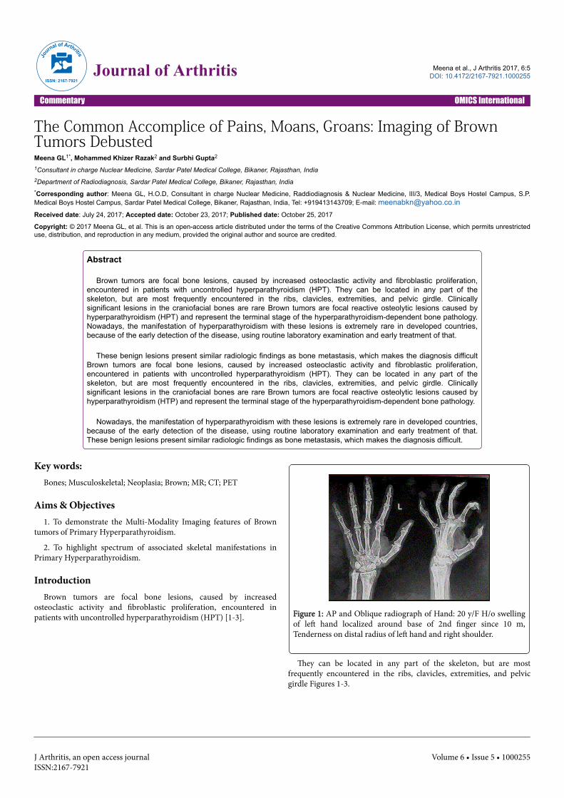
osteoclastic activity and fibroblastic proliferation, encountered inpatients with uncontrolled hyperparathyroidism (HPT) [1-3]. Figure 1: AP and Oblique radiograph of Hand: 20 y/F H/o swelling
of left hand localized around base of 2nd finger since 10 m,Tenderness on distal radius of left hand and right shoulder.
They can be located in any part of the skeleton, but are mostfrequently encountered in the ribs, clavicles, extremities, and pelvicgirdle Figures 1-3.
Journal of Arthritis
ISSN: 2167-7921Journal of Arthritis Meena et al., J Arthritis 2017, 6:5
DOI: 10.4172/2167-7921.1000255
Commentary OMICS International
J Arthritis, an open access journalISSN:2167-7921
Volume 6 • Issue 5 • 1000255

Figure 2: Brown tumor of Primary HPT (Hyperparathyroidism):Well-defined, purely lytic lesion in II nd metacarpal with noreactive bone. The cortex is thinned and expanded with no extraosseous extension. Ill defined lytic lesion in distal radius with nomatrix calcification or ossification. Serum PTH-- >2000 Serum Ca2++--- raised.
Figure 3: CT Coronal reformat showing iso dense mass lesion insuperior parathyroid glandular mass with intense up take on Tc 99scan Tc 99 scan-uptake in LT parathyroid gland adenoma.
Clinically significant lesions in the craniofacial bones are rareBrown tumors are focal reactive osteolytic lesions caused byhyperparathyroidism (HPT) and represent the terminal stage of thehyperparathyroidism-dependent bone pathology. Nowadays, themanifestation of hyperparathyroidism [4,5] with these lesions isextremely rare in developed countries, because of the early detection ofthe disease, using routine laboratory examination and early treatmentof that.
These benign lesions present similar radiologic findings as bonemetastasis, which makes the diagnosis difficult Brown tumors are focalbone lesions in Figure 4, caused by increased osteoclastic activity and
fibroblastic proliferation [6], encountered in patients withuncontrolled hyperparathyroidism (HPT).
Figure 4: Brown tumours of Primary Hyperparathyroidism: Lyticexpansile intramedullary lesions with peripheral rim involvingproximal Tibia and Talar dome.
They can be located in any part of the skeleton, but are mostfrequently encountered in the ribs, clavicles, extremities, and pelvicgirdle. Clinically significant lesions in the craniofacial bones [7] arerare Brown tumors are focal reactive osteolytic lesions caused byhyperparathyroidism (HTP) and represent the terminal stage of thehyperparathyroidism-dependent bone pathology. Nowadays, themanifestation of hyperparathyroidism with these lesions is extremelyrare in developed countries, because of the early detection Figure 5 ofthe disease, using routine laboratory examination and early treatmentof that. These benign lesions present similar radiologic findings asbone metastasis, which makes the diagnosis difficult.
Figure 5: T1 sag, T2 sag and T2 FS Coronals of ankle and foot:Brown tumors in Talar dome.
A solitary brown tumor might be confused with solitary bone cyst,aneurismal bone cyst, and giant cell tumor or giant cell [8] reparativegranuloma. With multiple brown tumors, differential diagnosisincludes: osteolytic metastasis, multiple myeloma, multiple bone cysts,etc. It is the presence of sclerotic margin that excludes metastasis.
Citation: Meena GL, Razak MK, Gupta S (2017) The Common Accomplice of Pains, Moans, Groans: Imaging of Brown Tumors Debusted. JArthritis 6: 255. doi:10.4172/2167-7921.1000255
Page 2 of 4
J Arthritis, an open access journalISSN:2167-7921
Volume 6 • Issue 5 • 1000255

Findings & Procedure DetailsThe Radio-Pathology correlates of Brown Tumors are described
with differential diagnosis. Plain Radiography, CT, MRI features ofBrown tumors and associated findings in Primary HPT are elucidated.Brown tumor [9] (also known as osteitis fibrosa cystica or rarelyosteoclastoma) is one of the manifestations of hyperparathyroidism inFigure 6. It represents a reparative cellular process, rather than aneoplastic process. Brown tumor (also known as osteitis fibrosa cysticaor rarely osteoclastoma) is one of the manifestations ofhyperparathyroidism [10]. It represents a reparative cellular process,rather than a neoplastic process. Brown tumors have a slightly greaterfrequency in primary than in secondary hyperparathyroidism (3%versus 2%). However, secondary hyperparathyroidism is much morecommon than primary hyperparathyroidism; therefore most of browntumors that are seen are associated with secondaryhyperparathyroidism.
Figure 6: Bone scan features are suggestive of metabolic bonedisease Super scan- note absent kidney visualization. Uptake isevident in tibia.
PathologyIn chronic renal disease, continual and excessive urinary calcium
excretion can lower serum calcium level and lead to a rise inparathyroid hormone secretion. This results in mobilization of skeletalcalcium through rapid osteoclastic turnover of bone to maintainnormal serum calcium levels [11]. In localized regions where bone lossis particularly rapid, hemorrhage, and reparative granulation tissue,with active, vascular, proliferating fibrous tissue may replace thenormal marrow contents, resulting in a brown tumor. Hemosiderinimparts the brown color (hence the name of the lesion).
Plain radiographWell-defined, purely lytic lesions that provoke little reactive bone.
The cortex may be thinned and expanded, but will not be penetrated.
CTAttenuation values on CT will be in the range of blood and fibrous
tissue.
MRIThe MRI appearance depends on the relative proportion of its
components. The lesions therefore may be solid, cystic, or mixed. Solidcomponents are intermediate to low intensity on T1- and T2-weightedimages, while the cystic components are hyper intense on T2-weightedimages and may have fluid-fluid levels [12,13]. T1 C+ (Gd): there canbe enhancement of the solid component and septa.
AngiographyLesions are usually hyper vascular.
Nuclear medicineBone scan often shows intense uptake Figures 7 and 8.
Figure 7: Pepperpot skull of Hyperparathyroidism.
Figure 8: High Resolution USG of neck for Parathyroid adenoma:HRUS of neck show a relatively large hypoechoic lesion in posterioraspect of right lobe of thyroid.
Citation: Meena GL, Razak MK, Gupta S (2017) The Common Accomplice of Pains, Moans, Groans: Imaging of Brown Tumors Debusted. JArthritis 6: 255. doi:10.4172/2167-7921.1000255
Page 3 of 4
J Arthritis, an open access journalISSN:2167-7921
Volume 6 • Issue 5 • 1000255

ConclusionThis exhibit serves as a resource for Imaging of Brown tumors what
a resident must know.
References1. Van Herden JA, Beahrs OH, Woolner LB (1977) The pathology and
surgical management of primary Hyperparathyroidism. Surg Clin NorthAm 57: 557-563.
2. Hsu CH, Liew PL, Wang W, Leung TK, Yang KM (2008) Enhanced FDGuptake in brown tumors mimics multiple skeletal metastases in a patientwith primary hyperparathyroidism. Acta Radiol 49: 949-950.
3. Jouan A, Zabraniecki L, Vincent V, Poix E, Fournié B (2008) An unusualpresentation of primary Hyperparathyroidism: Severe hypercalcemia andmultiple brown tumors. Joint Bone Spine 75: 209-211.
4. Joyce JM, Idea RJ, Grossman SJ, Liss RG, Lyons JB (1994) Multiple browntumors in hyperparathyroidism mimicking metastatic disease onradiograph and bone scan. Clin Nucl Med 19:630-635.
5. Kalambokis G, Economou G, Kamina S, Papachristou DJ, Bai M, et al.(2005) Multiple brown tumors of the ribs simulating malingnancy. JEndocrinol Invest 28: 738-740.
6. Tumeh SS, Kaplan WD (1985) Clinical significance of solitary rib lesionsin patients with extra skeletal malignancy. J NucI Med 26: 1140-1143.
7. Reséndiz-Colosia JA, Alvarado-Cabrero I, Flores-Díaz R, Juan MH,BarrosoBravo S, et al. (2008) Multiple maxillofacial brown tumorsasprimary hyperparathyroidism manifestation. Gac Med Mex 144: 155-160.
8. Su AW, Chen CF, Huang CK, Chen PC, Chen WM, et al. (2010) Primaryhyperparathyroidism with brown tumor mimicking metastatic bonemalignancy J Chin Med Assoc 73: 177-180.
9. Sager S, Aliyev A, Halac M, Oztürk T (2012) Positron emissiontomography/computed tomography imaging of brown tumors mimickingmultiple skeletal metastases in patient with primary hyperparathyroidismIndian J Endocrinol Metabol 16: 850-852.
10. Hong WS, Sung MS, Chun KA, Kim JY, Park SW, et al. (1978) Emphasison the MR imaging findings of brown tumors: a report of five casesSkeletal Radiol, R.I. Rynes, Merzig EG () Calcium pyrophosphate crystaldeposition disease and hyperparathyroidism: a controlled, prospectivestudy. J Rheumatol 5: 460-468.
11. Yashiro T, Kamoto T, Anaka R, Ito K, Hara H, et al. (1991) Prevalence ofchondrocalcinosis in patients with primary hyperparathyroidism inJapan. Endocrinol Jpn 38: 457-464.
12. WJ Dodds, Steinbach HL (1968) Primary hyperparathyroidism andarticular cartilage calcification. Am J Roentgenol 104: 884-892.
13. Jouan A, Zabraniecki L, Vincent V, Poix E, Fournie B (2008) An unusualpresentation of primary hyperparathyroidism: severe hypercalcemia andmultiple brown tumors Join Bone Spine 75: 209-211.
Citation: Meena GL, Razak MK, Gupta S (2017) The Common Accomplice of Pains, Moans, Groans: Imaging of Brown Tumors Debusted. JArthritis 6: 255. doi:10.4172/2167-7921.1000255
Page 4 of 4
J Arthritis, an open access journalISSN:2167-7921
Volume 6 • Issue 5 • 1000255