journal of american science 2017;13(1) ... facial resurfacing using locoregional flaps ahmed gamil...
TRANSCRIPT

Journal of American Science 2017;13(1) http://www.jofamericanscience.org
87
Postburn facial resurfacing using locoregional flaps
Ahmed Gamil Elsharkawy, Tarek Ashour and Ahmed Rezk El Naggar
Department of Plastic and Reconstructive Surgery, Faculty of Medicine; Cairo University, Cairo, Egypt [email protected]
Abstract: Postburn hypertrophic scar in the face is extremely challenging to the reconstructive surgeons. Numerous methods have been postulated for the management of the hypertrophic scars, but till now, the single best choice has not been established. Among these treatments are surgical excision with grafting however cosmetic results were suboptimal, others include pressure therapy, topical and intralesional corticosteroids, laser therapy, silicone gel sheeting and others aiming at decreasing collagen synthesis. In this study scar excision and resurfacing with locoregional flap was tried to provide pliable tissues with best colour/texture match. Eight patients with various degrees of hypertrophic scar in the face were reconstructed with fasciocutaneous supraclavicular artery flap in this study and their outcome was evaluated. [Ahmed Gamil Elsharkawy, Tarek Ashour and Ahmed Rezk El Naggar. Postburn facial resurfacing using locoregional flaps. J Am Sci 2017;13(1):87-92]. ISSN 1545-1003 (print); ISSN 2375-7264 (online). http://www.jofamericanscience.org. 12. doi:10.7537/marsjas130117.12. Key words: hypertrophic scar, supraclavicular flap, postburn scar management. 1. Introduction
Facial burn is one of the most destructive events that has a massive physical and psychological impact on burn trauma victim.1
Hypertrophic scars are defined as an elevated scar that do not extend beyond the wound boundaries.2 These scars are characterized by excessive proliferation of the dermal tissue, with excessive collagen deposition.3 Hypertrophic scars are troublesome to the patient and pose a challenge for plastic surgeons. The main goals of reconstructive surgery for these patients in general are first to restore the function, then to regain the aesthetic appearances.4
Multiple combined methods have been described for the management of the hypertrophic scars, but till now, the optimal choice has not been established. Among these treatments are surgical excision with grafting however cosmetic results were suboptimal, others include pressure therapy, topical and intralesional corticosteroids, laser therapy, intralesional interferon, silicone gel sheeting, onion extract gel and others aiming at decreasing collagen synthesis.5
In this study scar excision and resurfacing with locoregional flap was tried so as to provide pliable tissues with best colour match and avoid the need for lengthy microvascular surgeries. Many flaps were proposed including supraclavicular flap, submental flap and platysmal myocutaneous flap. In this study the supraclavicular region was chosen to get the best colour / texture match.
Supraclavicular flap is a fasciocutaneous flap based on supraclavicular artery, a branch derived from transverse cervical artery. Venous drainage is through the transverse cervical vein.6
The basic concepts for reconstruction of facial burn are:
Replace with a like tissue regarding colour and texture.
Multidisciplinary, realistic, well timed reconstructive plan discussed with the patient.
Function over cosmoses, but combine whenever possible.
Reconstruct areas of significant functional importance first, such as the mouth and lips to correct microstomia and the eyelids to avoid lower eyelid ectropion.7 2. Methods
The current study included eight patients (where 9 flaps were done) with various degrees of hypertrophic scar and keloid in the face resulting from flame burn.
They were four males and four female patients of various age groups from 22 to 60 with a mean age of 39 years.
All scars in this study were mature, and the time elapsed between the burn insult and the procedure done ranged from 6 to 12 months. All scars were subjected to the Vancouver burn scar assessment scale (VSS). 8 (Table 1).
The patients were assessed for the functional disability including microstomia and lower eyelid ectropion.
Multidisciplinary team was involved in full assessment of the patients, this team included in addition to plastic surgeon, physiotherapist, psychiatrist, skilful nursing staff and social workers.
All patients were counselled preoperatively about the anticipated results to avoid over expectations,
Journal of American Science 2017;13(1) http://www.jofamericanscience.org
88
visible scar over the donor site, the possibility of scar widening postoperatively and the possibility of using split thickness graft to close the donor site was explained.
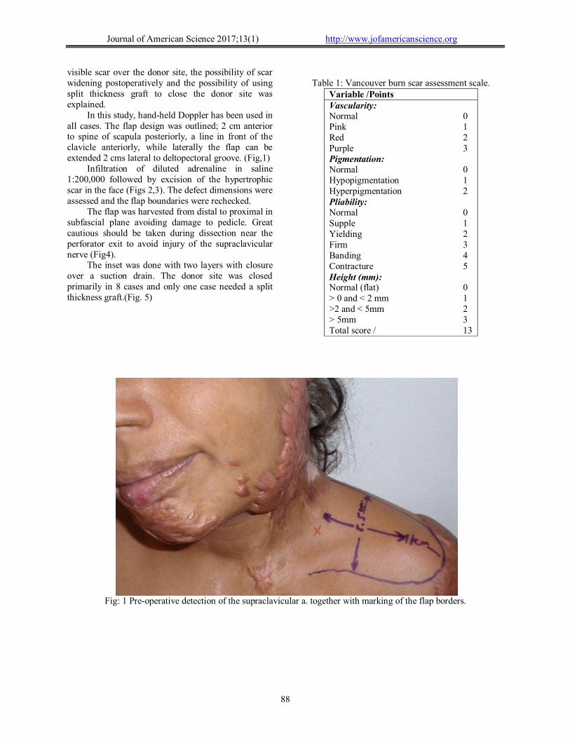
In this study, hand-held Doppler has been used in all cases. The flap design was outlined; 2 cm anterior to spine of scapula posteriorly, a line in front of the clavicle anteriorly, while laterally the flap can be extended 2 cms lateral to deltopectoral groove. (Fig,1)
Infiltration of diluted adrenaline in saline 1:200,000 followed by excision of the hypertrophic scar in the face (Figs 2,3). The defect dimensions were assessed and the flap boundaries were rechecked.
The flap was harvested from distal to proximal in subfascial plane avoiding damage to pedicle. Great cautious should be taken during dissection near the perforator exit to avoid injury of the supraclavicular nerve (Fig4).
The inset was done with two layers with closure over a suction drain. The donor site was closed primarily in 8 cases and only one case needed a split thickness graft.(Fig. 5)
Table 1: Vancouver burn scar assessment scale.
Variable /Points Vascularity: Normal 0 Pink 1 Red 2 Purple 3 Pigmentation: Normal 0 Hypopigmentation 1 Hyperpigmentation 2 Pliability: Normal 0 Supple 1 Yielding 2 Firm 3 Banding 4 Contracture 5 Height (mm): Normal (flat) 0 > 0 and < 2 mm 1 >2 and < 5mm 2 > 5mm 3 Total score / 13
Fig: 1 Pre-operative detection of the supraclavicular a. together with marking of the flap borders.

Journal of American Science 2017;13(1) http://www.jofamericanscience.org
89
Fig: 2, 3 Intra-operative view showing the defect created after excision of the hypertrophic scar in two cases
Fig: 4 Intra-operative view showing the perforator emerging from supraclacivicular artery together with supraclavicular nerve.
Fig: 5 Six months Postoperative photo showing well healed recipient and donor sites

Journal of American Science 2017;13(1) http://www.jofamericanscience.org
90
Postoperatively the flap was monitored for complications such as flap viability, wound dehiscence, infection and hematoma.
The scar developed at the boundaries of the flap was managed by application of silicon sheets with accepted results.
Photos were taken preoperatively, one month and then six month postoperatively.
A summary of clinical cases is shown in table (2).
Table: 2 clinical characteristics of the patient. VSS = Vancouver scar assessment scale. Case no.
Age/sex VSS Defect size/cm
Flap type Donor site closure
complications
1 25/F 11/13 10x15 supraclavicular Primary Nil 2 25/F 12/13 12x14 supraclavicular Primary Nil 3 34/M 9/13 8x9 supraclavicular Primary Wound dehiscence (recipient) 4 45/F 10/13 11x12 supraclavicular STSG Partial graft loss 5 21/M 11/13 9x11 supraclavicular Primary Widened donor scar 6 60/F 11/13 10x14 supraclavicular Primary Nil 7 52/M 10/13 10x13 supraclavicular Primary Nil 8 43/F 12/13 12x14 supraclavicular Primary Mild infection 9 29/M 11/13 7x9 supraclavicular Primary Widened donor scar Report of the cases
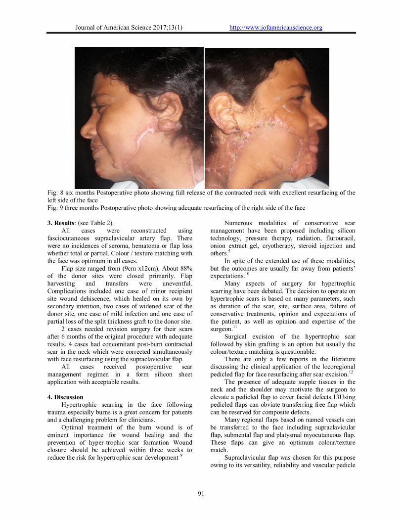
Case (1,2): 25 years old female patient who sustained a flame burn to faced and neck 20 year ago, her burns were allowed to heal spontaneously with resulting bilateral hypertrophic scarring of the face and multiple disabling contracture band of the neck (Fig s 6,7 ).
Supraclavicular artery flap utilized for release of the contracted neck together with bilateral resurfacing of the face (Figs.8,9). The patient had smooth postoperative recovery with no recorded complications in both donor and recipient sites.
Fig: 6,7Preoperative photos showing sever bilateral hypertrophic scars in the same patient
Journal of American Science 2017;13(1) http://www.jofamericanscience.org
91
Fig: 8 six months Postoperative photo showing full release of the contracted neck with excellent resurfacing of the left side of the face Fig: 9 three months Postoperative photo showing adequate resurfacing of the right side of the face 3. Results: (see Table 2).
All cases were reconstructed using fasciocutaneous supraclavicular artery flap. There were no incidences of seroma, hematoma or flap loss whether total or partial. Colour / texture matching with the face was optimum in all cases.
Flap size ranged from (9cm x12cm). About 88% of the donor sites were closed primarily. Flap harvesting and transfers were uneventful. Complications included one case of minor recipient site wound dehiscence, which healed on its own by secondary intention, two cases of widened scar of the donor site, one case of mild infection and one case of partial loss of the split thickness graft to the donor site.
2 cases needed revision surgery for their scars after 6 months of the original procedure with adequate results. 4 cases had concomitant post-burn contracted scar in the neck which were corrected simultaneously with face resurfacing using the supraclavicular flap.
All cases received postoperative scar management regimen in a form silicon sheet application with acceptable results. 4. Discussion
Hypertrophic scarring in the face following trauma especially burns is a great concern for patients and a challenging problem for clinicians.
Optimal treatment of the burn wound is of eminent importance for wound healing and the prevention of hyper-trophic scar formation Wound closure should be achieved within three weeks to reduce the risk for hypertrophic scar development 9
Numerous modalities of conservative scar management have been proposed including silicon technology, pressure therapy, radiation, flurouracil, onion extract gel, cryotherapy, steroid injection and others.5
In spite of the extended use of these modalities, but the outcomes are usually far away from patients’ expectations.10
Many aspects of surgery for hypertrophic scarring have been debated. The decision to operate on hypertrophic scars is based on many parameters, such as duration of the scar, site, surface area, failure of conservative treatments, opinion and expectations of the patient, as well as opinion and expertise of the surgeon.11
Surgical excision of the hypertrophic scar followed by skin grafting is an option but usually the colour/texture matching is questionable.
There are only a few reports in the literature discussing the clinical application of the locoregional pedicled flap for face resurfacing after scar excision.12
The presence of adequate supple tissues in the neck and the shoulder may motivate the surgeon to elevate a pedicled flap to cover facial defects.13Using pedicled flaps can obviate transferring free flap which can be reserved for composite defects.
Many regional flaps based on named vessels can be transferred to the face including supraclavicular flap, submental flap and platysmal myocutaneous flap. These flaps can give an optimum colour/texture match.
Supraclavicular flap was chosen for this purpose owing to its versatility, reliability and vascular pedicle

Journal of American Science 2017;13(1) http://www.jofamericanscience.org
92
stability. Elevation of neurocutaneous supraclavicular flap not only replaces the scared tissues but also can restore facial sensation. Initially sensation of the shoulder is maintained until cortical reintegration occurs with adequate patient training. 14
The other advantage of the supraclavicular flap is the hidden donor site which can be closed primarily in most cases.15 Conclusion
When comparing the regional pedicled flaps to the traditional conservative measures for scar management, it had been concluded that these flaps can offer the best chance of facial resurfacing after hypertrophic scar excision.
When compared to other locoregional pedicled flaps, supraclavicular flap showed better colour/texture match and sensation. Combining conservative measures after surgical correction can improve the aesthetic outcomes.
Currently what we are working on is to expand the flap to gain larger surface area for full facial resurfacing and to minimize donor site morbidity. References 1. Klein MB, Moore ML, Costa B, Engrav LH.
Primer on the management of face burns at the University of Washington. J Burn Care Rehabil 2005; 26:2.
2. English RS, Shenefelt PD. Keloids and hypertrophic scars. Dermatol Surg. 1999; 25(8):631-8.
3. Atiyeh BS. Nonsurgical management of hypertrophic scars: evidence-based therapies, standard practices, and emerging methods. Aesthetic Plast Surg. 2007 31(5): 468-92.
4. Shelley OP, Dziewulski P. Late management of burns. Surgery Oxford 2006; 24:15.
5. Mustoe TA, Cooter RD, Gold MH, Hobbs FD, Ramelet AA, Shakespeare PG, Stella M, Téot L, Wood FM, Ziegler UE. International clinical recommendations on scar management. International Advisory Panel on Scar Management. Plast Reconstr Surg. 2002 Aug; 110(2):560-71.
6. Lamberty BG. The supra-clavicular axial patterned flap. Br J Plast Surg. 1979; 32(3): 207-12.
7. Jorge LV, Dziewulski P. Principles of burn reconstruction: Face, scalp, and neck, up to date, Literature review current through: Nov 2016.
8. Nedelec B, Shankowsky HA, Tredget EE. Rating the resolving hypertrophic scar: comparison of the Vancouver Scar Scale and scar volume. J Burn Care Rehabil. 2000 May-Jun; 21(3): 205-12.
9. 9- Rabello FB, Souza CD, Farina JA. Update on hypertrophic scar treatment. Clinics (Sao Paulo). Clinics 2014 Aug; 69(8): 565–573.
10. Manuskiatti W, Richard EF. Treatment Response of Keloidal and Hypertrophic Sternotomy Scars, Comparison Among Intralesional Corticosteroid, 5-Fluorouracil, and 585-nm Flashlamp-Pumped Pulsed-Dye Laser Treatments. Arch Dermatol. 2002 Sep; 138(9): 1149-55.
11. Bloemen C T, Willem MV - Prevention and Curative Management of Hypertrophic Scar Formation. Journal of the International Society for Burn Injuries 35(4):463-75 · November 2008.
12. Brissett AE, Sherris DA. Scar contractures, hypertrophicscars, and keloids. Facial Plast Surg 2001; 17(4): 263–72.
13. Loghmani Shahriar, Mohammad Eidy, Mahdi Mohammadzadeh, Alireza Loghmani, Fahimeh Raigan. The Supraclavicular Flap for Reconstruction of Post-Burn Mentosternal Contractures. IRCMJ 2 April 2013.1600.
14. Granzow JW1, Suliman A, Roostaeian J, Perry A, Boyd JB. The supraclavicular artery island flap (SCAIF) for head and neck reconstruction: surgical technique and refinements. Otolaryngol Head Neck Surg. 2013 Jun;148(6):933-40.
15. Alves HR, Ishida LC, Ishida LH, Besteiro JM, Gemperli R, Faria JC, Ferreira MC. A clinical experience of the supraclavicular flap used to reconstruct head and neck defects in late-stage cancer patients. J Plast Reconstr Aesthet Surg. 2012 Oct; 65(10):1350-6.
1/17/2017