journal club 2007.6.21 endocrine review 2005; 26(5):599-614 autoimmune hypophysitis patrizio...
TRANSCRIPT

Journal Club 2007.6.21
Endocrine Review 2005; 26(5):599-614
Autoimmune Hypophysitis
Patrizio Caturegli, Craig Newschaffer, Alessandro Olivi, Martin G. Pomper, Peter C. Burger and Noel R. Rose
Departments of Pathology (P.C., P.C.B., N.R.R.), Epidemiology (C.N.), Neurosurgery (A.O.), Neuroradiology (M.G.P.), and Molecular Microbiology and Immunology (P.C., N.R.R.), The Johns Hopkins University, Baltimore, Maryland 21205
Diabetes and Endocrine Department,
Kameda medical center
Masuzawa Masahiro
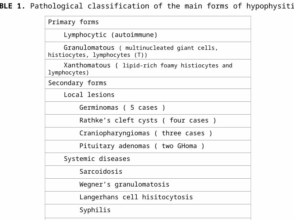
TABLE 1. Pathological classification of the main forms of hypophysitis
Primary forms
Lymphocytic (autoimmune)
Granulomatous ( multinucleated giant cells, histiocytes, lymphocytes (T))
Xanthomatous ( lipid-rich foamy histiocytes and lymphocytes)
Secondary forms
Local lesions
Germinomas ( 5 cases )
Rathke’s cleft cysts ( four cases )
Craniopharyngiomas ( three cases )
Pituitary adenomas ( two GHoma )
Systemic diseases
Sarcoidosis
Wegner’s granulomatosis
Langerhans cell hisitocytosis
Syphilis
Tuberculosis

LAH was first described in 1962 by Goudie and Pinkerton . They reported a 22-yr-old woman who died 14 months after her second delivery, probably because of adrenal insufficiency. Twelve months postpartum she felt increasingly tired and noticed
enlargement of her neck. She was admitted to the hospital for vomiting, diarrhea, and severe lower abdominal pain radiating to the right iliac fossa, and thus was brought to the operating room for suspected appendicitis. Surgery revealed an acutely inflamed, gangrenous appendix that had not ruptured. The appendix was removed, but 8 h later the patient developed circulatory shock and died. The autopsy showed a firm, enlarged thyroid gland infiltrated with lymphocytes, atrophic adrenal glands, and a small pituitary. The adenohypophysis was extensively infiltrated by lymphocytes and few plasma cells, aggregating in some areas to form lymphoid follicles. The neurohypophysis was normal.
Noting the presence of Hashimoto’s thyroiditis, a well-characterized autoimmune disease, the authors concluded that the coexistence of lymphocytic thyroiditis and mononuclear cells infiltration of the anterior pituitary was not fortuitous.
Historical Note
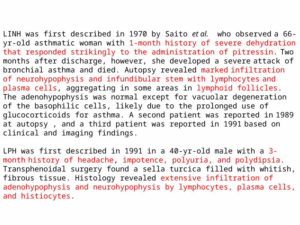
LINH was first described in 1970 by Saito et al. who observed a 66-yr-old asthmatic woman with 1-month history of severe dehydration that responded strikingly to the administration of pitressin. Two months after discharge, however, she developed a severe attack of bronchial asthma and died. Autopsy revealed marked infiltration of neurohypophysis and infundibular stem with lymphocytes and plasma cells, aggregating in some areas in lymphoid follicles. The adenohypophysis was normal except for vacuolar degeneration of the basophilic cells, likely due to the prolonged use of
glucocorticoids for asthma. A second patient was reported in 1989 at autopsy , and a third patient was reported in 1991 based on clinical and imaging findings.
LPH was first described in 1991 in a 40-yr-old male with a 3-month history of headache, impotence, polyuria, and polydipsia. Transphenoidal surgery found a sella turcica filled with whitish, fibrous tissue. Histology revealed extensive infiltration of adenohypophysis and neurohypophysis by lymphocytes, plasma cells, and histiocytes.

Classification of the autoimmune hypophysitis
• Lymphocytic adenohypophysitides (LAH)
inflammation limited to the anterior hypophysis
• Lymphocytic infundibuloneurohypophysitis (LINH)
inflammation limited to infundibular stem and posterior lobe
• Lymphocytic panhypophysitis (LPH)
inflammation occur both the adenohypophysis and the infundibuloneurohypophysis

Autopsy finding of the lymphocyte infiltration in the pituitary
Simmonds and Brandes200 pituitaries (sudden death): 21 cases(10%) lymphocytic infiltration.pars intermedia( 17 cases), anterior lobe( 2 cases), posterior lobe( 2 cases)
Zonchi and Dova150 pituitaries (usual cases): pars intermedia( 70 cases (47%))
Shanklin100 pituitarieswithin or near the pars intermedia( 43 cases(43%))
Scheithauer69 pituitaries (death during pregnancy, after abortion, or in the postpartum)five cases( exact anatomic location was not mentioned)
Conclusionlymphocytes in or near the pars intermedia; normal findinglymphocytes in the anterior or posterior lobe; pathological finding

TABLE 2. Classification of 379 patients with primary lymphocytic hypophysitis based on the anatomical location
A, Anterior hypophysis; N, infundibuloneurohypophysis; DI, diabetes insipidus.
No. of patients
LAH (n = 245)
Histology shows infiltrated A and normal N. 25
Histology shows infiltrated A. N was not recorded, but no symptoms of DI. 157
Clinic and imaging show A involvement, but no symptoms or radiological signs of DI. 51
Clinic shows A involvement, but not DI. Imaging was normal or not done. 12
LINH (n = 39)
Histology shows infiltrated N and normal A. 5
Histology shows infiltrated N. A was not recorded, but no symptoms of A involvement. 21
Clinics and imaging show N involvement, but no symptoms or signs of A involvement. 13
LPH (n = 95)
Histology shows infiltrated A and infiltrated N. 25
Histology shows infiltrated A. N was not recorded but symptoms or radiological signs of DI. 32
Histology shows infiltrated N. A was not recorded but symptoms of A involvement. 3
Clinics and imaging show A involvement and DI. 35

TABLE 3. Key variables included in the database of patients affected by AH
Sex Anatomic classification
Age Surgical treatment
Association with pregnancy Medical treatment
Association with other autoimmune diseases Other types of treatment
Symptoms at presentation Details on pathological findings
Endocrinological assessment Details on mass-reducing treatment
Imaging studies Details on hormone replacement
Pituitary antibodies Follow-up time
Method of diagnosis Status upon follow-up

Epidemiology
379 cases identified( 1962-2004)Geographic or ethnic variation
Japan: 130 cases( 34%)United State: 82 cases( 22%)United Kingdom: 28 cases( 7%)Canada: 19 cases( 5%)
IncidenceBuxton and Robertson( Nottingham, UK)
619 pituitary surgeries: 5 cases of AH(0.8%)Sautner et al. and Fehn et al.( Hamburg, Germany)
2500 pituitaries surgeries: 6 cases of AH(0.24%)Honneger et al.( Erlagen, Germany)
2362 specimens: 7 cases of AH(0.30%)Leung et al.
2000 patients( TSS): 13 cases of AH(0.65%)Authors
905 specimens( Johns Hopkins Hospital): 8 cases of AH(0.88%)Gender
LAH: 210 women, 35 men; F:M ratio, 6:1LINH: 20 women, 19 menLPH: 62 women, 33 men; F:M ratio, 1.9:1

FIG. 1. Distribution of symptom appearance in relation to delivery (indicated as wk 0) in AH. Note the clustering in late pregnancy and early postpartum. Most patients represented in the last month of pregnancy or in the first 2 months after delivery.
Of the total 210 women with LAH, 120 (57%) presented during pregnancy or postpartum.

(1) Pituitary size increases by about 30%(estrogen-driven hypertrophy and hyperplasia of the lactotrophs)→release of pituitary antigens↑
(2) The massive hyperestrogenemia of pregnancy changes the pattern of pituitary blood flow such that more blood derives from the systemic circulation and less from the hypothalamic-pituitary portal circulation→accessible to the immune system↑
(3) The immune system in the uterine environment changes significantly during pregnancy.
(4) Pregnancy influences the course of autoimmune diseases in different ways.improve; rheumatoid arthritis,Graves’ disease,type 1 diabetes mellitusworse; Wegener’s granulomatosisunpredictable; systemic lupus erythematosus,myasthenia gravisno change; scleroderma, Sjögren’s syndrome, thrombocytopenic purpura
Association between Pregnancy and AH
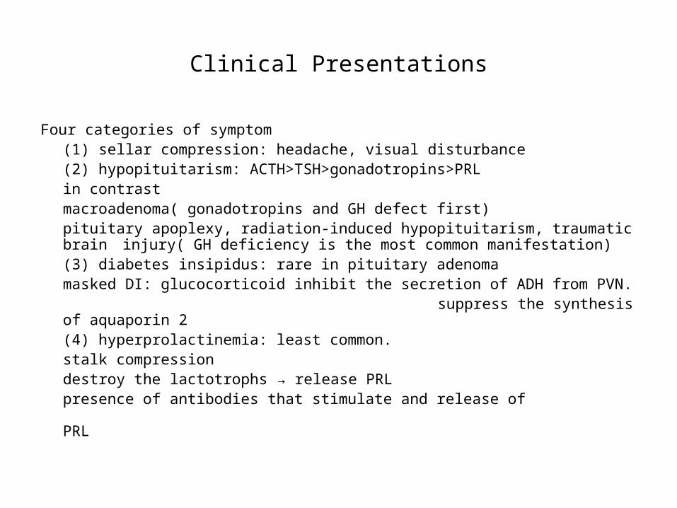
Clinical Presentations
Four categories of symptom(1) sellar compression: headache, visual disturbance(2) hypopituitarism: ACTH>TSH>gonadotropins>PRL
in contrastmacroadenoma( gonadotropins and GH defect first)pituitary apoplexy, radiation-induced hypopituitarism, traumatic
brain injury( GH deficiency is the most common manifestation) (3) diabetes insipidus: rare in pituitary adenoma
masked DI: glucocorticoid inhibit the secretion of ADH from PVN. suppress the synthesis of aquaporin 2
(4) hyperprolactinemia: least common.stalk compressiondestroy the lactotrophs → release PRLpresence of antibodies that stimulate and
release of PRL

TABLE 4. Percentages of patients with LAH, LINH, or LPH presenting with the symptoms indicated on the left
The three columns on the right report P values, which are based on pairwise comparisons using the Wilcoxon rank-sum test.
Symptom LAH (%) LINH (%) LPH (%) LAH vs. LINH LAH vs. LPH LINH vs. LPH
Headache 53 13 41 0.0001 0.045 0.0023
Visual disturbances 43 3 18 0.0001 0.0001 0.070
Hypocortisolism 42 8 19 0.0001 0.001 0.106
Hypothyroidism 18 0 17 0.005 0.871 0.007
Hypogonadism 12 3 14 0.078 0.669 0.057
Inability to lactate 11 0 5 0.028 0.094 0.146
Polydipsia-polyuria 1 98 83 0.0001 0.0001 0.025
Hyperprolactinemia 23 5 17 0.011 0.227 0.073
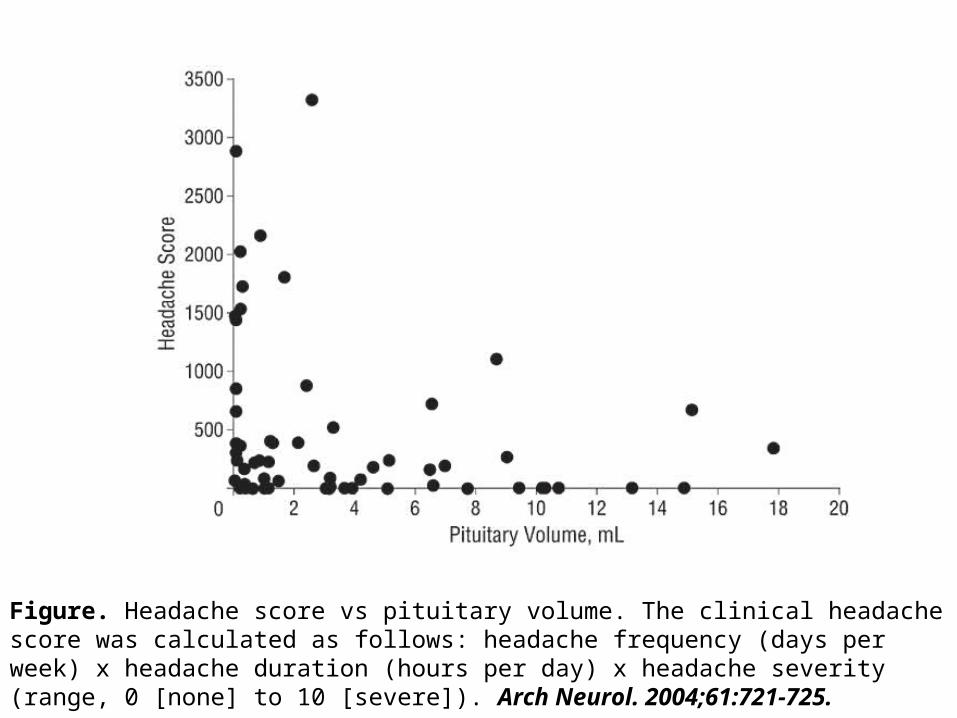
Figure. Headache score vs pituitary volume. The clinical headache score was calculated as follows: headache frequency (days per week) x headache duration (hours per day) x headache severity (range, 0 [none] to 10 [severe]). Arch Neurol. 2004;61:721-725.

FIG. 2. Duration of symptoms (in months) in patients with LAH not associated with pregnancy (LAHno preg), LAH associated with pregnancy (LAHpreg), lymphocytic infundibulo-neurohypophysitis (LINH), or lymphocytic panhypophysitis (LPH). Each box represents the middle 50% of the observations (interquartile range), bordered at the 25th and 75th percentiles, and contains a dotted line to indicate the median (50th percentile). The whisker lines extend from the box to data points that are equal to or less than 1.5 interquartile ranges. Extreme values outside the whisker lines are not shown. P values are based on pairwise comparisons using the Wilcoxon rank-sum test, performed after the Kruskal-Wallis test (P = 0.0203).
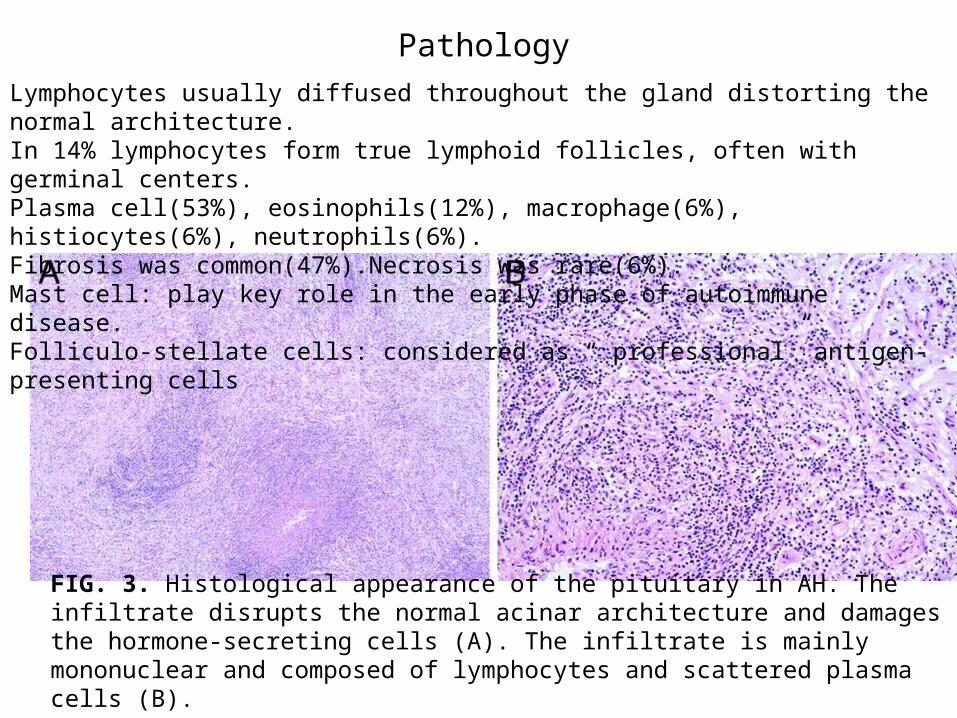
FIG. 3. Histological appearance of the pituitary in AH. The infiltrate disrupts the normal acinar architecture and damages the hormone-secreting cells (A). The infiltrate is mainly mononuclear and composed of lymphocytes and scattered plasma cells (B).
Pathology
Lymphocytes usually diffused throughout the gland distorting the normal architecture.In 14% lymphocytes form true lymphoid follicles, often with germinal centers.Plasma cell(53%), eosinophils(12%), macrophage(6%), histiocytes(6%), neutrophils(6%).Fibrosis was common(47%).Necrosis was rare(6%).Mast cell: play key role in the early phase of autoimmune disease.Folliculo-stellate cells: considered as “ professional” antigen-presenting cells

TABLE 5. Association between AH and other autoimmune diseases
Association was reported in 67 of the total 376 patients (18%). APS, Autoimmune polyglandular syndrome.
Associated condition No. of patients % of total AH patients
Hashimoto’s thyroiditis 28 7.4
APS type 2 7 1.8
Graves’ disease 6 1.6
Systemic lupus erythematosus 5 1.3
Sjögren’s syndrome 3 0.8
Type 1 diabetes 3 0.8
Optic neuritis 3 0.8
Autoimmune gastritis 2 0.5
Addison’s disease 2 0.5
Sarcoidosis 2 0.5
Primary biliary cirrhosis 1 0.3
Myocarditis 1 0.3
Temporal arteritis 1 0.3
Bechet’s disease 1 0.3
Erythema nodosum 1 0.3
Rheumatoid arthritis 1 0.3
Idiopathic thrombocytopenic purpura 1 0.3
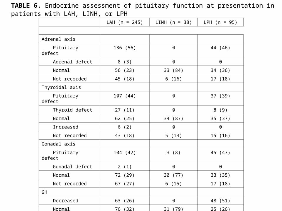
TABLE 6. Endocrine assessment of pituitary function at presentation in patients with LAH, LINH, or LPH
LAH (n = 245) LINH (n = 38) LPH (n = 95)
Adrenal axis
Pituitary defect 136 (56) 0 44 (46)
Adrenal defect 8 (3) 0 0
Normal 56 (23) 33 (84) 34 (36)
Not recorded 45 (18) 6 (16) 17 (18)
Thyroidal axis
Pituitary defect 107 (44) 0 37 (39)
Thyroid defect 27 (11) 0 8 (9)
Normal 62 (25) 34 (87) 35 (37)
Increased 6 (2) 0 0
Not recorded 43 (18) 5 (13) 15 (16)
Gonadal axis
Pituitary defect 104 (42) 3 (8) 45 (47)
Gonadal defect 2 (1) 0 0
Normal 72 (29) 30 (77) 33 (35)
Not recorded 67 (27) 6 (15) 17 (18)
GH
Decreased 63 (26) 0 48 (51)
Normal 76 (32) 31 (79) 25 (26)
Increased 4 (1) 8 (21) 0
Not recorded 100 (41) 0 22 (24)

Data represent number of patients, with percentages in parentheses.
PRL
Decreased 62 (25) 0 15 (16)
Normal 75 (31) 28 (72) 29 (30)
Increased 57 (23) 5 (13) 38 (40)
Not recorded 51 (21) 6 (15) 13 (14)
ADH
Decreased 0 38 (98) 90 (95)
Normal 47 (19.2) 0 5 (5)
Increased 1 (0.4) 0 0
Not recorded 197 (80.4) 1 (2) 0
TABLE 6. Endocrine assessment of pituitary function at presentation in patients with LAH, LINH, or LPH
LAH (n = 245) LINH (n = 38) LPH (n = 95)

TABLE 7. Pituitary antibody results in patients with LAH, LINH, or LPH
LAH (n = 245) LINH (n = 39) LPH (n = 95)
Not measured 190 32 70
Measured by immunofluorescence 39 4 19
Negative 25 5 17
Positive 14 (36%) 0 2 (10%)
Measured by immunoblotting 16 3 5
Negative 5 2 1
Positive 11 (68%) 1 (33%) 4 (80%)
Measured by ELISA 0 0 1
Negative 1
Positive 0
The specificity of pituitary antibodies is poor, as they have been found in various nonautoimmune pituitary diseases such as Cushing’s disease , pituitary adenomas, empty sella syndrome, and Sheehan syndrome , as well as in other autoimmune diseases such as type 1 diabetes, Hashimoto’s thyroiditis, and Graves’ disease.
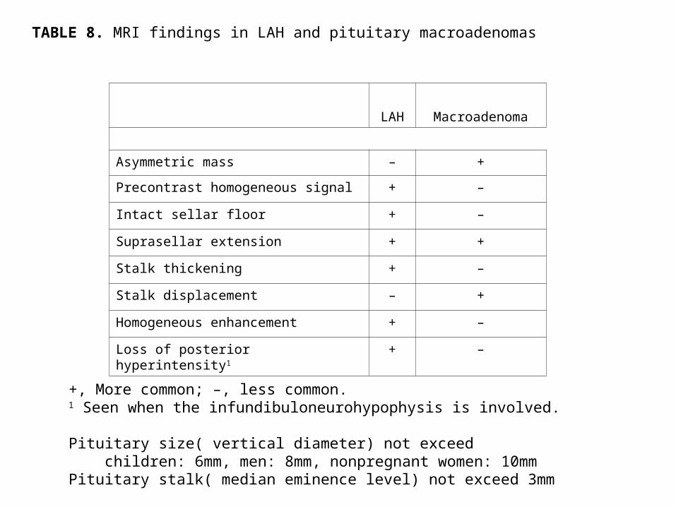
TABLE 8. MRI findings in LAH and pituitary macroadenomas
+, More common; –, less common. 1 Seen when the infundibuloneurohypophysis is involved.
Pituitary size( vertical diameter) not exceed children: 6mm, men: 8mm, nonpregnant women: 10mmPituitary stalk( median eminence level) not exceed 3mm
LAH Macroadenoma
Asymmetric mass – +
Precontrast homogeneous signal + –
Intact sellar floor + –
Suprasellar extension + +
Stalk thickening + –
Stalk displacement – +
Homogeneous enhancement + –
Loss of posterior hyperintensity1 + –
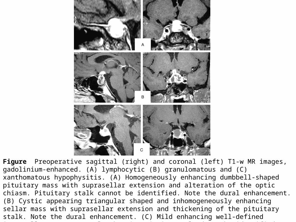
Figure Preoperative sagittal (right) and coronal (left) T1-w MR images, gadolinium-enhanced. (A) lymphocytic (B) granulomatous and (C) xanthomatous hypophysitis. (A) Homogeneously enhancing dumbbell-shaped pituitary mass with suprasellar extension and alteration of the optic chiasm. Pituitary stalk cannot be identified. Note the dural enhancement. (B) Cystic appearing triangular shaped and inhomogeneously enhancing sellar mass with suprasellar extension and thickening of the pituitary stalk. Note the dural enhancement. (C) Mild enhancing well-defined intrasellar tumor. Normal enhancing pituitary gland can be identified. Note the pituitary stalk displacement without stalk thickening. European Journal of Endocrinology, Vol 155, Issue 1, 101-107

Coronal (A, B) and sagittal (C, D) T1weighted MRI before (A, C) and after contrast (B, D). The pituitary is atrophic and the pituitary stalk grossly enlarged. The normal hyperintense signal of the posterior pituitary lobe on native T1weighted images is missing. After contrast the pituitary and the pituitary stalk are enhanced markedly and homogeneously. J Neurol Neurosurg Psychiatry 1998;64:693-694 ( May )

Figure Serial T1-weighted magnetic resonance imaging (MRI) of a patient with central diabetes insipidus (CDI) possibly due to lymphocytic infundibuloneurohypophysitis. T1-weighted MRI in December 2002 (A, D) revealed prominent pituitary-stalk thickening and neurohypophysial enlargement, indicating lymphocytic infundibuloneurohypophysitis, which caused CDI. MRI in March 2003 (B, E) demonstrated improvement in the pituitary-stalk thickening and neurohypophysial enlargement. MRI in June 2004 (C, F) revealed empty sella. A, B, C: coronal view; D, E, F: sagittal view. European Journal of Endocrinology, Vol 153, Issue 6, 989-990

Treatment
(1) Surgery
(2) Medical treatment
glucocorticoid: prednisone 20-60mg/day
methylprednisolone 120mg/day 2weeks
azathioprine
methotrexate
(1) Radiation therapy
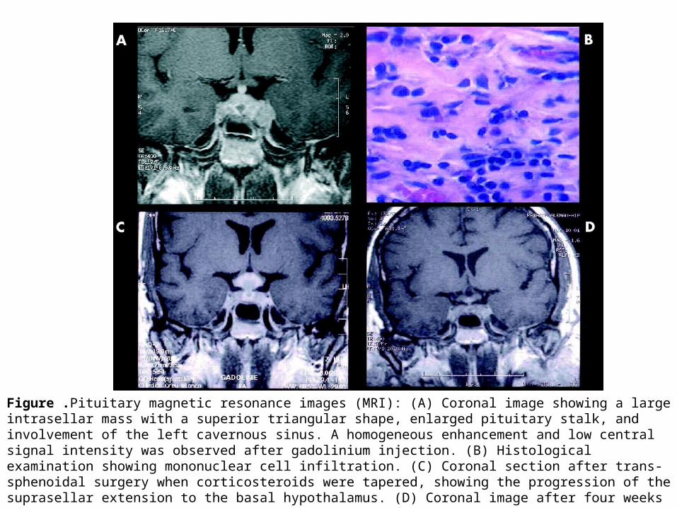
Figure .Pituitary magnetic resonance images (MRI): (A) Coronal image showing a large intrasellar mass with a superior triangular shape, enlarged pituitary stalk, and involvement of the left cavernous sinus. A homogeneous enhancement and low central signal intensity was observed after gadolinium injection. (B) Histological examination showing mononuclear cell infiltration. (C) Coronal section after trans-sphenoidal surgery when corticosteroids were tapered, showing the progression of the suprasellar extension to the basal hypothalamus. (D) Coronal image after four weeks of azathioprine treatment. There is no evidence of residual lesion and no pituitary stalk enlargement. Journal of Neurology Neurosurgery and Psychiatry 2003;74:1581-1583
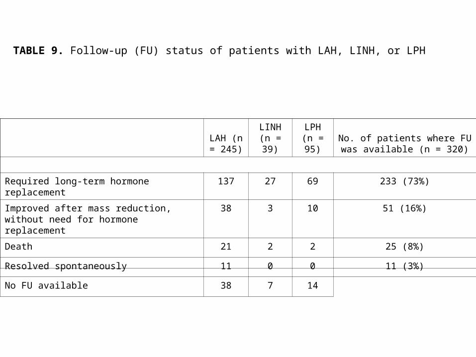
TABLE 9. Follow-up (FU) status of patients with LAH, LINH, or LPH
LAH (n = 245)
LINH (n = 39)
LPH (n = 95)
No. of patients where FU was available (n = 320)
Required long-term hormone replacement 137 27 69 233 (73%)
Improved after mass reduction, without need for hormone replacement
38 3 10 51 (16%)
Death 21 2 2 25 (8%)
Resolved spontaneously 11 0 0 11 (3%)
No FU available 38 7 14