joseph l. napoli and robert v. farese, jr. zhou, cl eric yen, ryan
TRANSCRIPT
Joseph L. Napoli and Robert V. Farese, Jr.Zhou, C. L. Eric Yen, Ryan S. Streeper, Michelle Y. S. Shih, Maureen A. Kane, Ping Murine SkinEssential for Retinoid Homeostasis in Retinol Esterification by DGAT1 IsRegulation, and Signaling:Lipids and Lipoproteins: Metabolism,
doi: 10.1074/jbc.M807503200 originally published online November 20, 20082009, 284:4292-4299.J. Biol. Chem.
10.1074/jbc.M807503200Access the most updated version of this article at doi:
.JBC Affinity SitesFind articles, minireviews, Reflections and Classics on similar topics on the
Alerts:
When a correction for this article is posted•
When this article is cited•
to choose from all of JBC's e-mail alertsClick here
Supplemental material:
http://www.jbc.org/content/suppl/2008/11/20/M807503200.DC1.html
http://www.jbc.org/content/284/7/4292.full.html#ref-list-1
This article cites 49 references, 19 of which can be accessed free at
at HA
RVA
RD U
NIV
ERSITY on O
ctober 28, 2014http://w
ww
.jbc.org/D
ownloaded from
at H
ARV
ARD
UN
IVERSITY
on October 28, 2014
http://ww
w.jbc.org/
Dow
nloaded from

Retinol Esterification by DGAT1 Is Essential for RetinoidHomeostasis in Murine Skin*□S
Received for publication, September 29, 2008, and in revised form, November 20, 2008 Published, JBC Papers in Press, November 20, 2008, DOI 10.1074/jbc.M807503200
Michelle Y. S. Shih‡§, Maureen A. Kane¶, Ping Zhou‡, C. L. Eric Yen‡, Ryan S. Streeper‡, Joseph L. Napoli¶,and Robert V. Farese, Jr.‡§!**1
From the ‡Gladstone Institute of Cardiovascular Disease, San Francisco, California 94158, the !Departments of Medicine and ofBiochemistry and Biophysics, the **Diabetes Center, and the §Biomedical Sciences Graduate Program, University of California,San Francisco, California 94158, and the ¶Department of Nutritional Sciences and Toxicology, University of California,Berkeley, California 94720-3104
Retinoic acid (RA) is a potent signalingmolecule that is essen-tial for many biological processes, and its levels are tightly reg-ulated by mechanisms that are only partially understood. Thesynthesis of RA from its precursor retinol (vitamin A) is animportant regulatory mechanism. Therefore, the esterificationof retinol with fatty acyl moieties to generate retinyl esters, themain storage form of retinol, may also regulate RA levels. Herewe show that the neutral lipid synthesis enzyme acyl-CoA:dia-cylglycerol acyltransferase 1 (DGAT1) functions as the majoracyl-CoA:retinol acyltransferase (ARAT) in murine skin.Whendietary retinol is abundant, DGAT1 deficiency results inelevated levels of RA in skin and cyclical hair loss; both areprevented by dietary retinol deprivation. Further, DGAT1-deficient skin exhibits enhanced sensitivity to topicallyadministered retinol. Deletion of the enzyme specifically inthe epidermis causes alopecia, indicating that the regulationof RA homeostasis by DGAT1 is autonomous in the epider-mis. These findings show that DGAT1 functions as an ARATin the skin, where it acts to maintain retinoid homeostasisand prevent retinoid toxicity. Our findings may have impli-cations for human skin or hair disorders treated with agentsthat modulate RA signaling.
Regulation of cellular proliferation and differentiation of epi-thelial tissues is crucial in embryonic development and in adulthomeostasis. Retinoic acid (RA)2 is a major regulator of theseprocesses (1) through its ability to serve as a ligand for RAnuclear receptors (RARs) (2). Since RA is such a potent signal-ing molecule, its levels must be tightly controlled. Indeed,
excess RA is highly teratogenic during embryonic developmentand may be toxic to adult tissues (3). Further, RA is used ther-apeutically for skin disorders, such as acne and psoriasis, andcertain cancers (4), but its uses are often limited by local andsystemic toxicity. Thus, understanding how RA levels are reg-ulated has important biological and clinical relevance.
The synthesis of RA from its precursor retinol, or vitamin A,is a major node in the regulation of RA levels (5). To generateRA, retinol is oxidized in two sequential reactions, catalyzed byretinol and retinal dehydrogenases (5), whose activities regulateRA homeostasis. We hypothesized that the availability of reti-nol for these reactions may also be regulated by the balancebetween retinol and retinyl esters. Indeed, the majority of reti-nol in the body is stored as retinyl esters, which are concen-trated in cytosolic lipid droplets of cells and serve as a localsource of retinol. Retinyl esters are also stored in major organs,such as liver and white adipose tissue (WAT), from which ret-inol can be mobilized to supply other tissues during increaseddemand. Thus, retinol esterificationmay participate in regulat-ing the retinol pool available for RA synthesis.
Retinol esterification is carried out by two distinct enzymaticactivities. One is mediated by lecithin:retinol acyltransferase(LRAT), which catalyzes the covalent joining of a fatty acylmoi-ety from lecithin (phosphatidylcholine) to retinol that is boundto cellular retinol-binding protein (CRBP) (6, 7). LRAT activityis crucial for maintaining tissue retinol stores. LRAT-null(Lrat!/!) mice have severe reductions in hepatic and lung reti-nyl ester levels (8–10), which are accompanied by testicularhypoplasia/atrophy (9) andblindness (8). Retinyl ester levels arenormal inWATand several other tissues, indicating alternativemechanisms for retinol esterification (9, 10). This esterificationis probablymediated in part by acyl CoA:retinol acyltransferase(ARAT) enzymes, which use fatty acyl-CoA and unbound reti-nol as substrates (11). Although many tissues exhibit ARATactivity (12), attempts to purify and clone an ARAT gene wereunsuccessful, and thus molecular tools to study ARAT activityhave been lacking. However, the enzyme encoded by Dgat1, anacyl CoA:diacylglycerol acyltransferase (DGAT), was recentlyreported to catalyze the ARAT reaction in vitro (13, 14). More-over, several tissues of Dgat1!/! mice had reduced ARATactivity, and retinol esterification was reduced in culturedmurine embryonic fibroblasts lacking DGAT1 (14). Mostrecently, a study of Dgat1!/! mice demonstrated a role for theenzyme in retinol absorption in the small intestine (15). Thus,
* This work was supported, in whole or in part, by National Institutes of HealthGrants DK-056084 (to R. F.) and DK36870 (to J. L. N.). This work was alsosupported by National Center for Research Resources extramural researchfacilities Grant C06 RR018928 and by the J. David Gladstone Institutes. Thecosts of publication of this article were defrayed in part by the payment ofpage charges. This article must therefore be hereby marked “advertise-ment” in accordance with 18 U.S.C. Section 1734 solely to indicate this fact.
□S The on-line version of this article (available at http://www.jbc.org) containssupplemental Table 1 and Figs. 1–3.
1 To whom correspondence should be addressed: The J. David GladstoneInstitutes, 1650 Owens St., San Francisco, CA 94158. Tel.: 415-734-2000;Fax: 415-355-0960; E-mail: [email protected].
2 The abbreviations used are: RA, retinoic acid; RAR, RA receptor; WAT, whiteadipose tissue; LRAT, lecithin:retinol acyltransferase; CRBP, cellular retinol-binding protein; ARAT, acyl-CoA:retinol acyltransferase; DGAT, acyl-CoA:diacylglycerol acyltransferase; Pn, postnatal day n.
THE JOURNAL OF BIOLOGICAL CHEMISTRY VOL. 284, NO. 7, pp. 4292–4299, February 13, 2009© 2009 by The American Society for Biochemistry and Molecular Biology, Inc. Printed in the U.S.A.
4292 JOURNAL OF BIOLOGICAL CHEMISTRY VOLUME 284 • NUMBER 7 • FEBRUARY 13, 2009
at HA
RVA
RD U
NIV
ERSITY on O
ctober 28, 2014http://w
ww
.jbc.org/D
ownloaded from
accumulating evidence indicates that the retinol esterificationactivity of DGAT1 is of biological, and possibly clinical,importance.
In the current study, we investigated whether retinol esteri-fication byDGAT1 is important inmurine skin.Dgat1!/!miceexhibit a pleiotropic phenotype, which includes resistance todiet-induced obesity and altered energy metabolism but alsoincludes prominent phenotypic findings in the skin (16–19).Retinoids play key roles in skin and hair biology (20), and excessretinoids induce epidermal hyperplasia, inhibit sebocyte prolif-eration and differentiation, and alter hair growth (21). Notably,the skin manifestations of Dgat1!/! mice, which include alo-pecia and sebaceous gland atrophy (18), resemble those of ret-inoid toxicity (22, 23). Thus, we hypothesized that DGAT1functions as an ARAT in murine skin and that the absence ofDGAT1 alters retinoid homeostasis. In this study,we tested thishypothesis by examining retinoid metabolism in the skin ofDGAT1-deficient mice.
EXPERIMENTAL PROCEDURES
MiceandDiets—MaleDgat1!/! andwild-typemice (C57BL/6Jgenetic background) were genotyped as described (16). Micewere housed in a pathogen-free barrier facility (12-h light/12-hdark cycle) and fed a retinoid-abundant chow diet (5053 Pico-LabDiet; Purina, St. Louis,MO), unless otherwise specified. Forstudies in which dietary retinol content was controlled,Dgat1"/! damswere fed a retinoid-deficient diet containing 10kcal% fat and #0.04 IU retinol/g (D03102201; Research Diets,New Brunswick, NJ) throughout gestation and suckling. Thefirst litters of Dgat1!/! and wild-type offspring were weanedand maintained on the retinoid-deficient diet until 3 weeksbefore sacrifice, when they were switched to a retinoid-suffi-cient diet containing 10 kcal% fat and 4 IU retinyl palmitate/g(D12450B; Research Diets). To generate retinoid-deficientmice, the second litters of Dgat1"/! dams fed a retinoid-defi-cient diet were weaned and maintained on the retinoid-defi-cient diet until sacrifice.
Dgat1flox/flox mice (mixed C57BL/6J and 129/SvJae), generatedas described in the supplemental material, were crossed with ker-atin 14-Cre mice (K14-Cre; mixed Swiss Webster; C57BL/6J;CBA/J; Jackson Laboratory, Bar Harbor, ME) (24). K14-Cre"DGAT1flox/" mice were crossed with Dgat1flox/flox mice togenerate male K14-Cre"DGAT1flox/", K14-Cre"DGAT1flox/flox,and Dgat1flox/flox littermates (mixed Swiss Webster; 129/SvJae;C57BL/6J; CBA/J) for studies. All experiments were approved bythe Committee on Animal Research of the University of Califor-nia, San Francisco.Genotyping of Cre and Floxed Dgat1 Alleles—Genomic DNA
was extracted from tail epidermis (mechanically separatedfrom the dermis after incubation of skin at 37 °C for 45min) anddermis, WAT, liver, and testes. Cre was detected with senseprimer 5$-GCGGTCTGGCAGTAAAAACTATC-3$ andantisense primer 5$-GTGAAACAGCATTGCTGTCACTT-3$ (100-bp product); interleukin-2 was detected by senseprimer 5$-CTAGGCCACAGAATTGAAAGATCT-3$ andantisense primer 5$-GTAGGTGGAAATTCTAGCATCA-TCC-3$ (324-bp product) and served as an internal PCR control.To identify various alleles ofDgat1, genomic PCR was performed
with forward primer 5$-CAGACATGGCAGCAGCAAATG-3$(in exon 15) and reverse primer 5$-TGCAAGTTGCTGCTGCC-ACCTG-3$ (in the 3$-untranscribed region). Thewild-typeDgat1allele yields an 895 bp band. The floxedDgat1 allele yields a 1002bpbandwhose absence indicatesCre-mediated recombination ofthe floxed Dgat1 allele and deletion of exons 14–17.Retinol Esterification Assays—Mouse skin was homogenized
with a Tissue Tearor (model 398, probe 9853G-04; BiospecProducts, Bartlesville, OK) in Buffer A (50 mM Tris-HCl, pH7.4, and 250 mM sucrose) containing proteinase inhibitors(Roche Applied Science). ARAT assays were performed withtotal protein homogenates (100 !g) in an assay mix containingBuffer A, 5 mM MgCl2, 1.25 mg/ml bovine serum albumin, 200!M all-trans-retinol (Sigma) in acetone, and 25 !M [14C]oleoyl-CoA (55.0 mCi/mmol). After 10 min at 37 °C, lipids wereextracted with chloroform/methanol (2:1, v/v) and separatedby silica gel G-60 TLC plates with hexane/ethyl ether/aceticacid (80:20:1). Retinyl ester, triacylglycerol, and cholesterolester bands were scraped, and radioactivity was measured byscintillation counting.Retinoid Analyses—Retinol, retinyl ester, and all-trans-RA
were quantified as described (25) with modifications. Mousetissue samples were harvested under yellow light and immedi-ately frozen in liquidN2. Tissues were homogenized on ice withice-cold 0.9% saline tomake a%25% homogenate. Tissues werehomogenized in ground glass vessels (Kontes; size 21) eithermanually or with a Heidolph motorized homogenizer (at 280rpm). For skin samples (200–400 mg), a portion (50–100 mg)would not homogenize and was subtracted to obtain tissuemass. Serumwas obtained by centrifuging blood at 7000& g for%7 min at 4 °C. Tissue homogenate or serum was added to adisposable glass culture tube (16 & 150 mm), and an internalstandard (50–100 nM 4,4-dimethyl-RA in 10ml of acetonitrile)was added, followed by the addition of 25 mM KOH in ethanol(1 ml for serum, 3 ml for tissue homogenates) and extractionwith 10 ml of hexane. The organic phase containing nonpolarretinoids (retinol and retinyl ester) was removed. HCl (4 M,60–180 ml) was added to the aqueous phase, and polar sub-stances (RA) were removed by extraction with 10ml of hexane.Organic phases were dried under nitrogen with gentle heatingat %25–30 °C in a water bath (N-EVAP 112; OrganomationAssociates, Berlin, MA). Extracts were resuspended in acetoni-trile according to analyte (RA in 60 ml, retinol/retinyl esterfrom liver in 1000ml; retinol/retinyl ester from all other tissuesin 150 ml). Only glass containers, pipettes, and calibratedsyringes were used to handle retinoid samples.Real Time PCR—Whole skin was homogenized, and total
RNAwas extractedwith theRNeasy fibrous tissueminikit (Qia-gen, Valencia, CA). RNA (5 !g) was reverse-transcribed withthe Superscript III First-Strand Synthesis Supermix kit(Invitrogen). Real time PCR was performed and analyzed withthe ABI Prism 7700 sequence detection system (Applied Bio-systems, Foster City, CA). Each 20-!l PCR contained 2 !l ofcDNA, 10 !l of 2& SYBR Green PCR Master Mix (AppliedBiosystems), and 10 pmol each of forward and reverse primer.Relative expression levels were calculated by the comparativeCT (cycle of threshold detection) method, as outlined in themanufacturer’s technical bulletin. Cyclophilin expression
DGAT1 and Retinoid Homeostasis in Skin
FEBRUARY 13, 2009 • VOLUME 284 • NUMBER 7 JOURNAL OF BIOLOGICAL CHEMISTRY 4293
at HA
RVA
RD U
NIV
ERSITY on O
ctober 28, 2014http://w
ww
.jbc.org/D
ownloaded from
served as control. The primers used for reverse transcription-PCR are listed in supplemental Table 1.Topical Retinol Treatments—All-trans-retinol (Sigma) was
dissolved in ethanol (0.5 nmol/!l, 1 nmol/!l, and 2 nmol/!l),and 50, 100, or 200 nmol was applied topically to the dorsalcephalad skin of shavedmice (n' 3/group). As control, ethanolalone was applied to dorsal caudal skin of the same mice. Fourdays after treatment, three skin regions from each retinol- andvehicle-treated area were harvested from eachmouse. One skinsection per regionwas stainedwith hematoxylin-eosin and ana-lyzed. Epidermal thickness was measured with SPOT advancedimaging software in three areas per skin section (ninemeasure-ments/mouse). To assess susceptibility to retinol toxicity, all-trans-retinol was dissolved in ethanol (1 nmol/ml), and 100nmol was applied topically to dorsal cephalad skin of shavedmice (n ' 5/group) daily for 3 consecutive days. As a control,ethanol was applied to dorsal caudal skin of the same mice.Histological Analyses—Whole middorsal skin was removed
from wild-type and Dgat1!/! mice, fixed in 4% paraformal-dehyde/phosphate-buffered saline at 4 °C overnight, andwashed for 15 min in phosphate-buffered saline and embed-ded in paraffin. Tissue sections (6 !m) were stained withhematoxylin-eosin.Adhesive Tape Test—A 1 & 1.5-inch piece of adhesive tape
(VWR,Batavia, IL)was pressed gently on the nape of the neck ofmice and pulled off in the direction of hair growth (toward thetail). The tape was photographed andweighed (before and afterstripping).Statistical Analyses—Values are reported as mean ( S.E.
Means were compared by t test or analysis of variance followedby the Bonferroni or Tukey test.
RESULTS
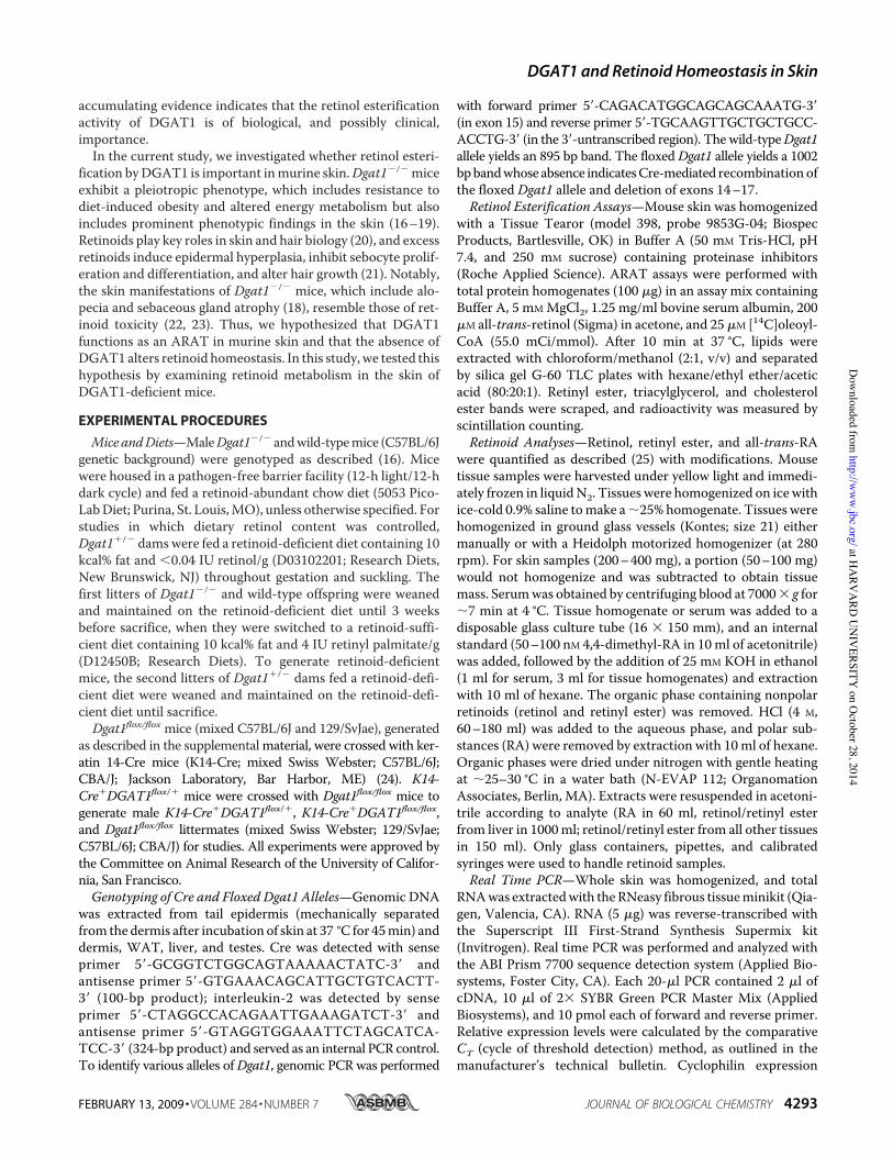
Altered Retinoid Metabolism in Dgat1!/! Skin—To deter-mine if DGAT1 contributes to retinol esterification in skin, wemeasuredARATactivity inwhole skin homogenates of 7-week-old wild-type andDgat1!/! mice fed a chow diet. ARAT activ-ity was reduced by %90% in Dgat1!/! skin (Fig. 1a). DGATactivity was reduced bymore than 95%. The activity of a controlenzyme, acyl-CoA:cholesterol acyltransferase, which cata-lyzes cholesterol ester synthesis, was slightly lower inDgat1!/! skin (Fig. 1a), possibly because of atrophy of seba-ceous glands, where acyl-CoA:cholesterol acyltransferasesare highly expressed (26).
To determine if the reduced ARAT activity inDgat1!/! skinaltered retinoid homeostasis, we measured retinoid levels inwhole skin of wild-type and Dgat1!/! mice fed a formulated,purified diet containing the amount of retinol recommended bythe American Institute of Nutrition (i.e. a retinol-sufficientdiet). Unesterified retinol was increased by %22%, and all-trans-RAwas increased by 40% inDgat1!/! skin (Fig. 1b). Reti-nyl ester levels were similar, probably reflecting contributionsfrom LRAT activity. Serum retinol (Fig. 1c) and liver retinoid(Fig. 1d) levels were similar in both genotypes of mice, suggest-ing that the increased all-trans-RA levels in Dgat1!/! skinresulted from local changes in retinoid metabolism.
All-trans-RA mediates many biological activities of retinolthrough transcriptional mechanisms (27). We therefore exam-
ined mRNA levels of two all-trans-RA target genes, CrbpI andcellular RA-binding protein II (CrabpII) (1, 27), in the syn-chronized telogen skin of 7-week-old chow-fed Dgat1!/!
and wild-type mice. CrbpI mRNA was increased 7-fold andCrabpIImRNA 2-fold inDgat1!/! skin (Fig. 1e). The mRNAlevels of the "-catenin and Vegf (vascular endothelial growthfactor) genes, which are hair cycle-regulated genes but nottargets of all-trans-RA, were similar in Dgat1!/! and wild-type skin (Fig. 1e).Enhanced EpidermalHyperplasia in Response to Topical Ret-
inol in Dgat1!/! Mice—To determine whether the biologicalactivity of retinol is enhanced in Dgat1!/! skin, we appliedretinol once to dorsal skin of shaved mice and examined the
FIGURE 1. Altered retinoid homeostasis in Dgat1!/! mice. a, reduced invitro ARAT activity in whole skin of Dgat1!/! mice (age 7 weeks, n ' 7/geno-type). *, p # 0.001 versus wild type. Retinyl esters (RE), triacylglycerols (TG),and cholesterol esters (CE) are the respective products of the ARAT, DGAT,and acyl-CoA:cholesterol acyltransferase reactions. b, retinol (ROL) and all-trans-retinoic acid (atRA) concentrations are increased in whole skin inDgat1!/! mice fed the retinoid-sufficient (RS) diet but not in those fed theretinoid-deficient (RD) diet (age 7.5–14 weeks, n ' 4 – 6/genotype). *, p # 0.05versus wild type; **, p # 0.001; #, p # 0.05 versus retinoid-sufficient diet.c, serum retinol concentrations are similar in wild-type and Dgat1!/! mice(age 7.5–14 weeks, n ' 4 – 6/genotype). *, p # 0.001 versus retinoid-sufficientdiet. d, hepatic retinyl ester and retinol concentrations are similar in wild-typeand Dgat1!/! mice (age 7.5–14 weeks, n ' 4 – 6/genotype). *, p # 0.05; **, p #0.01; #, p ' 0.01; †, p # 0.001 versus retinoid-sufficient diet. e, RA target geneexpression is increased in the whole skin of Dgat1!/! mice fed a retinoid-abundant chow diet. mRNA levels were quantified by real time PCR (age 7weeks, n ' 5– 6/genotype). *, p # 0.01; **, p # 0.05 versus wild type.
DGAT1 and Retinoid Homeostasis in Skin
4294 JOURNAL OF BIOLOGICAL CHEMISTRY VOLUME 284 • NUMBER 7 • FEBRUARY 13, 2009
at HA
RVA
RD U
NIV
ERSITY on O
ctober 28, 2014http://w
ww
.jbc.org/D
ownloaded from
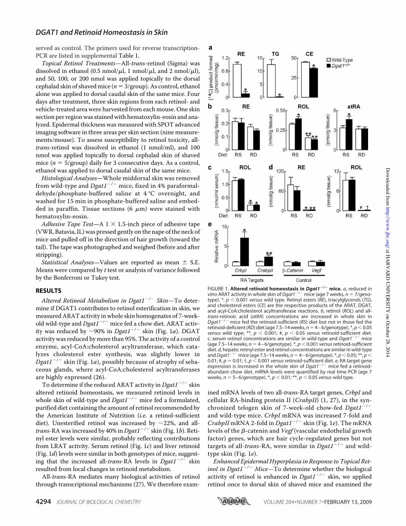
extent of retinoid-induced epidermal hyperplasia 4 days later.Retinol did not exert effects on wild-type skin but induced sig-nificant epidermal hyperplasia and increased epidermal thick-ness in Dgat1!/! skin (Fig. 2, a and b).
To determine if Dgat1!/! skin exhibits increased suscepti-bility to more chronic retinoid toxicity, we applied retinol top-ically for 3 consecutive days. Dgat1!/! skin exhibited severeirritation characteristic of retinoid toxicity, including ery-thema, severe skin scaling and cracking, and crusty lesions (28,29). In contrast, retinol caused only mild irritation (erythemaand some flaking) in wild-type skin (Fig. 2c).Retinoid Deprivation Prevents Alopecia of Dgat1!/! Mice—
Because excess retinoid activity in the skin causes alopecia (29,30), we hypothesized that the adult onset alopecia inDgat1!/!
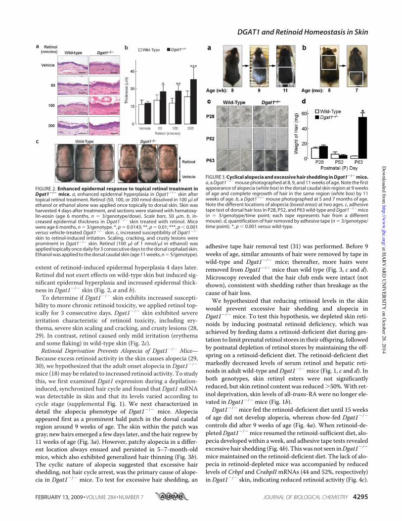
mice (18)may be related to increased retinoid activity. To studythis, we first examined Dgat1 expression during a depilation-induced, synchronized hair cycle and found that Dgat1mRNAwas detectable in skin and that its levels varied according tocycle stage (supplemental Fig. 1). We next characterized indetail the alopecia phenotype of Dgat1!/! mice. Alopeciaappeared first as a prominent bald patch in the dorsal caudalregion around 9 weeks of age. The skin within the patch wasgray; new hairs emerged a few days later, and the hair regrew by11 weeks of age (Fig. 3a). However, patchy alopecia in a differ-ent location always ensued and persisted in 5–7-month-oldmice, which also exhibited generalized hair thinning (Fig. 3b).The cyclic nature of alopecia suggested that excessive hairshedding, not hair cycle arrest, was the primary cause of alope-cia in Dgat1!/! mice. To test for excessive hair shedding, an
adhesive tape hair removal test (31) was performed. Before 9weeks of age, similar amounts of hair were removed by tape inwild-type and Dgat1!/! mice; thereafter, more hairs wereremoved from Dgat1!/! mice than wild type (Fig. 3, c and d).Microscopy revealed that the hair club ends were intact (notshown), consistent with shedding rather than breakage as thecause of hair loss.
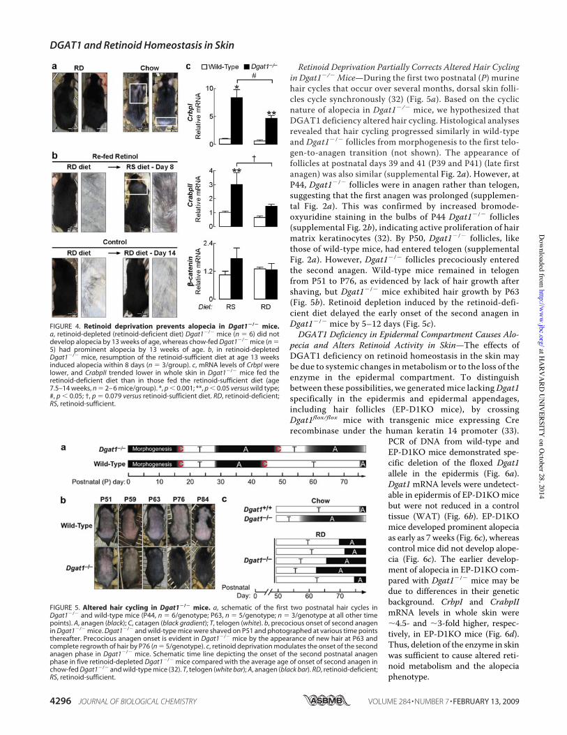
We hypothesized that reducing retinoid levels in the skinwould prevent excessive hair shedding and alopecia inDgat1!/! mice. To test this hypothesis, we depleted skin reti-noids by inducing postnatal retinoid deficiency, which wasachieved by feeding dams a retinoid-deficient diet during ges-tation to limit prenatal retinol stores in their offspring, followedby postnatal depletion of retinol stores by maintaining the off-spring on a retinoid-deficient diet. The retinoid-deficient dietmarkedly decreased levels of serum retinol and hepatic reti-noids in adult wild-type andDgat1!/! mice (Fig. 1, c and d). Inboth genotypes, skin retinyl esters were not significantlyreduced, but skin retinol content was reduced )50%.With ret-inol deprivation, skin levels of all-trans-RA were no longer ele-vated in Dgat1!/! mice (Fig. 1b).
Dgat1!/! mice fed the retinoid-deficient diet until 15 weeksof age did not develop alopecia, whereas chow-fed Dgat1!/!
controls did after 9 weeks of age (Fig. 4a). When retinoid-de-pletedDgat1!/! mice resumed the retinoid-sufficient diet, alo-pecia developed within a week, and adhesive tape tests revealedexcessive hair shedding (Fig. 4b). Thiswas not seen inDgat1!/!
micemaintained on the retinoid-deficient diet. The lack of alo-pecia in retinoid-depleted mice was accompanied by reducedlevels of CrbpI and CrabpIImRNAs (44 and 52%, respectively)in Dgat1!/! skin, indicating reduced retinoid activity (Fig. 4c).
FIGURE 2. Enhanced epidermal response to topical retinol treatment inDgat1!/! mice. a, enhanced epidermal hyperplasia in Dgat1!/! skin aftertopical retinol treatment. Retinol (50, 100, or 200 nmol dissolved in 100 !l ofethanol or ethanol alone was applied once topically to dorsal skin. Skin washarvested 4 days after treatment, and sections were stained with hematoxy-lin-eosin (age 6 months, n ' 3/genotype/dose). Scale bars, 50 !m. b, in-creased epidermal thickness in Dgat1!/! skin treated with retinol. Micewere age 6 months, n ' 3/genotype. *, p ' 0.0143; **, p ' 0.01; ***, p # 0.001versus vehicle-treated Dgat1!/! skin. c, increased susceptibility of Dgat1!/!
skin to retinol-induced irritation. Scaling, cracking, and crusty lesions wereprominent in Dgat1!/! skin. Retinol (100 !l of 1 nmol/!l in ethanol) wasapplied topically once daily for 3 consecutive days to the dorsal cephalad skin.Ethanol was applied to the dorsal caudal skin (age 11 weeks, n ' 5/genotype).
FIGURE 3. Cyclical alopecia and excessive hair shedding in Dgat1!/! mice.a, a Dgat1!/! mouse photographed at 8, 9, and 11 weeks of age. Note the firstappearance of alopecia (white box) in the dorsal caudal skin region at 9 weeksof age and complete regrowth of hair in the same region (white box) by 11weeks of age. b, a Dgat1!/! mouse photographed at 5 and 7 months of age.Note the different locations of alopecia (boxed areas) at two ages. c, adhesivetape test of dorsal hair loss in P28, P52, and P63 wild-type and Dgat1!/! mice(n ' 3/genotype/time point; each tape represents hair from a differentmouse). d, quantification of hair removed by adhesive tape (n ' 3/genotype/time point). *, p # 0.001 versus wild-type.
DGAT1 and Retinoid Homeostasis in Skin
FEBRUARY 13, 2009 • VOLUME 284 • NUMBER 7 JOURNAL OF BIOLOGICAL CHEMISTRY 4295
at HA
RVA
RD U
NIV
ERSITY on O
ctober 28, 2014http://w
ww
.jbc.org/D
ownloaded from
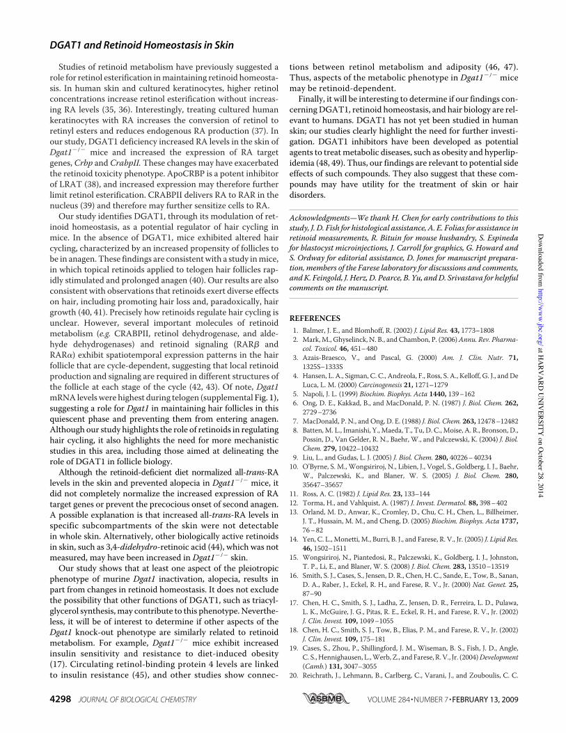
Retinoid Deprivation Partially Corrects Altered Hair Cyclingin Dgat1!/! Mice—During the first two postnatal (P) murinehair cycles that occur over several months, dorsal skin folli-cles cycle synchronously (32) (Fig. 5a). Based on the cyclicnature of alopecia in Dgat1!/! mice, we hypothesized thatDGAT1 deficiency altered hair cycling. Histological analysesrevealed that hair cycling progressed similarly in wild-typeand Dgat1!/! follicles from morphogenesis to the first telo-gen-to-anagen transition (not shown). The appearance offollicles at postnatal days 39 and 41 (P39 and P41) (late firstanagen) was also similar (supplemental Fig. 2a). However, atP44, Dgat1!/! follicles were in anagen rather than telogen,suggesting that the first anagen was prolonged (supplemen-tal Fig. 2a). This was confirmed by increased bromode-oxyuridine staining in the bulbs of P44 Dgat1!/! follicles(supplemental Fig. 2b), indicating active proliferation of hairmatrix keratinocytes (32). By P50, Dgat1!/! follicles, likethose of wild-type mice, had entered telogen (supplementalFig. 2a). However, Dgat1!/! follicles precociously enteredthe second anagen. Wild-type mice remained in telogenfrom P51 to P76, as evidenced by lack of hair growth aftershaving, but Dgat1!/! mice exhibited hair growth by P63(Fig. 5b). Retinoid depletion induced by the retinoid-defi-cient diet delayed the early onset of the second anagen inDgat1!/! mice by 5–12 days (Fig. 5c).DGAT1 Deficiency in Epidermal Compartment Causes Alo-
pecia and Alters Retinoid Activity in Skin—The effects ofDGAT1 deficiency on retinoid homeostasis in the skin maybe due to systemic changes inmetabolism or to the loss of theenzyme in the epidermal compartment. To distinguishbetween these possibilities, we generated mice lackingDgat1specifically in the epidermis and epidermal appendages,including hair follicles (EP-D1KO mice), by crossingDgat1flox/flox mice with transgenic mice expressing Crerecombinase under the human keratin 14 promoter (33).
PCR of DNA from wild-type andEP-D1KO mice demonstrated spe-cific deletion of the floxed Dgat1allele in the epidermis (Fig. 6a).Dgat1 mRNA levels were undetect-able in epidermis of EP-D1KOmicebut were not reduced in a controltissue (WAT) (Fig. 6b). EP-D1KOmice developed prominent alopeciaas early as 7 weeks (Fig. 6c), whereascontrol mice did not develop alope-cia (Fig. 6c). The earlier develop-ment of alopecia in EP-D1KO com-pared with Dgat1!/! mice may bedue to differences in their geneticbackground. CrbpI and CrabpIImRNA levels in whole skin were%4.5- and %3-fold higher, respec-tively, in EP-D1KO mice (Fig. 6d).Thus, deletion of the enzyme in skinwas sufficient to cause altered reti-noid metabolism and the alopeciaphenotype.
FIGURE 4. Retinoid deprivation prevents alopecia in Dgat1!/! mice.a, retinoid-depleted (retinoid-deficient diet) Dgat1!/! mice (n ' 6) did notdevelop alopecia by 13 weeks of age, whereas chow-fed Dgat1!/! mice (n '5) had prominent alopecia by 13 weeks of age. b, in retinoid-depletedDgat1!/! mice, resumption of the retinoid-sufficient diet at age 13 weeksinduced alopecia within 8 days (n ' 3/group). c, mRNA levels of CrbpI werelower, and CrabpII trended lower in whole skin in Dgat1!/! mice fed theretinoid-deficient diet than in those fed the retinoid-sufficient diet (age7.5–14 weeks, n ' 2– 6 mice/group). *, p # 0.001; **, p # 0.05 versus wild type;#, p # 0.05; †, p ' 0.079 versus retinoid-sufficient diet. RD, retinoid-deficient;RS, retinoid-sufficient.
FIGURE 5. Altered hair cycling in Dgat1!/! mice. a, schematic of the first two postnatal hair cycles inDgat1!/! and wild-type mice (P44, n ' 6/genotype; P63, n ' 5/genotype; n ' 3/genotype at all other timepoints). A, anagen (black); C, catagen (black gradient); T, telogen (white). b, precocious onset of second anagenin Dgat1!/! mice. Dgat1!/! and wild-type mice were shaved on P51 and photographed at various time pointsthereafter. Precocious anagen onset is evident in Dgat1!/! mice by the appearance of new hair at P63 andcomplete regrowth of hair by P76 (n ' 5/genotype). c, retinoid deprivation modulates the onset of the secondanagen phase in Dgat1!/! mice. Schematic time line depicting the onset of the second postnatal anagenphase in five retinoid-depleted Dgat1!/! mice compared with the average age of onset of second anagen inchow-fed Dgat1!/! and wild-type mice (32). T, telogen (white bar); A, anagen (black bar). RD, retinoid-deficient;RS, retinoid-sufficient.
DGAT1 and Retinoid Homeostasis in Skin
4296 JOURNAL OF BIOLOGICAL CHEMISTRY VOLUME 284 • NUMBER 7 • FEBRUARY 13, 2009
at HA
RVA
RD U
NIV
ERSITY on O
ctober 28, 2014http://w
ww
.jbc.org/D
ownloaded from

DISCUSSION
We show here that DGAT1 functions as an ARAT inmurineskin. In mice lacking DGAT1 activity that were fed a retinol-sufficient diet, RA levels were elevated in the skin, as were RARtarget genes, consistent with retinoid toxicity. These miceexhibit alopecia that is probably from increased shedding,which in turn may relate to sebaceous gland dysfunction (18).
Lowering retinol levels with a retin-oid-deficient diet lowered RA levels,resulted in the appearance of smalland possibly immature sebaceousglands,3 and prevented alopecia andabnormal hair cycling in Dgat1!/!
mice. Further, Dgat1!/! skin wasmore susceptible to toxicity fromtopically applied retinol. Theseresults demonstrate that DGAT1functions as an ARAT in vivo inmurine skin, where it plays animportant role in maintaining nor-mal retinoid homeostasis.
Studies of Lrat!/! mice revealedthat LRAT is the predominant reti-nol acyltransferase for maintainingadequate supplies of retinol (8–10,15) particularly in the liver, wheremost retinol is stored. Consistentwith these findings, our study showsthat Dgat1 inactivation did not pre-dispose mice to retinol deficiency.Retinyl ester levels in liver and othertissues (WAT, brown adipose tis-sue, skeletal muscle, and brain; notshown) and serum retinol levels inDgat1!/! mice fed a retinoid-suffi-cient or retinoid-deficient diet were
similar to levels of wild-type mice. In contrast to the storagefunction of LRAT, our study suggests that the ARAT activity ofDGAT1 functions in the epidermis primarily to limit local RAconcentrations and protect against retinoid toxicity (see modelin Fig. 7). Dgat1!/! skin exhibited enhanced epidermal hyper-plasia and irritation in response to a range of retinol concentra-tions that did not affectwild-type skin.Additionally, the cyclicalalopecia and excessive hair shedding in Dgat1!/! mice wereprobably due to excessive retinoid action, since lowering retinollevels in the skin through retinol deprivation largely preventedthese phenotypic manifestations. Supporting the idea thatARAT activity prevents retinoid toxicity, theKm for retinol as asubstrate for the ARAT reaction is higher than that for theLRAT reaction (34), and ARAT utilizes predominantlyunbound retinol as a substrate (12).
Our study adds to accumulating evidence that the ARATfunction of DGAT1 is biologically relevant in vivo. Recently,Wongsiriroj et al. (15) reported that Dgat1 functions as anARAT in murine intestine. Retinol absorption in the smallintestines of Dgat1!/! mice was impaired when the micewere challenged with pharmacological doses of retinol, sup-porting the idea that Dgat1 is important for handling freeretinol that exceeds the capacity of LRAT. Thus, studies withDgat1!/! mice have now demonstrated the importance ofARAT activity by DGAT1 in two tissues, the small intestineand skin.
3 M. Shih, unpublished observations.
FIGURE 6. Effects of Dgat1 deficiency on retinoid homeostasis are epidermis-autonomous. a, specificdeletion of Dgat1 in epidermis. Shown is PCR detection of the wild-type and floxed allele of Dgat1 and Cretransgene in genomic DNA. Interleukin-2 (IL-2) served as an internal PCR control. The absence of a PCR bandindicates Cre-mediated recombination. b, absence of Dgat1 mRNA in the epidermis of K14-Cre"Dgat1flox/flox
(EP-D1KO) mice. mRNA levels of Dgat1 in the tail epidermis and WAT were quantified by real time PCR. (age 13weeks, n ' 3– 6 mice/group). c, cyclical alopecia in EP-D1KO mice. Alopecia was detectable by 7.5 weeks of age.Note partial hair regrowth by 10.5 weeks of age. Alopecia was not observed in control mice. (n ' 6/genotype).d, increased mRNA expression of RA target genes in whole skin of EP-D1KO mice. mRNA levels were quantifiedby real time PCR (age 13 weeks, n ' 5/genotype). *, p # 0.0001; **, p ' 0.0002 versus control.
FIGURE 7. Model of how DGAT1 deficiency modulates RA signaling inskin. LRAT is the major pathway for maintaining adequate stores of retinol, asretinyl esters, in the skin. However, the ARAT activity of DGAT1 is importantfor generating retinyl esters when retinol levels are excessive and preventingretinol toxicity. In DGAT1 deficiency in murine skin, excess retinol is convertedto retinaldehyde and subsequently to RAs, which modulate gene expressionand hair cycling. If retinol is depleted from the diet, the effects of DGAT1deficiency are minimized.
DGAT1 and Retinoid Homeostasis in Skin
FEBRUARY 13, 2009 • VOLUME 284 • NUMBER 7 JOURNAL OF BIOLOGICAL CHEMISTRY 4297
at HA
RVA
RD U
NIV
ERSITY on O
ctober 28, 2014http://w
ww
.jbc.org/D
ownloaded from
Studies of retinoid metabolism have previously suggested arole for retinol esterification inmaintaining retinoid homeosta-sis. In human skin and cultured keratinocytes, higher retinolconcentrations increase retinol esterification without increas-ing RA levels (35, 36). Interestingly, treating cultured humankeratinocytes with RA increases the conversion of retinol toretinyl esters and reduces endogenous RA production (37). Inour study, DGAT1 deficiency increased RA levels in the skin ofDgat1!/! mice and increased the expression of RA targetgenes, Crbp and CrabpII. These changes may have exacerbatedthe retinoid toxicity phenotype. ApoCRBP is a potent inhibitorof LRAT (38), and increased expression may therefore furtherlimit retinol esterification. CRABPII delivers RA to RAR in thenucleus (39) and therefore may further sensitize cells to RA.
Our study identifies DGAT1, through its modulation of ret-inoid homeostasis, as a potential regulator of hair cycling inmice. In the absence of DGAT1, mice exhibited altered haircycling, characterized by an increased propensity of follicles tobe in anagen. These findings are consistentwith a study inmice,in which topical retinoids applied to telogen hair follicles rap-idly stimulated and prolonged anagen (40). Our results are alsoconsistent with observations that retinoids exert diverse effectson hair, including promoting hair loss and, paradoxically, hairgrowth (40, 41). Precisely how retinoids regulate hair cycling isunclear. However, several important molecules of retinoidmetabolism (e.g. CRABPII, retinol dehydrogenase, and alde-hyde dehydrogenases) and retinoid signaling (RAR" andRAR#) exhibit spatiotemporal expression patterns in the hairfollicle that are cycle-dependent, suggesting that local retinoidproduction and signaling are required in different structures ofthe follicle at each stage of the cycle (42, 43). Of note, Dgat1mRNA levelswere highest during telogen (supplemental Fig. 1),suggesting a role for Dgat1 in maintaining hair follicles in thisquiescent phase and preventing them from entering anagen.Although our study highlights the role of retinoids in regulatinghair cycling, it also highlights the need for more mechanisticstudies in this area, including those aimed at delineating therole of DGAT1 in follicle biology.
Although the retinoid-deficient diet normalized all-trans-RAlevels in the skin and prevented alopecia in Dgat1!/! mice, itdid not completely normalize the increased expression of RAtarget genes or prevent the precocious onset of second anagen.A possible explanation is that increased all-trans-RA levels inspecific subcompartments of the skin were not detectablein whole skin. Alternatively, other biologically active retinoidsin skin, such as 3,4-didehydro-retinoic acid (44), which was notmeasured, may have been increased in Dgat1!/! skin.
Our study shows that at least one aspect of the pleiotropicphenotype of murine Dgat1 inactivation, alopecia, results inpart from changes in retinoid homeostasis. It does not excludethe possibility that other functions of DGAT1, such as triacyl-glycerol synthesis,may contribute to this phenotype. Neverthe-less, it will be of interest to determine if other aspects of theDgat1 knock-out phenotype are similarly related to retinoidmetabolism. For example, Dgat1!/! mice exhibit increasedinsulin sensitivity and resistance to diet-induced obesity(17). Circulating retinol-binding protein 4 levels are linkedto insulin resistance (45), and other studies show connec-
tions between retinol metabolism and adiposity (46, 47).Thus, aspects of the metabolic phenotype in Dgat1!/! micemay be retinoid-dependent.
Finally, it will be interesting to determine if our findings con-cerningDGAT1, retinoid homeostasis, and hair biology are rel-evant to humans. DGAT1 has not yet been studied in humanskin; our studies clearly highlight the need for further investi-gation. DGAT1 inhibitors have been developed as potentialagents to treatmetabolic diseases, such as obesity and hyperlip-idemia (48, 49). Thus, our findings are relevant to potential sideeffects of such compounds. They also suggest that these com-pounds may have utility for the treatment of skin or hairdisorders.
Acknowledgments—We thank H. Chen for early contributions to thisstudy, J. D. Fish for histological assistance, A. E. Folias for assistance inretinoid measurements, R. Bituin for mouse husbandry, S. Espinedafor blastocyst microinjections, J. Carroll for graphics, G. Howard andS. Ordway for editorial assistance, D. Jones for manuscript prepara-tion, members of the Farese laboratory for discussions and comments,andK. Feingold, J. Herz, D. Pearce, B. Yu, andD. Srivastava for helpfulcomments on the manuscript.
REFERENCES1. Balmer, J. E., and Blomhoff, R. (2002) J. Lipid Res. 43, 1773–18082. Mark,M., Ghyselinck, N. B., and Chambon, P. (2006)Annu. Rev. Pharma-
col. Toxicol. 46, 451–4803. Azais-Braesco, V., and Pascal, G. (2000) Am. J. Clin. Nutr. 71,
1325S–1333S4. Hansen, L. A., Sigman, C. C., Andreola, F., Ross, S. A., Kelloff, G. J., and De
Luca, L. M. (2000) Carcinogenesis 21, 1271–12795. Napoli, J. L. (1999) Biochim. Biophys. Acta 1440, 139–1626. Ong, D. E., Kakkad, B., and MacDonald, P. N. (1987) J. Biol. Chem. 262,
2729–27367. MacDonald, P. N., and Ong, D. E. (1988) J. Biol. Chem. 263, 12478–124828. Batten,M. L., Imanishi, Y., Maeda, T., Tu, D. C.,Moise, A. R., Bronson, D.,
Possin, D., Van Gelder, R. N., Baehr, W., and Palczewski, K. (2004) J. Biol.Chem. 279, 10422–10432
9. Liu, L., and Gudas, L. J. (2005) J. Biol. Chem. 280, 40226–4023410. O’Byrne, S. M., Wongsiriroj, N., Libien, J., Vogel, S., Goldberg, I. J., Baehr,
W., Palczewski, K., and Blaner, W. S. (2005) J. Biol. Chem. 280,35647–35657
11. Ross, A. C. (1982) J. Lipid Res. 23, 133–14412. Torma, H., and Vahlquist, A. (1987) J. Invest. Dermatol. 88, 398–40213. Orland, M. D., Anwar, K., Cromley, D., Chu, C. H., Chen, L., Billheimer,
J. T., Hussain, M. M., and Cheng, D. (2005) Biochim. Biophys. Acta 1737,76–82
14. Yen, C. L., Monetti, M., Burri, B. J., and Farese, R. V., Jr. (2005) J. Lipid Res.46, 1502–1511
15. Wongsiriroj, N., Piantedosi, R., Palczewski, K., Goldberg, I. J., Johnston,T. P., Li, E., and Blaner, W. S. (2008) J. Biol. Chem. 283, 13510–13519
16. Smith, S. J., Cases, S., Jensen, D. R., Chen, H. C., Sande, E., Tow, B., Sanan,D. A., Raber, J., Eckel, R. H., and Farese, R. V., Jr. (2000) Nat. Genet. 25,87–90
17. Chen, H. C., Smith, S. J., Ladha, Z., Jensen, D. R., Ferreira, L. D., Pulawa,L. K., McGuire, J. G., Pitas, R. E., Eckel, R. H., and Farese, R. V., Jr. (2002)J. Clin. Invest. 109, 1049–1055
18. Chen, H. C., Smith, S. J., Tow, B., Elias, P. M., and Farese, R. V., Jr. (2002)J. Clin. Invest. 109, 175–181
19. Cases, S., Zhou, P., Shillingford, J. M., Wiseman, B. S., Fish, J. D., Angle,C. S., Hennighausen, L.,Werb, Z., and Farese, R. V., Jr. (2004)Development(Camb.) 131, 3047–3055
20. Reichrath, J., Lehmann, B., Carlberg, C., Varani, J., and Zouboulis, C. C.
DGAT1 and Retinoid Homeostasis in Skin
4298 JOURNAL OF BIOLOGICAL CHEMISTRY VOLUME 284 • NUMBER 7 • FEBRUARY 13, 2009
at HA
RVA
RD U
NIV
ERSITY on O
ctober 28, 2014http://w
ww
.jbc.org/D
ownloaded from

(2007) Horm. Metab. Res. 39, 71–8421. Zouboulis, C. C. (2001) Skin Pharmacol. Appl. Skin Physiol. 14, 303–31522. Berth-Jones, J., andHutchinson, P. E. (1995)Br. J. Dermatol. 132, 367–37523. Clarke, S. B., Nelson, A. M., George, R. E., and Thiboutot, D. M. (2007)
Dermatol. Clin. 25, 137–14624. Dassule, H. R., Lewis, P., Bei, M., Maas, R., and McMahon, A. P. (2000)
Development (Cambr.) 127, 4775–478525. Kane, M. A., Chen, N., Sparks, S., and Napoli, J. L. (2005) Biochem. J. 388,
363–36926. Meiner, V., Tam, C., Gunn, M. D., Dong, L. M.,Weisgraber, K. H., Novak,
S.,Myers, H.M., Erickson, S. K., and Farese, R. V., Jr. (1997) J. Lipid Res. 38,1928–1933
27. Fisher, G. J., and Voorhees, J. J. (1996) FASEB J. 10, 1002–101328. Elias, P. M. (1986) J. Am. Acad. Dermatol. 15, 797–80929. Look, J., Landwehr, J., Bauer, F., Hoffmann, A. S., Bluethmann, H., and
LeMotte, P. (1995) Am. J. Physiol. 269, E91–E9830. Foitzik, K., Spexard, T., Nakamura, M., Halsner, U., and Paus, R. (2005)
J. Invest. Dermatol. 124, 1119–112631. Hanakawa, Y., Matsuyoshi, N., and Stanley, J. R. (2002) J. Invest. Dermatol.
119, 27–3132. Muller-Rover, S., Handjiski, B., van derVeen, C., Eichmuller, S., Foitzik, K.,
McKay, I. A., Stenn, K. S., and Paus, R. (2001) J. Invest. Dermatol.117, 3–1533. Gritli-Linde, A., Hallberg, K., Harfe, B. D., Reyahi, A., Kannius-Janson,M.,
Nilsson, J., Cobourne, M. T., Sharpe, P. T., McMahon, A. P., and Linde, A.(2007) Dev. Cell 12, 99–112
34. Randolph, R. K., Winkler, K. E., and Ross, A. C. (1991) Arch. Biochem.Biophys. 288, 500–508
35. Randolph, R. K., and Simon, M. (1993) J. Biol. Chem. 268, 9198–920536. Kang, S., Duell, E. A., Fisher, G. J., Datta, S. C., Wang, Z.-Q., Reddy, A. P.,
Tavakkol, A., Yi, J. Y., Griffiths, C. E. M., Elder, J. T., and Voorhees, J. J.
(1995) J. Invest. Dermatol. 105, 549–55637. Kurlandsky, S. B., Duell, E. A., Kang, S., Voorhees, J. J., and Fisher, G. J.
(1996) J. Biol. Chem. 271, 15346–1535238. Herr, F. M., and Ong, D. E. (1992) Biochemistry 31, 6748–675539. Budhu, A. S., and Noy, N. (2002)Mol. Cell Biol. 22, 2632–264140. Bazzano, G., Terezakis, N., Attia, H., Bazzano, A., Dover, R., Fenton, D.,
Mandir, N., Celleno, L., Tamburro, M., and Jaconi, S. (1993) J. Invest.Dermatol. 101, 138S–142S
41. Bazzano, G. S., Terezakis, N., andGalen,W. (1986) J. Am. Acad. Dermatol.15, 880–883, 890–893
42. Everts,H. B., King, L. E., Jr., Sundberg, J. P., andOng,D. E. (2004) J. Investig.Dermatol. 123, 258–263
43. Everts,H. B., Sundberg, J. P., King, L. E., Jr., andOng,D. E. (2007) J. Investig.Dermatol. 127, 1593–1604
44. Randolph, R. K., and Simon, M. (1997) J. Lipid Res. 38, 1374–138345. Yang, Q., Graham, T. E., Mody, N., Preitner, F., Peroni, O. D., Zabolotny,
J. M., Kotani, K., Quadro, L., and Kahn, B. B. (2005)Nature 436, 356–36246. Zhang,M., Hu, P., Krois, C. R., Kane,M. A., andNapoli, J. L. (2007) FASEB
J. 21, 2886–289647. Ziouzenkova, O., Orasanu, G., Sharlach, M., Akiyama, T. E., Berger, J. P.,
Viereck, J., Hamilton, J. A., Tang, G., Dolnikowski, G. G., Vogel, S.,Duester, G., and Plutzky, J. (2007) Nat. Med. 13, 695–702
48. Zhao, G., Souers, A. J., Voorbach, M., Falls, H. D., Droz, B., Brodjian, S.,Lau, Y. Y., Iyengar, R. R., Gao, J., Judd, A. S., Wagaw, S. H., Ravn, M. M.,Engstrom, K. M., Lynch, J. K., Mulhern, M.M., Freeman, J., Dayton, B. D.,Wang, X., Grihalde, N., Fry, D., Beno, D. W., Marsh, K. C., Su, Z., Diaz,G. J., Collins, C. A., Sham, H., Reilly, R. M., Brune, M. E., and Kym, P. R.(2008) J. Med. Chem. 51, 380–383
49. Matsuda, D., and Tomoda, H. (2007) Curr. Opin. Investig. Drugs 8,836–841
DGAT1 and Retinoid Homeostasis in Skin
FEBRUARY 13, 2009 • VOLUME 284 • NUMBER 7 JOURNAL OF BIOLOGICAL CHEMISTRY 4299
at HA
RVA
RD U
NIV
ERSITY on O
ctober 28, 2014http://w
ww
.jbc.org/D
ownloaded from