jinghui hu studies on histomonas meleagridis and
TRANSCRIPT

JINGHUI HU
STUDIES ON HISTOMONAS MELEAGRIDIS AND HISTOMONIASIS IN CHICKENS AND TURKEYS (Under the direction of LARRY R. MCDOUGALD)
A series of studies has been conducted to expand knowledge of the pathogenicity,
epidemiology, and treatment of histomoniasis in chickens and turkeys.
The interaction of cecal coccidiosis with histomoniasis in chickens was investigated,
using concurrent infections. Cecal lesions from H. meleagridis were severe in all inoculated
control groups and did not appear to be affected by the introduction of Eimeria tenella
infections. However, the severity of liver lesions and number of birds positive for liver
lesions of H. meleagridis increased significantly with the presence of E. tenella. The positive
relationship between infections of cecal coccidiosis and H. meleagridis in chickens suggests
that, under field conditions, such dual exposure may contribute to increased clinical
outbreaks of histomoniasis in chickens.
The lateral transmission of H. meleagridis in turkeys was studied in floor pens in the
absence of the carrier cecal worm Heterakis gallinarum. One group received no exposure. In
other groups, either 10% (LE) or 25% (HE) of the birds were inoculated per cloaca with
cultured H. meleagridis (200,000 cells/bird). Inoculated birds died at 10-18 days post-
infection (DPI). Uninoculated-birds in the high exposure group (HE) died of histomoniasis
beginning 16 DPI, and continued to 100% mortality by day 23 DPI. Uninoculated birds in
low exposure group (LE) died beginning on day 19 DPI and continuing through day 31 DPI.
All but one LE birds alive on day 31 DPI had severe liver and cecal lesions of histomoniasis
at necropsy. There was no evidence of histomoniasis in unexposed birds at the end of
experiment. These results suggested that lateral transmission of histomoniasis through a

flock can occur readily through normal contact between infected birds and uninfected birds
and their droppings in the total absence of cecal worms.
Since no products are available for treatment of blackhead outbreaks, studies were
conducted on other antiprotozoal or antibiotic compounds. Five anticoccidials, six
antibiotics, and nine antiprotozoals drugs known from literature were tested for their efficacy
against Histomonas meleagridis in chickens. None of 5 anticoccidials (salinomycin,
diclazuril, nicarbazin, roxarsone, and lasalocid) and 6 antibiotics (bacitracin, apramycin,
penicillin, chlortetracycline, tylosin, sarafloxacin) had significant efficacy in control of
histomoniasis. Out of 9 antiprotozoals, the nitroimidazoles dimetridazole (control),
metronidazole, ornidazole, and tinidazole suppressed growth of H. meleagridis in vitro at 10
ug/ml or higher. There were also highly effective in chickens at 200 ppm in the feed,
preventing liver lesions and reducing cecal lesions to near zero. Paromomycin sulfate and
carbadox were weakly effective with the range tested but were ineffective in vivo.
Quinolinol, mebendazole, diloxanide furoate, and albendazole had no demonstrable efficacy
in vitro and were not tested in vivo.
A new phenomenon discovered during the culture of H. meleagridis was that cultured H.
meleagridis were readily agglutinated by horse serum at concentrations of 15% or higher.
The agglutinating factor (AF) was active when serum regardless of whether serum was heat-
inactivated, and was present in several batches of sera tested. The AF had a molecular
weight similar to that of serum globulins as shown by precipitation with 25-40% ammonium
sulfate. The AF was used as a means of obtaining Histomonas cells free of bacteria in the
following way: Histomonas cultures were pooled, washed twice with Hank’s balanced salt
solution (centrifuged at 1000g for 15 min and the supernatant decanted), and applied to a

nylon wool column. Horse serum was added (15%), and filtrates were agitated gently in Petri
plates, whereupon clusters of H. meleagridis formed. The aggregated suspensions were
collected by centrifugation, loaded onto a 2-layer sucrose gradient and centrifuged at 2500 g
for 10 min. A band of cells forming between the two layers was collected and washed 2 to 3
times with centrifugation. The resulting clumps of H. meleagridis were free of visible
bacteria.
INDEX WORDS: Histomonas meleagridis, Histomoniasis, Blackhead Disease,
Epidemiology, Pathology, Control, Chemotherapy, Purification,
Eimeria tenella, Culture in vitro, Chickens, Turkeys, Agglutinins.

STUDIES ON HISTOMONAS MELEAGRIDIS AND HISTOMONIASIS
IN CHICKENS AND TURKEYS
by
JINGHUI HU
B.S. Beijing Agricultural University, 1988
Ph.D. Beijing Agricultural University, 1994
A Dissertation Submitted to the Graduate Faculty of The University of
Georgia in Partial Fulfillment of the Requirements for the Degree
DOCTOR OF PHILOSOPHY
ATHENS, GEORGIA
2002

© 2002
Jinghui Hu
All Rights Reserved
STUDIES ON HISTOMONAS MELEAGRIDIS AND HISTOMONIASIS
IN CHICKENS AND TURKEYS
by
JINGHUI HU
Approve Major Professor: Larry R. McDougald
Committee: John R. Glisson Nicholas M. Dale Pedro N. Villegas Roger D. Wyatt Electrical Version Approved: Maureen Grasso Dean of the graduate School The University of Georgia December, 2002

iv
DEDICATION
To my wife, Meihua Qiao, for her understanding, encouragement, full support and
love to me. To my parents, Guanghuo Hu and Liangrong Chan, for encouraging me to aim
high and pursue graduate study. To my son, Michael Zhiyuan Hu, who was born while
studying for PhD in the University of Georgia.

v
ACKNOWLEDGEMENTS
I would like to express my deepest gratitude to Dr. Larry R. McDougald, my major
professor, for his guidance, support, and encouragement throughout my study. His help is
far beyond what I can describe with words.
Special thanks to my Doctoral Advisory Committee members, Dr. Nicholas Dale, Dr.
Roger D Wyatt, Dr. John R Glisson, and Dr. Pedro N Villegas, who were key players in my
entire graduate study and research. Their advice, technical support and ideas, were inspiring
and helpful. I sincerely appreciate the time these very busy people spent in evaluating and
editing my dissertation.
My years here are filled with many fond memories that I will always cherish. Thanks
for the benevolence of faculty, staff, and fellow students of the Department of Poultry
Science at the University of Georgia.

vi
TABLE OF CONTENTS
Page
Acknowledgements ..........................................................................................................v
Introduction .....................................................................................................................1
Literature review ..........................................................................................…...............3
Purification of Histomonas meleagridis cultured in vitro
via agglutinins in horse serum…….........................................................…........36
Direct lateral transmission of Histomonas meleagridis
in turkeys ...........................................................................................................47
The efficacy of some drugs with known antiprotozoal activity
against Histomonas meleagridis in chickens.................................... …............. 57
Effect of anticoccidials and antibiotics bn the control of black
Head disease in broiler breeder pullets ...............................................................68
Blackhead disease (Histomonas meleagridis) aggravated in broiler chickens
by concurrent infection with cecal coccidiosis (Eimeria tenella) .……………87
Conclusion …………………………………………………………………………... 101

1
INTRODUCTION
Histomoniasis, also frequently called blackhead (Cushman, 1893) or enterohepatitis
(Smith, 1895) is an infectious disease of gallinaceous birds that is caused by the protozoan,
Histomonas meleagridis. Histomoniasis can cause 90-100% mortality in turkeys, but is
relatively mild in chickens. Most studies were conducted before 1980, with little additional
research done during the past 20 years.
In recent years, there was a significant increase in field cases of clinically severe
histomoniasis in chickens, in both leghorn and broiler breeder pullets (Homer and Butcher,
1991; Luma et al., 1999). The possible causes for this increase would include (a) increased
virulence of organism, (b) increased susceptibility, (c) interaction of Histomonas with other
infections, (d) built-up cecal worm eggs in chicken houses, (e) change in management such
as feed restriction, and (f) lack of effective drugs to control the disease. But, the actual
reasons for the dramatic increase in clinical outbreaks are still unclear.
H. meleagridis is known to survive for a long period in cecal worm eggs, providing a
reservoir of infection from one year or place to another. Previous researcher concluded that
direct bird-to-bird transmission was unimportant in birds that were well fed and cared for
(Lund, 1956). But it is hard to explain that the rapid spread of histomoniasis through turkey
flocks resulting in 50-100% mortality in a few weeks. While there is no doubt that cecal
worm eggs may play important role in survival of histomonads in environmental conditions,
but there is no research to show how lateral transmission is important in histomoniasis
outbreaks in turkey or chicken flocks.

2
The increase in importance of blackhead disease comes at a time when use lack effective
drugs effective in treatment of H. meleagridis. The most effective drugs including
dimetridazole and ipronidazole are no longer registered for use in the USA due to action of
the Food and Drug Administration (McDougald, 1997).
The objectives of this project were to:
1. Study the conditions under which H. meleagridis spreads from bird to bird after
introduction of infection into a flock.
2. Demonstrate what, if any, influence parasites such as coccidia contributes to
pathogenicity of H. meleagridis in chickens.
3. Establish a system to screen drugs in vitro and in vivo and to evaluate the activity
of drugs against H. meleagridis, which are shown to be effective in control of
bacteria and other related protozoa
4. Characterize an agglutination factor in H. meleagridis which might be useful in
seperation of cells from other organisms for use in other studies.

3
LITERATURE REVIEW
1. Histomoniasis and Its Causative Agent
1.1. Histomoniasis in chickens and turkeys
Histomoniasis, also frequently called blackhead or enterohepatitis, is an infectious
disease of gallinaceous birds caused by the protozoan, Histomonas meleagridis. In 1893,
Cushman first brief described histomoniasis and (for no apparent reason) called it blackhead.
Smith (1895) described histomoniasis as infectious entero-hepatitis and gave a thorough
description of the disease after he necropsied about 50 turkeys. He also named the protozoan
parasite Amoeba meleagridis. In fresh preparations from ceca, he found the protozoan to be
circular or oval, 8-14 µm in diameter, and except for a granular nucleus, almost structureless.
Fixed and stained organisms were small oval bodies of 6 to 10 µm.
Tyzzer (1919) studied the morphology of the blackhead organism and described various
developing phases. The form observed in the periphery of early lesions was called the
invasive phase. These long forms with a single pseudopodium were frequently 30 µm in
length. In fixed tissues, some round forms 8-17 µm in diameter were observed. The
endoplasm of the invasive phase contained deeply stained particles within vacuoles and an
extranuclear body. The organisms in the vegetative phase had basophilic cytoplasm without
the inclusions, and a small amount of reticular or granular material was distributed around
the nucleus. Vegetative phase parasites were larger, varying from 12 - 21 µm. A so-called
resistant phase described by Tyzzer, was smaller, measuring 5 - 22 µm in diameter.

4
In 1920, Tyzzer noted its flagellate characteristics and changed the name of the parasite
to Histomonas meleagridis (Smith, 1895). Later, Tyzzer and Fabyan (1922) reported free
forms with one or two flagella in material from ceca of experimentally infected turkeys.
Tyzzer (1934) studied the movement of H. meleagridis in cecal discharges at 42 C and
described rhythmically beating flagella causing it to rotate counter-clock-wise one-fourth or
one-third of a turn at each stroke.
Histomoniasis was also recognized in chickens (Chester and Robin, 1900), but some
differences of the disease in chickens and turkeys were noted (Curtice, 1907a). Chickens had
less extensive liver lesions, and very few died. Tyzzer and Fabyan (1922) demonstrated that
the protozoan and nematode parasites responsible for histomoniasis in turkeys and chickens
were the same by feeding turkeys with (a) H. gallinarum ova of chicken origin, (b) liver
tissues from an infected chicken, and (c) soil from hen yards.
Histomoniasis develops in gallinaceous birds other than turkeys and chickens, including
the ruffed grouse, bobwhite quail, Japanese quail, the guinea fowl, pheasant, chukar
partridge, and peafowl (Graybill, 1925; Lund and Chute, 1972, 1973, 1974, 1974a; Tyzzer
and Fabyan, 1920;). Heterakis gallinarum, the carrier of H. meleagridis, can transfer the
disease from birds of one species to those of the same or another species (Lund and Chute,
1972, 1973, 1974, 1974a).
The enlarged ceca and extensive liver lesions characteristic of histomoniasis are well
described in the literature. The microscopic pathology of lesions of moribund and dead birds
was described by Tyzzer and Fabyan (1920) and Malewitz et al. (1958). Farmer et al. (1951)
gave a report on the progressive pathology of histomoniasis in turkeys following rectal
inoculation with cultured H. meleagridis.

5
The signs of histomoniasis in turkeys are ruffled feathers drooping wings, drowsiness,
birds huddling together, reduced food intake, reduced weight gain, and sulfur-yellow
droppings. Cheesy cecal core material may appear in the droppings. These clinical signs
develop from 12-15 days after infection and mortality may follow at 15 to 21 days after
infection. The course of the infection is usually less severe in chickens where unthriftiness,
diarrhea and cheesy cecal core material in feces are the most common signs.
The ceca are the primary sites of infection. This was demonstrated by Durant (1930) and
Delaplane and Stuart (1933) when they ligated the ceca and found that histomoniasis would
not develop when birds were exposed per os to infective worm ova. Farmer et al. (1951)
observed hyperemia, edema, and polymorphonuclear infiltration of the mucosa and
submuscosa of the ceca in early stages of infection. Later inflammatory reaction spread into
the muscularis mucosa and protozoa were seen singly and in “nests’ in the muscle layers.
Multinucleated giant cells were numerous and often contained histomonads (Malewitz et al.,
1958). The ceca were often filled with adherent, hard, dry, cheesy cores of serum proteins
and cellular debris. The serosal surface of the ceca frequently showed an inflammatory
reaction and peritonitis and was often quite severe.
The livers of diseased birds had dense areas of cellular infiltration, cloudy swelling,
degeneration, and necrosis of cells. There was congestion of the blood vessel and sinusoids.
In the necrotic areas, lymphocytes, macrophages, and giant cells were found, and the latter
often contained protozoa. The histomonads seen in lesions of the ceca and liver were
intercellular (Smith, 1915; Tyzzer, 1920).
6
On the liver surface, areas of necrotic and degenerated tissues were saucer-shaped,
yellowish to yellowish-green in appearance, and depressed below the surface especially in
advanced cases. Such lesions extended throughout the liver parenchyma.
Venkataratnam and Clarkson (1963) studied the effect of histomoniasis on the blood
cells of six-week old cockerels. A rise in total leukocytes was detected one day after
infection and reached a maximum count of 70,000 /mm3 10 days after infection. The increase
in leukocytes consisted mainly of heterophils, although monocytes and eosinophils increased
significantly during the recovery phase. Lymphocyte, basophils, and erythrocyte counts were
not changed. Total cell counts returned to normal levels 21 days after infection. Blood
changes in three week old chickens were similar (Wilson and Perie, 1967). The maximum
leukocyte count was 92,000 /mm3 on the tenth day. Basophils, eosinophils and monocytes
were significantly increased but there was no significant change in lymphocyte number.
McDougald and Hansen (1970) determined the effect of histomoniasis on several
plasma enzymes in chickens and turkeys. Substantial and sustained increases in glutamic-
oxalacetic transaminase (GOT) and lactic dehydrogenase (LDH) were observed in infected
turkeys after 9 or 12 days postinfection. GOT activity increased substantially and transiently
in chickens after 9 days if liver damage was present, and was below normal when infected
chickens had cecal but not liver damage. Cholinesterase was progressively depressed in
turkeys but not in chickens. Glutamic-pyruvic transaminase (GPT) remained same in all
instances. Glutamic dehydrogenase (GLDH) and malic dehydrogenase (MDH) were
evaluated in turkeys with liver damage; amylase was elevated and MDH and LDH were
depressed during cecal histomoniasis in chickens. In the absence of liver lesions, the fall in
these enzymes was associated with cecal lesions, probably due to loss of plasma into cecal
7
lumen (Beg and Clarkson, 1970). Al-Khateeb and Hansen (1973) reported that the number of
liver lesions was correlated significantly with GOT level and suggested that plasma GOT
could be used as clinical evidence for histomoniasis. In turkeys, the albumin concentration
fell and γ-globulin rose significantly during infection. The drop in albumin coincided with
the acute inflammation of cecal mucosa and with the appearance of large quantities of serum
protein in the cecal contents, the rise in γ-globulin appeared related to immunological
response.
1.2. Factors affecting pathogenicity of H. meleagridis
1.2.1. Age
Previous reports have suggested that age of birds was a factor in histomoniasis infection.
Curtice (1907b) found that 90% of the poults confined in an area became infected while only
20 percent of old turkeys confined in the same area became infected. However, Kendall
(1957) found that there was no significant difference in susceptibility to the disease in
turkeys 7 weeks to 20 months old when experimentally infected per os with H. gallinarum or
per rectum with H. meleagridis.
Histomoniasis in chickens, is usually more severe in younger birds (Milks, 1908;
Desowitz, 1951; and Ohara and Reid, 1961). Milks (1908) never diagnosed blackhead in
chicken flocks more than six weeks old. Desowitz (1951) studied young chicks by rectal
inoculation of infective material. Mortality was highest in the group inoculated at 21 days of
age and lowest in those inoculated at 34 days of age. Ohara and Reid (1961) found chickens
to be more susceptible to infection when they were fed H. gallinarum ova at 32 days of age
than at 1, 46, or 64 days of age, respectively. Results with rectal inoculation of H.

8
meleagridis also indicated that birds of 3-4 weeks old were more susceptible than old or
younger birds.
1.2.2. The interaction of histomonads with other pathogens or conditions
1.2.2.1. Coccidia:
Chappel (1973) reported that the histomonads appeared to affect the development of E.
tenella in 2 ways: (1) introduction of H. meleagridis at the same time or 1 day after E.
tenella apparently resulted in migration of the histomonads in mid-lamina propria
simultaneous to maturation of second-generation schizonts. The degree of pathology due to
rupture of schizonts was much greater than with coccidia alone, possibly due to weakening
of the lamina propria from histomonal migration. Oocyst production was reduced due to lack
of tissue suitable for gametogony. (2) Inoculation of H. meleagridis 3 day before E. tenella
apparently resulted in the destruction of second-generation schizonts situated adjacent to
multiplying histomonad colonies. Although gametocytes appeared little affected, oocyst
production was drastically reduced presumably due to a reduction in the number of parasites
producing sexual stages.
1.2.2.2. Bacteria:
H. meleagridis has a distinct requirement for certain species of bacteria for
pathogenicity in susceptible hosts, or even in for growth in vitro (McDougald and Reid,
1978). Tyzzer (1921) discovered that embryonated eggs of Heterakis might fail to develop in
young turkeys given access to only sterile feed and water, whereas the larvae developed
readily in poults having access to soil. In 1934, Tyzzer first recognized that the flora
accompanying the protozoa at the time of its isolation exercised an important influence on its
later propagation. A similar suggestion was made by Bishop (1938), who found that some

9
cultures failed to support histomonad growth due to the absence of ‘compatible’ flora. Doll
and Franker (1963) were the first to attempt to infect germ-free turkeys with Heterakis and
H. meleagridis. Only one of the 12 poults given about 1000 bacteria-free Heterakis eggs had
liver lesions when necropsied 17 days later, and 11 other poults had no macroscopic
evidences of survival of either Heterakis or Histomonas. No parasites were found in stained
sections of the livers of the germfree poults without visible lesions. Eleven of 12
conventional poults given similar infective doses of Heterakis eggs developed typical
histomoniasis and died 14 to 21 days after feeding. They suggested that the bacteria present
in conventional birds might contribute some heat-labile factor necessary for the growth of
the parasites.
Later, Franker and Doll (1964) found that both Heterakis and Histomonas sometimes
grew in poults that have only one or two species of bacteria. Bradley and Reid (1966) grew
H. meleagridis in poults with no intestinal flora except Escherichia coli. When poults were
supplied only heat-killed E. coli or an E. coli filtrate, H. meleagridis did not grow. They
viewed their findings as demonstrating the existence of a synergistic relationship between
the two organisms and suggested that the proper combination of organisms (H. meleagridis
and bacteria) must be present in order to produce histomoniasis. Springer et al. (1970)
concluded that the essential contributing factor of bacteria in the pathogenesis of H.
meleagridis involved neither pH nor oxidation-reduction potential in the intestine. However,
Kemp (1974) found evidence for a direct effect of bacteria on the environment of H.
meleagridis through the use of turkey poults with one surgically ligated cecum. The ceca
remained bacteriologically sterile when ligation was done prior to normal hatching. Such
altered birds became infected in the normal cecum but will remain refractory to infection in

10
the ligated cecum. The difference in the bacteria requirements in chickens and turkeys could
be significant with respect to the difference in pathogenicity of H. meleagridis in chickens
and turkeys. Further study on bacterial flora of other host will be valuable in elucidation of
the relationship of host flora and pathogenicity in the various host birds.
2. Transmission
2.1. Transmission aided by helminths
While studying the epidemiology of blackhead, Smith and Graybill (1920) observed the
disease in turkeys penned with chickens. Examination of cecal contents from dead birds
revealed the presence of Heterakis gallinarum, the chicken cecal worm, along with H.
meleagridis. They suggested that the causative organism may be ingested simultaneously
with Heterakis ova. Later, Graybill and Smith (1920) concluded that the cecal worm was an
important part in the transmission of histomoniasis, although the exact role of the worm was
not clearly understood. Blackhead was consistently produced by feeding embryonated ova of
the cecal worm. They regarded Heterakis papillosa as an accessory agent needed to break
down the resistance of the bird and prepare the cecal wall for invasion by histomonads.
It was Tyzzer (1926) who demonstrated that the protozoan was actually transmitted
within the Heterakis ova. Ova embryonated in 1.5% nitric acid to render them superficially
sterile, produced blackhead in turkeys. Infected, embryonated Heterakis ova are now
regarded as the most important source of histomonads for both chickens and turkeys, and
have been used experimentally as a means of infection.

11
Histomoniasis was transmitted to turkeys by intra-cecal injection with Heterakis larva
surface sterilized with a 50% solution of hydrogen peroxide (Swale, 1948). He concluded
that viable larvae were necessary to transmit and initiate disease. Previous attempts to
transmit Histomonas with unembryonated Heterakis ova were unsuccessful (Tyzzer, 1934).
Gibbs (1962) provided convincing morphological evidence of Histomonas in the
reproductive tract and eggs of Heterakis by light microscopy. He found histomonads in both
sexes. In male heterakids, histomonads were in the lumen of the gut, invading gut cells, and
among the sperm of the testis, vas deferens and seminal vesicle, and were identified in all
parts of reproductive tracts of female Heterakis. Histomonads were acquired and transmitted
by Heterakis for more than 10 days (Lund, 1971). Lund and Chute (1973) confirmed that the
female heterakid was capable of transmitting H. meleagridis from the bird to the cecal worm
egg without the intervention of the male except for fertilization.
Although Tyzzer (1934) was unable to infect turkeys by inoculation with male
heterakids, quite different results were obtained by Springer, Johnson and Reid (1969). They
fed turkey poults whole male or female worms, triturated worms and embryonated ova. The
female worms, containing unembryonated ova, did not infect the birds with blackhead, but
male worms transmitted infection in 9 of 11 poults. Triturated infections in birds with female
worms and 3 of 5 with male worms. These results suggested that some type of resting phase
is present in male worms and may be passed on to female copulation.
Lund and Chute (1972) found a positive correlation between the number of heterakids
lost per bird and the incidence of Histomonas infections and suggested that liberation of
histomonads occurred as the heterakids died. Several studies have shown that ova of H.
gallinarum remained positive for Histomonas as long as 2 to 3 years (Niimi, 1937; Farr,
12
1959, 1961; Lund, 1960). Lund and Chute (1970) reported that young chickens were 16
times as important as mature chickens in contaminating soil with Histomonas-bearing
Heterakis eggs. Compared with old turkeys, poults produced so few Heterakis eggs as to be
of no importance.
2.2. Direct transmission without helminths.
In his original report (1895), Smith postulated the direct infection of turkeys by
ingestion of Amoeba (=Histomonas) meleagridis. Pursuing Smith’s hypothesis, Moore
(1896) reported that histomoniasis was produced in turkeys by feeding them the droppings or
diseased tissues of severely affected birds. Later, Chester and Robin (1900) thought he had
produced histomoniasis in a chicken by a similar way. However, other investigators were
less successful in transmitting histomoniasis in chickens and turkeys via the oral route
(Curtice, 1907a; Tyzzer, 1919, 1920a, 1921; Tyzzer and Fabyan, 1920).
In 1922, Tyzzer and Fabyan produced “typical blackhead” in a poult that had received
fresh liver lesion material orally when it was 4, 6, and 16 days old. Subsequently, Tyzzer and
Collier (1925) produced blackhead in several 5 day old poults fed infected liver. Tyzzer
(1926) demonstrated that H. meleagridis could be transmitted “to some extent in nature by
directed ingestion of material contaminated with freshly passed discharges containing the
protozoan.”
Since that time, some investigators have produced blackhead by giving histomonads
orally, but the method was unreliable. Farmer and Stephenson (1949) found a low infection
rate using emulsified cecal lesions but no infection with emulsified liver lesions.
Horton-Smith and Long (1955, 1956), who found that H. meleagridis seldom survived
the low pH of the upper digestive tract demonstrated the irregularity of infection by the oral

13
route. Only after starving chickens for 18 hours was infection produced with one ml of cecal
filtrate given orally. A study of the starved digestive tract revealed an increase in pH. Further
elevation of the pH with one gram of a mixture of 40% calcium carbonate, 17% magnesium
trisilicate, and 43% colloidal kaolin given orally to starved birds greatly increased the
incidence of histomoniasis. Lund (1956) produced infections of the ceca in 43 of 109 poults,
6 - 9 weeks old, by giving orally 10,000 - 50,000 histomonads in 1 ml of saline. However,
only two birds developed liver lesions and only one died.
2.3. Earthworm transmission
The earthworm has been shown to serve as carrier of H. gallinarum larvae and a
reservoir for H. meleagridis (Lund, 1969). Curtice (1907b) first demonstrated the
transmission of blackhead with earthworms, but concluded, however, that the earthworm was
probably a carrier of infected soil and not necessarily a second host of the parasite.
Although not producing blackhead, H. prepicullum was transmitted by the dung-
earthworm (Scott 1913), Helodrilus parvus. Another cecal worm, H. papillosa was
transmitted by earthworm, Helodrilus gieseleri hempeli (Ackert, 1917). It was not
determined whether the relationship of nematodes was a casual association or true
parasitism.
Lund, Wehr, and Ellis (1963) reported that earthworms were actually biologic vectors of
H. gallinarum, and that, Histomonas could also be transmitted after a true parasitic
relationship. They observed numerous larvae emerging from pores of the body when worms
were warmed in the laboratory. When infected earthworms were fed to poults or young
pheasants, both cecal worm and blackhead infections occurred. Lund et al. (1966) also
demonstrated the infection of chickens with both parasites by feeding earthworms. Also,

14
cockerels and poults became infected with both Histomonas and Heterakis when they were
fed earthworms from soil where ring-necked pheasants were raised (Kemp et al., 1975).
3. Culture In Vitro
The first cultivation of H. meleagridis was by Drbohlav (1924), who reported that
coagulated white of egg covered with blood bouillon containing 1% peptone was better than
either blood agar overlaid with Locke’s solution or coagulated egg medium with Locke’s
solution. Growth of H. meleagridis was best at a pH of 7.2-7.8. Tyzzer (1934) described a
diphasic medium with rice powder and 5% horse serum in the fluid overlying agar-egg
albumen slants. He reported that the nature of the bacterial flora accompanying H.
meleagridis at the time of its initial isolation from cecal material had an important influence
on its propagation success. Bishop (1937) cultured the blackhead parasite from liver lesions
in inspissated horse serum slants. He later grew the same strain of histomonads with several
media including one consisting only of “horse serum diluted 1:8 with solid rice starch but
without a slope” (Bishop, 1938).
DeVolt (1943) developed a simpler, easily prepared medium (pH=9) consisting of
Locke’s solution with 2% turkey serum and 2% N/20 NaOH, autoclaved at 120 C for 20
minutes. Before use, each tube received a bit of sterile rice starch. He reported that certain
bacteria existed with cultures of the blackhead parasite in some degree of symbiosis.
Subsequently, other investigators modified DeVolt’s media in various ways. Lesser (1960a)
tested a number of substitutes for blood serum. Of these, the only one which gave results
approximating that of serum was fresh cream, either pasteurized or non-pasteurized,
sterilized by passage through a Selas filter (0.6 micron). The substitution of fresh cow milk
for cream was not as satisfactory. Lesser (1960b) later grew H. meleagridis in medium 199

15
diluted 10-fold with distilled water and supplemented with 10% filtered cream or serum and
0.05% NaHCO3. Lesser (1961b) found that cholesterol and its esters could replace cream in
the above medium. In a test of cholesterol esters, the sterate and palmitate supported
histomonad growth, but not as well as cream. Growth was poor with the acetate and benzoate
esters.
Lund et al. (1966) found that the pathogenicity of H. meleagridis had waned after some
500 passages in vitro and that its immunizing ability declined rapidly between 730 and 835
passages. The strain’s observable structure and activity remained unchanged after 1000
transfers, but it had almost lost its ability to grow in either chickens or turkeys (Lund, 1967).
Augustine et al. (1970) reported that no qualitative difference could be found by the indirect
fluorescent antibody method between fresh isolates of H. meleagridis and those maintained
in culture.
The most successful medium for cultivation of H. meleagridis is that of Dwyers (1970)
as modified by McDougald and Galloway (1973). The medium consisted of 85% medium
199, 5% chicken embryo extract, and 10% sheep or horse serum adjusted to pH 7.8.
Histomonads grow rapidly in this medium, and when used for diagnosis, the tests can be read
28-48 hrs after inoculation.
4. Diagnosis
4.1. Clinical signs
Affected birds become listless, walk slowly, stand with ruffled feathers and dropping
wings, and often have their head drawn to their bodies and their eyes closed. These signs are
not specific for histomoniasis. However, turkeys with histomoniasis usually void sulfur-
colored droppings and have the characteristic sulfur-colored stain on feathers near the vent.
16
Chickens less frequently void such droppings, which seem to be passed only by birds with
rather pronounced liver lesions. However, chicken often pass cores or core fragments with
blood in various amounts. Turkeys usually do not void cecal cores until recovery is
underway. Then, in most instances, the cores must be fragmented to be expelled.
4.2. Confirmation of organisms
A tentative confirmation of histomoniasis can be made at farm by inspection of the ceca
and liver of recently dead birds or sick ones killed for such examination. A conclusive
identification requires laboratory examination and, whenever possible, the demonstration of
H. meleagridis. There are four different types of confirmative tests which can be made
(McDougald, 1978).
1) Histomonads may be demonstrated in cecal fluid or mucosal scrapings by phase
contrast microscopy. The use of a warm stage (40 – 45 C) promotes observation of
ameboid movement.
2) Histomonads may be cultured in Dwyer’s medium. Samples of cecal contents or
scrapings must be obtained before much cooling has occurred. These may be
inoculated into culture medium or physiological saline (40 C) for holding
(McDougald and Galloway, 1973).
3) Positive cultures can be confirmed by cloacal inoculation of culture material into
young turkeys or chickens (1-3 weeks old). Characteristic lesions should appear upon
postmortem examination in about a week.
4) Histopathology has been of value in distinguishing histomoniasis from other diseases,
particularly mycoses. H. meleagridis stained weakly with H & E method. However,
the periodic acid-Schiff’s stain technique and Grocott’s stain give good

17
differentiation of H. meleagridis and albicans or other fungi in liver sections (Kemp
and Reid, 1966).
5. Prevention and Control
5.1. Chemotherapy and chemoprophylaxis
The early work on the chemotherapy of histomoniasis was focused on arsenical
compounds. After proof that the blackhead organism was a flagellate, Tyzzer (1923) tried
various arsenicals for the treatment of blackhead because of prior success of these
compounds against flagellates. Neoarsphenamine (sodium-3,3-diamino-4,4- dihydroxy-
arseno-benzene-n-methylene sulfate) injected intravenously or subcutaneously in turkeys
showed some favorable effect on the course of natural infections and lowered mortality.
However, this compound proved to be instable, was toxic for young turkeys, and was not
available in quantities suitable for treatment of commercial flocks. Tryparsamide (N-
phenylglycenamide-p-arsonic acid) could be given intravenously or subcutaneously in doses
as high as 1 g/kg of body weight without serious toxic effects. Prompt clinical improvement
usually followed injection, and mortality was greatly lowered. Tryparsamide was more
effective than neoarsphenamine (Tyzzer, 1923). McGuire and Morehouse (1952) screened a
large number of organic arsenicals. The best of these was 4-nitrobenzenearsonic acid
(Histostat, Dr. Salsbury’s Laboratories, Charles City, JA) administered in water or in feed
was highly effective at 0.0075-0.075% in feed, or 0.006-0.04% in water. There was some
evidence of growth stimulation with 0.01 to 0.03 % in feed. Welter and Clark (1961)
reported that p-ureidobenzenearsonic acid (Carbarsone, Whitmoyer, Philedelphia PA),
administered continuously starting one week before infection, reduced mortality and lesions

18
due to histomoniasis in turkeys. Similar results were obtained in challenge experiments by
Sullivan et al. (1964, 1965) when dietary treatments were initiated five days prior to
exposure. Worden and Wood (1973) tested carbarsone (0.075%) for adverse effects in
turkeys, and found no evidence of any effect upon food consumption or body weight gain
from day old to 24 weeks, and no macroscopic evidence of any organ damage. Peardon and
Eoff (1967) reported that carbarsone was effective against H. meleagridis in chickens when
given continuously at 0.025% or 0.05% in the feed. McDougald (1979) reported that
carbarsone and amprolium could be used in combination without interference in efficacy
against the target diseases (coccidiosis and blackhead).
DeVolt and Holst (1948) reported that iodochlorhydroxyqiun (‘Vioform’) showed
prophylactic activity against blackhead when used at 1 or 0.5 % in feed for 21 days. Later,
they compared vioform with chlorhydroxyqinoline, and found that chlorhydroxyqinoline was
more effective (DeVolt and Holst, 1949). However, in later trials against infections induced
by dosing Heterakis ova, efficacy of chlorhydroxyqinoline was marginal (DeVolt, 1950).
Lindquist (1962) reported that paromomycin sulfate at high levels (0.1-0.2% in feed)
provided 80% protection against heavy histomoniasis mortality of turkey poults, while
metronidazole [1-(2-hydroxyethyl)-2 methyl-5-nitroimidazole] in feed (0.038%) provided
100% protection.
The greatest activity is to be found among heterocyclic compounds, particular those
based upon a 5-membered ring. As a group, the nitrofuran have proved highly successful. In
a survey by McGregor (1953) furazolidone had prophylactic activity against Histomonas
infections when fed continuously at 0.0125% in feed. The compound had some therapeutic
activity if the dose level was raised to at least 0.016% but relapses occurred when medication

19
was withdrawn (Horton-Smith and Long, 1956). Jerstad (1957) reported that no adverse
effects on growth of poults when furazolidone at 0.02% in feed was given after poults were 7
weeks old; however, sexual maturity was delayed when the drug was fed to breeder stock.
Costello and Devolt (1956) studied on the effect of Furoxone [N-(5-nitro-2-furfuryl-idene)-
3-amino-2-oxazolidone] against blackhead and found that it had a protective action against
infectious enterohepatitis without any detrimental effect on growth during a 4-week period
of administration. However, feeding the drug at 0.033% over a longer period of time
significantly retarded the growth of the birds.
Nifursol (3, 5-dinitrosalicylic acid, 5-nitrofurfurylidene hydrazide), another nitrofuran
derivative, afforded excellent protection against blackhead disease at low feed
concentrations, with a high margin of safety in chickens and turkeys (Vatne et al., 1969a,
1969b, Sullivan et al., 1973.
Histomonacidal properties are especially well developed in 5-nitrothiazole derivatives
and it was among this group that drugs with real practical value for the control of blackhead
were first found (Waletzky et al., 1950). Joyner and Kendall (1955) reported that 2-amino-5-
nitrothiazole was effective for prevention at a concentration of 0.05% in feed and 0.1%
controlled established infections, provided that the disease had not progressed too far.
However, withdrawal of the drug allowed relapse.
Nithiazide, a substituted thiazolyl urea related to 2-amini-5-nitrothiazole was effective at
0.025-0.05% in feed (Cuckler and Malanga, 1956). Regardless of whether, poults were
infected by cecal inoculation of Histomonas meleagridis or oral dosing with Heterakis ova,
nithiazide was more effective than similar levels of 2-amini-5-nitrothiazole, and was better
tolerated. Nithiazide did not interfere with maturation or reproduction when fed

20
continuously for 62 weeks and was well tolerated at a level of 0.1% of the diet (Cuckler et
al. 1957).
Lucas (1961) reported that dimetridazole (1,2-dimethyl-5-nitroimidazole) was very
active against blackhead in turkeys. Compared to nithiazide and acinitrile (2-acetamino-5-
nitrothiazole), dimetridazole was more effective, providing 100% protection at levels of
0.012 and 0.025%. Subsequent reports by Lucas (1962, 1963a, 1963b) and McGuire et al.
(1964) further demonstrated a high activity of dimetridazole against histomoniasis.
Treatment with 0.05% (w/v) dimetridazole permanently suppressed the histomoniasis
infection in turkey poults infected via Heterakis gallinarum ova and treated after a delay of 2
days. After 5 days delay 1/62 died. Treatment for longer period did not increase the efficacy
(Lucas, 1963a). McGuire et al. (1964) reported that dimetridazole feed concentrations of
0.01-0.1% provided maximum blackhead preventive efficacy in chickens and turkeys.
Dimetridazole fed to laying hens for a period of seven months had no effect on egg
production. Levels of the drug in the albumen were increased as the level of dimetridazole
increased (Colvin et al., 1963). Dimetridazole was marketed as Emtryl (Salsbury
Laboratories, Charles City IA) and was the product of choice for blackhead treatment for
many years.
Mitrovic et al. (1968) tested a large number of 5-nitroimidazoles against H. meleagridis
infections in turkeys. The most effective was 1-methyl-2isopropyl-5-nitroimidazole
(ipronidazole), which was developed as a commercial product (Ipropran Holfman-
LaRoche, Inc. NJ) was twice as effective as dimetridazole and 4-8 times more effective than
other 5-nitroimidazoles. Ipronidazole was highly effective therapeutically, especially against

21
advanced infection when given in either feed or water at 0.025 or 0.0125%, respectively.
However, drug level, route and timing of therapy were important for best results (Sullivan et
al., 1973). For prevention of histomoniasis, the continuous administration of ipronidazole at
0.00625% in the feed was highly effective in preventing histomoniasis mortality (Mitrovic et
al., 1970, Sullivan, 1973). Dimetridazole and ronidazole fed at recommended levels had the
same prophylactic efficacy as ipronidazole, but carbarsone, nitarsone and 2-acetylamino-5-
nitrothiazole, were significantly less effective (Sullivan et al., 1973). Ipronidazole was also
an effective growth promoter for turkeys (Marusich et al., 1970).
Whitemore et al. (1968) and Peterson (1968) reported that ronidazole (1-methyl-2-
carbamoyloxymethyl-5-nitroimidazole) effectively prevented histomoniasis in turkeys.
Ronidazole at 30-60 ppm in feed prevented mortality and morbidity in poults. Treatment
with 30-40 ppm of ronidazole in water, initiated 10 days post-exposure and continued for at
least 7-10 days was effective as a treatment (Sullivan et al., 1977).
Hegngi et al. (1997) tested albendazole and febendazole for effectiveness in the
treatment and prevention of histomoniasis. Both drugs were found to be effective as a
preventive, but not as a treatment. When turkey poults were placed on contaminated litter,
treatment with albendazole and febendazole was associated with a significant increase in
body weight gain and a reduction in cacal and liver lesion scores. They concluded that the
prophylactic effect could be attributed to the destruction of the transport vector (Heterakis
larvae) rather than direct killing of Histomonas in the cacal lumen.
Callait et al. (2002) tested the efficacy of 10 drugs against H. meleagridis in vitro.
Febendazole, albendazole, and sulfadiazine were ineffective against H. meleagridis.
Nifursol, the only compound still authorized as a feed additive in Europe, is an inhibiting

22
agent but is not lethal in vitro. Roxarsone is effective at high concentration (200 µg/mL)
after a long exposure (48 hrs). Dimetridazole, metronidazole, ronidazole, tinidazole, and
furazolidone had lethal activity against H. meleagridis in vitro. Dimetridazole, which is very
effective in treatment and prevention of H. meleagridis, is not available in USA and Europe.
5.2. Prevention by management
Since primary means of transmission of histomoniasis occurs through the vehicle of
heterakid eggs, successful control measures are directed in large part toward reduction or
exclusion of the eggs (McDougald, 1997).
For turkeys, exclusion of domestic chickens from turkey-raising operations is essential,
since chickens may often harbor large numbers of egg-laying cecal worms. Young turkeys
should be kept isolated from mature turkeys (Harwood, 1954).
Young chickens, especially pullets, become infected in problem houses where worm
eggs have built up in number for several years. Anthelminthic therapy may have value in
preventing histomoniasis by reducing the contamination of the premises. However, most
poultry growing houses have earthen flows and contain considerable contamination,
challenging the effectiveness of this approach.
5.3. Immunization
Little is known concerning the mechanism of protective immunity against this disease.
Turkeys do not readily become resistant to reinfection with H. meleagridis. Tyzzer (1933)
reported that H. meleagridis maintained in culture for 2 years was no longer pathogenic to
chickens, but still stimulated some degree of protection immunity against reinfection. In later
attempts to immunize young turkeys by the same procedure, some degree of success was
attained (Tyzzer, 1934, 1936).

23
Lund (1959) tested immunogenicity of a nonpathogenic strain discovered in 1954.
Rectal inoculation of several thousand nonpathogenic histomonads on 2 or 3 consecutive
days afforded considerable protection against modest rectal challenges with pathogenic
histomonads 3 to 6 weeks later, but much less effective against pathogenic histomonads
introduced by feeding eggs of H. gallinarum. He speculated that an immunity barrier limited
to the surface of the cecal mucosa was established, and that the larvae of the cecal worms
often penetrated this barrier before liberating their histomonads, thus permitting blackhead to
develop. Immunization by the introduction of nonpathogenic histomonads via Heterakis eggs
was not successful.
Dwyer (1971) compared the antigens of 4 strains (one virulent, and three strains derived
after 12, 24, 52 weeks of cultivation in vitro, respectively) using gel diffusion method.
Analysis of bands revealed a common antigenic composition of the 4 strains. The
concentration and the number of precipitin lines increased with the length of cultivation.
These observations suggested a relationship between antigenicity and pathogenicity of
histomonads. Later, Dwyer and Honigberg (1972) confirmed the above results with the
more sensitive quantitative direct fluorescent antibody methods, which brought out
significant differences in antigenic composition among the parental strain and three
substrains.
Clarkson (1963) reported that a protective immunity was produced in drug-treated
turkeys and in fowls recovering spontaneously. These birds developed precipitating
antibodies in their sera to an antigen derived from H. meleagridis. Antigen was first detected
in cecal contents 4 days after infection and serum precipitins 7 days later. It was not possible
to transfer proctective immunity by injections of serum from immune to susceptible birds
24
(Clarkson, 1963). Kendall (1957) found no age immunity against histomoniasis in turkeys.
He found those five birds that recovered from a previous infection under sodium acetarsol
therapy was protected against reinfection via ova of H. gallinarum. Kendall found
pathogenic histomonads in the ceca of resistant birds. Other research workers have reported
the development of resistance in turkeys after recovery from natural infections (Sautter,
Pomeroy and Roepke, 1950), or under drug therapy (Brackett and Bliznick, 1949) but this
resistance was not always solid or long lasting. Thus, Lund (1959) recommended that
prophylactic drugs against histomoniasis must be given continuously during periods of risk.
6. References
Ackert, J. E. 1917. A means of transmitting the fowl nematode, Heterakis papillosa, Bloch.
Science 46:394.
Augustine, P. C. and E. E. Lund, 1970. Indirect fluorescent antibody tests comparing two
strains of Histomonas meleagridis and Histomonas wenrichi. J. Protozool. 17:97-99.
Bayon, H. P., and A. Bishop, 1937. Cultivation of Histomonas meleagridis from the liver
lesion of a hen. Nature 139:370.
Bishop, A., 1938. Histomonas meleagridis in domestic fowls (Gallus gallus). Cultivation
and experimental infection. Parasitol. 30:181.
Brackett, S. and A. Bliznick, 1949. The development of resistance to and the effect of some
new chemotherapeutic agents on enterohepatitis induced by the oral administration
of cecal worm ova to chickens and turkeys. J. Parasitol. 35:suppl. 16.
Bradley, R. E., and W. M. Reid. 1966. Histomonas meleagridis and several bacteria as
agents of infectious enterohepatitis in gnotobiotic turkeys Exp. Parasitol. 19:91-101.

25
Callait, M. P., C. Granier, C. Chauve, and L. Zenner, 2002. In vitro activity of therapeutic
drugs against Histomonas meleagridis (Smith, 1895). Poultry Sci. 81:1122-1127.
Chappel, L. R. 1973. The effect of Histomonas meleagridis on the development of Eimeria
tenella. J. Parasitol. 59:637-643.
Chester, F. D., and A. Robin. 1900. Enterohepatitis or blackhead of fowls. Twelfth Ann.
Rep. of Del. Agr. Exp. Sta. P:60-66.
Clarkson, M. J., 1963. Immunological responses to Histomonas meleagridis in the turkeys
and fowl. Immunity 6:156-168.
Colvin, L. B., C. R. Creger, and J. R. Couch, 1963. Effect of dimetridazole on the
performance of laying hens. Poultry Sci. 42:1103-1106.
Curtice, C. 1907a. The rearing and management of turkeys with special reference to the
blackhead disease. R. I. Agri. Exp. Sta. Bull. 123:1-64.
Curtice, C. 1907b. Further experiments in connection with the blackhead disease in turkeys.
R. I. Agr. Exp. Sta. Bull. 124:67-105.
Delaplane, J. P. and H. O. Stuart. 1933. Cecal abligation of turkeys by the use of clamps in
preventing enterohepatitis (blackhead) infection. J. Am. Vet. Med. Assn. 83:238-246.
Cushman, S. 1893. The production of turkeys. R. I. Agr. Sta. Bull. 25:89.
Desowitz, R. S. 1951. Age as a factor influencing fatal infections of histomoniasis in
chickens. J. Com. Path. and Therap. 61:231-236.
DeVolt, H. M., 1943. A new medium for the cultivation of Histomonas meleagridis. J.
Parasitol. 29:353.
DeVolt, H. M., and A. P. Holst, 1948. Preliminary report on the preventive action of vioform
against infectious enterohepatitis (blackhead) of turkeys. Poultry Sci. 27:356-358.

26
DeVolt, H. M., and A. P. Holst, 1949. Comparative value of chlorhydroxyqinoline and
vioform as preventives of blackhead (infectious enterohepatitis) of turkeys. Poultry
Sci. 28:641-643.
DeVolt, H. M., 1950. The different effect of artificially and naturally induced blackhead
(infectious enterohepatitis) of turkeys on the prophylactic action of one quinoline
derivative. Poultry Sci. 29:924-926.
Doll, J. P., and C. K. Franker. 1963. Experimental histomoniasis in gnotobiotic turkeys. I.
Infection and histopathology of the bacteria-free host. Jour. Parasitol. 49:411-414.
Drbohlav, J. J., 1924. The cultivation of the protozoon of blackhead. Journal of Medical
Research 44:411.
Durant, H. J. 1930. Blackhead in turkeys – surgical control by cecal abligation. Mo. Agri.
Exper. Sta. Res. Bul. 133:1-32.
Dwyer, D. M., 1970. An improved method for cultivating Histomonas meleagridis. J.
Parasitol. 56:191-192.
Dwyer, D. M., 1971. Immunologic analysis by gel diffusion of effects of prolonged
cultivation on Histomonas meleagridis (Smith). J. Protozool. 18:372-377.
Dwyer, D. M. and B. M. Honigberg. 1972. Immunologic analysis by quantitative fluorescent
antibody methods of effects of prolonged cultivation on Histomonas meleagridis
(Smith). Z. Parasitenk. 39:39-52.
Farmer, R. K. and J. Stephenson. 1949. Infectious enterohepatitis (blackhead) in turkeys: A
comparative study of methods of infection. J. Comp. Path. Therap. 59:119-127.
27
Farmer, R. K., D. L. Hughes, and G. Whiting. 1951. Infectious enterohepatitis (blackhead) in
turkeys: A study of the pathology of the artificially induced disease. J. Comp. Path.
Therap. 61:251-262.
Farr, M. 1959. Survival of Histomonas meleagridis and eggs of five species of turkey
nematodes outdoors in soil. J. Parasitol. 45:41.
Farr, M. 1961. Further observations on survival of the protozoan parasite Histomonas
meleagridis and eggs of poultry nematodes in the feces of infected birds. Cornell
Vet. 51:3.
Franker, G. K. And J. P. Doll. 1964. Experimental histomoniasis in gnotobiotic turkeys. II.
Effects of some cecal bacteria on pathogenesis. J. Parasitol. 50:636-640.
Gibbs, B. J. 1962. The occurrence of the protozoan parasite Histomonas meleagridis in the
adults and eggs of the cecal worm Heterakis gallinae. J. Protozool. 9:288-293.
Graybill, H. W. and T. Smith. 1920. Production of fatal blackhead in turkeys by feeding
embryonated eggs of Heterakis papillosa. J. Exp. Med. 31:647-655.
Graybill, H. W. 1925. Blackhead and other causes of loss of turkeys in California. U. Cal.
Agri. Exper. Sta. Circ. 291:1-14.
Harwood, P. M. A. 1954. Blackhead in turkeys. J. Agri. West. Aust. 3:225-227.
Hegngi, F. N., J. Doerr, T. S. Cummings, R. D. Schwartz, G. Saunders, A. Zajac, C. T.
Larsen and F. W. Pierson. The effectiveness of benzimidazole derivitives for the
treatment and prevention of histomoniasis (blackhead) in turkeys. Vet. Parasitol.
81:29-37.
Horton-Smith, G. and P. L. Long, 1955. The infection of chickens (Gallus gallus) with
suspension of the blackhead organism Histomonas meleagridis. Vet. Rec. 67:478.
28
Horton-Smith, G. and P. L. Long, 1956. Further observation on the chemotherapy of
histomoniasis (blackhead) in turkeys. J. Comp. Path. Therap. 66:378-388.
Joyner, L. P. and S. B. Kendall, 1955. The use of 2-amino-5-nitrothiazole in the control of
histomoniasis. Vet. Res. 67:180-183.
Kemp, R. L., 1974. The failure of Histomonas meleagridis to establish in germ-free ceca in
normal poults. Avian Dis. 18:452-455.
Kemp, R. L. and J. C. Franson, 1975. Transmission of Histomonas meleagridis to domestic
fowl by means of earthworms recovered from pheasant yard soil. Avian Dis. 19:741-
744.
Kendall, S. B., 1957. Some factors influencing resistance to histomoniasis in turkeys. Brit.
Vet. J. 133:435-439.
Lesser, E., 1960a. replacement of serum in the cultivation of Histomonas meleagridis. J.
Parasitol. 46:271.
Lesser, E., 1960b. Cultivation of Histomonas meleagridis in a modified tissue culture
medium. J. Parasitol. 46:686.
Lesser, E., 1961c. In vitro cultivation of Histomonas meleagridis free of demonstrable
bacteria. J. Protozool. 8:228.
Lesser, E., 1961b. Cholesterol in the cultivation of Histomonas meleagridis free of
demonstrable bacteria. J. Protozool. 8:suppl. 6.
Lindquist, W. D., 1962. Some effects of paramomycin sulfate on blackhead in turkeys. Am.
J. Vet. Res. 23:1053-1056.
Lucas, J. M. S., 1961. 1,2-dimethyl-5-nitroimidazole. I. Prophylactic activity against
experimental histomoniasis in turkeys. Vet. Rec. 73:465-467.

29
Lucas, J. M. S., 1962. Dimetridazole. II. Therapeutic activity and toxicity in the treatment of
experimental histomoniasis in turkeys. Vet. Rec. 74:759-763.
Lucas, J. M. S., 1963a. Dimetridazole. III. The permanent suppression of experimental
histomoniasis in turkeys following treatment. Vet. Rec. 75:695-696.
Lucas, J. M. S., 1963b. Dimetridazole. IV. Effect on growth in turkeys. Vet. Rec. 73:1102-
1103.
Lund, E. E. 1955. The progress of histomoniasis (blackhead) in turkeys as related to the size
of the infective dose. Poultry Sci. 34:127-130.
Lund, E. E. 1956. Oral transmission of Histomonas in turkeys. Poultry Sci. 35:900.
Lund, E. E., 1959. Immunizing action of a nonpathogenic strain of Histomonas against
blackhead in turkeys. Jour. Protozool.6: 182 - 185.
Lund, E. E. 1960. Factors influencing the survival of Heterakis and Histomonas on soil. J.
Parasitol. 46:suppl. 38.
Lund, E. E., E. E. Wehr, and D. J. Ellis. 1963. Role of earthworms in transmission of
Heterakis and Histomonas to turkeys and chickens. J. Parasitol. 49:50.
Lund, E. E., P. C. Augustine, and D. J. Ellis, 1966. Immunizing action of in vitro-attenuated
Histomonas meleagridis in chickens and turkeys. Experi. Parasitol. 18:403-407.
Lund, E. E. 1967. Acquired resistance to experimental Heterakis infection in chickens and
turkeys: effect on the transmission of Histomonas meleagridis. J. Helminth. 41:55-
62.
Lund, E. E. 1969. Histomoniasis. Veterinary Science and Comparative Med. 13:355-390.

30
Lund, E. E. and A. M. Chute, 1970. Relative importance of young and mature turkeys and
chickens in contaminating soil with Hstomonas-bearing Heterakis eggs. Avian Dis.
14:342-348.
Lund, E. E., 1971. Histomonas meleagridis and H. Wenrichi: Time of acquisition by
Heterakis gallinarum. Experi. Parasitol. 29:59-65.
Lund, E. E. and A. M. Chute, 1972. Transfer of ten-day Heterakis gallinarum larvae: effect
on retention and development of the heterakids, and liberation of Histomonas and
Parahistomonas. Experi. Parasitol. 31:361-369.
Lund, E. E. and A. M. Chute, 1973. The means of acquisition of Histomonas meleagridis by
eggs of Heterakis gallinarum. Parasitol. 66:335-342.
Lund, E. E., and A. M. Chute. 1974. Reciprocal transfer of Heterakis gallinarum Larvae
between Ring-necked pheasants and Japanese quail: Effects on H. gallinarum,
Histomonas meleagridis, and Parahistomonas wenrichi. Proceedings of the Helm.
Soc. Wash. 41:73-76.
Lund, E. E., and A. M. Chute. 1974a. Reproductive potential of Heterakis gallinarum in
various species of galliform birds: Implication for survival of H. gallinarum and
Histomonas meleagridis to recent times. Int. J. Parasitol. 4:455-461.
Malewitz, T. D., R. A.Runnels, and M. L. Calhoun. 1958. The pathology of experimental
produced histomoniasis in turkeys. Am. J. Vet. Res. 19:181-185.
Marusich, W. L., E. F. Ogrinz and M. Mitrovic, 1970. Ipronidazole, an antihistomonal agent,
as a turkey growth promotant. Poultry Sci. 49:98-101.
McDougald, L. R. and M. F. Hansen, 1970. Histomonas meleagridis: Effect on plasma
enzymes in chickens and turkeys. Exp. Parasitol. 27:299-235.

31
McDougald, L. R. and R. B. Galloway., 1973. Blackhead disease: in vitro isolation of
Histomonas meleagridis as a potentially useful diagnostic aid. Avian Dis. 17:847-
850.
McDougald, L. R., and W. M. Reid. 1978. Histomonas meleagridis and relatives. In:
Parasitic Protozoa. Vol. II. Academic Press, N.Y. 139-161.
McDougald, L. R., 1979. Efficacy and compatibility of amprolium and carbarsone against
coccidiosis and blackhead in turkeys. Poultry Sci. 58:76-80.
McDougald, L. R. 1997. Protozoa: other protozoan diseases of the intestinal tract. In Calnek,
B., Barnes, H. J., Beard, C. W., McDougald, L. R., and Saif, Y. M. (eds), Diseases of
Poultry, 10 ed., Iowa State University Press, Ames IA., pp. 890-899.
McGregor, J. K., 1953. Preliminary observations on the use of certain nitrofuran compounds
in the control of enterohepatitis (blackhead) in turkeys. J. Amer. Vet. Med. Assn.
122:312-314.
McGuire, W. C., M. W. Moeller, and N. F. Morehouse, 1964. The effect of dimetridazole on
growth and the prevention of histomoniasis in poultry. Poultry Sci. 43:864-871.
McGuire, W. C. and N. F. Morehouse, 1952. Chemotherapy studies of histomoniasis.
Poultry Sci. 31:603-609.
Milks, H. J. 1908. A preliminary report on some diseases of chickens. La. Agr. Exp. Sta.
Bull. 108:1-17.
Mitrovic, M., M. Hoffer, and E. G. Schildknecht, 1968. Antihistomonal activity of 1,2-
disubstituted 5-initroimidazoles. Antimicrobial Agents and Chemotherapy 445-448.
Mitrovic, M., and E. G. Schildknecht, 1970. Antihistomonal activity of ipronidazole in
turkeys. Poultry Sci. 49:86-92.

32
Moore, V. A. 1896. The direct transmission of infectious enterohepatitis in turkeys. Bureau
of Animal Industry, U.S.D.A., Circular 5:1-8.
Niimi, D. 1937. Studies of blackhead. II. Mode of infection. J. Japan. Soc. Vet. Sci. 16:23-
26.
Ohara, T., and W. M. Reid. 1961. Histomoniasis in chickens. Age of greatest susceptibility
and pathogenicity studies. Avian Dis. 5:355-361.
Peardon, D. L., and H. J. Eoff, 1767. Efficacy of p-ureidobenzenearsonic acid against
blackhead in chickens. Poultry Sci. 46:1108-1112.
Peterson, E. H., 1968. The efficacy of 1-methyl-2-carbamoyloxymethyl- 5-nitroimidazole
against enterohepatitis in experimental poults. Poultry Sci. 47:1245-1254.
Sautter, J. H., B. S. Pomeroy, and M. H. Roepke, 1950. Histomoniasis (entero-hepatitis) in
turkeys. II. Chemotherapy of experimental histomoniasis. Am. J. Vet. Res. 11:120-
129.
Scott, J. W. 1913. A new means of transmitting the fowl nematode Heterakis perspicillum.
Science 38:672-673.
Smith, T. 1895. An infectious disease among turkeys caused by protozoa (infectious
enterohepatitis). U.S. Dept. Agr. Bureau Animal Industry Bull. 8:1-38.
Smith, T. 1915. Further investigations into the etiology of the protozoan disease of turkeys
known as blackhead, enterohepatitis, typhlitis, etc. J. Med. Res. (33, n.s.) 28:243-
370.
Smith, T. and H. W. Graybill. 1920. Blackhead in chickens and its experimental production
by feeding embryonated eggs of Heterakis papillosa. J. Exp. Med. 32:143-152.

33
Springer, W. T., J. Johnson, and W. M. Reid, 1970. Histomoniasis in gnotobiotic chickens
and turkeys: Biological aspects of the role of bacteria in the etiology. Exp. Parasitol.
28:283-292.
Sullivan, T. W., J. R. Kingan, O. D. Grace and G. W. Kelly, 1964. Evaluation of four
compounds for the prevention of blackhead lesions and mortality in young turkeys.
Poultry Sci. 43:1367-1368.
Sullivan, T. W., J. H. Whitmore, O. D. Grace and J. R. Kingan, 1965. Effect of certain
blackhead preventive compounds on growth and their efficacy against histomoniasis
in turkeys. Poultry Sci. 44:1420.
Sullivan, T. W., R. J. Mitchell, and O. D. Grace 1973. Prophylactic efficacy of nifursol
against different levels of exposure to histomoniasis in turkeys 4 to 10 weeks of age.
Poultry Sci. 51:1956-1959.
Sullivan, T. W., O. D. Grace and R. J. Mitchell, 1973. Efficacy of ipronidazole in feed and
water against histomoniasis in turkeys 4 to 10 weeks of age. Poultry Sci. 52:1287-
1291.
Sullivan, T. W., O. D. Grace and A. Aksoy, 1977. Influence of level, timing and duration of
ronidazole water medication on histomoniasis in turkeys. Poultry Sci. 56:571-576.
Swales, W. E. 1948. Enterohepatitis (blackhead) in turkeys. II. Observations on transmission
by the cecal worm (Heterakis gallinae). Canad. J. Comp. Med. 12:97-100.
Tyzzer, E. E., 1919. Development phases of the protozoan of “blackhead” in turkeys. J.
Med. Res. 40:1-30.

34
Tyzzer, E. E. 1920a. The flagellate character and reclassification of the parasite producing
“blackhead” in turkeys – Histomonas (gen. nov.) meleagridis (Smith). J. Parasitol.
6:124-131.
Tyzzer, E. E. 1920b. Observations on the transimission of “blackhead” in turkeys. The
common fowl as a source of infection. J. Med. Res. 41:219-237.
Tyzzer, E. E., and M. Fabyan. 1920c. Further studies on “blackhead” in turkeys, with special
reference to transmission by inoculation. J. Inf. Dis. 27:207-239.
Tyzzer, E. E., M. Fabyan, and N. C. Foot, 1921. Further observation of “blackhead” in
turkeys. J. Inf. Dis. 29:268.
Tyzzer, E. E., and M. Fabyan. 1922. A further inquiry into the source of the virus in
blackhead of turkeys, together with the observations on the administration of ipecac
and of sulfur. J. Exp. Med. 35:791-812.
Tyzzer, E. E., 1923. Arsenical compounds in the treatment of blackhead in chickens and the
turkeys. Proc. Am. Acad. Arts. Sci. 69:189-264.
Tyzzer, E. E. and J. Collier. 1925. Induced and natural transmission of blackhead in the
absence of Heterakis. J. Inf. Dis. 37:265-276.
Tyzzer, E. E. 1926. Heterakis vesicularis (Frolich 1791): A vector of an infectious disease.
Proc. Soc. Exp. Biol. And Med. 23:708-709.
Tyzzer, E. E. 1933. The immunizing properties of an attenuated strain of Histomonas
meleagridis. J. Parasitol. 19:158-159.
Tyzzer, E. E. 1934. Studies on Histomoniasis, or “blackhead” infection in the chicken and
the turkey. Proc. Am. Acad. Arts and Sci. 69:190-264.

35
Tyzzer, E. E. 1936. A study of immunity produced by infection with attenuated culture-
strains of Histomonas meleagridis. J. Comp. Path. Ther. 49:285-303.
Vatne, R. D., R. R. Baron, and N. F. Morehouse, 1969a. Histomonastatic activity of nifursol
in turkeys. Poultry Sci. 48:590-596.
Vatne, R. D., R. R. Baron, and N. F. Morehouse, 1969b. Histomonastatic activity of nifursol
in chickens. Poultry Sci. 48:2157-2160.
Venkataratnam, A. and M. J. Clarkson. 1963. The effect of histomoniasis on the blood cells
of the fowl. Res. Vet. Sci. 4:603-607.
Welter, C. J., and D. T. Clark, 1961. The effect of p-ureidobenzenearsonic acid as a
preventive of histomoniasis in turkey poults. Poult. Sci. 40:144-147.
Whitemore, J. H., T. W. Sullivan, and O. D. Grace, 1968. Prophylactic efficacy of 1-methyl-
2-carbamoyloxymethyl-5-nitroimidazole against histomoniasis in young turkeys.
Poultry Sci. 47:428-430.
Wilson, S. G., and N. M. Perie. 1967. A study of the blood changes caused by Histomonas
meleagridis in chickens. Tijdshr. Diergeneesk. 91:509-522.
Worden, A. N., and E. C. Wood, 1973. The effect of Carbarsone (33.6% p-
ureidobenzenearsonic acid) on body weight gain, feed conversion and tissue arsenic
levels of turkey poults. J. Sci. Fd. Agric. 2.

36
PURIFICATION OF HISTOMONAS MELEAGRIDIS CULTURED IN VITRO VIA
AGGLUTININS IN HORSE SERUM
_______________________
1Jinghui Hu and L. R. McDougald. 2002. To be ubmitted to Parasitology
Research.

37
1. Abstract
Cultured Histomonas meleagridis were readily agglutinated by horse serum at concentrations
of 15% or higher. The agglutinating factor (AF) was active in several batches of serum,
regardless whether the complement was heat-inactivatedor not. The AF had a molecular
weight similar to that of serum globulins as shown by tests of proteins precipitated with 25-
70% ammonium sulfate. Our method of culturing Histomonas in vitro requires
contamination by bacteria, which flourish in the rich medium, making it difficult to obtain
cells free of bacteria for biochemical or immunological studies. The AF was used as a
means of obtaining Histomonas cells free of bacteria as follows: Histomonad cultures were
pooled, washed twice with Hanks balanced salt solution (centrifuged at 1000 g for 15 min
and the supernate decanted), and applied to a nylon wool column. Horse serum was added
(15%), and filtrates were agitated gently in Petri plates, where H. meleagridis cells
aggregated into large clumps. The aggregates were collected by centrifugation, loaded onto a
2-layer sucrose gradient and centrifuged at 2500 g for 10 min. A band of cells forming
between the two layers was collected and washed 2 to 3 times with centrifugation. The
resulting clumps of H. meleagridis were free of visible bacteria.
2. Introduction
Histomonas meleagridis, a causative agent of blackhead disease in turkeys and chickens, has
been successfully cultured in vitro (Lesser, 1960a, 1960b, 1963, Delappe, 1953, Devolt,
1943, McDougald and Galloway, 1973). However, the most successful methods require
extensive contamination with bacteria for good growth. Thus, it is difficult to obtain pure H.
meleagridis cells free of bacterial contamination for biochemical or immunological research.

38
Elimination of bacteria by gradient centrifugation is difficult because histomonads share a
density with some bacteria. Further, H. meleagridis cells are fragile, lysing easily with most
working conditions. The cells are amoeboid and pleomorphic, lending further complications
in their purification. During culture and subculture of H. meleagridis in a medium consisting
of Medium 199, chick embryo extract, and horse serum, we found that histomonad cells
aggregated into clusters in the presence of 15% or higher horse serum. The clusters did not
appear to include bacteria. We recognized this phenomenon as potentially useful in
separating H. meleagridis cells from contaminating bacteria, for use in biochemical or
immunological work.
3. Materials and Methods
Parasites. The parasites were cultured in modified Dwyer's Medium (Medium 199 in Hank’s
balanced sait solution, 85%; Chicken embryo extract, 5%; Horse serum, 10%; rice powder, 15
mg/50ml; pH 7.2) (McDougald, 1973). The cultures were harvested when the concentration of
H. meleagridis reached more than 200,000 per ml. Pooled cultures were washed twice in HBSS
+ gentamycin (20 ug/ml) by centrifugation at 1000 g for 15 min. The pellets were resuspended
in washing solution and loaded onto a nylon wool column (packed with nylon wool to depth of
2-3 cm, pre-washed with washing solution). The elute from this column (containing H.
meleagridis cells) was used for further purification steps.
Treatment of H. meleagridis with horse serum and gradient centrifugation: The elute
from the previous step was mixed with horse serum to a concentration of at least 15%. The
suspension was mixed well and distributed to clean Petri dishes, then agitated gently. Stock
sucrose solution (100 g sucrose, 120 ml of water, and 1 ml of phenol) was diluted with

39
water and used for layering (upper solution, 1 part sucrose + 3 parts water; lower solution 1
part sucrose + 2 parts water) (Hu, Jiang, and Zhu, 1995). The histomonad suspensions were
loaded onto sucrose in centrifuge tubes, then centrifuged at 2500 g for 10 min. A band
forming between the two layers was collected and washed 2-3 times in washing solution by
centrifugation at 1000 g for 10 min.
Agglutinin factor in horse serum. Proteins were precipitated sequentially from horse
serum by treatment with saturated ammonium sulfate solution (SASS) (25-70%). Batches of
30 ml horse serum were mixed with equal volumes of 0.01 M PBS (pH 7.2). Then 20 ml of
SASS were added dropwise as diluted serum was agitated gently by magnetic stirrer in an ice
bath. After an additional 10 min in ice, the mixture was centrifuged for 20 min at 3000 g.
The supernate was drawn off and used in the next step by addition of more SASS. The
precipitate was washed with the corresponding conc. of SASS. The precipitate was mixed
with 1/4 the initial vol of PBS and dialysed against PBS (1 L of PBS for 1 hr and overnight
against fresh PBS) to remove SASS. The conc. of SASS used were 25, 33, 40, 50, and 70%,
respectively in successive steps. Dialysed proteins were mixed with PBS to initial sample
volume. Reconstituted protein solutions were mixed with the H. meleagridis cells eluted
from the column at a conc of 15 or 30%. Aggregation assay was done as described above.
Aggregation properties of horse sera of different brands. Batches of horse serum (4)
were purchased from different suppliers and used test the agglutination properties as
described above, using horse serum at 15 and 30%.

40
4. Results and Discussion
Histomonads were readily clumped by natural agglutinins (AF) found in horse serum,
when the concentration of serum was 15% or higher. Similar results were obtained with 4
batches of horse serum from different suppliers, but not from bovine, chicken, or turkey
serum (Table 3.1). The AF was not observed in serum from chickens or turkeys, even after
infection with H. melelagridis. Heating of horse serum to inactivate complement had no
effect on the AF reaction. The nature of the AF is unknown. However, the AF could be
precipitated with 25-40% ammonium sulfate giving it characteristics similar to serum
globulins. The presence of AF in precipitated proteins was demonstrated after precipitates
were dialyzed to remove ammonium sulfate (Table 3.2).
Clumped H. meleagridis prepared in this way were essentially free of bacteria, and
were suitable for our study of biochemical, immunologic, and structural properties (Figure
3.1, 3.2, 3.3). Figure 3.1 show the histomonads cultured in vitro. Figure 3.2 showed the
clumping of histomonads. Figure 3.3 showed the purified histomonads.
The clumping of H. meleagridis cells by horse serum was surprising, as this serum
has been used at 5-10% in culture media for many years (McDougald and Galloway, 1973).
This phenomenon has provided us with a quick and easy procedure to clean cells from in
vitro culture for electron microscopy and other work. We did not test to prove sterility, as it
was not needed for our work, but we would not expect that the cultures be sterile. Tests are
underway to further characterize the AF, to demonstrate its presence in other sera, and to
expand the technique for production of cleaned H. melelagridis cells for other studies.
41
5. References
1. Delappe, I. P. 1953. Studies on Histomonas meleagridis. II. Influence of age of
original inoculum and pH on growth in various media. Experiment. Parasitol. 2:117-
124.
2. DeVolt, H. M. 1943. A new medium for the cultivation of Histomonas meleagridis.
J. Parasitol. 29:353-355.
3. Hu, J., J. S. Jiang, and Y. J. Zhu. 1995. Purification of Oocysts and Sporozoites of
Cryptosporidium muris. J. Chinese Vet. Med. 21(4):3-4.
4. Lesser, E. 1960a. Replacement of serum in the cultivition of Histomonas
meleagridis. J. Parasitol. 46:271.
5. Lesser, E. 1960b. Cultivation of Histomonas meleagridis in a modified tissue culture
medium. J. Parasitol. 46:684.
6. Lesser, E. 1963. Effect of amphotericin B on in vitro growth of Histomonas
meleagridis. J. Parasitol. 49:329.
7. McDougald, L. R. and R. B. Galloway. 1973. Blackhead disease: in vitro isolation of
Histomonas meleagridis as a potentially useful diagnostic aid. Avian Dis. 17:847-
850.

42
Table 3.1. The aggregation of Histomonas meleagridis by sera of various sources and
by 4 batches of horse serum.
Serum Source of serum Serum Conc.(%) Aggregation
Chicken Chickens infected with
H. meleagridis
15
30
No
No
Uninfected chickens 15
30
No
No
Turkey Turkeys infected with
H. meleagridis
15
30
No
No
Uninfected Turkeys 15
30
No
No
Bovine Life Technologies,
Grand island, NY.
15
30
No
No
Horse 1 Life Technologies,
Grand Island, NY.
15
30
Yes
Yes
Horse 2 Atlanta Biologicals,
Norcross, GA.
15
30
Yes
Yes
Horse 3 Pel-Freez Biologicals,
Rogers, AR
15
30
Yes
Yes
Horse 4 Sigma Chemical Co., St.
Louis, MO.
15
30
Yes
Yes
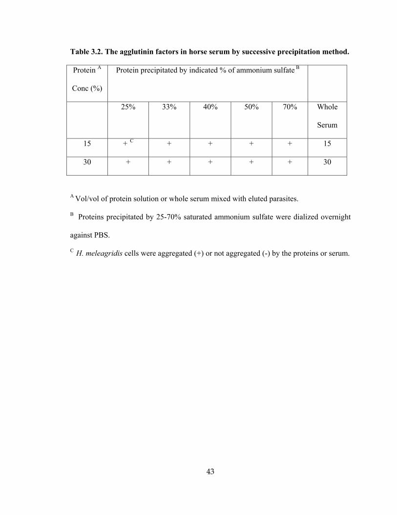
43
Table 3.2. The agglutinin factors in horse serum by successive precipitation method.
Protein A
Conc (%)
Protein precipitated by indicated % of ammonium sulfate B
25% 33%
40% 50% 70%
Whole
Serum
15 + C + + + + 15
30 + + + + + 30
A Vol/vol of protein solution or whole serum mixed with eluted parasites.
B Proteins precipitated by 25-70% saturated ammonium sulfate were dialized overnight
against PBS.
C H. meleagridis cells were aggregated (+) or not aggregated (-) by the proteins or serum.

44
Figure 3.1. Culture of Histomonas meleagridis in vitro(250X).

45
Figure 3.2. Clumping of histomonads when concentrations of horse serum are more than
15% (250X).
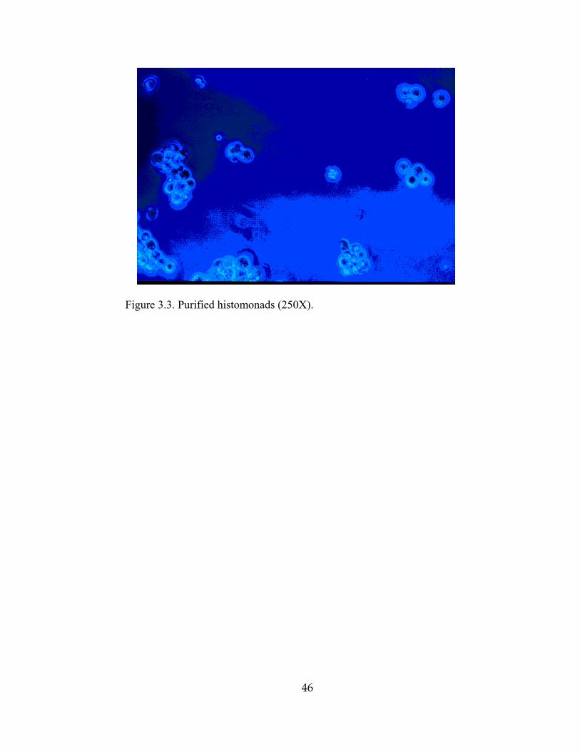
46
Figure 3.3. Purified histomonads (250X).

47
DIRECT LATERAL TRANSMISSION OF HISTOMONAS
MELEAGRIDIS IN TURKEYS
_______________________
1Jinghui Hu and L. R. McDougald. 2002. Accepted by Avian Disease.

48
1. Abstract
The lateral transmission of H. meleagridis in turkeys was studied in floor pens
without the presence of Heterakis gallinarum. Battery-reared poults (120) were transferred
at 2 wks of age to concrete-floored floor pens with fresh pine shavings litter (40/group). One
group received no exposure. In other groups, either 10% or 25% of the birds were
inoculated per cloaca with cultured H. meleagridis (200,000/bird), and placed in the pens as
seeder birds. Inoculated birds died at 10-18 days postinfection (DPI) showing typical liver
and cecal lesions of histomoniasis. Birds in the high exposure group (HE) died of
histomoniasis beginning 16 DPI, and continuing to 100% mortality by day 23 DPI. Birds in
the low-exposure group (LE) died beginning on day 19 DPI and continuing through day 31
DPI. All but one LE bird alive on day 31 DPI had severe liver and cecal lesions of
histomoniasis at necropsy. There was no evidence of histomoniasis in unexposed birds. No
cecal worms (H. gallinarum) were found at necropsy of dead birds or in unexposed birds at
the end of the experiment. Even though H. gallinarum is the only known reservoir for H.
meleagridis, these results suggest that lateral transmission of histomoniasis through a flock
can occur readily through normal contact between uninfected birds and infected birds and
their droppings in the total absence of cecal worms.
Key words: Histomonas meleagridis, turkeys, lateral transmission, histomoniasis,
blackhead disease

49
2. Introduction
Early work with blackhead disease of turkeys identified the cecal worm (Heterakis
gallinarum) as the main vector and reservoir of the causative protozoan Histomonas
meleagridis [1, 2, 3, 10]. Survival of H. meleagridis in droppings is brief [11]. However, the
infective protozoans are known to survive for long periods in cecal worm eggs, providing a
reservoir of infection from one year to another [1]. Blackhead infections result from
ingestion of embryonated cecal worm eggs or adult cecal worms, but not unembryonated
worm eggs [9]. In some areas, earthworms may consume cecal worm eggs in soil and thus
harbor the contained histomonads, transmitting the infection when the earthworm is
consumed by a turkey [6].
The infectivity of the lumen form of H. meleagridis in droppings of infected birds was
reportedly low [5] presumably because of the acidity of the crop and gizzard [4]. Lund [5]
concluded that direct bird-to-bird transmission was unimportant in birds that were well fed
and cared for. There is a general perception among scientists and poultry veterinarians that
infections arise only from ingestion of cecal worm eggs, leading some to question whether
other vectors may be involved where cecal worms cannot be demonstrated [8]. However, the
exposure to cecal worm eggs cannot explain the rapid spread of histomoniasis in turkey
flocks resulting in 50-90% mortality in a few weeks (H. L. Shivaprasaud, pers. comm. 2002;
Karen Burns, pers. com., 2002). We conducted the present study to determine whether H.
meleagridis could spread directly from infected bird to uninfected bird under practical
conditions, in the absence of Heterakis worms or other potential vectors.

50
3. Materials and methods
Experimental Animals: Day-old turkey poults (Nicholas) were obtained from a
commercial turkey hatchery and maintained until 2 weeks old in a steam-sterilized battery.
Unmedicated feed and water were provided ad libitum.
Parasites: Histomonas meleagridis were isolated from ceca of infected turkeys and
subcultured in modified Dwyer’s medium [7]. Seeder birds were exposed by cloacal
inoculation of 200,000 cells in 1 ml of culture medium via a blunt-tipped pipette. Exposed
birds were returned to the pen to commingle with the others.
Floor Pen Facility and Management: Three pens of a concrete-floored building were used.
No other animals were housed in the building during the experiment. The pens were
approximately 2.4 x 3 m, and separated by floor to ceiling chain link fencing. Black plastic
sheeting to a height of 1.2 m was used to further separate the pens. Fresh pine shaving litter
was added to a depth of 10 cm. Doors were kept closed. The study was conducted in the
winter, and the only arthropods observed were a few houseflies. The water supply (from
chlorinated municipal water) had a pH of 6.5-7. Feed was unmedicated turkey starter ration
formulated to meet National Research council (NRC) requirements. Pens were equipped
with nipple waterers and tube-type manual feeders. After transfer to pens, the birds were
given unmedicated feeder and water ad libitum.
Experimental design and procedures: The two-week old birds were divided into 3 groups
of 40 birds. Pen 1 was unexposed. In Pen 2, 4 birds (10%) were inoculated with H.
meleagridis (LE), and in Pen 3, 10 birds (25%) were inoculated with H. meleagridis (HE).
The birds were observed twice daily for morbidity and mortality. During daily care, the UE
pen was entered first, the LE pen second, and the HE pen last. Dead birds were examined for

51
presence of cecal or liver lesions of histomoniasis, and for any evidence of cecal worms. At
termination of the study at 31 DPI, all surviving birds, including UE controls, were killed
and examined for lesions of histomoniasis. Intestines were opened longitudinally and
examined grossly for infection with cecal or intestinal worms. Five handfuls of litter from
each pen were examined by scattering in a pan for any sign of arthropods.
4. Results and Discussion
The mortality of turkeys after introduction of H. meleagridis is shown in Table 4.1.
No birds died or became sick in the UE controls. Death losses from histomoniasis
began first with the inoculated birds in HE or LE pens. The inoculated seeder birds
died 10-16 DPI. Uninoculated turkeys in the (HE) group died 16-23 DPI, and all
were dead by day 23 DPI. Uninoculated turkeys in the LE group died from day 19
DPI to day 31 DPI. Yellow diarrhea was noted in the LE and HE pens. Morbidity
became common in LE and HE pens as the infections progressed. Necropsy of the
dead birds revealed lesions typical of histomoniasis in the liver and ceca. No worms
were found in ceca or intestines of dead birds. The experiment was terminated on
day 31 DPI because the surviving 11 LE birds suffered morbidity. At necropsy, 10 of
the 11 survivors had typical liver lesions and cecal lesions of histomoniasis. Only
one bird was normal. Examinations of the litter were negative for adults or larvae of
beetles or other arthropods.
These results indicated that the rapid spread of H. meleagridis within a turkey flock was
possible without the presence of cecal worms or other vectors. The birds in this study were

52
reared free from any exposure to worm eggs of any species, and without the arthropods or
invertebrates frequently found in poultry houses (hister beetles, darkling beetles,
earthworms, ants, and most flies). The only opportunity for the birds to become infected was
from usual litter pecking and coprophagy, and possibly from contamination of the feed by
infected pen-mates. The inoculum of H. meleagridis cultured in vitro contained only
histomonads and bacteria. The absence of cecal worms or other potential vectors was
confirmed by necropsy of death losses from histomoniasis, and necropsy of birds in the UE
pen at the end of experiment. Birds in the UE pen were negative for lesions of histomoniasis,
demonstrating the absence of vectors or reservoirs of infection in the experimental unit.
The feed and water were at no time interrupted during this study. Thus, previous
observations that feed deprivation was necessary to allow infection with H. meleagridis [4,5]
do not seem apropos. Even though histomonads do not survive more than a few minutes
outside the bird (in droppings), and most would be killed by acid conditions in the upper
digestive tract, it appears that sufficient numbers of organisms survive to precipitate
infection after direct ingestion. Infection of birds by this means helps to explain the rapid
dissemination of histomoniasis through commercial flocks.
When Norton et al. [8] were unable to find cecal worms associated with an outbreak of
blackhead in turkeys, they suggested that some other vector must be responsible for
spreading the infection through the flock. However, the present work suggests that the
infection could easily and rapidly disseminate after focal introduction of H. melelagridis into
the flock, not requiring all of the birds to ingest cecal worm eggs.
53
5. Reference
1. Farr, M. Further observations on survival of the protozoon parasite Histomonas
meleagridis and the eggs of the poultry nematode in the feces of infected birds.
Cornell Vet. 51:3-13. 1957.
2. Gibbs, B. J. The occurrence of the protozoan parasite Histomonas meleagridis in the
adults and eggs of the cecal worm Heterakis gallinae. J. Protozool. 9:288-293. 1962.
3. Grabill, H. W., and T. Smith. Production of fatal blackhead in turkeys by feeding
embryonated eggs of Heterakis papillosa. J. Exptl. Med. 34:647-662. 1920.
4. Horton-Smith, C., and P. L. Long. The infection of chickens (Gallus gallus) with
histomonad suspensions. Parasitol. 46:79-90.1956.
5. Lund, E. E. Oral transmission of Histomonas in turkeys. Poult. Sci. 35:900-904.
1956.
6. Lund, E. E., E. E. Wehr, and D. J. Ellis. Earthworm transmission of Heterakis and
Histomonas to turkeys and chickens. J. Parasitol. 52:899-902. 1966.
7. McDougald, L. R. and R. B. Galloway. Blackhead disease: in vitro isolation of
Histomonas meleagridis as a potentially useful diagnostic aid. Avian Dis. 17:847-
850. 1973.
8. Norton, R. A., F. D. Clark, and J. N. Beasley. An outbreak of histomoniasis in
turkeys infected with a moderate level of Ascaridia dissimilis but no Heterakis
gallinarum. Avian Dis. 43:342-348. 1999.
9. Springer, W. T., J. Johnson, and W. M. Reid. Transmission of histomoniasis with
male Heterakis gallinarum (Nematoda). Parasitol. 59, 401-405. 1969.

54
10. Tyzzer, E. E. Induced and natural transmission of blackhead in the absence of
Heterakis. J. Infectious Disease 37, 265-284. 1925.
11. Tyzzer, E. E. Studies on Histomoniasis, or blackhead infection in the chicken and the
turkey. Proc. Amer. Acad. Arts and Sci. 69:190-264. 1934.

55
Table 4.1. Mortality of turkeys after exposure to Histomonas meleagridis by infected
seeder birds in the absence of cecal worms or other vectors. Birds were unexposed
(UE), exposed to 10% seeders (LE), or exposed to 25% seeders (HE).
UE LE HE
DPI A Uninoc C Seeder B Uninoc.C. Seeder B Uninoc.C
10 0 1 0 2 0
11 0 0 0 2 0
12 0 0 0 1 0
13 0 0 0 1 0
14 0 1 0 3 0
16 0 0 0 1 D 3
17 0 0 0 - 9
18 0 2 D 0 - 3
19 0 - 2 - 8
20 0 - 7 - 3
21 0 - 2 - 1
22 0 - 2 - 0
23 0 - 7 - 3 D
25 0 - 2 - -
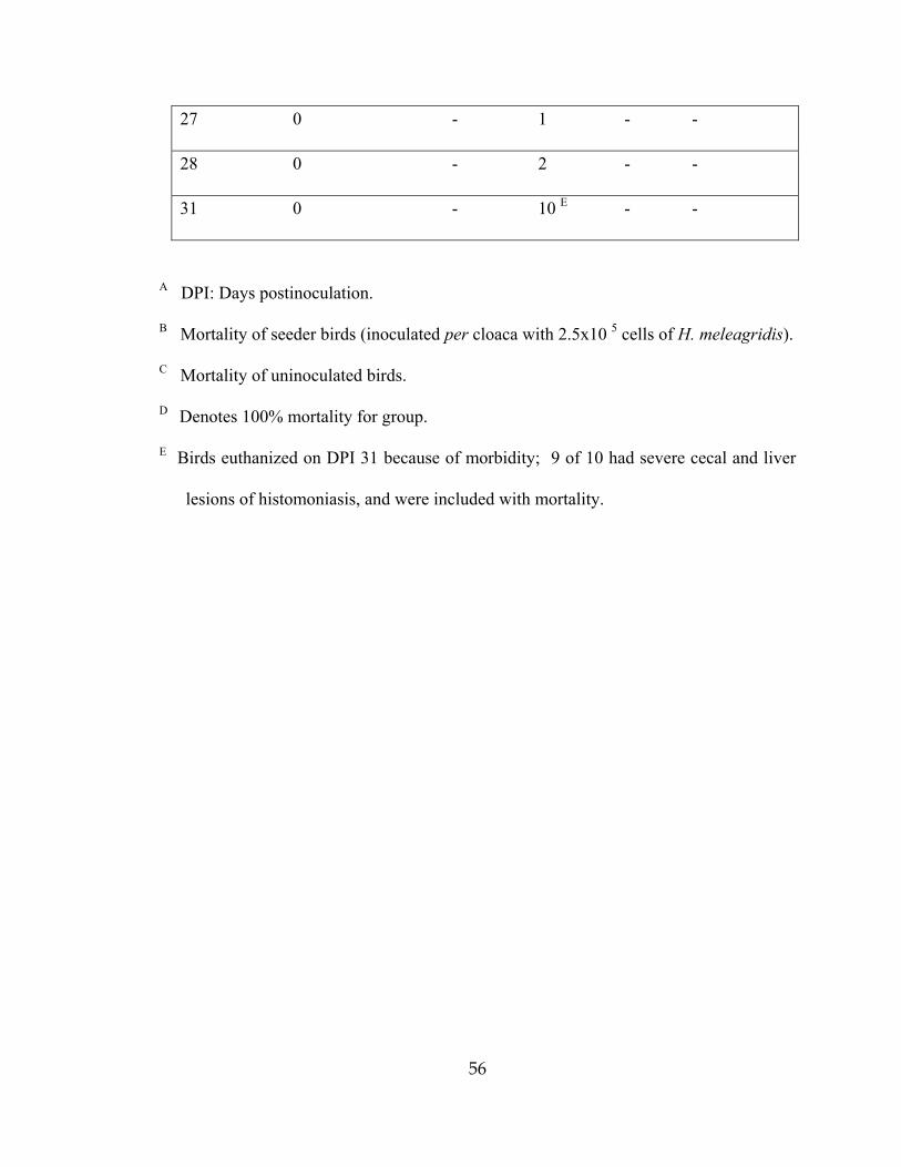
56
27 0 - 1 - -
28 0 - 2 - -
31 0 - 10 E - -
A DPI: Days postinoculation.
B Mortality of seeder birds (inoculated per cloaca with 2.5x10 5 cells of H. meleagridis).
C Mortality of uninoculated birds.
D Denotes 100% mortality for group.
E Birds euthanized on DPI 31 because of morbidity; 9 of 10 had severe cecal and liver
lesions of histomoniasis, and were included with mortality.

57
THE EFFICACY OF SOME DRUGS WITH KNOWN ANTIPROTOZOAL
ACTIVITY AGAINST HISTOMONAS MELEAGRIDIS IN CHICKENS
_______________________
1Jinghui Hu and L. R. McDougald. 2002. Submitted to Poultry Science.
58
1. Abstract
Nine known antiprotozoal drugs were tested in vitro and in vivo for activity against
Histomonas meleagridis. The nitroimidazoles dimetridazole, metronidazole, ornidazole, and
tinidazole suppressed the growth of H. meleagridis in vitro at 10 ug/ml or higher.
Paromomycin sulfate and carbadox were weakly effective within the range tested.
Quinolinol, mebendazole, diloxanide furoate, and albendazole had no demonstrable efficacy
in vitro. Drugs showing some activity were tested in young chickens inoculated with 2x10⁵
H. meleagridis/bird. Dimetridazole, metronidazole, ornidazole, and tinidazole were highly
effective at 200 ppm in feed, preventing liver lesions and reducing cecal lesions to near zero.
Paromomycin sulfate and carbadox were ineffective in vivo, with no improvement in liver or
cecal lesion scores compared to that of infected controls.
Keywords: Histomonas meleagridis, chemotherapy, antihistomonal, antiprotozoal,
dimetridazole, metronidazole, ornidazole, tinidazole, paromomycin, albendazole,
mebendazole, quinolinol, carbadox.
2. Introduction
Several products were at one time registered for prevention or treatment of blackhead
disease (histomoniasis) in the USA, including two arsenicals, two nitroimidazoles, and one
nitrofuran (McDougald, 1997b). However, recent regulatory action has removed all but
histostat (4-nitrophenyl-arsonic acid) from the market in the USA, leaving poultry producers
with no products to treat outbreaks of this disease (McDougald, 1997b). Outbreaks of
histomoniasis in chickens and turkeys continue to cause serious losses in young turkeys, and
in broiler breeder or layer pullets (Homer and Butcher, 1991; McDougald, 1997a). Previous

59
attempts at protection against blackhead by immunization were largely unsuccessful, thus it
is unlikely that vaccines will be developed (Lund, 1969). Management practices
(segregation of turkeys from chickens, disinfection of litter and soil, prevention of worm
infections, etc.) are important in preventing outbreaks, but overall have limited effectiveness.
Thus, we considered it important to test new drugs identified as having antiprotozoal
activity to identify possible candidates with antihistomonal activity.
Histomonas meleagridis, the causative agent of blackhead, is closely akin to other
amebas and flagellates (Smith, 1895, Tyzzer, 1919, 1920). Thus, compounds active against
Entamoeba or Trichomonas could be expected to have activity against H. meleagridis. From
the literature, we selected tinidazole, metronidazole (Bhopale et al., 1995), ornidazole
(Leimer et al., 1980), and diloxanide furoate (McAuley et al., 1992) as candidates. Some of
the benzimidazole anthelminthics also have antiprotozoal activity, including albendazole and
mebendazole (Katiyar et al., 1994). Paromomycin sulfate 5,7-diiodo-8-hydroxy-Quinoline
and carbadox are antibiotics or antibacterials with reported antiprotozoal activity (Barr et al.
,1994; Fournet et al., 1994). Dimetridazole, which was long used as the drug of choice for
treating blackhead outbreaks (McDougald, 1997b), was used as a postitive control in all
studies.
3. Materials and Methods
Drugs used: Dimetridazole, metronidazole, ornidazole, tinidazole, paromomycin sulfate,
mebendazole, albendazole, Diloxanide furoate, and 5,7-diiodo-8-hydroxy-Quinoline were
purchased commercially (Sigma-Aldrich, St. Louis, MO). Carbadox was supplied by Phibro
(New York, NY).

60
3.1 Experiments in vitro
Histomonas meleagridis were cultured in modified Dwyer's Medium (Medium 199 in HBSS,
85%; Chicken embryo extract, 5%; Horse serum, 10%; rice powder, 15 mg; pH 7.2)
(McDougald, 1973). The drugs were dissolved/suspended in 0.5 ml DMF or water, then
M199 was added for a concentration of 1000 µg/ml. Working dilutions of 100, 10, 1, 0.1,
and 0.01 µg/ml were prepared in culture flasks, each replicated in two flasks, with 9.5 ml
vol. Flasks were inoculated with 0.5 ml of seed culture containing approximately 2.5x 10⁵ of
H. meleagridis, and incubated at 40 C for 2 days. Parasites were counted by inverted phase
microscopy.
3.2. Experiments in vivo
Candidate compounds with significant activity in vitro were tested in chickens using a
previously established infection model (McDougald and Hu, 2001). Infective histomonads
were cultured in vitro as described above. Two-week-old broiler chickens were exposed by
intracloacal inoculation of 2 x 10⁵ H. meleagridis. Each bird also received 1x10⁴ oocysts of
cecal coccidia (Eimeria tenella). The compounds tested in vivo were dimetridazole,
metronidazole, ornidazole, tinidazole and paromomycin sulfate given in the feed at 200 or
400 ppm beginning one day before inoculation. Each treatment was replicated in 3
replicates of 10 birds each. The test was terminated 13 days postinoculation (DPI). Body wts of
birds before infection and at 13 DPI were recorded by pen unit. The liver and cecal lesions of
histomoniasis were scored at necropsy as previously described (McDougald and Hu, 2001).

61
Statistic analysis. The data were processed with General Linear Model Program of SAS.
The weight gains, liver lesion scores and cecal lesion scores were analyzed by ANOVA and
Duncan’s multiple range test for difference between means.
4. Results and Discussion
4.1. Experiments in vitro
All of the tested nitroimidazoles (dimetridazole, ornidazole, tinidazole, and metronidazole)
were highly active in vitro (Table 5.1) as shown by near complete suppression of growth of
H. meleagridis at 10 ug/ml or higher. Concentrations of 1 µg/ml or less were ineffective.
Paromomycin sulfate reduced the growth of parasites at 10 µg/ml, and was completely
effective at 100 µg/ml. Mebendazole, albendazole, Diloxanide furoate, and 5,7-diiodo-8-
hydroxy-Quinoline reduced growth of H. meleagridis at 100 µg/ml. Carbadox reduced
growth of histomonads at 100 but not 10 µg/ml. DMF had no effect on the growth of H.
meleagridis, but the use of DMF as a dissolving/suspending agent appeared to affect
observed endpoints with some compounds.
4.2. Experiments in vivo
Average wt gain of infected controls was depressed in comparison with uninfected controls
(Table 5.2). Treatment with Dimetridazole, metronidazole, ornidazole, tinidazole at 200 or
400 ppm improved wt gain relative to the infected control. Paromomycin sulfate treatment
provided no improvement in gain. The higher level (400 ppm) of tinidazole and
metronidazole, but not ornidazole, appeared to depress weights, possibly a sign of toxicity.
Liver scores were nil with dimetridazole, metronidazole, ornidazole, or tinidazole. The cecal
62
lesion scores were also nil or nearly nil with these compounds. Paromomycin sulfate was
inactive, with no reduction in liver or cecal lesion scores. Carbadox did not reduce liver or
cecal lesions of histomoniasis at 50 or 100 ppm.
These results indicated that dimetridazole, metronidazole, ornidazole, tinidazole were
highly effective in control of H. meleagridis in chickens, but paromomycin sulfate and
carbadox were ineffective at the tested levels.
Tests of compounds in vitro are probably reliable indicators of activity for selection of
candidates to test in animals. However, some compounds are active only after metabolism to
another form in the animal and would be missed with this system.
These results suggest that nitroimidazoles active against Entamoeba are also active
against Histomonas. However, compounds of other classes were inactive.
Benzamidazole anthelminthic compounds are sometimes used for prevention of blackhead
disease in chickens or turkeys (Hegngi et al., 1999). However, these compounds are not
effective against the causative organism H. meleagridis and are believed to work by killing
the cecal worm (Heterakis gallinarum) which carries the infection into cecal tissues.
Paromomycin reportedly has activity against a variety of protozoans, including H.
meleagridis (Lindquist, 1962) but had little activity against H. meleagridis in this study. The
earlier study used high levels of paromomycin in feed (1000-2000 ppm), much higher than
that used here (200-400 ppm) and higher than would be practical under commercial
conditions. The authors speculated that paromomycin might be working by reduce the
presence of bacteria necessary for pathogenicity of H. meleagridis, since they found no
improvement in efficacy with increased dosage. However, recent work suggests that
histomoniasis could not be controlled by antibiotic activity alone (Hu et al., 2002).

63
We found no useful activity of diloxanide furoate, carbadox or the quinoline compound.
Clearly, the results of testing with Entamoeba and other organisms can in some cases be
used as leads for activity against H. meleagridis, but in other cases is not a good indicator.
The poultry industry is in dire need of products for treatment or prevention of
histomoniasis in chickens and turkeys to replace dimetridazole and ipronidazole, recently de-
listed by the FDA. Considerable activity can be found among other antiprotozoal
compounds, but unfortunately the best ones are also nitroimidazoles. Other tested
antiprotozoal compounds did not possess useful activity against H. meleagridis.
5. References
1. Barr, S.C., Jamrosz, G.F., Hornbuckle, W.E., and Bowman,D.D., 1994. Use of
paromomycin for treatment of cryptosporidiosis in a cat. J. Am. Vet. Med. Assoc.
205, 1742-1743.
2. Bhopale, K. K., Pradhan, K. S., Masani, K. B., and Kaul, C. L., 1995. A comparative
study of experimental cecal amebiasis and the evaluation of amebicides. Ann. Trop.
Med. Parasit. 89, 253-259.
3. Bode, C., Schafer, C., Fukui, H., and Bode, J. C., 1997. Effect of treatment with
paromomycin on endotoxemia. Alcoholism-Clin. Exper. Res. 21, 1367-1373.
4. Fournet, A., Barrios, A.A., Munoz, V., Hocquemiller, R., Roblot, F., Cave, A.,
Richomme, P., and Bruneton, J., 1994. Antiprotozoal activity of quinoline alkaloids
isolated from Galipea longiflora, a bolivian plant used as a treatment for cutaneous
leishmaniasis. Phytotherapy Res. 8, 174-178.
64
5. Homer, B. L., and Butcher, G. D. 1991. Histomoniasis in leghorn pullets on a Florida
farm. Avian Dis. 35, 621-624.
6. Katiyar, S. K., Gordon, V. R., McLaughlin, G.L., and Edlind, T.D. 1994.
Antiprotozoal activities of benzimidazoles and correlations with beta-tubulin
sequence. Antimicrobial Agents and Chemotherapy 8, 2086-2090.
7. Hu, J. and McDougald, L. R., 2002. Effect of anticoccidials and antibiotics bn the
control of black head disease in broiler breeder pullets. J. Appl. Poultry Res. In press.
8. Leimer, R., Fermandez, F., Lotvin, B. R., Pereira, G. J., and Schenone, H., 1980.
Short-term treatment of acute intestinal amoebiasis with ornidazole. Acta Trop. 37,
266-270.
9. Lindquist, W. D., 1962. Some effects of paromomycin sulfate on blackhead in
turkeys. Am. J. Vet. Res. 23, 1053-1056.
10. Lund, E. E., 1969. Histomoniasis. Adv. Vet. Sci. Comp. Med., 13: 355-390.
11. McAuley, J. B., Herwaldt, B. L., Stokes, S. L., Becher, J. A., Roberts, J. M.,
Michelson, M. K., and Juranek, D. D., 1992. Diloxanide furoate for treating
asymptomatic Entamoeba histolytica cyst passers-14 years experience in the United
States. Clin. Infect. Dis. 15, 464-468.
12. McDougald, L. R., 1997a. Blackhead disease (histomoniasis) in chickens. Poult.
Dig. September, pp. 8-11.
13. McDougald, L. R., 1997b. Protozoa: other protozoan diseases of the intestinal tract.
in Calnek, B., Barnes, H. J., Beard, C. W., McDougald, L. R., and. Saif, Y. M. (eds.),
Diseases of poultry, 10 ed., Iowa state University Press, Ames IA., pp. 890-899.

65
14. Smith, T. 1895. An infectious disease among turkeys caused by protozoa (infectious
enterohepatitis). U.S. Dept. Agr. Bureau Animal Industry Bull. 8:1-38.
15. Tyzzer, E.E., 1919. Developmental phase of the protozoan of blackhead in turkeys. J.
Med. Res. 40, 1-30.
16. Tyzzer, E.E., 1920. The flagellate character and reclassification of the parasite
producing blackhead in turkeys, Histomonas (Gen. Nov.) meleagridis (Smith). J.
Parasitol. 6, 124-130.

66
Table 5.1. The effect of drugs on growth of Histomonas meleagridis in vitro with or
without Dimethyl-formamide (DMF).
Drugs DMF 100 ppm 10 ppm 1 ppm 0.1 ppm 0.01 ppm
Yes 0 0 1.20 12.83 8.46 Dimetridazole
No 0 0 1.98 39.11 83.17
Yes 0 0 6.17 73.46 86.96 Ornidazole
No 0 0 1.29 73.39 89.48
Yes 0 0 6.59 25.75 65.27 Tinidazole
No 0 0 69.74 85.41 94.64
Yes 0 0 24.94 37.99 68.88 Metronidazole
No 0 0 35.19 69.10 93.57
Yes 17.39 17.39 42.56 33.18 38.67 Paromomycin
No 0 1.49 71.78 91.58 75.74
Yes 40.72 36.93 40.92 44.91 43.71 Mebendazole
No 73.13 44.55 55.45 56.93 50.50
Yes 21.36 36.13 33.33 31.34 34.33 Diloxanide
furoate No 28.98 79.62 78.66 83.44 75.80
Albendazole Yes 18.99 21.05 47.60 46.00 85.35
Quinolinol Yes 10.53 19.91 27.92 40.50 72.54
Carbadox No 5.0 95.0 80.0 85.0 n.d
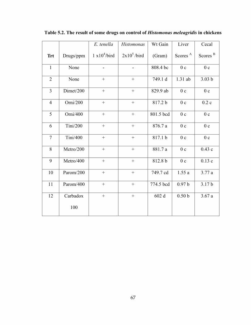
67
Table 5.2. The result of some drugs on control of Histomonas meleagridis in chickens
Trt
Drugs/ppm
E. tenella
1 x10⁴/bird
Histomonas
2x10⁵ /bird
Wt Gain
(Gram)
Liver
Scores A
Cecal
Scores B
1 None - - 808.4 bc 0 c 0 c
2 None + + 749.1 d 1.31 ab 3.03 b
3 Dimet/200 + + 829.9 ab 0 c 0 c
4 Orni/200 + + 817.2 b 0 c 0.2 c
5 Orni/400 + + 801.5 bcd 0 c 0 c
6 Tini/200 + + 876.7 a 0 c 0 c
7 Tini/400 + + 817.1 b 0 c 0 c
8 Metro/200 + + 881.7 a 0 c 0.43 c
9 Metro/400 + + 812.8 b 0 c 0.13 c
10 Parom/200 + + 749.7 cd 1.55 a 3.77 a
11 Parom/400 + + 774.5 bcd 0.97 b 3.17 b
12 Carbadox
100
+ + 602 d 0.50 b 3.67 a

68
EFFECT OF ANTICOCCIDIALS AND ANTIBIOTICS ON THE CONTROL OF
BLACKHEAD DISEASE IN BROILER BREEDER PULLETS
_______________________
1Jinghui Hu and L. R. McDougald. 2002. Journal of Applied Poultry
Research. In press.
69
1. Abstract
Broiler chicks inoculated with both Histomonas and cecal coccidia developed
moderately severe blackhead disease. Antibiotics tested at normal feed or water additive
levels had little effect on Histomonas lesions or weight gains. Bacitracin at 100, 200, or 300
g/ton reduced liver lesion scores somewhat (p<0.05), but had no other positive effects.
Apramycin at 300 ppm in water reduced liver lesion scores somewhat (p<0.05), but did not
improve cecal lesions or weight gains. Penicillin (100 ppm), chlortetracycline (100 ppm),
tylosin (110 ppm), and sarafloxacin (40 ppm), in water gave no improvement in liver or
cecal blackhead lesions. Weight gains were improved relative to infected controls with
penicillin, tylosin, or sarafloxacin treatment (p<0.05).
Five anticoccidials (salinomycin, diclazuril, nicarbazin, roxarsone, and lasalocid)
were tested at common use levels in two trials. Results were similar in both trials: Liver
lesion scores in the nicarbazin treatment were reduced (p<0.05) in comparison with controls
and other medicated groups, and the number of birds positive for liver lesions was lower
(p<0.05). Otherwise, anticoccidials had no effect on liver or cecal lesion scores or weight
gains. Control of coccidiosis by the anticoccidials (as shown by oocyst counts) varied from
product to product, but was not correlated with severity of blackhead lesions. These results
suggest that the effect of cecal coccidia on susceptibility of chickens to H. meleagridis is not
a simple function of mechanical damage to the cecal mucosa.

70
2. Introduction
Histomoniasis (blackhead disease) can cause 90-100% mortality in turkeys, but is
relatively mild in chickens [1,2]. However, recent outbreaks in broiler breeder pullets and
layer pullets in the Southeastern USA were severe, creating a significant deterrent to the
production of broiler hatching eggs and table eggs [3,4]. Outbreaks of blackhead in breeder
and layer pullets are characterized by extensive mortality, morbidity, poor uniformity, and
reduced potential for egg production. Morbidity and mortality from blackhead depends on
H. meleagridis spreading from the ceca to the liver, eventually causing destruction of that
organ. More typically, chickens suffer little liver involvement and the infection often passes
without notice. Chickens or turkeys become infected with H. meleagridis by swallowing
eggs of the cecal worm (Heterakis gallinarum). Cecal worms are widespread in poultry
houses, providing almost universal exposure of chickens. However, the reasons for the
present increase in clinical cases of histomoniasis in chickens are not clear. This increase in
clinical blackhead outbreaks comes only a few years after FDA's action barring the use of
nitroimidazole products (ipronidazole and dimetridazole) in the USA, leaving poultry
producers with no effective treatment.
We recently demonstrated that concurrent exposure to cecal coccidia (Eimeria
tenella) and H. meleagridis increased the severity of liver lesions caused by H. meleagridis
in chickens [5]. The mechanism for such interaction was not clear. Possible interactions
might include physical damage to the mucosa, a change in cecal microflora, and other
physiological or immunological factors. Blackhead disease is dependent upon the cecal
microflora for full pathogenicity [6,7].

71
Previously established interactions between H. meleagridis, bacteria, and coccidia
[5], suggest that the strategic use of anticoccidials and antibiotics could offer some relief in
outbreaks of blackhead. Because of misinformation and misplaced expectations, we felt that
it was important to test commonly used feed and water medications available to the poultry
industry, to determine whether any of these products had any significant effect on blackhead
disease. These products are not known to have significant antihistomonal activity, but might
have some indirect influence because of the complex relationship between H. meleagridis
and its host.
3. Materials and Methods
Parasites and Infections: Blackhead Parasites: A culture of H. meleagridis was isolated
from a field outbreak in commercial layer pullets in North Georgia in 1998. This isolate was
frozen in liquid nitrogen and retrieved for use by culturing in modified Dwyer's medium [8].
This medium consists of Medium 199, horse serum, chick embryo extract, and rice powder.
Thereafter, fresh cultures were isolated from young turkeys before each series of
experiments, to ensure maintenance of pathogenicity. Fresh cultures were pooled and
counted using a hemacytometer, and diluted to final working concentration in Medium 199.
The calculated dosage of H. meleagridis was inoculated into chickens per cloaca, using a
hypodermic syringe fitted with a blunt-tipped plastic pipette tip. Past experience suggested
that 10-15% of the birds do not become infected after inoculation in this way, probably
because the inoculum is voided by some birds. Liver and cecal lesions caused by H.
meleagridis were scored and recorded at necropsy, as previously described [5]. On this
scale, a score of 0 = normal, 1= minimal or barely detectable lesions, 2 = scattered but easily
72
detectable lesions, 3 = moderately severe lesions involving much of the organ, and 4 =
severe lesions involving the entire organ.
Cecal Coccidia: A field isolate of E. tenella (Athens, GA, 1997) was prepared for
use as previously described [2]. The oocysts were inoculated per os in 1 ml of water using a
10 ml syringe equipped with a plastic pipette tip. Our model, developed in previous
experiments [5], consisted of oral gavage with 10,000 oocysts of E. tenella on the same day
as intracloacloacal inoculation with 2.5 x 10⁵ cells of H. melelagridis. In some experiments,
a separate infected control was maintained, with only H. meleagridis inoculation.
3.1 Experimental Animals, Test Medicaments, and Experimental Design
Experimental Animals: Broiler chicks (Peterson x Arbor Acres) were hatched from
breeder flocks maintained at the UGA poultry research farm and used straight run. No
vaccinations were given. Unmedicated feed and water were given ad libitum. Chicks were
housed in steamed-cleaned, stainless steel cages until assignment to treatments at 2 weeks
old. Thereafter, birds were housed in cages with stainless steel droppings trays, feeders and
waterers (Petersime).
Feed Additives and water medication: Commercial premixes of anticoccidials and
antibiotics were used to prepare feeds with diclazuril, 1 ppm; lasalocid, 90 ppm; nicarbazin,
125 ppm; roxarsone, 50 ppm; salinomycin, 60 ppm; bacitracin, 50, 100, 200, or 300 ppm.
Apramycin wetable powder was mixed into tap water to produce 75, 150, or 300 ppm.
Penicillin, chlortetracycline, tylosin or sarafloxacin were given in the drinking water at 100,
100, 110, or 40 ppm, respectively.
Experiment I. Effect of bacitracin on blackhead disease in chickens: Four levels of
bacitracin (50, 100, 200, and 300 g/ton respectively) were tested in 3 replicates of 10 birds
73
each. Controls were unmedicated, uninfected (UUC), and unmedicated, infected with both
parasites (IIUC). Medicated feeds were given where indicated, from 14 days of age through
termination when birds were 28 days old. The birds were inoculated with H. meleagridis
and E. tenella at 15 days of age. Pen weights were recorded at inoculation and at necropsy at
28 days of age. Liver and cecal lesions were scored and recorded at necropsy [5].
Experiment II. The Effect of apramycin on blackhead disease in chickens. The
experiment included 5 treatments with 3 replicates of 10 birds each. The concentrations of
apramycin in water were 75, 150, and 300 g/ton, respectively. Controls were UUC and
IIUC. Medicated water was given from 14 to 28 days of age. The birds were inoculated
with H. meleagridis and/or E. tenella at 16 days of age. Pen weights were recorded on days
16 and 28. Liver and cecal lesions were scored and recorded at necropsy.
Experiment III. The effect of antibiotics on blackhead disease in chickens. This test
included 7 treatments with 3 replicates of 10 birds each. Antibiotics administered in water
were penicillin, 100 ppm; chlortetracycline, 100 ppm; tylosin, 100 ppm; and sarafloxacin,
40 ppm. Controls included UUC, unmedicated, inoculated with H. meleagridis only (IUC),
and inoculated with Histomonas + E. tenella (IIUC). Medicated water was given from 14 to
28 days of age. Birds were inoculated with at 15 days of age. Pen weights were recorded at
inoculation and at termination. Liver and cecal lesions were scored and recorded at necropsy
on day 28.
Experiment IV. The effect of anticoccidials on blackhead disease in chickens. Two
identical trials were conducted to evaluate effects of anticoccidials on histomoniasis. Eight
treatments included diclazuril, 1.0 ppm, salinomycin, 60 g/ton, lasalocid, 90 g/ton,
nicarbazin, 0.0125%, and roxarsone, 45.4 g/ton. Controls included UUC, IUC, and IIUC.
74
Each treatment was replicated in 3 groups of 10 birds each in two identical trials. Medicated
feeds were given from 14 days of age through termination when birds were 28 days old. The
birds were inoculated with H. meleagridis and E. tenella at 15 days of age. Pen weights were
recorded at inoculation and at necropsy at 28 days of age. Liver and cecal lesions were
scored and recorded at necropsy. In the second trial only, fecal samples were collected from
each pen on DPI 6-8, and oocysts of E. tenella were counted in aliquots using a McMaster
chamber.
3.2. Statistical analysis
Weight gains and lesion scores were analyzed by ANOVA for differences at P<0.05,
followed by Duncan's multiple range test to separate means, using the general Linear Model
program on SAS.
4. Results and Discussion
4.1. The effects of bacitracin on H. meleagridis in broiler chicks
Severe cecal lesions of blackhead developed in all birds exposed to H. meleagridis
(Table 6.1). Weight gains were significantly depressed in all the infected birds (p<0.05)
compared to UUC, and were not improved by bacitracin treatment. Liver lesion scores were
significantly reduced in groups treated with100, 200, 300 g/ton of bacitracin, but not in birds
treated with 50 gram/ton of bacitracin. However, cecal lesion scores and the number of birds
with liver lesion were unchanged in treated birds, in comparison with the infected control.
4.2. The effects of apramycin on H. meleagridis in broiler chicks
Weight gains were depressed in comparison with the UUC (P<0.05) in all H.
meleagridis-infected groups, regardless of treatment (Table 6.2). Cecal lesion scores were

75
numerically reduced at high concentrations of apramycin. Liver lesion scores were
significantly reduced by treatment with 300 ppm of apramycin, but not with other treatment
levels.
4.3. The effects of other antibiotics on H. meleagridis in broiler chicks
None of the tested antibiotics (penicillin, chlortetracycline, tylosin, or sarafloxacin)
was effective in reduction of liver or cecal lesions caused by blackhead (Table 6.3).
However, three products (penicillin, tylosin, or sarafloxacin) improved weight gains
(p<0.05), in comparison with IIUC (Treatment 3).
4.4. The effect of anticoccidials on H. meleagridis in broiler chicks
In Trial 1, weight gains were significantly (p<0.05) depressed in all Histomonas-
infected groups relative to the UUC, regardless of treatment (Table 6.4). Liver lesions in the
nicarbazin treatment were significantly reduced (p<0.05), compared with the IIUC, and the
number of birds positive for liver lesions was significantly lower. There was no change in
average liver lesion scores in other medicated groups compared to the IIUC. Cecal lesion
scores caused by blackhead were not reduced by any drug treatment. Oocysts produced were
not counted in Trial 1.
In Trial 2, weight gains were significantly (P<0.05) depressed in all blackhead-
infected birds in comparison with the UUC (Table 6.5). As in the first trial, liver lesions
were reduced by nicarbazin treatment (p<0.05), in comparison with the IIUC, but not in
comparison with other drug treatments. Cecal lesions caused by blackhead were not reduced
by anticoccidial treatment. Even though coccidial oocyst counts were greatly reduced by
diclazuril, salinomycin, roxarsone, and nicarbazin, only nicarbazin appeared to have an
effect on the number and severity of blackhead lesions in the liver.

76
Synergism of blackhead by Coccidia: Liver lesions caused by H. meleagridis were
significantly more severe in control birds inoculated with both H. meleagridis and E. tenella
(IIUC), as compared with those receiving only H. meleagridis (IUC)(Tables 6.3, 6.4, 6.5).
This confirms earlier observations on the synergism of these infections [5].
Although blackhead disease is not new in chicken flocks, and once accounted for up
to 5.5% of annual diagnostic cases in some laboratories [9], clinical outbreaks in chickens in
the USA and other countries have increased significantly in the past several years [3,4,10].
The reasons for this increase are not understood, but could include depressed immune
function, a change in the adaptation of H. meleagridis to chickens, or a change in the
virulence of H. meleagridis [5]. These infectious protozoans are carried inside the eggs of
the cecal worm (Heterakis gallinarum), and may lie dormant for many years [1,11]. We
believe that most commercial chicken facilities are contaminated with these worms, and
thus, with H. meleagridis [12]. Therefore, the reason for increases in clinical outbreaks in
chickens, with classical liver and cecal involvement, high mortality, and morbidity, are
unlikely to be a result of more widespread exposure.
Antibiotics might be expected to help control blackhead disease because of indirect
effects. Bacteria are required for the full pathogenicity of H. meleagridis in chickens and
turkeys, as shown by studies in bacteria-free and contaminated birds [6,7]. A combination of
Escherichia coli, Pseudomonas aeruginosa, Streptococcus, and Clostridium perfringens was
most successful and consistent in providing the necessary conditions for development of
blackhead disease in gnotobiotic chickens [7]. The interaction of bacteria and H. meleagridis
is less complicated in turkeys, where contamination of gnotobiotic birds with E. coli alone is
sufficient to support development of blackhead disease [6]. The results of our studies suggest
77
that while some of the tested products partly reduced the liver lesion scores or improved gain
when used at high levels, none was curative for blackhead disease. These studies suggest that
the tested antibiotics did not adequately control the bacterial species required for
pathogenicity of H. meleagridis. In future work, antibiotics could be administered from day
one, as a means of further altering the bacterial flora.
Anticoccidials highly effective against E. tenella could be expected to aid in
prevention of blackhead disease because of the demonstrated interaction between E. tenella
and H. meleagridis [5] and between E.adenoeides and H. meleagridis in turkeys [7]. Most
anticoccidials are known to allow some development of coccidia, even with sensitive
laboratory strains. An exception to this is diclazuril, which under laboratory conditions can
completely prevent development of E. tenella [13]. Thus, it was surprising that diclazuril
had no effect on development of blackhead in two trials, even though it was shown in Trial 2
that no oocysts were produced in infected birds. Nicarbazin, the only product showing an
effect on the liver lesions in both trials, was also highly effective against coccidia, but
allowed some oocysts to be produced. It is possible that nicarbazin has some innate
antihistomonal activity because of its chemical similarities to known antihistomonal
compounds (nitro-phenolic ring structure), but this has not been recognized in previous tests.
Thus, the mechanism for nicarbazin's interference with the synergistic effects of coccidia on
blackhead remains unclear.
Roxarsone is used in broilers in combination with other anticoccidials as an aid in
controlling E. tenella [14], or in combination with an anticoccidial vaccine [15]. Because of
its chemical similarity to other organic arsenic products with known antihistomonal activity,
roxarsone is sometimes given in the water during blackhead outbreaks in chickens. In this
78
study, we saw no evidence that roxarsone was of benefit in prevention of blackhead at the
normal feed additive level of 45 g/ton.
At present, no products are approved for treatment of blackhead infections in
chickens or turkeys. Only nitarsone (4-nitrophenylarsonic acid, 0.0175%) is used in feed for
prevention of blackhead. Exposure of chickens to the cecal worm vector (Heterakis
gallinarum) can be reduced by frequent worming with effective products [12].
5. Conclusions and Applications:
1) Some of the tested antibiotics had beneficial effects on weight gains in infected
chickens. However, none was effective in reducing the lesions or other pathology of
H. meleagridis at FDA-approved levels.
2) High levels of some products (300 g/ton of bacitracin or 300 ppm of apramycin)
reduced liver lesion scores, but had no other positive benefits.
3) Lesions and other signs of blackhead were not reduced by most anticoccidials,
regardless of the effectiveness of control of cecal coccidia.
4) Nicarbazin (0.0125% in feed) gave significant reduction in liver lesion scores in two
trials. The reason for this improvement was not clear.
6. References and Notes
1. Lund, E. E., 1969. Histomoniasis. Adv. Vet. Sci. Com. Med. 13: 355-390.
2. McDougald, L. R., 1997. Protozoa: other protozoan diseases of the intestinal tract.
Chapter 34, in B. Calnek, H. J. Barnes, C. W. Beard, L. R. McDougald, and Y. M.

79
Saif (eds). Diseases of poultry, 10 ed. Iowa state university press, Ames, IA. pp. 890-
899.
3. Homer B.L. and G. D. Butcher, 1991. Histomoniasis in leghorn pullets on a Florida
farm. Avian Dis. 35: 621-624
4. McDougald, L. R., 2000. New developments in research on blackhead disease in
chickens. World Poultry. 16(8):46-48.
5. McDougald, L.R. and J. Hu, 2001. Blackhead disease (Histomonas meleagridis)
aggravated in broiler chickens by concurrently infection with cecal coccidiosis
(Eimeria tenella). Avian Dis. 45: 307-312.
6. Bradley, R. E. and W. M. Reid, 1966. Histomonas meleagridis and several bacteria
as agents of infectious enterohepatitis in gnotobiotic turkeys. Exp. Parasitol. 19: 91-
101.
7. Springer, W. T., J. Johnson, and W. M. Reid, 1970. Histomoniasis in gnotobiotic
chickens and turkeys: Biological aspects of the role of bacteria in the etiology. Exp.
Parasitol. 28: 383-392.
8. McDougald, L., and R. B. Galloway, 1973. Blackhead disease: In vitro isolation of
Histomonas meleagridis as a potentially useful diagnostic aid. Avian Dis. 17: 847-
850.
9. Reid, W. M., 1962. Now it's blackhead in broilers. Broiler Business Magazine,
Watt Publishing Co., Mount Morris, IL. Sept., pp 20-21.
10. Banerjee, P.S. and C. L. Yadav, 2002. Infectious enterohepatitis in chickens in Uttar
Pradesh. Indian J. Anim. Sci. 71:30-31

80
11. Reid, W. M., 1967. Etiology and dissemination of the blackhead disease syndrome in
turkeys and chickens. Exp. Parasitol. 21, 249-275.
12. Hegngi, F. N., J. Doerr, T. S. Cummings, R. D. Schwartz, G. Saunders, A. Zajec, C.
T. Larsen, and F. W. Pierson., 1999. The effectiveness of benzimidazole derivatives
for the treatment and prevention of histomoniasis (blackhead) in turkeys. Vet.
Parasitol. 81: 29-37.
13. McDougald, L. R., G. F. Mathis, and B. P. Seibert, 1990. Anticoccidial efficacy of
diclazuril against recent field isolates of Eimeria from commercial poultry farms.
Avian Dis. 34:911-915.
14. McDougald, L. R., J. M. Gilbert, L. Fuller, A. Rotibi, M. Xie, and G. Zhu, 1992.
How much does roxarsone contribute to coccidiosis control in broilers when used in
combination with ionophores? J. Appl. Poultry Res. 1:172-179.
15. Chapman, H.D. and S.H. Fitzcoy, 1996. Effect of roxarsone and bacitracin
methylene disalicylate on the development of immunity to Eimeria in broilers given
a live coccidiosis vaccine. Poultry Sci. 75:1488-1492.

81
Table 6.1. Effect of bacitracin on blackhead disease in broiler-type chickens inoculated
with Histomonas meleagridis and Eimeria tenella.
Treatment Bacitracin
(g/ton) A
Infect.B Av Weight
gain (g)
Average
Liver lesionC
Ave
Cecal LesionC
1 0 -- 764.8 a 0 c 0 c
2 0 HC 615.9 b 0.93 a 3.66 ab
3 50 HC 604.2 b 0.51 ab 3.44 b
4 100 HC 621.7 b 0.34 bc 3.76 a
5 200 HC 647.1 b 0.31 bc 3.71 a
6 300 HC 635.9 b 0.44 bc 3.69 a
A Bacitracin methylene disalicylate mixed into the feed and given continuously from one
day prior to infection until termination.
B The infection model consisted of 104 oocysts of E. tenella (given by oral gavage) and 2.5 x
105 cultured cells of H. meleagridis (by intracloacal inoculation) given on the same day. H=
H. meleagridis, C= Coccidia (E. tenella)
C Lesions of histomoniasis, scored on a scale of 0-4 at necropsy on DPI 13-14 as previously
published [5]. Means with similar superscript letters are not significantly different (p<0.05).

82
Table 6.2. The effect of apramycin on blackhead disease in broiler-type chicks
inoculated with Histomonas meleagridis and Eimeria tenella.
Treatment InfectionA
ApramycinB
ppm Weight gain
(g)
Average Liver
Lesions C
Average
Cecal
Lesions C
1 ---- 0 857.5 a 0.00 c 0.00 b
2 HC 0 769.6 b 1.37 a 3.74 a
3 HC 75 702.8 c 0.77 ab 3.63 a
4 HC 150 786.8 b 1.18 ab 3.56 a
5 HC 300 701.0 c 0.47 bc 3.40 a
A The infection model consisted of 104 oocysts of E. tenella given by oral gavage (C) and
2.5 x 10⁵ cultured cells of H. meleagridis by intracloacal inoculation (H) given on the same
day.
B Apramycin mixed into the water and given continuously from one day pre-infection.
C Lesions of histomoniasis, scored on a scale of 0-4 at necropsy on DPI 13-14 as previously
published [5]. Means with similar superscript letters are not significantly different (p<0.05).

83
Table 6.3. Effects of antibiotics on blackhead disease in broiler-type chicks inoculated
with Histomoniasis meleagridis and Eimeria tenella.
Treatment
InfectionA
Antibiotics in
water B
Wt gain (g) Avg. Liver
Lesions C
Avg. Cecal
Lesions C
1 -- None 715.8 a 0.00 b 0.00 c
2 H-- None 609.3 bc 0.58 a 3.67 ab
3 HC None 586.8 c 0.82 a 3.63 ab
4 HC Penicillin
100 ppm
648.5 b 0.78 a 3.44 ab
5 HC Chlortetracycli
ne 100 ppm
617.9 bc 1.14 a 3.68 ab
6 HC Tylosin
110 ppm
658.2 b 0.91 a 3.90 a
7 HC Sarafloxacin
40 ppm
655.8 b 1.06 a 3.21 b
A The infection model consisted of 2.5 x 10⁵ cultured cells of H. meleagridis (H) by
intracloacal inoculation and/or 104 oocysts of E. tenella given by oral gavage (C) given on
the same day.
B Antibiotics mixed into the water and given continuously from one day preinfection.
C Lesions of histomoniasis, scored on a scale of 0-4 at necropsy on DPI 13-14 as previously
published [5]. Means with similar superscript letters are not significantly different (p<0.05).
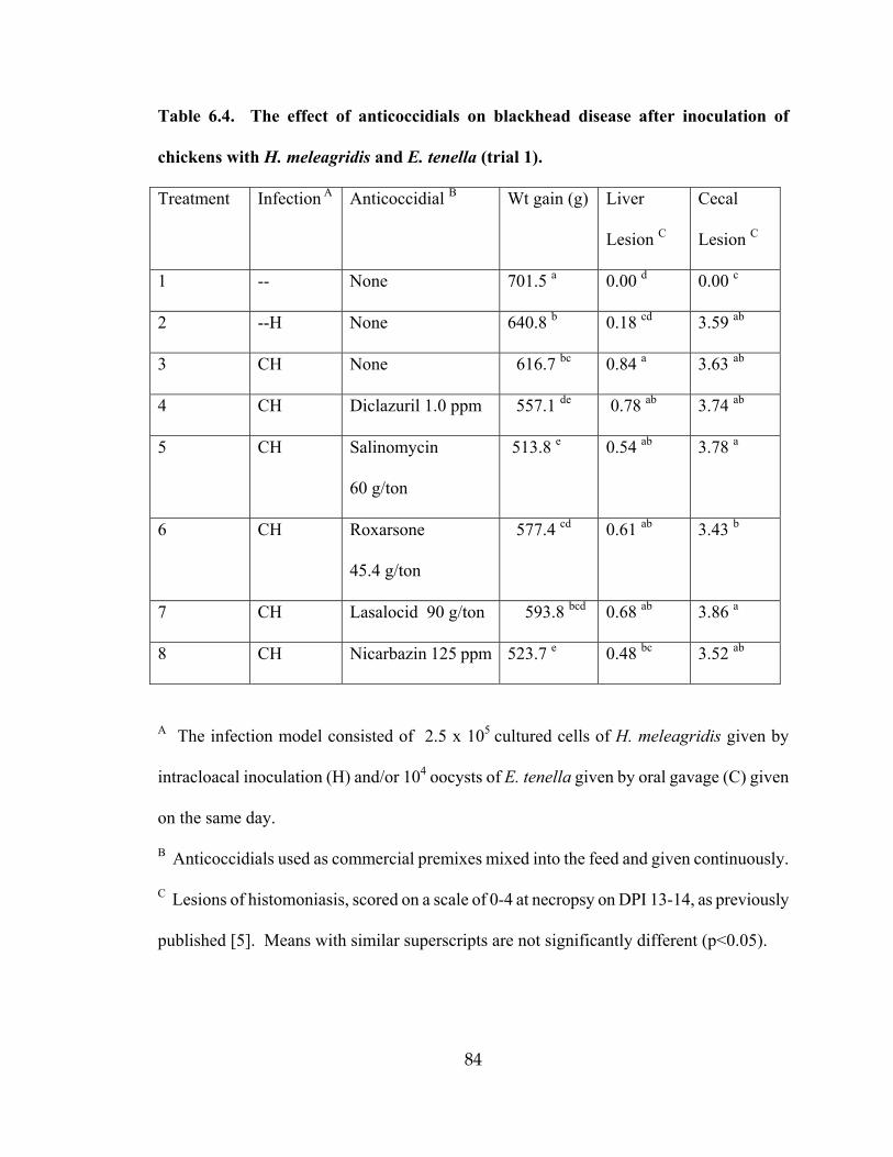
84
Table 6.4. The effect of anticoccidials on blackhead disease after inoculation of
chickens with H. meleagridis and E. tenella (trial 1).
Treatment Infection A Anticoccidial B Wt gain (g) Liver
Lesion C
Cecal
Lesion C
1 -- None 701.5 a 0.00 d 0.00 c
2 --H None 640.8 b 0.18 cd 3.59 ab
3 CH None 616.7 bc 0.84 a 3.63 ab
4 CH Diclazuril 1.0 ppm 557.1 de 0.78 ab 3.74 ab
5 CH Salinomycin
60 g/ton
513.8 e 0.54 ab 3.78 a
6 CH Roxarsone
45.4 g/ton
577.4 cd 0.61 ab 3.43 b
7 CH Lasalocid 90 g/ton 593.8 bcd 0.68 ab 3.86 a
8 CH Nicarbazin 125 ppm 523.7 e 0.48 bc 3.52 ab
A The infection model consisted of 2.5 x 10⁵ cultured cells of H. meleagridis given by
intracloacal inoculation (H) and/or 104 oocysts of E. tenella given by oral gavage (C) given
on the same day.
B Anticoccidials used as commercial premixes mixed into the feed and given continuously.
C Lesions of histomoniasis, scored on a scale of 0-4 at necropsy on DPI 13-14, as previously
published [5]. Means with similar superscripts are not significantly different (p<0.05).

85
Table 6.5. The effect of anticoccidials on blackhead disease after inoculation of
chickens with H. meleagridis and E. tenella (Trial 2).
Treatment
Infection 1
Anticocci-
dial 2
Wt gain
(g)
Oocyst
(x106)
Liver
Lesion 3
Cecal
Lesion 3
1 -- None 701.5 a ND 0.00 d 0.00 c
2 --H None 640.8 b ND 0.18 cd 3.59 ab
3 CH None 616.7 bc ND 0.84 a 3.63 ab
4 CH Diclazuril
1.0 ppm
557.1 de 0 0.78 ab 3.74 ab
5 CH Salinomycin
60 g/ton
513.8 e 21 0.54 ab 3.78 a
6 CH Roxarsone
45.4 g/ton
577.4 cd 285 0.61 ab 3.43 b
7 CH Lasalocid
90 g/ton
593.8 bcd 44 0.68 ab 3.86 a
8 CH Nicarbazin
0.0125%
523.7 e 4 0.48 bc 3.52 ab
1 The infection model consisted of "C" (104 oocysts of E. tenella given by oral gavage)
and/or "H" (2.5 x 10⁵ cultured cells of H. meleagridis by intracloacal inoculation) given on
the same day.
2 Anticoccidials as commercial premixes mixed into the feed and given continuously.
86
3 Lesions of histomoniasis, scored on scale of 0-4 at necropsy on DPI 13-14 as previously
published [5]. Means with similar superscript letters are not significantly different (p<0.05).

87
BLACKHEAD DISEASE (HISTOMONAS MELEAGRIDIS) AGGRAVATED
IN BROILER CHICKENS BY CONCURRENT INFECTION
WITH CECAL COCCIDIOSIS (EIMERIA TENELLA)
____________________________
L. R. McDougald and Jinghui Hu. 2001. Avian Dis. 45:307-312.

88
1. Abstract
The effect of concurrent cecal coccidiosis infections on severity of Histomonas
meleagridis (blackhead disease) in chickens was investigated in a series of experiments.
Cecal lesions from H. meleagridis were severe in all inoculated control groups and did not
appear to be affected by the introduction of Eimeria tenella infections. However, the
severity of liver lesions and number of birds positive for liver lesions of H. meleagridis
increased significantly with the presence of E. tenella. The increase was similar when 103 or
104 oocysts of E. tenella were given, and was the same when oocysts were given at the same
time as H. meleagridis or 4 days prior. The liver lesions increased directly as doses of H.
meleagridis increased from 7.5x 10³ cells to 30, 100 or 300 x 103, respectively, when E.
tenella were given along with H. melelagridis, but not when H. meleagridis were given
alone. Administration of a 3x dose of live coccidiosis vaccine, containing very low levels of
E. tenella, gave a small but significant boost to liver lesions, but at a much lower level than
that observed with larger doses of E. tenella.
The positive relationship between infections of cecal coccidiosis and H. meleagridis
in chickens suggests that such dual exposure may contribute to increased clinical outbreaks
of blackhead disease in chickens under field conditions.
2. Introduction
The flagellate Histomonas meleagridis is well known among parasitologists and poultry
veterinarians for its spectacular pathogenicity and virulence in turkeys, causing a disease
known as blackhead (6). Infectious cells carried inside eggs of the cecal worm Heterakis
gallinarum start in the ceca and spread to the liver, then birds may die from hepatic failure.

89
Other gallinacious birds, particularly chickens, serve as carriers because they harbor the
cecal worm, but rarely suffer the potentially fatal liver involvement with H. meleagridis. In
recent years an increase in field cases of clinically severe blackhead disease in chickens,
particularly leghorn and broiler breeder pullets (4, 5), has forced a reexamination of factors
affecting pathogenicity of H. meleagridis. Some possible causes of this increase are (a)
increased virulence of the organism, (b) increased susceptibility of chickens (either through
genetic changes or immunodeficiency), and (c) interaction of Histomonas with other
infections.
There is no literature on interaction of blackhead disease with other infectious disease
agents, except for a 1973 study on the effect of blackhead disease on the severity of
coccidiosis, wherein H. meleagridis given on or before inoculation with E. tenella appeared
to interfere with oocyst production (1). Thus, a series of experiments was designed to
determine whether concurrent cecal coccidiosis infections could increase the number or
severity of liver lesions in chickens.
3. Materials and Methods
Histomonas meleagridis cultures for experimental infections: A medium consisting of
chick embryo extract, horse serum, medium 199, and rice powder (8) was used to culture H.
meleagridis from a field outbreak of blackhead in Leghorn pullets. The culture was re-
isolated from turkeys prior to use in each experiment to maintain pathogenicity. Fresh
cultures were pooled and counted using a hemacytometer, and diluted to final working
concentration made in Medium 199 (Gibco), pH 7.4, 1 ml/dose. Inoculations of H.
meleagridis were made via the cloaca with a hypodermic syringe, using a blunt-tipped

90
plastic pipette tip in place of the needle. Past experience suggested that 10-15% of birds do
not become infected after inoculation in this way, probably because most of the inoculum is
voided.
Cecal Coccidia: A field isolate of E. tenella (Athens, GA, 1997) was prepared for use as
previously described (7). The oocysts were inoculated per os in 1 ml volume using a 10 ml
syringe and a plastic pipette tip in place of the needle.
Birds and facilities: Broiler chicks (Peterson x Arbor Acres) were hatched from breeder
flocks maintained at the UGA poultry research center and used straight run. No vaccinations
were given. Unmedicated feed and water were given ad libitum. Chicks were housed in
steam-cleaned, stainless steel cages until used at 10 or14 days of age.
Experimental Design: Treatments were replicated in 3 cages of 10 birds each, except for 6
replicates in the test of the live coccidiosis vaccine.
Concurrent infection with H. meleagridis and E. tenella: This experiment was repeated
three times. As the results were similar, the results of only one experiment are given.
Histomonas inoculations (105/bird) were given when birds were 14 days old. Oocysts of
Eimeria tenella were inoculated on day 10 (4 days prior to Histomonas), or on day 14. Also,
E. tenella were given at 103/bird, or at 104/bird. Controls were a) uninoculated, b) inoculated
with E. tenella only (103 or 104 oocysts), or c) inoculated with H. meleagridis only. The test
was terminated when birds were 24 days old, with record of weight gains (per cage) and
mortality. Birds were killed by cervical dislocation for necropsy. Liver and cecal lesions
were scored on a scale of 0-4 (Table 7.1). This is a subjective system, but is considered
reasonably accurate when used repeatedly by the same workers.
91
Titration of H. meleagridis inoculum in presence of E. tenella: Histomonas inoculations
were given on day 14 in graded doses of 7.5x10³, 30x103,, 105,or 3x10⁵ cells per dose, with
and without concurrent inoculation with 104 oocysts of E. tenella. Controls included a)
uninoculated or b) inoculated with E. tenella only. The test was terminated when birds were
28 days old, with record of weight gains (per cage), mortality, and necropsy of all birds.
Liver and cecal lesions were scored as above.
Interaction of H. meleagridis with a coccidiosis vaccine: A commercial live coccidiosis
vaccine (Immucox, Wingo, Inc., Watkinsville, GA), containing small numbers of live
Eimeria oocysts, was given to day-old chicks at the recommended level or at 3x. The
treatments included Histomonas inoculation with the vaccine at the two levels, Histomonas
alone, and uninoculated controls, each replicated in 6 cages of 10 birds each. The test was
terminated when birds were 28 days old, with data collected as in the other experiments,
above.
Statistical Evaluation: Data were analyzed, where appropriate, using the GLM procedures
on SAS (SAS Institute, Cary, NC), for significance at P<0.05.
4. Results
Interaction of H. meleagridis with E. tenella: Cecal lesions indicated that 79% of the birds
inoculated with H. meleagridis (Treatments 2, 4, 5, 7, and 8) became infected, causing a
reduction in weight gain of approximately 19% (Table 7.2). Inoculation with E. tenella
reduced final weights by 6 or 11%, but coccidian lesions were not apparent at the time of
necropsy. Cecal lesions from blackhead averaged 4.00 in positive birds of infected groups,
regardless of whether birds received one or both inocula. Liver lesions averaged 0.25 in

92
birds inoculated only with H. meleagridis, while those receiving concurrent E. tenella
inoculations averaged 0.97-1.14. The number of birds with liver lesions was higher in
groups inoculated with both parasites (69-91%) compared with those receiving only H.
meleagridis (19%). Severe liver lesions (scores of 3 or 4) were found only in birds
inoculated with both parasites. The results did not appear to be correlated with the time or
intensity of inoculation with E. tenella. Increased severity of liver lesions in the presence of
coccidia was highly significant in all combination exposure groups (P<0.05). A similar
pattern of infection and severity of liver lesions was seen in all three replicate studies.
Titration of H. meleagridis inoculum in the presence of E. tenella: Inoculation with H.
meleagridis produced cecal lesion scores averaging 3.47-3.83, with 73-93% positive at
necropsy (Table 7.3). Weight gain was reduced about 15% by H. meleagridis infections.
Neither the number of birds infected nor the severity of cecal lesions appeared to correlate
with the number of H. meleagridis in the inoculum in mono-infections. Liver lesions
averaged 0, 0.20, 0.19, or 0.23 for birds receiving 7.5x10³, 3x10⁴, 105 or 3x10⁵ histomonads,
respectively, with 0, 17, 13, or 17% positive for liver lesions. However, when E. tenella was
given along with H. meleagridis, the liver lesions increased significantly (P<0.05) in direct
proportion to inoculum dose (Table 7.3). The liver lesions averaged 0.14, 0.41, 0.47, or 0.86
for birds receiving 7.5x10³, 3x10⁴, 105 or 3x10⁵ histomonads, respectively, with 10, 30, 37 or
53% of birds positive for liver lesions.
Interaction of H. meleagridis with coccidiosis vaccine: Infection with H. meleagridis
caused cecal lesions averaging 3.4-3.7, with 90B92 percent of birds positive (Table 7.4).
Weight gain was reduced about 18% by blackhead infection. Liver lesions averaged 0.23 in
birds receiving only H. meleagridis, compared with 0.35 for those with H. meleagridis plus a
93
coccidiosis vaccine and 0.45 (Significant at P<0.05) for those receiving H. meleagridis plus
a 3x dose of vaccine . The number of birds positive for liver lesions was 13% with only H.
meleagridis, 25% with H. meleagridis plus the vaccine, and 32% in those receiving the 3x
dose of vaccine.
5. Discussion
Concurrent infections with E. tenella and H. meleagridis in chickens caused
increased severity of liver lesions and an increase in the percentage of birds that suffered
liver lesions. These effects were noted when coccidia were given at the same time or 4 days
prior to inoculation with H. meleagridis. Blackhead lesions were not more severe when 104
oocysts of E. tenella were given, as opposed to 103 oocyts. Liver infections were more
severe as the dose of H. meleagridis was increased from 7.5x10³, to 3x 10⁴, 105 or 3x 10⁵
cells. The severity of lesions was directly correlated with inoculation dose in birds receiving
both parasites.
Cecal lesions were severe in all groups inoculated with H. meleagridis, regardless of
the level of inoculation or the presence of E. tenella. Oral inoculation with a 3x dose of a
commercial live coccidiosis vaccine (containing fewer than 100 E. tenella/dose) also
enhanced number and severity of blackhead lesions in the liver when given at elevated doses,
although at a much lower level than higher doses of coccidia. This effect was considered
minor, in comparison with the results of the other experiments.
The interaction of cecal coccidiosis with blackhead disease could be expected
because of the disruption of cecal mucosa by E. tenella, perhaps making it easier for H.
meleagridis in the cecal tissues to enter the bloodstream and spread to the liver. Blackhead

94
can be transferred from bird to bird via whole blood collected from the veins draining the
ceca of infected turkeys (9). Thus, we could expect that other diseases or toxins that disrupt
the mucosa might also affect the spread of blackhead in the blood supply of the intestinal
tract, accounting for more field reports of severe infections in chickens. However, if this
was the only mechanism of interaction we might expect more severe liver lesions where
higher doses of coccidia were given, but this was not observed.
Because of the well known complexity of the interaction of H. meleagridis with
intestinal bacteria, the present observations could have other explanations. Previous work
with germfree birds has shown that H. meleagridis requires the presence of certain species of
bacteria for expression of pathogenicity in turkeys and chickens (10, 11). Coccidiosis is
known to cause major increases in the intestinal population of Clostridium perfringens and a
decrease in Bacterioides sp. (2).
A recent report suggests that cecal worms (Heterakis gallinarum), the carrier of H.
meleagridis, is widespread and common on broiler breeder pullet farms (3). Coccidia are
also prevalent in poultry(6,7), providing a high potential for the interaction of these two
diseases. However, the significant effects of coccidiosis on the course of H. meleagridis in
chickens do not completely explain the spectacular outbreaks of blackhead disease often
reported from commercial layer and breeder pullet flocks. Extensive liver lesions, high
morbidity, and mortality of 10-15% reported in young pullets suggest a rapidly developing
disease of high virulence. H. meleagridis isolated from such outbreaks and tested in our
laboratory show ordinary affinity for chickens (Unpublished observations). Immuno-
depression from viral infections such as Marek=s disease virus, chicken anemia virus, and
infectious bursal disease, which are commonly diagnosed in poultry, may contribute to the
95
field outbreaks. These viruses are responsible for an increased incidence and severity of
other diseases where T-cell function is important for primary and secondary protection.
Thus, for future research, to explain the increase in clinical blackhead disease reported from
commercial poultry it may be important to focus on the interaction mucosal damage, changes
in cecal microflora, and immunodeficiency produced by viral infections.
6. References
1. Chappell, L. R. 1993. The effect of Histomonas meleagridis on the development of
Eimeria tenella. J. Parasit. 59: 637-643.
2. Dykstra, D. D. and Reid, W. M. 1978. Monensin, Eimeria tenella and effects on the
bacterial populations in the ceca of gnotobiotic chickens. Poultry Sci. 57: 398-402.
3. Hegngi, F. N., Doerr, J., Cummings, T. S., Schwartz, R. D., Saunders, G., Zajac, A.,
Larsen, C. T., and Pierson, F. W. 1999. The effectiveness of benzimidazole
derivatives for the treatment and prevention of histomoniasis (blackhead) in turkeys.
Vet. Parasitol 81: 29-37.
4. Homer, B. L. and Butcher, G. D. 1991. Histomoniasis in leghorn pullets on a
Florida farm. Avian Dis. 35: 621-624.
5. Luma, G. L., Sander, J. E. Fuller, L., and McDougald, L. R. 1999. Histomoniasis in
commercial broiler-breeder flocks in northeast Georgia. Poultry Science, 78
(Suppl):117.

96
6. McDougald, L. R. 1997. Other protozoan diseases of the intestinal tract. Chapter
34 in B. W. Calnek, H. J. Barnes, C. W. Beard, L. R. McDougald and Y. M. Saif,
eds. Diseases of Poultry, 10th edition, Iowa State University Press, Ames, IA, pp.
890-899.
7. McDougald, L. R., Fuller, L., and R. Mattiello. 1997. A survey of coccidia on 43
poultry farms in Argentina. Avian Dis. 41: 923-929.
8. McDougald, L. R. and Galloway, R. B. 1973. Blackhead disease: In vitro isolation
of Histomonas meleagridis as a potentially useful diagnostic aid. Avian Dis. 17:
847-850.
9. McGuire, W. C. and Morehouse, N. F. 1958. Blood-induced blackhead. J. Parasit.
44: 292-296.
10. Reid, W. M. 1967. Etiology and dissemination of the blackhead disease syndrome
in turkeys and chickens. Exp. Parasitol. 21: 249-275.
11. Springer, W. T., Johnson, J., and Reid, W. M. 1970. Histomoniasis in gnotobiotic
chickens and turkeys: Biological aspects of the role of bacteria in the etiology. Exp.
Parasitol. 28: 383-392

97
Table 7.1. Subjective visual lesion scoring system for blackhead disease (Histomonas
meleagridis) in chicken ceca and livers.
________________________________________________________________________
Organ Score Observations
Cecum 0 Normal cecal mucosa and lumen contents
1 A few scattered small lesions visible from serosal or mucosal
surfaces, no blood or thickening of mucosal wall
2 Lesions prominent but discreet, some bleeding and/or
thickening of mucosa, lumen contents fairly normal
3 Cecum enlarged, opaque yellow appearance, walls thickened,
lesions confluent, blood or caseous abnormal lumen contents;
the entire cecum is involved
4 Cecum distended, yellow, dense caseous cores, cecal walls
thickened and fragile
Liver 0 Normal
1 A few small foci on surface of liver. Lesions are off-white
and variable in appearance
2 5-10 small foci, or 3-5 larger lesions/lobe
3 Lesions numerous, but much normal tissue still remaining
4 Lesions numerous throughout, coalescing, no normal tissue
_________________________________________________________________________
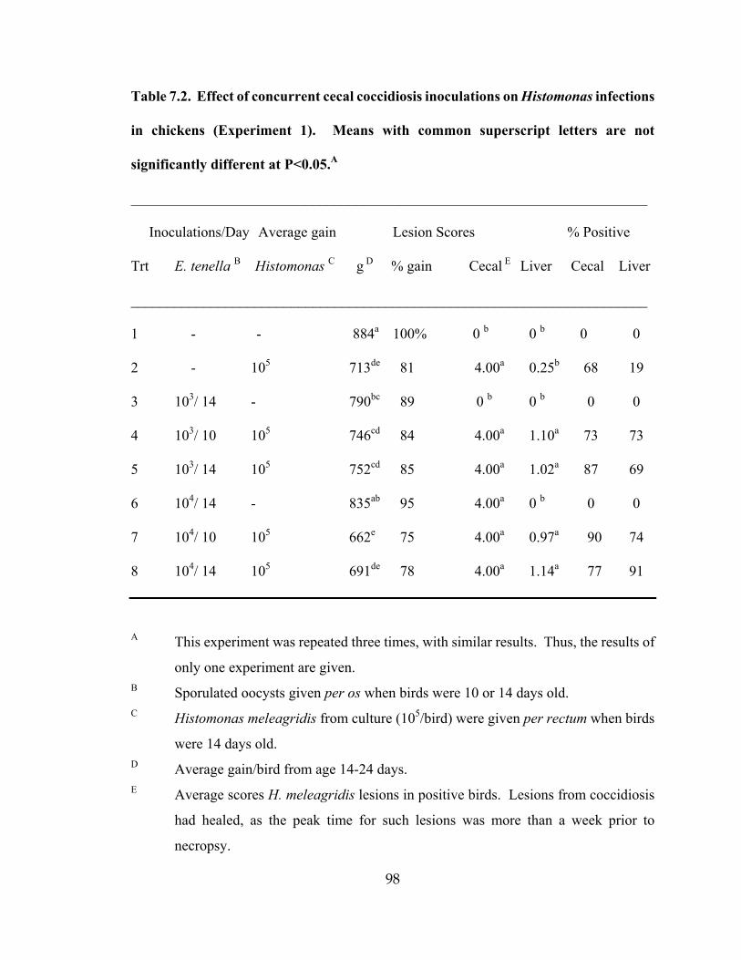
98
Table 7.2. Effect of concurrent cecal coccidiosis inoculations on Histomonas infections
in chickens (Experiment 1). Means with common superscript letters are not
significantly different at P<0.05.A
_______________________________________________________________________
Inoculations/Day Average gain Lesion Scores % Positive
Trt E. tenella B Histomonas C g D % gain Cecal E Liver Cecal Liver
_______________________________________________________________________
1 - - 884a 100% 0 b 0 b 0 0
2 - 105 713de 81 4.00a 0.25b 68 19
3 103/ 14 - 790bc 89 0 b 0 b 0 0
4 103/ 10 105 746cd 84 4.00a 1.10a 73 73
5 103/ 14 105 752cd 85 4.00a 1.02a 87 69
6 104/ 14 - 835ab 95 4.00a 0 b 0 0
7 104/ 10 105 662e 75 4.00a 0.97a 90 74
8 104/ 14 105 691de 78 4.00a 1.14a 77 91
A This experiment was repeated three times, with similar results. Thus, the results of
only one experiment are given.
B Sporulated oocysts given per os when birds were 10 or 14 days old. C Histomonas meleagridis from culture (105/bird) were given per rectum when birds
were 14 days old. D Average gain/bird from age 14-24 days.
E Average scores H. meleagridis lesions in positive birds. Lesions from coccidiosis
had healed, as the peak time for such lesions was more than a week prior to
necropsy.

99
Table 7.3. Titration of infective dose of Histomonas meleagridis in the presence and
absence of concurrent cecal coccidiosis infections (Experiment 3). Data entries with
similar superscript letters are not significantly different at P<0.05.
Inoculations Average gain Lesions % Positive
birds
Group E.
tenella A
Histomon
as B
g C % Cecal D
Liver Cecal Liver
1 B B 691a 100 0 c 0 d 0 0
2 + B 669ab 97 0.2 c 0 d 0 0
3 B 7.5 594 c 86 3.47 b 0 d 77 0
4 B 30 571 c 83 3.77 ab 0.20 c 83 17
5 B 100 618 bc 89 3.62 ab 0.19 c 73 13
6 B 300 550 c 80 3.73 ab 0.23 c 87 17
7 + 7.5 623 90 3.73 ab 0.14 c 73 10
8 + 30 597 bc 86 3.83 a 0.41 b 80 30
9 + 100 573 c 83 3.70 ab 0.47 b 87 37
10 + 300 595 c 86 3.6 ab 0.86 a 93 53
A Sporulated oocysts (104 /bird) given per os when birds were 10 or 14 days old.
B H. meleagridis cells (x 10 3/bird) from culture given per rectum to 14-day-old birds.
C Lesions primarily from H. meleagridis. Occasional light coccidiosis lesions.
D Average gain/bird from age 14-24 days.

100
Table 7.4. Effect of concurrent administration of live coccidiosis vaccine on severity of
Histomonas meleagridis infections in chickens. Data entries within columns with
similar superscript letters are not significantly different at P<0.05.
__________________________________________________________________________
Inoculations Average gain Lesion ScoreD % Positive
Trt VaccineA HistomonasB g C % Ceca Liver Ceca Liver
__________________________________________________________________________
1 B B 681a 100 0 b 0 c 0 0
2 B + 556b 82 3.5a .23b 92 13
3 1x + 579b 85 3.4a .35ab 90 25
4 3x + 583b 86 3.7a .45a 92 32
__________________________________________________________________________
A A commercial live vaccine containing low levels of Eimeria tenella (cecal coccidia)
and other species, given to day-old chicks at the recommended level (1x) or at three
times the recommended level (3x).
B H. meleagridis from cultures. Given per rectum at 105/bird at 14 days.
C Lesion scores graded subjectively on a scale of 0-4, for blackhead lesions.
D Average gain per bird calculated from day of inoculation to termination
(days 14-28).

101
CONCLUSION
1. Coccidiosis aggregated pathogenicity of histomoniasis in chickens. Cecal lesions did
not appear to be affected by the introduction of Eimeria tenella infections. However,
the severity of liver lesions and number of birds positive for liver lesions of H.
meleagridis increased significantly with the presence of E. tenella. The positive
relationship between infections of cecal coccidiosis and H. meleagridis in chickens
suggests that, under field conditions, such dual exposure may contribute to increased
clinical outbreaks of histomoniasis in chickens.
2. The lateral transmission of H. meleagridis in turkeys was studied in floor pens in the
absence of the carrier cecal worm Heterakis gallinarum. These results suggested that
lateral transmission of histomoniasis through a turkey flock can occur readily through
normal contact between infected birds and uninfected birds and their droppings in the
total absence of cecal worms.
3. Five anticoccidials, six antibiotics, and nine antiprotozoals drugs known from
literature were tested for their efficacy against Histomonas meleagridis in chickens.
None of 5 anticoccidials (salinomycin, diclazuril, nicarbazin, roxarsone, and lasalocid)
and 6 antibiotics (bacitracin, apramycin, penicillin, chlortetracycline, tylosin,
sarafloxacin) had significant efficacy in control of histomoniasis. Out of 9
antiprotozoals, only nitroimidazoles (dimetridazole, metronidazole, ornidazole, and
tinidazole) suppressed growth of H. meleagridis in vitro and in vivo.

102
4. A new phenomenon discovered during the culture of H. meleagridis was that cultured
H. meleagridis were readily agglutinated by horse serum at concentrations of 15% or
higher. The agglutinating factor (AF) was active when serum regardless of whether
serum was heat-inactivated, and was present in several batches of sera tested.
Histomonads were purified via AFfrom in vitro culture.