jaw locking after maxillofacial trauma - journal |...
TRANSCRIPT
106
Jaw locking after maxillofacial trauma
david B. Kamadjaja and r. SoesantoDepartment of Oral and Maxillofacial SurgeryFaculty of Dentistry Airlangga UniversitySurabaya - Indonesia
abstract
Thepurposeofthisreportistopresenttwocasesofjawlockingwithtwodifferentetiologies.Incase#1,jawlockingoccured5.5monthsafterasurgicalreductionandinternalfixationonthefracturedmaxillaandmandible.Someplainradiographicx-rayweremadebutfailedtogiveadequateinformationinestablishingthecauseoftrismus.Thethreedimensionalcomputedtomography(3D-CT)wasfinallymadeandabletohelpguidethepre-operativediagnosisandtreatment.Two-stepsgaparthroplastyweredonecomprisingagaparthroplastyleadingtoacceptableoutcome.Anadultpatientincase#2withahistoryoftraumaathischildhoodandbird-likefaceapprearanceclinically,wasunabletoopenthemouthsincethetimeofaccident.Thepatientwasdiagnosedwithbilateralankylosisoftemporomandibularjoints.Oneside(right)gaparthroplastywasdoneandresultedinnormalmouthopening.
Key words:trismus,Zygomafracture,Gapatrthroplasty,Threedimensionalcomputedtomography(3D-CT),Birdfaceappearance
Correspondence: David B. Kamadjaja, c/o: Bagian Ilmu Bedah Mulut, Fakultas Kedokteran Gigi Universitas Airlangga. Jln. Mayjend. Prof. Dr. Moestopo no. 47 Surabaya 60132, Indonesia. e-mail: [email protected]
introduction
Facial injuries are most commonly associated with falls, violence, motor vehicle accidents and sport-related trauma. Mandible, middle third area of the face, and temporomandibular region are frequently involved in such facial injuries. The complexity of the temporomandibular joint (TMJ), and its anatomical proximity to other craniofacial structures makes its diagnosis and treatment especially challenging.1
Facial fractures in children are relatively uncommon, it is probably due to the elasticity of pediatric skeleton. With respect to facial fractures, mandible is the second most frequently fractured bone, after nasal bone fracture. Hall in 19722 reported 20.7% incidence of mandible fracture in 495 patients younger than 14 years of age. Carroll etal.2 in 1987 studied 268 facial fractures, of which 26.5% were mandibular fracture. The involvement of condylar process is found higher than seen in the adult, ranging from 40% to 60%.3,4
The actual growth of the mandible occurs at the mandibular condyle and along the posterior surface of the ramus.5 Therefore it is understandable that fracture of the head of the condyle in the growing period may lead to joint ankylosis and might interfere with the mandibular growth and development. In delayed treatment of condylar fracture some potential complications might occur including the followings: 1) malocclusion; 2) temporomandibular joint dysfunction; 3) ankylosis, and 4) mandibular growth disturbances,
The middle third of the face consists of nasoethmoidal complex, maxilla and smaller bones attached to it together
with malar complex. It lies between the mandible and the cranial cavity and calvarium, in particular the frontal and sphenoid bones.1
Treatment of the midface and mandible fractures might result in unsatisfactory outcome in terms of aesthetic and function such as facial deformity and jaw movement restriction.
Fracture of the zygoma complex has a specific clinical symptom i.e. facial bone flattening. Flattening of the side of the face was noted in 57% of isolated zygomatic arch fractures in a study by ellis et al.2 Accompanying the zygomatic arch fractures might be trismus due to impingement of fractured segment on temporal muscle. This finding was noted in 45% of isolated zygomatic arch fractures.2
Post-fracture trismus, known as limitation of mouth opening which presented clinically in various degrees, from difficulty to inability in opening the mouth, may be associated with several conditions. The reason often cited for post-fracture trismus is impingement of the translating coronoid process of the mandible by the displaced zygomatic fragments.3 Trismus can also be caused by fractures of facial or mandible in which bone fragments have displaced and rotated and restricted the mandible movements. Problem of joint movements might also arise from bony ankylosis which is usually formed between the distal end of the fractured mandibular condyle and the surrounding bones.
Fractures of zygomatic compartments or complex and those involving temporo-mandibular joint are the most common types of fracture which are much related to the jaw movement problem, especially in severe trauma, in

107Kamadjaja and Soesanto: Jaw locking after maxillofacial trauma
which the fractured zygomatic complex is depressed in the direction of the coronoid process of the mandible and impairs the jaw movements.
In patients suspected with condylar fracture, a series of plain x-ray projections consisting of posteroanterior skull view, two lateral oblique, and Towne’s view are usually sufficient in supporting the diagnosis, but in other cases it might be difficult to adequately evaluate the cause of trismus. Chayra etal.5 found that panoramic radiograph had a higher accuracy in detecting all types of mandible fractures in 92%. In mid facial injury, the panoramic film gives little value, therefore a maxillofacial radiograph series become necessary.2 In case of trismus presenting in complex fractures of maxilla and mandible, plains radiographic projection might be difficult in finding the causes of the trismus.
With the advent of newer imaging techniques, such as computed tomography, identification of complex maxillofacial fractures can now be done with ease. Computed tomography (CT) has largely been supplanted in diagnosis of maxillofacial as it yields excellent bony detail of the facial skeleton in multiple views.2 Revolutionary progress of the CT technique which has been developed from two into three-dimension image, called 3D-CT, may represent the section of the patient that is scanned. This
leads to performing profile to profile for the entire image reconstruction process. It can be used as the best means for establishing the diagnosis and surgical guidance in maxillofacial traumas.
cases
Case #1: A-49 year old woman visited the Department of Oral and Maxillofacial Surgery at Dr. Soetomo General Hospital Surabaya asking for a treatment due to her trismus. The patient had a history of trauma caused by car traffic accident. The patient was seated just behind the driver and she had been thrown forward by the force to the driver chair. She underwent her first surgical treatment in a district hospital shortly after the accident. She also had a history of wearing intermaxillary fixation for 2 months post-operatively.
Clinically she appeared with midfacial flattening which was a characteristic sign of post-bilateral zygomatic bone fractures. Multiple fractures in the mandible, including the condylar fracture, were also suspected. Intraoral examination showed severe trismus, the maxillary and mandibular teeth were seen in occlusion but not in the proper alignment. The mandibular and maxillary arches were not in good relationship (Figure 1).
MALALIGNMENT LENGKUNG MANDIBULA DAN MAKSILA
figure 1. The patient presented with midface flattening and severe trismus (left); Intraorally, both mandible and maxilla arches collapsed medially.
figure 2. Preoperative posteroanterior skull x-ray (left); Water’s projection taken post-operatively (right).

108 Dent. J. (Maj. Ked. Gigi), Vol. 40. No. 3 July-September 2007: 106-113
figure 3. Panoramic view showing fixed paramedian fracture and right condylar fracture with the condylar head being displaced medially.
figure 4. Submentovertex radiograph showing the coronoid process free from bone obstacles (left); PA skull x-ray showing right condylar neck fracture, but no obvious bony ankylosis seen (center); Lateral skull view presenting unclear joint situation (right).
A B
DC
figure 5. Three D (CT) in series: (A) Occipital view shows clearly the position both mandible joints and coronoid processes. Both coronoid processes free from bone obstacles, the right condyle fractured and displaced, ankylosis is present. The left condyle slightly displaced laterally. (B) Anterior view: fractured of the left zygoma and median site of the mandible and fixed with miniplates. The mandible arch is repositioned in wider form. (C) The right joint displaced and attached to the arcus zygoma and (D) The left joint displaced outside from the glenoid fossae.

109Kamadjaja and Soesanto: Jaw locking after maxillofacial trauma
figure 6. Mouth opening ten days after surgery of right joint a gap arthroplasty and relapse 3 month afterward (left); Five days after left joint gap arthroplasty, the patient was able to open her mouth more easily.
figure 7. Panoramic radiograph showing an abnormal bone growth in both condyles areas and ankylosis on the right joint; the left condyles showing a gap between fossae and condyle indicating a pseudo arthrosis.
figure 8. Preoperative trismus (left); normal mouth opening three days after surgery.
110 Dent. J. (Maj. Ked. Gigi), Vol. 40. No. 3 July-September 2007: 106-113
Some pre and post-operative plain x-ray films were available at her presentation in our clinic, but they were not able to give adequate information as to point out the primary cause of the trismus. The gross bone fractures patterns could be seen in the pre-operative antero-posterior skull film showing multiple mandibular fractures of comminutive type between the midline and the right paramedian region. Water’s projection made after the surgery showed a bilateral zygomatic fracture involving the left fronto-zygoma suture. This multiple facial bone fractures had been treated surgically, internal fixation using miniplates osteosynthesis
were applied. The left zygoma was fixed with 2 miniplates over the left zygomatico-frontal and zygomatico-maxilla regions. The comminutive type of fracture on the mandible at the right paramedian region had been fixed with X-shape miniplate osteosynthesis (Figure 2).
Few plain x-rays were subsequently made which are: orthopantomogram (Figure 3), submentovertex, posteroanterior and lateral skull film (Figure 4) and yet unable to detect the primary cause of the existing trismus. In panoramic film the right condylar head of the mandible was clearly seen to displace out from the glenoid fossa and
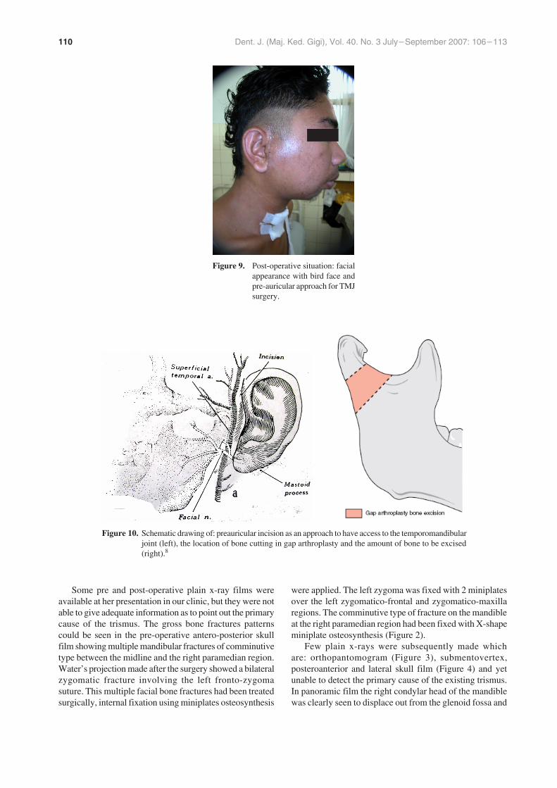
figure 10. Schematic drawing of: preauricular incision as an approach to have access to the temporomandibular joint (left), the location of bone cutting in gap arthroplasty and the amount of bone to be excised (right).8
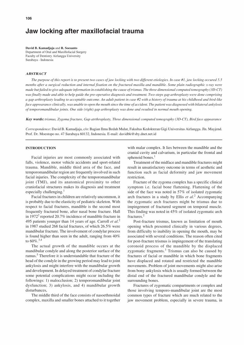
figure 9. Post-operative situation: facial appearance with bird face and pre-auricular approach for TMJ surgery.

111Kamadjaja and Soesanto: Jaw locking after maxillofacial trauma
was trapped medial to the ascending ramus of the mandible, but it did not seem to interfere with the joint movements.
The pre-operative schenario was made based on the clinical situation and the results of the plain imagings. The patient has sustained multiple trauma on her left body, shown by the fracture of the left fibula and radius bones, and some facial scars over the region of left lateral upper eye lid and lower lip. These clinical findings might indicate that the left site of the body including the face was subjected to blunt force during the accident. The chronology of the facial trauma suggested that before the face hit the object she had in reflex turned her head slightly to the left, therefore the mandible sustained fractures in the region of midline and right paramedian. The comminutive type of fracture, the lateral displacement of both ascending ramus, and the alteration of mandibular arch indicated that a powerful force must have struck this part of the mandible.
The mandible including the alveolar process and the teeth on both sides seemed to have displaced medially resulting in a narrowed mandible arch. The possible rationale to explain this condition is as follows. The fracture on the right parasymphysis region of the mandible may have caused the mandible to displace laterally. As some bone loss may have occurred on that part of the mandible, the effort to forcedly reposition the two segments in an end to end approximation have resulted in the mandible assuming an awkwardly narrow arch and the upper part of the mandible on both sides collapsing toward median line.
A series of three dimensional-Computed Tomogram was finally made to help establish the diagnosis of trismus. The result of 3D-CT showed that both condylar processes were fractured at the level of condylar neck in which the condylar heads were displaced laterally out from the glenoid fossa. It was clearly seen that bony ankylosis was formed in the region of the posterior part of the right zygomatic arch (Figure 5).
Case #2: A 24 year old man visited Department of Oral and Maxillofacial Surgery at Dr. Soetomo General Hospital Surabaya asking for a treatment to his trismus. Patient was unable to open his mouth since his childhood. The parent explained a history of trauma when he was 7 year old. Clinical evaluation showed that the patient had a bird-face profile indicating a hypoplastic mandible and an old scar was noted on his chin. Intraorally, the patient present with severe trismus, whereas the occlusion was normal (Figure 8). Panoramic showed old bilateral condylar fractures, the joints were seen to be covered with a massive bone which had grown irregulary replacing the curvature of both condylar articular surfaces. The right joint was seen to be more severely ankylotic compared to the left site as a thin radiolucency was seen surrounding the enlarged left joint (Figure 7). The surgery of gap arthroplasty was planned initially on the right joint as the suspected side of the cause of the trismus keeping in view of left gap arthroplasty if the mouth opening was not adequately obtained after the right arthroplasty.
case management
Case #1: The surgery was performed under general anesthesia. As the patient was unable to open her mouth, two options of intubation techniques were planned, fiber optic-guided intubation or tracheostomy. The fiber optic guided intubation was chosen and performed successfully.
Two step surgical treatments were planned. A gap arthroplasty of the right joint would be done first, if this surgery and post-operative physiotherapy cannot achieve the normal mouth opening, a gap arthroplasty of the left condyle is considered. Surgery for jaws realignment was also offered to the patient, but the patient refused this option as the primary concern was only the jaw locking.
After the right gap arthroplasty had been done, refracturing in the median part of the mandible was done to realign the mandible arch. Unfortunately, this part of the mandible had healed up well and the two fragments were fixed and united in new position, therefore surgical reconstruction for mandible realignment was ceased. The mandible was repositioned and fixed with 2 miniplates. Two Heister’s mouth opener were used to force the mouth to open and maintained for 24 hours with rubber mouth gag to avoid relapse of jaw locking. Post-operative physiotherapy exercise was done to help the patient achieve a maximum mouth opening.
Until one month after right joint gap arthroplasty, the patient was able to open her mouth, but 3 months after the surgery the jaw locking recurred (Figure 6). Left joint clicking and slight pain during mouth opening were reported. Reevaluation of the 3 D-CT brought us to the decision to do a left joint gap arthroplasty. Intra-operative direct visualization showed the left condylar neck had displaced laterally out from its fossae and formed ankylosis with the surrounding bony structure. Condylectomy was done and a normal mouth opening was directly achieved.
Immediately after the left joint surgery the patient was able to open her mouth much wider compared to before the surgery, allowing for a direct normal jaw function (Figure 6). Physiotherapy exercise was continued to overcome the masticatory muscles problem due to jaw locking which had been lasting since almost 1 year previously.
Case #2: The general anesthesia was performed via tracheostomy cannulation. A pre-auricular approach was used. The surgery of gap arthroplasty was done initially on the right joint as the suspected side of the cause of the trismus. After the joint had been osteotomized, using a mouth spreader the mouth was successfully forced to open, therefore the gap arthroplasty on the left joint was not done. An allograft material of premilen mesh graft was interposed into the gap to prevent it from developing re-ankylosis. A rubber mouth gag was placed between upper and lower teeth for 48 hours to avoid recurrence of the trismus due to masticatory muscle contraction. Five days after the surgery the patient was able to open his mouth normally (Figure 8 and 9).

112 Dent. J. (Maj. Ked. Gigi), Vol. 40. No. 3 July-September 2007: 106-113
discussion
In the treatment of facial trauma detailed observation of the facial skeleton situation is considerably important. The facial bones and mandible should be clearly observed from part to part to determine their involvement in the fracture. Occlusion should be used as primary guidance in establishing the diagnosis of facial and mandibular bones displacement as well as in the surgical reduction and fixation of the bones. Besides occlusion, the form of jaw arch can also be used as an important guidance during the procedure of jaws alignment if both jaws are found to be mal-aligned. In severe mandible fracture, reconstruction can be done by holding such anatomical landmarks as: the form of mandible arch, tooth to tooth contact, the curve of Spee, and the alignment of the bony structure in the body of the mandible.6
In severe maxillofacial trauma, post-operative complications may be in the form of two major problems, i.e. aesthetic and functions. The aesthetic problem is the most common complication especially when the middle third of the face is involved in. The most frequent aesthetic problem is flattening of the face due to the depression of malar bones or the whole middle third of the face as a result of Le Fort III fracture. Malocclusion and jaw locking or trismus are two major functional problems which may occur after surgical correction of maxillofacial fractures.
Case #1 presented unsatisfactory result of surgical correction of maxillofacial fractures where both post-operative aesthetic and functional problems existed. It is considered a difficult clinical situation in that both the maxilla and the mandible, as well as both the zygomatic bones, were involved in the injuries. In addition to flattening of the patient’s midface, the abnormal arch of the mandible has contributed to the altered appearance of the lower third of the face. The mandible has not been repositioned to its anatomical arch, the normal tubular form. This may be caused by the loss of anatomical landmarks due to some bone loss in the anterior region of the mandible. The effort to reposition the mandible using the occlusion guidance has made the mandible to assume an abnormally wider form on its inferior part and narrower arch on its superior or alveolar part (Figure 1).
In single jaw fracture, the healthy jaw can be used as a template during the surgical reconstruction. Simultaneous surgical treatment of facial and mandibular fractures might cause a mal-alignment due to the loss of anatomical landmark orientations as the normal mandible can no longer be used as a template for guiding the reconstruction procedure.
In a fresh jaw bone fracture, anatomical reposition of the bone fragments can usually be achieved but in other cases the bone ends may not be well aligned. Nevertheless, non-anatomical reduction of bone approximation is usually still acceptable so long as the occlusion is obtained.Such a situation was encountered in the first case.
In our opinion the surgical approach ofbottom to top as suggested by Fonseca7 is the advisable method which should be used when both jaws are involved in a severe facial trauma. Mandible reconstruction can be achieved relatively more easily than maxilla therefore mandible reconstruction should be done first. The maxilla can then be corrected following the template given by the mandible arch.
In the treatment of mandible fracture, application of arch bars is usually very helpful in achieving bone alignment, the displaced bone fragments will return to its anatomical alignment relatively easily after the arch bar has been applied. If some difficulty is encountered in the effort to realign the bone segments, bone chips or foreign body materials in the fracture line should be suspected. In consolidated fracture lines, removal of the fragments should be done to achieve an anatomical alignment of the mandible.
The jaw locking presented in this case was initially suspected to be caused either by: the fractured zygoma bone being rotated in counter clockwise in left zygoma or clockwise in the right zygoma restricting the movement of the mandible in the region of coronoid processes, or the fractured right condyle having restricted normal mandibular movement. According to the series of plain imagings both considerations were not supported in determining the primary cause of the existing trismus. A series of three dimensions computed tomography (3D-CT) could eventually determine the cause of jaw locking. It was found that the trismus is caused by ankylosis over the right joint which was formed by the displaced condylar neck laterally consolidating with the surrounding bone in the area of the right zygoma bone. Both coronoid processes were seen to be free from impingement by the displaced zygomas.
A gap arthroplasty at the right side was done as the initial step. The preauricular incision (Figure 10) was chosen in this case because it: provides good access to the temporomandibular joint area, is cosmetically acceptable, and minimal post-operative complications. In the procedure of gap arthroplasty some amount of bone at the condylar neck region was removed to create a gap between the ankylotic condyle and the distal segment of the mandible. This was done by making two horizontal bone cuts 1-1.5 cm apart just below the joint and the piece of bone was removed (Figure 10). Care should be taken not to damage the internal maxillary artery which runs below the neck of the conydle. Since a gap is created the procedure is known as gap arthroplasty. The left gap arthroplasty alone resulted in an acceptable mouth opening. The recurrence of jaw locking three months post operatively indicated that there existed some restriction of the movement at the left joint as well which may not be as severe as the right side. The left gap arthroplasty showed some bony ankylosis formed by the displaced right condylar neck and the surrounding bone. The unpleasant thing was that the patient had to go through two surgeries. If the right gap arthroplasty had not been able to open the mouth then we would have performed the

113Kamadjaja and Soesanto: Jaw locking after maxillofacial trauma
left arhtroplasty. The reason why the right gap arthroplasty alone could attain acceptable mouth opening in the first surgery remains unclear.
In case #2 the jaw locking problem was focused on the temporomandibular joint which was found to have ankylosis due to trauma during the period of childhood. Gap arthroplasty on the right side was done with good result. An allograft material inserted interpositionally between two segments was necessary to prevent the temporomandibular joint from re-ankylosing secondary to bone re-growing.
The bird face appearance presented clinically as the result of bilateral temporomandibular joint during the childhood period supported the initial theory about condylar cartilage as the primary center for mandibular growth. It was believed that the cartilage cap provided the driving force forward and downward for mandibular growth. Limitation of normal growth is related to an alteration in the normal condylar growth center.7 The theory described by Moss in year 1968,7 which is taken to be believed by some authors, giving a new understanding of mandibular growth described that the mandible being pushed down and forward as the result of changes in the growing soft tissue envelope, or functional matrix, surrounding it. The growth at the condylar center is secondary and compensatory to these primary changes. These 2 theories of condylar cartilage as the center of growth and functional matrix theory are 2 theories that cannot be separated. The case #2 presented a good example of bilateral condylar fractures
which was followed by mandible growth disturbances. This supported the first theory as well as that of Johnson and Moore in 19979 which explained that mandible growth and development began with the Meckel’s cartilage through the process of intramembranous development.
references
1. Rombach DM, Quinn PD. Trauma to the temporomandibular joint. In: Fonseca RJ, Walker RV, Betts NJ, editors. Oral and maxillofacial trauma. 2nd ed. Vol 1. Philadelphia. 1997. p. 527–70.
2. Carroll MJ, Hill CM, Mason DA. Facial fractures in children. Br Dent J 1987; 163:23.
3. Hall RK. Injuries of the face and jaw in children. Int J Oral Surg 1972; 1:65.
4. Amaratunga NA deS. The relation of age to the immobilization period required for healing in mandibular fractures. J Oral Maxillofac Surg 1987; 45:111.
5. Chayra GA, Meador LR, Laskin DM. Comparison of panoramic and standard radiographs for the diagnosis of mandibular fractures. J Oral Maxillofac Surg 1988; 44:677.
6. Profifit WR, Field HW, Ackerman JL, Bailey L’Tnya, Tulloch JFC. Contemporary orthodontics. 3rd ed. St Louis: Mosby An Imprint of elsevier; 2000. p. 41.
7. Fonseca RJ, Walker VW. Bett NJ, Barber HD. Oral and maxillofacial trauma. 2nd ed. Vol. 1. Philadelphia: 1991. p. 571–652.
8. Temporomandibular Joint Investigation and Surgery. Available at: www.fleshandbones.com/ readingroom/pdf/760.pdf. Accessed July 7th, 2007.
9. Johnson DR, Moore WJ. Anatomy for dental student. Hongkong: Oxford University Press; 1997. p. 144.