· january and february 2020 • volume 28, number 1 journal of swine health & production...
TRANSCRIPT

January and February 2020 • Volume 28, Number 1
Journal of
SWINE HEALTH & PRODUCTION
E�ects of oral administration of Bacillus subtilis C-3102 to nursing piglets
Menegat MB, DeRouchey JM, Woodworth JC, et al
Y enterocolitica serovar O:9 diagnosis on a sow farm with false-positive B suis serology
Free RA, Ladd M, Capsel R, et al
Potential to export fresh pork in the event of an ASF outbreak in the US
Roth JA
The Journal of the American Association of Swine Veterinarians

2 Journal of Swine Health and Production —January and February 2012
Journal of Swine Health and Production
JSHAP Sta�Terri O’SullivanExecutive Editor, [email protected]
Sherrie WebbAssociate Editor, [email protected]
Karen RichardsonPublications Manager, [email protected]
Tina SmithGraphic Designer, Advertising Coordinator, [email protected]
�e Journal of Swine Health and Production is published by the American Association of Swine Veterinarians.
Opinions expressed in this publication are those of the individual authors and do not necessarily re�ect the endorsement, o�cial attitude, or position of the American Association of Swine Veterinarians, the Journal of Swine Health and Production, or any Industry Support Council member.
�e Journal of Swine Health and Production is a refereed publication and is a bene�t of membership in the American Association of Swine Veterinarians. Subscriptions ($US) are available to non-members at $150.00 per year (six issues) for United States, Canada, and Mexico. �e cost is $185.00 for all countries outside North America. For inquiries regarding membership or subscriptions, please contact
AASV 830 26th Street, Perry, IA 50220-2328 Tel: 515-465-5255; Fax: 515-465-3832 Email: [email protected]
Editorial questions, comments, and inquiries should be addressed to Karen Richardson, Publications Manager: Tel: 519-856-2089; Email: [email protected]
Journal of Swine Health and Production is indexed in ISI Focus On: Veterinary Science & Medicine, and in CAB Abstracts, Euroscience VETLINE on CD-ROM
AASV O�cersNathan Winkelman President, [email protected]
Jeffrey HarkerPresident-elect, [email protected]
(ISSN 1537-209X) Volume 28, Number 1; January and February 2020 Copyright © 2020 American Association of Swine Veterinarians
Editorial BoardGlen AlmondNorth Carolina, [email protected]
Andréia G. ArrudaOhio, [email protected]
Russ DalySouth Dakota, [email protected]
Phil GaugerIowa, [email protected]
John HardingSaskatchewan, [email protected]
Daniel LinharesIowa, [email protected]
2 Journal of Swine Health and Production — January and February 2020
Mary Battrell Vice President, [email protected]
Scanlon DanielsImmediate Past President, [email protected]
Alex RamirezIowa, [email protected]
Yolande SeddonSaskatchewan, [email protected]
Mike TokachKansas, [email protected]
Jerry TorrisonMinnesota, [email protected]
Beth Young Sweden, [email protected]
AASV Sta�Harry SnelsonExecutive Director, [email protected]
Sue SchulteisAssociate Director, [email protected]
Dave BrownWebmaster/IT Specialist, [email protected]
Abbey Canon Director of Communications, [email protected]
Sherrie Webb Director of Animal Welfare, [email protected]
Laura Batista and Sandra PérezSpanish translatorsSerge MessierFrench translatorZvonimir PoljakConsulting Epidemiologist
DISCLAIMERScienti�c manuscripts published in the Journal of Swine Health and Production are peer reviewed. However, information on medications, feed, and management techniques may be speci�c to the research or commercial situation presented in the manuscript. It is the responsibility of the reader to use information responsibly and in accordance with the rules and regulations governing research or the practice of veteri-nary medicine in their country or region.

Journal of Swine Health and Production — Volume 28, Number 1 3
President’s message ....................................................................................5
Executive Director’s message ...................................................................9
Executive Editor’s message ................................................................... 11
E�ects of oral administration of Bacillus subtilis C-3102 to nursing piglets on preweaning growth performance, fecal consistency, and fecal microbes ....................................................12
Menegat MB, DeRouchey JM, Woodworth JC, et al
Conversion tables ..............................................................................20Diagnosis of Yersinia enterocolitica serovar O:9 in a commercial
2400-sow farm with false-positive Brucella suis serology using western blot, competitive ELISA, bacterial isolation, and whole genome sequencing ....................................................21
Free RA, Ladd M, Capsel R, et al
Potential to export fresh pork in the event of an African swine fever outbreak in the United States .................................31
Roth JA
News from the National Pork Board .................................................. 34
AASV news .............................................................................................. 36
AASV Foundation news ...................................................................... 39
Advocacy in action ................................................................................. 43
Author guidelines .................................................................................. 45
Author guidelines checklist ................................................................. 48
Vice-Presidential Candidate’s message .............................................. 50
Upcoming meetings .............................................................................. 55
AASV Resources online at
www.aasv.org
Author guidelineswww.aasv.org/shap/guidelines
Journal of Swine Health and Production
www.aasv.org/shap
Membership informationwww.aasv.org/
aasv/membership
Subscription information ecom.aasv.org/journal
Upcoming meetingswww.aasv.org/meetings
Table of contents
Cover photo courtesy of Dr Martin Choinière
Download this issue to your iPad or Android tablet at
www.aasv.org/shap/issues/
v28n1/v28n1jshap.pdf
“Each researcher is responsible for �nishing the scienti�c research within the budget and time constraints agreed upon, with the trial results presented to the AASV membership via lectures, proceedings papers, and ulti mately peer-reviewed journals such as Jour nal of Swine Health and Production.”
quoted �om the President’s message, page 5

Lineage 1contemporary & homologous
11 mLdose
93.2%reduced
lung lesions1,2
Safeno adverse
vaccine or site reactions3
26weeks
immunity4, 5
RESPIRATORY PRRS
SAFE, SMOOTH AND CONTEMPORARY PRRS PROTECTION
Expect more from your PRRS protocol.Learn more at elanco.us or talk with your Elanco representative.
The label contains complete use information, including cautions and warnings. Always read, understand and follow the label and use directions.
FULL VALUE FROM THE START A Full Value relationship starts with understanding your business. Managing disease challenges, minimizing variation, and mitigating mortality determines success. Elanco’s portfolio of solutions optimize sow and piglet health to get a Full Value pig from the start. Through continuous innovation, trusted solutions, and actionable insights, Elanco is invested
in helping you achieve the Full Value of every decision.
Prevacent, Elanco, Full Value Pork logo and the diagonal bar logo
are trademarks of Elanco or its a�liates.
©2019 Elanco.
PM-US-19-1886 | 345283264
1Elanco Animal Health. Data on file. 2Elanco Animal Health. Data on file. 3Elanco Animal Health. Data on file. 4Elanco Animal Health. Data on file.5Elanco Animal Health. Data on file.
Prevacent® PRRS has proven effective
against the respiratory form of PRRS in
healthy pigs 2 weeks of age or older. With
at least 26-weeks of demonstrated duration
of immunity, a single 1 mL Prevacent
dose is a safe and effective solution to
get Full Value from the start.1, 2, 3, 4, 5

5Journal of Swine Health and Production — Volume 28, Number 1
President’s message
“�e money distributed through the AASV Foundation in 2020 for swine student and
veterinarian scholarships and research grants will total more than $196,000!”
President's message continued on page 7
�e AASV Foundation: ensuring our future, creating a legacy
I am really very proud of our AASV Foun-dation (AASVF)! With the tremendous giving from many sources to reach our
$2 million goal in 2019, we can ful�ll the AASVF mission for our membership. �is message is to thank and highlight the gener-ous donors, review the Foundation’s giving programs and scholarships, and hopefully motivate and inspire continued philanthro-py now and into the future within this great organization.
�e mission of the AASVF is to empower swine veterinarians to achieve a higher level of personal and professional e�ectiveness by:
• enhancing the image of the swine veterinary profession,
• supporting the development and schol-arship of students and veterinarians interested in the swine industry,
• addressing long-range issues of the profession,
• supporting faculty and promoting ex-cellence in the teaching of swine health and production, and
• funding research with direct application to the profession.
To achieve this mission, AASVF funds are distributed as student scholarships and grants, swine veterinary debt relief scholar-ships, continuing education scholarships, swine research grants, and endowed lectures. �e money distributed through the AASV Foundation in 2020 for swine student and veterinarian scholarships and research grants will total more than $196,000!
Research grants. For many years, the AASVF has awarded $60,000 each year in research grants for a wide range of topics important to swine veterinarians. �ere is a great demand for research funding and the Foundation wishes it could provide more. When I chaired this subcommittee in 2017, there were 17 proposed research projects requesting over $350,000 of funding. Each researcher is responsible for �nishing the scienti�c research within the budget and time constraints agreed upon, with the trial results presented to the AASV membership via lectures, proceedings papers, and ulti-mately peer-reviewed journals such as Jour-nal of Swine Health and Production.
�ank you to the researchers who submit applications for funding and to the selec-tion committee for selecting the awardees. A listing of past research award recipients can be found at www.aasv.org/foundation/
research.htm.
Veterinary student scholarships. At our an-nual meeting, the AASV does an exemplary job of attracting about 130 veterinary stu-dents each year. �e student oral and poster scienti�c presenters are the benefactors of approximately $45,000 in scholarships, and travel stipends thanks to the generosity of Zoetis, Elanco, United Animal Health, and the AASV Foundation.
Merck Animal Health will provide $50,000 in 2020 to enable the AASVF to award $5000 to each of 10 second- and third-year veterinary students to assist with their edu-cational expenses.
�e David Schoneweis Memorial Scholar-ship will award a $1000 scholarship to a Kansas State University or Oklahoma State
University student participating in the AASV oral or poster presentations for the �rst time at our annual meeting in Atlanta.
Swine externship grants up to $500 are avail-able to any student AASV member who com-pletes a two-week externship at a swine prac-tice to help defray the cost of the externship.
Swine veterinarian scholarships. �e AASV Member Student Debt Relief Scholarship was established last year by the Conrad Schmidt and Family Endowment to annually award $5000 to a young swine veterinarian in pri-vate swine practice to o�set a portion of his or her student debt. As we all know, this is another area of signi�cant need for young veterinarians and an opportunity for the AASVF to give even more as additional funds are added to the Foundation co�ers.
Since 2008, the Alex Hogg Memorial Schol-arship of $10,000 has been available for wor-thy AASV candidates returning to graduate school. �e award is given on an as needed basis, without any recipients some years and 3 scholarship recipients in 2018. �anks to Mary Lou Hogg for this endowment.
American College of Welfare (ACAW) scholarship program. �e Foundation feels strongly that �nancial help for Board Certi-�ed Animal Welfare Swine Veterinarians is important for our industry and the veteri-nary community overall. �e AASVF will provide a reimbursement of up to $20,000 for travel, course fees, and textbook expense, with an additional $10,000 incentive to be paid upon completion of the ACAW Board Certi�cation.
Endowed Lectures. Each year the Founda-tion sponsors and pays the honorarium for the Howard Dunne and Alex Hogg Memo-rial Lecture at the annual meeting.

Journal of Swine Health and Production — January and February 20206
JobBill toClient
Media Type
LiveTrimBleed
Due To Pub
ZOPK8BIOS064ZOPK8BIOS064Zoetis Pork
8” x 10.5”8.5” x 11”8.75” x 11.25”
6/18/2018
Job Info
PORK CHECKOFF REPORT - FP4C
Notes
Producer
CD
AD
Copywriter
Production Artist
Proofreader
Account
Kelly/Colleen
Danielle
Jordan H.
None
Jeff/Jeannie K.
Alex L.
Kari K.
ApprovalsFontsGotham Narrow (Book, Bold, Book Italic)
ImagesWOLF_2_CMYK.tif (CMYK; 1056 ppi; 28.4%), Zoetis_logo_0k.eps (12.71%), Fostera_Gold_PCV_MH_horizon-tal_cmyk_0k.eps (47.42%)
Inks Cyan, Magenta, Yellow, Black
Fonts & Images
Zoetis Pork Fostera Gold Print Ad
ZOPK8BIOS064_FP3.indd
6-18-2018 3:57 PMSaved at From BR1007 Jeannie Kehoss / Jeannie KehossBy NonePrinted At
1
All trademarks are the property of Zoetis Services LLC or a related company or a licensor unless otherwise noted. © 2018 Zoetis Services LLC. All rights reserved. FSTRA-00129
Threats adapt. So should your protection.
Porcine circovirus Type 2 (PCV2) changes rapidly. Be sure your pigs are protected with Fostera® Gold PCV MH, the only vaccine that contains two PCV2 genotypes as well as long-lasting Mycoplasma hyopneumoniae coverage. All to help keep your pigs safe from lurking threats. Get the broadest antigenic and longest-lasting PCV2 coverage available with Fostera Gold PCV MH. ThreatsAdapt.com
S:8”S:10.5”
T:8.5”T:11”
B:8.75”B:11.25”
ZOPK8BIOS064_FP3.indd 1 6/18/18 3:57 PM

7Journal of Swine Health and Production — Volume 28, Number 1
Building the FoundationAnnual Foundation auction. �e awards program and auction on Monday night are what make this organization special. Founda-tion fundraising through contributions and the silent and live auctions averaged over $104,000 each of the last 5 years. �anks to Dr Butch Baker and the Auction Committee.
AASVF golf outing. �e annual golf outing is also a valuable bene�t for the Foundation. �anks to all the sponsors, volunteers, and golfers.
Giving programs. �ere are three giving programs created for establishing endow-ments for the AASV Foundation:
• Leman Fellows have generously contributed $1000 to the Foundation. Currently there are over 165 Leman Fellows. �ank you!
• Heritage Fellows have contributed $5000 or more and received a walnut plaque and lapel pin in recognition of their commitment to the future of swine veterinary medicine. So far there are 65 AASV Foundation Heritage Fel-lows. �ank you!
• �e AASV Foundation Legacy Fund provides an opportunity to recognize a principle donor, veterinary practice, or honoree through endowed contribu-tions of $50,000 (or more). Since 2016, there have been 9 Legacy Funds created, achieving the highest level of the giving programs. �ank you!
Additionally, Phibro Animal Health has matched up to $25,000 in Foundation contributions each year for the past 4 years. �ank you Phibro!
All three giving programs are endowed, meaning the principle investment is con-served and only the interest, dividends, or capital gains are utilized for philanthropic distribution through the Foundation. Each dollar contributed is an important invest-ment for our Foundation. Contributions can be made anytime via credit card or check to this 501(c)(3) charitable corporation and added toward your giving program. Dona-tions are tax deductible in the United States.
Please consider some of these tax saving strategies and methods as you plan your fu-ture giving:
• Gi� of stocks and other securities. Consider donating appreciated securi-ties, such as publicly traded stock, bonds,
or mutual funds to a quali�ed charity to help make your charitable dollars go farther. An appreciated security is an investment that has increased in value since its purchase. If you donate appreci-ated securities, you may be able to claim a charitable deduction for the full, fair market value of the securities and pay no capital gains tax on the transfer.
• Giving from an IRA. �e IRA Quali-�ed Charitable Distribution bene�t has now been made permanent. In short, if you are above the age of 70 ½, you can make a contribution (up to $100k annu-ally) transferred directly from your IRA account to a charity of your choice. Your gi� would count toward your minimum required distribution and would not be considered taxable income for you.
If you are not ready to make an outright gi� now but would like some of your property to pass to a charity a�er your lifetime, consider these options:
• Retirement Plans. Donating all or some of your unused retirement assets, such as your IRA or 401(k), is a great way to make a charitable gi�. Estate and income taxes can eat up a large portion of the money remaining in these tax deferred accounts. So, a retirement plan can be a tax-e�cient and simple way of including a charity in your estate plan, simply name them on the plan’s bene�-ciary designation form.
• Bequest. A bequest is a gi� of cash, se-curities, or other property made through your estate plan. You can make a bequest by including language in your will or trust leaving a portion of your estate to your favorite charity. You do not need to create a new will. Simply ask your attorney to prepare a codicil, a documentthat amends your original will.1
Consult with your professional advisor when considering any of these or other planning methods. Please contact the AASV Founda-tion Board or the AASV o�ce for any help with legacy planning.
Why we give - inspirational mes-sages from Legacy donors“�e mission of the AASV Foundation is the embodiment of what we want as members to be able to achieve. Contributing to the Foun-dation is key in continuing the building blocks that previous swine veterinarians so willfully gave to us.” – Dr Joe Connor, Joe and Callie Connor Legacy Fund.
“�e AASV has been like family to me. Con-tributing to the Legacy Fund was my way of paying it ‘forward’.” – Dr Teddi Wol�, �eo Paula (Teddi) Wol� Legacy Fund.
“So many AASV members have helped me in my career path as I continue to learn and de-velop every day. I just wanted to give something back to help others in the organization.” – Dr Paul Yeske, Paul and Lori Yeske Legacy Fund.
“�e organization keeps me educated and motivated. Its members are my mentors, col-leagues, past and present dear fri nds, and our industry’s future. My wife and I are proud and privileged to be able to donate.” – Dr Nathan Winkelman, Nathan L. Winkelman Legacy Fund.
“�e swine veterinary profession and US swine industry face many issues now and in the future. �e AASV Foundation (and proceeds �om this Practice Legacy Fund) will support fin ing long-term solutions to maintaining the US swine industry as the best in the world. We hope that the small role we play encourages other practices to support the AASV Founda-tion via a swine practice Legacy Fund.” – Dr Gordon Spronk, Pipestone Veterinary Ser-vices Practice Legacy Fund.
“Over the years, I have asked many AASV members to participate in the Leman and Heritage Fellows, and I’m a firm b liever in the old adage to ‘put your money where your mouth is’. Veterinary medicine has been very good to me during my career…, and I consid-ered creating a Legacy Fund as a worthy way of giving back.” – Dr KT Wright, Kenneth T. Wright Legacy Fund.
"I have stood on some 'wonderful shoulders' and have great veterinary fri nds! I am now required to show others how it is done. I am so graciously thankful to the AASV." - Dr Warren Wilson, Warren Wilson Family Legacy Fund.
�ank you all for supporting the AASVF. We truly are ensuring our future by creating a legacy.
Nathan Winkelman, DVM AASV President
Reference*1. Teblius M. Legacy gi� planning. www.mvma.org/bimonthlynewsletter. Minnesota Veterinary Medical Association. Published November 2019. Accessed November 15, 2019.
* Non-refereed reference.
President's message continued �om page 5
TechMix is a market leader of innovative, science-designed products that provide e�ective alternatives for veterinarians and producers. Our products provide support to the young pig, resulting in improved hydration status,
gut integrity, and overall performance. Our 3E program is an e�ective combination of products and practices proven to help pigs thrive.
@techmixglobal
Learn more about TechMix. Call 877-466-6455 or visit TechMixGlobal.com
Innovative solutions
for improved pig
performance.
EnvironmentEnrichmentEncouragement

9Journal of Swine Health and Production — Volume 28, Number 1
Executive Director’s message
“All-in-all, it was probably the best example I have seen of industry working
with state and federal government to achieve an outcome that
bene��ed all involved.”
Swine Fever Exercise for Agriculture Response
Back in September, the United States Department of Agriculture (USDA) conducted a functional African swine
fever (ASF) exercise code-named Swine Fever Exercise for Agriculture Response (SFEAR). �is 4-day event was the culmina-tion of a year-long e�ort focusing on our preparedness to deal with an introduction of ASF into the US swine herd. �is exercise was initiated as a result of a request made by the American Association of Swine Veterinarians, the National Pork Board, the National Pork Producers Council, and the Swine Health Information Center to USDA in the fall of 2018. �e USDA leadership agreed with the swine industry that there was a need to exercise our capabilities and response plans targeting ASF and dedicated the resources needed to pull it o�.
A functional exercise is di�erent than the more common tabletop exercise with which we are all familiar. �e goal of SFEAR was to engage all facets of the response plan at the industry, laboratory, state, and federal levels. It explored the challenges associated with how we would deal with an outbreak by designing an as close to real-world scenario as possible that would test our response strategies at all levels.
By engaging all levels of industry and gov-ernment, exercise participants were able to perform the activities they would be expected to conduct during a real outbreak. Veterinarians and producers dealt with recognizing a disease incursion, reporting those �ndings, and dealing with a foreign animal disease (FAD) response on their farm. Foreign animal disease diagnosticians were able to be on a farm, coordinate activities with the producer and veterinarian, collect samples, and submit samples to the diagnostic labs. State animal health o�cials got to work through the process of responding to an FAD outbreak in swine – addressing issues associ-ated with stop movements, deployment of resources, interactions with local and federal government bureaucracies, sample submis-sions, and permitting. Veterinarians, produc-ers, and animal health o�cials tackled the still unanswered questions regarding depopula-tion and carcass disposal while faced with the real-world scope of that challenge.
accepting actual �eld samples (ok, prunes instead of spleen. It was only an exercise a�er all) to test the sample submission and han-dling protocols. State and federal o�cials worked with the labs to coordinate sample delivery methods and the dissemination of lab results.
All-in-all, it was probably the best example I have seen of industry working with state and federal government to achieve an outcome that bene�tted all involved. So, what was the outcome of all this e�ort? You’ll be hearing much more about that going forward as multiple groups evaluate what they learned and prepare their a�er-action analysis. To me, however, the biggest achievement was the opportunity for all parties to actively engage. �e entire process promoted networking between industry, the labs, and animal health o�cials at the state and federal levels. �e SFEAR forced the consideration of issues that heretofore had only been described on paper or theorized on a tabletop.
During SFEAR, actual people stood face-to-face with actual pigs and had to make a decision. Each party had the opportunity, and responsibility, to explore each other’s objectives and work together to try to over-come the barriers identi�ed. On the upside, I do not think the exercise identi�ed any challenges about which we were not already aware. Did it provide answers to all those challenges? No. Many issues, some major, still remain unresolved. I think, however, as we go forward, all the parties involved have a better understanding of the plans in place, the challenges we face, and the barriers to solving those challenges. Hopefully, we also better understand the faces behind those plans and the reasons those challenges exist. �at understanding can hopefully foster enhanced cooperation, input, and accomplishment.
Harry Snelson, DVM Executive Director
I was impressed by the commitment of all those involved with the exercise. �e USDA contracted with a third-party company to facilitate the design and implementation of the exercise. �ey utilized subject mat-ter experts throughout the design process and conducted three large-scale planning meetings focused on ensuring that SFEAR was as realistic as possible. Fourteen of the top swine production states dedicated time and resources to the planning process and participated during the actual exercise. In addition, numerous swine producers and veterinarians donated their time, personnel, and resources as well as allowed their farms and data to be used to support the realism of SFEAR. �e veterinary diagnostic labs also actively participated – even to the point of

3FLEX®, ENTERISOL®, ENTERISOL SALMONELLA T/C®, INGELVAC MYCOMAX®, INGELVAC PRRS® and INGELVAC PROVENZA® are registered trademarks of Boehringer Ingelheim Vetmedica GmbH, used under license. The Newport Laboratories Logo® is a registered trademark of Newport Laboratories, Inc. ©2019 Boehringer Ingelheim Animal Health USA Inc., Duluth, GA. All Rights Reserved. US-POR-0130-2019A
For generations, we’ve worked side-by-side with producers and veterinarians to improve swine health. Whether we’re sharing industry expertise, providing technical support or bringing you leading-edge vaccine options, we take pride in being your trusted swine health partner in all phases of production. Because, like you, our satisfaction comes from a job well done. And a herd well raised. Read more at www.swineresource.com.
We’re proud of our company, but prouder of the company we keep.

11Journal of Swine Health and Production — Volume 28, Number 1
Executive Editor’s message
“Any type of con�ict always presents a learning opportunity and I am
thankful for the experience.”
An editor’s re�ection
I started my role as editor in 2012 and I �nd it hard to believe that we are now entering 2020! Editing a scienti�c jour-
nal is a privilege and highly rewarding, and as we embark on a new year, I wanted to share some re�ections about my experiences as an editor. I started my role as editor of the Journal of Swine Health and Production in March 2012, but given the timelines to publication my �rst editorial did not “hit the press” until July. I have admitted before that one of the toughest aspects of being the JSHAP editor is thinking of a topic for my message, and for this issue I am going to re�ect on a broad topic, the peer-review pro-cess. I have discussed the peer-review process in many of my past messages so why re�ect on the peer-review process again? Simply, it is what I do every day as editor, so it is hard for me to not re�ect on this process on a regular basis. �e other reason is the peer-review process inherently invites con�ict and for the most part, the con�ict is a good thing and results in a positive outcome – a published manuscript. Any type of con�ict always presents a learning opportunity and I am thankful for the experience.
Let me explain further, one challenge of being an editor is managing expected and unexpected con�ict that may result from the peer-review process. �ere are many types of con�ict, ie, con�ict of interest, con�ict of opinion, and con�ict between careful revi-sion and rapid publication to name a few. Over the years I have learned how to deal with unique situations and con�icts that have provided valuable learning opportuni-ties. Situations such as informing authors of suspected plagiarism, dealing with attempts to in�uence editorial decisions, delivering publication decisions, and receiving author feedback regarding that decision. So, it is likely not surprising that con�ict of opinion is probably the one area I spend a great deal of time re�ecting upon and hence, I take it very seriously.
�e scienti�c discussion that can occur be-tween an author and reviewer is a valuable exercise for both the reviewer and author(s) and, in my opinion, should stimulate the re�ective process for both parties. Not only do authors and reviewers disagree, but sometimes reviewers disagree with other reviewers. O�en such divergent opinions are well presented and re�ect the di�er-ent viewpoints that should be considered. Other times the opinions are not expressed well at all. Seeing a submitted manuscript move through the process and managing the process, plus or minus any con�ict that may arise, is highly rewarding.
How can my simple editor re�ection relate to the JSHAP readership? Well, I hope by sharing my thoughts that you can pause to take time as you face your day-to-day respon-sibilities to re�ect upon how con�ict, the good and the bad, shapes who you are today.
Terri O’Sullivan, DVM, PhD Executive Editor

MBM, SSD: Department of Diagnostic Medicine/Pathobiology, College of Veterinary Medicine, Kansas State University, Manhattan, Kansas.
JMD, JCW, MDT, RDG: Department of Animal Sciences and Industry, College of Agriculture, Kansas State University, Manhattan, Kansas.
Corresponding author: Dr Mariana B. Menegat, 1800 Denison Ave, I102 Mosier Hall, Kansas State University, Manhattan, KS 66506; Tel: 785-532-4288;
Email: [email protected].
This article is available online at http://www.aasv.org/shap.html.
Menegat MB, DeRouchey JM, Woodworth JC, Tokach MD, Goodband RD, Dritz SS. Effects of oral administration of Bacillus subtilis C-3102 to nursing piglets on preweaning growth performance, fecal consistency, and fecal microbes. J Swine Health Prod. 2020;28(1):12-20.
Original research Peer reviewed
E�ects of oral administration of Bacillus subtilis C-3102 to nursing piglets on preweaning growthperformance, fecal consistency, and fecal microbesMariana B. Menegat, DVM, PhD; Joel M. DeRouchey, PhD; Jason C. Woodworth, PhD; Mike D. Tokach, PhD; Robert D. Goodband, PhD; Steve S. Dritz, DVM, PhD
SummaryObjective: To evaluate the e�ects of daily oral dose of Bacillus subtilis C-3102 to nursing piglets on fecal consistency, fecal microbes, and preweaning performance in a controlled trial.
Materials and methods: A total of 26 litters of nursing piglets were assigned to receive a daily oral dose of placebo (n = 14 litters) or probiotic (n = 12 litters) for 18 days begin-ning on day 2 a�er birth until weaning on day 19. �e probiotic treatment was B subtilis C-3102 (Calsporin, Calpis Co Ltd). Treatments were applied orally once daily to individual piglets via 1 mL sugar-based gel
solution alone (placebo) or with B subtilis C-3102. Growth performance and litter size were measured on days 2, 9, 16, and 19. Fecal scoring and sampling were performed on days 2, 9, and 16 to categorize fecal con-sistency and conduct microbial analysis by isolation and enumeration method.
Results: �ere was no statistical di�erence (P > .05) on growth performance, litter size, mortality, and fecal consistency in the preweaning period between placebo- and probiotic-treated litters. �e numbers of B subtilis C-3102 (P < .001), total Bacillus species (P < .001), and total aerobes (P = .03) were increased in litters receiving probiotic
compared to placebo. �e numbers of Lactobacillus species, Enterococcus species, Clostridium perfri gens, and Enterobacteria-ceae were not in�uenced by treatment.
Implications: A daily oral dose of B subtilis C-3102 probiotic did not in�uence prewean-ing growth performance and fecal consistency of nursing piglets and only in�uenced Bacillusspecies fecal microbial population.
Keywords: swine, Bacillus subtilis, diarrhea, fecal bacterial population, suckling pigs
Received: April 12, 2019 Accepted: September 11, 2019
Resumen – E�ectos de la administración oral de Bacillus subtilis C-3102 a lechones lactantes sobre el rendimiento del creci-miento previo al destete, la consistencia fecal, y los microbios fecales
Objetivo: Evaluar los efectos de la dosis oral diaria de Bacillus subtilis C-3102 en lechones lactantes sobre la consistencia fecal, los microbios fecales y el rendimiento previo al destete en un ensayo controlado.
Materiales y métodos: Se asignó un total de 26 camadas de lechones lactantes para recibir una dosis oral diaria de placebo (n = 14 camadas) o probiótico (n = 12 ca-madas) durante 18 días a partir del día 2 después del nacimiento hasta el destete el día 19. El tratamiento probiótico fue
B subtilis C-3102 (Calsporin, Calpis Co Ltd). Los tratamientos, a base de 1 mL de solución de gel solo de azúcar (placebo) o con B subtilis C-3102, se aplicaron por vía oral una vez al día individualmente a cada lechón. El creci-miento y el tamaño de la camada se midieronlos días 2, 9, 16, y 19. La puntuación fecal y el muestreo se realizaron los días 2, 9, y 16 para clasi�car la consistencia fecal y realizar análisis microbianos mediante el método de aislamiento y enumeración.
Resultados: No hubo diferencia estadística (P > .05) en el crecimiento, el tamaño de la camada, la mortalidad y la consistencia fecal en el período previo al destete entre las camadas tratadas con placebo y con pro-bióticos. El número de B subtilis C-3102 (P < .001), el total de especies de Bacillus
(P < .001) y los aerobios totales (P = .03) aumentaron en las camadas que recibieron probióticos en comparación con el placebo. El tratamiento no in�uyó en el número de especies de Lactobacillus, Enterococcus, Clos-tridium perfri gens y Enterobacteriaceae.
Implicaciones: Una dosis oral diaria de probiótico B subtilis C-3102 no in�uyó en el rendimiento del crecimiento previo al destete y ni en la consistencia fecal de los lechones lactantes y solo in�uyó en la población micro-biana fecal de las especies de Bacillus.
Journal of Swine Health and Production — January and February 202012

Strategies to improve pig performance and preserve health while minimizing the use of antibiotics are of great interest
for the swine industry. �e preweaning period is particularly important to focus e�orts on improving piglet viability and survivability
as preweaning mortality rate typically ranges between 10% to 20% in commercial swine production.1 Moreover, diarrhea incidence in nursing piglets contributes to poor growth rate and low survivability before weaning as well as a rise in antibiotic use.2,3
Porcine gastrointestinal tract bacterial colo-nization begins at birth and in�uences the gastrointestinal tract structural, functional, and immunological maturation in neonatal piglets.4,5 Studies suggest establishing a healthy intestinal microbiota in early life might be essential for preventing pathogen colonization and immune system stimula-tion later in life.6-9 Dietary strategies meant to modulate piglet intestinal microbiota during the preweaning period can ultimately lead to these expected health bene�ts.
Probiotics are non-pathogenic live microor-ganisms that provided in adequate amounts can improve the intestinal microbial balance and confer a health bene�t to the host.10 Bacillus subtilis C-3102 is a nongenetically modi�ed strain of a gram-positive spore-forming bacteria used as a probiotic for swine. �e e�ects of B subtilis C-3102 on fecal mi-crobiota have been associated with increase of bene�cial bacteria population in sows, partic-ularly Lactobacillus species, and reduction of pathogenic bacteria population and diarrhea incidence in the nursing progeny.11,12 How-ever, to the best of the authors’ knowledge, the investigation of direct administration of this bacillary probiotic to nursing piglets has not previously been conducted.
�e objective of this study was to evaluate the e�ects of a daily oral dose of a bacillary probiotic administered to piglets during the nursing phase on fecal consistency, fecal mi-crobes, and preweaning performance.
Materials and methods�e Kansas State University Institutional Care and Use Committee approved the pro-tocol used in this experiment.
Facilities and health status�e experiment was conducted at the Kansas State University Swine Teaching and Re-search Center in Manhattan, Kansas during a 20-day period in December. �e facility was a farrow-to-�nish operation with approxi-mately 120 sows in a 5-week batch farrowing system. Replacement gilts were routinely introduced into the herd from the genetic supplier (DNA Genetics) a�er a quarantine period. Sows were individually housed in environmentally controlled and mechanically
ventilated gestation and farrowing barns. All sows were housed within a single gestation barn and a single farrowing room.
�e herd was free of porcine reproductive and respiratory syndrome virus and porcine epidemic diarrhea virus. Sows were routinely vaccinated on every reproductive cycle for parvovirus, leptospirosis, and erysipelas (Far-rowSure Gold, Zoetis), for enterotoxigenic Escherichia coli and Clostridium perfri gens type C (LitterGuard LT-C, Zoetis), and with a bacterin for Haemophilus parasuis. Piglets were vaccinated for porcine circovi-rus type 2 and Mycoplasma hyopneumoniae (Circumvent PCV-M G2, Merck Animal Health) at 1 and 8 weeks of age, and Law-sonia intracellularis (Porcilis Ileitis, Merck Animal Health) at 1 week of age. Sows and piglets were administered intramuscular antimicrobial treatment following veterinary directions in the occurrence of clinical signs of bacterial disease.
Animals, housing, and managementA total of 26 lactating sows (DNA 241, DNA Genetics; 2.5 average parity) and lit-ters (412 piglets DNA 241 × 600, DNA Genetics) were used in the study. Initially, a total of 28 sows and litters were allocated to the experiment, consisting of the maximum number of animals in the batch available at the time of experiment. Two sows were re-moved before the beginning of the study due to postpartum dysgalactia syndrome. Sows were individually housed in an environmen-tally controlled and mechanically ventilated farrowing house from day 110 of gestation to weaning on day 19 of lactation. Farrowing stalls were equipped with an individual wa-ter nipple and an electronic feeding system (Gestal Solo Feeders, Jyga Technologies). Sows were fed 2.7 kg of feed per day until farrowing and gradually transitioned to ad libitum feed intake a�er parturition. Farrow-ing stalls were equipped with a rubber mat and heating lamp for piglet comfort. Piglets had free access to sow milk and water and no creep feed was provided during lactation. Piglets were processed and cross-fostered to equalize litter size within 24 hours of birth.
TreatmentsTreatments were assigned to litters of nurs-ing piglets in a randomized complete block design based on sow parity and farrowing date. Within a farrowing date, sows were blocked by parity and litters were ran-domly assigned to 1 of 2 treatments using a
Résumé – E�ets de l’administration de Bacillus subtilis C-3102 à des porcelets en pouponnière sur les performances de croissance pré-sevrage, la consistance des fèces, et les microbes fécaux
Objectif: Évaluer les e�ets d’une dose orale quotidienne de Bacillus subtilis C-3102 à des porcelets en pouponnière sur la consistance des fèces, les microbes fécaux, et les perfor-mances pré-sevrage dans un essai contrôlé.
Matériels et méthodes: Vingt-six portées de porcelets en pouponnière furent assignées à recevoir une dose orale quotidienne de pla-cebo (n = 14 portées) ou un probiotique (n = 12 portées) pendant 18 jours débutant au jour 2 suivant la mise-bas jusqu’au sevrage au jour 19. Le traitement probiotique était B subtilis C-3102 (Calsporin, Calpis Co Ltd). Les traitements furent appliqués oralement une fois par jour individuellement aux porce-lets via 1 mL d’une solution en gel à base de sucre seulement (placebo) ou avec B subtilis C-3102. Les performances de croissance et la taille de la portée furent mesurées aux jours 2, 9, 16, et 19. Un pointage des fèces et des échantillonnages furent e�ectués aux jours 2, 9, et 16 a�n de caractériser la consistance des fèces et mener des analyses microbiologiques par des méthodes d’isolement et de dénombrement.
Résultats: Il n’y avait pas de di�érence statis-tiquement signi�cative (P > .05) dans les performances de croissance, la taille des litières, les mortalités, et la consistance fécale durant la période pré-sevrage entre les portées ayant reçu le placebo ou celles recevant le probiotique. Le nombre de B subtilis C-3102 (P < .001), le total d’espèces de Bacillus (P < .001), et le nombre total de bactéries aérobies (P = .03) étaient augmentés chez les portées recevant le probiotique comparativement à celles recevant le placebo. Le nombre d’espèces de Lactobacil-lus et d’Enterococcus, le nombre de Clostridium perfri gens, et d’Enterobacteriaceae n’était pas in�uencé par le traitement.
Implications: Une dose orale quotidienne de B subtilis C-3102 probiotique n’a pas in�uencé les performances de croissance pré-sevrage et la consistance des fèces de porce-lets en pouponnière et in�uença uniquement les populations microbiennes fécales des espèces de Bacillus.
13Journal of Swine Health and Production — Volume 28, Number 1

spreadsheet-based randomization procedure. Treatments consisted of providing a daily oral dose of a placebo (n = 14 litters) or a probiotic (n = 12 litters) to nursing piglets for a period of 18 days beginning on day 2 a�er birth until weaning on day 19 of lacta-tion. �e probiotic treatment was a probi-otic product containing B subtilis C-3102 (Calsporin, Calpis Co Ltd) provided at approximately 20 × 106 colony-forming units (CFU) per kg of body weight (BW). A daily dosage of 45.0 × 10⁶, 77.5 × 10⁶, and 108.3 × 10⁶ CFU/mL was used on days 2 to 8, 9 to 15, and 16 to 19, respectively. Treatments were applied orally to individual piglets using a dosing device once daily at ap-proximately 7am via 1 mL gel solution. �e gel solution was composed of a sugar-based carrier (Headstart, Animal Science Products, Inc) administered alone or with B subtilis C-3102 for placebo or probiotic treatments, respectively. �e preparation of the solution consisted of dissolving the carrier in warm water with or without B subtilis C-3102 while continuously mixing the solution with a magnetic stirrer. �e solution was prepared immediately before use. Both placebo and probiotic suspensions were analyzed for quanti�cation of B subtilis C-3102.
Growth performancePiglets were individually weighed, and litter size recorded on days 2, 9, 16, and 19 (wean-ing day). Piglet average daily gain (ADG) was calculated from piglet BW gain during each period: days 2 to 8, 9 to 15, 16 to 19, and 2 to 19. Preweaning mortality was calculated fromlitter size on days 2 and 19. Sow farrowing performance was recorded as number of pig-lets born, born alive, stillborn, and mummi-�ed. Sows were weighed on days 2 and 19 to calculate lactation BW loss. Sow feed intake was recorded daily from days 2 to 19 to calcu-late overall average lactation feed intake.
Fecal scoreFecal scoring was conducted on days 2, 9, and 16 to categorize the consistency of pig-lets’ feces per litter into the following catego-ries: hard feces, �rm formed feces, so� moist feces, so� unformed feces, and watery feces. Fecal score evaluation was conducted by a trained individual blind to treatments.
Fecal microbial analysisFecal samples were collected from piglets on days 2, 9, and 16 for microbial analysis. Fecal samples were freshly collected from piglets using sterile mini cotton tip swabs
and pooled by litter for analysis. Fecal sam-ples were kept at 4°C until analysis within 24 hours of collection.
Microbial analysis of fecal samples was performed by isolation and enumeration method of B subtilis C-3102, total Bacillus species, Lactobacillus species, Enterococcus species, Clostridium perfri gens, Salmonella species, Enterobacteriaceae, total aerobes, and total anaerobes.
For microbial plating, approximately 1 g of feces was suspended in 9 mL of anaerobic diluent and serial 10-fold dilutions were prepared according to procedures described previously.11 Aliquots of 0.05 mL of each dilution were inoculated into selective and non-selective media. All media were incubated at 37°C unless otherwise noted. Bacillus subtilis C-3102 were enumerated on tryptic soy broth with 2% agar a�er incuba-tion for 1 day.13 Total Bacillus species were enumerated by chromogenic method using a di�erential medium (92325 Bacillus Chro-moSelect Agar, Sigma-Aldrich) a�er incuba-tion for 1 day and spores were quanti�ed a�er incubation at 80°C for 15 minutes.12 Lactobacillus species were enumerated on modi�ed lactobacilli selective agar a�er an-aerobic incubation for 2 days.11 Enterococcus species were enumerated on triphenyltet-razolium chloride-acridine orange-thallous sulfate aesculin crystal violet agar a�er incu-bation for 2 days.11 Clostridium perfri gens were enumerated on neomycin-brilliant green-taurocholate-nagler agar a�er an-aerobic incubation for 3 days.11 Salmonella species were enumerated on mannitol lysine crystal violet brilliant green agar a�er incu-bation for 1 day.14 Enterobacteriaceae were enumerated on neomycin-brilliant green-taurocholate-blood agar a�er incubation for 1 day.11 Total aerobes were enumerated on trypticase soy agar a�er incubation for 2 days.11 Total anaerobes were enumerated on glucose blood liver agar and Eggerth-Gagnon agar a�er anaerobe incubation for 3 days.11 Limit of detection was 2 × 10² CFU/g. Microbial analysis was performed by the microbiology laboratory of Calpis America, Inc.
Statistical analysis�e experiment was a randomized complete block design with sow parity within farrow-ing date serving as the block and litter as the experimental unit. A total of 13 blocks were used with no replicates within block. Data
were analyzed using a linear mixed model with treatment included as �xed e�ect and block as random e�ect.
Model assumptions were met by evaluating studentized residuals and QQ plots. All response variables were analyzed assuming a normal distribution unless otherwise noted. Preweaning mortality was analyzed assum-ing a binomial distribution and fecal score assuming a multinomial distribution. For binomial responses, the logit link function was used and for fecal score the cumulative probit link function was used. Fecal score and fecal microbial analysis were analyzed as repeated measures. Piglet initial BW (day 2) was included as a covariate for piglet BW and ADG during lactation. Statistical mod-els were �t and pairwise comparisons were performed using the GLIMMIX procedure of SAS (SAS Institute Inc). Results were considered signi�cant at P < .05.
ResultsQuanti�cation of Bacillus subtilis C-3102Quanti�cation of B subtilis C-3102 in the oral suspension provided daily to piglets revealed undetectable levels in the placebo, and 7.9 × 10⁸, 10.4 × 10⁸, and 9.8 × 10⁸
CFU/mL in the probiotic treatment for days 2 to 8, 9 to 15, and 16 to 19, respectively.
PerformanceAnalysis of sow performance demonstrated no statistical di�erence on farrowing and lactation performance between treatments (Table 1). For nursing piglet performance, no statistical di�erence was observed in the preweaning period between treatments (Table 2).
Fecal scoreFecal score of nursing piglets was not in�u-enced by treatment (P = .92) or treatment by day (P = .30) interaction, as observed by the similar frequency distribution of fecal score categories on both placebo- and probiotic-treated litters within lactation day (Figure 1). Fecal score of nursing piglets was in�uenced (P < .001) by day of lactation, as observed by the shi� in frequency distribution of fe-cal score categories throughout the lactation period regardless of treatment (Figure 1). �e frequency of �rm formed and hard feces increased from day 2 to 9 of lactation, sug-gesting hardening of feces in the �rst week of study. �en, from day 9 to 16, the frequency
Journal of Swine Health and Production — January and February 202014

Table 1: Analysis of sow performance according to litter treatment*
Placebo Probiotic SEM P†
Parity 2.6 2.5 0.23 .30Total born, No. 17.7 17.5 0.90 .85Born alive, No. 16.5 16.3 0.64 .80Stillborn, No. 0.6 0.8 0.26 .59Mummified, No. 0.6 0.4 0.25 .33Lactation feed intake, kg 6.60 6.64 0.182 .57Lactation body weight loss, kg 6.25 6.24 2.659 .99
* A total of 26 lactating sows (DNA 241, DNA genetics) and litters were used with litter treatments consisting of providing a daily oral dose of a placebo (n = 14 litters) or a probiotic (n = 12 litters) to nursing piglets from day 2 after birth until weaning on day 19. The probiotic treatment was a direct-fed microbial containing Bacillus subtilis C-3102 (Calsporin, Calpis Co Ltd).
† Level of significance is P < .05 using linear mixed models.SEM = standard error of the mean.
Table 2: Effects of providing a daily oral dose of probiotics to nursing piglets during lactation on preweaning piglet performance*
Placebo Probiotic SEM P†
Body weight, kgd 2‡ 1.63 1.53 0.042 .07d 9 2.95 3.04 0.054 .30d 16 4.76 4.81 0.107 .78d 19 5.47 5.55 0.136 .67ADG, gd 2 to 8 196 208 7.75 .30d 9 to 15 259 252 9.51 .63d 16 to 19 226 247 21.38 .40d 2 to 19 205 209 7.15 .67Litter size, No.d 2 16.0 15.7 0.23 .31d 9 15.7 15.1 0.23 .07d 16 14.9 14.8 0.23 .80d 19 14.8 14.7 0.26 .92Mortality, %d 2 to 19 7.5 5.8 0.02 .51
* A total of 26 lactating sows (DNA 241, DNA genetics) and litters were used with litter treatments consisting of providing a daily oral dose of a placebo (n = 14 litters) or a probiotic (n = 12 litters) to nursing piglets from day 2 after birth until weaning on day 19. The probiotic treatment was a direct-fed microbial containing Bacillus subtilis C-3102 (Calsporin, Calpis Co Ltd).
† Level of significance is P < .05 using linear mixed models.‡ Piglet initial body weight included as a covariate for piglet body weight and ADG during lactation in the statistical analysis.SEM = standard error of the mean; ADG = average daily gain.
15Journal of Swine Health and Production — Volume 28, Number 1

Table 1: Analysis of sow performance according to li�er treatment*
Placebo Probiotic SEM P†
Parity 2.6 2.5 0.23 .30Total born, No. 17.7 17.5 0.90 .85Born alive, No. 16.5 16.3 0.64 .80Stillborn, No. 0.6 0.8 0.26 .59Mummi�ed, No. 0.6 0.4 0.25 .33Lactation feed intake, kg 6.60 6.64 0.182 .57Lactation body weight loss, kg 6.25 6.24 2.659 .99
* A total of 26 lactating sows (DNA 241, DNA genetics) and li�ers were used with li�er treatments consisting of providing a daily oral dose of a placebo (n = 14 li�ers) or a probiotic (n = 12 li�ers) to nursing piglets from day 2 a�er birth until weaning on day 19. �e probiotic treatment was a direct-fed microbial containing Bacillus subtilis C-3102 (Calsporin, Calpis Co Ltd).
† Level of signi�cance is P < .05 using linear mixed models.SEM = standard error of the mean.
Table 2: E�ects of providing a daily oral dose of probiotics to nursing piglets during lactation on preweaning piglet performance*
Placebo Probiotic SEM P†
Body weight, kgd 2‡ 1.63 1.53 0.042 .07d 9 2.95 3.04 0.054 .30d 16 4.76 4.81 0.107 .78d 19 5.47 5.55 0.136 .67ADG, gd 2 to 8 196 208 7.75 .30d 9 to 15 259 252 9.51 .63d 16 to 19 226 247 21.38 .40d 2 to 19 205 209 7.15 .67Li�er size, No.d 2 16.0 15.7 0.23 .31d 9 15.7 15.1 0.23 .07d 16 14.9 14.8 0.23 .80d 19 14.8 14.7 0.26 .92Mortality, %d 2 to 19 7.5 5.8 0.02 .51
* A total of 26 lactating sows (DNA 241, DNA genetics) and li�ers were used with li�er treatments consisting of providing a daily oral dose of a placebo (n = 14 li�ers) or a probiotic (n = 12 li�ers) to nursing piglets from day 2 a�er birth until weaning on day 19. �e probiotic treatment was a direct-fed microbial containing Bacillus subtilis C-3102 (Calsporin, Calpis Co Ltd).
† Level of signi�cance is P < .05 using linear mixed models.‡ Piglet initial body weight included as a covariate for piglet body weight and ADG during lactation in the statistical analysis.SEM = standard error of the mean; ADG = average daily gain.
15Journal of Swine Health and Production — Volume 28, Number 1

of soft moist and soft unformed feces increased, suggesting a shift to a looser fecal consistency in the second week of study.
Fecal microbial analysisFecal microbial analysis revealed an interaction between treatment and day of lactation on number of B subtilis C3102 (P < .001), total Bacillus species (P < .001), and total anaerobes (P = .03; Table 3). The numbers of B subtilis C3102 and total Ba-cillus species increased (P < .001) in litters receiving probiotic compared to placebo on days 9 and 16 of lactation. On day 2 of lactation, the detection of B subtilis C3102 also increased (P = .02) in probiotic litters compared to placebo litters, but total Bacillus species was similar (P = .17) between litter treatments. The levels of B subtilis C3102 and total Bacillus species in placebo litters gradually increased throughout lactation, whereas the levels in probiotic litters considerably increased from day 2 to 9 and then remained constant until day 16 (Table 3). The presence of B subtilis C3102 in fecal microflora of placebo litters is associated to the ubiquitous nature of the species and is within expectations, ie, at least 1 log10 CFU/g lower than fecal microflora of probiotic litters.13 The quantification of B subtilis C3102 in the placebo oral suspension was undetectable.
The levels of total anaerobes in placebo litters remained constant (P = .31) from day 2 to 9 and then decreased (P < .001) until day 16, whereas, the levels in probiotic litters increased (P = .05) from day 2 to 9 and then decreased (P < .001) until day 16. The number of total aerobes was influenced by treatment (P = .03) and day of lactation (P < .001). The number of total aerobes was increased (P = .03) in placebo litters compared to probiotic litters (8.79 vs 8.64 log10 CFU/g, respectively; standard error of the mean [SEM] = 0.046) and the levels decreased (P < .001) throughout lactation irrespective of treatment (9.30, 8.53, and 8.32 log10 CFU/g on days 2, 9, and 16, respectively; SEM = 0.066).
The number of Lactobacillus species, En-terococcus species, and Enterobacteriaceae were influenced (P < .001) by day of lactation (Table 3). The number of Lactobacillus species increased from day 2 to 9 and then decreased until day 16 of lactation (7.94, 8.85, and 8.47 CFU/g, respectively; SEM = 0.074; P < .001). The number of En-terococcus species (8.66, 7.42, and 6.06 CFU/g on days 2, 9, and 16, respectively;
SEM = 0.151) and Enterobacteriaceae (9.13, 8.33, and 7.36 CFU/g on days 2, 9, and 16, respectively; SEM = 0.074; P < .001) decreased throughout lactation.
The number of C perfri gens was not influenced (P = .33) by litter treatment and remained constant (P = .66) throughout lactation (Table 3). The fecal microbial analysis revealed nondetectable levels of Salmonella species in piglets’ feces with exception of one placebo litter sample on day 2 of lactation with 2.75 × 10⁷ CFU/g.
DiscussionBacterial colonization of the porcine gastrointestinal tract begins at birth and mainly comes from the sow and the environment surrounding the newborn piglet. The first 2 weeks of life have been reported as a developmental window for piglets,6 in which the gastrointestinal tract is undergoing critically important steps of development including structural, functional, and immunological maturation concomitantly with the establishment of the gut microbiota.4,5 The establishment of the gut microbiota in early stages of life exerts a longterm influence on pigs described as microbial imprinting,15 particularly in terms of pathogen colonization and immune system development on the adult pig.69 The evidence that gut microbiota is critically determined at early stages of life presents an opportunity to develop dietary strategies to modulate the gut microbiota of piglets and ultimately lead to an impact on lifetime performance. Because it is difficult to induce a change once the gut microbiota is established and stable,16 early after birth represents the best opportunity to modulate gut microbiota with dietary strategies.17 The delivery of probiotics has been recently appointed as a promising additive to piglet nutrition as studies have shown a beneficial impact on growth performance and health of nursing piglets orally supplemented with probiotics in the preweaning period.1821 However, to the best of the authors’ knowledge, this is the first published study with bacillary probiotics directly administered to nursing piglets.
The delivery of nutritional strategies to nursing piglets is often challenging, even for research purposes. Different strategies have been proposed for early administration of probiotics to piglets, including via sow milk, creep feeding, or suspension in water or milk replacers. The administration
of probiotics via sow milk provides dual benefits to sows and piglets, as probiotics are fed to sows and are able to modulate milk bacterial population through the enteromammary pathway.22 However, the origin of milk bacterial population is complex and influenced by the bacterial population on the sow skin and in the environment.23 Moreover, from a research standpoint, it is difficult to determine a standard amount of probiotic being delivered by the milk and consumed by the piglets during lactation. The traditional approach to nutritional supplementation of nursing piglets is via creep feeding. However, studies have shown that not all piglets consume creep feed and those that consume have low intake during the nursing period.24 Again, from a research standpoint, it is difficult to determine a standard amount of probiotic being consumed by the piglets in the creep feed during lactation. A new approach undertaken by recent studies on probiotic supplementation of nursing piglets consists of individual oral administration of the probiotic in liquid or gel suspension.1820 The approach is labor intensive for regular farm application, but practical for research purposes. Most importantly, the direct oral administration to individual piglets ensures the delivery of an accurate dose of probiotics to every piglet in a litter. The consistent delivery of probiotics to nursing piglets was the main reason for choosing the oral administration approach in the present study.
Sow performance at farrowing and during lactation was similar for placebo and probiotictreated litters which was expected and thereby not likely to influence the litter response to treatments. The nursing piglet performance in the preweaning period was not influenced by providing a daily oral dose of probiotic until weaning. In contrast, previous studies evaluating the effects of oral administration of probiotic to nursing piglets have found a growth rate improvement ranging from 7% to 15% in litters supplemented with probiotics from the first days after birth until 5 to 21 days of age.1821 The fecal consistency of nursing piglets was also not influenced by probiotic administration. The preweaning fecal consistency was mostly classified as firm formed feces and the frequency distribution of fecal score categories was similar in placebo and probiotictreated litters during the nursing period. In contrast to our study, a reduction in diarrhea incidence and severity along with improvement in growth
Journal of Swine Health and Production — January and February 202016

performance has been observed in previous studies where nursing piglets received early administration of probiotics.1821
The divergence between our study and the literature could be related to the use of different probiotic bacteria with distinct modes of action. In previous studies,1821 nursing piglets received lactic acid bacteriabased probiotics, including species of Lactobacillus and Enterococcus, whereas in the present study piglets received a Bacillusbased probiotic. Lactic acid bacteria are grampositive, nonsporulating bacteria that produce lactic acid as the main metabolic product of carbohydrate fermentation.25 The lactic acid produced by bacteria contributes to an acidic environment in the gastrointestinal tract to a level which influences growth of pathogenic bacteria. In addition, lactic acid bacteria colonize the intestine and inhibit pathogenic bacteria by competitive exclusion for nutrients or binding sites on the intestinal epithelium.26 Consequently, the reduction in pathogen load can contribute to an improvement in piglet growth rate.19
Bacillusbased probiotics such as the B sub-tilis C3102 used in the present study are grampositive, sporeforming bacteria that germinate but not proliferate in the gastrointestinal tract.25 The germination of B subtilis spores results in blocking pathogenic bacteria binding sites on the intestinal epithelium. However, the main mode of action of Bacillusbased probiotics is through the production of enzymes subtilisin and catalase as metabolites.27 The enzymes create a favorable environment for growth and colonization of beneficial bacteria in the gastrointestinal tract, particularly Lactobacillus species. However, in the present study the administration of B subtilis C3102 to nursing piglets did not elicit an increase in number of Lactobacillus species in the feces. This could explain the lack of probiotic effect on preweaning growth performance and fecal consistency of nursing piglets in the present study. Importantly, the normal microbial population of the piglets should be taken into consideration. In the present study, the number of Lactobacil-lus species in fecal microbial population of nursing piglets was almost equivalent to the
number of C perfri gens. The high levels of C perfri gens were not causing diarrhea in piglets and were considered within normal levels for the farm under study, as evaluated in other instances before and after the present study. It could be speculated that the dose of B subtilis C3102 used in this study was not enough to influence the high fecal levels of C perfri gens11 or to elicit an effect in the number of Lactobacillus species so as to outnumber C perfri gens.
The fecal microbial population of nursing piglets was moderately influenced by providing a daily oral dose of probiotic until weaning. The number of total Bacillus species increased in the fecal microbial population of piglets from probiotictreated litters compared to piglets from placebotreated litters. The increase in total Bacillus species was mainly driven by B subtilis C3102, which was expected to be found in increased number in fecal microbial population of litters receiving the probiotic. The presence of substantial levels of B sub-tilis C3102 in fecal microbial population of probiotictreated litters also substantiates
Figure 1: Effects of providing a daily oral dose of probiotics to nursing piglets during lactation on frequency distribution of fecal consistency assessed by litter fecal score. A total of 26 lactating sows (DNA 241, DNA genetics) and litters were used with litter treatments consisting of providing a daily oral dose of a placebo (n = 14 litters) or a probiotic (n = 12 litters) to nursing piglets from day 2 after birth until weaning on day 19. The probiotic treatment was a direct-fed microbial containing Bacillus subtilis C-3102 (Calsporin, Calpis Co Ltd). Fecal score evaluation was conducted by a trained individual blind to treatments to categorize the consistency of piglets’ feces per litter. Interactive and main effects of treatment and day evaluated using linear mixed models.
28.616.7
35.750.0
14.3 8.3
35.7 58.3
64.350.0
71.4
58.3
35.725.0 7.1
25.0
7.1 8.3
0
10
20
30
40
50
60
70
80
90
100
Placebo Probiotic Placebo Probiotic Placebo Probioticd 2 d 9 d 16
Freq
uenc
y of
feca
l sco
re, %
of l
i�er
s
Days in lactation
Hard feces Firm formed feces So� moist feces So� unformed feces Watery feces
17Journal of Swine Health and Production — Volume 28, Number 1

Table 3: Effects of providing a daily oral dose of probiotics to nursing piglets during lactation on fecal microbes*
Placebo Probiotic P†
Microbe, log10 CFU/g d 2 d 9 d 16 d 2 d 9 d 16 Day Treatment Treatment × DayBacillus subtilis C-3102 2.02bx 2.36by 3.20bz 2.24ax 5.55ay 5.74ay < .001 < .001 < .001 SEM 0.06 0.10 0.08 0.06 0.11 0.08 Detected/sampled, No. 2/14 7/14 14/14 7/12 12/12 12/12Total Bacillus species 2.44x 3.32by 3.75bz 2.67x 5.55ay 5.75ay < .001 < .001 < .001 SEM 0.13 0.10 0.12 0.13 0.11 0.12 Detected/sampled, No. 10/14 14/14 14/14 11/12 12/12 12/12Lactobacillus species 7.84 8.85 8.48 8.04 8.84 8.45 < .001 .62 .72 SEM 0.16 0.06 0.10 0.19 0.06 0.11 Detected/sampled, No. 14/14 14/14 14/14 11/11 12/12 12/12Enterococcus species 8.58 7.59 5.41 8.74 7.25 6.70 < .001 .18 .10 SEM 0.11 0.19 0.52 0.11 0.21 0.56 Detected/sampled, No. 13/13 14/14 12/14 10/10 12/12 12/12Clostridium perfringens 8.74 8.79 8.59 8.72 8.84 8.89 .66 .33 .40 SEM 0.02 0.13 0.15 0.02 0.14 0.17 Detected/sampled, No. 14/14 14/14 14/14 12/12 12/12 12/12Enterobacteriaceae 9.20 8.33 6.97 9.05 8.34 7.75 < .001 .16 .13 SEM 0.10 0.09 0.27 0.11 0.10 0.29 Detected/sampled, No. 14/14 14/14 14/14 12/12 12/12 11/12Total aerobes 9.32 8.64 8.41 9.28 8.42 8.24 < .001 .03 .66 SEM 0.09 0.09 0.09 0.10 0.10 0.10 Detected/sampled, No. 14/14 14/14 14/14 12/12 12/12 12/12Total anaerobes 9.68x 9.61x 9.27y 9.61y 9.76x 9.18z < .001 .99 .03 SEM 0.08 0.06 0.07 0.08 0.07 0.08 Detected/sampled, No. 14/14 14/14 14/14 12/12 12/12 12/12
* A total of 26 lactating sows (DNA 241, DNA genetics) and litters were used with litter treatments consisting of providing a daily oral dose of a placebo (n = 14 litters) or a probiotic (n = 12 litters) to nursing piglets from day 2 after birth until weaning on day 19. The probiotic treatment was a direct-fed microbial containing Bacillus subtilis C-3102 (Calsporin, Calpis Co Ltd). Microbial analysis of fecal samples was performed by isolation and enumeration method.
† Interactive and main effects of treatment and day. Level of significance is P < .05 using linear mixed models.a,b Indicate significant difference (P < .05) between treatments within each day.x,y,z Indicate significant difference (P < .05) between days within each treatment.CFU = colony-forming units; SEM = standard error of the mean.
our decision to orally dose piglets individually in this study as a means of ensuring the ingestion of the expected dose of probiotic by all piglets in the litters assigned to the probiotic treatment. The number of total aerobes was decreased in fecal microbial population of piglets receiving probiotic compared to piglets receiving placebo. Total aerobe count is commonly used as an indicator of general bacterial population in fecal samples.25 The decrease in number of total aerobes indicates the probiotic contributes to maintaining a low bacterial load in the
feces of nursing piglets and, consequently, in the environment.28,29 The number of total anaerobes was mostly similar in placebo or probiotictreated litters, with both achieving a decrease in number of total anaerobes at the end of lactation. Total anaerobe count is commonly used as an indicator of anaerobic populations in the posterior portion of the gastrointestinal tract, which includes Lactobacillus, Bacteroides, and Streptococcus species among others.25 In the present study, approximately 90% of the total anaerobes in both placebo or probiotictreated litters
consisted primarily of Lactobacillus species, which is in agreement with previous studies with young piglets.30
The number of Lactobacillus species, Entero-coccus species, C perfri gens, and Enterobacteriaceae in fecal microbial populations was not influenced by providing probiotics to nursing piglets. However, earlier studies have indicated the potential to increase Lactobacillus species and decrease Enterobacteriaceae in the fecal microbial population of sows in a beforeandafter study with B subtilis C3102.11
Journal of Swine Health and Production — January and February 202018

Recently, a study demonstrated a decrease in Clostridium species in the fecal microflora of oneweekold progeny of sows fed B subtilis C3102 probiotic following two sequential reproductive cycles.12 The lack of influence ofB subtilis C3102 on fecal populations of Lac-tobacillus species, Enterococcus species, C per-fri gens, and Enterobacteriaceae in nursing piglets in the present study could be due to the same hypothesized reason for the lack of effect on growth performance and fecal consistency: the dose of B subtilis C3102 was not enough to influence the fecal levels of Enterococcus species, C perfri gens, and Enterobacteriaceae or to elicit an increase in Lactobacillus species. Furthermore, the fecal population of these bacteria remaining unaffected by the probiotic treatment could be responsible for the lack of effect on preweaning growth performance and fecal consistency of nursing piglets during lactation. Finally, a variation in probiotic effect could be attributed to a multitude of factors, including environmental conditions and health status. In this regard, it has been suggested that growthpromoting effects of probiotics are more evident under conditions of environmental stress or health challenge,31
which were not experienced in the current study. The effects of B subtilis C3102 probiotic on preweaning performance should be evaluated under typical environmental stress and health challenges of commercial swine production in further studies.
ImplicationsUnder the conditions of this study, providing a daily oral dose of Bacillus subtilis C3102 probiotic to nursing piglets until weaning:
• Did not influence preweaning growth performance and fecal consistency.
• Influenced only total Bacillus species fecal microbial populations.
AcknowledgmentsAppreciation is expressed to Quality Technology International, Inc and Calpis America, Inc for technical support and partially funding the study. The authors are thankful to Dr Carine M. Vier, Dwight J. Shawk, and Dr Henrique S. Cemin for their valuable contributions throughout the experiment.
Conflict of interestNone reported.
DisclaimerScientific manuscripts published in the Jour-nal of Swine Health and Production are peer reviewed. However, information on medications, feed, and management techniques may be specific to the research or commercial situation presented in the manuscript. It is the responsibility of the reader to use information responsibly and in accordance with the rules and regulations governing research or the practice of veterinary medicine in their country or region.
References 1. Muns R, Nuntapaitoon M, Tummaruk P. Noninfectious causes of preweaning mortality in piglets. Livest Sci. 2016;184:4657. doi:10.1016/j.livsci.2015.11.0252. Luppi A. Swine enteric colibacillosis: diagnosis, therapy and antimicrobial resistance. Porcine Health Manag. 2017;3:16. doi:10.1186/s40813017006343. Kongsted H, Pedersen K, Hjulsager CK, Larsen LE, Pedersen KS, Jorsal SE, Bækbo P. Diarrhoea in neonatal piglets: a case control study on microbiological findings. Porcine Health Manag. 2018;4:17. doi:10.1186/s40813018009454. Everaert N, Van Cruchten S, Weström B, Bailey M, Van Ginneken C, Thymann T, Pieper R. A review on early gut maturation and colonization in pigs, including biological and dietary factors affecting gut homeostasis. Anim Feed Sci Technol. 2017;233:89103. doi:10.1016/j.anifeedsci.2017.06.0115. Pluske JR, Turpin DL, Kim JC. Gastrointestinal tract (gut) health in the young pig. Anim Nutr. 2018;4(2):187196. doi:10.1016/ j.aninu.2017.12.0046. Thompson CL, Wang B., Holmes AJ. The immediate environment during postnatal development has longterm impact on gut community structure in pigs. ISME J. 2008;2:739748. doi:10.1038/ismej.2008.297. Schmidt B, Mulder IE, Musk CC, Aminov RI, Lewis M, Stokes CR, Bailey M, Prosser JI, Gill BP, Pluske JR, Kelly D. Establishment of normal gut microbiota is compromised under excessive hygiene conditions. PLoS One. 2011;6:e28284. doi:10.1371/journal.pone.00282848. Merrifield CA, Lewis M, Berger B, Cloarec O, Heinzmann SS, Charton F, Krause L, Levin NS, Duncker S, Mercenier A, Holmes E, Bailey M, Nicholson JK. Neonatal environment exerts a sustained influence on the development of the intestinal microbiota and metabolic phenotype. ISME J. 2016;10:145157. doi:10.1038/ismej.2015.909. Dou S, GadonnaWidehem P, Rome V, Hamoudi D, Rhazi L, Lakhal L, Larcher T, BahiJaber N, PinonQuintana A, Guyonvarch A, HuërouLuron ILE, AbdennebiNajar L. Characterisation of earlylife fecal microbiota in susceptible and healthy pigs to postweaning diarrhoea. PLoS One. 2017;12:e0169851. doi:10.1371/journal.pone.016985110. Fuller R. Probiotics in man and animals. J Appl Bacteriol. 1989;66:365378. doi:10.1111/j.13652672.1989.tb05105.x
11. Maruta K, Miyazaki H, Tadano Y, Masuda S, Suzuki A, Takahashi H, Takahashi M. Effects of Bacillus subtilis C3102 intake on fecal flora of sows and on diarrhea and mortality rate of their piglets. Anim Sci Technol. 1996;67(5):403409.12. Kritas SK, Marubashi T, Filioussis G, Petridou E, Christodoulopoulos G, Burriel AR, Tzivara A, Theodoridis A, Pískoriková M. Reproductive performance of sows was improved by administration of a sporing bacillary probiotic (Bacillus subtilis C3102). J Anim Sci. 2015;93:405413. doi:10.2527/jas.2014765113. Marubashi T, Gracia MI, Vilà B, Bontempo V, Kritas SK, Piskoríková M. The efficacy of the probiotic feed additive Calsporin® (Bacillus subtilis C3102) in weaned piglets: combined analysis of four different studies. J Appl Anim Nutr. 2012;1(e2)15. doi:10.1017/jan.2012.114. Maruta K, Miyazaki H, Masuda S, Takahashi M, Marubashi T, Tadano Y, Takahashi H. Exclusion of intestinal pathogens by continuous feeding with Bacillus subtilis C3102 and its influence on the intestinal microflora of broilers. Anim Sci Technol. 1996;63(3):273280.15. Mach A, Berri M, Estellé J, Levenez F, Lemonnier G, Denis C, Leplat JJ, Chevaleyre C, Billon Y, Doré J, RogelGaillard C, Lepage P. Earlylife establishment of the swine gut microbiome and impact on host phenotypes. Environ Microbiol Rep. 2015;7:554569. doi:10.1111/17582229.1228516. Savage DC. Factors involved in colonization of the gut epithelial surface. Am J Clin Nutr. 1978;31:S131S135. doi:10.1093/ajcn/31.10.S13117. Wang M, Radlowski EC, Monaco MH, Fahey GC Jr, Gaskins HR, Donovan SM. Mode of delivery and early nutrition modulate microbial colonization and fermentation products in neonatal piglets. J Nutr. 2013;143:795803. doi:10.3945/jn.112.17309618. Zeyner A, Boldt E. Effects of a probiotic Enterococcus faecium strain supplemented from birth to weaning on diarrhoea patterns and performance of piglets. J Anim Physiol Anim Nutr. 2006;90:2531. doi:10.1111/j.14390396.2005.00615.x19. Liu C, Zhu Q, Chang J, Yin Q, Song A, Li Z, Wang E, Lu F. Effects of Lactobacillus casei and En-terococcus faecalis on growth performance, immune function and gut microbiota of suckling piglets. Arch Anim Nutr. 2017;71(2):120133. doi:10.1080/1745039X.2017.128382420. Sayan H, Assavacheep P, Angkanaporn K, Assavachep A. Effect of Lactobacillus salivarius ongrowth performance, diarrhea incidence, fecal bacterial population and intestinal morphology of suckling pigs challenged with F4+ enterotoxigenic Escherichia coli. Asian-Australas J Anim Sci. 2018;31(8):13081314. doi:10.5713/ajas.17.074621. Wang Y, Gong L, Wu YP, Cui ZW, Wang YQ, Huang Y, Zhang XP, Li WF. Oral administration of Lactobacillus rhamnosus GG to newborn piglets augments gut barrier function in preweaning piglets. J Zhejiang Univ Sci B. 2019;20(2):180192. doi:10.1631/jzus.B180002222. Rodriguez JM. The origin of human milk bacteria: is there a bacterial enteromammary pathway during late pregnancy and lactation? Adv Nutr. 2014;5:779784. doi:10.3945/an.114.00722923. Chen W, Mi J, Lv N, Gao J, Cheng J, Wu R, Ma J, Lan T, Liao X. Lactation stagedependency of the sow milk microbiota. Front Microbiol. 2018;9:945. doi:10.3389/fmicb.2018.00945
19Journal of Swine Health and Production — Volume 28, Number 1

24. Sulabo RC, Tokach MD, Dritz SS, Goodband RD, DeRouchey JM, Nelssen JL. Effects of varying creep feeding duration on the proportion of pigs consuming creep feed and neonatal pig performance. J Anim Sci. 2010;88:31543162. doi:10.2527/jas.2009213425. Buchanan R, Gibbons N. Bergey’s Manual of Determinative Bacteriology. 8th ed. Baltimore, Maryland: Williams & Wilkins Co; 1974:531533. 26. Yang F, Hou C, Zeng X, Qiao S. The use of lactic acid bacteria as a probiotic in swine diets. Pathogens. 2015;4:3445. doi:10.3390/pathogens401003427. Hosoi T, Ametani A, Kiuchi K, Kaminogawa S. Improved growth and viability of lactobacilli in the presence of Bacillus subtilis (natto), catalase, or subtilisin. Can J Microbiol. 2000;46:892897. doi:10.1139/w0007028. Siggers RH, Siggers J, Boye M, Thymann T, Mølbak L, Leser T, Jensen BB, Sangild PT. Early administration of probiotics alters bacterial colonization and limits dietinduced gut dysfunction and severity of necrotizing enterocolitis in preterm pigs. J Nutr. 2008;138:14371444. doi:10.1093/jn/138.8.143729. Luyckx K, Millet S, Van Weyenberg S, Herman L, Heyndrickx M, Dewulf J, De Reu K. Comparison of competitive exclusion with classical cleaning and disinfection on bacterial load in pig nursery units. BMC Vet Res. 2016;12:189. doi:10.1186/s129170160810930. Vervaeke IJ, Van Nevel CJ, Decuypere JA, Van Assohe PF. A comparison of two methods for obtaining anaerobic counts in different segments of the gastrointestinal tract of piglets. J Appl Bacteriol. 1973;36:397405. doi:10.1111/j.13652672.1973.tb04121.x31. Madec F, Bridoux N, Bounaix S, Jestin A. Measurement of digestive disorders in the piglet at weaning and related risk factors. Prev Vet Med. 1998;35:5372. doi:10.1016/S01675877(97)000573
Conversion tablesWeights and measures conversions
Common (US) Metric To convert Multiply by1 oz 28.35 g oz to g 28.4
1 lb (16 oz) 453.59 g lb to kg 0.452.2 lb 1 kg kg to lb 2.21 in 2.54 cm in to cm 2.54
0.39 in 1 cm cm to in 0.391 ft (12 in) 0.31 m ft to m 0.3
3.28 ft 1 m m to ft 3.281 mi 1.6 km mi to km 1.6
0.62 mi 1 km km to mi 0.621 in2 6.45 cm2 in2 to cm2 6.45
0.16 in2 1 cm2 cm2 to in2 0.161 ft2 0.09 m2 ft2 to m2 0.09
10.76 ft2 1 m2 m2 to ft2 10.81 ft3 0.03 m3 ft3 to m3 0.03
35.3 ft3 1 m3 m3 to ft3 351 gal (128 fl oz) 3.8 L gal to L 3.8
0.264 gal 1 L L to gal 0.261 qt (32 fl oz) 946.36 mL qt to L 0.9533.815 fl oz 1 L L to qt 1.1
Temperature equivalents (approx)°F °C32 050 1060 15.561 16
65 18.3
70 21.1
75 23.880 26.682 2885 29.490 32.2
102 38.8103 39.4104 40.0105 40.5106 41.1212 100
˚F = (˚C × 9/5) + 32˚C = (˚F - 32) × 5/9
Conversion chart, kg to lb (approx)Pig size Lb KgBirth 3.3-4.4 1.5-2.0
Weaning 7.7 3.5
11 5
22 10
Nursery 33 15
44 20
55 25
66 30
Grower 99 45
110 50
132 60
Finisher 198 90
220 100
231 105
242 110
253 115
Sow 300 135
661 300
Boar 794 360
800 3631 tonne = 1000 kg 1 ppm = 0.0001% = 1 mg/kg = 1 g/tonne 1 ppm = 1 mg/L
Journal of Swine Health and Production — January and February 202020

RAF, BJM: US Department of Agriculture, Animal and Plant Health Inspection Service, Veterinary Services, Raleigh, North Carolina.
ML: Smithfield Foods, Inc, Rose Hill, North Carolina.
RC, LC, JH, KL: US Department of Agriculture, Animal and Plant Health Inspection Service, National Veterinary Services Laboratories, Ames, Iowa.
MJN, JK: North Carolina Department of Agriculture and Consumer Services Veterinary Division, Raleigh, North Carolina.
Corresponding author: Dr Ross Free, USDA APHIS Veterinary Services, 920 Main Campus Drive Suite 200, Raleigh, North Carolina, 27606; Tel: 9198557712; Email: [email protected].
This article is available online at http://www.aasv.org/shap.html.
Free RA, Ladd M, Capsel R, Cox L, Hicks J, Lantz K, Neault MJ, Kittrell J, Meade BJ. Diagnosis of Yersinia enterocolitica serovar O:9 in a commercial 2400sow farm with falsepositive Brucella suis serology using western blot, competitive ELISA, bacterial isolation, and whole genome sequencing. J Swine Health Prod. 2020;28(1):2130.
21Journal of Swine Health and Production — Volume 28, Number 1
Case reportPeer reviewed
Diagnosis of Yersinia enterocolitica serovar O:9 in a commercial 2400-sow farm with false-positive Brucella suis serology using western blot, competitive ELISA, bacterial isolation, and whole genome sequencingRoss A. Free, DVM; Mark Ladd, DVM; Randy Capsel, PhD; Linda Cox, MS; Jessica Hicks; Kristina Lantz, PhD; Michael J. Neault, DVM; James Kittrell, DVM; Barry J. Meade, DVM
SummaryDespite eradication of swine brucellosis from US commercial swine, Brucella suis still exists in feral swine. Therefore, brucellosis surveillance occurs to detect and eliminate any disease introduction from feral swine to domestic swine. As serology for swine brucellosis has imperfect specificity, falsepositive serological reactions (FPSRs) occur and true brucellosis infection must be ruled out. In this case report, we detail a process to rule out B suis infection in a commercial sow herd using additional diagnostics including bacterial culture, whole genome sequencing, western blot, and competitive enzymelinked immunosorbent assay. It was determined Yersinia enterocolitica serovar O:9 caused the FPSRs.
Keywords: swine, Brucella suis, falsepositive, Yersinia enterocolitica, serology
Received: May 7, 2019 Accepted: October 7, 2019
Resumen – Diagnóstico de Yersinia entero-colitica serovar O:9 en una granja comercial de 2400 cerdas con un falso-positivo de serología de Brucella suis usando Western blot, ELISA competitiva, aislamiento bacteriano, y secuenciación del genoma completo
A pesar de la erradicación de la brucelosis porcina de los cerdos comerciales de EE UU, Brucella suis todavía existe en los cerdos salvajes. Por lo tanto, la vigilancia de la brucelosis se usa para detectar y eliminar cualquier introducción de enfermedad de cerdos salvajes a cerdos domésticos. Como la serología para la brucelosis porcina tiene una sensibilidad imperfecta, se producen reacciones serológicas falsaspositivas (FPSRs), y se debe descartar una verdadera infección por brucelosis. En este reporte de caso, detallamos un proceso para descartar la infección por B suis en una piara de cerdas comerciales utilizando diagnósticos adicionales que incluyen cultivo bacteriano, secuenciación del genoma completo, western blot y ensayo competitivo de inmunoadsorción ligado a enzimas. Se determinó que Yersinia enteroco-litica serovar O:9 causó las FPSRs.
Résumé – Détection de Yersinia entero-colitica serovar O:9 dans une ferme com-merciale de 2400 truies présentant des résultats faux-positifs à Brucella suis par sérologie en utilisant l’immunobuvardage, un ELISA compétitif, l’isolement bacté-rien, et le séquençage du génome entier
Malgré l’éradication de la brucellose porcine chez les porcs américains commerciaux, Bru-cella suis est présent chez les porcs sauvages. Ainsi, la surveillance pour la brucellose porcine existe afin de détecter et d’éliminer toute transmission de la maladie des porcs sauvages aux porcs domestiques. Étant donné que le test sérologique pour la brucellose a une sensibilité imparfaite, des réactions faussementpositives (FPSRs) se produisent, et une véritable infection brucellique doit être exclue. Dans le présent rapport de cas, nous détaillons un processus pour exclure l’infection à B suis dans un troupeau commercial de truies en utilisant des méthodes diagnostiques additionnelles incluant la culture bactérienne, le séquençage du génome complet, l’immunobuvardage, et une épreuve ELISA compétitive. Il fut déterminé que Yersinia enterocolitica O:9 était responsable des FPSRs.

Journal of Swine Health and Production — January and February 202022
Swine brucellosis was eradicated in the US commercial swine herd in 2011 when Texas was added as the final vali
dated brucellosisfree state.1 In spite of this eradication success, Brucella suis continues to exist in a wildlife carrier, feral swine.2,3
Brucella suis presents a risk of disease reintroduction to domestic swine via contact with feral swine and presents an ongoing risk of zoonotic disease to people who have contact with blood or other body fluids from infected swine.4 Therefore, swine brucellosis disease surveillance programs exist at US slaughter plants to allow prompt detection and removal of infected domestic swine and to provide assurance to international trading partners that US commercial swine herds are brucellosisfree.
Falsepositive serological reactions (FPSRs) are common when testing for swine brucellosis, and Yersinia enterocolitica serovar O:9 appears to be the most common cause of these falsepositive tests due to the similar lipopolysaccharide (LPS) Oantigens in both organisms.5,6 Additionally, Y enterocolitica serovar O:9 has also been shown to cause FPSRs in cattle that are serologically tested for Brucella abortus for the same reason.7 Many researchers have sought to create serologic tests79 that cancel out crossreactivity and either prevent or rule out these FPSRs.
In spite of these efforts, there is still no dependable serological test for the diagnosis of swine brucellosis in an individual animal.10 Hence, ruling out a true swine brucellosis infection in a seropositive animal or herd comes at a considerable cost to the swine producer due to time spent under quarantine and to the state or federal government due to required additional testing to ensure a herd is not infected with B suis.11,12 In the absence of an alternative method, a serologic surveillance program with specificity less than 100% will continue to be used and FPSRs will need to be investigated. This case report details a diagnostic workup to rule out swine brucellosis in a herd with FPSRs, and Y enterocolitica serovar O:9 was isolated and deemed to be the cause of the FPSRs.
Case descriptionInitial herd investigationIn February 2017, the National Veterinary Services Laboratories (NVSL) notified the North Carolina Veterinary Services office of a swine brucellosis reactor animal found by slaughter surveillance. Serology results
from the cull sow collected at slaughter revealed fluorescent polarization assay (FPA) values of 85/80 Delta millipolarization units (mP; each sample was analyzed twice for comparison and reported as two values, eg, 85/80; negative reference range: < 10 Delta mP, suspect reference range: 1020 Delta mP, and positive reference range: > 20 Delta mP) and complement fixation (CF) value of 2+ at a 1:10 dilution (negative reference range: no complement fixation occurs at a 1:10 dilution). This animal was traced to a 2400sow farm in North Carolina. The source herd did not have clinical signs suggesting swine brucellosis infection. The herd was kept in closed buildings and potential exposure to feral swine was considered negligible. Pigs that were weaned from the source farm were destined for market production only after shipment to a nursery and then to a finishing unit. No females weaned from this farm were kept as replacement gilts. Serological testing of 160 breeding females was conducted within the source herd. The herd was placed under quarantine due to the positive herd test serology and replacement females could not enter and cull sows could not leave during the investigation period. The finishing units that ultimately received pigs from the sow farm flow could move pigs to slaughter under permit during the investigation period.
In order to differentiate an FPSR situation from a truly infected swine brucellosis herd, the North Carolina Department of Agriculture, the US Department of Agriculture, and the herd veterinarian agreed that 4 of the sows with high titers should be humanely euthanized by the herd veterinarian and necropsied at the North Carolina Veterinary Diagnostic Laboratory System (NCVDL). Because the herd had no clinical signs of swine brucellosis, more sows were not sacrificed for tissue collection, thus preventing unnecessary loss to the producer. Tissues sampled from each euthanized sow were submitted to the NVSL for culture. Tissue samples included mandibular lymph nodes, retropharyngeal lymph nodes, hepatic lymph nodes, internal iliac lymph nodes, superficial inguinal lymph nodes, mesenteric lymph nodes, kidney, and tonsil. These tissues were examined in order to maximize the likelihood of isolating B suis if it was present in the animals. Three of the 4 euthanized sows were pregnant and fetal lung, amniotic fluid, and placenta samples were submitted for culture (Table 1).
If B suis was isolated from the collected tissues, wholeherd depopulation and further tissue collection would be the likely outcome. More samples for culture (placenta and milk) would become available as sows farrowed, which would provide further evidence of a negative herd status and further prevent the need for sacrificing additional animals. Resampling of the remaining seropositive sows in the source herd was accomplished 39 days after initial samples were taken and titers were compared (Table 2). Onetime milk and placenta samples were collected from 8 sows with titers when they farrowed and were submitted for isolation of B suis at the NCVDL (Table 3). A third set of serological testing was completed on 8 of the seropositive sows on the source farm between 88 to 104 days after the initial herd test (Table 2).
Serological sampling of breeding females in the source herd and from swine in epidemiologically linked herds was conducted to approximate a 95% confidence level of finding an infected animal assuming a 2% herd prevalence and 90% diagnostic test sensitivity. The brucellosis card test (NVSL SOPSERO0020) was used for sample screening and FPA (NVSL SOPSERO0021) and CF (NVSL SOPSERO0015) were used as confirmatory tests. In addition, for selected secondary sow samples, a competitive enzymelinked immunosorbent assay (cELISA; NVSL SOPSERO0023; Boehringer Ingelheim Svanova) was performed as a potential highly specific differential test. All serological testing was conducted using standard operating procedures administered by the NVSL which are controlled documents and available through the NVSL Quality Assurance program section (nvsl.mastercon-
Testing of the 160 breeding females at the source herd identified 35 animals as card positive, and these positive serum samples were sent to the NVSL for confirmatory testing. Of these 35 cardpositive animals, 23 animals were positive in both the FPA and the CF, 3 animals were suspect in the FPA and positive in CF (Table 2). Four animals were positive in the FPA and negative in the CF, and the remaining 5 animals were negative in both the FPA and the CF. Of the 26 sows resampled from the source farm, 21 had a decrease in the mean FPA value and 20 had a decrease or no change in the CF value (Table 2). Half of the 26 animals were negative in the cELISA (Table 2).

23Journal of Swine Health and Production — Volume 28, Number 1
Table 1: Serologic titers and culture results from four sows that were euthanized and tissues collected to determine swine brucellosis status
Sow ID (Parity)
Card test
FPA, Delta mP
CF value (dilution) MLN RLN Tonsil ALN Kidney* AF* PL* FL*
1979 (5) Pos 98/92 2+ (1:60) YE NI YE NI NI NI NI NI5218 (0) Pos 94/96 4+ (1:80) YE YE YE YE NI NI NI NI2870 (4) Pos 52/47 3+ (1:10) NI NI YE NI NI NS NS NS2672 (5) Pos 91/100 2+ (1:10) NI NI YE NI NI NI NI NI
* Brucella isolation attempt only, no Yersinia isolation attempt.ID = identification; FPA = fluorescent polarization assay; mP = millipolarization units; CF = complement fixation (cold) test; MLN = mandibularlymph node; RLN = retropharyngeal lymph node; ALN = additional lymph nodes; AF = amniotic fluid; PL = placenta; FL = fetal lung; Pos = positive; YE = Yersinia enterocolitica; NI = no isolation of Brucella suis or Y enterocolitica; NS = not submitted (sow not pregnant).
Serologic investigation of epide-miologically linked herdsThe source herd received replacement gilts from a single 2400sow multiplier herd. Serological testing was conducted on 164 animals from the multiplier herd. Gilts from the multiplier were sent to a nursery and then a finisher before arriving at the source herd for breeding. The multiplier finisher (7920head farm) that supplied gilts to the source farm also had serum collected from 167 gilts. The source herd had no boars, but had received semen from 2 boar studs in the previous 12 months. The 2 boar studs, which housed 430 and 532 boars, had 143 and 150 animals sampled, respectively.
During quarantine, farms within 2.4 km of the quarantined herd were identified by the North Carolina Department of Agriculture. Five farms in this radius were commercial finishing units, and 2 were backyard swine producers. The backyard swine producers each had 1 breeding female on their respective farms.
All boars tested from the first boar stud were negative in the card test. Two boars from the second boar stud were positive in the card test; these samples were shipped to the NVSL for confirmatory serological testing and both samples were negative in the FPA and the CF test.
Serological testing of 164 animals from the multiplier herd indicated 32 of the 164 breeding females were positive using the card test. These positive samples were sent to the NVSL for confirmatory serological testing, and all 32 samples were negative using the FPA and the CF tests. Serum samples collected from the 167 gilts at the finisher farm resulted in 2 samples being positive using
the card test. These samples were sent to the NVSL for confirmatory serological testing and were both negative using the FPA and the CF test. The 2 breeding females from the backyard swine producers were found to be serologically negative for brucellosis. All epidemiologically linked herds were considered negative for swine brucellosis.
Brucella culture testingAt the NVSL, culture for B suis was performed as previously described,13 with a modification for the use of a blender to homogenize tissues. At the NCVDL, tissues were aseptically placed in a sterile plastic bag with trypticasesoy broth and macerated for up to 10 minutes. A sterile swab was used to inoculate the following media: 1) Brucella serum tryptose agar plate (made inhouse) composed of horse serum (5 mL/500 mL of prepared media), polymixin B (1.5 mL/500 mL of prepared media), cyclohexamide (2.5 mL/500 mL of prepared media), and bacitracin (1 mL/500 mL of prepared media); 2) Brucella crystal violet tryptose agar plate (made inhouse) composed of 1% crystal violet solution (0.7 mL/500 mL of prepared media), polymixin B (1.5 mL/500 mL of prepared media), cycloheximide (2.5 mL/500 mL of prepared media), and bacitracin (1 mL/500 mL of prepared media); and 3) Brucella selective tryptose agar plate composed of heat inactivated horse serum (25 mL/500 mL of prepared media) and Brucella Selective Supplement (Oxoid Bru-cella Selective Supplement, ThermoFisher Scientific; 10 mL/500 mL of prepared media) containing 2500 IU of polymyxin B, 12,500 IU of bacitracin, 50 mg of cycloheximide, 2.5 mg of nalidixic acid, 50,000 IU of nystatin, and 10 mg of vancomycin.
The plates were incubated at 37°C in 5% to 7% CO2. Plates were examined daily for 14 days. Any colonies with a morphology consistent with Brucella species would have been subcultured to a blood agar plate and examined by Gram stain. Isolates exhibiting the typical Brucella Gram stain (gramnegative coccobacilli, or short rods) would have been further tested by performing a Koster’s stain, an oxidase test, and inoculating a triple sugar iron (TSI) slant and a urea slant. If presumptive tests were positive for Brucella species, the isolate would have been forwarded to the NVSL.
Yersinia culture testingBacterial culturing for Yersinia at the NVSL was conducted by cutting tissues into 1 to 2 mm pieces with sterile scissors or sterile scalpels and put into peptone sorbitol bile broth (PSBB; made inhouse) in a 1:10 ratio and thoroughly vortexed. The PSBB consisted of 8.23 g sodium phosphate dibasic anhydrous (SigmaAldrich), 1.2 g sodium phosphate monobasic monohydrate (Avantor), 1.5 g bile salts mixture (Becton, Dickinson and Co), 5 g sodium chloride (Fisher Scientific), 10 g Dsorbitol (SigmaAldrich), 5 g Bacto peptone (Becton, Dickinson and Co), and was brought to 1000 mL with sterile water.14 The PSBB was incubated at 10°C for 10 to 12 days.
After incubation was complete, the PSBB was thoroughly vortexed. A swab was used to sample the PSBB and then plated directly onto MacConkey (MAC; Remel) and Yer-sinia Selective agar (cefsulodinirgasannovobiocin; CIN; Remel) and streaked for isolation. Also, 0.1 mL PSBB was transferred to 0.9 mL of 5% potassium hydroxide (SigmaAldrich) in normal saline and vortexed. This

Journal of Swine Health and Production — January and February 202024
was plated onto MAC and CIN agar using a swab and streaked for isolation. Another 0.1 mL PSBB was transferred to 0.9 mL normal saline and swabbed on MAC and CIN agar and streaked for isolation. All plates were incubated at 30°C for 1 to 2 days.
After incubation the plates were read and suspect colonies were streaked on trypticase soy agar with 5% sheep blood agar plates (Remel) which were incubated at 30°C for 1 to 2 days. Isolated colonies were identified by Matrix Assisted Laser Desorption/IonizationTime of Flight (MALDITOF)
Table 2: Chronological decrease of titers in sows seropositive for swine brucellosis
Sow ID Initial FPA, Delta mP*
Follow-up FPA, Delta mP*
Final FPA, Delta mP*
Initial CF value
(dilution)
Follow-up CF value
(dilution)
Final CF value
(dilution)cELISA,
%I†Follow-up
cELISA, %I†
5128 21/20 25/26 NS 2+ (1:80) 2+ (1:40) NS 31.5 NS4613 14/15 34/31 NS 3+ (1:10) 1+ (1:40) NS 40.1 NS5053 117/113 72/74 36/31 2+ (1:160) 1+ (1:80) 2+ (1:10) 76.0 28.84505 21/19 9/10 NS 2+ (1:10) Neg (1:10) NS 20.2 NS5097 16/16 10/9 7/6 3+ (1:10) Neg (1:10) Neg (1:10) 12.0 -4.94029 42/40 19/20 NS Neg (1:10) Neg (1:10) NS 28.6 NS3647 34/35 22/30 NS 1+ (1:10) 1+ (1:10) NS 29.8 NS4177 32/25 20/19 NS 1+ (1:40) 1+ (1:10) NS 28.7 NS2623 29/28 17/16 8/7 Neg (1:10) Neg (1:10) Neg (1:10) 25.9 4.654146 86/89 39/51 NS 4+ (1:40) 1+ (1:20) NS 54.1 NS3284 41/46 27/23 NS 3+ (1:10) 1+ (1:10) NS 32.3 NS2169 24/27 13/13 -1/-2 4+ (1:20) 1+ (1:10) Neg (1:10) 23.6 10.84026 34/29 32/28 NS 2+ (1:10) 2+ (1:10) NS 52.0 NS5004 70/66 54/53 NS 4+ (1:40) 1+ (1:20) NS 40.6 NS4259 54/52 25/22 NS 3+ (1:20) 1+ (1:10) NS 26.1 NS5142 50/50 29/26 10/11 3+ (1:20) Neg (1:10) Neg (1:10) 21.7 -20.74539 25/21 15/13 5/5 4+ (1:40) 1+ (1:10) Neg (1:10) 29.0 1.65191 117/115 79/70 NS 2+ (1:160) 1+ (1:40) NS 84.1 NS4530 61/61 41/37 NS 3+ (1:40) 3+ (1:10) NS 81.2 NS4167 18/18 42/41 NS 2+ (1:10) 3+ (1:20) NS 61.8 NS3160 49/42 24/26 NS 2+ (1:10) 3+ (1:10) NS 25.6 NS3163 50/47 55/52 NS 3+ (1:10) 2+ (1:80) NS 64.5 NS3153 73/74 69/63 NS Neg (1:10) 1+ (1:10) NS 48.8 NS4216 47/50 22/19 NS 4+ (1:20) 2+ (1:10) NS 35.7 NS4702 23/19 27/28 5/4 Neg (1:10) 2+ (1:10) Neg (1:10) 20.1 10.05217 19/23 19/18 6/6 2+ (1:10) 2+ (1:10) Neg (1:10) 20.0 -1.9
* FPA reference ranges: < 10 Delta mP = negative; 10-20 Delta mP = suspect; > 20 Delta mP = positive.† cELISA reference ranges: ≥ 30% inhibition = positive; < 30% inhibition = negative.ID = identification; FPA = fluorescence polarization assay; mP = millipolarization units; CF = complement fixation (cold) test; cELISA = competitive enzyme-linked immunosorbent assay; I = inhibition; NS = not submitted; Neg = negative.
using Bruker Biotyper software (Bruker Daltonics) on a Bruker Autoflex MALDITOF (Bruker Daltonics).
For bacterial culturing for Yersinia at the NCVDL, tissues were aseptically placed in a sterile plastic bag with trypticasesoy broth and macerated using a stomacher for up to 10 minutes. A sterile swab was used to inoculate a MAC agar plate and a CIN agar plate. The plates were incubated at 30°C in ambient air for 48 hours. The swab was also used to inoculate a sterile tube containing phosphate buffered saline (PBS; pH 7.4).
This tube was stored at 2°C to 8°C for up to 21 days with weekly subcultures to MAC and CIN agar plates which were also incubated at 30°C for 48 hours.
Original plates and plates from weekly subcultures were observed for colonies exhibiting morphologies consistent with Yersinia species. Suspicious colonies, if they had been found, would have been further tested by inoculating biochemicals including a TSI slant, a urea slant, and two sulfide, indole, motility tubes (one at 30°C and one at 37°C). Oxidase and indole tests would also

25Journal of Swine Health and Production — Volume 28, Number 1
differentiate between Yersinia and Brucella antibodies. Antigens were prepared from B abortus strain 2308 and strain RB51, and from Y enterocolitica serovar O:8 (Y en-terocolitica subspecies enterocolitica ATCC 51871) and serovar O:9 (Y enterocolitica subspecies enterocolitica ATCC 55075), using a cell lysis extraction kit (CellLytic B cell lysis solution, SigmaAldrich) according to the manufacturer’s directions followed by centrifugation at 5018g. The supernatant was retained with subsequent filtration using a 0.2 µm syringe filter. The antigen preparations were a crude extract containing outer membrane and cytoplasmic proteins. Resulting suspensions were tested for inactivation. Precast 4% to 12% Novex BisTris gels (12 well, 1 mm thickness, ThermoFisher Scientific) were used for SDSPAGE separation of proteins. Respective protein suspensions were prepared by the addition of 60 µL of sample buffer (4x NuPage LDS Sample Buffer, ThermoFisher Scientific) to 180 µL of antigen. Preparations were heated at 70°C for 10 minutes prior to loading 15 µL into preassigned gel lanes. The approximate protein concentrations for each respective antigen well was B abortus 2308 = 9 mg; B abortus RB51 = 8 mg; Y enterocolitica serovar O:8 = 40 mg; Y enterocolitica serovar O:9 = 25 mg.
Electrophoresis was conducted in an Invitrogen XCell SureLock MiniCell system (ThermoFisher Scientific) at a constant current of 125 mA for 35 minutes. A control gel to be used as a western blot comparative standard was prepared by including Invitrogen SeeBlue Plus2 prestained molecular
standard (ThermoFisher Scientific) to serve as a marker for molecular weight determination in one lane of the respective gel.
Electrophoretic transfer of proteins onto nitrocellulose was performed using the Invitrogen XCell II Blot Module (ThermoFisher Scientific) and Invitrogen NuPAGE transfer buffer (ThermoFisher Scientific) at 160 mA for 1 hour. After transfer, membranes were blocked with PBS (pH 7.0) with 0.5% Tween 20 plus 2% bovine serum albumin (PBST+BSA) at room temperature for 1 to 2 hours with rocking. Membranes were washed 3 times with PBS plus 0.5% Tween 20 (PBST). Nitrocellulose sheets were then cut into 3 sections, with each section containing duplicate antigen lanes, for incubation with swine sera. Swine sera were diluted at either 1:50 or 1:200 in PBST+BSA and incubated with the membranes at room temperature on a rocker platform for approximately 60 minutes. Membranes were washed 3 times with PBST.
Membranes were incubated for approximately 3 minutes at room temperature on a rocker with Pierce peroxidase conjugated Protein A (ThermoFisher Scientific) diluted 1:20,000 in PBST+BSA. Membranes were then washed 3 times with PBST. Membranes were developed in Sigma TMB Substrate (SigmaAldrich) according to the manufacturer’s directions.
Bacterial isolation resultsThree of the 4 sows at the source farm that were euthanized after the herd quarantine were pregnant, and none of the 4 sows had gross lesions on necropsy. Brucella suis was
have been performed. If presumptive tests were consistent with Yersinia, an Analytical Profile Index 20E (bioMérieux, Inc) would have been set up.
Yersinia isolate sequencing and serovar determinationOne isolate of Y enterocolitica from sow 1979 and sow 5218 were streaked on blood agar plates and incubated at 37°C for 18 to 24 hours. Genomic DNA was extracted from each isolate using the Promega Maxwell RSC 48 instrument with the Maxwell RSC whole blood DNA kit (Promega). Isolates were sequenced on an Illumina MiSeq (Illumina) using 2 × 250 paired end chemistry and the NexteraXT (Illumina) library preparation kit. Each isolate was aligned using the BurrowsWheeler AlignerMEM algorithm to reference genomes for Y entero-colitica serovar O:3 strain Y11 (GenBank accession NC_017564), Y enterocolitica serovar O:8 strain 8081 (GenBank accession NC_008800), and Y enterocolitica serovar O:9 strain 105.5R(r) (GenBank accession CP002246). Alignments and annotation were viewed using Integrative Genomics Viewer version 2.3.97. Samtools was used to output depth of coverage at each position, which was used to determine percent coverage of the Oantigen clusters. In addition, the Genome Annotation Toolkit’s Unified Genotyper was used to call singlenucleotide polymorphisms for determining percent identity of the Oantigen clusters.
Western blot testingTwelve serum samples from seropositive sows were subjected to western blot testing to
Table 3: Initial serologic titers and culture results from sows immediately post-partum
Sow ID Initial FPA, Delta mP Initial CF value (dilution) Milk culture Placenta culture2623 29/28 Neg (1:10) NI NI5053 117/113 2+ (1:160) NS NI4539 25/21 4+ (1:40) NI NI2169 24/27 4+ (1:20) NI NI5217 19/23 2+ (1:10) NI* NS5142 50/50 3+ (1:20) NI* NS5097 16/16 3+ (1:10) NI* NS4702 23/19 Neg (1:10) NI* NI
* Isolation of Yersinia enterocolitica attempted in addition to Brucella suis isolation attempt.ID = identification; FPA = fluorescent polarization assay; mP = millipolarization units; CF = complement fixation (cold) test; NI = no isolation ofBrucella suis; NS = not submitted.

Journal of Swine Health and Production — January and February 202026
not isolated from any tissue sample from the 4 euthanized sows. Yersinia enterocolitica was isolated from all 4 sows, with tonsil being the most common tissue of successful isolation (4 of 4 animals). One animal also yielded Y enterocolitica from the mandibular lymph node. Yersinia enterocolitica was isolated from multiple lymph nodes of the fourth animal including mandibular, suprapharyngeal, internal iliac, and superficial inguinal nodes (Table 1).
Alignment to the Oantigen cluster of Y enterocolitica serovar O:3 had 18% coverage with 99.6% identity, Y enterocolitica serovar O:8 had 58% coverage with 97.8% identity, and Y enterocolitica serovar O:9 had 100% coverage with 99.97% identity. Alignments of both isolates with the Oantigen cluster are consistent with an identification as Y enterocolitica serovar O:9 as previously described.15 The regions of Y enterocolitica serovar O:3 and Y en-terocolitica serovar O:8 Oantigen clusters with sequence coverage correspond directly with genes that are homologous to genes present in the Y enterocolitica serovar O:9 Oantigen cluster. Unique regions of the Oantigen clusters showed no sequence coverage, consistent with absence of the O:3 and O:8 Oantigen clusters.
Western blot evaluationThe 1:50 serum dilution resulted in an overload of antibody preventing clear interpretation of the blot results. There was excessive smearing observed at the bottom of the Yersinia antigen lanes and across other lanes on the blot. Multiple protein band reactivity against B abortus strain RB51 antigen was observed with the 1:50 serum dilutions and is normally not observed. This was attributed to nonspecific binding due to the overload of antibody. A 1:200 serum dilution improved the ability to decipher banding patterns and reduce smearing and nonspecific binding (Figure 1). However, due to very high antibody levels to Yersinia the incubation time was kept to a minimum, with the reactivity resulting in heavy staining with moderate smearing between the 38 and 14 kDa molecular weight ranges in both the Y enterocolitica serovar O:8 and Y enterocolitica serovar O:9 antigen lanes. Immunoreactivity was observed against multiple protein bands in both the Y enterocolitica serovar O:8 and Y enteroco-litica serovar O:9 antigen lanes with strong reactivity noted at bands of approximately 35, 28, 20, 12, and 5 kDa molecular weight.
Moderate to strong immunoreactivity was also observed in both the Y enterocolitica serovar O:8 and Y enterocolitica serovar O:9 antigen lanes corresponding to molecular weights of approximately 98, 62, 60, 58, and 50 kDa. Moderate staining intensity accompanied by smearing was observed against multiple proteins of the B abortus strain 2308 antigen in ranges between 28 and 90 kDa, but of less intensity than observed against both the Yersinia antigen proteins.
Reactivity to a single protein band (approximately 38 kDa) within the B abortus strain RB51 antigen was consistently observed for all sow samples. Stronger immunoreactivity against both the Y enterocolitica serovar O:8 and Y enterocolitica serovar O:9 low and midmolecular weight antigens in comparison to lower reactivity observed within the two Brucella antigen lanes were indicative of positive Yersinia antibody reactivity. In addition, comparing results obtained with a control blot using brucellosis positive bovine field samples, bovine positive control serum, and Y enterocolitica serovar O:8 and Y enterocolitica serovar O:9 control serum, there was a lack of strong reactivity of the sow serum to low to medium molecular weight proteins (3 to 28 kDa) against the B abortus strain 2308 antigen (Figure 2). The sow serum also resulted in a greater number of protein bands staining within both Yersinia antigen lanes as compared to results observed with the brucellosis control sera to the Yersinia antigen preparation. The reactivity of the sow sera to a single protein band in the B abortus strain RB51 antigen at approximately the 38 kDa molecular weight range was also consistent with that observed in the control blot using the Y enterocolitica serovar O:8 and Y enterocolitica serovar O:9 control serum.
As was noted in the control blot the Brucella control and field serum samples react with a higher molecular weight RB51 antigen at approximately 49 kDa. This higher molecular weight RB51 protein band was not visible from the swine sera tested on the immunoblot procedure. Strong reactivity to both Yersinia antigens, the lack of similar reactivity to the B abortus strain 2308 antigen, and specific reactivity to the B abortus strain RB51 antigen 38 kDa protein band indicated the sow sera contained high levels of Yersinia antibody.
After culture results became available on the euthanized sows, coupled with the western blot results and declining serologic titers,
the herd received a partial quarantine release that enabled the herd to move cull sows direct to slaughter but not to buying stations and to receive replacement gilts. Brucella was not isolated from any milk or placental samples taken from farrowing sows between 88 and 104 days from the initial herd test (Table 3). Yersinia was not isolated from any of these samples (Table 3). Once culture results became available on milk and placenta samples, the herd received a full quarantine release.
DiscussionThis report describes the difficulties associated with FPSRs for swine brucellosis. These FPSRs cause significant economic costs to both the producer and the state government due to time spent under quarantine, labor for followup testing, and costs associated with confirmatory diagnostic tests including serology and culture. This case reveals potential methods for dealing with this situation in the future. Serologic titers in this case report did decline over time and can be used as evidence for FPSRs as has previously been discussed.5 However, this is not ideal as the herd must remain under quarantine during the waiting period between serial sampling. Yersinia enterocolitica was readily cultured from the tissues of sows with swine brucellosis titers, but this requires the sacrifice of productive females from the herd.
As Y enterocolitica has been isolated from bovine raw milk samples,16,17 attempts were made to isolate the organism from some of the postpartum milk samples. Swabbing the tonsils of swine has been shown to be a possible method of isolating Y enterocolitica from carrier swine.18,19 Reports of similar cases in species other than swine have cultured Y enterocolitica from the feces of infected animals.20,21 However, these methods would not rule out the potential for dual infection with Yersinia and Brucella, and therefore would not be a suitable test for ruling out FPSRs. It should be noted that newer cellmediated assays11,12,22 have shown promise when used to rule out FPSRs, however, they were not utilized for this investigation.
The use of a developmental western blot assay in this investigation added to the evidence that the herd was not infected with B suis. However, interpretation is somewhat subjective and does not provide ample evidence by itself for a diagnosis of FPSR and subsequent quarantine release. Western

27Journal of Swine Health and Production — Volume 28, Number 1
Figure 1: Western blot of 2 sow samples (sows 4539 and 4529) tested using 2 Brucella and 2 Yersinia antigen preparations. Lanes 1and 5: Brucella abortus strain 2308 antigen; Lanes 2 and 6: B abortus strain RB51 antigen; Lanes 3 and 7: Yersinia enterocolitica serotype O:8 antigen; Lanes 4 and 8: Y enterocolitica serotype O:9 antigen. Sow serum was diluted 1:200 in blot blocker. Note strong reactivity in Yersinia antigen lanes corresponding to approximate 35, 28, 20, 12, and 5 kD molecular weights. Moderate reactivity in Yersinia antigen lanes corresponding to approximate molecular weights ranging between 48 and 62 kD. A strong band of reactivity is noted in the B abortus RB51 antigen lane corresponding to an approximately 38 kD protein band.
1 2 3 4 5 6 7 8
188 kDa
98 kDa
62 kDa
49 kDa
38 kDa
28 kDa
17 kDa
14 kDa
6 kDa
3 kDa
Sample 118, ID 4539 Sample 113, ID 4529

Journal of Swine Health and Production — January and February 202028
Figure 2: Western blot using control sera for evaluation of expected results for analysis. Lanes 1, 5, and 9: Yersinia enterocolitica serotype O:8 antigen; Lanes 2, 6, and 10: Brucella abortus strain RB51 antigen; Lanes 3, 7, and 11: B abortus strain 2308 antigen; Lanes 4, 8, and 12: pre-stained molecular weight marker. Control sera used were: Lanes 1-4: B abortus bovine field sample; Lanes 5-8: B abortus 12-H (high positive control serum); Lanes 9-12: Yersinia enterocolitica O:8 positive rabbit control serum. Serum was diluted 1:100 in blot blocker. Strong homologous reactivity was evident in lane 9 of the Yersinia control serum at the 38, 28, and 3-5 kD range. Specific reactivity of the Yersinia control serum was noted at the 38 kDa protein band to the B abortus RB51 antigen and moderate reaction at the 28 kDa range for both the B abortus RB51 and Strain 2308 antigens. Of significant interest was the distinction noted of the B abortus field sample and control serum reacting to the 49 kDa protein of the RB51 antigen. Multiple bands of reactivity to high molecular weight proteins visible against the homologous Yersinia control serum evident in Lane 9 that is not as prevalent in Lanes 1 and 5 with the Brucella control serum. Strong contrast of reactivity is noted in the 30-50 kDa range between Lanes 3, 7, and 11 with the varying control serum, indicating strong reactions of the Brucella positive serum and lack of reaction of the Yersinia control serum.
Bovine �eld sample B abortus 12-H 1:100 Y enterocolitica 0:8 1:100
1 2 3 4 5 6 7 8 9 10 11 12
188 kDa
98 kDa
62 kDa
49 kDa
38 kDa
28 kDa
17 kDa
14 kDa
6 kDa
3 kDa

29Journal of Swine Health and Production — Volume 28, Number 1
blot results from our study were consistent with previous studies 23,24 using Brucella positive bovine control serum resulting in intense protein band staining between 29 and 68 kDa against a smooth Brucella antigen preparation. In addition, during development of the western blot assay Brucella positive bovine field samples and Brucella positive bovine control serum were evaluated against Y enterocolitica serovar O:8 antigen. The brucellosis positive bovine samples had limited reactivity detected at approximately 38, 48, and 98 kDa against the Y enterocolitica O:8 antigen. These results are similar to a previous study25 indicating little to no reaction of B abortus positive serum against Y enterocolit-ica O:8 LPS and proteins, whereas there was greater crossreactivity against Y enterocolitica O:9 LPS and proteins. The use of Y enteroco-litica O:8 control serum against the Y entero-colitica O:8 antigen during assay development consistently resulted in blot staining detected at locations corresponding to approximately 12 to 15, 20, 28, 35, 48, 62, and 98 kDa molecular weight proteins. These developmental test results indicated that positive brucellosis bovine serum samples would have limited reaction to the Y enterocolitica O:8 antigen. In contrast, crossreactions of either Yersinia O:8 or O:9 antibody would be expected against both Yersinia antigens. Inclusion of the O:8 antigen in our study allowed us an additional component to decipher the level and characteristics of possible cross reactions if Brucella antibody was present. The high level of reactivity observed against both Yersinia antigens supported the presence of high level of Yer-sinia antibody. Additional supporting information for the lack of Brucella antibody was indicated by the lack of strong reactivity in the midmolecular weight range (2849 kDa) against the B abortus strain 2308 antigen as is observed with the use of positive control serum. In our study, an additional higher molecular weight band (approximately 98 kDa) was observed with the NVSL B abortus strain 2308 antigen not previously reported with immunoblot procedures.
Use of the B abortus strain RB51 antigen in this study provided information related to possible antibody reactions against core Brucella proteins. Results indicated specific differences between reactions of Yersinia antibody reacting at approximately 38 kDa versus Brucella antibody which indicated reactivity with a protein band at approximately 49 kDa. This variance may provide additional support in the future for differentiating Yersinia from Brucella immunological reactions in these situations.
One difficulty associated with use of western blot is unknown antibody titers that may be present in field samples. During antigen standardization trials this variable antibody titer of field samples continued to result in difficulties establishing antigen concentrations that would provide clear blot results and yet ensure adequate sensitivity. Decreasing protein concentrations of the Brucella antigens allowed better delineation of banding patterns from Yersinia-positive samples, but still results in variable smearing. Initially, higher concentrations of the Yersinia antigens proved useful for low titer brucellosis serum samples but does present continued difficulties when encountering Yersinia field samples containing high antibody titers. This may result in having to repeat immunoblot testing if serum samples were over or underdiluted during initial testing and may add time onto the testing period. As further work proceeds with immunoblot procedures it may be possible to determine an initial serum dilution based upon a correlation with brucellosis serological results.
The amount of additional diagnostics performed in this investigation was extensive since the implications for the company and the state pork industry would have been immeasurable if the herd would have truly been infected with swine brucellosis. Therefore, the efforts were necessary to rule out swine brucellosis infection and to prevent unnecessary depopulation of the herd.
Implications• Due to imperfect specificity, other
diagnostics were used to rule out B suis infection.
• A joint effort was needed to determine herd status and relieve the burden of quarantine.
• Several diagnostic tools helped confirm FPSR for B suis and remove the herd quarantine.
AcknowledgmentsWe thank the North Carolina Department of Agriculture and Consumer Services Veterinary Diagnostic Laboratory System for performing diagnostic necropsies, tissue collection, bacteriology and serology to assist in this investigation. We also thank Christine Quance at NVSL for performing the Brucella cultures for this investigation. Finally, we thank Suelee RobbeAusterman for reviewing the manuscript.
Conflict of interestNone reported.
DisclaimerScientific manuscripts published in the Jour-nal of Swine Health and Production are peer reviewed. However, information on medications, feed, and management techniques may be specific to the research or commercial situation presented in the manuscript. It is the responsibility of the reader to use information responsibly and in accordance with the rules and regulations governing research or the practice of veterinary medicine in their country or region.
References*1. Animal and Plant Health Inspection Service. Brucellosis in swine; Add Texas to list of validated brucellosisfree states. Fed Reg. 2011;76:97.2. Corn J, Cumbee JC, Barfoot R, Erickson GA. Pathogen exposure in feral swine populations geographically associated with high densities of transitional swine premises and commercial swine production. J Wildl Dis. 2009;43:713721.3. Gresham C, Gresham CA, Duffy MJ, Faulkner CT, Patton S. Increased prevalence of Brucella suis and Pseudorabies virus antibodies in adults of an isolated feral swine population in coastal South Carolina. J Wildl Dis. 2002;38:653656.4. Glynn MK, Lynn TV. Brucellosis. J Am Vet Med Assoc. 2008;233(6):900908.5. Jungersen G, Sørensen V, Giese S, Stack J, Riber U. Differentiation between serological responses to Bru-cella suis and Yersinia enterocolitica serotype O:9 after natural or experimental infection in pigs. Epidemiol Infect. 2006;134:347357.6. Lindberg AA, Haeggman S, Karlson K, Carlsson HE, Mair N. Enzyme immunoassay of the antibody response to Brucella and Yersinia enteroco-litica 09 infections in humans. J Hyg. 1982;88:295307.7. Weynants V, Tibor A, Denoel PA, Saegerman C, Godfroid J, Thiange P, Letesson JJ. Infection of cattle with Yersinia enterocolitica O:9 a cause of the false positive serological reactions in bovine brucellosis diagnostic tests. Vet Microbiol. 1996;48:101112.8. Nielsen K, Smith P, Yu WL, Elmgren C, Halbert G, Nicoletti P, Perez B, Conde S, Samartino L, Nicola A, Bermudez R, Renteria T. Validation of a second generation competitive enzyme immunoassay (cELISA) for the diagnosis of brucellosis in various species of domestic animals. Vet Immunol Immunopathol. 2008;125:246250.9. Nielsen K, Smith P, Yu W, Nicoletti P, Jungersen G, Stack J, Godfroid J. Serological discrimination by direct enzyme immunoassay between the antibody response to Brucella sp. and Yersinia enterocolitica O:9 in cattle and pigs. Vet Immunol Immunopathol. 2006;109:6978.10. Ragan V, Vroegindewey G, Babcock S. International standards for brucellosis prevention and management. Rev Sci Tech. 2013;32:189198.11. DiestePérez L, Blasco JM, de Miguel MJ, Marin CM, Barberán M, CondeAlvarez R, Moriyón I, Muñoz PM. Performance of skin tests with allergens from B melitensis B115 and rough B abor-tus mutants for diagnosing swine brucellosis. Vet Microbiol. 2014;168:161168.

Journal of Swine Health and Production — January and February 202030
12. DiestePerez L, Blasco JM, de Miguel MJ, Moriyón I, Muñoz PM. Diagnostic performance of serological tests for swine brucellosis in the presence of false positive serological reactions. J Microbiol Methods. 2015;111:5763.*13. Alton GG, Jones LM, Angus RD, Verger JM. Techniques for the brucellosis laboratory. Paris, France: INRA Publications; 1988:1361. 14. Atlas, R. Handbook of microbiological media. 4th ed. Boca Raton, FL: CRC Press; 2010:1365.15. Garzetti D, Susen R, Fruth A, Tietze E, Heesemann J, Rakin A. A molecular scheme for Yersinia enterocolitica pathoserotyping derived from genomewide analysis. Int J Med Microbiol. 2014;304:275283.16. Jamali H, Paydar M, Radmehr B, Ismail S. Prevalence, characterization, and antimicrobial resistance of Yersinia species and Yersinia enterocolitica isolated from raw milk in farm bulk tanks. J Dairy Sci. 2015;98:798803.17. Jayarao BM, Donaldson SC, Straley BA, Sawant AA, Hegde NV, Brown JL. A survey of foodborne pathogens in bulk tank milk and raw milk consumption among farm families in Pennsylvania. J Dairy Sci. 2006;89:24512458.18. Van Damme I, Berkvens D, De Zutter L. Effect of sampling and short isolation methodologies on the recovery of human pathogenic Yersinia en-terocolitica from pig tonsils. Foodborne Pathog Dis. 2012;9:600606.
19. Van Damme I, Berkvens D, Baré J, De Zutter L. Influence of isolation methods on the occurrence of plasmidcarrying Yersinia enterocolitica serotype O:3 in slaughter pig tonsils, faeces and carcass surface swabs. Int J Food Microbiol. 2013;164:3235.20. Chenais E, Bagge E, Lambertz ST, Artursson K. Yersinia enterocolitica serotype O:9 cultured from Swedish sheep showing serologically falsepositive reactions for Brucella melitensis. Infect Ecol Epide-miol. 2012;2:17.21. O’Grady D, Kenny K, Power S, Egan J, Ryan F. Detection of Yersinia enterocolitica serotype O:9 in the faeces of cattle with false positive reactions in serological tests for brucellosis in Ireland. Vet J. 2016;216:133135.22. Riber U, Jungersen G. Cellmediated immune responses differentiate infections with Brucella suis from Yersinia enterocolitica serotype O:9 in pigs. Vet Immunol Immunopathol. 2007;116:1325. 23. Edmonds MD, Ward FM, O’Hara TM, Elzer PH. Use of western immunoblot analysis for testing moose serum for Brucella suis biovar 4 specific antibodies. J Wildl Dis. 1999;35:591595.
24. Schumaker BA, Mazet JA, Gonzales BJ, Elzer PH, Hietala SK, Ziccardi MH. Evaluation of the western immunoblot as a detection method for Brucella abortus exposure in elk. J Wildl Dis. 2010;46:8794.25. Gu WP, Wang X, Qiu HY, Luo X, Xiao YC, Tang LY, Kan B, Xu JG, Jing HQ. Comparison of lipopolysaccharide and protein immunogens from pathogenic Yersinia enterocolitica bioserotype 1B/O:8 and 2/O:9 using SDSPAGE. Biomed Envi-ron Sci. 2012;25(3):282290.
* Nonrefereed references.

Center for Food Security and Public Health, College of Veterinary Medicine, Iowa State University, Ames, Iowa.
Corresponding author: Dr James A. Roth, Center for Food Security and Public Health, College of Veterinary Medicine, Iowa State University, Ames, IA 50011, Tel: 5152948459, Email: [email protected].
This article is available online at http://www.aasv.org/shap.html.
Roth JA. Potential to export fresh pork in the event of an African swine fever outbreak in the United States. J Swine Health Prod. 2020;28(1):3133.
31Journal of Swine Health and Production — Volume 28, Number 1
CommentaryPeer reviewed
Potential to export fresh pork in the event of an African swine fever outbreak in the United StatesJames A. Roth, DVM, PhD, DACVM
SummaryAn African swine fever (ASF) outbreak in the United States would result in the loss of fresh pork exports and a decrease in pig price. The World Organisation for Animal Health (OIE) Terrestrial Animal Health Code provides a potential opportunity for packers, working with swine production systems and the US Department of Agriculture (USDA), to maintain a significant portion of exports during an ASF outbreak through a combination of producer biosecurity and surveillance, packers only accepting pigs from production systems that meet specific requirements, and the USDA developing veterinary certificates for export stating the pork shipment meets the OIE requirements.
Keywords: swine, African swine fever, biosecurity, surveillance, pork exports.
Received: May 7, 2019 Accepted: September 20, 2019
Resumen – Potencial para exportar carne de cerdo fresca en caso de un brote de peste porcina Africana en los Estados Unidos
Un brote de peste porcina Africana (ASF) en los Estados Unidos provocaría la pérdida de las exportaciones de carne de cerdo fresca y una disminución en el precio del cerdo. El Código de Salud de los Animales Terrestres de la Organización Mundial de Sanidad Animal (OIE) brinda una oportunidad potencial para que los empacadores, que trabajan con los sistemas de producción porcina y el Departamento de Agricultura de los Estados Unidos (USDA por sus cifras en inglés), mantengan una parte significativa de las exportaciones durante un brote de ASF mediante una combinación de bioseguridad y vigilancia del productor, los empacadores solo aceptan cerdos de sistemas de producción que cumplan requisitos específicos, y el USDA desarrolla certificados veterinarios para exportación que declaran que el envío de carne de cerdo cumple con los requisitos de la OIE.
Résumé – Potentiel d’exportation de porc frais advenant une épidémie de peste por-cine Africaine aux États-Unis
Une épidémie de peste porcine Africaine (ASF) aux ÉtatsUnis résulterait eu une interdiction des exportations de porc frais et une diminution du prix du porc. Le Code de santé des animaux terrestres de l’Office international des épizooties (OIE) fournit une opportunité potentielle pour les conditionneurs, travaillant avec les systèmes de production porcine et le US Department of Agriculture (USDA), à maintenir une portion significative des exportations durant une épidémie d’ASF grâce à une combinaison de surveillance et mesures de biosécurité par les producteurs, les conditionneurs acceptant uniquement des porcs provenant de systèmes de production qui se conforment à des exigences spécifiques, et le USDA développant des certificats vétérinaires pour l’exportation stipulant que la cargaison de porc rencontre les exigences de l’OIE.
The emergence and spread of African swine fever (ASF) in Europe and Asia
have caused increasing concern that the virus may find its way to the United States. The US swine industry has been very successful in increasing pork exports in recent years. Exports represented 25.7% of total US pork production in 2018.1 A major factor in the success of exports is that the United States is free of ASF, classical swine fever (CSF), and footandmouth disease (FMD). If any of these diseases were to infect an animal in the United States, there would likely be an immediate loss of export markets. However, in the case of an incursion of ASF, there are steps that could be taken to attempt to maintain some export markets. The international sanitary standards for trade in animals and animal products are
described in the World Organisation for Animal Health (OIE) Terrestrial Animal Health Code (TAHC). 2 The relevant OIE TAHC section containing recommendations for importation of pork from countries not free of ASF is found in the sidebar (next page).
These recommendations provide a potential opportunity for packers to work with swine production systems and the US Department of Agriculture (USDA) to maintain a significant portion of pork exports through an ASF Export Requirements Program. Effective biosecurity and surveillance will be essential. This will require coordination, planning, preparation, and investments by producers, packers, and the USDA in advance of an ASF outbreak.
Experience with the currently circulating strains of ASF virus (ASFV) in Europe and Asia is instructive for designing effective biosecurity and surveillance. While people are not affected, ASFV is highly contagious for swine and can spread rapidly in pig populations if pigs are exposed to infected blood or carcasses. Recent experience in Europe indicates that under field conditions, transmission of a highly virulent ASFV genotype II strain can be a slow process when animals are in direct contact with infected animals if exposure to blood and carcasses is avoided.3 Therefore, animals that are suspected of being infected with ASFV and dead animals should be removed from pens as soon as they are observed.

Journal of Swine Health and Production — January and February 202032
OIE Terrestrial Animal Health Code (2019)2 Article 15.1.15
Recommendations for importation from countries or zones not free from ASF
For fresh meat of domestic and captive wild pigs
Veterinary Authorities should require the presentation of an international veterinary certificate attesting that:
1. The entire consignment of fresh meat comes from animals which originated from herds in which surveillance in accordance with Articles 15.1.28 to 15.1.30 demonstrates that no case of ASF has occurred in the past three years. This period can be reduced to 12 months when the surveillance demonstrates that there is no evidence of tick involvement in the epidemiology of the infection. In addition, samples from a statistically representative number of animals were tested for ASF, with negative results;
2. The entire consignment of fresh meat comes from animals which have been slaughtered in an approved slaughterhouse/abattoir, have been subjected with favourable results to ante and postmortem inspections in accordance with Chapter 6.3;
3. Necessary precautions have been taken after slaughter to avoid contact of the fresh meat with any source of ASFV.
Program to maintain exportsThe success of a program for maintaining exports will depend on convincing trading partners that the pork offered for export meets the recommendations in the OIE TAHC and any additional requirements that the trading partner may have. If ASF is reported in the United States, a trading partner must have a strong desire to import US pork for them to accept these pork products. If ASF continues to spread in Europe and Asia, there could be a worldwide shortage of pork. A country that is ASF negative may be very reluctant to agree to accept pork from an ASFpositive country, even if it meets all the OIE TAHC recommendations. However, a country that is already ASF positive has very little at risk by accepting pork that meets the requirements recommended in the OIE TAHC Article 15.1.15. The ASFVpositive country would already have lost their ability to export pork and would have a high degree of confidence that pork meeting the OIE requirements is free of ASFV. The USDA and industry should work with trading partners before an ASF outbreak to convince them that measures are in place to meet the OIE TAHC Article 15.1.15 recommendations so that if an outbreak occurs, pork exports to that country could continue with minimal disruption for packing plants and production systems participating in the ASF Export Requirements Program.
Industry, state, and federal officials have collaborated with swine disease experts to develop the Secure Pork Supply (SPS) Continuity of Business Plan.4 The SPS plan is in place and provides opportunities for producers to voluntarily prepare before an outbreak of FMD, CSF, or ASF. This will better position pig premises with animals that have no evidence of infection to move animals to processing or another pig production premises under a movement permit issued by regulatory officials, and to maintain business continuity for the swine industry, including producers, haulers, and packers, during an FMD, CSF, or ASF outbreak. Meeting the requirements of the voluntary SPS plan should provide a high degree of confidence that a swine production system remains free of ASF. Third party auditing of compliance with the SPS plan components, including biosecurity, would provide increased confidence among trading partners.
The differences between the SPS plan and an ASF Export Requirements Program are illustrated in their respective purpose
statements. The purpose of the SPS plan is to demonstrate with a high degree of confidence that a herd in a control area is not infected with the foreign animal disease so that animals can be permitted to move to another production system or to slaughter. The purpose of an ASF Export Requirements Program would be to demonstrate with a high degree of confidence that pork from a participating packing plant does not contain ASF virus so that it can be exported.
Funding provided by USDA Animal and Plant Health Inspection Service (APHIS) Veterinary Services and the National Pork Board to develop and implement the SPS plan and increased funding in the 2018 Farm Bill for the National Animal Health Laboratory Network (NAHLN) have put the United States in an excellent position to develop an ASF Export Requirements Program that could maintain a portion of pork export markets in the event of an ASF outbreak. Some of the Farm Bill funding could be used to enhance NAHLN lab testing capabilities, including validation, for ASF antibody and virus detection and to help fund an ASF surveillance program. The 2019 OIE TAHC Articles 15.1.28 to 15.1.30 describe the internationally accepted surveillance strategies for ASF.2
ASF surveillance and testingWhen ASF was introduced into Latvia in January 2014, most outbreaks were associated with swill feeding or feeding potentially
contaminated fresh grass or crops. African swine fever virus had a very low transmission rate and clear evidence of pigtopig transmission during the early stage of infection was lacking.5 A case report of an ASF outbreak in a large commercial pig farm in Latvia in 2017 concluded that failure to fulfill biosecurity requirements due to human behavior was the main vulnerability for virus introduction. They also concluded that early detection of ASF by passive surveillance is crucial. In risk areas, they recommended that dead pigs be compulsorily tested for ASFV even if farm mortality is below the normal threshold.6 Given the potential slow rate of spread within a herd and the high mortality rate for the strain circulating in Europe and Asia, mandatory polymerase chain reaction (PCR) testing of all dead animals for ASFV may be the most effective method for early detection of a highly virulent ASFV strain. However, the natural evolution of the ASFV genotype II circulating in CentralEastern Europe has led to different ASF clinical forms, from acute to subclinical, coexisting in the field.7 Surveillance will need to be designed to detect ASFV strains that may circulate in a US outbreak.
Currently, the United States has historical freedom from ASF as defined by 2019 OIE TAHC Article 15.1.4. Routine testing for ASF in a production system may not be necessary until the first case of ASF is found in the United States. Testing a representative sampling of pigs in the production system for antibody at the beginning of an outbreak

33Journal of Swine Health and Production — Volume 28, Number 1
would give assurance that the ASFV has not been circulating in the herd. Beginning when ASF is first diagnosed in the United States, testing of all dead pigs by PCR for ASFV should provide a high degree of confidence that there are no cases of highly virulent ASF in the production system. Testing prior to shipment to slaughter also would reduce the chances that a packing plant would become contaminated with ASFV. It should be relatively easy to demonstrate that production systems in which all phases of production are housed in biosecure buildings are free from exposure to potential tick vectors, an important component of prevention of ASFV transmission.
Next stepsIf the United States has a case of ASF in either domestic or feral swine, packing plants that wish to continue to export pork would likely need to accept pigs only from production systems that participate in the SPS plan and the ASF Export Requirements Program in order to provide assurances that fresh meat from the plant did not contact any potential source of ASFV. Other packing plants may accept pigs from producers that do not meet the biosecurity and surveillance requirements of these programs, however their pork would not be eligible for export.
International veterinary certificates for export that no longer state that the United States is free of ASF would need to be developed and ready to use by USDA APHIS and Food Safety Inspection Service. They would need a statement to the effect that all pork in the shipment meets the OIE TAHC recommendations for importing pork from an ASFpositive country. The USDA will need a way to validate that production systems and packing plants meet these requirements in order to include that statement on an international veterinary export certificate. Third party auditing of compliance with the SPS plan and ASF Exports Requirement Program components, including biosecurity, would provide increased confidence for USDA that the requirements are being met.
Being prepared to continue exporting pork from packing plants and production systems voluntarily participating in the ASF Export Requirements Program will require advance planning and will incur costs for producers, packers, and the USDA. However, the financial impact of losing all pork exports would be devastating for pork producers, associated industries, and the tax base for porkproducing states. Retaining a portion of pork exports in the event of an ASF outbreak would help support pork prices for all producers.
ImplicationsImplementation of an ASF Export Requirements Program could:
• Preserve some export markets during an ASF outbreak.
• Reduce the economic impact of an ASF outbreak.
Acknowledgments Jane Galyon, MS, Center for Food Security and Public Health, College of Veterinary Medicine, Iowa State University, assisted with editing and formatting this manuscript. This work was partially supported by the Iowa State University Presidential Chair in Veterinary Microbiology and Preventive Medicine.
Conflict of interestNone reported.
DisclaimerScientific manuscripts published in the Jour-nal of Swine Health and Production are peer reviewed. However, information on medications, feed, and management techniques may be specific to the research or commercial situation presented in the manuscript. It is the responsibility of the reader to use information responsibly and in accordance with the rules and regulations governing research or the practice of veterinary medicine in their country or region.
References*1. U.S. pork exports remain steady in 2018 despite challenges [news release]. Des Moines, IA: National Pork Board. March 11, 2019. https://www.pork.org/news/u-s-pork-exports-remain-steady-2018-despite-challenges/. Accessed May 6, 2019.*2. World Organisation for Animal Health. Chapter 15.1. Infection with Africa swine fever virus. In: Terrestrial Animal Health Code. 28th ed. Pairs, France: World Organisation for Animal Health; 2019. http://www.oie.int/index.php?id=169&L=0&htmfile=chapitre_asf.htm. Accessed May 6, 2019.3. Chenais E, Depner K, Guberti V, Dietze K, Viltrop A, Stahl K. Epidemiological considerations on African swine fever in Europe 20142018. Porcine Health Manag. 2019;5(6):110. *4. Secure Pork Supply Plan. www.securepork.org. Accessed May 6, 2019.5. Olsevskis E, Guberti V, Serzants M, Westergaard J, Gallardo C, Rodze I, Depner K. African swine fever introduction into the EU in 2014: experience of Latvia. Res Vet Sci. 2016;105:2830. 6. Lamberga K, Serzants M, Olsevskis E. African swine fever outbreak investigations in a large commercial pig farm in Latvia: a case report. Berl Munch Tierarztl Wochenschr. 2018. doi:10.2376/00059366180317. Gallardo C, FernándezPinero J, Arias M. African swine fever (ASF) diagnosis, an essential tool in the epidemiological investigation. Virus Res. 2019:271:197676. doi:10.1016/j.virusres.2019.197676
* Nonrefereed references.

Journal of Swine Health and Production — January and February 202034
News from the National Pork Board
Pork Checkoff works with DHS on ASF disinfectant study The Department of Homeland Security (DHS) Science and Technology Directorate (S&T) and the National Pork Board recently entered into a collaborative agreement to assess potential methods to disinfect and decontaminate surfaces of African swine fever (ASF) virus. As a proactive measure to address the spread of ASF virus occurring now in other parts of the world, scientists at S&T’s Plum Island Animal Disease Center (PIADC) will evaluate commercial disinfectants and methods to decontaminate porous and nonporous surfaces typically associated with swine production facilities.
The research will be conducted through a funded cooperative research and development agreement between S&T PIADC and
the National Pork Board, a program funded by pork producers and sponsored by the USDA. This is part of the work being done by an interagency ASF Task Force to develop vaccines, improve diagnostic tests, and disinfectant testing for this emerging disease threat.
“America’s pig farmers continue to invest in seeking ways to keep ASF and other foreign animal diseases out of this country by partnering with groups such as those at Plum Island,” said Dave Pyburn, senior vice president of science and technology at the National Pork Board. “We’re committed to doing what’s needed to keep our nation’s pig herd protected and our industry safe from this global threat.”
For more about this collaboration and others, contact Dr Dave Pyburn at DPyburn@
pork.org or call 5152232634.
Study: pork in foreign luggage poses high ASF riskWith African swine fever (ASF) now in more than 50 countries, the United States remains keenly focused on preventing it from entering the country. The latest efforts to keep it at bay includes a new study that looked at the possibility of ASF virus entering via infected pork smuggled in airline passenger luggage.
“We knew that the risk of ASF virus entering the United States is certainly a concern from people traveling or in feedstuffs from infected countries,” said Dave Pyburn, senior vice president of science and technology at the Pork Checkoff. “This study specifically looked at the risks of ASF being introduced through infected pork in travelers’ luggage.”
The study, funded by the Pork Checkoff and the Swine Health Information Center, found that the risk of ASF entering the country is much higher (183.33%) than 2 years ago when the disease first spread into Western Europe and Asia.
The study results also showed that 5 US airports (see graphic), and especially 2 of them, pose the most risk for incoming travelers with ASF.
To read more about this study, visit www.
nature.com. To discuss further with the National Pork Board, contact Dr Dave Pyburn at [email protected] or call 5152232634.

35Journal of Swine Health and Production — Volume 28, Number 1
National Pork Board names two new vice presidentsThe National Pork Board recently announced Bryan Humphreys as vice president of producer, state, and industry relations and Jacque Matsen as vice president of strategic communications. Both assumed their new roles on December 16.
Humphreys was most recently with the Ohio Pork Council where he served as CEO since 2014. Matsen was most recently the administrator of the Iowa Economic Development Authority where she served in the marketing & communications division.
To reach them, please contact BHumphreys@
pork.org or [email protected].
Second Pig Welfare Symposium deemed success The 2019 Pig Welfare Symposium held in Minneapolis last November saw a record crowd with more than 300 attendees in person or online that represented 16 countries. The event drew pig farmers, veterinarians, animal welfare experts, social scientists, and other pork industry leaders to share information and discuss what’s new and happening in pig welfare in the United States and elsewhere.
“Our goal was to create a forum for producers, veterinarians, packers, researchers and other stakeholders to share ideas, learn from each other, and to foster dialogue on pig welfare,” said Sara Crawford, assistant vice president of
animal welfare for the National Pork Board. “We think that we achieved that.”
Premeeting seminars focused on reaching students coming into the industry and Spanish speakers. Breakout sessions featured important welfare topics like pain management, vulnerable animals, and environmental enrichment. One of the most popular topics from the first Pig Welfare Symposium made a return as well: the relationship between euthanasia and caretaker mental health. Postsymposium workshop topics included Day One Pig Care, Euthanasia, Low Stress Pig Handling, and Preparing for a Foreign Animal Disease. In addition,
22 research posters were presented, 10 of which were from students.
The next Pig Welfare Symposium will be held in 2021. To get information regarding presentations from the 2019 symposium, continue to check www.pork.org/pws. For more information, contact Dr Sara Crawford at [email protected] or 5152232790.
Checkoff messages resonate with dietitiansThe National Pork Board recently had another successful engagement with the Academy of Nutrition and Dietetics’ 2019 Food and Nutrition Conference and Expo (FNCE). Attendees had the opportunity to experience a virtual reality farm tour showcasing modern pork production, sample delicious pork recipes, grab a Pork Checkoffbranded giveaway and educational handouts, and sign up for the chance to join an inperson registered dietitian farm tour.
A growing number of meeting attendees proactively sought out the National Pork Board’s booth for the pork samples and
premium giveaways, praising the quality of both offerings with comments like, “Your thermometers are the best giveaways,” and “Finally, real food!” The Checkoff team reported that traffic in the booth was up by nearly 50% as the team welcomed nutrition experts into the booth to learn more about pork nutrition, proper pork cooking temperature and the industry’s commitment to sustainability and responsibility through We Care.
A few key metrics from the FNCE meeting included signing up more than 100 registered dietitians who said they were interested in attending inperson farm tours in 2020,
distributing 2700 samples of teriyaki pork skewers and pork pocket sandwiches, circulating 900 pork thermometers with 145°F messaging and 1000 insulated pork bags, and distributing educational handouts and recipe cards with information on purchasing pork, lean pork choices, responsible antibiotic use, and sustainability practices.
For more information, contact Adria Huseth at [email protected] or 5152232632.
Bryan Humphreys, vice president of producer, state, and industry relations.
Jacque Matsen, vice president of strategic communications.

Journal of Swine Health and Production — January and February 202036
AASVA A S V N E W S
AASV engages FFA students at national conventionOnce again, AASV took advantage of the opportunity to promote the swine veterinary profession to the throng of agriculturefocused high school and college students and their instructors at the 92nd National FFA Convention and Expo in Indianapolis, Indiana. The 2019 event marked the twelfth year of AASV’s participation in the convention, which hosted more than 70,000 attendees from across the country.
The National FFA organization describes itself as “the premier youth organization dedicated to preparing members for leadership and careers in the science, business and technology of agriculture.”(convention.
ffa.org/about/) The fact that several AASV officers, district directors, and committee leaders participated in FFA during their youth appears to support this claim. The AASV Student Recruitment Committee directs the effort to interact with this massive gathering of students from which future swine veterinarians are likely to arise.
Drs Todd Wolff, Brad Schmitt, and Tom Burkgren represented the association during the 3day event, visiting with students and instructors about what it is like and what it
takes to be a swine veterinarian. They shared posters and information about swine diseases, biosecurity, and production practices, and passed out copies of AASV’s swine career brochure to students interested in pursuing a career in veterinary medicine. For the ag educators in the crowd, they offered AASV’s
advisor packet of educational resources to use in the classroom.
It may be a few years before these efforts result in new AASV members, but it is clear that AASV has a “vision for the future.”
Photos courtesy of Sue Kimpston

37Journal of Swine Health and Production — Volume 28, Number 1
AASV committee leaders and Board of Directors meetThe Board of Directors of the American Association of Swine Veterinarians met in West Des Moines, Iowa on October 2, 2019 following a meeting of Committee Chairs on October 1. Highlights of the meetings include:
• A new eLetter feature, “Get to Know your AASV Leadership,” will help introduce you to AASV district directors and officers.
• It is important for AASV members to also hold American Veterinary Medical Association (AVMA) membership to ensure that AASV continues to qualify for voting representation as an allied veterinary organization in the AVMA House of Delegates.
• The AASV will conduct a salary survey in 2020.
• The AASV membership directory will no longer be printed but will remain up to date on the AASV website at www.aasv.
org/members/only/directory.php.
• The Transboundary and Emerging Diseases Committee developed a document, Establishing Mycoplasma hyopneumoniae herd status classific tion criteria for breeding herds. The objective of this document is to propose an updated Mycoplasma hyopneumoniae breeding herd status classification system that includes a set of diagnostic guidelines to help determine the exposure and shedding status of herds. The document can be found on the AASV website at www.aasv.org/members/
only/committee/CTED.php. • The Porcine Reproductive and Respira
tory Syndrome (PRRS) Task Force revised the PRRS herd classification document, which will be posted online at a later date.
• The Board approved an increase in 2020 membership dues from $235 to $255.
• Program Planning Chair Dr Jeff Harker announced the 2020 annual meeting will be held in Atlanta, Georgia and feature Dr Betsy Charles as a motivational speaker.
• The Student Recruitment Committee developed PowerPoint slides describing the benefits of AASV student membership. These slides can be found on the Student Recruitment Committee page at www.aasv.
org/members/only/committee/
StudentRecruitmentCommittee.
php and used in conjunction with any presentation.
Read the complete minutes of the Board meeting on the AASV website at www.aasv.
org/aasv/board.htm.
AASV forms new committee for early career veterinariansThe AASV Board of Directors established an Early Career Committee to strengthen the value of AASV membership for early career swine veterinarians, assess the needs of early career veterinarians, identify resources needed by those veterinarians, guide AASV leadership and staff in developing resources, and develop future AASV leaders. The Early Career Committee will represent early career swine veterinarians less than 10 years post veterinary graduation.
Proposed Committee Mission Statement:
To strengthen the value of AASV membership for early career veterinarians (less than 10 years post veterinary graduation) by assessing their needs, identifying resources, and guiding AASV leadership and staff o develop those resources that will assist AASV members early in their careers.
The Early Career Committee will meet on Saturday morning, March 7, in Atlanta, Georgia, during the AASV annual meeting.
Interested in joining the committee? Contact Dr Abbey Canon, Director of Communications, at [email protected].
AASV updates committee guidelinesThe AASV Board of Directors approved the updated AASV committee guidelines, which now include more descriptive membership categories and a conflict of
interest statement. The updated committee guidelines can be found on the AASV committee webpage at www.aasv.org/
aasv/committee.php.

HYATT REGENCY ATLANTA 265 Peachtree Street NE , Atlanta, Georgia
www.aasv.org/annmtg
AASV Annual Meeting March 7-10, 2020 Atlanta, Georgia
2020: A VISION FOR THE FUTURE
Registration opens in late December

39Journal of Swine Health and Production — Volume 28, Number 1
AASVF O U N D AT I O N N E W S
Scholarships available for members pursuing ACAW certificationTwo AASV members have already benefited from scholarships supporting their efforts to achieve board certification in the American College of Animal Welfare (ACAW). Will you be the next?
Recognizing the need for swine veterinarians to be leaders in the field of animal welfare, the AASV Foundation continues to accept applications from AASV members seeking board certification in the ACAW. Applicants must have a DVM or VMD degree and at least 5 years of continuous membership in the AASV.
To apply, the applicant must submit a curriculum vitae, an ACAWapproved program plan, and three (3) letters of reference (one of which must come from the applicant’s mentor). There is no submission due date, but there is a limit to the amount of funding available each year. A selection committee reviews applications as they are received.
The scholarship will provide annual reimbursements for actual expenses related to the ACAW program, including travel, course fees, and textbooks, with a maximum
reimbursement amount of $20,000. Reimbursement will not cover lost income. An incentive payment of $10,000 will be issued upon successful and timely completion of the ACAW Board Certification.
For more information, contact the AASV office by phone, 5154655255, or email, [email protected].
Swine veterinarians invited to apply for Hogg ScholarshipThe American Association of Swine Veterinarians Foundation is pleased to offer the Hogg Scholarship, established to honor the memory of longtime AASV member and swine industry leader Dr Alex Hogg. Applications for the $10,000 scholarship will be accepted until January 31, 2020, and the scholarship recipient will be announced on Sunday, March 8 during the Foundation Luncheon at the AASV 2020 Annual Meeting in Atlanta, Georgia.
The intent of the scholarship is to assist a swine veterinarian in his or her efforts to return to school for graduate education (resulting in a master’s degree or higher) in an academic field of study related to swine health and production. Twelve swine practitioners, recognized at aasv.org/foundation/
hoggscholars, have been awarded this prestigious scholarship since it was established in 2008.
Dr Alex Hogg’s career serves as the ideal model for successful applicants. After twenty years in mixed animal practice, Dr Hogg
pursued a master’s degree in veterinary pathology. He subsequently became a swine extension veterinarian and professor at the University of Nebraska. Upon “retirement,” Dr Hogg capped off his career with his work for MVP Laboratories. Always an enthusiastic learner, at age 75 he graduated from the Executive Veterinary Program offered at the University of Illinois.
Hogg Scholarship Application Requirements An applicant for the Hogg Scholarship shall have:
1. Three or more years of experience as a swine veterinarian, either in a private practice or in an integrated production setting
2. Five or more years of continuous membership in the American Association of Swine Veterinarians
Applicants are required to submit the following for consideration as a Hogg Scholar:
1. Current curriculum vitae2. Letter of intent detailing his or her
plans for graduate education and future plans for participation and employment within the swine industry
3. Two letters of reference from AASV members attesting to the applicant’s qualifications to be a Hogg Scholar
The scholarship application requirements are also outlined on the AASV website at www.
aasv.org/foundation/hoggscholarship.
Applications and requests for information may be addressed to:
AASV Foundation 830 26th Street Perry, IA 50220 Tel: 5154655255 Email: [email protected]
AASV Foundation news continued on page 41

pharmgate.com 1(800) 380-6099
Protect your pigs’ health and performance – and your profits – with fast, flexible Aivlosin® Water Soluble. Aivlosin® delivers the potent, latest-generation macrolide tylvalosin. It achieves peak blood levels in ≤ 2 hours for effective control of SRD and ileitis.
Save time, stress and expense with Aivlosin® Water Soluble compared to injectable treatment alternatives. Treat with no needles, less stress, and no withdrawal period.
Ask your veterinarian about Aivlosin® Water Soluble or visit pharmgate.com.
Important Safety Information: Available under prescription only. AIVLOSIN is indicated for the control of swine respiratory disease (SRD) associated with Bordetella bronchiseptica, Haemophilus parasuis, Pasteurella multocida and Streptococcus suis, or porcine proliferative enteropathy (PPE) associated with Lawsonia intracellularis, in groups of swine in buildings experiencing an outbreak of either disease. For use only in drinking water of pigs. Not for use in lactating or pregnant females, or males and females intended for breeding. People with known hypersensitivity to tylvalosin tartrate should avoid contact with this product. When used in accordance with label directions, no withdrawal period is required before slaughter for human consumption.
©2019 Pharmgate Animal Health, LLC. Aivlosin is a registered trademark of ECO Animal Health Ltd., London, U.K.
1096-0819
(tylvalosin)Water Soluble Granules
A Cost-Effective, Low-Stress Injection Site
Isn’t an Injection Site
PAH_Aivlosin_Ad_JournalOfSwineHealth_8.5x11_FINAL_111819.indd 1 11/18/2019 12:48:25 PM

41Journal of Swine Health and Production — Volume 28, Number 1
How’s your vision?As AASV heads into its next 50 years, the AASV Foundation Auction Committee is “Creating the Vision” for another outstanding auction fundraiser. Auction Cochairmen Drs John Waddell and Rodney “Butch” Baker are encouraging AASV members to set their sights on the future by supporting the foundation’s mission to ensure the future for swine veterinarians and AASV for the next 50 years.
The auction proceeds are a major source of revenue to support foundation programs that include scholarships, swine research grants, travel stipends for veterinary students, swine externship grants, tuition grants at the Swine Medicine Education Center, ACAW board certification efforts, and more.
Take a look at the items up for bid at www.
aasv.org/foundation and make plans to bid on your favorites. With the ClickBid
mobile bidding app, you won’t need to be in Atlanta to participate: you can make bids on your phone or mobile device from anywhere! Keep in mind that monetary donations are also welcome and will count towards the total auction proceeds.
It’s easy and fun! And, most importantly, you’ll be supporting the foundation – and the future of swine veterinary medicine – with every dollar you spend, since all the auction items have been donated.
The auction items will be on display in Atlanta on auction day, Monday, March 9. It will be “allin, alldone” for the silent auction at 7:00 pm Eastern Daylight Time but be sure to stay for the exciting live auction following the AASV Awards Reception later that evening. We are counting on your support!
Debt relief scholarship available to young practitionersFor the second year, the AASV Foundation will award a $5000 scholarship to an AASV member engaged in private practice who is between 2 and 5 years postgraduation from veterinary school and who carries a significant student debt burden.
The scholarship was initiated with a $110,000 contribution to the foundation by the Conrad Schmidt and Family Endowment. Dr Schmidt, a charter member of AASV, explained, “Together, Judy and I noticed that many new DVM graduates interested in swine medicine begin their professional life with heavy educational debt obligations. It is our desire to help AASV members who have dedicated their professional skills to swine herd health and production. We hope that this endowment will grow over time to assist in reducing the educational debt load of AASV members as they begin their professional journeys.”
The Schmidts also expressed their hope that the contribution will prompt additional donors to join them in the effort to reduce the debt load of young veterinarians by endowing similar scholarships for other sectors of the profession such as corporate practice, technical services, and academia.
Applications are being accepted through January 31 for the scholarship to be awarded during the AASV Annual Meeting in Atlanta, Georgia. The application form is available at aasv.org/foundation/debtrelief.php. The following criteria will be used to select the scholarship recipient:
1. Joined AASV as a student enrolled in an AVMArecognized college of veterinary medicine.
2. Attended the AASV Annual Meeting as a student.
3. Maintained continuous membership in AASV since graduation from veterinary school.
4. Is at least 2 years and at most 5 years postgraduation from veterinary school.
5. Has been engaged in private veterinary practice, 50% or more devoted to swine, providing onfarm service directly to independent pork producers. Veterinarians who work for production companies, pharmaceutical companies, or universities are not eligible for this scholarship.
6. Has a significant student debt burden. For more information, contact the AASV Foundation by email, [email protected], or phone, 5154655255.
AASV Foundation news continued from page 39NADA 141-336
(62.5% w/w Tylvalosin as Tylvalosin Tartrate)
Water Soluble GranulesUse only as directed. For use only in the drinking water of pigs. Not for use in lactating or pregnant females, or males and females intended for breeding. CAUTION:Federal law restricts this drug to use by or on the order of a licensed veterinarian.PRODUCT DESCRIPTION:Aivlosin® (tylvalosin tartrate) Water Soluble Granules is a water soluble granular powder for oral use by administration in the drinking water.ANTIBIOTIC CLASSIFICATION:Tylvalosin, the active ingredient in Aivlosin® Water Soluble Granules, is a macrolide antibiotic.INDICATIONS:For Swine:Control of porcine proliferative enteropathy (PPE) associated with Lawsonia intracellularis infection in groups of swine in buildings experiencing an outbreak of PPE.Control of swine respiratory disease (SRD) associated with Bordetella bronchiseptica, Haemophilus parasuis, Pasteurella multocida, and Streptococcus suis in groups of swine in buildings experiencing an outbreak of SRD.DOSAGE AND ADMINISTRATION:Prepare drinking water medicated with 50 parts per million tylvalosin. Administer continuously in drinking water for five (5 consecutive days.Galvanized metal adversely affects the stability of tylvalosin n water and may reduce the effectiveness of the product. Prepar a fresh batch of medicated stock solution or medicated drinking water daily.MIXING DIRECTIONS:Direct Mixing: When mixing the product directly into the drinking water system, the contents of the sachet should be sprinkled onto the surface of the water and mixed slowly and thoroughly for at least 3 minutes. Prepare a fresh batch of medicated drinking water daily.Stock Solution:When preparing a stock solution, the recommended concentration is one 40-gram sachet per US gallon, or one 160-g sachet per four (4) US gallons or one 400-gram sachet per 10 US gallons. Sprinkle sachet contents onto the surface of the water of the stock solution and mix slowly and thoroughly for at least 10 minutes. Use the stock solution for dilution into the drinking water system as soon as it is prepared. Add one (1) fluid ounce of this stock solution pe 131 fluid ounces (1 US gallon, 3 fluid ounces) of drinking wate to provide a final concentration of 50 ppm. If using an automatic ater proportioner, set the flow rate to add stock solution at a rate of fluid ounce per 131 fluid ounces of drinking water (1:131). Pre re a fresh batch of medicated stock solution daily.ANTIBACTERIAL WARNINGS: Use of antibacterial drugs in the absence of a susceptible bacterial infection is unlikely to provide benefit to treated animals and may increase the development of drug-resistant pathogenic bacteria.USER SAFETY WARNINGS:NOT FOR USE IN HUMANS.KEEP OUT OF REACH OF CHILDREN.May cause skin irritation. Tylvalosin tartrate has been shown to cause hypersensitivity reactions in laboratory animals.People with known hypersensitivity to tylvalosin tartrate should avoid contact with this product. In case of accidental ingestion, seek medical advice. When handling Aivlosin® Water Soluble Granules and preparing medicated drinking water, avoid direct contact with the eyes and skin.The Safety Data Sheet contains more detailed occupational safety information.PRECAUTIONS: Not for use in lactating or pregnant females, or males and females intended for breeding. The effects of tylvalosin on swin reproductive performance, pregnancy, and lactation have not been determined.ADVERSE REACTIONS IN ANIMALS: No adverse reactions related to the drug were observed during clinical or target animal safety trials. To report suspected adverse reactions in animals, contact the ASPCA Animal Product Safety Service at 1-800-345-4735 or the FDA at 1-888-FDA-VETS.EFFECTIVENESS: Swine:Control of Porcine Proliferative Enteropathy (PPE): A multi-location challenge model study was conducted to confirm the effectiveness o Aivlosin® Water Soluble Granules for the control of PPE associated with Lawsonia intracellularis. Pigs were challenged by intragastric gavage with a mucosal homogenate containing a North American isolate of Lawsonia intracellularis isolated in 2005 that induces representative disease in challenged pigs. When at least 15% of the study pigs were showing signs of infection based on abnormal fecal scores, pigs were provided water containing tylvalosin at an inclusion rate of 50 ppm for five consecutive days, or were provided non-medicate water. Effectiveness was evaluated using clinical scores (pi demeanor score, abdominal appearance score, and fecal score) and clinically-validated gross PPE lesion scores. A conclusion of the effectiveness of 50 ppm tylvalosin for the control of PP was determined based on a statistically significant (p = 0.0103 improvement in the clinically-validated gross PPE lesion scores in the 50 ppm tylvalosin-treated group compared to the non-medicated group.Control of Swine Respiratory Disease (SRD): The effectiveness of Aivlosin® Water Soluble Granules for the control of swine respiratory disease (SRD) associated with Bordetella bronchiseptica, Haemophilus parasuis, Pasteurella multocida and Streptococcus suis was investigated in a natural fiel infection study conducted in the United States (three study sites) and Canada (one study site). Day 0 was defined when at leas 15% of the candidate pigs were deemed clinically affected wit SRD (moderate or severe respiratory score, moderate or severe depression score, and rectal temperature greater than or equal to 104.0 °F). On Day 0 a total of 980 pigs were enrolled and randomly assigned to a tylvalosin-treated group (50 ppm tylvalosin in drinking water for 5 consecutive days) or a non-medicated control group. Treatment success was evaluated on Day 7 and was defined as pig with normal or mild respiratory score, normal or mild depression score, and rectal temperature less than 104.0 °F. The proportion of pigs meeting the definition of treatment success was numericall higher in the tylvalosin-treated group (48.5%) compared to the proportion of pigs meeting the definition of treatment success n the non-medicated control group (41.6%), and the observed differenc was statistically significant (p=0.0353)ANIMAL SAFETY: Swine:Margin of safety: Aivlosin® Water Soluble Granules given orally in drinking water at 0, 50, 150 and 250 ppm tylvalosin (0, 1X, 3X and 5X the labeled dose, respectively) to 8 healthy pigs per treatment group over 15 days (3X the labeled duration) did not result in drug-induced clinical signs, gross pathologic lesions, histopathologic lesions or clinically-relevant clinical pathology abnormalities.For technical assistance or to obtain a Safety Data Sheet, call Pharmgate Animal Health at 1-800-380-6099. To report suspected adverse drug events, contact the ASPCA Animal Product Safety Service at 1-800-345-4735 or FDA at 1-888-FDA-VETS.
Approved by FDA.
Aivlosin® is a registered trademark of ECO Animal Health Ltd.
Pharmgate.com

Journal of Swine Health and Production — January and February 202042
Up to $60,000 research funding available; proposals due January 17As part of its mission to fund research with direct application to the profession, the American Association of Swine Veterinarians Foundation is accepting research proposals to be considered for funding in 2020. Proposals are due January 17, 2020 and may request a maximum of $30,000 per project. Up to $60,000 will be awarded across two or more projects. The announcement of projects selected for funding will take place at the AASV Foundation Luncheon in Atlanta, Georgia on Sunday, March 8, 2020. Awardees will be notified in advance.
Proposed research should fit one of the five action areas stated in the AASV Foundation mission statement (see sidebar).
The instructions for submitting proposals are available on the AASV Foundation website at aasv.org/foundation/2020/
research.php. Proposals may be submitted by mail or email (preferred).
A panel of AASV members will evaluate and select proposals for funding, based on the following scoring system:
• Potential benefit to swine veterinarians/swine industry (40 points)
• Probability of success within timeline (35 points)
• Scientific/investigative quality (15 points)
• Budget justification (5 points)• Originality (5 points)
A summary of the research funded by the foundation over the past 13 years is available at aasv.org/foundation/research.
For more information, or to submit a proposal:
AASV Foundation 830 26th Street Perry, IA 502202328 Tel: 5154655255; Fax: 5154653832 Email: [email protected]
AASV Foundation Mission StatementThe mission of the AASV Foundation is to empower swine veterinarians to achieve a higher level of personal and professional effectiveness by:
• enhancing the image of the swine veterinary profession
• supporting the development and scholarship of students and veterinarians interested in the swine industry
• addressing longrange issues of the profession
• supporting faculty and promoting excellence in the teaching of swine health and production
• funding research with direct application to the profession

43Journal of Swine Health and Production — Volume 28, Number 1
Advocacy in action
“Audit criteria must be valid measures of pig well-being, achieve reliable
outcomes, and be feasible to evaluate.”
What does a successful audit look like?
At a recent task force meeting to discuss the future of the Common Swine Industry Audit (CSIA), the
question “What does a successful audit look like?” was posed to the group. The question helped the group to assess the overall audit objective and desired outcomes for all levels of the supply chain. While success may be defined differently for each stakeholder, outcomes are all linked and build upon each other.
First, the success of the CSIA is largely dependent upon the audit tool. The audit process serves as a snapshot in time and so the audit tool must be designed to accurately reflect the status of pig wellbeing on the farm and provide insight into how current management practices and farm culture influence pig care and wellbeing. Audit criteria must be valid measures of pig wellbeing, achieve reliable outcomes, and be feasible to evaluate. The audit tool must also be clear and concise. Audit tool clarity and conciseness help to build producer confidence in the audit tool.
Audit tool clarity and conciseness are also important to promote consistency between auditors and between farms, as auditors are tasked with interpreting and applying audit
criteria in a variety of farm settings. The Professional Animal Auditor Certification Organization (PAACO) trains auditors how to interpret and apply audit tool criteria to help further reduce inter and intraauditor variability. Minimal auditor variability builds producer confidence in auditor competency and the overall auditing process.
Second, the audit process accurately verifies how internal processes and management procedures are working on the farm. At the farm level, the ultimate desired outcome is that there is good pig welfare on the farm due to good management practices occurring when and how they should throughout the year, not just in preparation for an audit. If a deficiency is identified through the audit process, producers are expected to develop and implement corrective actions. However, corrective actions should not be limited to words on paper. They must also be adopted into the daytoday culture on the farm and affiliated sites.
Finally, building trust through the supply chain is critical to the success of the CSIA and audit process. When the audit tool is designed to accurately measure the current status of the farm, packers and customers have confidence that onfarm practices are in place to promote good pig wellbeing. The process also helps to provide confidence that existing internal training programs and industry programs, such as Pork Quality Assurance (PQA) Plus and Transport Quality Assurance, are working.
As new research becomes available and technology continues to advance, it is important to reassess the CSIA tool and implement necessary updates to ensure the overall objective of accurately measuring animal wellbeing and maintaining packer and customer trust is still being achieved. The CSIA Task Force, composed of producers, veterinarians, and packers with additional input from auditors and others in the supply chain, is responsible for annually reviewing and updating the audit tool.
In addition to influencing CSIA tool content, veterinarians also play an integral role in educating producers about any changes made to CSIA and influencing farm culture related to animal care. Conducting PQA Plus site assessments is one avenue for education and influence. Veterinarians can assist producers with developing corrective actions for areas found to need improvement within the site assessment. Similarly, veterinarians can assist producers with interpreting audit results and developing any needed corrective actions. Veterinarians seeking additional resources to help them assist their producer clients should consider PQA Plus Advisor training or PAACO’s swine welfare auditor certification training (www.animalauditor.org).
While there are many components to providing assurances and building trust throughout the supply chain, the CSIA is an important tool for providing a snapshot of management practices and onfarm pig wellbeing. Ultimately, the CSIA Task Force felt success could be measured by having a clear and concise audit tool that accurately and consistently measures onfarm pig wellbeing, that farms employ a culture that protects and promotes pig wellbeing every day, and packers and customers have confidence that industry programs are working.
Sherrie Webb, MSc Director of Swine Welfare

Circumvent® G2 has withstood the toughest test there is...
TIME.Circumvent® G2 provides the monumental protection herds need in the ever-evolving fight against circovirus.
Find the protection you need at DrivenByPrevention.com.
merck-animal-health-usa.com • 800-521-5767© 2019 Intervet Inc., doing
business as Merck Animal Health, a subsidiary of Merck & Co. Inc.
All rights reserved.US-CRV-190800001
49654-1_Circumvent_Monumental_FullPg_AASV_FA_cp.indd 1 8/16/19 2:07 PM

Author guidelinesJournal of Swine Health and Production Author GuidelinesJournal descriptionThe Journal of Swine Health and Production ( JSHAP) is published bimonthly by the American Association of Swine Veterinarians (AASV) and is freely available online. The journal accepts manuscripts for peer review that encompass the many domains of applied swine health and production, ie, the diagnosis, treatment, management, prevention and eradication of swine diseases, swine welfare and behavior, nutrition, public health, epidemiology, food safety, biosecurity, pharmaceuticals, antimicrobial use and resistance, reproduction, growth, systems flow, economics, and facility design.
Types of papersThe Journal of Swine Health and Production currently accepts manuscripts that meet the descriptions and formatting requirements defined in Table 1.
Policies and proceduresAnimal care and welfareFor animal experiments performed in research facilities or on commercial farms, include a statement indicating that the studies were reviewed and approved by an institutional animal care and use committee or equivalent. For case reports and studies performed under field conditions, in which animals are not manipulated beyond what would be required for diagnostic purposes, it must be clear that housing was adequate and that the animals were humanely cared for. If the study is exempt from animal care and use approval (eg, use of diagnostic records), authors need to clearly state the reasons in the manuscript. Place welfare statements in a paragraph immediately after the “Materials and methods” heading or equivalent position depending on genre.
AuthorshipAccording to the International Committee of Medical Journal Editors, all listed authors must have participated sufficiently to take public responsibility for the work. Individuals should only be listed as authors if
contributions have been made in each of the following areas1:
1) Conception and design, acquisition of data, or analysis and interpretation of the data,
2) Drafting the manuscript or revising it critically for important intellectual content,
3) Approval of the version of the manuscript to be published, and
4) Agreement to be accountable for all aspects for the work, ensuring questions related to accuracy and integrity are investigated and resolved.
EthicsAuthors are expected to observe high standards with respect to research and publication ethics. Fabrication, falsification, or plagiarism in proposing, performing, or reviewing research, or in reporting research results is considered research misconduct.2 All cases of research misconduct will be investigated and addressed accordingly.
Conflict of interestAuthors are required to declare the presence of any personal, professional, or financial relationships that could potentially be construed as a conflict of interest for the submitted manuscript, regardless of genre. This declaration is placed just before the reference section, and provides information concerning authors who profit in some way from publication of the paper. For example, one or more of the authors may be employed by a pharmaceutical company that manufactures a drug or vaccine tested in the study reported. Other examples include consultancies, stock ownership, honoraria, paid expert testimony, patent applications/registrations, and grants or other funding. If there is no conflict of interest to declare, the statement under the “Conflict of interest” heading is “None reported.”
Copyright transferWhen a manuscript is submitted to the JSHAP, a prereview copyright agreement and disclosure statement must be signed by all authors. It is the responsibility of the corresponding author to secure these signatures.
This form is available from the publications manager. Scan and email signed copies to Karen Richardson at [email protected]. When the manuscript is accepted for publication, the corresponding author will be required to transfer copyright to the AASV, with the exceptions of US government employees whose work is in the public domain and portions of manuscripts used by permission of another copyright holder. Anyone acknowledged by name in the manuscript will need to sign an acknowledgment permission form.
Prior publicationWe do not republish materials previously published in refereed journals. Sections of theses and extension publications that may be of value to our readership will be considered. Prior publication of an abstract only (eg, in a proceedings book) is generally acceptable.
PermissionsIf copyrighted material is used, advise the editors of this at the time of manuscript submission. Authors are responsible for securing permission to use copyrighted art or text, including the payment of fees.
Publication feesThere is no fee for publication of manuscripts in the JSHAP.
Manuscript preparationFile typesAll manuscripts must be submitted as a Microsoft Word document using 1inch margins, Times New Roman 12point font (unless otherwise specified), and left justification with doublespacing throughout. Include continuous page and line numbers. Do not use numbered or bulleted lists in the summary or the text. Do not include tables or figures in this file, but do include table and figure references, such as (Table 1) or (Figure 1), within the text. Software programs that automatically create endnotes, footnotes, and references should be avoided in the final submitted version of the manuscript as the embedded formatting cannot be read by the publication software.
Updated May 2019
45Journal of Swine Health and Production — Volume 28, Number 1

Table 1: Manuscript genres and formatting requirements currently accepted by the Journal of Swine Health and Production.
Genre Description
Maximum words Maximum No.
Other requirements*Abstract Manuscript
Figures and
Tables ReferencesOriginal Research
Reports the results of original research on topics that
are within journal scope.
250 4000 As needed
35 –
Brief Communication
Documents observations made in a narrowly
defined research area or a mini-review of a subject area.
50 2000 2 15 –
Case Report
Describes an unusual or interesting case.
100 3000 As needed
As needed
Manuscript should not exceed 20 pages including figures,
tables, and references.Case Study
Describes unusual or interesting cases occurring
on two or more farms.
100 3000 As needed
As needed
Manuscript should not exceed 20 pages including figures,
tables, and references.Literature Review
Review of the published scientific literature about
a specific topic area in which important advances have
been made in the past five years and is of current interest.
200 5000 As needed
As needed but most refer-
ences should be recent (within
5 yrs) and avoid use of
non-refereed references and personal com-munications.
Manuscript should not exceed 30 pages including figures,
tables, and references.
Production Tool
Describes a practical, state-of-the-art technique
for improving an individual swine enterprise or the swine industry at large.
100 3000 As needed
As needed
Manuscript should not exceed 20 pages including figures,
tables, and references.
Diagnostic Note
Describes methods of diagnosis for swine diseases.
A brief literature review may be included and use of
non-refereed references and personal communications
is not restricted.
100 3000 As needed
As needed
Manuscript should not exceed 20 pages including figures,
tables, and references.
Practice Tip Describes new technological methods likely to be of use
to swine practitioners.
100 3000 As needed
As needed
Manuscript should not exceed 20 pages including figures,
tables, and references.Peer-Reviewed Commentary
Commentary on diagnostic, research, or production techniques used in the
field of swine health and production.
100 3000 As needed
As needed
Manuscript should not exceed 20 pages
including figures, tables, and references.
Journal of Swine Health and Production — January and February 202046

Table 1: Continued
Genre Description
Maximum words Maximum No.
Other requirements*Abstract Manuscript
Figures and
Tables ReferencesLetter to the Editor (LTE)
Offers comment or useful critique on materials
published in the journal.
- 500 0 5 The decision to publish an LTE rests solely with
the executive editor. Letters referring to a published article will be forwarded to the author of the article, and both the original
letter and the response will be published in the same issue if possible.
Letters to the Editor are not peer-reviewed but are subject to editorial
changes.
* Page limits are for Microsoft Word documents using 1-inch margins, Times New Roman 12-point font (unless otherwise specified), and left justification with double-spacing throughout.
If the manuscript includes tables, create and submit them in a second Microsoft Word document titled “Art”. Multiple tables can be submitted in a single Word document.
If the manuscript includes figures (graphs or images), submit each figure in a separate file titled as the respective figure number. Graphs created in Microsoft Excel should be submitted in the original .xls file(s). A graph created in statistics software can be submitted as a .pdf file. Photographs and images need to be high resolution .jpg files. Figure caption and legend texts should be submitted in a Microsoft Word file titled “Art” (included with Tables if applicable).
Supplementary materials are accepted for online only publication and should be formatted according to these guidelines.
Sample templates have been created for each genre to assist authors in formatting their manuscript and can be accessed at www.
aasv.org/shap/guidelines.
General styleManuscripts must be written in English and use American spelling and usage. The JSHAP uses the AMA Manual of Style for guidance on general style and form.3 Please review the complete author guidelines and
author checklist at www.aasv.org/shap/
guidelines for full details on journal formatting requirements for submitted manuscripts.
Manuscript submissionSubmission instructionsAll submissions must be accompanied by a cover letter. The cover letter should be on official letterhead, not exceed 1 page, and include the following information:
• a statement acknowledging the manuscript is not currently under consideration for publication elsewhere,
• a statement that all coauthors have reviewed and approve the manuscript submission,
• the intended genre of the submitted manuscript,
• a brief description of how the manuscript relates to the scope of JSHAP (optional),
• suggestions for potential reviewers of the submitted manuscript (optional), and
• signature of the corresponding author.
All manuscript files should be submitted to the JSHAP publications manager via email: [email protected].
Unless given alternate instructions at the time of submission, we will correspond with the corresponding author.
Questions about manuscript submission or status can be directed to the JSHAP publications manager:
Karen Richardson Journal of Swine Health and Production c/o American Association of Swine Veterinarians 830 26th Street Perry, IA 50220 Tel: 5198562089 Email: [email protected]
References1. International Committee of Medical Journal Editors. Recommendations for the conduct, reporting, editing, and publication of scholarly work in medical journals. http://www.icmje.org/icmje-recommendations.pdf. Updated December 2017. Accessed June 20, 2018.2. Office of Science and Technology Policy. Federal policy on research misconduct. Fed Regist. 2000;65(6):7626076264. 3. Iverson C, Christiansen S, Flanagin A, Fontanarosa PB, Glass RM, Gregoline B, Lurie SJ, Meyer HS, Winker MA, Young RK, eds. AMA Manual of Style: A Guide for Authors and Editors. 10th ed. New York, New York: Oxford University Press. 2007.
47Journal of Swine Health and Production — Volume 28, Number 1

Journal of Swine Health and Production — January and February 202048
JSHAP Author Guideline ChecklistTitle Page
□ My manuscript is a Word document with double spacing, footer page numbers, continuous line numbers, and Times New Roman 12 pt font.
□ I have provided a short title of 90 characters or less (including spaces).□ I have included the genre of publication.□ I have created a title that is concise, specific, and informative without using abbreviations.□ I have properly formatted the author byline.○ Alpha B. Charlie, degree, degree; Juliett K. Lima, degree; Mike N. Oscar, degree○ List only the highest level of degree or professional certification except if additional degree denotes a different field of study or a spe
cialty degree, license, certification or credentials. □ I have properly formatted the author affiliations.○ ABC, MNO: department, college, institution, City, State or Country. (State only if in the United States)○ JKL: company, City, State or Country. (State only if in the United States)
□ I have properly formatted the Corresponding Author information.○ Corresponding author: Dr Alpha B. Charlie, street address, City, State Zip; Tel: 5555555555; Email: [email protected].
Summary□ I have included a Summary not exceeding the word limit for the genre:○ 250 words for original research including these subheadings – Objective(s), Materials and methods, Results, and Implication(s).○ 200 words for literature review. No subheadings needed.○ 100 words for case report, case study, production tool, diagnostic note, practice tip, or peerreviewed commentary. No subheadings
needed.○ 50 words for brief communication. No subheadings needed.
□ I have defined abbreviations at the first mention of the term being abbreviated in the summary.□ I have only introduced abbreviations if they are used again in the summary and have used the abbreviation whenever the term is men
tioned in the summary except at the beginning of a sentence.□ I have included “swine” as the first keyword with up to 4 additional words or phrases for a total of 5 keywords.
Manuscript Body□ I have included the required sections for the genre of manuscript.□ I have defined abbreviations at the first mention of the term being abbreviated in the body of the manuscript except in titles, headings,
and subheadings.□ I have only introduced abbreviations if they are used again in the manuscript body and have used the abbreviation whenever the term is
mentioned in the manuscript body except at the beginning of a sentence or as the sole term in headings and subheadings.□ I have included an animal care and use statement at the beginning of the Materials and methods section.□ I have provided the manufacturer’s name for all equipment and reagents used in my study.□ When P values are reported, I have capitalized and italicized the P and have not included a zero to the left of the decimal point. The
numerical value is rounded to 2 or 3 digits to the right of the decimal point with the smallest being P < .001. □ I have included spaces around signs of operation (+, <, >, =, etc).□ I have used commas to separate all parts of a series (eg, green, red, and yellow).□ I have spelled out all units of measure unless they are accompanied by a numerical value.□ I have not used numbered or bulleted lists in the manuscript.□ I have used brackets to indicate a parenthetical expression within a parenthetical expression: ([ ]).
Implications□ I have included up to 3 bulleted implications, each with a maximum of 80 characters or less (including spaces). This section is exempt
only for literature review and practice tip manuscripts.
Acknowledgments□ I have mentioned any individuals, companies, or funding sources that I would like to acknowledge. □ I have disclosed all conflicts of interest for this paper. If none exist, I have included the statement “None reported.”□ I have included the JSHAP disclaimer.

49Journal of Swine Health and Production — Volume 28, Number 1
References□ I have checked that all reference numbers in the manuscript are listed in sequential order.□ I have formatted reference numbers in the manuscript as superscripts placed after periods and commas and before colons and semico
lons.□ I have properly formatted references according to the table in the author guidelines.□ I have italicized and abbreviated all journal titles according to the US National Library of Medicine rules (www.nlm.nih.gov/pubs/fact
sheets/constructitle.html) and catalog (www.ncbi.nlm.nih.gov/nlmcatalog/journals). □ I have provided complete page numbers in all references (eg, 120128, not 1208). □ I have used a hyphen to separate page numbers in all references. □ I have identified all nonrefereed references with an asterisk (*) to the left of the reference list number and have included the following
notation at the end of the reference list.○ * Nonrefereed references.
Tables□ I have included all tables in an “Art” file separate from the manuscript (may include figure legends).□ I have created tables that stand alone from the manuscript (ie, they do not rely on explanatory materials from the manuscript) and are
numbered in the order they are referenced in the text.□ My table titles are brief, in sentence case with only the first word capitalized, and do not end with a period.□ I have created my tables using Microsoft Word.□ I have included the appropriate unit of measure when appropriate for each row and column. □ I have no missing data in my tables (eg, empty cell, hyphen, period) and used the numeral “0” to indicate the value of the data is zero or
“NA” to denote not available, not analyzed, or not applicable and have defined the abbreviation accordingly in the abbreviations footnote.□ I have used parentheses instead of the ± symbol throughout my table (eg, “1 (3.5)” rather than 1 ± 3.5”).□ I have used footnotes to explain data in the table using symbols in the designated order (*†‡§¶) and doubled the symbols in that order if
more were needed.□ When appropriate, I have provided a footnote to describe the level of significance and the statistical method of analysis used.□ When appropriate, I have used lower case letters as superscripts to designate significant differences and have created a footnote to ex
plain the level of significance and the statistical method used.□ I have defined all abbreviations used in the table in the last footnote, which does not use a footnote symbol.□ I have ensured the abbreviations used in the table are consistent with any abbreviations used in the manuscript.
Figures□ I have included all figure legends in an “Art” file separate from the manuscript (may include tables).□ I have created figures that stand alone from the manuscript (ie, they can be understood without referencing information from the manu
script) and are numbered in the order they are referenced in the text.□ My figure title is descriptive, brief, and followed by the legend and abbreviations. The legend includes a brief description of treatments,
level of significance, P values, and the statistical method used. All abbreviations used in the figure are defined.□ I have created a separate file for each figure in the acceptable file types (ie, .xls, .pdf, or .jpg).□ All axes are labeled with a description followed by the unit of measure, when needed, separated by a comma.

Vice-Presidential candidate
Dr Brent Pepin
I have always had a natural inclination to all science and veterinary medicine topics, although I could not put to words
what to specifically focus on until my time as an undergraduate student. At that time, I started working for a local swine veterinarian. Although he probably did not realize it at the time, Dr Nate Winkelman sparked a lifechanging inspiration in me: my desire to pursue a career in swine medicine.
I appreciated the focus on the scientific application of medicine and research. I found the mindset of not only thinking of the individual pig but also the larger scale of the whole herd and public health invigorating. I enjoyed the passion of doing what is best for the pig and the producer. These aspects and more drove my interest in becoming a swine practitioner. I genuinely feel swine medicine is the true melting pot of medicine, health, and applied scientific rigor.
As someone who did not grow up on a swine farm, I cannot express how much the American Association of Swine Veterinarians (AASV) meant to me as I went through veterinary school. Because of AASV’s financial support for students attending the annual meeting, I was able to expand my pig knowledge and be exposed to experts from across the industry. I was also fortunate to receive the AASV and National Pork Industry Foundation stipend to gain more exposure to the swine veterinarian life. As a student, I competed in both the AASV student oral and poster contests and was an active member of Iowa State University’s AASV student chapter. These experiences built my immense respect and gratitude to the AASV organization and fueled my desire to return the favor to the association.
Attending veterinary school at Iowa State University allowed me a variety of handson experience in all stages of production and research. During veterinary school, I also completed my master’s degree in Veterinary Preventive Medicine under Dr Zimmerman focused on swine population surveillance methods. My master’s degree further developed my interest and understanding of the research principles we depend on as practitioners in the field. Upon graduation,
I joined the Postville Veterinary Clinic, where I worked as a swine and cattle practitioner before joining Pipestone Veterinary Services in 2018.
Since graduating from veterinary school, I remain actively involved in giving back to the industry. I was selected to participate in the Iowa Veterinary Medical Association’s Power of Ten Leadership committee, served as cochair of the AASV Communications committee, am an Operation Main Street speaker, and participated in the AASV program planning committee. Currently, I also serve on the Minnesota Swine Emergency Disease Management Committee (EDMC). Finally, I have spoken in the popular Practice Tips seminar at the annual meeting multiple times.
The nomination for AASV vice president is unbelievably humbling. If elected, my vision would be to ensure the swine industry has tools and knowledge in place to quickly and appropriately respond to a new transboundary or foreign animal disease threat, including but not limited to African swine fever (ASF). Specific objectives I have related to and beyond that vision are:
• By participating in EDMC and working with my Pipestone colleagues, it is clear that we must strive to prepare for the current ASF threat as well as the next one. These preparations include but are not limited to scientifically viable and validated options for depopulation and disposal methods.
• The threat of ASF has demonstrated how various members of the swine industry can come together to reach a common goal. Practitioners, university researchers, independent producers, production systems, feed companies, state government representatives, and others have come together to help protect our nation’s swine herd. I want to continue to foster these relationships into the future as we all are interdependent for pig wellbeing.
• I understand the importance of continuing AASV’s objective to recruit and keep younger generations of veterinarians involved in growing both the swine industry and the AASV organization. Younger veterinarians, like myself, and the next generation are going to be the future of this organization, and I want to see this group have greater involvement in shaping AASV’s trajectory.
• I was greatly impacted by AASV as a student and we, as an organization, must maintain this dedication to students. We cannot lose this focus as the industry continues to transform. If it is possible to improve this already incredible aspect of AASV, I will work to do so.
Thank you for taking the time to read my candidate message. Know that if elected, I will serve to the best of my ability. I hope you will consider me for the role of your AASV vice president.
Journal of Swine Health and Production — January and February 202050

Vice-Presidential candidate
Dr Mike Senn
I am humbled and honored for the AASV vice president nomination. When asked if I would accept the nomination, I wel
comed another opportunity to continue to serve the organization that has contributed to both my personal and professional development. The AASV has been my professional anchor, not only for top continuing education at each annual meeting, but also for the advocacy and strong relationships that it provides.
My involvement in agriculture began as a youth being raised on a diversified livestock and crop farm in Kansas and continues as the 4th generation engaged in its operation. My wife Stephanie and I have two children, Annika and Jakob, who are a senior and junior in high school.
Participation in 4H and FFA as a youth set the foundation for volunteerism and leadership and I have served as a club and chapter officer, member of the veterinary school book store board of directors, county fair 4H superintendent, and county fair board president. The AASV has continued giving opportunities to serve, including two terms on the board of directors, committee member, chair of the Foreign Animal Disease Committee (now Committee on Transboundary and Emerging Diseases), and student presentation judge. All these roles have provided me with the opportunity to not only serve, but also develop professional relationships and friendships that I will always cherish.
After obtaining my DVM from Kansas State University in 1991, I had the great opportunity to practice 4 years in 2 multiveterinarian mixed animal practices in Minnesota and Kansas. The mentorship that I received from AASV members during my early career and throughout my career confirmed my passion for swine medicine and AASV. My wish for lifelong learning next lead me to Iowa State University as an Adjunct Instructor and Extension Swine Veterinarian while completing my MS in Swine Production Medicine in 1996. The mentoring and guidance of Drs Brad Thacker, Jim McKean, Jeff Zimmerman, and many other faculty and colleagues
provided a strong basis for critical thinking and clinical research that have been instrumental to my career in veterinary medicine. Upon graduation from Iowa State University, I managed the health and biosecurity for the breeding stock division of a rapidly growing pork production system in the Midwest focusing on disease prevention, surveillance, and epidemiology in established and new farms. In 1998, I joined Pharmacia and Upjohn as a Technical Services Veterinarian, which lead to a nearly 20year career with multiple mergers and acquisitions and company names ending with a leadership role with Zoetis in 2018. During this time, I provided technical support for products and I focused on clinical research, antimicrobial resistance monitoring, antibiotic regulatory issues, and emerging infectious disease surveillance. Since leaving Zoetis, I have continued to work as an independent consultant focusing in the livestock and animal health sectors.
In 2001, I became a Foreign Animal Disease Diagnostician at Plum Island Animal Disease Center. This experience not only provided firsthand experience with devastating transboundary pathogens, but also concerns about preparedness and vulnerabilities of livestock in the United States and the world. The introduction of a transboundary disease, whether accidental or intentional, is a real and significant threat to the animals and clients that we serve. As an organization, we must continue to keep up and enhance our relationships with governmental agencies and producer groups to assure adequate organized surveillance and response programs. The AASV must continue taking a leadership role in the discussions at all levels of the process for these collaborations to best serve the swine industry.
One of the missions of AASV is to create opportunities that inspire personal and professional growth and interaction. I am pleased to see that the AASV board formed the Early Career Committee at their fall meeting to assess the needs of our recently graduated members. The transition from veterinary school to a postgraduation career is a challenging time, especially for those with
low levels of mentoring and peer support. As those needs are assessed by the new committee, I expect new opportunities for our membership to step up and give guidance, coaching, and mentorship to this vital group of future leaders.
The challenges that AASV faces today, including changing regulatory issues, emerging and transboundary disease, animal wellbeing, and others will continue, and new challenges will surely evolve. Through the collaborative efforts of its members, AASV has and will continue to adapt, organize, and lead responses to these challenges. I am honored and excited for the opportunity to continue to serve this organization and address future challenges.
51Journal of Swine Health and Production — Volume 28, Number 1

MVP17015/USA/0817 ©Phibro Animal Health Corporation. Phibro, Phibro logo design, MVP adjuvants, Tailor-Made,MJPPRS and Emulsibac are trademarks owned by or licensed to Phibro Animal Health Corporation or its affiliates.

9781119350859
June 2019 | Hardback
1136 pages | $249.99
Provides a fully revised Eleventh Edition of the definitive reference to swine health and disease
Diseases of Swine has been the definitive reference on swine health and
disease for over 60 years. This new edition has been completely revised
to include the latest information, developments, and research in the
field. Now with full color images throughout, this comprehensive and
authoritative resource has been redesigned for improved consistency
and readability, with a reorganized format for more intuitive access
to information.
Diseases of Swine covers a wide range of essential topics on swine
production, health, and management, with contributions from more than
100 of the foremost international experts in the field. This revised edition
makes the information easy to find and includes expanded information
on welfare and behavior.
A key reference for anyone involved in the swine industry, Diseases of
Swine, Eleventh Edition:
Presents a thorough revision to the gold-standard reference on pig health and disease
Features full color images throughout the book
Includes information on the most current advances in the field
Provides comprehensive information on swine welfare and behavior
Offers a reorganized format to make the information more accessible
Written for veterinarians, academicians, students, and individuals and agencies responsible for swine health and
public health, Diseases of Swine, Eleventh Edition is an essential guide to swine health.
www.wiley.com/learn/vetmedicine
DISEASES OF
SWINE11th EditionEdited by Jeffrey J. Zimmerman, Locke A. Karriker, Alejandro Ramirez, Kent J. Schwartz, Gregory W. Stevenson and Jianqiang Zhang
Enjoy 20% discount! Use code AASV at wiley.com

Journal of Swine Health and Production — Volume 28, Number 1 55
Upcoming meetings2020 Pig Ski SeminarFebruary 1214, 2020 (WedFri) Copper Mountain, Colorado For registration or more information: Lori Yeske Pig Group 39109 375th Ave Saint Peter, MN 56082 Tel: 5073811647 Email: [email protected] Web: www.pigski.com
American Association of Swine Veterinarians 51st Annual MeetingMarch 710, 2020 (SatTue) Hyatt Regency Atlanta Atlanta, Georgia
For more information: American Association of Swine Veterinarians 830 26th Street Perry, Iowa
Tel: 5154655255 Email: [email protected] Web: www.aasv.org/annmtg
26th International Pig Veterinary Society CongressJune 25, 2020 (TueFri) Florianopolis, Brazil
For more information: Tel: +55 31 3360 3663 Email: [email protected] Web: www.ipvs2020.com
For additional information on upcoming meetings: www.aasv.org/meetings
World Pork ExpoJune 35, 2020 (WedFri) Hosted by the National Pork Producers Council (NPPC) Iowa State Fairgrounds Des Moines, Iowa
For more information: National Pork Producers Council Tel: 5152788012 Fax: 5152788014 Web: www.worldpork.org
International Conference on Pig SurvivabilityOctober 2829, 2020 (WedThu) Omaha, Nebraska Hosted by Iowa State University, Kansas State University, and Purdue University
For more information: Email: [email protected] Web: www.piglivability.org/conference

AASV Resources online at www.aasv.org
Photo courtesy of Tina Smith
Photo Corner
AASV Industry Support Council
®
The Journal of Swine Health and Production is made possible by the generous support of these
Industry Support Council members.

of so� moist and so� unformed feces in-creased, suggesting a shi� to a looser fecal consistency in the second week of study.
Fecal microbial analysisFecal microbial analysis revealed an interac-tion between treatment and day of lacta-tion on number of B subtilis C-3102 (P < .001), total Bacillus species (P < .001), and total anaerobes (P = .03; Table 3). �e numbers of B subtilis C-3102 and total Ba-cillus species increased (P < .001) in litters receiving probiotic compared to placebo on days 9 and 16 of lactation. On day 2 of lacta-tion, the detection of B subtilis C-3102 also increased (P = .02) in probiotic litters com-pared to placebo litters, but total Bacillus species was similar (P = .17) between litter treatments. �e levels of B subtilis C-3102 and total Bacillus species in placebo litters gradually increased throughout lactation, whereas the levels in probiotic litters con-siderably increased from day 2 to 9 and then remained constant until day 16 (Table 3). �e presence of B subtilis C-3102 in fecal mi-cro�ora of placebo litters is associated to the ubiquitous nature of the species and is within expectations, ie, at least 1 log10 CFU/g lower than fecal micro�ora of probiotic litters.13 �e quanti�cation of B subtilis C-3102 in the placebo oral suspension was undetectable.
�e levels of total anaerobes in placebo lit-ters remained constant (P = .31) from day 2 to 9 and then decreased (P < .001) until day 16, whereas, the levels in probiotic lit-ters increased (P = .05) from day 2 to 9 and then decreased (P < .001) until day 16. �e number of total aerobes was in�uenced by treatment (P = .03) and day of lactation (P < .001). �e number of total aerobes was increased (P = .03) in placebo litters compared to probiotic litters (8.79 vs 8.64 log10 CFU/g, respectively; standard error of the mean [SEM] = 0.046) and the levels decreased (P < .001) throughout lactation irrespective of treatment (9.30, 8.53, and 8.32 log10 CFU/g on days 2, 9, and 16, respectively; SEM = 0.066).
�e number of Lactobacillus species, En-terococcus species, and Enterobacteriaceae were in�uenced (P < .001) by day of lacta-tion (Table 3). �e number of Lactobacillus species increased from day 2 to 9 and then decreased until day 16 of lactation (7.94, 8.85, and 8.47 CFU/g, respectively; SEM = 0.074; P < .001). �e number of En-terococcus species (8.66, 7.42, and 6.06 CFU/g on days 2, 9, and 16, respectively;
SEM = 0.151) and Enterobacteriaceae (9.13, 8.33, and 7.36 CFU/g on days 2, 9, and 16, respectively; SEM = 0.074; P < .001) decreased throughout lactation.
�e number of C perfri gens was not in�u-enced (P = .33) by litter treatment and re-mained constant (P = .66) throughout lacta-tion (Table 3). �e fecal microbial analysis revealed non-detectable levels of Salmonella species in piglets’ feces with exception of one placebo litter sample on day 2 of lactation with 2.75 × 10⁷ CFU/g.
DiscussionBacterial colonization of the porcine gastro-intestinal tract begins at birth and mainly comes from the sow and the environment surrounding the newborn piglet. �e �rst 2 weeks of life have been reported as a devel-opmental window for piglets,6 in which the gastrointestinal tract is undergoing critically important steps of development including structural, functional, and immunological maturation concomitantly with the estab-lishment of the gut microbiota.4,5 �e estab-lishment of the gut microbiota in early stages of life exerts a long-term in�uence on pigs described as microbial imprinting,15 particu-larly in terms of pathogen colonization and immune system development on the adult pig.6-9 �e evidence that gut microbiota is critically determined at early stages of life presents an opportunity to develop dietary strategies to modulate the gut microbiota of piglets and ultimately lead to an impact on lifetime performance. Because it is di�cult to induce a change once the gut microbiota is established and stable,16 early a�er birth represents the best opportunity to modulate gut microbiota with dietary strategies.17 �e delivery of probiotics has been recently appointed as a promising additive to piglet nutrition as studies have shown a bene�cial impact on growth performance and health of nursing piglets orally supplemented with probiotics in the preweaning period.18-21 However, to the best of the authors’ knowl-edge, this is the �rst published study with bacillary probiotics directly administered to nursing piglets.
�e delivery of nutritional strategies to nursing piglets is o�en challenging, even for research purposes. Di�erent strategies have been proposed for early administra-tion of probiotics to piglets, including via sow milk, creep feeding, or suspension in water or milk replacers. �e administration
of probiotics via sow milk provides dual bene�ts to sows and piglets, as probiotics are fed to sows and are able to modulate milk bacterial population through the entero-mammary pathway.22 However, the origin of milk bacterial population is complex and in�uenced by the bacterial population on the sow skin and in the environment.23 Moreover, from a research standpoint, it is di�cult to determine a standard amount of probiotic being de-livered by the milk and consumed by the piglets during lactation. �e traditional approach to nutritional supplementation of nursing piglets is via creep feeding. How-ever, studies have shown that not all piglets consume creep feed and those that consume have low intake during the nursing period.24 Again, from a research standpoint, it is dif-�cult to determine a standard amount of probiotic being consumed by the piglets in the creep feed during lactation. A new approach undertaken by recent studies on probiotic supplementation of nursing piglets consists of individual oral administration of the probiotic in liquid or gel suspension.18-20 �e approach is labor intensive for regular farm application, but practical for research purposes. Most importantly, the direct oral administration to individual piglets ensures the delivery of an accurate dose of probiotics to every piglet in a litter. �e consistent de-livery of probiotics to nursing piglets was the main reason for choosing the oral adminis-tration approach in the present study.
Sow performance at farrowing and dur-ing lactation was similar for placebo- and probiotic-treated litters which was expected and thereby not likely to in�uence the litter response to treatments. �e nursing piglet performance in the preweaning period was not in�uenced by providing a daily oral dose of probiotic until weaning. In contrast, pre-vious studies evaluating the e�ects of oral ad-ministration of probiotic to nursing piglets have found a growth rate improvement rang-ing from 7% to 15% in litters supplemented with probiotics from the �rst days a�er birth until 5 to 21 days of age.18-21 �e fecal consistency of nursing piglets was also not in�uenced by probiotic administration. �e preweaning fecal consistency was mostly clas-si�ed as �rm formed feces and the frequency distribution of fecal score categories was simi-lar in placebo- and probiotic-treated litters during the nursing period. In contrast to our study, a reduction in diarrhea incidence and severity along with improvement in growth
Journal of Swine Health and Production — January and February 202016

performance has been observed in previous studies where nursing piglets received early administration of probiotics.18-21
�e divergence between our study and the literature could be related to the use of di�erent probiotic bacteria with distinct modes of action. In previous studies,18-21 nursing piglets received lactic acid bacteria-based probiotics, including species of Lactobacillus and Enterococcus, whereas in the present study piglets received a Bacillus-based probiotic. Lactic acid bacteria are gram-positive, non-sporulating bacteria that produce lactic acid as the main metabolic product of carbohydrate fermentation.25 �e lactic acid produced by bacteria contributes to an acidic environment in the gastrointestinal tract to a level which in�uences growth of pathogenic bacteria. In addition, lactic acid bacteria colonize the intestine and inhibit pathogenic bacteria by competitive exclusion for nutrients or binding sites on the intestinal epithelium.26 Consequently, the reduction in pathogen load can contribute to an improve-ment in piglet growth rate.19
Bacillus-based probiotics such as the B sub-tilis C-3102 used in the present study are gram-positive, spore-forming bacteria that germinate but not proliferate in the gas-trointestinal tract.25 �e germination of B subtilis spores results in blocking pathogenic bacteria binding sites on the intestinal epithe-lium. However, the main mode of action of Bacillus-based probiotics is through the pro-duction of enzymes subtilisin and catalase as metabolites.27 �e enzymes create a favorable environment for growth and colonization of bene�cial bacteria in the gastrointestinal tract, particularly Lactobacillus species. However, in the present study the administration of B subtilis C-3102 to nursing piglets did not elicit an increase in number of Lactobacillus species in the feces. �is could explain the lack of probiotic e�ect on preweaning growth performance and fecal consistency of nursing piglets in the present study. Importantly, the normal microbial population of the piglets should be taken into consideration. In the present study, the number of Lactobacil-lus species in fecal microbial population of nursing piglets was almost equivalent to the
number of C perfri gens. �e high levels of C perfri gens were not causing diarrhea in piglets and were considered within normal levels for the farm under study, as evaluated in other instances before and a�er the present study. It could be speculated that the dose of B subtilis C-3102 used in this study was not enough to in�uence the high fecal levels of C perfri gens11 or to elicit an e�ect in the number of Lactobacillus species so as to out-number C perfri gens.
�e fecal microbial population of nursing pig-lets was moderately in�uenced by providing a daily oral dose of probiotic until weaning. �e number of total Bacillus species increased in the fecal microbial population of piglets from probiotic-treated litters compared to piglets from placebo-treated litters. �e increase in total Bacillus species was mainly driven by B subtilis C-3102, which was expected to be found in increased number in fecal microbial population of litters receiving the probiotic. �e presence of substantial levels of B sub-tilis C-3102 in fecal microbial population of probiotic-treated litters also substantiates
Figure 1: E�ects of providing a daily oral dose of probiotics to nursing piglets during lactation on frequency distribution of fecal consistency assessed by li�er fecal score. A total of 26 lactating sows (DNA 241, DNA genetics) and li�ers were used with li�er treatments consisting of providing a daily oral dose of a placebo (n = 14 li�ers) or a probiotic (n = 12 li�ers) to nursing piglets from day 2 a�er birth until weaning on day 19. �e probiotic treatment was a direct-fed microbial containing Bacillus subtilis C-3102 (Calsporin, Calpis Co Ltd). Fecal score evaluation was conducted by a trained individual blind to treatments to categorize the consistency of piglets’ feces per li�er. Interactive and main e�ects of treatment and day evaluated using linear mixed models.
28.616.7
35.750.0
14.3 8.3
35.7 58.3
64.350.0
71.4
58.3
35.725.0 7.1
25.0
7.1 8.3
0
10
20
30
40
50
60
70
80
90
100
Placebo Probiotic Placebo Probiotic Placebo Probioticd 2 d 9 d 16
Freq
uenc
y of
feca
l sco
re, %
of l
i�er
s
Days in lactation
Hard feces Firm formed feces So� moist feces So� unformed feces Watery feces
17Journal of Swine Health and Production — Volume 28, Number 1

Table 3: E�ects of providing a daily oral dose of probiotics to nursing piglets during lactation on fecal microbes*
Placebo Probiotic P†
Microbe, log10 CFU/g d 2 d 9 d 16 d 2 d 9 d 16 Day Treatment Treatment × DayBacillus subtilis C-3102 2.02bx 2.36by 3.20bz 2.24ax 5.55ay 5.74ay < .001 < .001 < .001 SEM 0.06 0.10 0.08 0.06 0.11 0.08 Detected/sampled, No. 2/14 7/14 14/14 7/12 12/12 12/12Total Bacillus species 2.44x 3.32by 3.75bz 2.67x 5.55ay 5.75ay < .001 < .001 < .001 SEM 0.13 0.10 0.12 0.13 0.11 0.12 Detected/sampled, No. 10/14 14/14 14/14 11/12 12/12 12/12Lactobacillus species 7.84 8.85 8.48 8.04 8.84 8.45 < .001 .62 .72 SEM 0.16 0.06 0.10 0.19 0.06 0.11 Detected/sampled, No. 14/14 14/14 14/14 11/11 12/12 12/12Enterococcus species 8.58 7.59 5.41 8.74 7.25 6.70 < .001 .18 .10 SEM 0.11 0.19 0.52 0.11 0.21 0.56 Detected/sampled, No. 13/13 14/14 12/14 10/10 12/12 12/12Clostridium perfringens 8.74 8.79 8.59 8.72 8.84 8.89 .66 .33 .40 SEM 0.02 0.13 0.15 0.02 0.14 0.17 Detected/sampled, No. 14/14 14/14 14/14 12/12 12/12 12/12Enterobacteriaceae 9.20 8.33 6.97 9.05 8.34 7.75 < .001 .16 .13 SEM 0.10 0.09 0.27 0.11 0.10 0.29 Detected/sampled, No. 14/14 14/14 14/14 12/12 12/12 11/12Total aerobes 9.32 8.64 8.41 9.28 8.42 8.24 < .001 .03 .66 SEM 0.09 0.09 0.09 0.10 0.10 0.10 Detected/sampled, No. 14/14 14/14 14/14 12/12 12/12 12/12Total anaerobes 9.68x 9.61x 9.27y 9.61y 9.76x 9.18z < .001 .99 .03 SEM 0.08 0.06 0.07 0.08 0.07 0.08 Detected/sampled, No. 14/14 14/14 14/14 12/12 12/12 12/12
* A total of 26 lactating sows (DNA 241, DNA genetics) and li�ers were used with li�er treatments consisting of providing a daily oral dose of a placebo (n = 14 li�ers) or a probiotic (n = 12 li�ers) to nursing piglets from day 2 a�er birth until weaning on day 19. �e probiotic treatment was a direct-fed microbial containing Bacillus subtilis C-3102 (Calsporin, Calpis Co Ltd). Microbial analysis of fecal samples was performed by isolation and enumeration method.
† Interactive and main e�ects of treatment and day. Level of signi�cance is P < .05 using linear mixed models.a,b Indicate signi�cant di�erence (P < .05) between treatments within each day.x,y,z Indicate signi�cant di�erence (P < .05) between days within each treatment.CFU = colony-forming units; SEM = standard error of the mean.
our decision to orally dose piglets individu-ally in this study as a means of ensuring the ingestion of the expected dose of probiotic by all piglets in the litters assigned to the probiotic treatment. �e number of total aerobes was decreased in fecal microbial population of piglets receiving probiotic compared to piglets receiving placebo. Total aerobe count is commonly used as an indica-tor of general bacterial population in fecal samples.25 �e decrease in number of total aerobes indicates the probiotic contributes to maintaining a low bacterial load in the
feces of nursing piglets and, consequently, in the environment.28,29 �e number of total anaerobes was mostly similar in placebo- or probiotic-treated litters, with both achieving a decrease in number of total anaerobes at the end of lactation. Total anaerobe count is commonly used as an indicator of anaerobic populations in the posterior portion of the gastrointestinal tract, which includes Lactobacillus, Bacteroides, and Streptococcus species among others.25 In the present study, approximately 90% of the total anaerobes in both placebo- or probiotic-treated litters
consisted primarily of Lactobacillus species, which is in agreement with previous studies with young piglets.30
�e number of Lactobacillus species, Entero-coccus species, C perfri gens, and Enterobacte-riaceae in fecal microbial populations was not in�uenced by providing probiotics to nursing piglets. However, earlier studies have indi-cated the potential to increase Lactobacillus species and decrease Enterobacteriaceae in the fecal microbial population of sows in a be-fore-and-a�er study with B subtilis C-3102.11
Journal of Swine Health and Production — January and February 202018

Recently, a study demonstrated a decrease in Clostridium species in the fecal micro�ora of one-week-old progeny of sows fed B subtilis C-3102 probiotic following two sequential reproductive cycles.12 �e lack of in�uence of B subtilis C-3102 on fecal populations of Lac-tobacillus species, Enterococcus species, C per-fri gens, and Enterobacteriaceae in nursing piglets in the present study could be due to the same hypothesized reason for the lack of e�ect on growth performance and fecal consistency: the dose of B subtilis C-3102 was not enough to in�uence the fecal levels of Enterococcus species, C perfri gens, and Enterobacteriaceae or to elicit an increase in Lactobacillus species. Furthermore, the fecal population of these bacteria remain-ing una�ected by the probiotic treatment could be responsible for the lack of e�ect on preweaning growth performance and fecal consistency of nursing piglets during lacta-tion. Finally, a variation in probiotic e�ect could be attributed to a multitude of factors, including environmental conditions and health status. In this regard, it has been sug-gested that growth-promoting e�ects of pro-biotics are more evident under conditions of environmental stress or health challenge,31 which were not experienced in the current study. �e e�ects of B subtilis C-3102 probi-otic on preweaning performance should be evaluated under typical environmental stress and health challenges of commercial swine production in further studies.
ImplicationsUnder the conditions of this study, providing a daily oral dose of Bacillus subtilis C-3102 probiotic to nursing piglets until weaning:
• Did not in�uence preweaning growth performance and fecal consistency.
• In�uenced only total Bacillus species fecal microbial populations.
AcknowledgmentsAppreciation is expressed to Quality Tech-nology International, Inc and Calpis Amer-ica, Inc for technical support and partially funding the study. �e authors are thankful to Dr Carine M. Vier, Dwight J. Shawk, and Dr Henrique S. Cemin for their valuable contributions throughout the experiment.
Con�ict of interestNone reported.
DisclaimerScienti�c manuscripts published in the Jour-nal of Swine Health and Production are peer reviewed. However, information on medica-tions, feed, and management techniques may be speci�c to the research or commercial situation presented in the manuscript. It is the responsibility of the reader to use infor-mation responsibly and in accordance with the rules and regulations governing research or the practice of veterinary medicine in their country or region.
References 1. Muns R, Nuntapaitoon M, Tummaruk P. Non-infectious causes of pre-weaning mortality in piglets. Livest Sci. 2016;184:46-57. doi:10.1016/j.livsci.2015.11.0252. Luppi A. Swine enteric colibacillosis: diagnosis, therapy and antimicrobial resistance. Porcine Health Manag. 2017;3:16. doi:10.1186/s40813-017-0063-43. Kongsted H, Pedersen K, Hjulsager CK, Lars-en LE, Pedersen KS, Jorsal SE, Bækbo P. Diarrhoea in neonatal piglets: a case control study on microbi-ological �ndings. Porcine Health Manag. 2018;4:17. doi:10.1186/s40813-018-0094-54. Everaert N, Van Cruchten S, Weström B, Bailey M, Van Ginneken C, �ymann T, Pieper R. A review on early gut maturation and colonization in pigs, including biological and dietary factors a�ecting gut homeostasis. Anim Feed Sci Technol. 2017;233:89-103. doi:10.1016/j.anifeedsci.2017.06.0115. Pluske JR, Turpin DL, Kim J-C. Gastro-intestinal tract (gut) health in the young pig. Anim Nutr. 2018;4(2):187-196. doi:10.1016/ j.aninu.2017.12.0046. �ompson CL, Wang B., Holmes AJ. �e im-mediate environment during postnatal development has long-term impact on gut community structure in pigs. ISME J. 2008;2:739-748. doi:10.1038/ismej.2008.297. Schmidt B, Mulder IE, Musk CC, Aminov RI, Lewis M, Stokes CR, Bailey M, Prosser JI, Gill BP, Pluske JR, Kelly D. Establishment of normal gut microbiota is compromised under excessive hygiene conditions. PLoS One. 2011;6:e28284. doi:10.1371/journal.pone.00282848. Merri�eld CA, Lewis M, Berger B, Cloarec O, Heinzmann SS, Charton F, Krause L, Levin NS, Duncker S, Mercenier A, Holmes E, Bailey M, Nicholson JK. Neonatal environment exerts a sus-tained in�uence on the development of the intesti-nal microbiota and metabolic phenotype. ISME J. 2016;10:145-157. doi:10.1038/ismej.2015.909. Dou S, Gadonna-Widehem P, Rome V, Ham-oudi D, Rhazi L, Lakhal L, Larcher T, Bahi-Jaber N, Pinon-Quintana A, Guyonvarch A, Huërou-Luron ILE, Abdennebi-Najar L. Characterisation of early-life fecal microbiota in susceptible and healthy pigs to post-weaning diarrhoea. PLoS One. 2017;12:e0169851. doi:10.1371/journal.pone.016985110. Fuller R. Probiotics in man and ani-mals. J Appl Bacteriol. 1989;66:365-378. doi:10.1111/j.1365-2672.1989.tb05105.x
11. Maruta K, Miyazaki H, Tadano Y, Masuda S, Suzuki A, Takahashi H, Takahashi M. E�ects of Bacillus subtilis C-3102 intake on fecal �ora of sows and on diarrhea and mortality rate of their piglets. Anim Sci Technol. 1996;67(5):403-409.12. Kritas SK, Marubashi T, Filioussis G, Petri-dou E, Christodoulopoulos G, Burriel AR, Tzivara A, �eodoridis A, Pískoriková M. Reproductive performance of sows was improved by adminis-tration of a sporing bacillary probiotic (Bacillus subtilis C-3102). J Anim Sci. 2015;93:405-413. doi:10.2527/jas.2014-765113. Marubashi T, Gracia MI, Vilà B, Bontempo V, Kritas SK, Piskoríková M. �e e�cacy of the probiotic feed additive Calsporin® (Bacillus subtilis C-3102) in weaned piglets: combined analysis of four di�erent studies. J Appl Anim Nutr. 2012;1(e2)1-5. doi:10.1017/jan.2012.114. Maruta K, Miyazaki H, Masuda S, Takahashi M, Marubashi T, Tadano Y, Takahashi H. Exclusion of intestinal pathogens by continuous feeding with Bacillus subtilis C-3102 and its in�uence on the intestinal micro�ora of broilers. Anim Sci Technol. 1996;63(3):273-280.15. Mach A, Berri M, Estellé J, Levenez F, Lem-onnier G, Denis C, Leplat JJ, Chevaleyre C, Bil-lon Y, Doré J, Rogel-Gaillard C, Lepage P. Early-life establishment of the swine gut microbiome and impact on host phenotypes. Environ Microbiol Rep. 2015;7:554-569. doi:10.1111/1758-2229.1228516. Savage DC. Factors involved in coloniza-tion of the gut epithelial surface. Am J Clin Nutr. 1978;31:S131-S135. doi:10.1093/ajcn/31.10.S13117. Wang M, Radlowski EC, Monaco MH, Fa-hey GC Jr, Gaskins HR, Donovan SM. Mode of delivery and early nutrition modulate microbial colonization and fermentation products in neonatal piglets. J Nutr. 2013;143:795-803. doi:10.3945/jn.112.17309618. Zeyner A, Boldt E. E�ects of a probiotic Entero-coccus faecium strain supplemented from birth to weaning on diarrhoea patterns and performance of piglets. J Anim Physiol Anim Nutr. 2006;90:25-31. doi:10.1111/j.1439-0396.2005.00615.x19. Liu C, Zhu Q, Chang J, Yin Q, Song A, Li Z, Wang E, Lu F. E�ects of Lactobacillus casei and En-terococcus faecalis on growth performance, immune function and gut microbiota of suckling piglets. Arch Anim Nutr. 2017;71(2):120-133. doi:10.1080/1745039X.2017.128382420. Sayan H, Assavacheep P, Angkanaporn K, As-savachep A. E�ect of Lactobacillus salivarius on growth performance, diarrhea incidence, fecal bacterial population and intestinal morphology of suckling pigs challenged with F4+ enterotoxi-genic Escherichia coli. Asian-Australas J Anim Sci. 2018;31(8):1308-1314. doi:10.5713/ajas.17.074621. Wang Y, Gong L, Wu Y-P, Cui Z-W, Wang Y-Q, Huang Y, Zhang X-P, Li W-F. Oral administration of Lactobacillus rhamnosus GG to newborn piglets augments gut barrier function in pre-weaning pig-lets. J Zhejiang Univ Sci B. 2019;20(2):180-192. doi:10.1631/jzus.B180002222. Rodriguez JM. �e origin of human milk bac-teria: is there a bacterial entero-mammary pathway during late pregnancy and lactation? Adv Nutr. 2014;5:779-784. doi:10.3945/an.114.00722923. Chen W, Mi J, Lv N, Gao J, Cheng J, Wu R, Ma J, Lan T, Liao X. Lactation stage-dependency of the sow milk microbiota. Front Microbiol. 2018;9:945. doi:10.3389/fmicb.2018.00945
19Journal of Swine Health and Production — Volume 28, Number 1

24. Sulabo RC, Tokach MD, Dritz SS, Goodband RD, DeRouchey JM, Nelssen JL. E�ects of varying creep feeding duration on the proportion of pigs consuming creep feed and neonatal pig performance. J Anim Sci. 2010;88:3154-3162. doi:10.2527/jas.2009-213425. Buchanan R, Gibbons N. Bergey’s Manual of Determinative Bacteriology. 8th ed. Baltimore, Mary-land: Williams & Wilkins Co; 1974:531-533. 26. Yang F, Hou C, Zeng X, Qiao S. �e use of lactic acid bacteria as a probiotic in swine diets. Pathogens. 2015;4:34-45. doi:10.3390/pathogens401003427. Hosoi T, Ametani A, Kiuchi K, Kaminogawa S. Improved growth and viability of lactobacilli in the presence of Bacillus subtilis (natto), catalase, or subtili-sin. Can J Microbiol. 2000;46:892-897. doi:10.1139/w00-07028. Siggers RH, Siggers J, Boye M, �ymann T, Møl-bak L, Leser T, Jensen BB, Sangild PT. Early admin-istration of probiotics alters bacterial colonization and limits diet-induced gut dysfunction and severity of necrotizing enterocolitis in preterm pigs. J Nutr. 2008;138:1437-1444. doi:10.1093/jn/138.8.143729. Luyckx K, Millet S, Van Weyenberg S, Her-man L, Heyndrickx M, Dewulf J, De Reu K. Com-parison of competitive exclusion with classical clean-ing and disinfection on bacterial load in pig nursery units. BMC Vet Res. 2016;12:189. doi:10.1186/s12917-016-0810-930. Vervaeke IJ, Van Nevel CJ, Decuypere JA, Van Assohe PF. A comparison of two methods for obtaining anaerobic counts in di�erent segments of the gastro-intestinal tract of piglets. J Appl Bacteriol. 1973;36:397-405. doi:10.1111/j.1365-2672.1973.tb04121.x31. Madec F, Bridoux N, Bounaix S, Jestin A. Measurement of digestive disorders in the pig-let at weaning and related risk factors. Prev Vet Med. 1998;35:53-72. doi:10.1016/S0167-5877(97)00057-3
Conversion tablesWeights and measures conversions
Common (US) Metric To convert Multiply by1 oz 28.35 g oz to g 28.4
1 lb (16 oz) 453.59 g lb to kg 0.452.2 lb 1 kg kg to lb 2.21 in 2.54 cm in to cm 2.54
0.39 in 1 cm cm to in 0.391 � (12 in) 0.31 m � to m 0.3
3.28 � 1 m m to � 3.281 mi 1.6 km mi to km 1.6
0.62 mi 1 km km to mi 0.621 in2 6.45 cm2 in2 to cm2 6.45
0.16 in2 1 cm2 cm2 to in2 0.161 �2 0.09 m2 �2 to m2 0.09
10.76 �2 1 m2 m2 to �2 10.81 �3 0.03 m3 �3 to m3 0.03
35.3 �3 1 m3 m3 to �3 351 gal (128 � oz) 3.8 L gal to L 3.8
0.264 gal 1 L L to gal 0.261 qt (32 � oz) 946.36 mL qt to L 0.9533.815 � oz 1 L L to qt 1.1
Temperature equivalents (approx)°F °C32 050 1060 15.561 16
65 18.3
70 21.1
75 23.880 26.682 2885 29.490 32.2
102 38.8103 39.4104 40.0105 40.5106 41.1212 100
˚F = (˚C × 9/5) + 32˚C = (˚F - 32) × 5/9
Conversion chart, kg to lb (approx)Pig size Lb KgBirth 3.3-4.4 1.5-2.0
Weaning 7.7 3.5
11 5
22 10
Nursery 33 15
44 20
55 25
66 30
Grower 99 45
110 50
132 60
Finisher 198 90
220 100
231 105
242 110
253 115
Sow 300 135
661 300
Boar 794 360
800 3631 tonne = 1000 kg 1 ppm = 0.0001% = 1 mg/kg = 1 g/tonne 1 ppm = 1 mg/L
Journal of Swine Health and Production — January and February 202020

RAF, BJM: US Department of Agriculture, Animal and Plant Health Inspection Service, Veterinary Services, Raleigh, North Carolina.
ML: Smith�eld Foods, Inc, Rose Hill, North Carolina.
RC, LC, JH, KL: US Department of Agriculture, Animal and Plant Health Inspection Service, National Veterinary Services Laboratories, Ames, Iowa.
MJN, JK: North Carolina Department of Agriculture and Consumer Services Veterinary Division, Raleigh, North Carolina.
Corresponding author: Dr Ross Free, USDA APHIS Veterinary Services, 920 Main Campus Drive Suite 200, Raleigh, North Carolina, 27606; Tel: 919-855-7712; Email: [email protected].
�is article is available online at http://www.aasv.org/shap.html.
Free RA, Ladd M, Capsel R, Cox L, Hicks J, Lantz K, Neault MJ, Kittrell J, Meade BJ. Diagnosis of Yersinia enterocolitica serovar O:9 in a commercial 2400-sow farm with false-positive Brucella suis serology using western blot, competitive ELISA, bacterial isolation, and whole genome sequencing. J Swine Health Prod. 2019;28(1):21-30.
21Journal of Swine Health and Production — Volume 28, Number 1
Case reportPeer reviewed
Diagnosis of Yersinia enterocolitica serovar O:9 in a commercial 2400-sow farm with false-positive Brucella suis serology using western blot, competitive ELISA, bacterial isolation, and whole genome sequencingRoss A. Free, DVM; Mark Ladd, DVM; Randy Capsel, PhD; Linda Cox, MS; Jessica Hicks; Kristina Lantz, PhD; Michael J. Neault, DVM; James Kittrell, DVM; Barry J. Meade, DVM
SummaryDespite eradication of swine brucellosis from US commercial swine, Brucella suis still exists in feral swine. �erefore, brucellosis surveillance occurs to detect and eliminate any disease introduction from feral swine to domestic swine. As serology for swine brucellosis has imperfect speci�city, false-positive serological reactions (FPSRs) occur and true brucellosis infection must be ruled out. In this case report, we detail a process to rule out B suis infection in a commer-cial sow herd using additional diagnostics including bacterial culture, whole genome sequencing, western blot, and competitive enzyme-linked immunosorbent assay. It was determined Yersinia enterocolitica serovar O:9 caused the FPSRs.
Keywords: swine, Brucella suis, false-posi-tive, Yersinia enterocolitica, serology
Received: May 7, 2019 Accepted: October 7, 2019
Resumen – Diagnóstico de Yersinia entero-colitica serovar O:9 en una granja comercial de 2400 cerdas con un falso-positivo de serología de Brucella suis usando Western blot, ELISA competitiva, aislamiento bacteriano, y secuenciación del genoma completo
A pesar de la erradicación de la brucelosis porcina de los cerdos comerciales de EE UU, Brucella suis todavía existe en los cer-dos salvajes. Por lo tanto, la vigilancia de la brucelosis se usa para detectar y eliminar cualquier introducción de enfermedad de cerdos salvajes a cerdos domésticos. Como la serología para la brucelosis porcina tiene una sensibilidad imperfecta, se producen reac-ciones serológicas falsas-positivas (FPSRs), y se debe descartar una verdadera infección por brucelosis. En este reporte de caso, detal-lamos un proceso para descartar la infección por B suis en una piara de cerdas comerciales utilizando diagnósticos adicionales que incluyen cultivo bacteriano, secuenciación del genoma completo, western blot y ensayo competitivo de inmunoadsorción ligado a enzimas. Se determinó que Yersinia enteroco-litica serovar O:9 causó las FPSRs.
Résumé – Détection de Yersinia entero-colitica serovar O:9 dans une ferme com-merciale de 2400 truies présentant des résultats faux-positifs à Brucella suis par sérologie en utilisant l’immunobuvardage, un ELISA compétitif, l’isolement bacté-rien, et le séquençage du génome entier
Malgré l’éradication de la brucellose porcine chez les porcs américains commerciaux, Bru-cella suis est présent chez les porcs sauvages. Ainsi, la surveillance pour la brucellose porcine existe a�n de détecter et d’éliminer toute transmission de la maladie des porcs sauvages aux porcs domestiques. Étant don-né que le test sérologique pour la brucellose a une sensibilité imparfaite, des réactions faussement-positives (FPSRs) se produisent, et une véritable infection brucellique doit être exclue. Dans le présent rapport de cas, nous détaillons un processus pour exclure l’infection à B suis dans un troupeau com-mercial de truies en utilisant des méthodes diagnostiques additionnelles incluant la cul-ture bactérienne, le séquençage du génome complet, l’immunobuvardage, et une épreuve ELISA compétitive. Il fut déterminé que Yersinia enterocolitica O:9 était responsable des FPSRs.

Journal of Swine Health and Production — January and February 202022
Swine brucellosis was eradicated in the US commercial swine herd in 2011 when Texas was added as the �nal vali-
dated brucellosis-free state.1 In spite of this eradication success, Brucella suis continues to exist in a wildlife carrier, feral swine.2,3
Brucella suis presents a risk of disease re-introduction to domestic swine via contact with feral swine and presents an ongoing risk of zoonotic disease to people who have con-tact with blood or other body �uids from infected swine.4 �erefore, swine brucellosis disease surveillance programs exist at US slaughter plants to allow prompt detection and removal of infected domestic swine and to provide assurance to international trading partners that US commercial swine herds are brucellosis-free.
False-positive serological reactions (FPSRs) are common when testing for swine brucel-losis, and Yersinia enterocolitica serovar O:9 appears to be the most common cause of these false-positive tests due to the similar li-popolysaccharide (LPS) O-antigens in both organisms.5,6 Additionally, Y enterocolitica serovar O:9 has also been shown to cause FPSRs in cattle that are serologically tested for Brucella abortus for the same reason.7 Many researchers have sought to create sero-logic tests7-9 that cancel out cross-reactivity and either prevent or rule out these FPSRs.
In spite of these e�orts, there is still no de-pendable serological test for the diagnosis of swine brucellosis in an individual animal.10 Hence, ruling out a true swine brucellosis infection in a seropositive animal or herd comes at a considerable cost to the swine producer due to time spent under quaran-tine and to the state or federal government due to required additional testing to ensure a herd is not infected with B suis.11,12 In the absence of an alternative method, a serologic surveillance program with speci�city less than 100% will continue to be used and FPSRs will need to be investigated. �is case report details a diagnostic work-up to rule out swine brucellosis in a herd with FPSRs, and Y enterocolitica serovar O:9 was isolated and deemed to be the cause of the FPSRs.
Case descriptionInitial herd investigationIn February 2017, the National Veterinary Services Laboratories (NVSL) noti�ed the North Carolina Veterinary Services o�ce of a swine brucellosis reactor animal found by slaughter surveillance. Serology results
from the cull sow collected at slaughter re-vealed �uorescent polarization assay (FPA) values of 85/80 Delta millipolarization units (mP; each sample was analyzed twice for comparison and reported as two values, eg, 85/80; negative reference range: < 10 Delta mP, suspect reference range: 10-20 Delta mP, and positive reference range: > 20 Delta mP) and complement �xation (CF) value of 2+ at a 1:10 dilution (negative reference range: no complement �xation occurs at a 1:10 dilution). �is animal was traced to a 2400-sow farm in North Carolina. �e source herd did not have clinical signs sug-gesting swine brucellosis infection. �e herd was kept in closed buildings and potential exposure to feral swine was considered negli-gible. Pigs that were weaned from the source farm were destined for market production only a�er shipment to a nursery and then to a �nishing unit. No females weaned from this farm were kept as replacement gilts. Serological testing of 160 breeding females was conducted within the source herd. �e herd was placed under quarantine due to the positive herd test serology and replacement females could not enter and cull sows could not leave during the investigation period. �e �nishing units that ultimately received pigs from the sow farm �ow could move pigs to slaughter under permit during the investi-gation period.
In order to di�erentiate an FPSR situation from a truly infected swine brucellosis herd, the North Carolina Department of Agricul-ture, the US Department of Agriculture, and the herd veterinarian agreed that 4 of the sows with high titers should be humanely euthanized by the herd veterinarian and necropsied at the North Carolina Veterinary Diagnostic Laboratory System (NCVDL). Because the herd had no clinical signs of swine brucellosis, more sows were not sac-ri�ced for tissue collection, thus preventing unnecessary loss to the producer. Tissues sampled from each euthanized sow were submitted to the NVSL for culture. Tissue samples included mandibular lymph nodes, retropharyngeal lymph nodes, hepatic lymph nodes, internal iliac lymph nodes, super�cial inguinal lymph nodes, mesenteric lymph nodes, kidney, and tonsil. �ese tissues were examined in order to maximize the likeli-hood of isolating B suis if it was present in the animals. �ree of the 4 euthanized sows were pregnant and fetal lung, amniotic �uid, and placenta samples were submitted for culture (Table 1).
If B suis was isolated from the collected tis-sues, whole-herd depopulation and further tissue collection would be the likely out-come. More samples for culture (placenta and milk) would become available as sows farrowed, which would provide further evi-dence of a negative herd status and further prevent the need for sacri�cing additional animals. Resampling of the remaining se-ropositive sows in the source herd was ac-complished 39 days a�er initial samples were taken and titers were compared (Table 2). One-time milk and placenta samples were collected from 8 sows with titers when they farrowed and were submitted for isolation of B suis at the NCVDL (Table 3). A third set of serological testing was completed on 8 of the seropositive sows on the source farm between 88 to 104 days a�er the initial herd test (Table 2).
Serological sampling of breeding females in the source herd and from swine in epide-miologically linked herds was conducted to approximate a 95% con�dence level of �nd-ing an infected animal assuming a 2% herd prevalence and 90% diagnostic test sensitiv-ity. �e brucellosis card test (NVSL SOP-SERO-0020) was used for sample screening and FPA (NVSL SOP-SERO-0021) and CF (NVSL SOP-SERO-0015) were used as con�rmatory tests. In addition, for selected secondary sow samples, a competitive en-zyme-linked immunosorbent assay (cELISA; NVSL SOP-SERO-0023; Boehringer Ingel-heim Svanova) was performed as a potential highly speci�c di�erential test. All serologi-cal testing was conducted using standard operating procedures administered by the NVSL which are controlled documents and available through the NVSL Quality Assur-ance program section (nvsl.mastercon-
Testing of the 160 breeding females at the source herd identi�ed 35 animals as card positive, and these positive serum samples were sent to the NVSL for con�rmatory testing. Of these 35 card-positive animals, 23 animals were positive in both the FPA and the CF, 3 animals were suspect in the FPA and positive in CF (Table 2). Four ani-mals were positive in the FPA and negative in the CF, and the remaining 5 animals were negative in both the FPA and the CF. Of the 26 sows resampled from the source farm, 21 had a decrease in the mean FPA value and 20 had a decrease or no change in the CF value (Table 2). Half of the 26 animals were nega-tive in the cELISA (Table 2).

23Journal of Swine Health and Production — Volume 28, Number 1
Table 1: Serologic titers and culture results from four sows that were euthanized and tissues collected to determine swine brucellosis status
Sow ID (Parity)
Card test
FPA, Delta mP
CF value (dilution) MLN RLN Tonsil ALN Kidney* AF* PL* FL*
1979 (5) Pos 98/92 2+ (1:60) YE NI YE NI NI NI NI NI5218 (0) Pos 94/96 4+ (1:80) YE YE YE YE NI NI NI NI2870 (4) Pos 52/47 3+ (1:10) NI NI YE NI NI NS NS NS2672 (5) Pos 91/100 2+ (1:10) NI NI YE NI NI NI NI NI
* Brucella isolation a�empt only, no Yersinia isolation a�empt.ID = identi�cation; FPA = �uorescent polarization assay; mP = millipolarization units; CF = complement �xation (cold) test; MLN = mandibularlymph node; RLN = retropharyngeal lymph node; ALN = additional lymph nodes; AF = amniotic �uid; PL = placenta; FL = fetal lung; Pos = positive; YE = Yersinia enterocolitica; NI = no isolation of Brucella suis or Y enterocolitica; NS = not submi�ed (sow not pregnant).
Serologic investigation of epide-miologically linked herds�e source herd received replacement gilts from a single 2400-sow multiplier herd. Se-rological testing was conducted on 164 ani-mals from the multiplier herd. Gilts from the multiplier were sent to a nursery and then a �nisher before arriving at the source herd for breeding. �e multiplier �nisher (7920-head farm) that supplied gilts to the source farm also had serum collected from 167 gilts. �e source herd had no boars, but had received semen from 2 boar studs in the previous 12 months. �e 2 boar studs, which housed 430 and 532 boars, had 143 and 150 animals sampled, respectively.
During quarantine, farms within 2.4 km of the quarantined herd were identi�ed by the North Carolina Department of Agriculture. Five farms in this radius were commercial �nishing units, and 2 were backyard swine producers. �e backyard swine producers each had 1 breeding female on their respec-tive farms.
All boars tested from the �rst boar stud were negative in the card test. Two boars from the second boar stud were positive in the card test; these samples were shipped to the NVSL for con�rmatory serological testing and both samples were negative in the FPA and the CF test.
Serological testing of 164 animals from the multiplier herd indicated 32 of the 164 breeding females were positive using the card test. �ese positive samples were sent to the NVSL for con�rmatory serological testing, and all 32 samples were negative using the FPA and the CF tests. Serum samples col-lected from the 167 gilts at the �nisher farm resulted in 2 samples being positive using
the card test. �ese samples were sent to the NVSL for con�rmatory serological testing and were both negative using the FPA and the CF test. �e 2 breeding females from the backyard swine producers were found to be serologically negative for brucellosis. All epidemiologically linked herds were consid-ered negative for swine brucellosis.
Brucella culture testingAt the NVSL, culture for B suis was per-formed as previously described,13 with a modi�cation for the use of a blender to ho-mogenize tissues. At the NCVDL, tissues were aseptically placed in a sterile plastic bag with trypticase-soy broth and macer-ated for up to 10 minutes. A sterile swab was used to inoculate the following media: 1) Brucella serum tryptose agar plate (made in-house) composed of horse serum (5 mL/500 mL of prepared media), polymixin B (1.5 mL/500 mL of prepared media), cy-clohexamide (2.5 mL/500 mL of prepared media), and bacitracin (1 mL/500 mL of prepared media); 2) Brucella crystal violet tryptose agar plate (made in-house) com-posed of 1% crystal violet solution (0.7 mL/500 mL of prepared media), polymixin B (1.5 mL/500 mL of prepared media), cycloheximide (2.5 mL/500 mL of prepared media), and bacitracin (1 mL/500 mL of prepared media); and 3) Brucella selective tryptose agar plate composed of heat inactivated horse serum (25 mL/500 mL of prepared media) and Brucella Selective Supplement (Oxoid Bru-cella Selective Supplement, �ermoFisher Scienti�c; 10 mL/500 mL of prepared me-dia) containing 2500 IU of polymyxin B, 12,500 IU of bacitracin, 50 mg of cyclohexi-mide, 2.5 mg of nalidixic acid, 50,000 IU of nystatin, and 10 mg of vancomycin.
�e plates were incubated at 37°C in 5% to 7% CO2. Plates were examined daily for 14 days. Any colonies with a morphology consistent with Brucella species would have been subcultured to a blood agar plate and examined by Gram stain. Isolates exhibit-ing the typical Brucella Gram stain (gram-negative coccobacilli, or short rods) would have been further tested by performing a Koster’s stain, an oxidase test, and inoculat-ing a triple sugar iron (TSI) slant and a urea slant. If presumptive tests were positive for Brucella species, the isolate would have been forwarded to the NVSL.
Yersinia culture testingBacterial culturing for Yersinia at the NVSL was conducted by cutting tissues into 1 to 2 mm pieces with sterile scissors or sterile scalpels and put into peptone sorbitol bile broth (PSBB; made in-house) in a 1:10 ratio and thoroughly vortexed. �e PSBB con-sisted of 8.23 g sodium phosphate dibasic anhydrous (Sigma-Aldrich), 1.2 g sodium phosphate monobasic monohydrate (Avan-tor), 1.5 g bile salts mixture (Becton, Dick-inson and Co), 5 g sodium chloride (Fisher Scienti�c), 10 g D-sorbitol (Sigma-Aldrich), 5 g Bacto peptone (Becton, Dickinson and Co), and was brought to 1000 mL with ster-ile water.14 �e PSBB was incubated at 10°C for 10 to 12 days.
A�er incubation was complete, the PSBB was thoroughly vortexed. A swab was used to sample the PSBB and then plated directly onto MacConkey (MAC; Remel) and Yer-sinia Selective agar (cefsulodin-irgasan-no-vobiocin; CIN; Remel) and streaked for iso-lation. Also, 0.1 mL PSBB was transferred to 0.9 mL of 5% potassium hydroxide (Sigma-Aldrich) in normal saline and vortexed. �is

Journal of Swine Health and Production — January and February 202024
was plated onto MAC and CIN agar using a swab and streaked for isolation. Another 0.1 mL PSBB was transferred to 0.9 mL normal saline and swabbed on MAC and CIN agar and streaked for isolation. All plates were incubated at 30°C for 1 to 2 days.
A�er incubation the plates were read and suspect colonies were streaked on trypticase soy agar with 5% sheep blood agar plates (Remel) which were incubated at 30°C for 1 to 2 days. Isolated colonies were identi-�ed by Matrix Assisted Laser Desorption/Ionization-Time of Flight (MALDI-TOF)
Table 2: Chronological decrease of titers in sows seropositive for swine brucellosis
Sow ID Initial FPA, Delta mP*
Follow-up FPA, Delta mP*
Final FPA, Delta mP*
Initial CF value
(dilution)
Follow-up CF value
(dilution)
Final CF value
(dilution)cELISA,
%I†Follow-up
cELISA, %I†
5128 21/20 25/26 NS 2+ (1:80) 2+ (1:40) NS 31.5 NS4613 14/15 34/31 NS 3+ (1:10) 1+ (1:40) NS 40.1 NS5053 117/113 72/74 36/31 2+ (1:160) 1+ (1:80) 2+ (1:10) 76.0 28.84505 21/19 9/10 NS 2+ (1:10) Neg (1:10) NS 20.2 NS5097 16/16 10/9 7/6 3+ (1:10) Neg (1:10) Neg (1:10) 12.0 -4.94029 42/40 19/20 NS Neg (1:10) Neg (1:10) NS 28.6 NS3647 34/35 22/30 NS 1+ (1:10) 1+ (1:10) NS 29.8 NS4177 32/25 20/19 NS 1+ (1:40) 1+ (1:10) NS 28.7 NS2623 29/28 17/16 8/7 Neg (1:10) Neg (1:10) Neg (1:10) 25.9 4.654146 86/89 39/51 NS 4+ (1:40) 1+ (1:20) NS 54.1 NS3284 41/46 27/23 NS 3+ (1:10) 1+ (1:10) NS 32.3 NS2169 24/27 13/13 -1/-2 4+ (1:20) 1+ (1:10) Neg (1:10) 23.6 10.84026 34/29 32/28 NS 2+ (1:10) 2+ (1:10) NS 52.0 NS5004 70/66 54/53 NS 4+ (1:40) 1+ (1:20) NS 40.6 NS4259 54/52 25/22 NS 3+ (1:20) 1+ (1:10) NS 26.1 NS5142 50/50 29/26 10/11 3+ (1:20) Neg (1:10) Neg (1:10) 21.7 -20.74539 25/21 15/13 5/5 4+ (1:40) 1+ (1:10) Neg (1:10) 29.0 1.65191 117/115 79/70 NS 2+ (1:160) 1+ (1:40) NS 84.1 NS4530 61/61 41/37 NS 3+ (1:40) 3+ (1:10) NS 81.2 NS4167 18/18 42/41 NS 2+ (1:10) 3+ (1:20) NS 61.8 NS3160 49/42 24/26 NS 2+ (1:10) 3+ (1:10) NS 25.6 NS3163 50/47 55/52 NS 3+ (1:10) 2+ (1:80) NS 64.5 NS3153 73/74 69/63 NS Neg (1:10) 1+ (1:10) NS 48.8 NS4216 47/50 22/19 NS 4+ (1:20) 2+ (1:10) NS 35.7 NS4702 23/19 27/28 5/4 Neg (1:10) 2+ (1:10) Neg (1:10) 20.1 10.05217 19/23 19/18 6/6 2+ (1:10) 2+ (1:10) Neg (1:10) 20.0 -1.9
* FPA reference ranges: < 10 Delta mP = negative; 10-20 Delta mP = suspect; > 20 Delta mP = positive.† cELISA reference ranges: ≥ 30% inhibition = positive; < 30% inhibition = negative.ID = identi�cation; FPA = �uorescence polarization assay; mP = millipolarization units; CF = complement �xation (cold) test; cELISA = competitive enzyme-linked immunosorbent assay; I = inhibition; NS = not submi�ed; Neg = negative.
using Bruker Biotyper so�ware (Bruker Dal-tonics) on a Bruker Auto�ex MALDI-TOF (Bruker Daltonics).
For bacterial culturing for Yersinia at the NCVDL, tissues were aseptically placed in a sterile plastic bag with trypticase-soy broth and macerated using a stomacher for up to 10 minutes. A sterile swab was used to inoculate a MAC agar plate and a CIN agar plate. �e plates were incubated at 30°C in ambient air for 48 hours. �e swab was also used to inoculate a sterile tube containing phosphate bu�ered saline (PBS; pH 7.4).
�is tube was stored at 2°C to 8°C for up to 21 days with weekly subcultures to MAC and CIN agar plates which were also incu-bated at 30°C for 48 hours.
Original plates and plates from weekly sub-cultures were observed for colonies exhibit-ing morphologies consistent with Yersinia species. Suspicious colonies, if they had been found, would have been further tested by inoculating biochemicals including a TSI slant, a urea slant, and two sul�de, indole, motility tubes (one at 30°C and one at 37°C). Oxidase and indole tests would also

25Journal of Swine Health and Production — Volume 28, Number 1
di�erentiate between Yersinia and Brucella antibodies. Antigens were prepared from B abortus strain 2308 and strain RB51, and from Y enterocolitica serovar O:8 (Y en-terocolitica subspecies enterocolitica ATCC 51871) and serovar O:9 (Y enterocolitica subspecies enterocolitica ATCC 55075), us-ing a cell lysis extraction kit (CellLytic B cell lysis solution, Sigma-Aldrich) according to the manufacturer’s directions followed by centrifugation at 5018g. �e supernatant was retained with subsequent �ltration using a 0.2 µm syringe �lter. �e antigen prepara-tions were a crude extract containing outer membrane and cytoplasmic proteins. Result-ing suspensions were tested for inactivation. Precast 4% to 12% Novex Bis-Tris gels (12 well, 1 mm thickness, �ermoFisher Scien-ti�c) were used for SDS-PAGE separation of proteins. Respective protein suspensions were prepared by the addition of 60 µL of sample bu�er (4x NuPage LDS Sample Buf-fer, �ermoFisher Scienti�c) to 180 µL of antigen. Preparations were heated at 70°C for 10 minutes prior to loading 15 µL into pre-assigned gel lanes. �e approximate protein concentrations for each respective antigen well was B abortus 2308 = 9 mg; B abortus RB51 = 8 mg; Y enterocolitica serovar O:8 = 40 mg; Y enterocolitica serovar O:9 = 25 mg.
Electrophoresis was conducted in an Invi-trogen XCell SureLock Mini-Cell system (�ermoFisher Scienti�c) at a constant cur-rent of 125 mA for 35 minutes. A control gel to be used as a western blot comparative standard was prepared by including Invit-rogen SeeBlue Plus2 prestained molecular
standard (�ermoFisher Scienti�c) to serve as a marker for molecular weight determina-tion in one lane of the respective gel.
Electrophoretic transfer of proteins onto nitrocellulose was performed using the Invi-trogen XCell II Blot Module (�ermoFisher Scienti�c) and Invitrogen NuPAGE transfer bu�er (�ermoFisher Scienti�c) at 160 mA for 1 hour. A�er transfer, membranes were blocked with PBS (pH 7.0) with 0.5% Tween 20 plus 2% bovine serum albumin (PBST+BSA) at room temperature for 1 to 2 hours with rocking. Membranes were washed 3 times with PBS plus 0.5% Tween 20 (PBST). Nitrocellulose sheets were then cut into 3 sections, with each section con-taining duplicate antigen lanes, for incuba-tion with swine sera. Swine sera were diluted at either 1:50 or 1:200 in PBST+BSA and incubated with the membranes at room tem-perature on a rocker platform for approxi-mately 60 minutes. Membranes were washed 3 times with PBST.
Membranes were incubated for approxi-mately 3 minutes at room temperature on a rocker with Pierce peroxidase conjugated Protein A (�ermoFisher Scienti�c) diluted 1:20,000 in PBST+BSA. Membranes were then washed 3 times with PBST. Mem-branes were developed in Sigma TMB Substrate (Sigma-Aldrich) according to the manufacturer’s directions.
Bacterial isolation results�ree of the 4 sows at the source farm that were euthanized a�er the herd quarantine were pregnant, and none of the 4 sows had gross lesions on necropsy. Brucella suis was
have been performed. If presumptive tests were consistent with Yersinia, an Analytical Pro�le Index 20E (bioMérieux, Inc) would have been set up.
Yersinia isolate sequencing and serovar determinationOne isolate of Y enterocolitica from sow 1979 and sow 5218 were streaked on blood agar plates and incubated at 37°C for 18 to 24 hours. Genomic DNA was extracted from each isolate using the Promega Max-well RSC 48 instrument with the Maxwell RSC whole blood DNA kit (Promega). Iso-lates were sequenced on an Illumina MiSeq (Illumina) using 2 × 250 paired end chem-istry and the NexteraXT (Illumina) library preparation kit. Each isolate was aligned using the Burrows-Wheeler Aligner-MEM algorithm to reference genomes for Y entero-colitica serovar O:3 strain Y11 (GenBank accession NC_017564), Y enterocolitica serovar O:8 strain 8081 (GenBank accession NC_008800), and Y enterocolitica serovar O:9 strain 105.5R(r) (GenBank accession CP002246). Alignments and annotation were viewed using Integrative Genomics Viewer version 2.3.97. Samtools was used to output depth of coverage at each position, which was used to determine percent cover-age of the O-antigen clusters. In addition, the Genome Annotation Toolkit’s Uni�ed Genotyper was used to call single-nucleotide polymorphisms for determining percent identity of the O-antigen clusters.
Western blot testingTwelve serum samples from seropositive sows were subjected to western blot testing to
Table 3: Initial serologic titers and culture results from sows immediately post-partum
Sow ID Initial FPA, Delta mP Initial CF value (dilution) Milk culture Placenta culture2623 29/28 Neg (1:10) NI NI5053 117/113 2+ (1:160) NS NI4539 25/21 4+ (1:40) NI NI2169 24/27 4+ (1:20) NI NI5217 19/23 2+ (1:10) NI* NS5142 50/50 3+ (1:20) NI* NS5097 16/16 3+ (1:10) NI* NS4702 23/19 Neg (1:10) NI* NI
* Isolation of Yersinia enterocolitica a�empted in addition to Brucella suis isolation a�empt.ID = identi�cation; FPA = �uorescent polarization assay; mP = millipolarization units; CF = complement �xation (cold) test; NI = no isolation ofBrucella suis; NS = not submi�ed.

Journal of Swine Health and Production — January and February 202026
not isolated from any tissue sample from the 4 euthanized sows. Yersinia enterocolitica was isolated from all 4 sows, with tonsil be-ing the most common tissue of successful isolation (4 of 4 animals). One animal also yielded Y enterocolitica from the mandibu-lar lymph node. Yersinia enterocolitica was isolated from multiple lymph nodes of the fourth animal including mandibular, supra-pharyngeal, internal iliac, and super�cial inguinal nodes (Table 1).
Alignment to the O-antigen cluster of Y enterocolitica serovar O:3 had 18% cover-age with 99.6% identity, Y enterocolitica serovar O:8 had 58% coverage with 97.8% identity, and Y enterocolitica serovar O:9 had 100% coverage with 99.97% iden-tity. Alignments of both isolates with the O-antigen cluster are consistent with an identi�cation as Y enterocolitica serovar O:9 as previously described.15 �e regions of Y enterocolitica serovar O:3 and Y en-terocolitica serovar O:8 O-antigen clusters with sequence coverage correspond directly with genes that are homologous to genes present in the Y enterocolitica serovar O:9 O-antigen cluster. Unique regions of the O-antigen clusters showed no sequence coverage, consistent with absence of the O:3 and O:8 O-antigen clusters.
Western blot evaluation�e 1:50 serum dilution resulted in an over-load of antibody preventing clear interpreta-tion of the blot results. �ere was excessive smearing observed at the bottom of the Yersinia antigen lanes and across other lanes on the blot. Multiple protein band reactivity against B abortus strain RB51 antigen was observed with the 1:50 serum dilutions and is normally not observed. �is was attributed to non-speci�c binding due to the overload of antibody. A 1:200 serum dilution improved the ability to decipher banding patterns and reduce smearing and nonspeci�c binding (Figure 1). However, due to very high anti-body levels to Yersinia the incubation time was kept to a minimum, with the reactivity resulting in heavy staining with moderate smearing between the 38 and 14 kDa molecu-lar weight ranges in both the Y enterocolitica serovar O:8 and Y enterocolitica serovar O:9 antigen lanes. Immunoreactivity was observed against multiple protein bands in both the Y enterocolitica serovar O:8 and Y enteroco-litica serovar O:9 antigen lanes with strong reactivity noted at bands of approximately 35, 28, 20, 12, and 5 kDa molecular weight.
Moderate to strong immunoreactivity was also observed in both the Y enterocolitica serovar O:8 and Y enterocolitica serovar O:9 antigen lanes corresponding to molecular weights of approximately 98, 62, 60, 58, and 50 kDa. Moderate staining intensity accom-panied by smearing was observed against multiple proteins of the B abortus strain 2308 antigen in ranges between 28 and 90 kDa, but of less intensity than observed against both the Yersinia antigen proteins.
Reactivity to a single protein band (approxi-mately 38 kDa) within the B abortus strain RB51 antigen was consistently observed for all sow samples. Stronger immunoreactivity against both the Y enterocolitica serovar O:8 and Y enterocolitica serovar O:9 low and mid-molecular weight antigens in compari-son to lower reactivity observed within the two Brucella antigen lanes were indicative of positive Yersinia antibody reactivity. In addition, comparing results obtained with a control blot using brucellosis positive bo-vine �eld samples, bovine positive control serum, and Y enterocolitica serovar O:8 and Y enterocolitica serovar O:9 control serum, there was a lack of strong reactivity of the sow serum to low to medium molecular weight proteins (3 to 28 kDa) against the B abortus strain 2308 antigen (Figure 2). �e sow serum also resulted in a greater number of protein bands staining within both Yersinia antigen lanes as compared to results observed with the brucellosis control sera to the Yersinia antigen preparation. �e reactivity of the sow sera to a single protein band in the B abortus strain RB51 antigen at approximately the 38 kDa molecular weight range was also consis-tent with that observed in the control blot using the Y enterocolitica serovar O:8 and Y enterocolitica serovar O:9 control serum.
As was noted in the control blot the Brucella control and �eld serum samples react with a higher molecular weight RB51 antigen at approximately 49 kDa. �is higher mo-lecular weight RB51 protein band was not visible from the swine sera tested on the immunoblot procedure. Strong reactivity to both Yersinia antigens, the lack of similar reactivity to the B abortus strain 2308 anti-gen, and speci�c reactivity to the B abortus strain RB51 antigen 38 kDa protein band indicated the sow sera contained high levels of Yersinia antibody.
A�er culture results became available on the euthanized sows, coupled with the western blot results and declining serologic titers,
the herd received a partial quarantine release that enabled the herd to move cull sows di-rect to slaughter but not to buying stations and to receive replacement gilts. Brucella was not isolated from any milk or placental samples taken from farrowing sows between 88 and 104 days from the initial herd test (Table 3). Yersinia was not isolated from any of these samples (Table 3). Once culture re-sults became available on milk and placenta samples, the herd received a full quarantine release.
Discussion�is report describes the di�culties associ-ated with FPSRs for swine brucellosis. �ese FPSRs cause signi�cant economic costs to both the producer and the state government due to time spent under quarantine, labor for follow-up testing, and costs associated with con�rmatory diagnostic tests including serology and culture. �is case reveals poten-tial methods for dealing with this situation in the future. Serologic titers in this case report did decline over time and can be used as evidence for FPSRs as has previously been discussed.5 However, this is not ideal as the herd must remain under quarantine during the waiting period between serial sampling. Yersinia enterocolitica was readily cultured from the tissues of sows with swine brucel-losis titers, but this requires the sacri�ce of productive females from the herd.
As Y enterocolitica has been isolated from bovine raw milk samples,16,17 attempts were made to isolate the organism from some of the post-partum milk samples. Swabbing the tonsils of swine has been shown to be a pos-sible method of isolating Y enterocolitica from carrier swine.18,19 Reports of similar cases in species other than swine have cultured Y enterocolitica from the feces of infected ani-mals.20,21 However, these methods would not rule out the potential for dual infection with Yersinia and Brucella, and therefore would not be a suitable test for ruling out FPSRs. It should be noted that newer cell-mediated assays11,12,22 have shown promise when used to rule out FPSRs, however, they were not utilized for this investigation.
�e use of a developmental western blot assay in this investigation added to the evi-dence that the herd was not infected with B suis. However, interpretation is somewhat subjective and does not provide ample evidence by itself for a diagnosis of FPSR and subsequent quarantine release. Western

27Journal of Swine Health and Production — Volume 28, Number 1
Figure 1: Western blot of 2 sow samples (sows 4539 and 4529) tested using 2 Brucella and 2 Yersinia antigen preparations. Lanes 1and 5: Brucella abortus strain 2308 antigen; Lanes 2 and 6: B abortus strain RB51 antigen; Lanes 3 and 7: Yersinia enterocolitica serotype O:8 antigen; Lanes 4 and 8: Y enterocolitica serotype O:9 antigen. Sow serum was diluted 1:200 in blot blocker. Note strong reactivity in Yersinia antigen lanes corresponding to approximate 35, 28, 20, 12, and 5 kD molecular weights. Moderate reactivity in Yersinia antigen lanes corresponding to approximate molecular weights ranging between 48 and 62 kD. A strong band of reactivity is noted in the B abortus RB51 antigen lane corresponding to an approximately 38 kD protein band.
1 2 3 4 5 6 7 8
188 kDa
98 kDa
62 kDa
49 kDa
38 kDa
28 kDa
17 kDa
14 kDa
6 kDa
3 kDa
Sample 118, ID 4539 Sample 113, ID 4529

Journal of Swine Health and Production — January and February 202028
Figure 2: Western blot using control sera for evaluation of expected results for analysis. Lanes 1, 5, and 9: Yersinia enterocoliticaserotype O:8 antigen; Lanes 2, 6, and 10: Brucella abortus strain RB51 antigen; Lanes 3, 7, and 11: B abortus strain 2308 antigen; Lanes 4, 8, and 12: pre-stained molecular weight marker. Control sera used were: Lanes 1-4: B abortus bovine �eld sample; Lanes 5-8: B abortus 12-H (high positive control serum); Lanes 9-12: Yersinia enterocolitica O:8 positive rabbit control serum. Serum was diluted 1:100 in blot blocker. Strong homologous reactivity was evident in lane 9 of the Yersinia control serum at the 38, 28, and 3-5 kD range. Speci�c reactivity of the Yersinia control serum was noted at the 38 kDa protein band to the B abortus RB51 antigen and moderate reaction at the 28 kDa range for both the B abortus RB51 and Strain 2308 antigens. Of signi�cant interest was the distinction noted of the B abortus �eld sample and control serum reacting to the 49 kDa protein of the RB51 antigen. Multiple bands of reactivity to high molecular weight proteins visible against the homologous Yersinia control serum evident in Lane 9 that is not as prevalent in Lanes 1 and 5 with the Brucella control serum. Strong contrast of reactivity is noted in the 30-50 kDa range between Lanes 3, 7, and 11 with the varying control serum, indicating strong reactions of the Brucella positive serum and lack of reaction of the Yersinia control serum.
Bovine �eld sample B abortus 12-H 1:100 Y enterocolitica 0:8 1:100
1 2 3 4 5 6 7 8 9 10 11 12
188 kDa
98 kDa
62 kDa
49 kDa
38 kDa
28 kDa
17 kDa
14 kDa
6 kDa
3 kDa

29Journal of Swine Health and Production — Volume 28, Number 1
blot results from our study were consistent with previous studies 23,24 using Brucella positive bovine control serum resulting in intense protein band staining between 29 and 68 kDa against a smooth Brucella antigen preparation. In addition, during development of the western blot assay Brucella positive bovine �eld samples and Brucella positive bovine control serum were evaluated against Y enterocolitica serovar O:8 antigen. �e bru-cellosis positive bovine samples had limited reactivity detected at approximately 38, 48, and 98 kDa against the Y enterocolitica O:8 antigen. �ese results are similar to a previous study25 indicating little to no reaction of B abortus positive serum against Y enterocolit-ica O:8 LPS and proteins, whereas there was greater cross-reactivity against Y enterocolitica O:9 LPS and proteins. �e use of Y enteroco-litica O:8 control serum against the Y entero-colitica O:8 antigen during assay development consistently resulted in blot staining detected at locations corresponding to approximately 12 to 15, 20, 28, 35, 48, 62, and 98 kDa mo-lecular weight proteins. �ese developmental test results indicated that positive brucellosis bovine serum samples would have limited reaction to the Y enterocolitica O:8 antigen. In contrast, cross-reactions of either Yersinia O:8 or O:9 antibody would be expected against both Yersinia antigens. Inclusion of the O:8 antigen in our study allowed us an additional component to decipher the level and charac-teristics of possible cross reactions if Brucella antibody was present. �e high level of reac-tivity observed against both Yersinia antigens supported the presence of high level of Yer-sinia antibody. Additional supporting infor-mation for the lack of Brucella antibody was indicated by the lack of strong reactivity in the mid-molecular weight range (28-49 kDa) against the B abortus strain 2308 antigen as is observed with the use of positive control serum. In our study, an additional higher mo-lecular weight band (approximately 98 kDa) was observed with the NVSL B abortus strain 2308 antigen not previously reported with immunoblot procedures.
Use of the B abortus strain RB51 antigen in this study provided information related to possible antibody reactions against core Brucella proteins. Results indicated speci�c di�erences between reactions of Yersinia anti-body reacting at approximately 38 kDa versus Brucella antibody which indicated reactivity with a protein band at approximately 49 kDa. �is variance may provide additional support in the future for di�erentiating Yersinia from Brucella immunological reactions in these situations.
One di�culty associated with use of western blot is unknown antibody titers that may be present in �eld samples. During antigen standardization trials this variable antibody titer of �eld samples continued to result in di�culties establishing antigen concentra-tions that would provide clear blot results and yet ensure adequate sensitivity. Decreas-ing protein concentrations of the Brucella antigens allowed better delineation of band-ing patterns from Yersinia-positive samples, but still results in variable smearing. Initially, higher concentrations of the Yersinia anti-gens proved useful for low titer brucellosis serum samples but does present continued di�culties when encountering Yersinia �eld samples containing high antibody titers. �is may result in having to repeat immu-noblot testing if serum samples were over- or under-diluted during initial testing and may add time onto the testing period. As further work proceeds with immunoblot procedures it may be possible to determine an initial se-rum dilution based upon a correlation with brucellosis serological results.
�e amount of additional diagnostics per-formed in this investigation was extensive since the implications for the company and the state pork industry would have been im-measurable if the herd would have truly been infected with swine brucellosis. �erefore, the e�orts were necessary to rule out swine brucellosis infection and to prevent unneces-sary depopulation of the herd.
Implications• Due to imperfect speci�city, other
diagnostics were used to rule out B suis infection.
• A joint e�ort was needed to determine herd status and relieve the burden of quarantine.
• Several diagnostic tools helped con�rm FPSR for B suis and remove the herd quarantine.
AcknowledgmentsWe thank the North Carolina Department of Agriculture and Consumer Services Veterinary Diagnostic Laboratory System for performing diagnostic necropsies, tis-sue collection, bacteriology and serology to assist in this investigation. We also thank Christine Quance at NVSL for performing the Brucella cultures for this investigation. Finally, we thank Suelee Robbe-Austerman for reviewing the manuscript.
Con�ict of interestNone reported.
DisclaimerScienti�c manuscripts published in the Jour-nal of Swine Health and Production are peer reviewed. However, information on medica-tions, feed, and management techniques may be speci�c to the research or commercial situation presented in the manuscript. It is the responsibility of the reader to use infor-mation responsibly and in accordance with the rules and regulations governing research or the practice of veterinary medicine in their country or region.
References*1. Animal and Plant Health Inspection Service. Brucellosis in swine; Add Texas to list of validated brucellosis-free states. Fed Reg. 2011;76:97.2. Corn J, Cumbee JC, Barfoot R, Erickson GA. Pathogen exposure in feral swine populations geo-graphically associated with high densities of transi-tional swine premises and commercial swine produc-tion. J Wildl Dis. 2009;43:713-721.3. Gresham C, Gresham CA, Du�y MJ, Faulkner CT, Patton S. Increased prevalence of Brucella suis and Pseudorabies virus antibodies in adults of an isolated feral swine population in coastal South Carolina. J Wildl Dis. 2002;38:653-656.4. Glynn MK, Lynn TV. Brucellosis. J Am Vet Med Assoc. 2008;233(6):900-908.5. Jungersen G, Sørensen V, Giese S, Stack J, Riber U. Di�erentiation between serological responses to Bru-cella suis and Yersinia enterocolitica serotype O:9 a�er natural or experimental infection in pigs. Epidemiol Infect. 2006;134:347-357.6. Lindberg AA, Haeggman S, Karlson K, Carlsson HE, Mair N. Enzyme immunoassay of the antibody response to Brucella and Yersinia enteroco-litica 09 infections in humans. J Hyg. 1982;88:295-307.7. Weynants V, Tibor A, Denoel PA, Saegerman C, Godfroid J, �iange P, Letesson JJ. Infection of cat-tle with Yersinia enterocolitica O:9 a cause of the false positive serological reactions in bovine brucellosis diagnostic tests. Vet Microbiol. 1996;48:101-112.8. Nielsen K, Smith P, Yu WL, Elmgren C, Halbert G, Nicoletti P, Perez B, Conde S, Samar-tino L, Nicola A, Bermudez R, Renteria T. Valida-tion of a second generation competitive enzyme immunoassay (cELISA) for the diagnosis of bru-cellosis in various species of domestic animals. Vet Immunol Immunopathol. 2008;125:246-250.9. Nielsen K, Smith P, Yu W, Nicoletti P, Jungersen G, Stack J, Godfroid J. Serological discrimination by direct enzyme immunoassay between the antibody response to Brucella sp. and Yersinia enterocolitica O:9 in cattle and pigs. Vet Immunol Immunopathol. 2006;109:69-78.10. Ragan V, Vroegindewey G, Babcock S. Interna-tional standards for brucellosis prevention and man-agement. Rev Sci Tech. 2013;32:189-198.11. Dieste-Pérez L, Blasco JM, de Miguel MJ, Marin CM, Barberán M, Conde-Alvarez R, Mori-yón I, Muñoz PM. Performance of skin tests with allergens from B melitensis B115 and rough B abor-tus mutants for diagnosing swine brucellosis. Vet Microbiol. 2014;168:161-168.

Journal of Swine Health and Production — January and February 202030
12. Dieste-Perez L, Blasco JM, de Miguel MJ, Moriyón I, Muñoz PM. Diagnostic performance of serological tests for swine brucellosis in the presence of false positive serological reactions. J Microbiol Methods. 2015;111:57-63.*13. Alton GG, Jones LM, Angus RD, Verger JM. Techniques for the brucellosis laboratory. Paris, France: INRA Publications; 1988:13-61. 14. Atlas, R. Handbook of microbiological media. 4th ed. Boca Raton, FL: CRC Press; 2010:1365.15. Garzetti D, Susen R, Fruth A, Tietze E, Heesemann J, Rakin A. A molecular scheme for Yersinia enterocolitica patho-serotyping derived from genome-wide analysis. Int J Med Microbiol. 2014;304:275-283.16. Jamali H, Paydar M, Radmehr B, Ismail S. Preva-lence, characterization, and antimicrobial resistance of Yersinia species and Yersinia enterocolitica isolated from raw milk in farm bulk tanks. J Dairy Sci. 2015;98:798-803.17. Jayarao BM, Donaldson SC, Straley BA, Sawant AA, Hegde NV, Brown JL. A survey of foodborne pathogens in bulk tank milk and raw milk consumption among farm families in Pennsyl-vania. J Dairy Sci. 2006;89:2451-2458.18. Van Damme I, Berkvens D, De Zutter L. E�ect of sampling and short isolation methodologies on the recovery of human pathogenic Yersinia en-terocolitica from pig tonsils. Foodborne Pathog Dis. 2012;9:600-606.
19. Van Damme I, Berkvens D, Baré J, De Zutter L. In�uence of isolation methods on the occurrence of plasmid-carrying Yersinia enterocolitica serotype O:3 in slaughter pig tonsils, faeces and carcass surface swabs. Int J Food Microbiol. 2013;164:32-35.20. Chenais E, Bagge E, Lambertz ST, Artursson K. Yersinia enterocolitica serotype O:9 cultured from Swedish sheep showing serologically false-positive reactions for Brucella melitensis. Infect Ecol Epide-miol. 2012;2:1-7.21. O’Grady D, Kenny K, Power S, Egan J, Ryan F. Detection of Yersinia enterocolitica serotype O:9 in the faeces of cattle with false positive reactions in serological tests for brucellosis in Ireland. Vet J. 2016;216:133-135.22. Riber U, Jungersen G. Cell-mediated immune responses di�erentiate infections with Brucella suis from Yersinia enterocolitica serotype O:9 in pigs. Vet Immunol Immunopathol. 2007;116:13-25. 23. Edmonds MD, Ward FM, O’Hara TM, Elzer PH. Use of western immunoblot analysis for testing moose serum for Brucella suis biovar 4 speci�c antibodies. J Wildl Dis. 1999;35:591-595.
24. Schumaker BA, Mazet JA, Gonzales BJ, El-zer PH, Hietala SK, Ziccardi MH. Evaluation of the western immunoblot as a detection method for Brucella abortus exposure in elk. J Wildl Dis. 2010;46:87-94.25. Gu WP, Wang X, Qiu HY, Luo X, Xiao YC, Tang LY, Kan B, Xu JG, Jing HQ. Comparison of lipopolysaccharide and protein immunogens from pathogenic Yersinia enterocolitica bio-serotype 1B/O:8 and 2/O:9 using SDS-PAGE. Biomed Envi-ron Sci. 2012;25(3):282-290.
* Non-refereed references.

Center for Food Security and Public Health, College of Veterinary Medicine, Iowa State University, Ames, Iowa.
Corresponding author: Dr James A. Roth, Center for Food Security and Public Health, College of Veterinary Medicine, Iowa State University, Ames, IA 50011, Tel: 515-294-8459, Email: [email protected].
�is article is available online at http://www.aasv.org/shap.html.
Roth JA. Potential to export fresh pork in the event of an African swine fever outbreak in the United States. J Swine Health Prod. 2020;28(1):31-33.
31Journal of Swine Health and Production — Volume 28, Number 1
CommentaryPeer reviewed
Potential to export fresh pork in the event of an African swine fever outbreak in the United StatesJames A. Roth, DVM, PhD, DACVM
SummaryAn African swine fever (ASF) outbreak in the United States would result in the loss of fresh pork exports and a decrease in pig price. �e World Organisation for Animal Health (OIE) Terrestrial Animal Health Code provides a potential opportunity for packers, working with swine production sys-tems and the US Department of Agriculture (USDA), to maintain a signi�cant portion of exports during an ASF outbreak through a combination of producer biosecurity and surveillance, packers only accepting pigs from production systems that meet speci�c requirements, and the USDA developing veterinary certi�cates for export stating the pork shipment meets the OIE requirements.
Keywords: swine, African swine fever, biosecurity, surveillance, pork exports.
Received: May 7, 2019 Accepted: September 20, 2019
Resumen – Potencial para exportar carne de cerdo fresca en caso de un brote de peste porcina Africana en los Estados Unidos
Un brote de peste porcina Africana (ASF) en los Estados Unidos provocaría la pérdida de las exportaciones de carne de cerdo fresca y una disminución en el precio del cerdo. El Código de Salud de los Animales Terrestres de la Organización Mundial de Sanidad Animal (OIE) brinda una oportunidad potencial para que los empacadores, que trabajan con los sistemas de producción porcina y el Departamento de Agricultura de los Estados Unidos (USDA por sus cifras en inglés), mantengan una parte signi�cativa de las exportaciones durante un brote de ASF mediante una combinación de bioseguridad y vigilancia del productor, los empacadores solo aceptan cerdos de sistemas de producción que cumplan requisitos especí�cos, y el USDA desarrolla certi�cados veterinarios para expor-tación que declaran que el envío de carne de cerdo cumple con los requisitos de la OIE.
Résumé – Potentiel d’exportation de porc frais advenant une épidémie de peste por-cine Africaine aux États-Unis
Une épidémie de peste porcine Africaine (ASF) aux États-Unis résulterait eu une interdiction des exportations de porc frais et une diminution du prix du porc. Le Code de santé des animaux terrestres de l’O�ce international des épizooties (OIE) fournit une opportunité potentielle pour les con-ditionneurs, travaillant avec les systèmes de production porcine et le US Department of Agriculture (USDA), à maintenir une por-tion signi�cative des exportations durant une épidémie d’ASF grâce à une combinai-son de surveillance et mesures de biosécurité par les producteurs, les conditionneurs ac-ceptant uniquement des porcs provenant de systèmes de production qui se conforment à des exigences spéci�ques, et le USDA développant des certi�cats vétérinaires pour l’exportation stipulant que la cargaison de porc rencontre les exigences de l’OIE.
The emergence and spread of African swine fever (ASF) in Europe and Asia
have caused increasing concern that the virus may �nd its way to the United States. �e US swine industry has been very successful in in-creasing pork exports in recent years. Exports represented 25.7% of total US pork produc-tion in 2018.1 A major factor in the success of exports is that the United States is free of ASF, classical swine fever (CSF), and foot-and-mouth disease (FMD). If any of these diseases were to infect an animal in the Unit-ed States, there would likely be an immediate loss of export markets. However, in the case of an incursion of ASF, there are steps that could be taken to attempt to maintain some export markets. �e international sanitary standards for trade in animals and animal products are
described in the World Organisation for Ani-mal Health (OIE) Terrestrial Animal Health Code (TAHC). 2 �e relevant OIE TAHC section containing recommendations for im-portation of pork from countries not free of ASF is found in the sidebar (next page).
�ese recommendations provide a potential opportunity for packers to work with swine production systems and the US Depart-ment of Agriculture (USDA) to maintain a signi�cant portion of pork exports through an ASF Export Requirements Program. E�ective biosecurity and surveillance will be essential. �is will require coordination, planning, preparation, and investments by producers, packers, and the USDA in ad-vance of an ASF outbreak.
Experience with the currently circulating strains of ASF virus (ASFV) in Europe and Asia is instructive for designing e�ective bi-osecurity and surveillance. While people are not a�ected, ASFV is highly contagious for swine and can spread rapidly in pig popula-tions if pigs are exposed to infected blood or carcasses. Recent experience in Europe indicates that under �eld conditions, trans-mission of a highly virulent ASFV genotype II strain can be a slow process when animals are in direct contact with infected animals if exposure to blood and carcasses is avoided.3 �erefore, animals that are suspected of be-ing infected with ASFV and dead animals should be removed from pens as soon as they are observed.

Journal of Swine Health and Production — January and February 202032
OIE Terrestrial Animal Health Code (2019)2 Article 15.1.15
Recommendations for importation from countries or zones not free from ASF
For fresh meat of domestic and captive wild pigs
Veterinary Authorities should require the presentation of an international veterinary certi�cate attesting that:
1. �e entire consignment of fresh meat comes from animals which originated from herds in which surveillance in accordance with Articles 15.1.28 to 15.1.30 demon-strates that no case of ASF has occurred in the past three years. �is period can be reduced to 12 months when the surveillance demonstrates that there is no evidence of tick involvement in the epidemiology of the infection. In addition, samples from a sta-tistically representative number of animals were tested for ASF, with negative results;
2. �e entire consignment of fresh meat comes from animals which have been slaugh-tered in an approved slaughterhouse/abattoir, have been subjected with favourable results to ante- and post-mortem inspections in accordance with Chapter 6.3;
3. Necessary precautions have been taken a�er slaughter to avoid contact of the fresh meat with any source of ASFV.
Program to maintain exports�e success of a program for maintaining exports will depend on convincing trading partners that the pork o�ered for export meets the recommendations in the OIE TAHC and any additional requirements that the trading partner may have. If ASF is reported in the United States, a trading part-ner must have a strong desire to import US pork for them to accept these pork products. If ASF continues to spread in Europe and Asia, there could be a worldwide shortage of pork. A country that is ASF negative may be very reluctant to agree to accept pork from an ASF-positive country, even if it meets all the OIE TAHC recommendations. How-ever, a country that is already ASF positive has very little at risk by accepting pork that meets the requirements recommended in the OIE TAHC Article 15.1.15. �e ASFV-positive country would already have lost their ability to export pork and would have a high degree of con�dence that pork meet-ing the OIE requirements is free of ASFV. �e USDA and industry should work with trading partners before an ASF outbreak to convince them that measures are in place to meet the OIE TAHC Article 15.1.15 rec-ommendations so that if an outbreak occurs, pork exports to that country could continue with minimal disruption for packing plants and production systems participating in the ASF Export Requirements Program.
Industry, state, and federal o�cials have collaborated with swine disease experts to develop the Secure Pork Supply (SPS) Con-tinuity of Business Plan.4 �e SPS plan is in place and provides opportunities for produc-ers to voluntarily prepare before an outbreak of FMD, CSF, or ASF. �is will better posi-tion pig premises with animals that have no evidence of infection to move animals to processing or another pig production prem-ises under a movement permit issued by regulatory o�cials, and to maintain business continuity for the swine industry, includ-ing producers, haulers, and packers, during an FMD, CSF, or ASF outbreak. Meeting the requirements of the voluntary SPS plan should provide a high degree of con�dence that a swine production system remains free of ASF. �ird party auditing of compliance with the SPS plan components, including biosecurity, would provide increased con�-dence among trading partners.
�e di�erences between the SPS plan and an ASF Export Requirements Program are illustrated in their respective purpose
statements. �e purpose of the SPS plan is to demonstrate with a high degree of con-�dence that a herd in a control area is not infected with the foreign animal disease so that animals can be permitted to move to an-other production system or to slaughter. �e purpose of an ASF Export Requirements Program would be to demonstrate with a high degree of con�dence that pork from a participating packing plant does not contain ASF virus so that it can be exported.
Funding provided by USDA Animal and Plant Health Inspection Service (APHIS) Veterinary Services and the National Pork Board to develop and implement the SPS plan and increased funding in the 2018 Farm Bill for the National Animal Health Laboratory Network (NAHLN) have put the United States in an excellent position to develop an ASF Export Requirements Pro-gram that could maintain a portion of pork export markets in the event of an ASF out-break. Some of the Farm Bill funding could be used to enhance NAHLN lab testing ca-pabilities, including validation, for ASF an-tibody and virus detection and to help fund an ASF surveillance program. �e 2019 OIE TAHC Articles 15.1.28 to 15.1.30 describe the internationally accepted surveillance strategies for ASF.2
ASF surveillance and testingWhen ASF was introduced into Latvia in January 2014, most outbreaks were associated with swill feeding or feeding potentially
contaminated fresh grass or crops. African swine fever virus had a very low transmis-sion rate and clear evidence of pig-to-pig transmission during the early stage of infec-tion was lacking.5 A case report of an ASF outbreak in a large commercial pig farm in Latvia in 2017 concluded that failure to ful-�ll biosecurity requirements due to human behavior was the main vulnerability for virus introduction. �ey also concluded that early detection of ASF by passive surveillance is crucial. In risk areas, they recommended that dead pigs be compulsorily tested for ASFV even if farm mortality is below the normal threshold.6 Given the potential slow rate of spread within a herd and the high mortality rate for the strain circulating in Europe and Asia, mandatory polymerase chain reaction (PCR) testing of all dead animals for ASFV may be the most e�ective method for early detection of a highly virulent ASFV strain. However, the natural evolution of the ASFV genotype II circulating in Central-Eastern Europe has led to di�erent ASF clinical forms, from acute to subclinical, coexisting in the �eld.7 Surveillance will need to be designed to detect ASFV strains that may circulate in a US outbreak.
Currently, the United States has historical freedom from ASF as de�ned by 2019 OIE TAHC Article 15.1.4. Routine testing for ASF in a production system may not be necessary until the �rst case of ASF is found in the United States. Testing a representative sampling of pigs in the production system for antibody at the beginning of an outbreak

33Journal of Swine Health and Production — Volume 28, Number 1
would give assurance that the ASFV has not been circulating in the herd. Beginning when ASF is �rst diagnosed in the United States, testing of all dead pigs by PCR for ASFV should provide a high degree of con-�dence that there are no cases of highly viru-lent ASF in the production system. Testing prior to shipment to slaughter also would reduce the chances that a packing plant would become contaminated with ASFV. It should be relatively easy to demonstrate that production systems in which all phases of production are housed in biosecure build-ings are free from exposure to potential tick vectors, an important component of preven-tion of ASFV transmission.
Next stepsIf the United States has a case of ASF in either domestic or feral swine, packing plants that wish to continue to export pork would likely need to accept pigs only from produc-tion systems that participate in the SPS plan and the ASF Export Requirements Program in order to provide assurances that fresh meat from the plant did not contact any potential source of ASFV. Other packing plants may accept pigs from producers that do not meet the biosecurity and surveillance requirements of these programs, however their pork would not be eligible for export.
International veterinary certi�cates for export that no longer state that the United States is free of ASF would need to be devel-oped and ready to use by USDA APHIS and Food Safety Inspection Service. �ey would need a statement to the e�ect that all pork in the shipment meets the OIE TAHC rec-ommendations for importing pork from an ASF-positive country. �e USDA will need a way to validate that production systems and packing plants meet these requirements in order to include that statement on an international veterinary export certi�cate. �ird party auditing of compliance with the SPS plan and ASF Exports Requirement Program components, including biosecurity, would provide increased con�dence for USDA that the requirements are being met.
Being prepared to continue exporting pork from packing plants and production systems voluntarily participating in the ASF Export Requirements Program will require advance planning and will incur costs for producers, packers, and the USDA. However, the �nan-cial impact of losing all pork exports would be devastating for pork producers, associated industries, and the tax base for pork-produc-ing states. Retaining a portion of pork ex-ports in the event of an ASF outbreak would help support pork prices for all producers.
ImplicationsImplementation of an ASF Export Require-ments Program could:
• Preserve some export markets during an ASF outbreak.
• Reduce the economic impact of an ASF outbreak.
Acknowledgments Jane Galyon, MS, Center for Food Security and Public Health, College of Veterinary Medicine, Iowa State University, assisted with editing and formatting this manuscript. �is work was partially supported by the Iowa State University Presidential Chair in Veterinary Microbiology and Preventive Medicine.
Con�ict of interestNone reported.
DisclaimerScienti�c manuscripts published in the Jour-nal of Swine Health and Production are peer reviewed. However, information on medica-tions, feed, and management techniques may be speci�c to the research or commercial situation presented in the manuscript. It is the responsibility of the reader to use infor-mation responsibly and in accordance with the rules and regulations governing research or the practice of veterinary medicine in their country or region.
References*1. U.S. pork exports remain steady in 2018 despite challenges [news release]. Des Moines, IA: National Pork Board. March 11, 2019. https://www.pork.org/news/u-s-pork-exports-remain-steady-2018-despite-challenges/. Accessed May 6, 2019.*2. World Organisation for Animal Health. Chapter 15.1. Infection with Africa swine fever virus. In: Terrestrial Animal Health Code. 28th ed. Pairs, France: World Organisation for Ani-mal Health; 2019. http://www.oie.int/index.php?id=169&L=0&htmfile=chapitre_asf.htm. Accessed May 6, 2019.3. Chenais E, Depner K, Guberti V, Dietze K, Vil-trop A, Stahl K. Epidemiological considerations on African swine fever in Europe 2014-2018. Porcine Health Manag. 2019;5(6):1-10. *4. Secure Pork Supply Plan. www.securepork.org. Accessed May 6, 2019.5. Olsevskis E, Guberti V, Serzants M, Westergaard J, Gallardo C, Rodze I, Depner K. African swine fever introduction into the EU in 2014: experience of Lat-via. Res Vet Sci. 2016;105:28-30. 6. Lamberga K, Serzants M, Olsevskis E. African swine fever outbreak investigations in a large com-mercial pig farm in Latvia: a case report. Berl Munch Tierarztl Wochenschr. 2018. doi:10.2376/0005-9366-180317. Gallardo C, Fernández-Pinero J, Arias M. African swine fever (ASF) diagnosis, an essential tool in the epidemiological investigation. Virus Res. 2019:271:197676. doi:10.1016/j.virusres.2019.197676
* Non-refereed references.

Journal of Swine Health and Production — January and February 202034
News from the National Pork Board
Pork Checko� works with DHS on ASF disinfectant study �e Department of Homeland Security (DHS) Science and Technology Director-ate (S&T) and the National Pork Board recently entered into a collaborative agree-ment to assess potential methods to disinfect and decontaminate surfaces of African swine fever (ASF) virus. As a proactive measure to address the spread of ASF virus occurring now in other parts of the world, scientists at S&T’s Plum Island Animal Disease Center (PIADC) will evaluate commercial disinfectants and methods to decontaminate porous and non-porous surfaces typically associated with swine production facilities.
�e research will be conducted through a funded cooperative research and develop-ment agreement between S&T PIADC and
the National Pork Board, a program funded by pork producers and sponsored by the USDA. �is is part of the work being done by an interagency ASF Task Force to de-velop vaccines, improve diagnostic tests, and disinfectant testing for this emerging disease threat.
“America’s pig farmers continue to invest in seeking ways to keep ASF and other foreign animal diseases out of this country by partnering with groups such as those at Plum Island,” said Dave Pyburn, senior vice president of science and technology at the National Pork Board. “We’re committed to doing what’s needed to keep our nation’s pig herd protected and our industry safe from this global threat.”
For more about this collaboration and oth-ers, contact Dr Dave Pyburn at DPyburn@
pork.org or call 515-223-2634.
Study: pork in foreign luggage poses high ASF riskWith African swine fever (ASF) now in more than 50 countries, the United States remains keenly focused on preventing it from entering the country. �e latest e�orts to keep it at bay includes a new study that looked at the possibility of ASF virus enter-ing via infected pork smuggled in airline passenger luggage.
“We knew that the risk of ASF virus enter-ing the United States is certainly a concern from people traveling or in feed-stu�s from infected countries,” said Dave Pyburn, senior vice president of science and technology at the Pork Checko�. “�is study speci�cally looked at the risks of ASF being introduced through infected pork in travelers’ luggage.”
�e study, funded by the Pork Checko� and the Swine Health Information Center, found that the risk of ASF entering the country is much higher (183.33%) than 2 years ago when the disease �rst spread into Western Europe and Asia.
�e study results also showed that 5 US air-ports (see graphic), and especially 2 of them, pose the most risk for incoming travelers with ASF.
To read more about this study, visit www.
nature.com. To discuss further with the Na-tional Pork Board, contact Dr Dave Pyburn at [email protected] or call 515-223-2634.

35Journal of Swine Health and Production — Volume 28, Number 1
National Pork Board names two new vice presidents�e National Pork Board recently an-nounced Bryan Humphreys as vice president of producer, state, and industry relations and Jacque Matsen as vice president of strategic communications. Both assumed their new roles on December 16.
Humphreys was most recently with the Ohio Pork Council where he served as CEO since 2014. Matsen was most recently the administrator of the Iowa Economic Devel-opment Authority where she served in the marketing & communications division.
To reach them, please contact BHumphreys@
pork.org or [email protected].
Second Pig Welfare Symposium deemed success �e 2019 Pig Welfare Symposium held in Minneapolis last November saw a record crowd with more than 300 attendees in per-son or online that represented 16 countries. �e event drew pig farmers, veterinarians, animal welfare experts, social scientists, and other pork industry leaders to share informa-tion and discuss what’s new and happening in pig welfare in the United States and else-where.
“Our goal was to create a forum for producers, veterinarians, packers, researchers and other stakeholders to share ideas, learn from each other, and to foster dialogue on pig welfare,” said Sara Crawford, assistant vice president of
animal welfare for the National Pork Board. “We think that we achieved that.”
Pre-meeting seminars focused on reach-ing students coming into the industry and Spanish speakers. Breakout sessions featured important welfare topics like pain manage-ment, vulnerable animals, and environmen-tal enrichment. One of the most popular topics from the �rst Pig Welfare Symposium made a return as well: the relationship between euthanasia and caretaker mental health. Post-symposium workshop topics included Day One Pig Care, Euthanasia, Low Stress Pig Handling, and Preparing for a Foreign Animal Disease. In addition,
22 research posters were presented, 10 of which were from students.
�e next Pig Welfare Symposium will be held in 2021. To get information regarding presentations from the 2019 symposium, continue to check www.pork.org/pws. For more information, contact Dr Sara Crawford at [email protected] or 515-223-2790.
Checko� messages resonate with dietitians�e National Pork Board recently had another successful engagement with the Academy of Nutrition and Dietetics’ 2019 Food and Nutrition Conference and Expo (FNCE). Attendees had the opportunity to experience a virtual reality farm tour show-casing modern pork production, sample delicious pork recipes, grab a Pork Checko�-branded giveaway and educational handouts, and sign up for the chance to join an in-person registered dietitian farm tour.
A growing number of meeting attendees proactively sought out the National Pork Board’s booth for the pork samples and
premium giveaways, praising the quality of both o�erings with comments like, “Your thermometers are the best giveaways,” and “Fi-nally, real food!” �e Checko� team reported that tra�c in the booth was up by nearly 50% as the team welcomed nutrition experts into the booth to learn more about pork nutrition, proper pork cooking temperature and the industry’s commitment to sustainability and responsibility through We Care.
A few key metrics from the FNCE meeting included signing up more than 100 registered dietitians who said they were interested in attending in-person farm tours in 2020,
distributing 2700 samples of teriyaki pork skewers and pork pocket sandwiches, circu-lating 900 pork thermometers with 145°F messaging and 1000 insulated pork bags, and distributing educational handouts and recipe cards with information on purchasing pork, lean pork choices, responsible antibi-otic use, and sustainability practices.
For more information, contact Adria Huseth at [email protected] or 515-223-2632.
Bryan Humphreys, vice president of producer, state, and industry relations.
Jacque Matsen, vice president of strategic communications.

Journal of Swine Health and Production — January and February 202036
AASVAASVAASVAASVAASVAASVAASVA A S V N E W S
AASV engages FFA students at national conventionOnce again, AASV took advantage of the opportunity to promote the swine veterinary profession to the throng of agriculture-focused high school and college students and their instructors at the 92nd National FFA Convention and Expo in In-dianapolis, Indiana. �e 2019 event marked the twel�h year of AASV’s participation in the convention, which hosted more than 70,000 attendees from across the country.
�e National FFA organization describes itself as “the premier youth organization dedicated to preparing members for leader-ship and careers in the science, business and technology of agriculture.”(convention.
ffa.org/about/) �e fact that several AASV o�cers, district directors, and com-mittee leaders participated in FFA during their youth appears to support this claim. �e AASV Student Recruitment Commit-tee directs the e�ort to interact with this massive gathering of students from which future swine veterinarians are likely to arise.
Drs Todd Wol�, Brad Schmitt, and Tom Burkgren represented the association during the 3-day event, visiting with students and instructors about what it is like and what it
takes to be a swine veterinarian. �ey shared posters and information about swine diseas-es, biosecurity, and production practices, and passed out copies of AASV’s swine career brochure to students interested in pursuing a career in veterinary medicine. For the ag educators in the crowd, they o�ered AASV’s
advisor packet of educational resources to use in the classroom.
It may be a few years before these e�orts result in new AASV members, but it is clear that AASV has a “vision for the future.”
Photos courtesy of Sue Kimpston

37Journal of Swine Health and Production — Volume 28, Number 1
AASV committee leaders and Board of Directors meet�e Board of Directors of the American Association of Swine Veterinarians met in West Des Moines, Iowa on October 2, 2019 following a meeting of Committee Chairs on October 1. Highlights of the meetings include:
• A new e-Letter feature, “Get to Know your AASV Leadership,” will help in-troduce you to AASV district directors and o�cers.
• It is important for AASV members to also hold American Veterinary Medical Association (AVMA) membership to ensure that AASV continues to qualify for voting representation as an allied veterinary organization in the AVMA House of Delegates.
• �e AASV will conduct a salary survey in 2020.
• �e AASV membership directory will no longer be printed but will remain up to date on the AASV website at www.aasv.
org/members/only/directory.php.
• �e Transboundary and Emerging Diseases Committee developed a document, Establishing Mycoplasma hyopneumoniae herd status classific tion criteria for breeding herds. �e objec-tive of this document is to propose an updated Mycoplasma hyopneumoniae breeding herd status classi�cation system that includes a set of diagnostic guidelines to help determine the expo-sure and shedding status of herds. �e document can be found on the AASV website at www.aasv.org/members/
only/committee/CTED.php. • �e Porcine Reproductive and Respira-
tory Syndrome (PRRS) Task Force revised the PRRS herd classi�cation document, which will be posted online at a later date.
• �e Board approved an increase in 2020 membership dues from $235 to $255.
• Program Planning Chair Dr Je� Harker announced the 2020 annual meeting will be held in Atlanta, Georgia and fea-ture Dr Betsy Charles as a motivational speaker.
• �e Student Recruitment Committee developed PowerPoint slides describing the bene�ts of AASV student membership. �ese slides can be found on the Student Recruitment Committee page at www.aasv.
org/members/only/committee/
StudentRecruitmentCommittee.
php and used in conjunction with any presentation.
Read the complete minutes of the Board meeting on the AASV website at www.aasv.
org/aasv/board.htm.
AASV forms new committee for early career veterinarians�e AASV Board of Directors established an Early Career Committee to strengthen the value of AASV membership for early career swine veterinarians, assess the needs of early career veterinarians, identify resources needed by those veterinarians, guide AASV leadership and sta� in developing resources, and develop future AASV leaders. �e Early Career Committee will represent early career swine veterinarians less than 10 years post veterinary graduation.
Proposed Committee Mission Statement:
To strengthen the value of AASV membership for early career veterinarians (less than 10 years post veterinary graduation) by assessing their needs, identifying resources, and guiding AASV leadership and staff o develop those resources that will assist AASV members early in their careers.
�e Early Career Committee will meet on Saturday morning, March 7, in Atlanta, Georgia, during the AASV annual meeting.
Interested in joining the committee? Con-tact Dr Abbey Canon, Director of Commu-nications, at [email protected].
AASV updates committee guidelines�e AASV Board of Directors approved the updated AASV committee guidelines, which now include more descriptive membership categories and a con�ict of
interest statement. �e updated committee guidelines can be found on the AASV committee webpage at www.aasv.org/
aasv/committee.php.

HYATT REGENCY ATLANTA 265 Peachtree Street NE , Atlanta, Georgia
www.aasv.org/annmtg
AASV Annual Meeting March 7-10, 2020 Atlanta, Georgia
2020: A VISION FOR THE FUTURE
Registration opens in late December

39Journal of Swine Health and Production — Volume 28, Number 1
AASVAASVAASVAASVAASVAASVAASVF O U N D AT I O N N E W S
Scholarships available for members pursuing ACAW certi�cationTwo AASV members have already bene�ted from scholarships supporting their e�orts to achieve board certi�cation in the American College of Animal Welfare (ACAW). Will you be the next?
Recognizing the need for swine veterinar-ians to be leaders in the �eld of animal wel-fare, the AASV Foundation continues to accept applications from AASV members seeking board certi�cation in the ACAW. Applicants must have a DVM or VMD degree and at least 5 years of continuous membership in the AASV.
To apply, the applicant must submit a cur-riculum vitae, an ACAW-approved program plan, and three (3) letters of reference (one of which must come from the applicant’s mentor). �ere is no submission due date, but there is a limit to the amount of funding available each year. A selection committee reviews applications as they are received.
�e scholarship will provide annual re-imbursements for actual expenses related to the ACAW program, including travel, course fees, and textbooks, with a maximum
reimbursement amount of $20,000. Reim-bursement will not cover lost income. An incentive payment of $10,000 will be issued upon successful and timely completion of the ACAW Board Certi�cation.
For more information, contact the AASV o�ce by phone, 515-465-5255, or email, [email protected].
Swine veterinarians invited to apply for Hogg Scholarship�e American Association of Swine Veteri-narians Foundation is pleased to o�er the Hogg Scholarship, established to honor the memory of longtime AASV member and swine industry leader Dr Alex Hogg. Ap-plications for the $10,000 scholarship will be accepted until January 31, 2020, and the scholarship recipient will be announced on Sunday, March 8 during the Foundation Luncheon at the AASV 2020 Annual Meet-ing in Atlanta, Georgia.
�e intent of the scholarship is to assist a swine veterinarian in his or her e�orts to return to school for graduate education (re-sulting in a master’s degree or higher) in an academic �eld of study related to swine health and production. Twelve swine practitioners, recognized at aasv.org/foundation/
hoggscholars, have been awarded this pres-tigious scholarship since it was established in 2008.
Dr Alex Hogg’s career serves as the ideal model for successful applicants. A�er twenty years in mixed animal practice, Dr Hogg
pursued a master’s degree in veterinary pathology. He subsequently became a swine extension veterinarian and professor at the University of Nebraska. Upon “retirement,” Dr Hogg capped o� his career with his work for MVP Laboratories. Always an enthusias-tic learner, at age 75 he graduated from the Executive Veterinary Program o�ered at the University of Illinois.
Hogg Scholarship Application Requirements An applicant for the Hogg Scholarship shall have:
1. �ree or more years of experience as a swine veterinarian, either in a private practice or in an integrated production setting
2. Five or more years of continuous mem-bership in the American Association of Swine Veterinarians
Applicants are required to submit the fol-lowing for consideration as a Hogg Scholar:
1. Current curriculum vitae2. Letter of intent detailing his or her
plans for graduate education and future plans for participation and employment within the swine industry
3. Two letters of reference from AASV members attesting to the applicant’s quali�cations to be a Hogg Scholar
�e scholarship application requirements are also outlined on the AASV website at www.
aasv.org/foundation/hoggscholarship.
Applications and requests for information may be addressed to:
AASV Foundation 830 26th Street Perry, IA 50220 Tel: 515-465-5255 Email: [email protected]
AASV Foundation news continued on page 41

pharmgate.com 1(800) 380-6099
Protect your pigs’ health and performance – and your pro�ts – with fast, �exible Aivlosin® Water Soluble. Aivlosin® delivers the potent, latest-generation macrolide tylvalosin. It achieves peak blood levels in ≤ 2 hours for effective control of SRD and ileitis.
Save time, stress and expense with Aivlosin® Water Soluble compared to injectable treatment alternatives. Treat with no needles, less stress, and no withdrawal period.
Ask your veterinarian about Aivlosin® Water Soluble or visit pharmgate.com.
Important Safety Information: Available under prescription only. AIVLOSIN is indicated for the control of swine respiratory disease (SRD) associated with Bordetella bronchiseptica, Haemophilus parasuis, Pasteurella multocida and Streptococcus suis, or porcine proliferative enteropathy (PPE) associated with Lawsonia intracellularis, in groups of swine in buildings experiencing an outbreak of either disease. For use only in drinking water of pigs. Not for use in lactating or pregnant females, or males and females intended for breeding. People with known hypersensitivity to tylvalosin tartrate should avoid contact with this product. When used in accordance with label directions, no withdrawal period is required before slaughter for human consumption.
©2019 Pharmgate Animal Health, LLC. Aivlosin is a registered trademark of ECO Animal Health Ltd., London, U.K.
1096-0819
(tylvalosin)Water Soluble Granules
A Cost-Effective, Low-Stress Injection Site
Isn’t an Injection Site
PAH_Aivlosin_Ad_JournalOfSwineHealth_8.5x11_FINAL_111819.indd 1 11/18/2019 12:48:25 PM

41Journal of Swine Health and Production — Volume 28, Number 1
How’s your vision?As AASV heads into its next 50 years, the AASV Foundation Auction Committee is “Creating the Vision” for another outstand-ing auction fundraiser. Auction Co-chairmen Drs John Waddell and Rodney “Butch” Baker are encouraging AASV members to set their sights on the future by supporting the founda-tion’s mission to ensure the future for swine veterinarians and AASV for the next 50 years.
�e auction proceeds are a major source of revenue to support foundation programs that include scholarships, swine research grants, travel stipends for veterinary students, swine externship grants, tuition grants at the Swine Medicine Education Center, ACAW board certi�cation e�orts, and more.
Take a look at the items up for bid at www.
aasv.org/foundation and make plans to bid on your favorites. With the ClickBid
mobile bidding app, you won’t need to be in Atlanta to participate: you can make bids on your phone or mobile device from anywhere! Keep in mind that monetary donations are also welcome and will count towards the total auction proceeds.
It’s easy and fun! And, most importantly, you’ll be supporting the foundation – and the future of swine veterinary medicine – with every dollar you spend, since all the auction items have been donated.
�e auction items will be on display in Atlan-ta on auction day, Monday, March 9. It will be “all-in, all-done” for the silent auction at 7:00 pm Eastern Daylight Time but be sure to stay for the exciting live auction following the AASV Awards Reception later that evening. We are counting on your support!
Debt relief scholarship available to young practitionersFor the second year, the AASV Foundation will award a $5000 scholarship to an AASV member engaged in private practice who is between 2 and 5 years post-graduation from veterinary school and who carries a signi�-cant student debt burden.
�e scholarship was initiated with a $110,000 contribution to the foundation by the Conrad Schmidt and Family En-dowment. Dr Schmidt, a charter member of AASV, explained, “Together, Judy and I noticed that many new DVM graduates interested in swine medicine begin their professional life with heavy educational debt obligations. It is our desire to help AASV members who have dedicated their professional skills to swine herd health and production. We hope that this endowment will grow over time to assist in reducing the educational debt load of AASV members as they begin their professional journeys.”
�e Schmidts also expressed their hope that the contribution will prompt additional donors to join them in the e�ort to reduce the debt load of young veterinarians by en-dowing similar scholarships for other sectors of the profession such as corporate practice, technical services, and academia.
Applications are being accepted through January 31 for the scholarship to be awarded during the AASV Annual Meeting in Atlan-ta, Georgia. �e application form is available at aasv.org/foundation/debtrelief.php. �e following criteria will be used to select the scholarship recipient:
1. Joined AASV as a student enrolled in an AVMA-recognized college of veteri-nary medicine.
2. Attended the AASV Annual Meeting as a student.
3. Maintained continuous membership in AASV since graduation from veterinary school.
4. Is at least 2 years and at most 5 years post-graduation from veterinary school.
5. Has been engaged in private veterinary practice, 50% or more devoted to swine, providing on-farm service directly to in-dependent pork producers. Veterinarians who work for production companies, pharmaceutical companies, or universi-ties are not eligible for this scholarship.
6. Has a signi�cant student debt burden.
For more information, contact the AASV Foundation by email, [email protected], or phone, 515-465-5255.
AASV Foundation news continued �om page 39NADA 141-336
(62.5% w/w Tylvalosin as Tylvalosin Tartrate)
Water Soluble GranulesUse only as directed. For use only in the drinking water of pigs. Not for use in lactating or pregnant females, or males and females intended for breeding. CAUTION:Federal law restricts this drug to use by or on the order of a licensed veterinarian.PRODUCT DESCRIPTION:Aivlosin® (tylvalosin tartrate) Water Soluble Granules is a water soluble granular powder for oral use by administration in the drinking water.ANTIBIOTIC CLASSIFICATION:Tylvalosin, the active ingredient in Aivlosin® Water Soluble Granules, is a macrolide antibiotic.INDICATIONS:For Swine:Control of porcine proliferative enteropathy (PPE) associated with Lawsonia intracellularis infection in groups of swine in buildings experiencing an outbreak of PPE.Control of swine respiratory disease (SRD) associated with Bordetella bronchiseptica, Haemophilus parasuis, Pasteurella multocida, and Streptococcus suis in groups of swine in buildings experiencing an outbreak of SRD.DOSAGE AND ADMINISTRATION:Prepare drinking water medicated with 50 parts per million tylvalosin. Administer continuously in drinking water for five (5 consecutive days.Galvanized metal adversely affects the stability of tylvalosin n water and may reduce the effectiveness of the product. Prepar a fresh batch of medicated stock solution or medicated drinking water daily.MIXING DIRECTIONS:Direct Mixing: When mixing the product directly into the drinking water system, the contents of the sachet should be sprinkled onto the surface of the water and mixed slowly and thoroughly for at least 3 minutes. Prepare a fresh batch of medicated drinking water daily.Stock Solution:When preparing a stock solution, the recommended concentration is one 40-gram sachet per US gallon, or one 160-g sachet per four (4) US gallons or one 400-gram sachet per 10 US gallons. Sprinkle sachet contents onto the surface of the water of the stock solution and mix slowly and thoroughly for at least 10 minutes. Use the stock solution for dilution into the drinking water system as soon as it is prepared. Add one (1) fluid ounce of this stock solution pe 131 fluid ounces (1 US gallon, 3 fluid ounces) of drinking wate to provide a final concentration of 50 ppm. If using an automatic ater proportioner, set the flow rate to add stock solution at a rate of fluid ounce per 131 fluid ounces of drinking water (1:131). Pre re a fresh batch of medicated stock solution daily.ANTIBACTERIAL WARNINGS: Use of antibacterial drugs in the absence of a susceptible bacterial infection is unlikely to provide benefit to treated animals and may increase the development of drug-resistant pathogenic bacteria.USER SAFETY WARNINGS:NOT FOR USE IN HUMANS.KEEP OUT OF REACH OF CHILDREN.May cause skin irritation. Tylvalosin tartrate has been shown to cause hypersensitivity reactions in laboratory animals.People with known hypersensitivity to tylvalosin tartrate should avoid contact with this product. In case of accidental ingestion, seek medical advice. When handling Aivlosin® Water Soluble Granules and preparing medicated drinking water, avoid direct contact with the eyes and skin.The Safety Data Sheet contains more detailed occupational safety information.PRECAUTIONS: Not for use in lactating or pregnant females, or males and females intended for breeding. The effects of tylvalosin on swin reproductive performance, pregnancy, and lactation have not been determined.ADVERSE REACTIONS IN ANIMALS: No adverse reactions related to the drug were observed during clinical or target animal safety trials. To report suspected adverse reactions in animals, contact the ASPCA Animal Product Safety Service at 1-800-345-4735 or the FDA at 1-888-FDA-VETS.EFFECTIVENESS: Swine:Control of Porcine Proliferative Enteropathy (PPE): A multi-location challenge model study was conducted to confirm the effectiveness o Aivlosin® Water Soluble Granules for the control of PPE associated with Lawsonia intracellularis. Pigs were challenged by intragastric gavage with a mucosal homogenate containing a North American isolate of Lawsonia intracellularis isolated in 2005 that induces representative disease in challenged pigs. When at least 15% of the study pigs were showing signs of infection based on abnormal fecal scores, pigs were provided water containing tylvalosin at an inclusion rate of 50 ppm for five consecutive days, or were provided non-medicate water. Effectiveness was evaluated using clinical scores (pi demeanor score, abdominal appearance score, and fecal score) and clinically-validated gross PPE lesion scores. A conclusion of the effectiveness of 50 ppm tylvalosin for the control of PP was determined based on a statistically significant (p = 0.0103 improvement in the clinically-validated gross PPE lesion scores in the 50 ppm tylvalosin-treated group compared to the non-medicated group.Control of Swine Respiratory Disease (SRD): The effectiveness of Aivlosin® Water Soluble Granules for the control of swine respiratory disease (SRD) associated with Bordetella bronchiseptica, Haemophilus parasuis, Pasteurella multocida and Streptococcus suis was investigated in a natural fiel infection study conducted in the United States (three study sites) and Canada (one study site). Day 0 was defined when at leas 15% of the candidate pigs were deemed clinically affected wit SRD (moderate or severe respiratory score, moderate or severe depression score, and rectal temperature greater than or equal to 104.0 °F). On Day 0 a total of 980 pigs were enrolled and randomly assigned to a tylvalosin-treated group (50 ppm tylvalosin in drinking water for 5 consecutive days) or a non-medicated control group. Treatment success was evaluated on Day 7 and was defined as pig with normal or mild respiratory score, normal or mild depression score, and rectal temperature less than 104.0 °F. The proportion of pigs meeting the definition of treatment success was numericall higher in the tylvalosin-treated group (48.5%) compared to the proportion of pigs meeting the definition of treatment success n the non-medicated control group (41.6%), and the observed differenc was statistically significant (p=0.0353)ANIMAL SAFETY: Swine:Margin of safety: Aivlosin® Water Soluble Granules given orally in drinking water at 0, 50, 150 and 250 ppm tylvalosin (0, 1X, 3X and 5X the labeled dose, respectively) to 8 healthy pigs per treatment group over 15 days (3X the labeled duration) did not result in drug-induced clinical signs, gross pathologic lesions, histopathologic lesions or clinically-relevant clinical pathology abnormalities.For technical assistance or to obtain a Safety Data Sheet, call Pharmgate Animal Health at 1-800-380-6099. To report suspected adverse drug events, contact the ASPCA Animal Product Safety Service at 1-800-345-4735 or FDA at 1-888-FDA-VETS.
Approved by FDA.
Aivlosin® is a registered trademark of ECO Animal Health Ltd.
Pharmgate.com

Journal of Swine Health and Production — January and February 202042
Up to $60,000 research funding available; proposals due January 17As part of its mission to fund research with direct application to the profession, the American Association of Swine Veterinarians Foundation is accepting research proposals to be considered for funding in 2020. Proposals are due January 17, 2020 and may request a maximum of $30,000 per project. Up to $60,000 will be awarded across two or more projects. �e announcement of projects selected for funding will take place at the AASV Foundation Luncheon in Atlanta, Georgia on Sunday, March 8, 2020. Awardees will be noti�ed in advance.
Proposed research should �t one of the �ve action areas stated in the AASV Foundation mission statement (see sidebar).
�e instructions for submitting propos-als are available on the AASV Foundation website at aasv.org/foundation/2020/
research.php. Proposals may be submitted by mail or email (preferred).
A panel of AASV members will evaluate and select proposals for funding, based on the following scoring system:
• Potential bene�t to swine veterinarians/swine industry (40 points)
• Probability of success within timeline (35 points)
• Scienti�c/investigative quality (15 points)
• Budget justi�cation (5 points)• Originality (5 points)
A summary of the research funded by the foundation over the past 13 years is available at aasv.org/foundation/research.
For more information, or to submit a proposal:
AASV Foundation 830 26th Street Perry, IA 50220-2328 Tel: 515-465-5255; Fax: 515-465-3832 Email: [email protected]
AASV Foundation Mission Statement�e mission of the AASV Foundation is to empower swine veterinarians to achieve a higher level of personal and professional e�ectiveness by:
• enhancing the image of the swine veterinary profession
• supporting the development and scholarship of students and veterinarians interested in the swine industry
• addressing long-range issues of the profession
• supporting faculty and promoting excellence in the teaching of swine health and production
• funding research with direct application to the profession

43Journal of Swine Health and Production — Volume 28, Number 1
Advocacy in action
“Audit criteria must be valid measures of pig well-being, achieve reliable
outcomes, and be feasible to evaluate.”
What does a successful audit look like?
At a recent task force meeting to discuss the future of the Common Swine Industry Audit (CSIA), the
question “What does a successful audit look like?” was posed to the group. �e question helped the group to assess the overall audit objective and desired outcomes for all levels of the supply chain. While success may be de�ned di�erently for each stakeholder, outcomes are all linked and build upon each other.
First, the success of the CSIA is largely dependent upon the audit tool. �e audit process serves as a snapshot in time and so the audit tool must be designed to accurately re�ect the status of pig well-being on the farm and provide insight into how current management practices and farm culture in-�uence pig care and well-being. Audit crite-ria must be valid measures of pig well-being, achieve reliable outcomes, and be feasible to evaluate. �e audit tool must also be clear and concise. Audit tool clarity and concise-ness help to build producer con�dence in the audit tool.
Audit tool clarity and conciseness are also important to promote consistency between auditors and between farms, as auditors are tasked with interpreting and applying audit
criteria in a variety of farm settings. �e Pro-fessional Animal Auditor Certi�cation Or-ganization (PAACO) trains auditors how to interpret and apply audit tool criteria to help further reduce inter- and intra-auditor vari-ability. Minimal auditor variability builds producer con�dence in auditor competency and the overall auditing process.
Second, the audit process accurately veri�es how internal processes and management procedures are working on the farm. At the farm level, the ultimate desired outcome is that there is good pig welfare on the farm due to good management practices occur-ring when and how they should throughout the year, not just in preparation for an audit. If a de�ciency is identi�ed through the audit process, producers are expected to develop and implement corrective actions. However, corrective actions should not be limited to words on paper. �ey must also be adopted into the day-to-day culture on the farm and a�liated sites.
Finally, building trust through the supply chain is critical to the success of the CSIA and audit process. When the audit tool is designed to accurately measure the current status of the farm, packers and customers have con�dence that on-farm practices are in place to promote good pig well-being. �e process also helps to provide con�dence that exist-ing internal training programs and industry programs, such as Pork Quality Assurance (PQA) Plus and Transport Quality Assur-ance, are working.
As new research becomes available and tech-nology continues to advance, it is important to reassess the CSIA tool and implement necessary updates to ensure the overall ob-jective of accurately measuring animal well-being and maintaining packer and customer trust is still being achieved. �e CSIA Task Force, composed of producers, veterinar-ians, and packers with additional input from auditors and others in the supply chain, is responsible for annually reviewing and up-dating the audit tool.
In addition to in�uencing CSIA tool content, veterinarians also play an integral role in edu-cating producers about any changes made to CSIA and in�uencing farm culture related to animal care. Conducting PQA Plus site assessments is one avenue for education and in�uence. Veterinarians can assist producers with developing corrective actions for areas found to need improvement within the site assessment. Similarly, veterinarians can assist producers with interpreting audit results and developing any needed corrective actions. Veterinarians seeking additional resources to help them assist their producer clients should consider PQA Plus Advisor training or PAACO’s swine welfare auditor certi�cation training (www.animalauditor.org).
While there are many components to providing assurances and building trust throughout the supply chain, the CSIA is an important tool for providing a snapshot of management practices and on-farm pig well-being. Ultimately, the CSIA Task Force felt success could be measured by having a clear and concise audit tool that accurately and consistently measures on-farm pig well-being, that farms employ a culture that pro-tects and promotes pig well-being every day, and packers and customers have con�dence that industry programs are working.
Sherrie Webb, MSc Director of Swine Welfare

Circumvent® G2 has withstood the toughest test there is...
TIME.Circumvent® G2 provides the monumental protection herds need in the ever-evolving fight against circovirus.
Find the protection you need at DrivenByPrevention.com.
merck-animal-health-usa.com • 800-521-5767© 2019 Intervet Inc., doing
business as Merck Animal Health, a subsidiary of Merck & Co. Inc.
All rights reserved.US-CRV-190800001
49654-1_Circumvent_Monumental_FullPg_AASV_FA_cp.indd 1 8/16/19 2:07 PM

Author guidelinesJournal of Swine Health and Production Author GuidelinesJournal description�e Journal of Swine Health and Production ( JSHAP) is published bi-monthly by the American Association of Swine Veterinar-ians (AASV) and is freely available online. �e journal accepts manuscripts for peer review that encompass the many domains of applied swine health and production, ie, the diagnosis, treatment, management, prevention and eradication of swine diseases, swine welfare and behavior, nutrition, public health, epidemiology, food safety, biosecu-rity, pharmaceuticals, antimicrobial use and resistance, reproduction, growth, systems �ow, economics, and facility design.
Types of papers�e Journal of Swine Health and Production currently accepts manuscripts that meet the descriptions and formatting requirements de�ned in Table 1.
Policies and proceduresAnimal care and welfareFor animal experiments performed in re-search facilities or on commercial farms, include a statement indicating that the studies were reviewed and approved by an institutional animal care and use committee or equivalent. For case reports and studies performed under �eld conditions, in which animals are not manipulated beyond what would be required for diagnostic purposes, it must be clear that housing was adequate and that the animals were humanely cared for. If the study is exempt from animal care and use approval (eg, use of diagnostic records), authors need to clearly state the reasons in the manuscript. Place welfare statements in a paragraph immediately a�er the “Materials and methods” heading or equivalent posi-tion depending on genre.
AuthorshipAccording to the International Committee of Medical Journal Editors, all listed authors must have participated su�ciently to take public responsibility for the work. Indi-viduals should only be listed as authors if
contributions have been made in each of the following areas1:
1) Conception and design, acquisition of data, or analysis and interpretation of the data,
2) Dra�ing the manuscript or revising it critically for important intellectual content,
3) Approval of the version of the manu-script to be published, and
4) Agreement to be accountable for all aspects for the work, ensuring questions related to accuracy and integrity are investigated and resolved.
EthicsAuthors are expected to observe high stan-dards with respect to research and publica-tion ethics. Fabrication, falsi�cation, or plagiarism in proposing, performing, or reviewing research, or in reporting research results is considered research misconduct.2 All cases of research misconduct will be in-vestigated and addressed accordingly.
Con�ict of interestAuthors are required to declare the presence of any personal, professional, or �nancial rela-tionships that could potentially be construed as a con�ict of interest for the submitted manuscript, regardless of genre. �is declara-tion is placed just before the reference section, and provides information concerning authors who pro�t in some way from publication of the paper. For example, one or more of the authors may be employed by a pharmaceuti-cal company that manufactures a drug or vaccine tested in the study reported. Other examples include consultancies, stock owner-ship, honoraria, paid expert testimony, patent applications/registrations, and grants or other funding. If there is no con�ict of interest to declare, the statement under the “Con�ict of interest” heading is “None reported.”
Copyright transferWhen a manuscript is submitted to the JSHAP, a pre-review copyright agreement and disclosure statement must be signed by all authors. It is the responsibility of the cor-responding author to secure these signatures.
�is form is available from the publications manager. Scan and email signed copies to Karen Richardson at [email protected]. When the manuscript is accepted for publication, the corresponding author will be required to transfer copyright to the AASV, with the ex-ceptions of US government employees whose work is in the public domain and portions of manuscripts used by permission of another copyright holder. Anyone acknowledged by name in the manuscript will need to sign an acknowledgment permission form.
Prior publicationWe do not republish materials previously published in refereed journals. Sections of theses and extension publications that may be of value to our readership will be considered. Prior publication of an abstract only (eg, in a proceedings book) is generally acceptable.
PermissionsIf copyrighted material is used, advise the editors of this at the time of manuscript sub-mission. Authors are responsible for securing permission to use copyrighted art or text, including the payment of fees.
Publication fees�ere is no fee for publication of manu-scripts in the JSHAP.
Manuscript preparationFile typesAll manuscripts must be submitted as a Microso� Word document using 1-inch margins, Times New Roman 12-point font (unless otherwise speci�ed), and le� justi-�cation with double-spacing throughout. Include continuous page and line numbers. Do not use numbered or bulleted lists in the summary or the text. Do not include tables or �gures in this �le, but do include table and �gure references, such as (Table 1) or (Figure 1), within the text. So�ware pro-grams that automatically create endnotes, footnotes, and references should be avoided in the �nal submitted version of the manu-script as the embedded formatting cannot be read by the publication so�ware.
Updated May 2019
45Journal of Swine Health and Production — Volume 28, Number 1

Table 1: Manuscript genres and forma�ing requirements currently accepted by the Journal of Swine Health and Production.
Genre Description
Maximum words Maximum No.
Other requirements*Abstract Manuscript
Figures and
Tables ReferencesOriginal Research
Reports the results of original research on topics that
are within journal scope.
250 4000 As needed
35 –
Brief Communication
Documents observations made in a narrowly
de�ned research area or a mini-review of a subject area.
50 2000 2 15 –
Case Report
Describes an unusual or interesting case.
100 3000 As needed
As needed
Manuscript should not exceed 20 pages including �gures,
tables, and references.Case Study
Describes unusual or interesting cases occurring
on two or more farms.
100 3000 As needed
As needed
Manuscript should not exceed 20 pages including �gures,
tables, and references.Literature Review
Review of the published scienti�c literature about
a speci�c topic area in which important advances have
been made in the past �ve years and is of current interest.
200 5000 As needed
As needed but most refer-
ences should be recent (within
5 yrs) and avoid use of
non-refereed references and personal com-munications.
Manuscript should not exceed 30 pages including �gures,
tables, and references.
Production Tool
Describes a practical, state-of-the-art technique
for improving an individual swine enterprise or the swine industry at large.
100 3000 As needed
As needed
Manuscript should not exceed 20 pages including �gures,
tables, and references.
Diagnostic Note
Describes methods of diagnosis for swine diseases.
A brief literature review may be included and use of
non-refereed references and personal communications
is not restricted.
100 3000 As needed
As needed
Manuscript should not exceed 20 pages including �gures,
tables, and references.
Practice Tip Describes new technological methods likely to be of use
to swine practitioners.
100 3000 As needed
As needed
Manuscript should not exceed 20 pages including �gures,
tables, and references.Peer-Reviewed Commentary
Commentary on diagnostic, research, or production techniques used in the
�eld of swine health and production.
100 3000 As needed
As needed
Manuscript should not exceed 20 pages
including �gures, tables, and references.
Journal of Swine Health and Production — January and February 202046

Table 1: Continued
Genre Description
Maximum words Maximum No.
Other requirements*Abstract Manuscript
Figures and
Tables ReferencesLe�er to the Editor (LTE)
O�ers comment or useful critique on materials
published in the journal.
- 500 0 5 �e decision to publish an LTE rests solely with
the executive editor. Le�ers referring to a published article will be forwarded to the author of the article, and both the original
le�er and the response will be published in the same issue if possible.
Le�ers to the Editor are not peer-reviewed but are subject to editorial
changes.
* Page limits are for Microso� Word documents using 1-inch margins, Times New Roman 12-point font (unless otherwise speci�ed), and le� justi�cation with double-spacing throughout.
If the manuscript includes tables, create and submit them in a second Microso� Word document titled “Art”. Multiple tables can be submitted in a single Word document.
If the manuscript includes �gures (graphs or images), submit each �gure in a separate �le titled as the respective �gure number. Graphs created in Microso� Excel should be submitted in the original .xls �le(s). A graph created in statistics so�ware can be submit-ted as a .pdf �le. Photographs and images need to be high resolution .jpg �les. Figure caption and legend texts should be submit-ted in a Microso� Word �le titled “Art” (in-cluded with Tables if applicable).
Supplementary materials are accepted for online only publication and should be for-matted according to these guidelines.
Sample templates have been created for each genre to assist authors in formatting their manuscript and can be accessed at www.
aasv.org/shap/guidelines.
General styleManuscripts must be written in English and use American spelling and usage. �e JSHAP uses the AMA Manual of Style for guidance on general style and form.3 Please review the complete author guidelines and
author checklist at www.aasv.org/shap/
guidelines for full details on journal format-ting requirements for submitted manuscripts.
Manuscript submissionSubmission instructionsAll submissions must be accompanied by a cover letter. �e cover letter should be on o�cial letterhead, not exceed 1 page, and include the following information:
• a statement acknowledging the manu-script is not currently under consider-ation for publication elsewhere,
• a statement that all co-authors have reviewed and approve the manuscript submission,
• the intended genre of the submitted manuscript,
• a brief description of how the manu-script relates to the scope of JSHAP (optional),
• suggestions for potential reviewers of the submitted manuscript (optional), and
• signature of the corresponding author.
All manuscript �les should be submitted to the JSHAP publications manager via email: [email protected].
Unless given alternate instructions at the time of submission, we will correspond with the corresponding author.
Questions about manuscript submission or status can be directed to the JSHAP publica-tions manager:
Karen Richardson Journal of Swine Health and Production c/o American Association of Swine Veteri-narians 830 26th Street Perry, IA 50220 Tel: 519-856-2089 Email: [email protected]
References1. International Committee of Medical Journal Editors. Recommendations for the conduct, report-ing, editing, and publication of scholarly work in medical journals. http://www.icmje.org/icmje-recommendations.pdf. Updated December 2017. Accessed June 20, 2018.2. O�ce of Science and Technology Policy. Fed-eral policy on research misconduct. Fed Regist. 2000;65(6):76260-76264. 3. Iverson C, Christiansen S, Flanagin A, Fontan-arosa PB, Glass RM, Gregoline B, Lurie SJ, Meyer HS, Winker MA, Young RK, eds. AMA Manual of Style: A Guide for Authors and Editors. 10th ed. New York, New York: Oxford University Press. 2007.
47Journal of Swine Health and Production — Volume 28, Number 1

Journal of Swine Health and Production — January and February 202048
JSHAP Author Guideline ChecklistTitle Page
□ My manuscript is a Word document with double spacing, footer page numbers, continuous line numbers, and Times New Roman 12 pt font.
□ I have provided a short title of 90 characters or less (including spaces).□ I have included the genre of publication.□ I have created a title that is concise, speci�c, and informative without using abbreviations.□ I have properly formatted the author byline.○ Alpha B. Charlie, degree, degree; Juliett K. Lima, degree; Mike N. Oscar, degree○ List only the highest level of degree or professional certi�cation except if additional degree denotes a di�erent �eld of study or a spe-
cialty degree, license, certi�cation or credentials. □ I have properly formatted the author a�liations.○ ABC, MNO: department, college, institution, City, State or Country. (State only if in the United States)○ JKL: company, City, State or Country. (State only if in the United States)
□ I have properly formatted the Corresponding Author information.○ Corresponding author: Dr Alpha B. Charlie, street address, City, State Zip; Tel: 555-555-5555; Email: [email protected].
Summary□ I have included a Summary not exceeding the word limit for the genre:○ 250 words for original research including these subheadings – Objective(s), Materials and methods, Results, and Implication(s).○ 200 words for literature review. No subheadings needed.○ 100 words for case report, case study, production tool, diagnostic note, practice tip, or peer-reviewed commentary. No subheadings
needed.○ 50 words for brief communication. No subheadings needed.
□ I have de�ned abbreviations at the �rst mention of the term being abbreviated in the summary.□ I have only introduced abbreviations if they are used again in the summary and have used the abbreviation whenever the term is men-
tioned in the summary except at the beginning of a sentence.□ I have included “swine” as the �rst keyword with up to 4 additional words or phrases for a total of 5 keywords.
Manuscript Body□ I have included the required sections for the genre of manuscript.□ I have de�ned abbreviations at the �rst mention of the term being abbreviated in the body of the manuscript except in titles, headings,
and subheadings.□ I have only introduced abbreviations if they are used again in the manuscript body and have used the abbreviation whenever the term is
mentioned in the manuscript body except at the beginning of a sentence or as the sole term in headings and subheadings.□ I have included an animal care and use statement at the beginning of the Materials and methods section.□ I have provided the manufacturer’s name for all equipment and reagents used in my study.□ When P values are reported, I have capitalized and italicized the P and have not included a zero to the le� of the decimal point. �e
numerical value is rounded to 2 or 3 digits to the right of the decimal point with the smallest being P < .001. □ I have included spaces around signs of operation (+, <, >, =, etc).□ I have used commas to separate all parts of a series (eg, green, red, and yellow).□ I have spelled out all units of measure unless they are accompanied by a numerical value.□ I have not used numbered or bulleted lists in the manuscript.□ I have used brackets to indicate a parenthetical expression within a parenthetical expression: ([ ]).
Implications□ I have included up to 3 bulleted implications, each with a maximum of 80 characters or less (including spaces). �is section is exempt
only for literature review and practice tip manuscripts.
Acknowledgments□ I have mentioned any individuals, companies, or funding sources that I would like to acknowledge. □ I have disclosed all con�icts of interest for this paper. If none exist, I have included the statement “None reported.”□ I have included the JSHAP disclaimer.

49Journal of Swine Health and Production — Volume 28, Number 1
References□ I have checked that all reference numbers in the manuscript are listed in sequential order.□ I have formatted reference numbers in the manuscript as superscripts placed a�er periods and commas and before colons and semico-
lons.□ I have properly formatted references according to the table in the author guidelines.□ I have italicized and abbreviated all journal titles according to the US National Library of Medicine rules (www.nlm.nih.gov/pubs/fact-
sheets/constructitle.html) and catalog (www.ncbi.nlm.nih.gov/nlmcatalog/journals). □ I have provided complete page numbers in all references (eg, 120-128, not 120-8). □ I have used a hyphen to separate page numbers in all references. □ I have identi�ed all non-refereed references with an asterisk (*) to the le� of the reference list number and have included the following
notation at the end of the reference list.○ * Non-refereed references.
Tables□ I have included all tables in an “Art” �le separate from the manuscript (may include �gure legends).□ I have created tables that stand alone from the manuscript (ie, they do not rely on explanatory materials from the manuscript) and are
numbered in the order they are referenced in the text.□ My table titles are brief, in sentence case with only the �rst word capitalized, and do not end with a period.□ I have created my tables using Microso� Word.□ I have included the appropriate unit of measure when appropriate for each row and column. □ I have no missing data in my tables (eg, empty cell, hyphen, period) and used the numeral “0” to indicate the value of the data is zero or
“NA” to denote not available, not analyzed, or not applicable and have de�ned the abbreviation accordingly in the abbreviations footnote.□ I have used parentheses instead of the ± symbol throughout my table (eg, “1 (3.5)” rather than 1 ± 3.5”).□ I have used footnotes to explain data in the table using symbols in the designated order (*†‡§¶) and doubled the symbols in that order if
more were needed.□ When appropriate, I have provided a footnote to describe the level of signi�cance and the statistical method of analysis used.□ When appropriate, I have used lower case letters as superscripts to designate signi�cant di�erences and have created a footnote to ex-
plain the level of signi�cance and the statistical method used.□ I have de�ned all abbreviations used in the table in the last footnote, which does not use a footnote symbol.□ I have ensured the abbreviations used in the table are consistent with any abbreviations used in the manuscript.
Figures□ I have included all �gure legends in an “Art” �le separate from the manuscript (may include tables).□ I have created �gures that stand alone from the manuscript (ie, they can be understood without referencing information from the manu-
script) and are numbered in the order they are referenced in the text.□ My �gure title is descriptive, brief, and followed by the legend and abbreviations. �e legend includes a brief description of treatments,
level of signi�cance, P values, and the statistical method used. All abbreviations used in the �gure are de�ned.□ I have created a separate �le for each �gure in the acceptable �le types (ie, .xls, .pdf, or .jpg).□ All axes are labeled with a description followed by the unit of measure, when needed, separated by a comma.

Vice-Presidential candidate
Dr Brent Pepin
I have always had a natural inclination to all science and veterinary medicine top-ics, although I could not put to words
what to speci�cally focus on until my time as an undergraduate student. At that time, I started working for a local swine veterinar-ian. Although he probably did not realize it at the time, Dr Nate Winkelman sparked a life-changing inspiration in me: my desire to pursue a career in swine medicine.
I appreciated the focus on the scientific application of medicine and research. I found the mindset of not only thinking of the in-dividual pig but also the larger scale of the whole herd and public health invigorating. I enjoyed the passion of doing what is best for the pig and the producer. �ese aspects and more drove my interest in becoming a swine practitioner. I genuinely feel swine medicine is the true melting pot of medicine, health, and applied scienti�c rigor.
As someone who did not grow up on a swine farm, I cannot express how much the Ameri-can Association of Swine Veterinarians (AASV) meant to me as I went through vet-erinary school. Because of AASV’s �nancial support for students attending the annual meeting, I was able to expand my pig knowl-edge and be exposed to experts from across the industry. I was also fortunate to receive the AASV and National Pork Industry Foundation stipend to gain more exposure to the swine veterinarian life. As a student, I competed in both the AASV student oral and poster contests and was an active mem-ber of Iowa State University’s AASV student chapter. �ese experiences built my immense respect and gratitude to the AASV organiza-tion and fueled my desire to return the favor to the association.
Attending veterinary school at Iowa State University allowed me a variety of hands-on experience in all stages of production and research. During veterinary school, I also completed my master’s degree in Veterinary Preventive Medicine under Dr Zimmerman focused on swine population surveillance methods. My master’s degree further devel-oped my interest and understanding of the research principles we depend on as practi-tioners in the �eld. Upon graduation,
I joined the Postville Veterinary Clinic, where I worked as a swine and cattle practi-tioner before joining Pipestone Veterinary Services in 2018.
Since graduating from veterinary school, I remain actively involved in giving back to the industry. I was selected to participate in the Iowa Veterinary Medical Association’s Power of Ten Leadership committee, served as co-chair of the AASV Communications com-mittee, am an Operation Main Street speaker, and participated in the AASV program plan-ning committee. Currently, I also serve on the Minnesota Swine Emergency Disease Man-agement Committee (EDMC). Finally, I have spoken in the popular Practice Tips seminar at the annual meeting multiple times.
The nomination for AASV vice president is unbelievably humbling. If elected, my vision would be to ensure the swine industry has tools and knowledge in place to quickly and appropriately respond to a new transbound-ary or foreign animal disease threat, includ-ing but not limited to African swine fever (ASF). Speci�c objectives I have related to and beyond that vision are:
• By participating in EDMC and working with my Pipestone colleagues, it is clear that we must strive to prepare for the current ASF threat as well as the next one. �ese preparations include but are not limited to scienti�cally viable and validated options for depopulation and disposal methods.
• �e threat of ASF has demonstrated how various members of the swine industry can come together to reach a common goal. Practitioners, university researchers, independent producers, production systems, feed companies, state government representatives, and others have come together to help protect our nation’s swine herd. I want to continue to foster these relationships into the future as we all are interdepen-dent for pig well-being.
• I understand the importance of continuing AASV’s objective to recruit and keep younger generations of veterinarians involved in growing both the swine industry and the AASV organization. Younger veterinarians, like myself, and the next generation are going to be the future of this organiza-tion, and I want to see this group have greater involvement in shaping AASV’s trajectory.
• I was greatly impacted by AASV as a student and we, as an organization, must maintain this dedication to students. We cannot lose this focus as the industry continues to transform. If it is possible to improve this already incredible aspect of AASV, I will work to do so.
�ank you for taking the time to read my candidate message. Know that if elected, I will serve to the best of my ability. I hope you will consider me for the role of your AASV vice president.
Journal of Swine Health and Production — January and February 202050
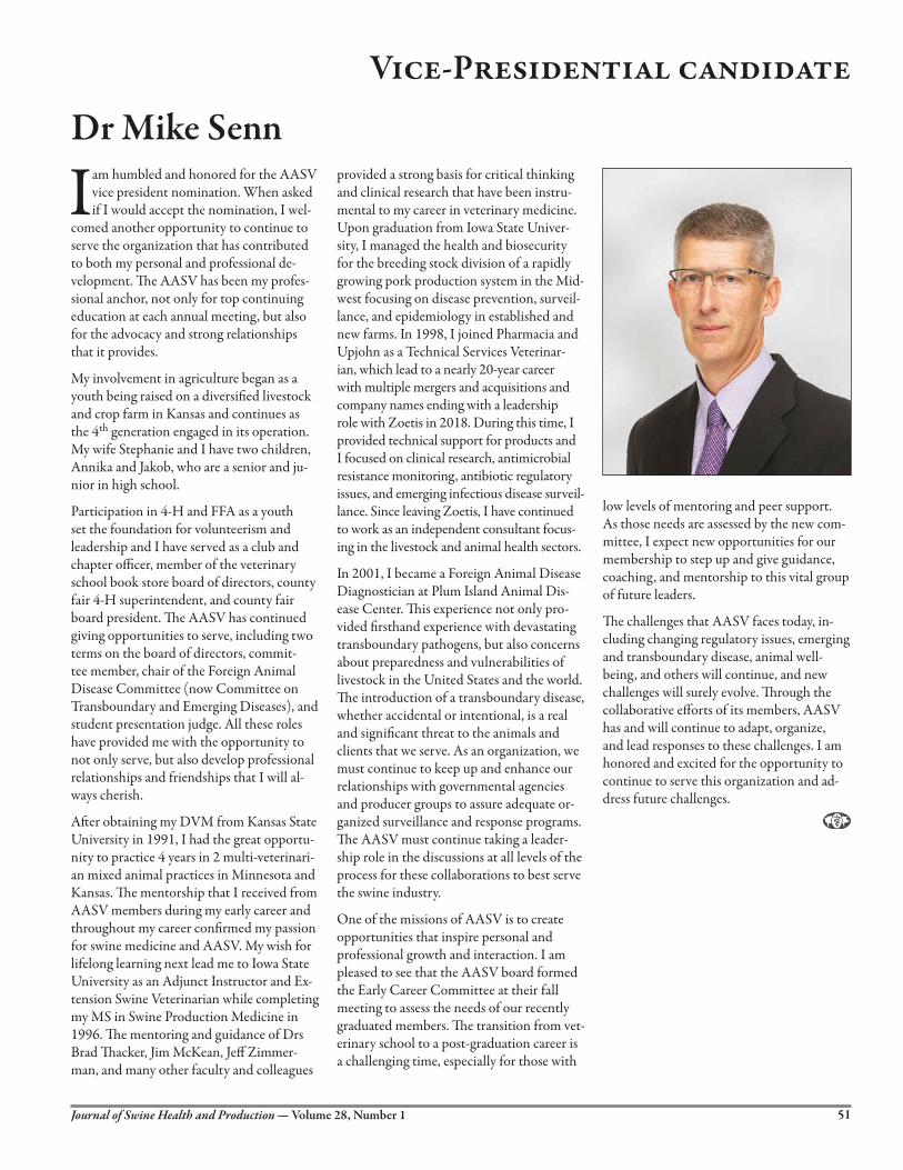
Vice-Presidential candidate
Dr Mike Senn
I am humbled and honored for the AASV vice president nomination. When asked if I would accept the nomination, I wel-
comed another opportunity to continue to serve the organization that has contributed to both my personal and professional de-velopment. �e AASV has been my profes-sional anchor, not only for top continuing education at each annual meeting, but also for the advocacy and strong relationships that it provides.
My involvement in agriculture began as a youth being raised on a diversi�ed livestock and crop farm in Kansas and continues as the 4th generation engaged in its operation. My wife Stephanie and I have two children, Annika and Jakob, who are a senior and ju-nior in high school.
Participation in 4-H and FFA as a youth set the foundation for volunteerism and leadership and I have served as a club and chapter o�cer, member of the veterinary school book store board of directors, county fair 4-H superintendent, and county fair board president. �e AASV has continued giving opportunities to serve, including two terms on the board of directors, commit-tee member, chair of the Foreign Animal Disease Committee (now Committee on Transboundary and Emerging Diseases), and student presentation judge. All these roles have provided me with the opportunity to not only serve, but also develop professional relationships and friendships that I will al-ways cherish.
A�er obtaining my DVM from Kansas State University in 1991, I had the great opportu-nity to practice 4 years in 2 multi-veterinari-an mixed animal practices in Minnesota and Kansas. �e mentorship that I received from AASV members during my early career and throughout my career con�rmed my passion for swine medicine and AASV. My wish for lifelong learning next lead me to Iowa State University as an Adjunct Instructor and Ex-tension Swine Veterinarian while completing my MS in Swine Production Medicine in 1996. �e mentoring and guidance of Drs Brad �acker, Jim McKean, Je� Zimmer-man, and many other faculty and colleagues
provided a strong basis for critical thinking and clinical research that have been instru-mental to my career in veterinary medicine. Upon graduation from Iowa State Univer-sity, I managed the health and biosecurity for the breeding stock division of a rapidly growing pork production system in the Mid-west focusing on disease prevention, surveil-lance, and epidemiology in established and new farms. In 1998, I joined Pharmacia and Upjohn as a Technical Services Veterinar-ian, which lead to a nearly 20-year career with multiple mergers and acquisitions and company names ending with a leadership role with Zoetis in 2018. During this time, I provided technical support for products and I focused on clinical research, antimicrobial resistance monitoring, antibiotic regulatory issues, and emerging infectious disease surveil-lance. Since leaving Zoetis, I have continued to work as an independent consultant focus-ing in the livestock and animal health sectors.
In 2001, I became a Foreign Animal Disease Diagnostician at Plum Island Animal Dis-ease Center. �is experience not only pro-vided �rsthand experience with devastating transboundary pathogens, but also concerns about preparedness and vulnerabilities of livestock in the United States and the world. �e introduction of a transboundary disease, whether accidental or intentional, is a real and signi�cant threat to the animals and clients that we serve. As an organization, we must continue to keep up and enhance our relationships with governmental agencies and producer groups to assure adequate or-ganized surveillance and response programs. �e AASV must continue taking a leader-ship role in the discussions at all levels of the process for these collaborations to best serve the swine industry.
One of the missions of AASV is to create opportunities that inspire personal and professional growth and interaction. I am pleased to see that the AASV board formed the Early Career Committee at their fall meeting to assess the needs of our recently graduated members. �e transition from vet-erinary school to a post-graduation career is a challenging time, especially for those with
low levels of mentoring and peer support. As those needs are assessed by the new com-mittee, I expect new opportunities for our membership to step up and give guidance, coaching, and mentorship to this vital group of future leaders.
�e challenges that AASV faces today, in-cluding changing regulatory issues, emerging and transboundary disease, animal well-being, and others will continue, and new challenges will surely evolve. �rough the collaborative e�orts of its members, AASV has and will continue to adapt, organize, and lead responses to these challenges. I am honored and excited for the opportunity to continue to serve this organization and ad-dress future challenges.
51Journal of Swine Health and Production — Volume 28, Number 1

MVP17015/USA/0817 ©Phibro Animal Health Corporation. Phibro, Phibro logo design, MVP adjuvants, Tailor-Made,MJPPRS and Emulsibac are trademarks owned by or licensed to Phibro Animal Health Corporation or its affiliates.

9781119350859
June 2019 | Hardback
1136 pages | $249.99
Provides a fully revised Eleventh Edition of the definitive reference to swine health and disease
Diseases of Swine has been the definitive reference on swine health and
disease for over 60 years. This new edition has been completely revised
to include the latest information, developments, and research in the
field. Now with full color images throughout, this comprehensive and
authoritative resource has been redesigned for improved consistency
and readability, with a reorganized format for more intuitive access
to information.
Diseases of Swine covers a wide range of essential topics on swine
production, health, and management, with contributions from more than
100 of the foremost international experts in the field. This revised edition
makes the information easy to find and includes expanded information
on welfare and behavior.
A key reference for anyone involved in the swine industry, Diseases of
Swine, Eleventh Edition:
Presents a thorough revision to the gold-standard reference on pig health and disease
Features full color images throughout the book
Includes information on the most current advances in the field
Provides comprehensive information on swine welfare and behavior
Offers a reorganized format to make the information more accessible
Written for veterinarians, academicians, students, and individuals and agencies responsible for swine health and
public health, Diseases of Swine, Eleventh Edition is an essential guide to swine health.
www.wiley.com/learn/vetmedicine
DISEASES OF
SWINE11th EditionEdited by Jeffrey J. Zimmerman, Locke A. Karriker, Alejandro Ramirez, Kent J. Schwartz, Gregory W. Stevenson and Jianqiang Zhang
Enjoy 20% discount! Use code AASV at wiley.com


Journal of Swine Health and Production — Volume 28, Number 1 55
Upcoming meetings2020 Pig Ski SeminarFebruary 12-14, 2020 (Wed-Fri) Copper Mountain, Colorado For registration or more information: Lori Yeske Pig Group 39109 375th Ave Saint Peter, MN 56082 Tel: 507-381-1647 Email: [email protected] Web: www.pigski.com
American Association of Swine Veterinarians 51st Annual MeetingMarch 7-10, 2020 (Sat-Tue) Hyatt Regency Atlanta Atlanta, Georgia
For more information: American Association of Swine Veterinarians 830 26th Street Perry, Iowa
Tel: 515-465-5255 Email: [email protected] Web: www.aasv.org/annmtg
26th International Pig Veterinary Society CongressJune 2-5, 2020 (Tue-Fri) Florianopolis, Brazil
For more information: Tel: +55 31 3360 3663 Email: [email protected] Web: www.ipvs2020.com
For additional information on upcoming meetings: www.aasv.org/meetings
World Pork ExpoJune 3-5, 2020 (Wed-Fri) Hosted by the National Pork Producers Council (NPPC) Iowa State Fairgrounds Des Moines, Iowa
For more information: National Pork Producers Council Tel: 515-278-8012 Fax: 515-278-8014 Web: www.worldpork.org
International Conference on Pig SurvivabilityOctober 28-29, 2020 (Wed-�u) Omaha, Nebraska Hosted by Iowa State University, Kansas State University, and Purdue University
For more information: Email: [email protected] Web: www.piglivability.org/conference

AASV Resources online at www.aasv.org
Photo courtesy of Tina Smith
Photo Corner
AASV Industry Support Council
®
The Journal of Swine Health and Production is made possible by the generous support of these
Industry Support Council members.