janice m. beitz, - njha · restarted infliximab in 4 weeks after surgery; others ... these patients...
TRANSCRIPT

Janice M. Beitz, PhD, RN, CS, CNOR, CWOCN-AP, CRNP, MAPWCA, ANEF, FAAN
Professor of Nursing, WOCNEP Director School of Nursing-Camden
Rutgers University, Camden NJ

Participants will: 1) Identify common conditions associated with
non-wound-related polypharmacy. 2) Determine data for drugs that impair wound
healing processes. 3) Recognize drugs that may contribute to wound
healing. 4) Explain drugs that may cause wound reactions. 5) Describe clinical practices mitigating drug
effects

Normal wound healing processes Chronic disease prevalence and wounds Drugs and wound healing/wound healing
Impairment Common and uncommon drug offenders Clinical implications for wound professionals

Human body “wired” to heal
Despite many obstacles, most wounds heal
Not here to discuss this comforting reality
Here to discuss pharmacologic impact on wound healing and wound generation
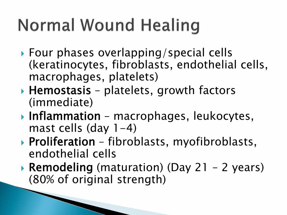
Four phases overlapping/special cells (keratinocytes, fibroblasts, endothelial cells, macrophages, platelets)
Hemostasis – platelets, growth factors (immediate)
Inflammation – macrophages, leukocytes, mast cells (day 1-4)
Proliferation – fibroblasts, myofibroblasts, endothelial cells
Remodeling (maturation) (Day 21 – 2 years) (80% of original strength)
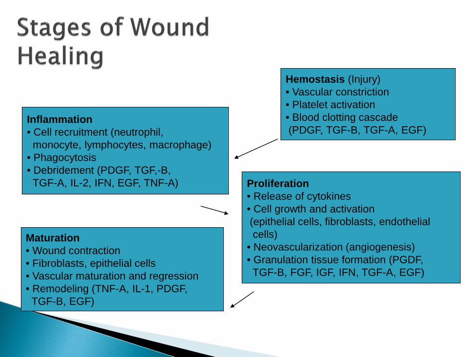
Hemostasis (Injury) • Vascular constriction • Platelet activation • Blood clotting cascade (PDGF, TGF-B, TGF-A, EGF)
Inflammation • Cell recruitment (neutrophil, monocyte, lymphocytes, macrophage) • Phagocytosis • Debridement (PDGF, TGF,-B, TGF-A, IL-2, IFN, EGF, TNF-A) Proliferation
• Release of cytokines • Cell growth and activation (epithelial cells, fibroblasts, endothelial cells) • Neovascularization (angiogenesis) • Granulation tissue formation (PGDF, TGF-B, FGF, IGF, IFN, TGF-A, EGF)
Maturation • Wound contraction • Fibroblasts, epithelial cells • Vascular maturation and regression • Remodeling (TNF-A, IL-1, PDGF, TGF-B, EGF)

Defined as wound that is physiologically impaired due to: ◦ Inadequate angiogenesis ◦ Impaired innervation ◦ Impaired cellular migration
Medications can affect any aspect of wound healing

In 2015, top 10 leading causes of death accounted for approximately 75% of all US deaths
In 2015, 2,712,630 Americans died (86,212 more than 2014) (CDC, 2017)

*Chronic Disorders (CDC, 2017)
1) Heart Disease* 2) Cancer* 3) Chronic Lower
Respiratory Disease* 4) Unintentional injuries 5) Stroke*
6) Alzheimer’s Disease* 7) Diabetes* 8) Influenza/pneumonia 9) Kidney Disease* 10) Suicide

As of 2012, half of all American adults had one or more chronic diseases
Constitutes 117 million Americans Obesity – serious health disorder –
approaching 50% are obese or overweight Diabetes mellitus type 2 – pandemic Risk behaviors: Little or no exercise, poor
dietary habits (fat, calories, salt), smoking (1 in 5 adults), alcohol abuse (CDC, 2017)
Arthritis affects 53 million Americans

86% of all US healthcare spending in 2010 was for people with one or more chronic diseases
In Americans over 65, 3 of 4 have multiple chronic conditions.
93% of total Medicare spending in 2012 was for people with multiple chronic conditions (CDC, 2017; CMS, 2012)

Pressure injuries – focus of today’s conference
Venous ulcers Arterial ulcers Neuropathic (diabetic) ulcers Vasculitic and “other” ulcers Just about ALL wound patients are receiving
medication therapy

Wound healing affected by many drugs and disease processes
Nearly 50% of Americans take one prescription drug monthly
Twenty percent take three drugs or more a month
Eleven percent take five or more drugs (CDC, 2017)
Thirty-six million Americans use herbals yearly (Ranade & Collins, 2014)
U.S. herbal use grew for 12th straight year (Crane, 2016)


Anticoagulants Antimicrobials Aspirin/NSAIDs ◦ NSAIDs impair
fibroblasts; weaken wound contraction with long-term use ◦ (Guo et al, 2010)
Povidone/Iodine Colchicine
Dakin’s solution ◦ Useful and safe if
used diluted and for short-term
Glucocorticoids Immunosuppressive
agents Anti-angiogenesis
agents

Antineoplastic agents ◦ Reduce RBC and WBC
presence ◦ Damage keratinocyte ◦ May decrease VEGF
and angiogenesis Colchicine ◦ Reduces granulocyte
migration ◦ Reduces fibroblast
synthesis
Vasoconstrictors ◦ Decrease tissue
perfusion Anti-rheumatoid
drugs ◦ Methotrexate:
cytotoxic to T cells and macrophages
Nicotine and smoking (But NRT does NOT impair healing)

Kaduta et al (2015) retrospective record review of 1036 elective orthopedic surgery patients
Looked at risk factors for SSI and DWH Risk factors were foot/ankle surgery, total knee
arthroplasty, and rheumatoid arthritis (RA) disease duration
Looked at conventional synthetic DMARDs; looked at biologic DMARDs as variables
Neither were risk factors Why?? – drugs stopped 2-4 weeks before surgery Restarted infliximab in 4 weeks after surgery; others
(Entanercept, Adalimumab, Tocilizumab, Golimumab) restarted after healing of surgical wounds

Gaucher et al, 2017 53-year-old male with sarcoidosis and
steroid-induced diabetes Receiving methotrexate and prednisone Developed LLE cellulitis Stopped prednisone Treated cellulitis Four rounds of skin grafting: stopped
methotrexate Fifth skin graft was successful

Use is expanding ◦ Bevacizumab (Monoclonal antibody; VEGF-A
inhibitor) ◦ Aflibercept (VEGF-A, VEGF-B) ◦ Sunitinib (Tyrosine kinase inhibitor)
Can cause impaired wound healing, osteo-necrosis of jaw, hand-foot skin reaction, hand-foot syndrome, and bleeding

Notorious inhibitors of wound healing Notorious for systemic effects (hyperglycemia,
osteoporosis, mood changes) Steroids affect cells by altering gene expression
after crossing cell membrane Consequently affect almost every phase of wound
healing Degree of inhibition related to potency of steroid Long-term usage impact is the challenge: bottom
line is immune modulation (and associated derived risks)

Delay in removal of bacteria and foreign bodies ◦ Decreased neutrophil and macrophage activity
Decrease in epithelial regeneration and granulation activity (caused by steroids anti-mitotic activity)
Decrease in fibroblast activity Over time thinned epidermis inhibits wound
contraction Yet no problem with acute surgical healing if
not long-term use (Treadwell, 2013; Wang et al, 2013)
And when used topically may help healing (e.g., stasis dermatitis)

Work by inhibiting Cyclooxygenase (COX) COX affects arachidonic acid and
prostaglandins; blocking has serious effects NSAIDs – have well known effect on
delaying bone healing Krischak et al (2007): Found diclofenac
inhibited fibroblasts after use in 10 rats (lab testing)
Can affect ligament health too

Retrospective study of all orthopedic patients with femur, tibia, and/or humerus fractures between October 2009 and September 2011 – University of Tennessee Level 1 Trauma Center
1,901 patients with LBFs Assessed for complications: Nonunion/malunion,
infection 60 patients had complications Logistic regression calculated ORs Patient more likely to have complication if received
NSAIDs postop (OR 2.17) or if they were smokers (OR 3.19)
Recommend avoidance of NSAID use in traumatic LBF

Large retrospective study of adult GI surgery patients between 2008 and 2012
Among 398,752 patients, 55% underwent colorectal surgery and 45% had non-colorectal GI surgery.
Five percent of all received ketorolac (IV) These patients had higher odds of re-intervention
(OR 1.20), emergency room visit (OR 1.44) and 30-day readmission (OR 1.11) and readmission for anastomotic complications (OR 1.2)
Use great caution when using IV ketorolac in patients undergoing GI surgery

Tested 42 rats with oral sildenafil (10mg/kg) in 1cc distilled water via NG tube vs. sodium chloride injection in intraperitoneum (21 exp; 21 control)
Created an ischemic skin wound on rats’ abdomens Checked healing at days 3,5,10 on 7 rats in each
group Theoretically sildenafil (PDE-5 inhibitor) should
help healing Sildenafil significantly reduced re-epithelialization,
neovascularization, granulation tissue and number of inflammatory cells on day 3
Increased inflammatory cells on day 10 Oral vs. Topical sildenafil: have differential effects
on wounds


Hemorrheologics (e.g., pentoxifylline (Trental)
Hormones (estrogen): topical Phenytoin (think gums): topical Prostaglandins Zinc Vitamins A and C

Topical “Natural” Medications ◦ Aloe vera ◦ Curcumin ◦ Ginger ◦ Medicinal Honey ◦ Mucilage (in Slippery Elm) ◦ Witch Hazel
Off Label Topical Drugs (in the literature) ◦ Calcium Channel Blockers ◦ Topical Regular Insulin ◦ Topical Nitroglycerin ◦ Topical Dilantin

Polypharmacy is the norm Co-morbidities are common Drugs involved in all wound patients care

94-year-old female admitted from home; cared for by son who is devoted to her; sits at bedside; multiple co-morbidities including dementia, poor nutrition, immobility and frailty
Consultation for wound care team: has pressure injuries on sacrum, bilateral hips and sternum
Also has “rash” on extremities and trunk Multiple medications; nothing new except
recently began Aricept (donepezil)


• One of most common adverse reactions • Overall incidence rate of 2-3% in hospitalized
patients • Almost any (1:1000 hospitalized patients (Ijaz,
2015)) medication can induce skin reactions • Selected drug classes have rates as high as 5% (Lee
& Thomson) • Some reactions are immunological; most are not
(thankfully) • Categorized by predictability (pharmacological) or
immune basis

Type A: 85-90% of adverse drug reactions (ADEs); predictable from known pharmacologic properties of a drug. Examples: •Diarrhea – Antibiotics •Gastritis – NSAIDS •Kidney toxicity – Aminoglycosides (Kaniwa et al, 2013)

Type B: 10-15% of ADEs hypersensitivity: Immunologic or other patho-mechanisms; have signs/symptoms different from action of drug usually not predictable. Examples: Exaggerated sensitivity to known drug reactions – tinnitus from low dose aspirin (Kaniwa et al, 2013)

Type I: Cased by drug/antigen specific IgE that links with mast cells and basophils – immediate release of histamine/leukotrienes get urticaria, angioedema, anaphylaxis (aspirin, penicillins) Type II: Cytotoxic reactions based on IgG or IgM – mediated mechanisms antibody ruptures cell (blood cell dyscrasias like hemolytic anemia and thrombocytopenia)

Type III: Mediated by intravascular immune complexes. Antibodies and drug antigens in circulation. Phagocytes remove complexes and ends up in skin, kidneys, etc. (serum sickness, vasculitis) Type IV: Mediated by T cells; cause “delayed” hypersensitivity (contact dermatitis, SJS and TENS)

Antacids Muscle Relaxants Antihistamines (oral) Nitrates Atropine Nystatin Benzodiazepines Oral Contraceptives Corticosteroids Propanolol Digoxin Spironolactone Ferrous Sulfate Theophylline Insulin Thyroid Hormones Laxatives Vitamins Local Anesthetics

Exanthems Fixed Drug Eruptions (Allergic) Blistering Psoriasisiform Immune Mediated (SJS and TEN) Hematologic/Vasculitic
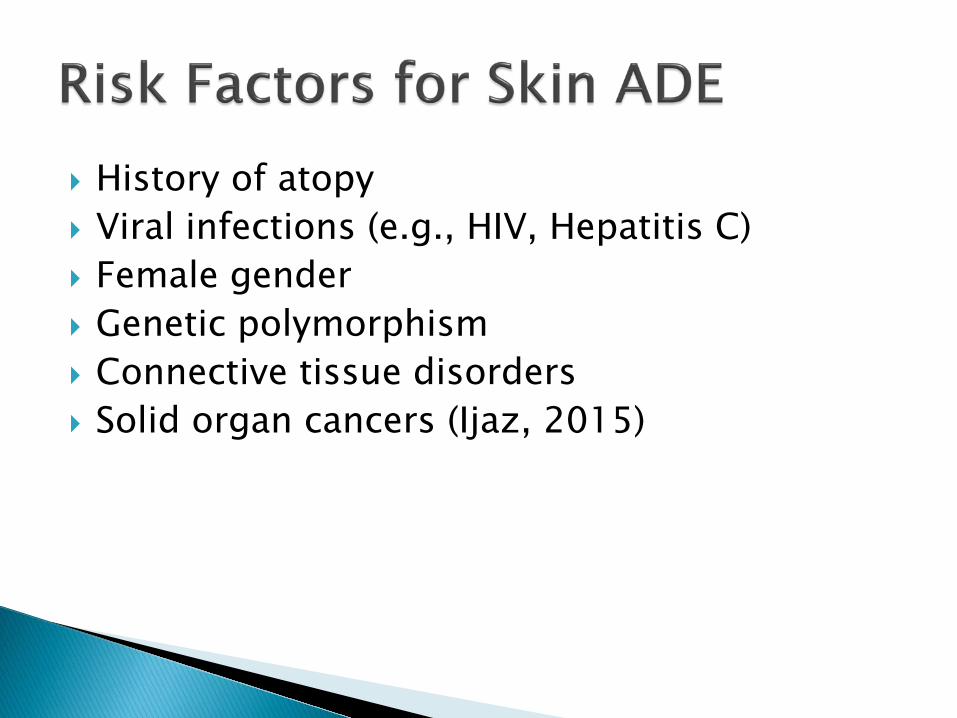
History of atopy Viral infections (e.g., HIV, Hepatitis C) Female gender Genetic polymorphism Connective tissue disorders Solid organ cancers (Ijaz, 2015)


• Allopurinol • Antimicrobials (PCN, Cephalosporins, Erythromycin,
Gentamicin, Anti-TB Drugs, Nitrofurantoin, Sulfa) • Barbiturates • Captopril • Carbamazepine • Furosemide • Gold Salts • Lithium • Phenothiazine • Phenytoin • Thiazides

• ACE Inhibitors • Allopurinol • Antimicrobials (Sulfa, Tetracyclines, Cephalosporins, PCN,
Clindamycin, Trimethoprim, metronidazole) • Barbiturates • Benzodiazepines • Calcium Channel Blockers • Carbamazepine • Fluconazole • Lamotrigine • NSAIDs • Paclitaxel • Proton Pump Inhibitors (Omeprazole, Lansoprazole) • Salicylates • Terbinafine • *Reaction at same site or sites each time drug is taken

ACE Inhibitors (captopril, enalapril) antibiotics (cephalosporins, penicillins, sulfa
agents, tetracyclines, vancomycin) gold/sodium aurothiolamar lithium loop diuretics (eg, furosemide, bumetanide) nonsteroidal anti-inflammatory drugs
(NSAIDs) penicillamine thiazide diuretics (eg, hydrochlorothiazide)

• ACE-I • Beta Blockers • Chloroquine • Digoxin • Gold • Interferons • Lithium • NSAIDs • Terbinafine • Tetracyclines • TNF-Alpha Antagonists


SJS TEN
Barbiturates* Allopurinol
Beta-Lactams Anti-TB Drugs
Carbamazepine* Barbiturates*
Chloropropamide Carbamazepine*
Co-Trimoxazole Gold* Gold* Griseofulvin H2 Antagonists Lamotrigine*
Lamotrigine* Leflunomide* Leflunomide* Macrolides Nitrofurantoin
NSAIDs NSAIDs* Phenothiazines Penicillins Phenytoin* Phenytoin*
Rifampicin Salicylates Sulfonamides* Sulfonamides* Tetracyclines* Tetracyclines* Thiazides
* Can cause both SJS and TEN

• Allopurinol • Aspirin • Beta-Lactam Antibiotics • Carbamazepine • Co-trimoxazole • Diltiazem • Erythromycin • Furosemide • Gold • Hydralazine • Methotrexate • NSAIDs • PTU • Sulfasalazine • Sulfonamides • Thiazides • Thrombolytic Agents

Take detailed accurate medication history Note use of all OTC medications especially
herbals Note injections – including vaccines or contrast
media Note time of medication relative to onset of
wound/skin problems (ADE) Take detailed medical history: Any history of
drug sensitivity, contact dermatitis, connective tissue disease, atopy (asthma, eczema), previous wound healing delays

Examine skin eruption closely and determine if drug-related
Educate patient about avoidance of drug in future; record clearly in history
If serious enough, Medic Alert bracelet Notify pertinent regulatory authorities if
serious reaction (FDA FAERS (Federal Adverse Event Reporting System: www.fda.gov)

WISN: Warfarin-induced skin necrosis HIT Syndrome WISN: occurs 3 to 5 days after dose of
warfarin; often in patient with Protein C and Protein S deficiences
Red painful plaques Progress possibly to hemorrhagic blisters,
ulcers, necrosis (Clinard et al)


HIT Syndrome (Specifically HIT II) o Get loss of heparin due to Immune complex (HIT
antibodies) o Get destruction of platelets from antibody complexes
(Trautman et al, 2010) o Decreased platelets < 150,000 o Get arterial and venous thrombosis o Necrosis of skin in fatty areas as abdomen, thighs
– can also be blisters, purpura o Diagnosis: Use “4Ts Score” (Thrombocytopenia, timing of
platelet fall, thrombosis and sequelae, other causes for thrombocytopenia) (Coutre, 2015)


Harkainen et al (2014) in a Finnish study of 463 patients looked at hospital records over 12 month time
Total of 180 ADEs in 125 patients (27%) Of these 74(41%) were preventable; 95%
caused temporary harm Most common ADE was abnormal blood
potassium Risk was increased by duration of care and
polypharmacy

Consider impact of hidden malnutrition (Protein insufficiency) on drug metabolism (protein binding and drug toxicities); does patient have fatigue, pain, mouth ulcers?
Consider common drugs of age groups treated (in wound care and wound clinics mostly older) ◦ Rheumatoid diseases and DMARDS (methotrexate
and sulfasalazine etc.) Consider polypharmacy and need for
“deprescribing”

Effects of Aging on Drug Metabolism and Excretion ◦ With aging, liver function decreases by 40% so
drugs can be “stored” and cause toxicity ◦ Kidney function decreases with age; better to use
creatinine clearance rather than creatinine level in elderly to monitor levels; affects drug excretion ◦ Selected drugs with greater harm in elderly:
Antipsychotics (haloperidol) Hypnotics (diazepam) Diuretics (furosemide) (Kaufman, 2015)

Usage of other “traditional” therapies 1) Need to ask if patient is consuming any
herbal products (teas, liquid extracts, capsules)
2) Need to ask patient is applying any herbal topical preparation to wound
3) Potential for herbal-drug interaction: “Natural” does not mean safe
4) Does the patient space herbals away in time from other drugs (St. John’s Wort, Ginkgo biloba, etc.)

Plant-based systems continue to play role in healthcare of 80% of world’s developing countries
Called “phytomedicines” Affordable and usually no to minimal side
effects Level of evidence varies greatly

Mainstream Markets Natural/Health Markets
Cranberry Garlic Saw Palmetto Soy Ginkgo Biloba Milk Thistle Black Cohosh Echinachea St. John’s Wort Ginseng
Flax seeds Wheat and Barley grass Turmeric Aloe Vera Blue Green Algae Milk Thistle Elderberry Saw Palmetto Echinachea Cranberry

Need to identify detailed information on herbals used (dose, form, topical etc.)
Need to identify “red-flag” medications for potential interactions (warfarin, digoxin, lithium, cyclosporine, protease inhibitors)
Educate patient with wounds on safety, dosing, and potential toxicities of non-prescription pharmaceuticals (Ranade and Collins, 2014)

Reduce polypharmacy for wound patients by “de-prescribing”; interact with primary care provider
Remember that polypharmacy is not only removing excess drugs but that polypharmacy is also going to more than one pharmacy (Gillette et al, 2015); educate patients about the risk
Put on your ARMOR and LOOK at the wound patient

Mnemonic Meaning (Haque, 2008)
A: Assess R: Review M: Minimize O: Optimize R: Reassess
A: Beers criteria; Beta blockers, Pain meds; Antipsychotics
R: D-drug interaction; D-disease interaction; ADEs
M: #of meds related to functional status
O: for renal/hepatic status R: functional/cognitive
status in one week from any changes and periodically

Update oneself about alternative adjuncts to wound healing
Repurposed approved drugs: Erythropoietin (EPO); excellent review of animal studies using exogenous EPO (Hamed et al, 2014); topical EPO accelerates wound healing
Phyto-extracts in wound healing-excellent overview (Ghosh and Gaba, 2013) 450 plant species have wound healing properties
Hydralazine has anti-angiogenesis effects (study done in rats and chicken models) (Zheng et al, 2016)

Discussed multiple chronic conditions affecting wound patients
Explored selected data for drugs that impair wound healing
Offered implications for informed clinical practice

Anderson K., Hamm, R.L. (2014). Factors that impair healing. Journal of American College of Clinical Wound Specialists, 4, 84-91.
Arslantas, R., & Arslantas, M. (2015). Adverse effect of sildenafil on healing ischemic wounds: Results of an In vivo study. Ostomy Wound Management, 61(9), 32-37.
Armstrong, D., & Meyr, A.J. (2016). Wound healing and risk factors for non-healing. UpToDate, Retrieved 12/1/2016 from www.uptodate.com
Barnard, A.R., Regan, M., Burke, F.D., Chung, K.C., & Wilgis, E.F. (2012). Wound healing with medications for rheumatoid arthritis in hand surgery. International Scholarly Research Network, 2012, Article ID 251962, 5 pgs.

Barrios, R.L., & Arbiser, J.L. (2011). Effectiveness of gentian violet and similar products commonly used to treat pyodermas. Dermatology Clinics, 29, 69-73.
Bauer, K.A. (2016). Protein C deficiency: clinical manifestations and diagnosis. UptoDate. Retrieved January 2, 2017.
Beitz, J. (2017). Breaking bad: Medications and wound healing. Ostomy Wound Management, 63(3), 18-35.
Benhadou, F., Del Marmol, V. (2013). The mTor inhibitors and the skin wound healing. EWMA Journal, 13(1), 20-22.

Bircher, A.J. (2017). Exanthematous (morbilliform) drug eruption. UpToDate, Retrieved January 2, 2017 from www.uptodate.com.
Centers for Disease Control (2017). Health: United States, 2016. Retrieved August 25, 2017 from www.cdc.gov.
Chin, K., & Cordell, B. (2013). The effect of tea tree oil (melaleuca alternifolia) on wound healing using a dressing model. Journal of Alternative and Complementary Medicine, 942-945.
Choueri, T.K. Sonpavde, G. (2016). Toxicity of molecularly targeted antiangiogenic agents: Noncardiovascular effects. UptoDate, Retrieved December 15, 2016 from www.uptodate.com.
Clinard, V., Smith, J. D. (2014). Drug-induced skin disorders. U.S. Pharmacist. Retrieved November 20, 2014 from www.medscape.com/viewarticle/763495_print.

Common Side Effects, Allergies and reactions to antibiotics. Drugs.com. Retrieved November 20, 2014 from www.drugs.com/article/antibiotic-sideeffects-allergies-reactions.html.
Cooper, KL. (2012). Drug reaction, skin care, skin lost. Critical Care Nurse, 32(4), 52-59.
Crane, M. (2016). U.S. herbal supplement sales climb 7.5% in 2015. Nutritional Outlook, Retrieved August 8, 2017 from www.nutritionaloutlook.com
Douglas, H.E. (2010). TGF-B in wound healing: A review. Journal of Wound Care, 19(8), 403-
Dowd, S.E., Wolcott, R., Kennedy, J., Jones., C., & Cox, S.B. (2011). Molecular diagnostic and personalized medicine in wound care: Assessment of outcomes. Journal of Wound Care, 20(5), 232-239.

Gaucher, S. Nicolas, C., Piveteau. O. Philippe, H.J. Blanche, P. (2017). Sarcoidosis and wound healing after cellulitis of the lower limb: Is methotrexate responsible for skin graft failure? Wounds, 29(8), 229-230.
Gillette, C., Prunty, L, Wolcott, J Brodel-Zaugg, K. (2015). A new lexicon for polypharmacy: Implications for research, practice, and education. Research in Social and Administrative Pharmacy, 11(3), 468-471.
Ghosh, P.K., & Gaba, A. (2013). Phyto-extracts in wound healing. Journal of Pharmacy and Pharmaceutical Science, 16(5), 760-820.
Guo, S. & DiPietro, L. (2010). Factors affecting wound healing. Journal of Dental Research, 89(3), 218-229.
Greener, M. (2009). NSAIDs applied to the skin: a very topical subject. Nurse Prescribing, 7(7), 294-299.
Hamed, S., Bennett, C.L., Demiot, C., Vulmann, Y., Teot, L., Desmouliere, A. (2014). Erythropoietin, a novel repurposed drug: An innovative treatment for wound healing in patients with diabetes mellitus. Wound Repair and Regeneration, 22, 23-33.
Harding, K. (2014). Innovation and wound healing. Proceedings of the international surgical wound forum. Journal of Wound Care, 24(4), 7-13.
Harding, K. (2014). Innovation and wound healing. Proceedings of the international surgical wound forum. Journal of Wound Care, 24(4), 7-13.
Haque, R. (2009). ARMOR: A tool to evaluate polypharmacy in elderly persons. Annals of Long-Term Care, June, 26-30.
Harkainen, M., Kervinen, M. Ahonen, J. Voutilainen, A. Turonen, H., & Vehvilaninen-Julkiunen, K. (2014). Patient-specific risk factors of adverse drug events in adult inpatinets-Evidence detected using the Global Trigger Tool method. Journal of Clinical Nursing, 24, 582-591.
Harris, C.L., & Fraser, C. (2004). Malnutrition in the institutionalized elderly: The effects on wound healing. Ostomy Wound Management, 50(10), 54-63.
Hashemi, S.A., Madani, S.A., & Abediankenari, S. (2015). The review on properties of aloe vera in healing of cutaneous wounds. BioMed Research International, 2015, Article ID 714216, 6 pages.

Hollister, C., & Li, V.W. (2007). Using angiogenesis in chronic wound care with becaplermin and oxidized regenerated cellulose/collagen. Nursing Clinics of North America, 42, 457-465.
Ijaz, N. (2015). Cutaneous drug rashes. British Journal of Hospital Medicine, 11(11), C166-169.
Jeffcoach, D.R., Sams, V.G., Lawson, C.M., Enderson, B.L., Smith, S.T., Kline, H., Barlow, P.B., Wylie, D.R., Krumenacker, L.A., McMillen, J.C., Pyda, J., Daley, B.J., & University of Tennessee Medical Center, Department of Surgery (2014). Nonsterioidal anti-inflammatory drugs’ impact on nonunion and infection rates in long-bone fractures. Journal of Trauma Acute Care Surgery, 76, 779-783.
Kadota, Y., Nishida, K., Hashizume, K., Nasu, Y., Nakahara, R., Kanazawa, T., Ozawa, M., Harapa, R., Machida, T., Ozaki, T. (2016). Risk factors for surgical site infections and delayed wound healing after orthopedic surgery in rheumatoid arthritis patients. Modern Rheumatology, 26(1), 68-74.
Kaniwa, N. & Saito, Y. (2013). Pharmacogenomics of severe cutaneous adverse reactions and drug-induced liver injury. Journal of Human Genetics, 58, 317-326.
Karukonda, SRK, Flynn, TC, Boh, EE, McBurney, EI, Russo, G & Millikan, LE. (2000). The effects of drugs on wound healing. Part I. International Journal of Dermatology, 39(4), 250-257.
Karukonda, SRK, Flynn, TC, Boh, EE, McBurney, EI, Russo, G & Millikan, LE. (2000). The effects of drugs on wound healing. Part II. International Journal of Dermatology, 39(5), 321-333.

Kaufman, G. (2015). Multiple medicines: The issues surrounding polypharmacy. NRC, 17(4), 198-203.
Khalil, H., Cullen, M., Chambers, H., Carroll, M., Walker, J., (2015). Elements affecting wound healing time: An evidence-based analysis. Wound Repair and Regeneration, 23, 550-556.
Krischak, G.D., Augat, P., Claes, L., Kinzl, L., & Beck, A. (2007). The effects of non-steroidal anti-inflammatory drug application on incisional wound healing in rats. Journal of Wound Care, 16(2), 76-8.
Lee, A. & Thomson, J. (2006). Drug-induced skin reactions. Adverse Drug Reactions, 2, 125-156.
Lindstrom, A., Ooyen, C., Lynch, M. E., Blumenthal, M. (2013). Herb supplement sales increase from 5.5% in 2012; Herbal supplements sales rise for the 9th consecutive year; turmeric sales sump 40% in natural channel. Herbal Gram, 99, 60-65.

Maggiore, R. J., Dale, W., Gross, C. P., Feng, T., Ten, W.P., Mohire, S. G., Owusu, C., Klepin, H.D., Lichtman, S. M., Gajra, A., Ramani, R., Katheria, V., Zavala, L., Hurria, A., and Cancer and Aging Research Group (2014). Polypharmacy and potentially inappropriate medication use in older adults with cancer undergoing chemotherapy: Effect on chemotherapy-related toxicity and hospitalization during treatment. JAGS, 62, 1505-1512.
Marrs, J., & Zubal, B.A. (2009). Oncology nursing in a new era: Optimizing treatment with bevacizumab. Clinical Journal of Oncology Nursing, 13(5), 564-572.
Maver, T., Maver, V., Kleinschek, K., Smrke, D., & Kreft, S. (2015). A review of herbal medicines in wound healing. International Journal of Dermatology, 54, 740-751.
McIntyre, K. (2015). An oncology nurses/ guide to new targeted agents for metastatic colorectal cancer. Clinical Journal of Oncology Nursing, 19(5), S71-S79.
Nedorost, S. T. & Stevens, S. R. (2011). Diagnosis and treatment of allergic skin disorders in the elderly. Drugs Aging, 18(11), 827-835.
Nijhuis, W., Houwing, R., Van der Zwet, W., Jansman, F. (2012). A randomized trial of honey barrier cream versus zinc oxide ointment. British Journal of Nursing, 21(20), S10-S13.

Non-steroidal anti-inflammatory drugs and their skin side effects. (2014). Dermnet NZ, Retrieved November 20, 2014 from www.dermnetnz.org/reactions/nsaids.html.
Peart, J. (2015). Influence of psychosocial factors on coping and living with a venous leg ulcer. Community Wound Care, June, 521-527.
Pichler, W. (2003). Delayed drug hypersensitivity reactions. Archives of Internal Medicine, 139, 683-693.
Pichler, W. (2016). Drug allergy: Classification and clinical features. UpToDate, Retrieved December 15, 2016 from www.uptodate.com.

Poetker, D. M. & Reh, D. (2010). A comprehensive review of the adverse effects of systemic corticosteroids. Otolaryngology Clinics of North America, 43, 753-768.
Price, P.E. (2008). Education, psychology and compliance. Diabetes/Metabolism Research and Reviews, 24(Suppl 1), S101-S105.
Ranade, D. & Collins, N. (2014). Nutrition 411: An introduction to herbs for wound healing professionals. Ostomy Wound Management, 60(6), 10 pages. Retrieved 2/5/2016 from www.o-wm.com.
Rochon, P. A. (2016). Drug prescribing for older adults. UpToDate, Retrieved December 15, 2016 from www.uptodate.com.
Ryan T. (2003). Use of herbal medicines in wound healing. International Journal of Lower Extremity Wounds, 2(1), 22-24.
Samel, A. D. & Chu, C. Y. (2016). Drug eruptions. UpToDate, Retrieved December 15, 2016 from www.uptodate.com

Scott, I.A., Hilmer, S.N., Reeve, E., Potter, K., LeCouteur, D., Rigby, D., Gnjidic, D., Delmar, C.B., Roughead, E.E., Page, A., Jansen, J., & Martin, J. (2015). Reducing inappropriate polypharmacy the process of deprescribing. JAMA Internal Medicine, 175(5), 27-834.
Shirin, J. (2015).Polypharmacy, the elderly, and deprescribing. Consultant Pharmacist, 30(9), 527-532.
Smith, R. (2008). The effects of medications in wound healing. Podiatry Management, August, 195-202
Sowicz, T. J. (2010). Atopic dermatitis: An evaluation of two clinical guidelines. Dermatology Nursing, 22(5), 9-11.
Teng, M., Huang, Y., & Zhang, H. (2014). Application of stem cells in wound healing: An update. Wound Repair and Regeneration, 22, 151-160.
Treadwell, T. (2013). Editorial Message: Corticosteroids and wound healing. Wounds, 25(10), 2 pages. Retrieved 12/28/2015 from www.wounds.research.com

Vodovotz, Y. (2010). Translational systems biology of inflammation and healing. Wound Repair and Regeneration, 18, 3-7.
Wallace, A. & Taylor, C. (2011). Recognizing how chemotherapy side effects can affect stoma care. Cancer Nursing Practice, 10(2), 20-25.
Wang, A., Armstrong, E.J., Armstrong, A.W. (2013). Corticosteroids and wound healing: Clinical considerations in the perioperative period. American Journal of Surgery, 206(3) 410-417.
Ward, K. E., Archambault, R., Mersfelder, T. L. (2010). Severe adverse skin reactions to nonsteroidal anti-inflammatory drugs: A review of the literature. American Journal of Health-System Pharmacy, 67, 206-213.

Wigston, C., Hassan, S., Turvey, S., Bosanquet, D., Richards, A., Holloway, S., & Harding, K. (2013). Impact of medications and lifestyle factors on wound healing: A pilot study. Wounds UK, 9(1), 22-28.
Zhang, Q., Lin, Z., Lin, X., Tang, L., Luo, H., Li, H., Zhang, Y., & Luo, W. (2016). In vitro and in vivo study of hydralazine, a potential anti-angiogenic agent. European Journal of Pharmacology, 779, 138-146.
Zhu, J. & Weingart, S. N. (2016). Prevention of adverse drug events in hospitals. UpToDate, Retrieved December 15, 2016 from www.uptodate.com.
N.B. See additional references and information in Beitz (2017)