jalal jalal shokouhi – md editor in chief of iranian society of radiology...
TRANSCRIPT

Jalal Jalal Shokouhi – MDJalal Jalal Shokouhi – MDEditor in Chief of Iranian Society of RadiologyEditor in Chief of Iranian Society of Radiology
jalaljalalshokouhi@hotmail/gmail.com www.medimage.ir
Imaging of epilepsis

Seizure is the clinical manifestations of an abnormal Seizure is the clinical manifestations of an abnormal and excessive excitation of a population of and excessive excitation of a population of neurons in the brain.neurons in the brain.
Epilepsy is recurrent on provoked seizures.Epilepsy is recurrent on provoked seizures.

Etiology of seizures and epilepsy is idiopathic Etiology of seizures and epilepsy is idiopathic cryptogenic in 65.5% , Vascular 10.9% , congenital cryptogenic in 65.5% , Vascular 10.9% , congenital 8%, Traumatic 5.5% , neoplastic 4.1% , 8%, Traumatic 5.5% , neoplastic 4.1% , degenerative 3.5%, and infectious 2.5% (hauser degenerative 3.5%, and infectious 2.5% (hauser wa,et al).wa,et al).

Risk factors for epilepsy that imaging can show Risk factors for epilepsy that imaging can show them, are:them, are:
Head injuries in military and civilian populations, Head injuries in military and civilian populations, Stroke, Embolic risk factors, left ventricular Stroke, Embolic risk factors, left ventricular hypertrophy, Encephalitis, Bacterial meningitis, hypertrophy, Encephalitis, Bacterial meningitis, Aseptic meningitis, Alzheimer disease, multiple Aseptic meningitis, Alzheimer disease, multiple sclerosis, Alcohol, Heroin and Marijuana sclerosis, Alcohol, Heroin and Marijuana intoxication.intoxication.

For treatment control,For treatment control,
also also
imaging could be in plan. imaging could be in plan.

ConclusionConclusion: Although EEG is the most specific : Although EEG is the most specific test for diagnosis but MRI is the imaging tool of test for diagnosis but MRI is the imaging tool of choice , especially in partial epilepsy. choice , especially in partial epilepsy.

PET , SPECT, MEG are important for PET , SPECT, MEG are important for localization of seizure focus often used in localization of seizure focus often used in pre-surgical evaluations.pre-surgical evaluations.

Epilepsy diagnosisEpilepsy diagnosis
ClinicClinic EEG, Brain mapping EEG, Brain mapping CT, CTACT, CTA MRI, MRA, MRVMRI, MRA, MRV MagnetographyMagnetography Mulecular, Spect, PetMulecular, Spect, Pet

CT and MRI in EpilepsyCT and MRI in Epilepsy

Pathology and causes:Pathology and causes:
1.Developmental disorders
• Neuronal migration disorders
• Hamartomas
• Vascular malformations,…
• Neuronal migration disorders include the following:
1.Tuberous sclerosis2.Focal cortical dysplasias3.Polymicrogyria4.Schizencephaly5.Heterotopias6.Lissencephaly (agyria-pachygria)7.Hemimegalencephaly8.Microdysgenesis

GREY MATTER HETEROTOPIA, BAND HETEROTOPIA, NODULAR GREY MATTER HETEROTOPIA, BAND HETEROTOPIA, NODULAR SUBEPANDIMALSUBEPANDIMAL

VASCULAR ANOMALY, PARIETAL VASCULAR ANOMALY, PARIETAL TEMPORAL AND OCCIPITALTEMPORAL AND OCCIPITAL

ENHANCED VASCULAR ANOMALY BILATERAL SCHIZENCEPHALIC CLEFTS
CLEFT IN SAGITAL

2.Traumatic2.Traumatic
• Hematomas, foreign bodies, penetrating injuries, Hematomas, foreign bodies, penetrating injuries, depressed fractures, brain edema, …depressed fractures, brain edema, …
3.Vascular, hemorrhagic and ischemic 3.Vascular, hemorrhagic and ischemic
• Cerebrovascular disease, including stroke, porencephaly, Cerebrovascular disease, including stroke, porencephaly, ……

SITES OF TRAUMATIC LESIONS SITES OF TRAUMATIC LESIONS

HEMATOMASHEMATOMAS

ANEURYSMANEURYSM

ANEURYSMSANEURYSMS

VASCULAR ANOMALYVASCULAR ANOMALY

VASCULAR ANOMALYVASCULAR ANOMALY

ISCHEMIC & HEMORRHAGICISCHEMIC & HEMORRHAGIC

VASCULAR ANOMALYVASCULAR ANOMALY

ISCHEMIC CHANGESISCHEMIC CHANGES

44..NeoplasmsNeoplasms
• GliomasGliomas
• Mixed neuronoglial tumorsMixed neuronoglial tumors
• Dysembryoplastic neuroepithelial tumorDysembryoplastic neuroepithelial tumor
• OthersOthers
5.Infections5.Infections
• Infections (bacterial, viral, fungal, parasitic diseases) Infections (bacterial, viral, fungal, parasitic diseases) &&
• Immune-mediated disorders (Rasmussen`s syndrome)Immune-mediated disorders (Rasmussen`s syndrome)
6.Mesial temporal sclerosis(hippocampus)6.Mesial temporal sclerosis(hippocampus)

SPECT, HYPER PERFUSION 12 SECONDS AFTER SPECT, HYPER PERFUSION 12 SECONDS AFTER EPILEPSIES, TRACER WAS INJECTEDEPILEPSIES, TRACER WAS INJECTED

HIPPOCAMPAL ATROPHY, MESIAL TEMPORAL SCLEROSISHIPPOCAMPAL ATROPHY, MESIAL TEMPORAL SCLEROSIS

ASTROCYTOMA IN THE RIGHT MESIAL TEMPORAL LOBEASTROCYTOMA IN THE RIGHT MESIAL TEMPORAL LOBE
COVERNOUS HEMANGIOMA IN THE RIGHT MESIAL TEMPORAL LOBE
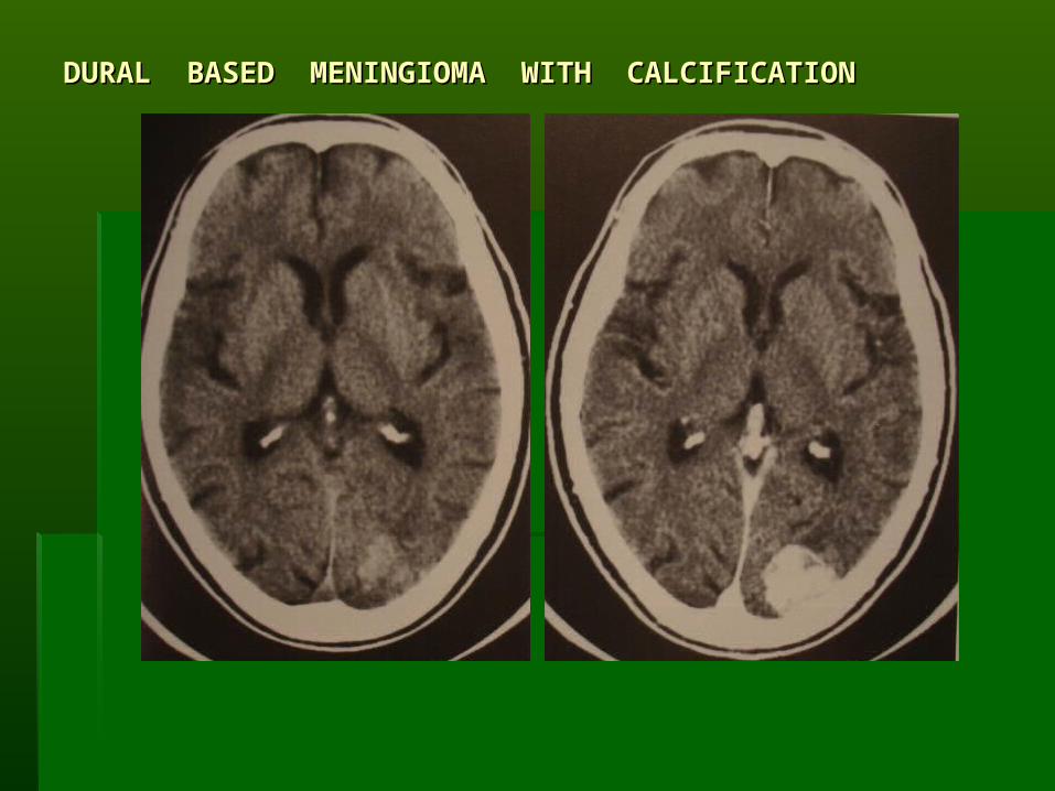
DURAL BASED MENINGIOMA WITH CALCIFICATIONDURAL BASED MENINGIOMA WITH CALCIFICATION

Cysticercosis+hydatidCysticercosis+hydatid

A NEOPLASM A NEOPLASM

ANOTHER NEOPLASM WITH SEVERE ANOTHER NEOPLASM WITH SEVERE EDEMA AND MIDLINE SHIFTEDEMA AND MIDLINE SHIFT

A HUGE SIZED NEOPLASM WITH BILATERAL A HUGE SIZED NEOPLASM WITH BILATERAL EDEMAEDEMA

HIPPOCAMPUS MESIAL TEMPORAL SCLEROSIS

LEFT FRONTAL LEFT FRONTAL AND TEMPORAL AND TEMPORAL HYPOMETABOLISM HYPOMETABOLISM IN A: TEMPORAL IN A: TEMPORAL LOBE EPILEPSYLOBE EPILEPSYPET. PET.
CRONAL SPECT- CRONAL SPECT- HMPAO SCAN HMPAO SCAN SHOWING: SHOWING: INCREASED INCREASED PERFUSION IN THE PERFUSION IN THE LEFT FRONTAL LOBE LEFT FRONTAL LOBE IN RASMUSSEN`S IN RASMUSSEN`S ENCEPHALITIS+ ENCEPHALITIS+ HYPERPERFUSION IN HYPERPERFUSION IN AXIALAXIAL
Axial Coro

Epilepsy induced by atrophyEpilepsy induced by atrophy