jak2 is a therapeutic target in … is a therapeutic target in myeloproliferative neoplasms by neha...
TRANSCRIPT

JAK2 IS A THERAPEUTIC TARGET
IN MYELOPROLIFERATIVE
NEOPLASMS
by
Neha Bhagwat
A Dissertation
Presented to the Faculty of the Louis V. Gerstner, Jr.
Graduate School of Biomedical Sciences,
Memorial Sloan-Kettering Cancer Center
in Partial Fulfillment of the Requirements for the Degree of
Doctor of Philosophy
New York, NY
November, 2013
___________________ _____________ Ross L. Levine MD Date Dissertation Mentor

Copyright by Neha Bhagwat 2013

iii
ABSTRACT
Myeloproliferative neoplasms (MPN) are clonal hematological disorders characterized by
dysregulated proliferation of one or more mature myeloid lineages. The identification of
activating somatic mutations in tyrosine kinase JAK2 and in the thrombopoietin receptor
gene (MPL) in a majority of patients with MPN led to clinical development and FDA
approval of JAK kinase inhibitors such as ruxolitinib for the treatment of these diseases.
JAK2 inhibitor therapy improves MPN-associated splenomegaly and systemic symptoms
but does not significantly decrease or eliminate the MPN clone in most patients. We
therefore sought to characterize mechanisms by which MPN cells persist despite chronic
inhibition of JAK2. Our studies showed that MPN cells could survive in the context of
chronic JAK inhibitor exposure by reactivating the JAK-STAT pathway via the
formation of heterodimers between JAK2 and other JAK kinases. This finding was
recapitulated in murine models as well as in samples from MPN patient treated with
ruxolitinib. Reactivation of the JAK-STAT pathway in inhibitor persistent cells was
facilitated by stabilization of phosphorylated JAK2 by Type I inhibitors and associated
with increased expression of JAK2. This inherent insensitivity of MPN cells to JAK
inhibitors led us to evaluate the requirement of JAK2 in naïve and inhibitor persistent
MPN cells. Genetic deletion of JAK2 in in vivo model of ET/MF revealed an
indispensable role for JAK2 in MPN pathogenesis. Further, RNAi and genetic loss of
function experiments revealed that inhibitor persistent cells remain dependent on JAK2
for their survival. Based on these data, we evaluated Hsp90 inhibitors, which target JAK2
degradation, and found that combination of JAK and Hsp90 inhibitors was more
efficacious than JAK inhibitor monotherapy in murine models. Importantly, Hsp90

iv
inhibition was able to overcome JAK inhibitor persistence in pre-clinical models and in
primary samples. These findings indicate that JAK2 is a bona fide therapeutic target for
MPN and combinatorial strategies or JAK inhibitors that can overcome persistence have
the potential to improve therapeutic efficacy in patients with MPN.

v
TABLE OF CONTENTS
LIST OF FIGURES ........................................................................................................ vii LIST OF TABLES ........................................................................................................... ix LIST OF ABBREVIATIONS .......................................................................................... x CHAPTER ONE: INTRODUCTION ............................................................................. 1
GENETIC ALTERATIONS IN PH- MPN ................................................................................ 2 BIOLOGY OF JAK-STAT PATHWAY ................................................................................. 6 JAK-STAT PATHWAY ACTIVATION IN MPN .................................................................... 9 JAK-STAT PATHWAY ACTIVATION IN OTHER MALIGNANCIES ....................................... 12 CLINICAL ADVANCES IN JAK INHIBITORS ....................................................................... 13 LIMITATIONS OF JAK2 INHIBITORS ................................................................................. 18 SCOPE OF THESIS ............................................................................................................ 21
CHAPTER TWO: MATERIALS AND METHODS .................................................. 23 REAGENTS AND CELL LINES ............................................................................................ 23 IN VITRO INHIBITOR ASSAYS ........................................................................................... 24 WESTERN BLOTTING AND IMMUNOPRECIPITATIONS ....................................................... 24 KNOCKDOWN EXPERIMENTS ........................................................................................... 25 QUANTITATIVE RT–PCR ANALYSES .............................................................................. 26 IN VITRO KINASE ASSAYS ................................................................................................ 27 FLOW CYTOMETRY ......................................................................................................... 27 PATIENT SAMPLES .......................................................................................................... 27 MURINE MODELS AND ANALYSIS OF MICE ...................................................................... 28
CHAPTER THREE: RESEARCH I ............................................................................. 31 DEVELOPMENT OF JAK INHIBITOR PERSISTENT CELL LINES ........................................... 32 REACTIVATION OF JAK-STAT SIGNALING IN PERSISTENT CELLS ................................... 36 HETERODIMERIC TRANSACTIVATION OF JAK2 BY OTHER JAK KINASES ........................ 40 REVERSIBILITY OF PERSISTENCE WITH DRUG WITHDRAWAL ........................................... 51 THERAPEUTIC STRATEGIES TO OVERCOME PERSISTENCE ................................................ 63
CHAPTER FOUR: RESEARCH II .............................................................................. 66 JAK2 IS REQUIRED FOR INITIATION OF MPLW515L-INDUCED DISEASE ......................... 67 JAK2 PLAYS A CRITICAL ROLE IN SURVIVAL OF MPN MUTANT CLONE ........................... 72 DELETION OF JAK2 IS MORE EFFECTIVE THAN JAK INHIBITOR TREATMENT IN VIVO ...... 76 PERSISTENT CELLS REMAIN DEPENDENT ON JAK2 .......................................................... 78 GENETIC OR PHARMACOLOGICAL LOSS OF JAK2 CAN OVERCOME PERSISTENCE IN VIVO: 86 JAK2 IS REQUIRED FOR NORMAL MYELOPOIESIS AND STEM CELL FUNCTION .................. 91

vi
CHAPTER FIVE: DISCUSSION .................................................................................. 94 OVERCOMING JAK INHIBITOR PERSISTENCE ................................................................... 95 ROLE OF JAK2 IN MPN PATHOGENESIS ......................................................................... 96 TARGETING JAK2 IN NAÏVE AND PERSISTENT MPN CELLS ............................................. 98 ROLE OF CYTOKINES IN RESPONSE TO JAK INHIBITOR THERAPY .................................. 102 MAJOR HURDLES IN JAK INHIBITOR MONOTHERAPY .................................................... 105 TARGETING ALTERNATE PATHWAYS IN MPN ............................................................... 107 THE FUTURE OF MPN THERAPY .................................................................................... 109
BIBLIOGRAPHY ......................................................................................................... 110

vii
LIST OF FIGURES FIG 1.1 JAK-STAT signaling in MPN
FIG 2.1 Schematic of retroviral bone marrow transplant model
FIG 3.1 Generation of JAK inhibitor persistent cells
FIG 3.2 Persistent cells show lower apoptosis in response to JAK inhibition
FIG 3.3 Persistent cells are insensitive to multiple JAK inhibitors
FIG 3.4 Single cell clones can be made persistent to JAK inhibitors
FIG 3.5 Persistent cells show reactivation of downstream signaling
FIG 3.6 Ruxolitinib treated MPN patient samples are insensitive to JAK inhibitors
FIG 3.7 JAK2 is reactivated in persistent cells
FIG 3.8 Gene expression analysis shows reactivation of JAK-STAT pathway
FIG 3.9 JAK1 and TYK2 are activated in persistent cells
FIG 3.10 Persistent cells have heterodimers between JAK2 and JAK1/TYK2
FIG 3.11 JAK inhibitor persistence in a murine model of ET/MF
FIG 3.12 Primary MPN samples have heterodimers between JAK2 and JAK1/TYK2
FIG 3.13 Knockdown of JAK1/TYK2 can partially overcome persistence
FIG 3.14 Heterodimeric complex in persistent cells is insensitive to kinase inhibition
FIG 3.15 Persistence is reversible with drug withdrawal
FIG 3.16 Resensitized cells have lower levels of activated JAK2
FIG 3.17 Resensitized cells lose heterodimers between JAK2 and JAK1/TYK2
FIG 3.18 Changes in JAK2 expression in persistent and resensitized cells
FIG 3.19 Increased JAK2 expression with inhibitor treatment in vivo
FIG 3.20 Epigenetic changes at JAK2 locus in persistent cells
FIG 3.21 Overexpression of JAK2 is not sufficient to induce persistence
FIG 3.22 Post-transcriptional stabilization of JAK2 in persistent cells
FIG 3.23 Persistent cells remain sensitive to type II JAK inhibitors
FIG 3.24 Model of JAK inhibitor persistence
FIG 4.1 JAK2-/- cells have a survival disadvantage in competitive transplants
FIG 4.2 JAK2 is required for initiation of MPLW515L-induced disease
FIG 4.3 Residual disease is due to cells with intact JAK2

viii
FIG 4.4 JAK2 plays a critical role in survival of MPN clone
FIG 4.5 Deletion of JAK2 leads to significant histopathological improvement
FIG 4.6 Loss of JAK2 leads to depletion of mutant cells in myeloid lineages
FIG 4.7 Deletion of JAK2 is more efficacious that kinase inhibitor treatment
FIG 4.8 JAK inhibitor persistent cells remain dependent on JAK2
FIG 4.9 Persistent cells are sensitive to Hsp90 inhibitors
FIG 4.10 Efficacy of JAK and Hsp90 inhibitor combination treatment
FIG 4.11 Combination treatment is more efficient at pathway inhibition
FIG 4.12 Histopathologic improvements with combination therapy
FIG 4.13 Efficacy of long-term combination treatment
FIG 4.14 Deletion of JAK2 can overcome persistence
FIG 4.15 Deletion of JAK2 prevents disease relapse
FIG 4.16 Hsp90 inhibition can overcome persistence in vivo
FIG 4.17 JAK2 is required for normal hematopoiesis

ix
LIST OF TABLES
TABLE 1.1 Frequency of common mutations in non-CML MPN
TABLE 1.2 JAK inhibitors in clinical development for MPN

x
LIST OF ABBREVIATIONS 5-FU Fluorouracil
ALL Acute Lymphoblastic Leukemia
AMKL Acute Megakaryocytic Leukemia
AML Acute Myeloid Leukemia
ATP Adenosine Triphosphate
B-ALL B-cell Acute Lymphoblastic Leukemia
BM Bone Marrow
BMT Bone Marrow Transplant
BSA Bovine Serum Albumin
ChIP Chromatin Immunoprecipitation
CHX Cycloheximide
CML Chronic Myeloid Leukemia
CMML Chronic Myelomonocytic Leukemia
CMP Common Myeloid Progenitor
COMFORT Controlled Myelofibrosis Study with Oral JAK Inhibitor Treatment
DNA Deoxyribonucleic Acid
Epo Erythropoietin
ET Essential Thrombocythemia
FBS Fetal Bovine Serum
FDA Food and Drug Administration
GFP Green Fluorescent Protein
GMP Granulocyte Macrophage Progenitor
H3K4me3 Histone 3 trimethyl Lysine 4
H3K9me3 Histone 3 trimethyl Lysine 9
H3Y41 Histone 3 Tyrosine 41
HDAC Histone Deacetylase
Hh Hedgehog
HSC Hematopoietic Stem Cell
IFN Interferon

xi
IL Interleukin
LSK Lin- ckit+ sca1+
LT-HSC Long Term Hematopoietic Stem Cell
MBP Myelin Basic Protein
MEP Megakaryocyte Erythroid Progenitor
MF Myelofibrosis
MPN Myeloproliferative Neoplasm
mRNA Messenger RNA
PB Peripheral Blood
PBS Phosphate Buffered Saline
Ph Philadelphia Chromosome
pI:pC polyI:polyC
PMF Primary Myelofibrosis
PV Polycythemia Vera
qPCR Quantitative Polymerase Chain Reaction
RNA Ribonucleic Acid
RNAi RNA Interference
siRNA Short Interfering RNA
ST-HSC Short Term Hematopoietic Stem Cell
TNF Tumor Necrosis Factor
WBC White Blood Cell

1
CHAPTER ONE
INTRODUCTION
Myeloproliferative neoplasms (MPN) are clonal hematological disorders characterized
by dysregulated proliferation of one or more mature myeloid lineages. They can be
classified into two groups: Philadelphia chromosome positive (Ph+) MPN, which
includes chronic myeloid leukemia (CML) and the Ph- diseases, which include
polycythemia vera (PV), essential thrombocythemia (ET) and primary myelofibrosis
(PMF). The prevalence of non-CML MPN is estimated to be around 300,000 patients in
the United States (Mehta et al., 2013). CML is characterized by the presence of the BCR-
ABL fusion protein and neutrophilia. A hallmark feature of PV is an elevated hematocrit
whereas ET is defined by an increased platelet count. PMF patients usually present with
enlarged spleens, extramedullary hematopoiesis accompanied by bone marrow fibrosis
and constitutional symptoms such as weight-loss, fevers and fatigue. A subset of patients
with PV and ET can also progress to secondary myelofibrosis (MF). Due to elevated
2
blood counts, patients with MPN suffer complications including thrombosis, hemorrhage
and infection. Over time, these patients develop progressive bone marrow failure and
may also transform to acute myeloid leukemia (AML) with a particularly poor
prognosis.(Mesa et al., 2005)
In 1951, the hematologist William Dameshek recognized that these distinct disorders
actually share certain common features, which he stated as follows - ‘It is possible that
these various conditions—'myeloproliferative disorders'—are all somewhat variable
manifestations of proliferative activity of the bone marrow cells, perhaps due to a hitherto
undiscovered stimulus. This may affect the marrow cells diffusely or irregularly with the
result that various syndromes, either clear-cut or transitional, result.’ (Dameshek, 1951).
GENETIC ALTERATIONS IN PH- MPN
The genetic basis for this commonality observed by Dameshek and others became clear
in 2005, with the identification of a recurrent somatic mutation in the cytosolic tyrosine
kinase, JAK2. The Janus family of kinases (JAK) is involved in the transduction of
cytokine-mediated signals in a number of cell types and regulates cytokine-dependent
gene expression, in part by activating the signal transducers and activators of
transcription (STATs). The JAK-STAT pathway can interact with the receptor tyrosine
kinase/Ras/MAPK pathway and also result in activation of the PI3K signaling pathway
leading to complex biological consequences (Rane and Reddy, 2000; Shuai et al., 1994).
The most common genetic mutation found in MPN is a guanine to thymine transversion
that results in the substitution of a phenylalanine in place of a valine at position 617 in the

3
pseudokinase domain of JAK2 (Baxter et al., 2005; James et al., 2005; Kralovics et al.,
2005; Levine et al., 2005b). The JAK2V617F mutation occurs in approximately 95% of
patients with PV and 40% - 60% of patients with ET or MF (Levine et al., 2007). It has
also been identified in a small proportion of patients with chronic myelomonocytic
leukemia (CMML) and a subtype of myelodysplastic syndrome known as refractory
anemia with ringed sideroblasts and thrombocytosis (Levine et al., 2005a; Schmitt-Graeff
et al., 2008).
Gain of function mutations in the SH2 region in exon 12 of JAK2 were also identified in
JAK2V617F negative cases of PV. Expression of these mutants in Ba/F3 cell lines and in
retroviral bone marrow transplant models caused a phenotype similar to JAK2V617F
(Scott et al., 2007). Taken together, mutations in the JAK2 gene occur in almost 100% of
patients with PV.
Mutational analysis of the JAK-STAT signaling pathway in JAKV617F negative cases of
MPN led to the discovery of somatic mutations at codon 515 in the thrombopoietin
receptor (MPL) in a small proportion of ET and MF patients (W515L/K) (Pikman et al.,
2006). The W515 residue is located in an amphipathic region between the transmembrane
and cytoplasmic domains of the receptor. This region is thought to be involved in
maintaining the receptor in an inactive, closed conformation in the absence of ligand
(Staerk et al., 2006). Similar to JAK2 alterations, mutation of this gene results
constitutive activation of JAK2 and downstream STATs (Pikman et al., 2006).

4
Genetic aberrations in the adaptor protein, LNK, which is a known negative regulator of
the JAK-STAT pathway, have also been reported in a small proportion of JAK2/MPL
negative ET/MF patients (Oh et al., 2010). Mutations in the E3 ubiquitin ligase, CBL,
have also been reported in MPN and have been associated with a poor prognosis (Aranaz
et al., 2012; Grand et al., 2009; Schwaab et al., 2012).
The frequency of occurrence of the most common mutations found in the non-CML MPN
is provided in Table 1. Aside from the JAK-STAT signaling pathway, other mutations in
several epigenetic regulators have been observed recently in MPN patients (mostly at a
frequency <10%) including, IDH1, IDH2, TET2, EZH2, DNMT3A, ASXL1, SF3B1,
IKZF1 and others (Shih et al., 2012). Most of these co-occur with the JAK2/MPL
mutations and their role in disease pathogenesis remains under active investigation.

5

6
BIOLOGY OF JAK-STAT PATHWAY
Four mammalian JAKs have been identified, namely JAK1, JAK2, JAK3, and TYK2
(Wilks et al. 1991, Harpur et al. 1992, Takahashi et al. 1994, Krowleski et al. 1990).
These proteins are characterized by the presence of two highly homologous domains at
the carboxyl terminus: the catalytic kinase domain (JH1) along with a pseudokinase
(JH2) domain. JAK1, JAK2 and TYK2 are ubiquitously expressed as compared to JAK3,
which is primarily expressed in hematopoietic cells (Kawamura et al., 1994). Cytokine
receptors that interact with JAK kinases include homodimeric and heterodimeric Type I
receptors that bind hormones, interleukins or colony-stimulating factors, and
heterodimeric Type II receptors that bind interferons and IL-10-family cytokines
(Leonard and O'Shea, 1998). The different JAK family members can form heterodimeric
as well as homodimeric complexes depending on the specific cytokine receptor and
transduce downstream signaling.
The crucial function of the JAK kinases in development, and in hematopoiesis in
particular, has been elucidated from knockout mouse models. JAK1 mediates signaling of
several pro-inflammatory cytokines such as IL-1, IL-6 and tumor necrosis factor alpha
(TNFα). JAK1 knockout mice die perinatally and have impaired lymphocyte
development (Rodig et al., 1998). JAK3 associates only with the common gamma chain
(γc) found on lymphocytes and JAK3 deficient mice have a severe defect of B, T and NK
cells (Nosaka et al., 1995; Park et al., 1995; Thomis et al., 1995). TYK2 can associate
with JAK1 and JAK2 and mediates signaling of cytokines including IL-12, IL-22 and

7
interferon alpha/beta. TYK2 knockout mice are viable but have impaired immunological
responses to certain infections (Karaghiosoff et al., 2000)
JAK2 is the only member that signals as a homodimer and associates with single chain
receptors such as erythropoietin, thrombopoetin, growth hormone and granulocyte colony
stimulating factor (G-CSF). Mice lacking JAK2 die in embryogenesis due to failure of
definitive hematopoiesis (Neubauer et al., 1998; Parganas et al., 1998). This result
phenocopies loss of erythropoietin (Epo) or the Epo receptor (EpoR). Cells from JAK2-/-
mice do not respond to thrombopoietin, IL-3, GM-CSF and IFNγ indicating the
requirement for JAK2 for these cytokine receptors (Parganas et al., 1998).
The JH2 domain of JAK2 was recently shown to possess catalytic activity and
autophosphorylate Ser523 and Tyr570, which are known negative regulatory sites in
JAK2 (Ungureanu et al., 2011). There is also evidence that the JH2 domain can stimulate
the catalytic activity of the JH1 kinase domain. Mutations in the JH2 domain of JAK3
lead to loss of kinase function and an immunodeficient phenotype (Russell et al., 1995).
Further, although deletion of the pseusokinase domain of JAK2 results in increased basal
activity of the kinase, the hypersensitivity to cytokine stimulation observed in
JAK2V617F mutant cells is lost upon deletion of the entire JH2 domain. Also, the basal
activity of the deletion mutants is significantly lower than that of the cytokine stimulated
full length JAK2 kinase (Saharinen and Silvennoinen, 2002; Saharinen et al., 2000).
These observations suggest an additional positive regulatory role of the pseudokinase
domain of the JAK kinases. The V617F mutation abrogates the catalytic activity of the

8
pseudokinase domain, thereby relieving the autoinhibitory regulation on JAK2.
Interestingly, MPN patients with the JAK2V617F mutation have decreased
phosphorylation at Tyr570, one of the negative regulatory sites in JAK2. (Ungureanu et
al., 2011).
The crustal structure of the wild type and JAK2V617F mutant pseudokinase structure
showed that the V617F mutation rigidified a critical helix in the JH2 domain and
stabilized the stimulatory interaction necessary for activation of the JH1 domain
(Bandaranayake et al., 2012). Thus, both loss of the inhibitory function along with gain of
the stimulatory function of the JH2 domain might contribute to constitutive pathway
activation in JAK2 mutant cells. The recently resolved crystal structure of the JAK1
pseudokinase domain included the polypeptide chain connecting the SH2 domain with
the pseudokinase domain (SH2-PK linker) (Toms et al., 2013). This linker encompasses
the region where PV-associated exon 12 mutations are found in JAK2. Structural and
mutational analysis revealed that this linker plays a critical role in mediating a
conformational change that is required for kinase activation upon cytokine stimulation
(Toms et al., 2013; Zhao et al., 2009b). Thus, the JH2 domain acts as a cytokine-
inducible switch that can regulate the catalytic activity of the JH1 kinase domain.

9
JAK-STAT PATHWAY ACTIVATION IN MPN
The critical role of this pathway in disease pathogenesis has been borne out in several
pre-clinical models. Expression of the JAK2V617F allele in vitro transforms
hematopoietic cells to cytokine independent growth as well as makes them hypersensitive
to cytokine stimulation (Levine et al., 2005b). Transformation is dependent on
coexpression of a cognate receptor such as erythropoietin (EPO-R), thrombopoietin
(MPL) or the granulocyte colony-stimulating factor (GCSF-R) receptor (Lu et al., 2008).
In vivo expression of JAK2V617F, either in a bone marrow transplant or genetic knockin
model results in fully penetrant myeloproliferative disease, marked by elevated
hematocrit/platelets and extramedullary hematopoiesis leading to splenomegaly,
comparable to human PV. (Akada et al., 2010; Bumm et al., 2006; Lacout et al., 2006; Li
et al., 2010; Marty et al., 2010; Mullally et al., 2010; Wernig et al., 2006; Zaleskas et al.,
2006). Bone marrow and spleen cells isolated from diseased mice are able to form
erythroid colonies in methylcellulose in the absence of cytokines and are hypersensitive
to erythropoietin, which is a clinical feature of PV (Wernig et al. 2006, Lacout et al.
2006). Mutational analysis of patient samples as well as functional studies in genetic
models have revealed the cell of origin in this disease to be a long-term hematopoietic
stem cell (Jamieson et al., 2006; Mullally et al., 2010).
Expression of MPLW515L/K in murine Ba/F3 and 32D cells led to cytokine independent
growth and constitutive phosphorylation of JAK2 and downstream effectors, STAT5,
STAT3, AKT and p44/42 MAPK in the absence of ligand stimulation (Pikman et al.,
2006). Reconstitution of MPLW515L in vivo in a bone marrow transplant model results

10
in a myelofibrosis-like disease characterized by following features: (i) Short latency (ii)
Severe leukocytosis and thrombocytosis (iii) Marked splenomegaly and increased
reticulin fibrosis (iv) Expansion of megakaryocyte/erythrocyte progenitors (Koppikar et
al., 2010; Pikman et al., 2006). Primary cells isolated from diseased mice are able to form
megakaryocytic colonies in the absence of cytokines in methycellulose culture, which is a
feature of clinical ET. (Pikman et al., 2006).
In addition to activating the STAT family of transcription factors, JAK2 can also regulate
gene expression via epigenetic mechanisms (Fig 1.1). Both wild type and mutant JAK2
have been found to translocate into the nucleus in hematopoietic cell lines and primary
cells (Dawson et al., 2009; Rinaldi et al., 2010). It can directly phosphorylate tyrosine 41
on histone H3 and disrupt the binding of the repressive factor, HP1α, to this site (Dawson
et al., 2009). This mechanism has been shown to regulate expression of several oncogenic
target genes, including JAK2 itself (Rui et al., 2010a). Another study showed that mutant
JAK2 can bind and phosphorylate the protein arginine methyltransferase, PRMT5, with a
much higher affinity than wildtype JAK2 (Liu et al., 2011). This phosphorylation disrupts
its interaction with methylosome protein 50 (MEP50), which leads to significant
reduction of global histone arginine methylation and gene expression changes that affect
erythroid differentiation and clonogenic activity. These studies demonstrate additional
non-canonical roles of JAK2 that might also contribute to oncogenesis.

11

12
JAK-STAT PATHWAY ACTIVATION IN OTHER MALIGNANCIES
In addition to MPN-associated mutations, which have been extensively described and
studied, the JAK-STAT pathway is aberrantly activated in a number of other
malignancies. Activating translocations involving JAK2 have been described in both
lymphoid and myeloid cancers(Lacronique et al., 1997; Peeters et al., 1997). A recurrent
mutation at residue 683 (JAK2 R683G/S), is present in a significant proportion of patients
with Down syndrome associated acute lymphoblastic leukemia (ALL) (Bercovich et al.,
2008; Kearney et al., 2009; Mullighan et al., 2009). Similar to the V617F mutation, this
residue lies in the pseudokinase domain of JAK2 and results in constitutive pathway
activation when mutated. Along with JAK2, gain of function mutations in JAK1 and
JAK3 have been reported in Ph- pediatric and adult ALL and are associated with a high-
risk disease subtype with a poor overall prognosis(Flex et al., 2008; Mullighan et al.,
2009). Mutations in JAK2 and JAK3 have also been identified in cell lines as well as
patients with acute megakaryoblastic leukemia (AMKL), a rare subtype of myeloid
leukemia (Mercher et al., 2006; Walters et al., 2006).
Activating mutations and overexpression of cytokine receptors like CRLF2 and IL7R,
which lead to hyperactivation of the JAK-STAT pathway have recently been reported in
ALL(Harvey et al., 2010; Shochat et al., 2011; Yoda et al., 2010; Zenatti et al., 2011).
Additionally, JAK2 is found to be amplified by gain of chromosome 9p24 in a significant
proportion of Non-Hodgkin’s lymphoma. (Joos et al., 2000). The JAK-STAT pathway is
dysregulated in non-hematological cancers as well, including breast, lung and head and
neck. Elevated cytokine secretion by tumor and stromal cells and increased receptor

13
expression seem to be the underlying mechanism for pathway activation in this context
(Sansone and Bromberg, 2012). These reports point to an increasingly important role of
the JAK-STAT pathway in the pathogenesis of a variety of cancers, thus making it an
attractive therapeutic target.
CLINICAL ADVANCES IN JAK INHIBITORS
Prior to the discovery of these activating mutations in the JAK-STAT pathway, patients
with MPN were treated with conventional therapies including phlebotomy, hyroxyurea,
anagrelide or splenectomy, which help alleviate symptoms. However, the only curative
option is allogeneic bone marrow transplant, which is associated with significant
morbidity and mortality (Ballen et al., 2010).
Following the identification of activating mutations in the JAK-STAT pathway, there was
considerable effort put into the development of small molecule inhibitors that can target
the kinase activity of JAK2. The remarkable success of Abl kinase inhibitors such as
imatinib in the treatment of CML provided a strong rationale for pursuing a similar
therapeutic strategy in other MPN. Further, since JAK2/MPL negative patients with ET
and MF also display an activated JAK-STAT gene expression signature, targeting this
pathway has broad therapeutic implications in MPN. The clinical experience with some
of the more promising drugs is described below

14
Ruxolitinib
The dual JAK1/JAK2 inhibitor ruxolitinib (INCB18424, Jakafi®) is FDA-approved for
the treatment of myelofibrosis and is in late-stage clinical trials for intermediate and high-
risk PV. It potently inhibits kinase activity of JAK1 and JAK2 in cell-free assays and
reduces downstream STAT3/STAT5 signaling in MPN cell lines. It also inhibits cell
proliferation and induces apoptosis in cells lines and hematopoietic progenitor cells
isolated from MPN patients (Quintas-Cardama et al., 2010). In a MPLW515L-driven
bone marrow transplant model of ET/MF, treatment with INCB16562 (a structurally
related JAK2 inhibitor) significantly reduced blood counts, splenomegaly, serum
cytokines and improved survival (Koppikar et al., 2010). The clinical efficacy of
ruxolitinib was evaluated in two Phase II/III trials for PMF and secondary post-ET/PV
MF: the Controlled MyeloFibrosis study with ORal JAK inhibiTor) trials, which
compared ruxolitionib to either placebo (COMFORT-I) or best available therapy
(COMFORT-II). Patients treated with ruxolitinib experienced significant alleviation of
constitutional symptoms, reductions in splenomegaly and levels of circulating
inflammatory cytokines (Harrison et al., 2012; Verstovsek et al., 2010; Verstovsek et al.,
2012b). Ruxolitinib also improved overall survival with sustained responses with
continued treatment and manageable side-effects (Verstovsek et al., 2012a). Further,
preliminary data from a Phase II study of ruxolitinib in PV patients also suggests
significant reductions in white blood counts and improvement in constitutional symptoms
(Verstovsek et al., 2012d)

15
SAR302503 Another drug in late-stage clinical testing is SAR302503 (formerly TG101348), which is
more specific towards JAK2 and JAK2V617F as compared to other JAK kinases.
Further, the compound inhibited ex vivo hematopoietic colony growth in MPN patients
(Lasho et al., 2008)and was efficacious in a mouse bone marrow transplant driven by
JAK2V617F (Wernig et al., 2008). The clinical experience with SAR302503 in a Phase II
study showed improvements in splenomegaly and constitutional symptoms, and durable
reduction in mutant allele burdens in patients with intermediate or high-risk
myelofibrosis, PV, or ET, and in patients with ruxolitinib-resistant or intolerant
myelofibrosis (Pardanani et al., 2011b). SAR302503 is also being investigated in a Phase
III trial in patients with intermediate or high-risk myelofibrosis.
CYT387 CYT387 is a Type I JAK1/2 inhibitor that was shown to suppress growth and
downstream signaling in MPN cell lines as well as inhibit growth of erythroid colonies
from PV patients (Pardanani et al., 2009). It was also efficacious in murine models of PV
causing reductions in blood counts, spleen sizes and circulating cytokines (Tyner et al.,
2010). In a phase I/II clinical trial for myelofibrosis, Pardanani and colleagues reported
improvements in patient constitutional symptoms and reduction in splenomegaly.
Additionally, about 70% of the patients enrolled in this trial became transfusion
independent for prolonged periods suggesting this agent may have different effects on
erythroid response compared to other agents in this class (Pardanani et al., 2013).

16
In addition to these compounds, there are a number of other inhibitors including CEP701,
SB1518, LY2784544, NS018, AZD1480, and BMS911543 undergoing pre-clinical and
clinical testing primarily in hematological malignancies but there is little data available
regarding the efficacy of these inhibitors thus far (Table 2).
All the JAK inhibitors tested thus far in clinical trials have been ATP mimetic type I
inhibitors, which are defined by their ability to bind the region occupied by the adenine
ring of ATP in the active conformation of the kinase (Zhang et al., 2009). Treatment with
type I JAK inhibitors leads inhibition of kinase activity accompanied by a paradoxical
increase in activation loop phosphorylation of JAK2 (Andraos et al., 2012). On the other
hand, type II inhibitors target the ATP-binding pocket as well a hydrophobic region that
is only exposed when the DFG motif in the activation loop is in the ‘out’ or inactive
conformation. BBT-594, a type II inhibitor of JAK2 can stabilize its inactive
conformation and lead to decreased JAK2 phosphorylation and inhibition of downstream
signaling (Andraos et al., 2012). However, this class of inhibitors has not been
investigated in pre-clinical models so far.

17

18
LIMITATIONS OF JAK2 INHIBITORS
JAK inhibitors have been remarkably effective at ameliorating constitutional symptoms,
reducing splenomegaly and improving survival in MPN patients. However, they have had
limited efficacy in significantly reducing the mutant allele burden. They have also not
successfully reduced cytopenias or reversed bone marrow fibrosis.
Similar results have also been observed in pre-clinical murine models of MPN. Mullally
et al. (Mullally et al., 2010) generated a JAK2V617F knock-in mouse that had disease
features of human PV. Treatment of the primary JAK2V617F knock-in mice with
TG101348 for 6 weeks reduced spleen weights and improved histopathology in inhibitor
treated mice compared to vehicle treated mice. The authors then purified Lin- ckit+
Sca1+ cells, which are enriched in hematopoietic stem cells; from the vehicle treated and
JAK inhibitor treated primary mice and transplanted equal number of cells into lethally
irradiated secondary recipients. All the secondary recipients however showed complete
hematological reconstitution along with increased hematocrits, suggesting that inhibitor
treatment was not effective in eradicating or even reducing the number of MPN-initiating
cells. Further, longer treatment duration of 10 weeks was also not enough to eliminate the
disease initiating cells as seen by increased hematocrits in tertiary recipients three weeks
after transplantation suggesting that JAK inhibitor therapy was not curative in this model.
We observed similar results in the MPLW515L GFP driven mouse bone marrow
transplant model that mimics many features of human ET/PMF (Koppikar et al., 2010).
Four weeks of treatment with INCB016562, another JAK2 inhibitor, reduced blood
counts, improved survival and histopathology of treated mice; however, it did not reduce

19
GFP percentage (a measure of the mutant allele burden) in peripheral blood or bone
marrow in treated mice. Further, disease rapidly relapsed following cessation of
treatment, again demonstrating the absence of long-term cure (Koppikar et al., 2010).
There are numerous hypotheses regarding the limited efficacy of JAK inhibitors in MPN,
some of which are discussed below:
Acquisition of secondary mutations
One of the best-studied mechanisms of resistance to tyrosine kinase inhibitors is the
acquisition of secondary mutations in the protein being targeted. Instances include Abl-
kinase inhibitors in CML (Gorre et al., 2001), EGFR inhibitors in lung cancer (Kobayashi
et al., 2005; Pao et al., 2005) and others. Most commonly, these mutations occur in the
kinase domain and interfere with drug binding. Based on this assumption, several groups
conducted in vitro studies to identify possible genetic mechanisms that might develop
with long-term usage of JAK inhibitors. A saturation mutagenesis screen performed in
JAK2V617 mutant cells identified five non-synonymous mutations in the JAK2 kinase
domain that conferred resistance to ruxolitinib (Deshpande et al., 2012). Further, these
mutations displayed cross-resistance to other JAK2 kinase inhibitors such as CYT387,
TG101348, CEP701 and AZD1480. Another group isolated several other mutations in
TEL-JAK2 mutant cells, in which JAK2 is constitutive activated via the fusion of its
pseudokinase and kinase domain to the PNT oligomerization domain of TEL (Marit et
al., 2012). These alterations primarily conferred resistance to JAK Inhibitor I, a
commercially available pan-JAK inhibitor but did not affect response to other clinical
inhibitors indicating that these might be compound specific mutations. Neither group

20
isolated the putative gatekeeper mutations at position M929, which is predicted to confer
resistance to ATP-competitive inhibitors, suggesting that these screens did not achieve
complete saturation. Upon testing the gatekeeper mutation, they found it conferred
modest resistance to ruxolitinib and JAK inhibitor I.
Since JAK2 is mutated in other hematological malignancies including B-ALL, Weigert et
al. utilized a similar approach in JAK2R683G mutant cell lines using a novel JAK2
inhibitor, NVP-BVB808 and identified the same alleles as previous studies (Weigert et
al., 2012b). They also demonstrated that these alterations conferred varying degrees of
resistance to other clinically relevant JAK inhibitors in JAK2V617F mutant cells.
All the mutations identified thus far are located in the kinase domain of JAK2. A number
of the mutations occur at residues located in the ATP binding pocket of JAK2 that have
been shown to interact with JAK inhibitor I based on the crystallographic analyses (Lucet
et al., 2006) and presumably would interact with other Type I JAK inhibitors. They are
also relatively few in number compared to those identified in BCR-ABL mutant cells in
response to imatinib treatment (Azam et al., 2003) suggesting that a few critical residues
might be involved in mediating resistance. Of note, no mutations in JAK2 have been
reported in MPN patients treated with JAK inhibitors to date.
Insufficient pathway inhibition The JAK-STAT pathway is a crucial regulator of hematopoiesis and JAK2 is the major
kinase required for erythropoietin receptor signaling and normal red blood development
(Neubauer et al., 1998; Parganas et al., 1998). The JAK inhibitors in clinical development

21
are not specific for mutant JAK2 and can also efficiently inhibit wild type JAK2.
Therefore, using doses that are capable of inhibiting mutant JAK2 activity is bound to
also have adverse effects on normal hematopoiesis. This has been borne out in the clinic,
where JAK2 inhibitors have been associated with dose limiting toxicities including
anemia and thrombocytopenia (Verstovsek et al., 2010). This is unlike other highly
efficacious kinase inhibitors, which are highly specific for the mutant protein such as
vemurafenib in BRAFV600E mutant melanoma, or others like imatinib in CML that can
be tolerated at high doses. This inability to sufficiently inhibit the pathway at clinically
tolerable doses might also explain the lack of second site mutations in patients.
Activation of alternate pathways
Another strategy adopted by cancer cells to overcome targeted therapies is the activation
of alternate, bypass pathways. In EGFR mutant non-small cell lung cancer, treatment
with EGFR inhibitors can lead to amplification of c-MET, which activates downstream
PI3K signaling in and EGFR-independent manner (Engelman et al., 2007). EGFR mutant
cell lines can also persist in the context of chronic EGFR and downstream pathway
inhibition by signaling via the IGF-1 receptor pathway (Sharma et al., 2010). Such a
mechanism is a possibility in MPN as well and is under active investigation.
SCOPE OF THESIS
In order to gain a better understanding of the underlying mechanism of effects of JAK2
inhibitors in MPN, we attempted to model this phenomenon in the laboratory. We
chronically exposed JAK2/MPL mutant cell lines to JAK inhibitors and demonstrated that

22
MPN cells can survive in the context of chronic JAK inhibitor exposure by reactivating
the JAK-STAT pathway via the formation of heterodimers between JAK2 and other JAK
kinases. This finding was recapitulated in murine models as well as in samples from
MPN patient treated with ruxolitinib. Reactivation of the JAK-STAT pathway in inhibitor
persistent cells is facilitated by stabilization of phosphorylated JAK2 by Type I
inhibitors, which can be overcome by novel Type II inhibitors that engage JAK2 in its
inactive conformation. These findings led us to evaluate the requirement of JAK2 in
naïve and inhibitor persistent MPN cells. Genetic deletion of JAK2 in in vivo model of
ET/MF revealed an indispensable role for JAK2 in MPN pathogenesis. Further, RNAi
and genetic loss of function experiments revealed that inhibitor persistent cells remain
dependent on JAK2 for their survival. Based on these data, we evaluated Hsp90
inhibitors, which target JAK2 degradation, and found that combination of JAK and
Hsp90 inhibitors was more efficacious than JAK inhibitor monotherapy. Importantly,
Hsp90 inhibition was able to overcome JAK inhibitor persistence in pre-clinical models
and in primary samples. These findings indicate that JAK2 is a bona fide therapeutic
target for MPN and combinatorial strategies or JAK inhibitors that can overcome
persistence have the potential to improve therapeutic efficacy in patients with MPN.

23
CHAPTER TWO
MATERIALS AND METHODS
REAGENTS AND CELL LINES
The pan JAK inhibitor, JAK Inhibitor I, was purchased from Calbiochem (catalogue no.
420097). The JAK1 and JAK2 specific inhibitor INCB18424 was purchased from
Chemietek. PU-H71 (8-(6-iodobenzo[d][1.3]dioxol-5-ylthio)-9-(3-(isopropyl
amino)propyl)-9H-purine-6-amine) was synthesized as reported previously(Marubayashi
et al., 2010). BBT-594 was a gift from T. Radimerski. Stock aliquots (1 mM) were
prepared in DMSO and diluted in appropriate medium before use. Antibodies used for
western blotting and immunoprecipitation included phosphorylated and total JAK2,
STAT3, mitogen-activated protein kinase, AKT and phosphoSTAT5 (Cell Signaling
Technologies). Total STAT5 antibody was purchased from Santa Cruz Biotechnology,
and actin from EMD Chemicals. JAK1 and TYK2 antibodies were purchased from BD
Transduction. Pan phosphotyrosine antibody was purchased from Millipore. The
24
generation and maintenance of Ba/F3 hMPLW515L and Ba/F3 EpoR-V617F cells have
been described previously(Pikman et al., 2006). The JAK2V617F-positive human
leukemic cell line SET-2 was grown in RPMI 1640 medium with 20% heat-inactivated
serum, whereas UKE-1 (also JAK2V617F-positive) cells were grown in RPMI 1640 with
10% fetal calf serum, 10% horse serum and 1 µM hydrocortisone. Cycloheximide was
purchased from Sigma. Patient mononuclear cells were grown in MEM Alpha + 20%
fetal calf serum.
IN VITRO INHIBITOR ASSAYS
Viable cells were plated in triplicate at 10,000 cells per well in 96-well tissue culture
treated plates in 200 µl medium with increasing concentrations of the JAK2 inhibitor or
PU-H71. Inhibitor assays at 48 h were assessed with the cell viability luminescence assay
CellTiter-Glo (Promega; catalogue no. G7571). Results were normalized to growth of
cells in medium containing an equivalent volume of DMSO. The effective concentration
at which 50% inhibition in proliferation occurred was determined with GraphPad Prism
5.0 software.
WESTERN BLOTTING AND IMMUNOPRECIPITATIONS
For Western blot analysis, cells were harvested after treatment inhibitor, washed in ice-
cold PBS containing sodium orthovanadate, and collected in lysis buffer (150 mM NaCl,
20mM Tris (pH 7.4-7.5), 5mM EDTA, 1% Triton-X, 10% Glycerol) containing Protease
Arrest (G-Biosciences), Phosphatase Inhibitor Cocktail II (EMD Chemicals). Protein was
quantified using the Bio-Rad Bradford protein estimation and 30 – 50ug was loaded per
well in 4%–12% Bis-Tris electrophoresis gels (Invitrogen). Protein was transferred on to

25
0.45micron nitrocellulose membranes and blocked in TBS with 0.5% Tween-20 with 5%
non-fat milk.
For immunoprecipitation experiments, cells were harvested either at steady-state
conditions or after 4 h of incubation with a JAK2 inhibitor. Protein was normalized with
the Bradford dye, and 500–1,000 µg of total protein was incubated overnight with the
appropriate antibody, followed by incubation with Protein G-agarose beads (EMD
Chemicals) for a further 2 h. After incubation, cells were washed three times with cold
PBS and boiled with Laemmli buffer for 10 min. Supernatant was loaded onto gels
processed similar to western blotting.
KNOCKDOWN EXPERIMENTS
siRNA oligonucleotides targeting JAK1 and TYK2 were purchased from Invitrogen and
used in accordance with the manufacturer’s instructions. The two siRNA
oligonucleotides used for JAK1 knockdown were 5′-
GCACAGAAGACGGAGGAAAUGGUAU-3′ (JAK1VHS41387) and 5′-
GCCUUAAGGAAUAUCUUCCAAAGAA-3′ (JAK1VHS41388). The siRNA sequence
for TYK2 included a combination of two oligonucleotides (5′-
UUCUCAUGGACUGUCUUCAGAAUGG-3′ (TYK2VHS41729) and 5′-
GCAGCAAGUAUGAUGAGCAAGCUUU-3′ (TYK2VHS41246)). Scrambled siRNA
was purchased from Dharmacon (D-001206-13-20). Cells were transfected with 20uM of
scrambled siRNA, siJAK1, siTYK2, or both siJAK1 and siTYK2. Viability assays were
set up 24 h after transfection and harvested after 48 h. Cells were harvested at 72 h after
transfection to verify knockdown and assess downstream signalling. Persistent cells were

26
cultured in the presence of inhibitor during the entire experiment. shRNA
oligonucleotides against JAK2 and TYK2 were gifts from L. Staudt and T. Look,
respectively. shRNA target sequences used for knockdown of JAK2 were 5′-
CTCTTCGAGTGGATCAAATAA-3′ (shRNA 1) and 5′-
GCAGAATTAGCAAACCTTATA-3′ (shRNA 2). The target sequence for shRNA
against TYK2 was 5′-CGTGAGCCTAACCATGATCTT-3′. Lentiviral particles were
generated with the use of standard procedures. Cells were spinfected with virus and
selected with puromycin. Cell viability was monitored with trypan blue (for JAK2
knockdown studies), and cells were harvested 10 days after selection in puromycin.
Persistent cells were cultured in the presence of respective inhibitors during the entire
experiment.
QUANTITATIVE RT–PCR ANALYSES
Total RNA was extracted with the RNeasy Mini Kit (Qiagen), and cDNA was
synthesized with the Verso cDNA Kit (Thermo Scientific). Quantitative PCR was
performed with FastStart Universal SYBR Green Master (Roche) with the following
primer sequences: mouse JAK2, 5′-GATGGCGGTGTTAGACATGA-3′ (forward) and
5′-TGCTGAATGAATCTGCGAAA-3′ (reverse); mouse β-actin, 5′-
GATCTGGCACCACACCTTCT-3′ (forward) and 5′-CCATCACAATGCCTGTGGTA-
3′ (reverse); human JAK2, 5′-TCTTTCTTTGAAGCAGCAAG-3′ (forward) and 5′-
CCATGCCAACTGTTTAGCAA-3′ (reverse); human HPRT1, 5′-
AGATGGTCAAGGTCGCAAG-3′ (forward) and 5′-
GTATTCATTATAGTCAAGGGCATATC-3′ (reverse).

27
IN VITRO KINASE ASSAYS
Protein was harvested from naive and persistent SET-2 cells and used for in vitro kinase
assays. Endogenous JAK2 protein was precipitated with anti-JAK2 antibody (Santa Cruz;
catalog no. sc-34480) and Protein G-Sepharose gel. For JAK2 activity assay, the
immunoprecipitated JAK2 was incubated with myelin basic protein in a buffer containing
25 mM Tris-HCl pH 7.5, 10 mM MgCl2, 5 µM ATP and 2 mM dithiothreitol. The
reaction was incubated at 25 °C with 1 and 10 nM INCB18424 for 1 h and stopped by
addition of the SDS sample loading buffer. Samples were run under reducing conditions
on SDS–PAGE gels and immunoblotted using a pan phosphotyrosine antibody
(Millipore).
FLOW CYTOMETRY
Bone marrow and spleen cells were filtered; red blood cells were lysed and washed in
phosphate-buffered saline (PBS). Cells were incubated with the following antibodies for
30 minutes on ice in PBS + 2% BSA. For staining of myeloid progenitors, the antibodies
used were CD11b, Gr-1, Ter119, CD3, CD4, NK1.1, B220, CD19 conjugated to
APCCy7, c-kit-PE, Sca-1-PECy7, e450-CD16/32, e660-CD34. For chimerism and
mature leukocyte staining, the antibodies used were CD45.1-e450, CD45.2-APC, CD11b-
PECy7, Gr-1-PE. Data was collected on LSRFortessa (BD Biosciences) and analysis was
performed on FlowJo.
PATIENT SAMPLES
The Institutional Review Boards of Memorial Sloan Kettering Cancer Center and M. D.
Anderson Cancer Center approved sample collection and all experiments. Informed

28
consent was obtained from all human subjects before study. Mononuclear cells were
freshly extracted using Ficoll separation from peripheral blood and used for studies. Cells
were treated with 150nm ruxolitinib for 6 hours or 500nM PU-H71 for 16 hours and used
for western blot analysis.
MURINE MODELS AND ANALYSIS OF MICE
JAK2f/f mice were a kind gift from Kay-Uwe Wagner. They were back-crossed into
C57BL/6 for 7 generations and then crossed to Mx1-Cre also in a C57BL/6 background.
For JAK2 deletion studies, bone marrow cells from CD45.2 JAK2f/f Mx1-Cre positive
and negative mice were enriched using CD117 microbeads from Miltenyi and transduced
with viral supernatants containing MSCV-hMPLW515L-GFP. 1 million transduced cells
along with 500,000 CD45.1 c-kit+ bone marrow cells were injected into the tail veins of
lethally irradiated female CD45.1 mice. Nonlethal submandibular bleeds were performed
14-21 days after transplantation to assess engraftment and chimerism. For initiation
experiments, mice received 4 intra-peritoneal doses of 100ul polyI:polyC (1mg/ml) every
other day starting at 14 days post-tail vein injection. For maintenance experiments, mice
received pIpC injections starting 19 days following tail vein injection. All mice were
sacrificed 3 months after tail vein injection for histological analysis and flow cytometry.
For ruxolitinib experiments, mice were randomized to receive vehicle (20% Captisol in
citrate buffer), 60mg/kg ruxolitinib twice daily by oral gavage or polyI:polyC by IP
injection at day 18 after tail vein injection. All mice were sacrificed 6 weeks later for
further analysis.

29

30
For ruxolitinib and PU-H71 combination studies, bone marrow cells were isolated from
5-FU treated Balb/C donor mice, transduced with hMPLW515L-IRES-GFP retrovirus and
injected into lethally irradiated Balb/C female recipients. Disease establishment was
assessed at Day 14 based on blood counts from submandibular bleeds. Mice were
randomized to receive vehicle, 30mg/kg ruxolitinib twice daily by oral gavage, 90 mg/kg
ruxolitinib twice daily by oral gavage and 30mg/kg ruxolitinib with 75mg/kg PU-H71
thrice weekly by intra-peritoneal injection. All mice were bled at day 14 following start
of treatment. Two mice from each arm were sacrificed for further analysis. At the two-
week time point, a subset of mice receiving 30mg/kg ruxolitinib alone also started
receiving 75mg/kg PU-H71. Also, the ruxolitinib dose was increased to 90mg/kg in a
subset of mice receiving combination treatment. At 4 weeks from the start of drug
treatment, all mice were sacrificed for further analysis. For in vivo experiments,
ruxolitinib was synthesized by the Bradner laboratory at the Dana-Farber Cancer
Research Institute and PU-H71 was synthesized by the Chiosis laboratory at Memorial
Sloan-Kettering Cancer Center.
Animal care was in strict compliance with institutional guidelines established by the
Memorial Sloan-Kettering Cancer Center, the Guide for the Care and Use of Laboratory
Animals and the Association for Assessment and Accreditation of Laboratory Animal
Care International. For histopathology, tissues were fixed in 4% paraformaldehyde and
then embedded in paraffin for analysis. Tissue samples were stained using hematoxylin
and eosin as well as Gordon and Sweeds stain for reticulin fibers (ammoniacal silver
procedure).

31
CHAPTER THREE
HETERODIMERIC TRANSACTIVATION
OF JAK2 AS A MECHANISM OF
PERSISTENCE
JAK2 inhibitors have been approved for the treatment of myelofibrosis (MF) and are in
late-stage clinical testing for treating patients with intermediate and high-risk
polycythemia vera. These drugs have been remarkably effective at alleviating
constitutional symptoms, reducing spleen size, decreasing levels of circulating
inflammatory cytokines and improving overall quality of life. However, they do not
significantly affect the mutant allele burden or reverse cytopenias and bone marrow
fibrosis in MF. Additionally, they are not curative and patients must continue on drug
treatment for durable responses. Similar results have been observed in pre-clinical models
of MPN. This lack of an initial response or development of genetic resistance argues for
inherent insensitivity of MPN cells to JAK inhibitors, a phenomenon we termed

32
‘persistence’. In order to understand this process, we began to model how JAK2/MPL
mutant cells might survive in the context of chronic JAK2 inhibitor exposure, which is
described in this chapter.
DEVELOPMENT OF JAK INHIBITOR PERSISTENT CELL LINES
We cultured SET-2/UKE-1 (JAK2V617F-positive leukemia) cells and Ba/F3 cells
expressing JAK2V617F (EporVF) or MPLW515L (WL) cells in increasing
concentrations of ruxolitinib (INCB18424) or a commercially available pan-JAK
inhibitor, JAK Inhibitor I, for 4–6 weeks. In each case we found that JAK2/MPL-mutant
cells could survive and proliferate at inhibitor concentrations that were 5-10 fold higher
than the IC50 values of the parental naïve cell line (Fig 3.1). JAK2-inhibitor-persistent
(JAK2Per) cells had an attenuated apoptotic response to ruxolitinib as compared to the
naïve cells (Fig 3.2). JAK2 resequencing confirmed the absence of second-site mutations
in all JAK2Per cell lines. JAK2Per cells were also insensitive to structurally divergent JAK
inhibitors, including SAR302503 (also known as TG101348), a JAK2-selective inhibitor
in late-stage clinical trials (Fig 3.3). These data indicate that JAK2Per cells are insensitive
to different JAK inhibitors regardless of previous exposure to that inhibitor.

33

34

35

36
Our findings are consistent either with the selection of a subpopulation of pre-existing,
persistent cells, as previously posited for epidermal growth factor receptor (EGFR)
inhibitor-insensitive ‘drug-tolerant persisters’ (Sharma et al., 2010), or with the
acquisition of persistence by naive, inhibitor-sensitive cells. To distinguish between these
possibilities, we derived single-cell clones of inhibitor-naive JAK2/MPL mutant cell
lines. Each clonally derived naive cell line was sensitive to JAK inhibitors and retained
the capacity to become persistent over time to different JAK inhibitors (Fig 3.4). These
data depict a general capacity for persistence in the absence of clonal selection.
REACTIVATION OF JAK-STAT SIGNALING IN PERSISTENT CELLS
Activation of a parallel pathway in order to circumvent inhibition of the main driver
pathway is a common mechanism of resistance to targeted therapies in cancer (Engelman
et al., 2007; Johannessen et al., 2010). Therefore, we examined downstream STAT
signaling in the persistent cells. We observed robust reactivation of STAT3, STAT5 and
MAPK in the persistent cells at drug concentrations sufficient to abrogate signaling in the
naïve cells (Fig 3.5). We were able to validate these findings in primary MPN samples as
well. Ex vivo treatment of mononuclear cells from patients chronically treated with
ruxolitinib demonstrated sustained downstream signaling at inhibitor concentrations that
inhibited signaling in naive MPN patient samples (Fig 3.6).

37

38

39

40
Next, we examined whether persistence was associated with constitutive JAK2
activation. We observed persistent phosphorylation of JAK2 in persistent cells at drug
concentrations that inhibited JAK2 activation in naïve cells (Fig 3.7). Further, gene
expression analysis showed that the expression of known JAK–STAT target genes was
maintained in JAKPer cells, whereas these genes were suppressed with acute treatment of
inhibitor-naive parental cells (Fig 3.8). These data indicate that the persistent cells
continue to signal via the JAK-STAT pathway and do not activate a bypass pathway as a
mechanism of resistance.
HETERODIMERIC TRANSACTIVATION OF JAK2 BY OTHER JAK KINASES
Given that JAK inhibitors should inhibit JAK2 autophosphorylation, we reasoned that
other kinases might associate with and phosphorylate JAK2 in persistent cells. Although
the erythropoietin receptor (EpoR) and MPL predominantly signal through JAK2,
previous studies have shown that many cytokine receptors signal through JAK kinase
heterodimers (Ihle and Gilliland, 2007). We therefore assessed the activation status of
JAK1, JAK3 and TYK2 in naive and persistent SET-2 and WL cells. We observed
increased phosphorylation of JAK1 in JAK2Per cells in comparison with parental cells,
whereas TYK2 was constitutively phosphorylated in both parental and JAK2Per cells (Fig
3.9). Accordingly, immunoprecipitation studies demonstrated that JAK1 and TYK2
associated with phosphoJAK2 in persistent cells, but not in the respective parental cells
(Fig 3.10).

41

42

43

44
45
We then assessed whether this phenomenon was observed in vivo in a murine model of
ET/MF. We treated mice engrafted with MPLW515L-mutant murine bone marrow
(Koppikar et al., 2010) with vehicle or with ruxolitinib. Treatment with ruxolitinib was
associated with decreased splenomegaly; however, the proportion of malignant cells was
not decreased on treatment with JAK inhibitor (Fig 3.11a), in concordance with our
previous results (Koppikar et al., 2010). Further, we noted increased JAK2
phosphorylation and increased association between JAK1 and JAK2 in hematopoietic
cells from diseased mice treated with ruxolitinib (Fig 3.11b and c). We were able to
extend this observation in primary samples as well where we saw a similar association
between phosphoJAK2 and JAK1 or TYK2 in ruxolitinib-treated patient samples but not
in inhibitor-naive patient samples (Fig 3.12).

46

47

48
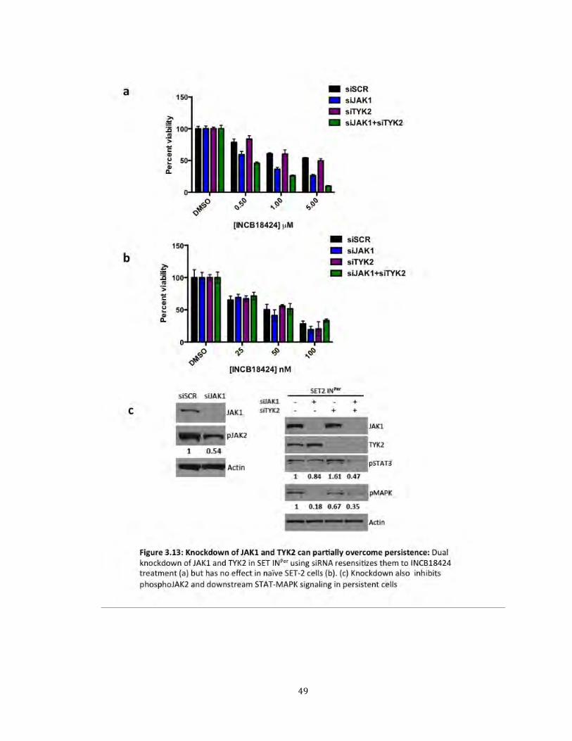
We then asked how dependent the persistent cells were on other JAK kinases by using
siRNA targeting JAK1 and TYK2. Knockdown of JAK1 and TYK2 increased the
sensitivity of persistent cells to JAK inhibitors (Fig 3.13a), whereas the parental cells
remained unaffected by JAK1 and TYK2 knockdown (Fig 3.13b). Additionally, loss of
JAK1 and TYK2 led to decreased downstream signaling and decreased JAK2
phosphorylation in the persistent cells (Fig 3.13c).
We performed in vitro kinase assays to examine kinase activity of JAK2 in inhibitor
persistent cells. JAK2 was immunoprecipitated from naïve and ruxolitinib persistent
SET-2 cells and its catalytic activity was assessed based on the phosphorylation of a
generic substrate, myelin basic protein (MBP). This assay revealed that the
JAK2 heterodimeric complex was more active in persistent cells as compared to the
parental naïve cells (Fig 3.14a). Further, JAK2 in persistent cells could phosphorylate
MBP at concentrations of ruxolitinib sufficient to inhibit JAK2 kinase activity in naive
SET-2 cells (Fig 3.14b). To determine whether JAK1-mediated phosphorylation of JAK2
was insensitive to ruxolitinib, we co-expressed a constitutively active mutant form of
JAK1 (JAK1V658F) with kinase-dead JAK2 (JAK2K882E) in JAK2-deficient γ2A cells.
We observed that JAK1 could transphosphorylate JAK2 in this context and this
phosphorylation could not be completely inhibited by ruxolitinib treatment (Fig 3.14c).
These data suggest that the heterodimer complex in persistent cells retains kinase activity
that is relatively insensitive to JAK inhibitors
49

50

51
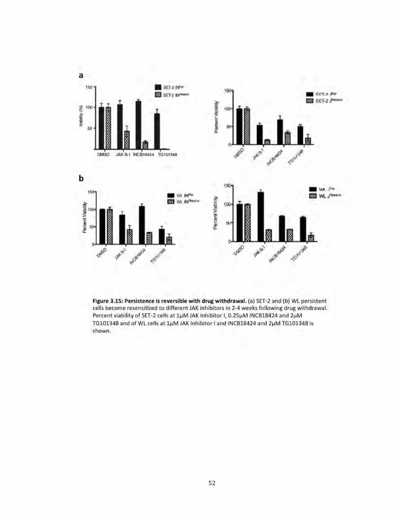
REVERSIBILITY OF PERSISTENCE WITH DRUG WITHDRAWAL
Another important observation we made was that persistence was reversible. After
washing out the drug, the persistent cells became sensitive to inhibitor in 2-4 weeks (Fig
3.15). Treatment with inhibitor led to reduction in phosphorylated JAK2, similar to naïve
cells (Fig 3.16). Furthermore, the resensitized cells no longer showed JAK1 or TYK2
association with phosphoJAK2 (Fig 3.17). Resensitized (JAK2Resens) cells were sensitive
to all three JAK inhibitors, suggesting that patients with MPN may respond to retreatment
with the same or different inhibitor following a brief drug withdrawal.
52

53

54

55
We also observed that JAK2 messenger RNA and JAK2 protein levels were higher in
persistent cells than in parental cells, and returned to lower levels with resensitization
(Fig 3.18). Treatment with ruxolitinib was associated with an increase in JAK2 mRNA
and JAK2 protein expression in MPLW515L-transduced mice. Similarly, comparison of
expression of JAK2 in granulocytes from MPN patients prior to and during ruxolitinib
treatment revealed an increase in JAK2 mRNA levels following drug exposure. (Fig 3.19)
The reversible nature of this phenomenon led us to speculate that there might an
epigenetic basis to the mechanism. Chromatin immunoprecipitation (ChIP) analysis of
the JAK2 locus showed a significant increase in H3K4me3, a modification associated
with active promoters, and a decrease in H3K9me3, a mark more typically associated
with inactive heterochromatin, in persistent cells in comparison with parental cells (Fig
3.20a), which is consistent with a change to a more active chromatin state at the JAK2
locus. However, global H3K4me3 levels in naive and persistent cells remained
unchanged, which is consistent with specific effects on H3K4me3 at the JAK2 locus in
persistent cells (Fig 3.20b).

56

57

58

59
Next, we asked whether overexpression of JAK2 was sufficient to induce persistence in
MPN cells. We generated Ba/F3 stable cells overexpressing MPL-W515L with and
without ectopic JAK2 expression and cultured them in increasing concentrations of
ruxolitinib. Upregulation of JAK2 protein levels did not increase the IC50 of the parental
cells. Although ectopic expression of JAK2 did result in a slight acceleration of the
generation of JAK inhibitor persistence, the viability, growth characteristics, and IC50 of
these cells were similar to cells expressing only MPLW515L with endogenous JAK2 by
4-5 weeks at which time both cell lines were fully persistent (Fig 3.21a) We also made
single cell clones of Ba/F3 EpoR JAK2VF-HA-FLAG cells expressing different amounts
of JAK2 from the transgene due to differences in integration/copy number. These cells
had differing amounts of JAK2, but we did not find any correlation between levels of
JAK2 and IC50 for ruxolitinib (Fig 3.21b). These data suggest that increased JAK2
expression contributes to persistence, but is not sufficient to cause rapid inhibitor
persistence without chronic (2-4 week) JAK inhibitor exposure.

60

61
Given that JAK2 protein levels, and particularly phosphoJAK2 levels, increased with
persistence, we examined whether JAK2 inhibitor persistence was also associated with
post-transcriptional stabilization of total and activated JAK2. We have previously shown
that JAK2 levels decline rapidly on treatment with cycloheximide in JAK2-mutant cells
(Marubayashi et al., 2010). As expected, we noted a time-dependent decrease in
phosphoJAK2 and total JAK2 levels in naive and resensitized WL/SET-2 cells; however,
exposure to cycloheximide did not result in a significant decline in JAK2, or more
notably in phosphoJAK2, in JAK inhibitor persistent cells (Fig 3.22). These data suggest
that chronic treatment with inhibitor results in the stabilization of activated JAK2, which,
combined with increased JAK2 mRNA expression, facilitates the formation of
heterodimers.

62

63
THERAPEUTIC STRATEGIES TO OVERCOME PERSISTENCE
The JAK inhibitors currently in clinical development are Type I inhibitors, which bind
the ATP-binding pocket of JAK2 in the ‘active’ conformation. A recent paper reported
that this mode of binding leads to stabilization of activation loop phosphorylation,
thereby resulting in increased levels of phosphorylated JAK2 (Andraos et al., 2012).
Based on these findings, we asked whether this mechanism was contributing to
development of persistence. We therefore tested the efficacy of BBT-594, a novel type II
inhibitor that engages JAK2 in its inactive conformation and does not contribute to
stabilization of activated JAK2. Treatment with BBT-594 inhibited the proliferation of
persistent cells to a similar extent as the naïve cells (Fig 3.23a). Additionally, activation
of JAK2 and downstream STAT signaling was efficiently inhibited in both naïve and
persistent cells by this compound (Fig 3.23b). Thus, novel agents that bind JAK2 in a
different conformation can be used to overcome JAK inhibitor persistence in MPN.
Taken together, our results suggest that kinase inhibitor persistence can occur through
reversible changes in JAK2 expression and stabilization of activated JAK2 by Type I
inhibitors, which facilitates transphosphorylation by other JAK kinases (Fig 3.24). The
outstanding question remains whether these persistent cells remain dependent on JAK2
for their survival. The next chapter discusses the requirement of JAK2 in naïve and
persistent MPN cells and how this can be leveraged therapeutically to overcome
persistence.

64

65

66
CHAPTER FOUR
REQUIREMENT OF JAK2 IN NAÏVE
AND PERSISTENT MPN CELLS
Pre-clinical and clinical studies have shown that JAK inhibitors are not curative in MPN
and do not effectively reduce the size of the MPN clone. This might be due to incomplete
pathway inhibition at clinically tolerable doses, presence of other disease alleles or
incomplete dependence on JAK2 by the MPN clone. Second site mutations in JAK2,
which might explain the limited efficacy of these drugs, have not been reported in
patients chronically treated with ruxolitinib. In vitro mutagenesis screens have not
identified recurrent resistance alleles of JAK2 at a significant frequency; a majority of
cells can persist in the presence of chronic exposure to a JAK inhibitor (Deshpande et al.,
2012; Marit et al., 2012). The previous chapter elucidated the underlying mechanism for
development of persistence, where JAK2 is activated via the formation of heterodimers
with other JAK kinases including JAK1 and TYK2 (Koppikar et al., 2012). This
phenomenon was observed in cells lines, mouse models as well as in primary samples.
67
This inherent insensitivity of MPN cells to JAK inhibitors led us to evaluate the
requirement of JAK2 in MPN pathogenesis in an in vivo murine model of ET/MF. We
also tested whether JAK inhibitor persistent cells remain dependent on JAK2 for their
survival. Finally, we assessed the efficacy of therapeutic strategies that target degradation
of total JAK2 protein rather than simply inhibition of its kinase activity.
JAK2 IS REQUIRED FOR INITIATION OF MPLW515L-INDUCED DISEASE
Retroviral expression of mutant MPLW515L in hematopoietic cells in vivo results in the
development of a highly penetrant, lethal MPN, characterized by leukocytosis,
thrombocytosis, extramedullary hematopoiesis and extensive bone marrow fibrosis
(Pikman et al., 2006). We decided to evaluate the effect of loss of JAK2 on disease
development in this model. Germline deletion of JAK2 results in embryonic lethality due
to lack of definitive hematopoiesis (Neubauer et al., 1998; Parganas et al., 1998). We
therefore utilized a conditional knockout approach in which JAK2 could be deleted in an
inducible and hematopoietic-specific manner by Cre-recombinase expressed under the
control of the Mx1 promoter (Khn et al., 1995). Bone marrow cells from JAK2f/f Mx1-
Cre+ and Mx1-Cre- mice expressing the CD45.2 congenic marker were transduced with a
GFP-tagged MPLW515L retrovirus and transplanted into irradiated CD45.1 recipients
along with equal number of CD45.1 support bone marrow. Two weeks following
transplantation, we determined engraftment by the presence of GFP positive cells in
peripheral blood. Before the mice developed overt disease in terms of elevated blood
counts, JAK2 was deleted by injection of polyI:polyC (pI:pC). Evaluation of peripheral
blood chimerism revealed that JAK2 deleted cells had a significant survival disadvantage

68
against the CD45.1 wildtype bone marrow (Fig. 4.1). In these mice, white blood cell and
platelet counts remained normal (Fig 4.2a and b) and mutant allele burden measured by
percentage of GFP positive cells was significantly reduced compared to controls (Fig
4.2c). Spleen and liver sizes were also significantly reduced in mice with bone marrow
lacking JAK2 (Fig 4.2d,e). Additionally, bone marrow fibrosis, a hallmark feature of this
MF model, was absent in JAK2 deleted mice (Fig 4.2f). One mouse in the Mx+ cohort
had incomplete deletion of JAK2 as can be seen by presence of the floxed allele in
peripheral blood (Fig 4.3a). This mouse had elevated blood counts and an enlarged spleen
(Fig 4.3b,c) indicating that any residual disease in this model was due to transduced cells
with intact JAK2. These data suggest that JAK2 function is required for all aspects of
disease development in MPL-mediated disease.

69

70

71

72
JAK2 PLAYS A CRITICAL ROLE IN SURVIVAL OF MPN MUTANT CLONE
We then wanted to determine the requirement of JAK2 in maintenance of the disease
clone in the MPLW515L model. Disease establishment usually takes four to six weeks
following transplantation of MPLW515L-transduced bone marrow. JAK2 was excised by
administration of pI:pC at this time point. Similar to previous results, the JAK2 deleted
cells had a significant survival disadvantage as measured by peripheral blood chimerism.
Loss of JAK2 at this stage of disease resulted in significant reduction in leukocytosis and
platelet counts (Fig 4.4a,b). Spleen sizes were also significantly smaller (Fig 4.4d). We
also observed a significant reduction in mutant allele burden, in terms of GFP+ cells (Fig
4.4c). Of note, this reduction in mutant allele burden is not seen even with maximal
kinase inhibition in this same model. Examination of the remaining GFP+ mutant cells in
the bone marrow revealed incomplete excision of JAK2 since we were able to detect the
floxed JAK2 allele in GFP+ sorted cells by PCR (Fig 4.4e). Thus, similar to the previous
result, residual disease was due to mutant cells with intact JAK2. Further, deletion of
JAK2 led to significant decrease in extramedullary hematopoiesis, restoration of splenic
architecture and complete loss of bone marrow fibrosis (Fig 4.5). There was also a
reduction in the megakaryocytic-erythroid progenitor (MEP) compartment and CD11b+
Gr1+ myeloid lineages (Fig 4.6), which are expanded in this model of MPN. These data
demonstrate that conditional deletion of JAK2 after establishment of disease can prevent
further progression. Thus, JAK2 is required for maintenance of the mutant MPN clone in
this model.

73

74

75

76
DELETION OF JAK2 IS MORE EFFECTIVE THAN JAK INHIBITOR TREATMENT IN VIVO
We decided to directly compare the efficacy of genetic loss of JAK2 to JAK kinase
inhibitor in our mouse model. Bone marrow from JAK2f/f Mx1-Cre+ mice was
retrovirally transduced with MPLW515L-IRES-GFP and transplanted into lethally
irradiated recipients. After disease establishment, mice were randomized to either receive
vehicle, 60mg/kg ruxolitinib twice daily or pI:pC to delete JAK2. As reported previously
(Koppikar et al., 2010), although drug treatment improved blood counts, there was no
reduction in mutant allele burden in terms of GFP positive cells. In contrast, deletion of
JAK2 led to significant decrease in the percentage of GFP+ cells in the bone marrow (Fig
4.7a). Deletion of JAK2 was also more effective at reducing blood counts and spleen size
as compared to drug treatment (Fig 4.7b,c). Analysis of myeloid and progenitor
populations revealed that loss of JAK2 leads to significant reduction in MEP and
CD11b+Gr+ proportions with a significant decrease in the contribution of mutant (GFP+)
cells (Fig 4.7d). These results indicate that deletion of JAK2 is superior to JAK2 kinase
inhibitor treatment alone at reducing disease burden in this model.

77

78
PERSISTENT CELLS REMAIN DEPENDENT ON JAK2
Using a genetic loss of function model of JAK2, we demonstrated that JAK2 is
indispensable for development and maintenance of MPLW515L-induced disease in vivo.
We then investigated whether JAK inhibitor persistent cells that can survive in the
presence of chronic drug exposure remain dependent on expression of JAK2. Knockdown
of JAK2 using two different short hairpins in naïve and persistent cell lines led to growth
suppression (Fig 4.8a) and inhibition of downstream STAT3/STAT5 signaling (Fig 4.8b).
We then asked whether we could leverage this dependency therapeutically to overcome
inhibitor persistence. We have previously shown that JAK2 is an Hsp90 client protein
and treatment with PU-H71, an Hsp90 inhibitor, leads to degradation of total and
activated JAK2 and inhibition of downstream signaling in MPN cells (Marubayashi et al.,
2010). We found that JAK inhibitor persistent cells remained sensitive to PU-H71 (Fig
4.9a) and drug treatment led to efficient degradation of JAK2 and abrogation of
downstream signaling (Fig 4.9b). These data indicate that JAK2 can serve as a scaffold
for transactivation and downstream signaling even in the context of inhibition of kinase
activity.

79

80

81
COMBINATION OF JAK AND HSP90 INHIBITION IS MORE EFFICACIOUS THAN JAK
INHIBITOR MONOTHERAPY: Based on cell line data and our previous work, we decided to
test the efficacy of a combination of ruxolitinib and PU-H71 in the MPLW515L model of
ET/MF. The dosing regimens tested included vehicle, two different doses of ruxolitinib
monotherapy, combined JAK/HSP90 inhibition from the onset and ruxolitinib followed
by the addition of PU-H71 after initial response (Fig 4.10a). At 2 weeks following start of
treatment, the combination group displayed a significant reduction in white blood cell
and platelet counts (Fig. 4.10b,c) compared to either low-dose or high-dose ruxolitinib
alone. Combination treatment also led to further reduction in spleen size compared to
ruxolitinib monotherapy (Fig 4.10d). We also observed a decrease in total and
phosphorylated JAK2 levels and more potent inhibition of downstream signaling
effectors including STAT3, STAT5 and MAPK by immunoblotting and
immunohistochemistry in the combination treatment arm (Fig 4.11). Combination
treatment also led to histopathological improvement in terms of reduction in bone
marrow and spleen cellularity, decrease in megakaryocytes as well as a reduction in bone
marrow fibrosis (Fig 4.12). These results were consistent over the entire 4-week drug
trial, with significant improvements in blood counts and organomegaly seen in
combination arms as compared to ruxolitnib alone. The ruxolitinib dose was increased
from low dose (30mg/kg) to high dose (90mg/kg) in a subset of the combination treated
mice, which proved to be the most efficacious strategy (Fig 4.13).

82

83

84

85

86
GENETIC OR PHARMACOLOGICAL LOSS OF JAK2 CAN OVERCOME PERSISTENCE IN VIVO:
We then asked whether genetic deletion of JAK2 following initial ruxolitinib treatment
could be effective at reducing disease burden. Although ruxolitinib treatment improved
blood counts and reduced spleen size, we did not observe a reduction in mutant allele
burden indicating persistence of MPN clone. Deletion of JAK2 following 2 weeks of
ruxolitinib treatment led to significant reduction in mutant allele burden (Fig 4.14a),
blood counts (Fig 4.14b) and spleen size (Fig 4.14c) as compared to mice that continued
to receive drug. Further, deletion of JAK2 after long-term ruxolitinib treatment (6weeks)
prevented disease relapse; blood counts and spleen weights remained low for up to 3
weeks following cessation of treatment (Figure 4.15a,b). The percentage of GFP positive
cells, which remained high after 6 weeks of ruxolitnib treatment, was also decreased by
deletion of JAK2 (Fig. 4.15c). These results indicate that deletion of JAK2 in vivo can
successfully eliminate mutant MPN cells, which cannot be achieved solely by kinase
inhibition of JAK2.

87

88

89
These results could also be recapitulated by targeting of JAK2 by pharmacological
degradation. After two weeks of 30mg/kg ruxolitinib treatment, we added 75/mg PU-H71
treatment to a subset of mice. Two weeks of combination treatment resulted in significant
reduction of leukocytosis and thrombocytosis (Fig 4.16a,b). Addition of PU-H71 to
ruxolitnib monotherapy also led to a further decrease in splenomegaly (Fig 4.16c).
We have previously demonstrated that mononuclear cells from patients treated with
ruxolitnib are insensitive to JAK2 kinase inhibition ex vivo and exhibit persistent
downstream signaling even after treatment with a JAK inhibitor (Fig 3.6). We asked
whether treatment with an Hsp90 inhibitor could overcome this persistence. Mononuclear
cells were isolated from two different MF patients that were receiving ruxolitinib therapy.
Treatment of these cells with PU-H71 led to degradation of total and activated JAK2 and
abrogation of downstream STAT and MAPK signaling (Fig 4.16d). Thus, PU-H71 is
effective in inhibiting JAK-STAT signaling in primary samples that are insensitive to
JAK inhibition. Taken together, our results indicate that Hsp90 inhibition can overcome
JAK inhibitor persistence in cell lines, pre-clinical models and primary MPN samples.

90

91
JAK2 IS REQUIRED FOR NORMAL MYELOPOIESIS AND STEM CELL FUNCTION
Thus far, our data indicate that JAK2 has an indispensable role in the survival and
maintenance of the MPN mutant clone. Complete loss of JAK2, as can be achieved by
genetic deletion, can potentially be curative in this disease. However, loss of JAK2 in
normal adult hematopoietic lineages has not been investigated in detail. This has
important implications for JAK inhibitor therapy since in the clinic; JAK inhibition is
associated with dose-limiting cytopenias. We therefore decided to use the conditional
knockout model of JAK2 to evaluate the role of JAK2 in normal hematopoiesis. We
performed competitive bone marrow transplant experiments in which CD45.2 JAK2f/f
Mx1-Cre+ or Cre- cells along with equal number of CD45.1 wild type cells were injected
into CD45.1 irradiated donor mice. JAK2 was deleted by administration of pI:pC
following bone marrow engraftment and chimerism of mature myeloid, B and T lineages
was monitored in peripheral blood for 16 weeks. Deletion of JAK2 led to significant
reduction in overall CD45.2 chimerism (Fig 4.17a). Further examination revealed a
dramatic reduction in CD11b+ Gr1+ myeloid lineages within 3 weeks of JAK2 deletion.
B and T cell lineages showed a more gradual decline over the 16-week period (Fig 4.17
b,c,d,e). We also evaluated the effect of loss of JAK2 in hematopoietic stem and
progenitor cells and found a significant reduction in CD45.2 contribution to the myeloid
progenitor (Lin-ckit+sca1-), short-term (Lin-ckit+sca1+CD48-CD150-) and long-term
(Lin-ckit+sca1+CD48-CD150+) stem cell compartment (Fig 4.17f). Taken together, these
results suggest that JAK2 is required for survival of mature myeloid lineages and also
plays an important role in maintenance of the stem cell and progenitor compartment..
Importantly, white blood cell counts, platelet counts and hematocrit remained normal in

92
these mice (data not shown) indicating that in this experimental system, support cells
with intact JAK2 can sustain normal hematopoiesis. Since MPN patients are also
chimeric for the JAK2V617F mutation (Jamieson et al., 2006), our results indicate that
long-term treatment with mutant-specific JAK inhibitors or agents that degrade JAK2 in a
tumor-specific manner will likely be well-tolerated and could eliminate primitive disease-
initiating cells in MPN patients.
Taken together, these data demonstrate that despite the rapid development of persistence
in MPN cells with chronic JAK inhibitor exposure, these cells still remain highly
dependent on JAK2 for their growth and survival. Treatment with an Hsp90 inhibitor
such as PU-H71, which degrades JAK2 specifically in mutant cells, can successful
overcome persistence in cells lines, mouse models as well as in primary samples. Thus,
JAK2 remains a bona fide target for therapy in MPN and strategies that combine JAK
kinase inhibition with JAK2 degradation can be beneficial for patients.

93

94
CHAPTER FIVE
DISCUSSION
The majority of patients with the classical MPN; PV, ET and MF harbor somatic
activating mutations in the tyrosine kinase JAK2, thereby making it an attractive
therapeutic target in these diseases. However, the relative contribution of JAK2 signaling
to disease phenotype, malignant proliferation, and disease progression has not been fully
delineated. The dual JAK1/JAK2 inhibitor, ruxolitinib, was approved by the FDA in
2011 for the treatment of MF, and several other compounds are in late-stage clinical
testing. Although these drugs alleviate many of constitutional symptoms in patients, they
have been ineffective at reducing the MPN mutant clone. The previous two chapters
discuss (i) mechanisms by which MPN cells can survive in spite of chronic inhibition of
JAK2 kinase activity and (ii) establish that JAK2 plays a critical role in MPN
pathogenesis and disease phenotype. In this chapter, I discuss some of the biological and
clinical implications of these findings.

95
OVERCOMING JAK INHIBITOR PERSISTENCE
We show that MPN cells can persist in the presence of chronic JAK inhibition by
reactivating downstream STAT-MAPK signaling via the formation of heterodimers
between JAK2 and the JAK kinases JAK1 and TYK2. This phenomenon is observed in
MPN cell lines, murine models and primary samples from patients treated with
ruxolitinib. Additionally, this phenomenon is reversible suggesting that MPN patients
could benefit from being re-exposed to drug after a brief withdrawal.
The formation of heterodimers is facilitated by the stabilization of phosphorylated JAK2
by Type I inhibitors such as ruxolitinib, which engage JAK2 in its active conformation.
In MPN cell lines, persistence can be overcome by type II inhibitors of JAK2, which
stabilize JAK2 in its inactive conformation and lead to a decrease in activation loop
phosphorylation. In our studies, cells that were persistent to ruxolitinib and JAK Inhibitor
I remained sensitive to treatment with BBT-594, a type II JAK inhibitor, and downstream
signaling was inhibited at similar concentrations as the parental cells. Experiments
evaluating the efficacy of newer, orally bioavailable type II JAK inhibitors in pre-clinical
models of MPN are ongoing. It will also be important to investigate whether MPN cells
can become persistent to this new class of inhibitors.
De novo mechanisms of resistance to JAK inhibitors should also be considered when
assessing efficacy. We observed that mononuclear cells isolated from patients being
treated with ruxolitinib are insensitive to ex vivo treatment with JAK inhibitors as
compared to inhibitor-naïve controls. In a recent report, Kalota et al. reported similar
findings in that granulocytes from myelofibrosis patients are relatively insensitive to ex

96
vivo JAK2 inhibition in terms of reduction in levels of phosphorylated STAT3 and
STAT5 as compared to patients with PV and ET as well as normal controls (Kalota et al.,
2013). Interestingly, this was observed in JAK2 inhibitor-naïve patients suggesting that
certain subgroups of patients might have de novo mechanisms of resistance/persistence to
JAK inhibitors. For example, pre-existing heterodimers between JAK2 and other JAK
kinases in these patients might be contributing to this phenomenon, which should be
further investigated.
ROLE OF JAK2 IN MPN PATHOGENESIS
The JAK2V617F mutation has been identified in the majority of patients with MPN. In
vivo expression of this allele in murine bone marrow transplant systems and in genetic
knockin models recapitulates many features of human MPN disease. This suggests that
this pathway plays an important role in disease pathogenesis. However, kinase inhibition
of JAK2 has not resulted in impressive molecular responses in preclinical and clinical
setting, as has been observed with ABL kinase inhibitors in CML. Although MPN
patients experience an improvement in constitutional symptoms, treatment with JAK
inhibitors is not curative and does not decrease the size of the MPN clone. Additionally,
the occurrence of secondary resistance mutations in response to chronic inhibition is
often considered a hallmark of effective targeted therapy. This has been observed in
numerous cases including ABL kinase inhibitors in CML (Gorre et al., 2001), EGFR
inhibitors in lung cancer (Pao et al., 2005) and FLT3 inhibitors in acute myeloid leukemia
(Smith et al., 2012). However, this has not been the case in the use of JAK2 inhibitors in
the treatment of MPN. There have been no reports of second site mutations in JAK2 in

97
patients treated with ruxolitinib. In vitro genetic screens in JAK2 mutant cell lines have
identified alleles that confer resistance to JAK2 inhibitors (Weigert et al., 2012a)
(Deshpande et al., 2012; Marit et al., 2012). However, these do not occur at a significant
frequency and a majority of cells can persist/survive in the presence of inhibitor without
acquiring second site mutations. These observations led us to question if JAK2
represented a ‘driver’ lesion in MPN and whether targeting JAK2 remained a viable
therapeutic strategy.
In our genetic studies, we demonstrate that JAK2 is essential in an in vivo model of
ET/MF by deleting JAK2 in an inducible, tissue-specific manner at different stages of
disease development. Loss of JAK2 leads to significant improvements in blood counts,
organomegaly and bone marrow fibrosis. More importantly, deletion of JAK2 results in a
dramatic reduction in mutant allele burden, which is not seen with maximal JAK2 kinase
inhibition in this model (Koppikar et al., 2010). Further, JAK inhibitor persistent cell
lines remain dependent on JAK2 expression for their growth and survival. Knockdown of
JAK2 using short hairpins leads to decrease in cell proliferation and inhibition of
downstream signaling in naïve and persistent MPN cells. These data suggest that MPN
cell lines require JAK2, at least in part as a scaffold to maintain downstream signaling,
even in the context of inhibition of its catalytic activity. Thus, targeting JAK2 remains a
viable therapeutic option in the treatment of MPN and novel strategies that result in
degradation/loss of total protein should be evaluated.

98
TARGETING JAK2 IN NAÏVE AND PERSISTENT MPN CELLS
There are several active agents that have been found to degrade JAK2 and inhibit
downstream signaling in MPN. The following sections discuss some of these approaches.
Hsp90 inhibitors
Heat shock protein 90 (Hsp90) is a molecular chaperone protein that plays a critical role
in maintaining protein homeostasis in response to various stimuli such as genomic
instability, proteotoxic stress, changes in nutrient and oxygen levels. Cancer cells are
particularly dependent on Hsp90 since this protein plays an important role in regulating
the stability and function of mutated or amplified oncoproteins as well as protecting the
cell from various stresses brought about by malignant transformation (Neckers and
Workman, 2012). Hsp90 client proteins include several receptors, kinases and
transcription factors involved in cancer such as HER2 (Miller et al., 1994), EML4-ALK,
mutant EGFR (Normant et al., 2011; Shimamura et al., 2005), mutant BRAF (Grbovic et
al., 2006) and activated AKT (Solit et al., 2003). Inhibition of Hsp90 by small molecules
leads to degradation of its client proteins and in the case of oncogenic targets, growth
inhibition or cytotoxicity in the cells that are dependent on them.
Our lab has previously demonstrated that JAK2 is a highly sensitive Hsp90 client protein
and treatment of MPN cells with Hsp90 inhibitors leads to degradation of JAK2 and
inhibition of downstream signaling at clinically achievable doses. PU-H71, a novel
purine-scaffold Hsp90 inhibitor, was highly efficacious in pre-clinical models of MPN
and resulted in lineage-specific reduction of myeloproliferation and increased survival.

99
Although both mutant and wild type JAK2 are Hsp90 client proteins, PU-H71 is
preferentially retained in mutant cells, thus making it a tumor-selective inhibitor
(Marubayashi et al., 2010). Another Hsp90 inhibitor, AUY922, has been shown to
overcome genetic resistance to JAK inhibitors in cell lines and xenograft models of JAK2
mutant B-ALL (Weigert et al., 2012a). AUY922 was also synergistic with the JAK
inhibitor, TG101348, in inducing apoptosis in primary MPN cells. It was also effective
against JAK inhibitor resistant cell lines (Fiskus et al., 2011). We found that the JAK
inhibitor persistent cell lines remained sensitive to PU-H71 with IC50 values similar to the
parental naïve cells. These findings also spurred us to test the efficacy of combination
treatment with PU-H71 and ruxolitinib in pre-clinical models of MPN. We found that
addition of an Hsp90 inhibitor to ruxolitinib either at the beginning or following JAK
inhibitor therapy led to significant improvements in leukocytosis, splenomegaly and bone
marrow fibrosis as compared to JAK inhibitor monotherapy, without adverse side effects.
We also observed more potent inhibition of downstream STAT-MAPK signaling in
splenocytes from mice receiving PU-H71/ruxolitinib combination treatment. Importantly,
we demonstrated that persistent signaling observed in cells from ruxolitinib treated MPN
patients, could be inhibited by treatment with PU-H71. Of note, PU-H71 treatment led to
degradation of JAK1 in MPN cells, suggesting that this drug could interfere with
formation of heterodimers associated with persistence in these cells. Taken together,
these findings suggest that PU-H71 can overcome JAK inhibitor persistence in MPN cells
and provide a compelling rationale for combining these inhibitors in clinical trials for
MPN.

100
HDAC inhibitors
Several studies have reported higher expression and enzymatic activity of histone
deacetylases (HDACs) in primary MPN cells, which correlates with the degree of
splenomegaly in patients (Skov et al., 2012; Wang et al., 2008). In addition to regulating
gene expression via the deacetylation of histone substrates, HDACs can influence the
function of myriad other proteins in cells including a number of transcription factors such
as p53 (Luo et al., 2000; Murphy et al., 1999), STAT3 (Yuan et al., 2005) and hormone
receptors (Gaughan et al., 2002; Wang et al., 2001). Importantly, Hsp90 is also a
substrate of HDAC6 and aberrant Hsp90 acetylation has been show to affect maturation
of client proteins (Kovacs et al., 2005) (Bali et al., 2005). HDAC inhibitor treatment
results in degradation of JAK2 and inhibition of growth and downstream signaling in
MPN cells (Guerini et al., 2007; Wang et al., 2009). This effect might be due to
disruption of the binding of JAK2 and HSP90 and through additional mechanisms
relating to the pleiotropic role of HDAC proteins on gene expression and protein
trafficking in MPN cells. In separate phase I/II trials with the HDAC inhibitors
panobinostat or givinostat, MF patients experienced improvement in systematic
symptoms and reduction in splenomegaly (DeAngelo et al., 2013; Finazzi et al., 2013;
Mascarenhas et al., 2011; Rambaldi et al., 2010). Cotreatment of MPN cells with the
JAK2 inhibitor TG101209 and panobinostat led to synergistic induction of apoptosis in
MPN cells (Wang et al., 2009). Similarly, combination of panobinostat with ruxolitinib
showed greater activity than either agent alone in a JAK2V617 murine model (Evrot et
al., 2013). There is an ongoing Phase I trial assessing the efficacy of combined ruxolitinib
and panobinostat treatment.

101
Based on our studies, we hypothesized that epigenetic regulation of JAK2 expression
might also contribute to development of persistence since we observed an increase in
activated chromatin marks at the JAK2 locus in persistent cells. JAK2 expression was
also increased at the protein and mRNA level. This might be due to a positive feedback
loop in which activated JAK2 can promote its own expression by phosphorylating histone
H3Y41 at the JAK2 locus (Rui et al., 2010b) Previous work attributed persistence in
EGFR inhibitor-insensitive ‘drug-tolerant persisters’ to an altered chromatin state, which
made these cells highly sensitive to HDAC inhibitors (Sharma et al., 2010). In the future,
we plan on evaluating whether HDAC inhibitors can overcome persistence by altering
transcriptional regulation of JAK2 and other target genes.
Inhibition of STATs
Expression of JAK2V617F and MPLW515L results in constitutive activation of the
STAT family of transcription factors, mainly STAT3 and STAT5, leading to increased
expression of STAT target genes involved in survival and proliferation including Bcl-xL,
Ccnd1 and Myc. Recently, several groups have reported that STAT5 is indispensable for
development of JAK2V617F-mediated transformation and disease development
(Funakoshi-Tago et al., 2010; Walz et al., 2012; Yan et al., 2012). Deletion of both
isoforms of STAT5, STATa and STAT5b, resulted in normalization of blood counts,
spleen size and myeloid progenitor expansion. Further, loss of STAT5 also led to
abrogation of Epo-independent erythroid colony formation, a hallmark feature of PV
(Yan et al., 2012). Conversely, in vivo expression of a constitutively active isoform of

102
STAT5 leads to a lethal myeloproliferative phenotype (Kato et al., 2005). Unpublished
work from our laboratory shows that genetic deletion of STAT3 in the MPLW515L
model reduces blood counts and splenomegaly and improves survival. Importantly, we
demonstrate that JAK inhibitor persistent cells do not engage an alternate pathway but in
fact reactivate downstream STAT signaling via formation of heterodimers between JAK2
and JAK1/TYK2. Taken together, these results suggest that STAT5 plays a crucial role in
MPN pathogenesis and represents an attractive target for therapeutic intervention.
Cell-based screening assays have led to the identification of several molecule inhibitors
of STAT5 (Nelson et al., 2011a). One of these compounds, pimozide, decreases STAT5
phosphorylation and expression of STAT5 target genes as well as induces cell cycle
arrest and apoptosis in BCR-ABL and JAK2V617F mutant MPN cells (Bar-Natan et al.,
2012; Nelson et al., 2011b). Importantly, it remains effective in the presence of the
T315I/Bcr-Abl mutation, which confers resistance to most available Abl kinase inhibitors
(Nelson et al., 2011b). Other strategies to target the STATs include decoy
oligonucleotides that compete the protein away from its target sequences (Sen et al.,
2012), (Wang et al., 2011), small molecules that prevent DNA binding (Turkson et al.,
2005) and inhibitors of STAT dimerization, which is essential for DNA binding (Page et
al., 2012; Schust et al., 2006). Experiments evaluating the efficacy of these approaches in
pre-clinical MPN models, particularly in JAK inhibitor persistent cells, are warranted.
ROLE OF CYTOKINES IN RESPONSE TO JAK INHIBITOR THERAPY
Since our data suggest that MPN cells can rapidly become insensitive to currently
available JAK inhibitors, understanding exactly how patients benefit from these drugs

103
remains an outstanding question in the field. MPN patients have high levels of pro-
inflammatory serum cytokines, which predict response to therapy and correlate with
shortened survival (Tefferi et al., 2011; Vaidya et al., 2012). An important observation
made by several groups is that JAK kinase inhibitor treatment in MF patients and in
murine models is associated with reduction in the elevated levels of cytokines (Koppikar
et al., 2010; Tyner et al., 2010; Verstovsek et al., 2012c). This suggests that some of the
clinical benefits of JAK inhibitors might be due to suppression of systemic inflammation
and not from complete inhibition of constitutive signaling in MPN cells.
Increasing evidence suggests that both autocrine/paracrine as well as stromal secretion of
cytokines plays an important role in the development of the MPN phenotype. Fleischman
and colleagues reported that JAK2V617F mutant cells have increased expression of
inflammatory cytokine tumor necrosis factor alpha (TNFα). Exposure to this cytokine in
vitro leads to preferential expansion of JAK2V617F mutant cells compared to normal
cells in mice and in primary samples. Further, reconstitution of JAK2V617 in a TNFα
null background resulted in amelioration of disease (Fleischman et al., 2011). Cytokines
like hepatocyte growth factor (HGF) and interleukin-11 (IL-11) were found to be
overexpressed in PV patients and contributed to growth of erythroid colonies, although
their effect was independent of JAK2V617F expression (Boissinot et al., 2011). Another
study showed that JAK2 mutant myeloid cells have increased secretion of oncostatin M
(OSM), which can stimulate production of angiogenic cytokines by fibroblasts and
endothelial cells in the stroma (Hoermann et al., 2012). Ectopic overexpression of
thrombopoietin in hematopoietic cells is sufficient to induce myeloproliferation and bone

104
marrow fibrosis in mice (Kakumitsu et al., 2005; Villeval et al., 1997; Yan et al., 1996).
Recent studies have also shown that cytokines including interleukin-6 (IL-6), fibroblast
growth factor (FGF) and chemokine C-X-C-motif ligand 10 (CXCL-10)/IFN-γ-inducible
10-kD protein (IP-10) can promote survival of JAK2V617F mutant cells. Using a
coculture platform, this group demonstrated that these cytokines were secreted by bone
marrow-derived stromal cells. Further, these cytokines were able to provide a significant
survival advantage to MPN cells in response to JAK inhibitor treatment (Manshouri et
al., 2011). Taken together, these studies suggest that cytokines secreted by both mutant
and stromal cells play an important role in MPN pathogenesis and their pleiotropic
effects must be considered when assessing the response to JAK inhibitors and other MPN
therapies.
Since clinically available JAK inhibitors are not mutant specific, they might be inhibiting
cytokine signaling in normal cells with wild type JAK2, which might also contribute to
their efficacy. This hypothesis can be investigated by using inhibitor resistant JAK2
alleles, such as the Y931C mutation reported by Hornakova et al. (Hornakova et al., 2011)
and others. Testing the efficacy of JAK inhibitors in murine bone marrow transplant
models where either diseased or non-mutant cells coexpress an inhibitor resistance allele
will allow us to distinguish between JAK2 inhibition in mutant versus normal cells.
Further, this hypothesis might also explain the improved efficacy of JAK and Hsp90
inhibitor combination treatment where ruxolitinib might be inhibiting pro-inflammatory
cytokines in the non-mutant stromal cells and PU-H71 might be providing mutant-
specific inhibition of JAK2.

105
JAK1 is also an important mediator of signaling by several pro-inflammatory cytokines
such as the interferons and IL-6 (Rodig et al., 1998). Although ruxolitinib can also
efficiently inhibit JAK1 in cell-free assays, our data suggest that JAK1 forms a
heterodimer with JAK2, which is insensitive to inhibition by ruxolitinib. However,
further structural studies of the JAK1/JAK2 heterodimers will be required to fully
understand this mechanism. Interestingly, SAR302503, a more specific JAK2 inhibitor,
does not lead to a significant reduction in serum cytokines indicating that JAK1 might be
the main mediator of cytokine signaling in MPN (Pardanani et al., 2011a). Conditional
knockout murine models of JAK1 will also be useful in elucidating the contribution of
JAK1 to disease phenotype and to the response to inhibitor therapy.
MAJOR HURDLES IN JAK INHIBITOR MONOTHERAPY
Our experiments with the genetic deletion of JAK2 in a murine model of ET/MF
demonstrated the critical role of JAK2 in pathogenesis and phenotype of MPN. Thus,
targeting JAK2 remains a viable option in the treatment of these diseases. However, the
JAK-STAT pathway is a crucial regulator of hematopoiesis and JAK2 is the major kinase
required for erythropoietin receptor signaling and normal red blood development
(Neubauer et al., 1998; Parganas et al., 1998). Our studies demonstrate that JAK2 plays
an important role in myelopoiesis and maintenance of the stem cell compartment in adult
hematopoietic tissues. The JAK inhibitors in current clinical development are not
specific for mutant JAK2 and can also efficiently inhibit wild type JAK2. Therefore,
using doses that are capable of inhibiting mutant JAK2 activity is bound to also have
adverse effects on normal hematopoiesis. This has been borne out in the clinic, where

106
JAK2 inhibitors have been associated with dose limiting toxicities including anemia and
thrombocytopenia (Verstovsek et al., 2010). Thus, the limited efficacy of JAK inhibitors
might also be due to insufficient inhibition of the pathway at clinically tolerable doses.
However, the crystal structure of the wild type and JAK2V617F mutant pseudokinase
domain was recently resolved. This should help inform rational drug design of molecules
that can selectively inhibit mutant JAK2, thereby sparing normal hematopoiesis. The
JAK2V617F mutation is particularly amenable to mutant-specific targeting since it
occurs in a vast majority of patients with MPN.
There are some evidence from mouse models and from MPN patients that expression of
JAK2V617 in hematopoietic stem cells does not lead to significant expansion of this
compartment, but rather to increased proliferation of more differentiated myeloid and
erythroid lineages (Anand et al., 2011; Li et al., 2010). However, several studies have
shown that long-term hematopoietic stem cells are the disease-initiating cells in mice and
in MPN patients (James et al., 2008; Mullally et al., 2012). Also, the JAK2V617F
mutation has been shown to be a late genetic event in a subset of MPN patients
(Kralovics et al., 2006) and mutations in other genes such as TET2, ASXL1 and EZH2
might be responsible for the early clonal expansion associated with these diseases
(Vainchenker et al., 2011). Taken together, these findings suggest that JAK2
monotherapy might not be sufficient to eradicate the disease initiating cells in at least a
subset of MPN patients.

107
TARGETING ALTERNATE PATHWAYS IN MPN
Pre-clinical and clinical data suggest that currently available JAK inhibitors have a
limited therapeutic window and will likely not be curative for MPN. Therefore, there is a
need to identify additional pathways that might be involved in the development and
maintenance of the MPN mutant clone, which could then be targeted in combination with
JAK2 for improved therapeutic benefit for MPN patients. In addition to enhanced JAK-
STAT signaling, MPN cells display activation of other oncogenic pathways including
MAPK and mTOR/PI3K signaling. In a phase I/II trial evaluating the efficacy of the
mTOR inhibitor, everolimus, 60% of MF patients experienced improvement in
constitutional symptoms and decrease in spleen enlargement albeit to a lesser degree than
observed with JAK inhibitors. However, it did not lead to a decrease in mutant allele
burden or significant changes in the cytokine profile of these patients (Guglielmelli et al.,
2011). Cotreatment with an mTOR and JAK inhibitor had synergistic activity against
JAK2 mutant cell lines and reduced Epo-independent colony formation of cells from PV
patients (Bogani et al., 2013). In another study, treatment of cultured as well as primary
MPN cells with the dual PI3K/mTOR inhibitor BEZ235 combined with the JAK inhibitor
SAR302503 had synergistic effects on induction of apoptosis and inhibition of colony
growth in cultured and primary MPN cells as compared to normal CD34+ cells (Fiskus et
al., 2013). BEZ235 was also effective against a cell line that had been made resistant to
TG101209 (Fiskus et al., 2013). Similar results were also reported with the combination
of JAK2 inhibitors with a MEK inhibitor, AZD6244 (Fiskus, 2010; Suryani, 2012).
Treatment with an allosteric Akt inhibitor, MK-2206 led to cell growth and induction of
apoptosis in MPN cell lines, along with decreased colony formation in primary MF cells.

108
It was also efficacious in pre-clinical murine models, resulting in decreased
organomegaly and megakaryocyte burden (Khan et al., 2013)
Recently, developmental pathways such as Hedgehog, Wnt and Notch have been shown
to play a role in development of myeloid malignancies and remain an active area of
research as possible therapeutic targets. Targeting β-catenin by either genetic deletion or
pharmacological inhibition in combination with imatinib treatment abrogates disease-
initiating cells in a Bcr-Abl model of CML (Heidel et al., 2012). The Hedgehog (Hh)
pathway is also reported to play an important role in the maintenance of the stem cell
compartment in CML. (Dierks et al., 2008; Zhao et al., 2009a). Combination treatment
with a hedgehog inhibitor vismodegib and ABL kinase inhibitor, ponatinib was found to
be efficacious in xenograft models of therapy-resistant BCR-ABL positive leukemia
(Katagiri et al., 2013). Preliminary data from our lab indicates that this pathway is
activated in the MPLW515L murine BMT model of ET/MF as well as in primary MPN
samples. Importantly, in pre-clinical models, combination treatment with a Smoothened
inhibitor, LDE-225 and ruxolitinib was more effective at reducing blood counts and bone
marrow fibrosis as compared to ruxolitinib alone. In Phase I trials of the Hh inhibitor PF-
04449913 in hematological malignancies including MPN, 4/5 MF patients attained stable
disease while 1 experienced a clinical response including a reduction in spleen size
(Jamieson, 2011). There are several early stage clinical trials testing the efficacy of
combination therapies with JAK and Hh inhibitors in myelofibrosis, which might provide
additional benefits for patients.

109
THE FUTURE OF MPN THERAPY
Although JAK kinase inhibitors have been approved for treatment of MPN, current
agents are not effective at reducing the size of the MPN clone and therefore do not offer
the potential for long term remissions or cure. MPN cells can rapidly develop persistent
to chronic kinase inhibition but remain dependent on expression of JAK2 for growth and
survival. Agents that lead to JAK2 degradation (Hsp90 inhibitors or histone deacetylase
inhibitors), inhibition of downstream targets (STAT inhibitors) or that retain the ability to
inhibit JAK2 in persistent cells (type II JAK inhibitors) have the potential to improve
therapeutic efficacy in patients with MPN. However, since JAK2 plays a critical role in
normal hematopoiesis, the development of mutant-specific JAK inhibitors that will spare
normal cells might offer a viable therapeutic option to increase efficacy and therapeutic
window. Targeting other proliferative and pro-survival pathways such as MAPK and
Akt/PI3K/mTOR, which are activated in MPN cells, might also be beneficial. Since JAK
inhibitors by themselves do not seem to eliminate disease-initiating MPN cells, drugs
targeting developmental pathways that are involved in the maintenance of the stem cell
compartment should also be evaluated. Finally, it would also be desirable to inhibit
cytokine signaling by targeting JAK1 and other JAK kinases in normal and mutant cells
to reduce systemic inflammation and constitutional symptoms associated with MPN,
particularly MF. In conclusion, findings from our lab and others suggest that a
combination treatment regimen that targets all these aspects of disease will likely be most
beneficial and potentially curative for patients with MPN.

110
BIBLIOGRAPHY
Akada, H., Yan, D., Zou, H., Fiering, S., Hutchison, R.E., and Mohi, M.G. (2010). Conditional expression of heterozygous or homozygous Jak2V617F from its endogenous promoter induces a polycythemia vera-like disease. Blood 115, 3589-3597.
Anand, S., Stedham, F., Gudgin, E., Campbell, P., Beer, P., Green, A.R., and Huntly, B.J. (2011). Increased basal intracellular signaling patterns do not correlate with JAK2 genotype in human myeloproliferative neoplasms. Blood 118, 1610-1621.
Andraos, R., Qian, Z., Bonenfant, D., Rubert, J., Vangrevelinghe, E., Scheufler, C., Marque, F., Regnier, C.H., De Pover, A., Ryckelynck, H., et al. (2012). Modulation of activation-loop phosphorylation by JAK inhibitors is binding mode dependent. Cancer discovery 2, 512-523.
Aranaz, P., Hurtado, C., Erquiaga, I., Miguéliz, I., Ormazábal, C., Cristobal, I., García-Delgado, M., Novo, F.J., and Vizmanos, J.L. (2012). CBL mutations in myeloproliferative neoplasms are also found in the gene’s proline-rich domain and in patients with the V617FJAK2. Haematologica 97, 1234-1241.
Azam, M., Latek, R.R., and Daley, G.Q. (2003). Mechanisms of autoinhibition and STI-571/imatinib resistance revealed by mutagenesis of BCR-ABL. Cell 112, 831-843.
Bali, P., Pranpat, M., Bradner, J., Balasis, M., Fiskus, W., Guo, F., Rocha, K., Kumaraswamy, S., Boyapalle, S., Atadja, P., et al. (2005). Inhibition of histone deacetylase 6 acetylates and disrupts the chaperone function of heat shock protein 90: a novel basis for antileukemia activity of histone deacetylase inhibitors. The Journal of biological chemistry 280, 26729-26734.
Ballen, K.K., Shrestha, S., Sobocinski, K.A., Zhang, M.J., Bashey, A., Bolwell, B.J., Cervantes, F., Devine, S.M., Gale, R.P., Gupta, V., et al. (2010). Outcome of transplantation for myelofibrosis. Biology of blood and marrow transplantation : journal of the American Society for Blood and Marrow Transplantation 16, 358-367.
Bandaranayake, R.M., Ungureanu, D., Shan, Y., Shaw, D.E., Silvennoinen, O., and Hubbard, S.R. (2012). Crystal structures of the JAK2 pseudokinase domain and the pathogenic mutant V617F. Nature structural & molecular biology 19, 754-759.

111
Bar-Natan, M., Nelson, E.A., Walker, S.R., Kuang, Y., Distel, R.J., and Frank, D.A. (2012). Dual inhibition of Jak2 and STAT5 enhances killing of myeloproliferative neoplasia cells. Leukemia 26, 1407-1410.
Baxter, E.J., Scott, L.M., Campbell, P.J., East, C., Fourouclas, N., Swanton, S., Vassiliou, G.S., Bench, A.J., Boyd, E.M., Curtin, N., et al. (2005). Acquired mutation of the tyrosine kinase JAK2 in human myeloproliferative disorders. The Lancet 365, 1054-1061.
Bercovich, D., Ganmore, I., Scott, L.M., Wainreb, G., Birger, Y., Elimelech, A., Shochat, C., Cazzaniga, G., Biondi, A., Basso, G., et al. (2008). Mutations of JAK2 in acute lymphoblastic leukaemias associated with Down's syndrome. Lancet 372, 1484-1492.
Bogani, C., Bartalucci, N., Martinelli, S., Tozzi, L., Guglielmelli, P., Bosi, A., and Vannucchi, A.M. (2013). mTOR inhibitors alone and in combination with JAK2 inhibitors effectively inhibit cells of myeloproliferative neoplasms. PloS one 8, e54826.
Boissinot, M., Cleyrat, C., Vilaine, M., Jacques, Y., Corre, I., and Hermouet, S. (2011). Anti-inflammatory cytokines hepatocyte growth factor and interleukin-11 are over-expressed in Polycythemia vera and contribute to the growth of clonal erythroblasts independently of JAK2V617F. Oncogene 30, 990-1001.
Bumm, T.G.P., Elsea, C., Corbin, A.S., Loriaux, M., Sherbenou, D., Wood, L., Deininger, J., Silver, R.T., Druker, B.J., and Deininger, M.W.N. (2006). Characterization of Murine JAK2V617F-Positive Myeloproliferative Disease. Cancer Res 66, 11156-11165.
Dameshek, W. (1951). Some speculations on the myeloproliferative syndromes. Blood 6, 372-375.
Dawson, M.A., Bannister, A.J., Gottgens, B., Foster, S.D., Bartke, T., Green, A.R., and Kouzarides, T. (2009). JAK2 phosphorylates histone H3Y41 and excludes HP1alpha from chromatin. Nature 461, 819-822.
DeAngelo, D.J., Mesa, R.A., Fiskus, W., Tefferi, A., Paley, C., Wadleigh, M., Ritchie, E.K., Snyder, D.S., Begna, K., Ganguly, S., et al. (2013). Phase II trial of panobinostat, an oral pan-deacetylase inhibitor in patients with primary myelofibrosis, post-essential thrombocythaemia, and post-polycythaemia vera myelofibrosis. British journal of haematology 162, 326-335.

112
Deshpande, A., Reddy, M.M., Schade, G.O., Ray, A., Chowdary, T.K., Griffin, J.D., and Sattler, M. (2012). Kinase domain mutations confer resistance to novel inhibitors targeting JAK2V617F in myeloproliferative neoplasms. Leukemia 26, 708-715.
Dierks, C., Beigi, R., Guo, G.-R., Zirlik, K., Stegert, M.R., Manley, P., Trussell, C., Schmitt-Graeff, A., Landwerlin, K., Veelken, H., et al. (2008). Expansion of Bcr-Abl-Positive Leukemic Stem Cells Is Dependent on Hedgehog Pathway Activation. Cancer Cell 14, 238-249.
Engelman, J.A., Zejnullahu, K., Mitsudomi, T., Song, Y., Hyland, C., Park, J.O., Lindeman, N., Gale, C.M., Zhao, X., Christensen, J., et al. (2007). MET amplification leads to gefitinib resistance in lung cancer by activating ERBB3 signaling. Science 316, 1039-1043.
Evrot, E., Ebel, N., Romanet, V., Roelli, C., Andraos, R., Qian, Z., Doelemeyer, A., Dammassa, E., Sterker, D., Cozens, R., et al. (2013). JAK1/2 and pan-deacetylase inhibitor combination therapy yields improved efficacy in preclinical mouse models of JAK2V617F-driven disease. Clinical cancer research : an official journal of the American Association for Cancer Research.
Finazzi, G., Vannucchi, A.M., Martinelli, V., Ruggeri, M., Nobile, F., Specchia, G., Pogliani, E.M., Olimpieri, O.M., Fioritoni, G., Musolino, C., et al. (2013). A phase II study of Givinostat in combination with hydroxycarbamide in patients with polycythaemia vera unresponsive to hydroxycarbamide monotherapy. British journal of haematology 161, 688-694.
Fiskus, W., Manepalli, Rekha Rao, Balusu, Ramesh, Bhalla, Kapil N. (2010). Synergistic Activity of Combinations of JAK2 Kinase Inhibitor with PI3K/mTOR, MEK or PIM Kinase Inhibitor Against Human Myeloproliferative Neoplasm Cells Expressing JAK2V617F. Blood ASH Annual Meeting Abstracts 2010 116: 798
Fiskus, W., Verstovsek, S., Manshouri, T., Rao, R., Balusu, R., Venkannagari, S., Rao, N.N., Ha, K., Smith, J.E., Hembruff, S.L., et al. (2011). Heat shock protein 90 inhibitor is synergistic with JAK2 inhibitor and overcomes resistance to JAK2-TKI in human myeloproliferative neoplasm cells. Clinical cancer research : an official journal of the American Association for Cancer Research 17, 7347-7358.
Fiskus, W., Verstovsek, S., Manshouri, T., Smith, J.E., Peth, K., Abhyankar, S., McGuirk, J., and Bhalla, K.N. (2013). Dual PI3K/AKT/mTOR inhibitor BEZ235 synergistically enhances the activity of JAK2 inhibitor against cultured and primary human myeloproliferative neoplasm cells. Molecular Cancer Therapeutics.

113
Fleischman, A.G., Aichberger, K.J., Luty, S.B., Bumm, T.G., Petersen, C.L., Doratotaj, S., Vasudevan, K.B., LaTocha, D.H., Yang, F., Press, R.D., et al. (2011). TNFalpha facilitates clonal expansion of JAK2V617F positive cells in myeloproliferative neoplasms. Blood 118, 6392-6398.
Flex, E., Petrangeli, V., Stella, L., Chiaretti, S., Hornakova, T., Knoops, L., Ariola, C., Fodale, V., Clappier, E., Paoloni, F., et al. (2008). Somatically acquired JAK1 mutations in adult acute lymphoblastic leukemia. The Journal of experimental medicine 205, 751-758.
Funakoshi-Tago, M., Tago, K., Abe, M., Sonoda, Y., and Kasahara, T. (2010). STAT5 activation is critical for the transformation mediated by myeloproliferative disorder-associated JAK2 V617F mutant. The Journal of biological chemistry 285, 5296-5307.
Gaughan, L., Logan, I.R., Cook, S., Neal, D.E., and Robson, C.N. (2002). Tip60 and histone deacetylase 1 regulate androgen receptor activity through changes to the acetylation status of the receptor. The Journal of biological chemistry 277, 25904-25913.
Gorre, M.E., Mohammed, M., Ellwood, K., Hsu, N., Paquette, R., Rao, P.N., and Sawyers, C.L. (2001). Clinical resistance to STI-571 cancer therapy caused by BCR-ABL gene mutation or amplification. Science 293, 876-880.
Grand, F.H., Hidalgo-Curtis, C.E., Ernst, T., Zoi, K., Zoi, C., McGuire, C., Kreil, S., Jones, A., Score, J., Metzgeroth, G., et al. (2009). Frequent CBL mutations associated with 11q acquired uniparental disomy in myeloproliferative neoplasms. Blood 113, 6182-6192.
Grbovic, O.M., Basso, A.D., Sawai, A., Ye, Q., Friedlander, P., Solit, D., and Rosen, N. (2006). V600E B-Raf requires the Hsp90 chaperone for stability and is degraded in response to Hsp90 inhibitors. Proceedings of the National Academy of Sciences of the United States of America 103, 57-62.
Guerini, V., Barbui, V., Spinelli, O., Salvi, A., Dellacasa, C., Carobbio, A., Introna, M., Barbui, T., Golay, J., and Rambaldi, A. (2007). The histone deacetylase inhibitor ITF2357 selectively targets cells bearing mutated JAK2V617F. Leukemia 22, 740-747.
Guglielmelli, P., Barosi, G., Rambaldi, A., Marchioli, R., Masciulli, A., Tozzi, L., Biamonte, F., Bartalucci, N., Gattoni, E., Lupo, M.L., et al. (2011). Safety and efficacy of everolimus, a mTOR inhibitor, as single agent in a phase 1/2 study in patients with myelofibrosis. Blood 118, 2069-2076.

114
Harrison, C., Kiladjian, J.J., Al-Ali, H.K., Gisslinger, H., Waltzman, R., Stalbovskaya, V., McQuitty, M., Hunter, D.S., Levy, R., Knoops, L., et al. (2012). JAK inhibition with ruxolitinib versus best available therapy for myelofibrosis. The New England journal of medicine 366, 787-798.
Harvey, R.C., Mullighan, C.G., Chen, I.M., Wharton, W., Mikhail, F.M., Carroll, A.J., Kang, H., Liu, W., Dobbin, K.K., Smith, M.A., et al. (2010). Rearrangement of CRLF2 is associated with mutation of JAK kinases, alteration of IKZF1, Hispanic/Latino ethnicity, and a poor outcome in pediatric B-progenitor acute lymphoblastic leukemia. Blood 115, 5312-5321.
Heidel, F.H., Bullinger, L., Feng, Z., Wang, Z., Neff, T.A., Stein, L., Kalaitzidis, D., Lane, S.W., and Armstrong, S.A. (2012). Genetic and pharmacologic inhibition of beta-catenin targets imatinib-resistant leukemia stem cells in CML. Cell Stem Cell 10, 412-424.
Hoermann, G., Cerny-Reiterer, S., Herrmann, H., Blatt, K., Bilban, M., Gisslinger, H., Gisslinger, B., Mullauer, L., Kralovics, R., Mannhalter, C., et al. (2012). Identification of oncostatin M as a JAK2 V617F-dependent amplifier of cytokine production and bone marrow remodeling in myeloproliferative neoplasms. FASEB journal : official publication of the Federation of American Societies for Experimental Biology 26, 894-906.
Hornakova, T., Springuel, L., Devreux, J., Dusa, A., Constantinescu, S.N., Knoops, L., and Renauld, J.-C. (2011). Oncogenic JAK1 and JAK2-activating mutations resistant to ATP-competitive inhibitors. Haematologica 96, 845-853.
Ihle, J.N., and Gilliland, D.G. (2007). Jak2: normal function and role in hematopoietic disorders. Current Opinion in Genetics & Development 17, 8-14.
James, C., Mazurier, F., Dupont, S., Chaligne, R., Lamrissi-Garcia, I., Tulliez, M., Lippert, E., Mahon, F.-X., Pasquet, J.-M., Etienne, G., et al. (2008). The hematopoietic stem cell compartment of JAK2V617F-positive myeloproliferative disorders is a reflection of disease heterogeneity. Blood 112, 2429-2438.
James, C., Ugo, V., Le Couedic, J.-P., Staerk, J., Delhommeau, F., Lacout, C., Garcon, L., Raslova, H., Berger, R., Bennaceur-Griscelli, A., et al. (2005). A unique clonal JAK2 mutation leading to constitutive signalling causes polycythaemia vera. Nature 434, 1144-1148.

115
Jamieson, C., Cortes, Jorge E., Oehler, Vivian, Baccarani, Michele, Kantarjian, Hagop M., Papayannidis, Cristina, Rice, Kristen N., Zhang, Xiaoxi, Shaik, Naveed, Courtney, Rachel, Levin, Wendy J., Martinelli, Giovanni (2011). Phase 1 Dose-Escalation Study of PF-04449913, An Oral Hedgehog (Hh) Inhibitor, in Patients with Select Hematologic Malignancies. Blood ASH Annual Meeting Abstracts 2011 118: 424
Jamieson, C.H.M., Gotlib, J., Durocher, J.A., Chao, M.P., Mariappan, M.R., Lay, M., Jones, C., Zehnder, J.L., Lilleberg, S.L., and Weissman, I.L. (2006). The JAK2 V617F mutation occurs in hematopoietic stem cells in polycythemia vera and predisposes toward erythroid differentiation. 103, 6224-6229.
Johannessen, C.M., Boehm, J.S., Kim, S.Y., Thomas, S.R., Wardwell, L., Johnson, L.A., Emery, C.M., Stransky, N., Cogdill, A.P., Barretina, J., et al. (2010). COT drives resistance to RAF inhibition through MAP kinase pathway reactivation. Nature 468, 968-972.
Joos, S., Küpper, M., Ohl, S., von Bonin, F., Mechtersheimer, G., Bentz, M., Marynen, P., Möller, P., Pfreundschuh, M., Trümper, L., et al. (2000). Genomic Imbalances Including Amplification of the Tyrosine Kinase Gene JAK2 in CD30+ Hodgkin Cells. Cancer Research 60, 549-552.
Kakumitsu, H., Kamezaki, K., Shimoda, K., Karube, K., Haro, T., Numata, A., Shide, K., Matsuda, T., Oshima, K., and Harada, M. (2005). Transgenic mice overexpressing murine thrombopoietin develop myelofibrosis and osteosclerosis. Leukemia research 29, 761-769.
Kalota, A., Jeschke, G.R., Carroll, M., and Hexner, E.O. (2013). Intrinsic resistance to JAK2 inhibition in myelofibrosis. Clinical cancer research : an official journal of the American Association for Cancer Research 19, 1729-1739.
Karaghiosoff, M., Neubauer, H., Lassnig, C., Kovarik, P., Schindler, H., Pircher, H., McCoy, B., Bogdan, C., Decker, T., Brem, G., et al. (2000). Partial impairment of cytokine responses in Tyk2-deficient mice. Immunity 13, 549-560.
Katagiri, S., Tauchi, T., Okabe, S., Minami, Y., Kimura, S., Maekawa, T., Naoe, T., and Ohyashiki, K. (2013). Combination of ponatinib with Hedgehog antagonist vismodegib for therapy-resistant BCR-ABL1-positive leukemia. Clinical cancer research : an official journal of the American Association for Cancer Research 19, 1422-1432.
Kato, Y., Iwama, A., Tadokoro, Y., Shimoda, K., Minoguchi, M., Akira, S., Tanaka, M., Miyajima, A., Kitamura, T., and Nakauchi, H. (2005). Selective activation of STAT5

116
unveils its role in stem cell self-renewal in normal and leukemic hematopoiesis. The Journal of experimental medicine 202, 169-179.
Kawamura, M., McVicar, D.W., Johnston, J.A., Blake, T.B., Chen, Y.Q., Lal, B.K., Lloyd, A.R., Kelvin, D.J., Staples, J.E., and Ortaldo, J.R. (1994). Molecular cloning of L-JAK, a Janus family protein-tyrosine kinase expressed in natural killer cells and activated leukocytes. Proceedings of the National Academy of Sciences of the United States of America 91, 6374-6378.
Kearney, L., Gonzalez De Castro, D., Yeung, J., Procter, J., Horsley, S.W., Eguchi-Ishimae, M., Bateman, C.M., Anderson, K., Chaplin, T., Young, B.D., et al. (2009). Specific JAK2 mutation (JAK2R683) and multiple gene deletions in Down syndrome acute lymphoblastic leukemia. Blood 113, 646-648.
Khan, I., Huang, Z., Wen, Q., Stankiewicz, M.J., Gilles, L., Goldenson, B., Schultz, R., Diebold, L., Gurbuxani, S., Finke, C.M., et al. (2013). AKT is a therapeutic target in myeloproliferative neoplasms. Leukemia 27, 1882-1890.
Khn, R., Schwenk, F., Aguet, M., and Rajewsky, K. (1995). Inducible gene targeting in mice. Science 269, 1427-1429.
Kobayashi, S., Boggon, T.J., Dayaram, T., Janne, P.A., Kocher, O., Meyerson, M., Johnson, B.E., Eck, M.J., Tenen, D.G., and Halmos, B. (2005). EGFR mutation and resistance of non-small-cell lung cancer to gefitinib. The New England journal of medicine 352, 786-792.
Koppikar, P., Abdel-Wahab, O., Hedvat, C., Marubayashi, S., Patel, J., Goel, A., Kucine, N., Gardner, J.R., Combs, A.P., Vaddi, K., et al. (2010). Efficacy of the JAK2 inhibitor INCB16562 in a murine model of MPLW515L-induced thrombocytosis and myelofibrosis. Blood 115, 2919-2927.
Koppikar, P., Bhagwat, N., Kilpivaara, O., Manshouri, T., Adli, M., Hricik, T., Liu, F., Saunders, L.M., Mullally, A., Abdel-Wahab, O., et al. (2012). Heterodimeric JAK-STAT activation as a mechanism of persistence to JAK2 inhibitor therapy. Nature 489, 155-159.
Kovacs, J.J., Murphy, P.J., Gaillard, S., Zhao, X., Wu, J.T., Nicchitta, C.V., Yoshida, M., Toft, D.O., Pratt, W.B., and Yao, T.P. (2005). HDAC6 regulates Hsp90 acetylation and chaperone-dependent activation of glucocorticoid receptor. Molecular cell 18, 601-607.

117
Kralovics, R., Passamonti, F., Buser, A.S., Teo, S.-S., Tiedt, R., Passweg, J.R., Tichelli, A., Cazzola, M., and Skoda, R.C. (2005). A Gain-of-Function Mutation of JAK2 in Myeloproliferative Disorders. The New England journal of medicine 352, 1779-1790.
Kralovics, R., Teo, S.-S., Li, S., Theocharides, A., Buser, A.S., Tichelli, A., and Skoda, R.C. (2006). Acquisition of the V617F mutation of JAK2 is a late genetic event in a subset of patients with myeloproliferative disorders. Blood 108, 1377-1380.
Lacout, C., Pisani, D.F., Tulliez, M., Gachelin, F.M., Vainchenker, W., and Villeval, J.-L. (2006). JAK2V617F expression in murine hematopoietic cells leads to MPD mimicking human PV with secondary myelofibrosis. Blood 108, 1652-1660.
Lacronique, V., Boureux, A., Della Valle, V., Poirel, H., Quang, C.T., Mauchauffé, M., Berthou, C., Lessard, M., Berger, R., Ghysdael, J., et al. (1997). A TEL-JAK2 Fusion Protein with Constitutive Kinase Activity in Human Leukemia. Science 278, 1309-1312.
Lasho, T.L., Tefferi, A., Hood, J.D., Verstovsek, S., Gilliland, D.G., and Pardanani, A. (2008). TG101348, a JAK2-selective antagonist, inhibits primary hematopoietic cells derived from myeloproliferative disorder patients with JAK2V617F, MPLW515K or JAK2 exon 12 mutations as well as mutation negative patients. Leukemia, published online.
Leonard, W.J., and O'Shea, J.J. (1998). JAKS AND STATS: Biological Implications*. Annual Review of Immunology 16, 293-322.
Levine, R.L., Loriaux, M., Huntly, B.J., Loh, M.L., Beran, M., Stoffregen, E., Berger, R., Clark, J.J., Willis, S.G., Nguyen, K.T., et al. (2005a). The JAK2V617F activating mutation occurs in chronic myelomonocytic leukemia and acute myeloid leukemia, but not in acute lymphoblastic leukemia or chronic lymphocytic leukemia. Blood 106, 3377-3379.
Levine, R.L., Pardanani, A., Tefferi, A., and Gilliland, D.G. (2007). Role of JAK2 in the pathogenesis and therapy of myeloproliferative disorders. Nature reviews Cancer 7, 673-683.
Levine, R.L., Wadleigh, M., Cools, J., Ebert, B.L., Wernig, G., Huntly, B.J.P., Boggon, T.J., Wlodarska, I., Clark, J.J., Moore, S., et al. (2005b). Activating mutation in the tyrosine kinase JAK2 in polycythemia vera, essential thrombocythemia, and myeloid metaplasia with myelofibrosis. Cancer Cell 7, 387-397.

118
Li, J., Spensberger, D., Ahn, J.S., Anand, S., Beer, P.A., Ghevaert, C., Chen, E., Forrai, A., Scott, L.M., Ferreira, R., et al. (2010). JAK2 V617F impairs hematopoietic stem cell function in a conditional knock-in mouse model of JAK2 V617F–positive essential thrombocythemia. Blood 116, 1528-1538.
Liu, F., Zhao, X., Perna, F., Wang, L., Koppikar, P., Abdel-Wahab, O., Harr, M.W., Levine, R.L., Xu, H., Tefferi, A., et al. (2011). JAK2V617F-mediated phosphorylation of PRMT5 downregulates its methyltransferase activity and promotes myeloproliferation. Cancer Cell 19, 283-294.
Lu, X., Huang, L.J.-S., and Lodish, H.F. (2008). Dimerization by a Cytokine Receptor Is Necessary for Constitutive Activation of JAK2V617F. Journal of Biological Chemistry 283, 5258-5266.
Lucet, I.S., Fantino, E., Styles, M., Bamert, R., Patel, O., Broughton, S.E., Walter, M., Burns, C.J., Treutlein, H., Wilks, A.F., et al. (2006). The structural basis of Janus kinase 2 inhibition by a potent and specific pan-Janus kinase inhibitor. Blood 107, 176-183.
Luo, J., Su, F., Chen, D., Shiloh, A., and Gu, W. (2000). Deacetylation of p53 modulates its effect on cell growth and apoptosis. Nature 408, 377-381.
Manshouri, T., Estrov, Z., Quintas-Cardama, A., Burger, J., Zhang, Y., Livun, A., Knez, L., Harris, D., Creighton, C.J., Kantarjian, H.M., et al. (2011). Bone marrow stroma-secreted cytokines protect JAK2(V617F)-mutated cells from the effects of a JAK2 inhibitor. Cancer Res 71, 3831-3840.
Marit, M.R., Chohan, M., Matthew, N., Huang, K., Kuntz, D.A., Rose, D.R., and Barber, D.L. (2012). Random mutagenesis reveals residues of JAK2 critical in evading inhibition by a tyrosine kinase inhibitor. PloS one 7, e43437.
Marty, C., Lacout, C., Martin, A., Hasan, S., Jacquot, S., Birling, M.-C., Vainchenker, W., and Villeval, J.-L. (2010). Myeloproliferative neoplasm induced by constitutive expression of JAK2V617F in knock-in mice. Blood 116, 783-787.
Marubayashi, S., Koppikar, P., Taldone, T., Abdel-Wahab, O., West, N., Bhagwat, N., Caldas-Lopes, E., Ross, K.N., xF, nen, M., et al. (2010). HSP90 is a therapeutic target in JAK2-dependent myeloproliferative neoplasms in mice and humans. The Journal of Clinical Investigation 120, 3578-3593.

119
Mascarenhas, J., Mercado, A., Rodriguez, A., Lu, M., Kalvin, C., Li, X., Petersen, B., Najfeld, V., Goldberg, J.D., and Hoffman, R. (2011). Prolonged Low Dose Therapy with a Pan-Deacetylase Inhibtor, Panobinostat (LBH589), in Patients with Myelofibrosis. ASH Annual Meeting Abstracts 118, 794-.
Mehta, J., Wang, H., Iqbal, S.U., and Mesa, R. (2013). Epidemiology of myeloproliferative neoplasms in the United States. Leukemia & lymphoma.
Mercher, T., Wernig, G., Moore, S.A., Levine, R.L., Gu, T.L., Frohling, S., Cullen, D., Polakiewicz, R.D., Bernard, O.A., Boggon, T.J., et al. (2006). JAK2T875N is a novel activating mutation that results in myeloproliferative disease with features of megakaryoblastic leukemia in a murine bone marrow transplantation model. Blood 108, 2770-2779.
Mesa, R.A., Li, C.Y., Ketterling, R.P., Schroeder, G.S., Knudson, R.A., and Tefferi, A. (2005). Leukemic transformation in myelofibrosis with myeloid metaplasia: a single-institution experience with 91 cases. Blood 105, 973-977.
Miller, P., Schnur, R.C., Barbacci, E., Moyer, M.P., and Moyer, J.D. (1994). Binding of benzoquinoid ansamycins to p100 correlates with their ability to deplete the erbB2 gene product p185. Biochemical and biophysical research communications 201, 1313-1319.
Mullally, A., Lane, S.W., Ball, B., Megerdichian, C., Okabe, R., Al-Shahrour, F., Paktinat, M., Haydu, J.E., Housman, E., Lord, A.M., et al. (2010). Physiological Jak2V617F Expression Causes a Lethal Myeloproliferative Neoplasm with Differential Effects on Hematopoietic Stem and Progenitor Cells. Cancer Cell 17, 584-596.
Mullally, A., Poveromo, L., Schneider, R.K., Al-Shahrour, F., Lane, S.W., and Ebert, B.L. (2012). Distinct roles for long-term hematopoietic stem cells and erythroid precursor cells in a murine model of Jak2V617F-mediated polycythemia vera. Blood 120, 166-172.
Mullighan, C.G., Zhang, J., Harvey, R.C., Collins-Underwood, J.R., Schulman, B.A., Phillips, L.A., Tasian, S.K., Loh, M.L., Su, X., Liu, W., et al. (2009). JAK mutations in high-risk childhood acute lymphoblastic leukemia. Proceedings of the National Academy of Sciences 106, 9414-9418.
Murphy, M., Ahn, J., Walker, K.K., Hoffman, W.H., Evans, R.M., Levine, A.J., and George, D.L. (1999). Transcriptional repression by wild-type p53 utilizes histone deacetylases, mediated by interaction with mSin3a. Genes & development 13, 2490-2501.

120
Neckers, L., and Workman, P. (2012). Hsp90 Molecular Chaperone Inhibitors: Are We There Yet? Clinical Cancer Research 18, 64-76.
Nelson, E.A., Sharma, S.V., Settleman, J., and Frank, D.A. (2011a). A chemical biology approach to developing STAT inhibitors: molecular strategies for accelerating clinical translation. Oncotarget 2, 518-524.
Nelson, E.A., Walker, S.R., Weisberg, E., Bar-Natan, M., Barrett, R., Gashin, L.B., Terrell, S., Klitgaard, J.L., Santo, L., Addorio, M.R., et al. (2011b). The STAT5 inhibitor pimozide decreases survival of chronic myelogenous leukemia cells resistant to kinase inhibitors. Blood 117, 3421-3429.
Neubauer, H., Cumano, A., M¸ller, M., Wu, H., Huffstadt, U., and Pfeffer, K. (1998). Jak2 Deficiency Defines an EssentialDevelopmental Checkpoint in DefinitiveHematopoiesis. Cell 93, 397-409.
Normant, E., Paez, G., West, K.A., Lim, A.R., Slocum, K.L., Tunkey, C., McDougall, J., Wylie, A.A., Robison, K., Caliri, K., et al. (2011). The Hsp90 inhibitor IPI-504 rapidly lowers EML4-ALK levels and induces tumor regression in ALK-driven NSCLC models. Oncogene 30, 2581-2586.
Nosaka, T., van Deursen, J.M., Tripp, R.A., Thierfelder, W.E., Witthuhn, B.A., McMickle, A.P., Doherty, P.C., Grosveld, G.C., and Ihle, J.N. (1995). Defective lymphoid development in mice lacking Jak3. Science 270, 800-802.
Oh, S.T., Simonds, E.F., Jones, C., Hale, M.B., Goltsev, Y., Gibbs, K.D., Merker, J.D., Zehnder, J.L., Nolan, G.P., and Gotlib, J. (2010). Novel mutations in the inhibitory adaptor protein LNK drive JAK-STAT signaling in patients with myeloproliferative neoplasms. Blood 116, 988-992.
Page, B.D., Khoury, H., Laister, R.C., Fletcher, S., Vellozo, M., Manzoli, A., Yue, P., Turkson, J., Minden, M.D., and Gunning, P.T. (2012). Small molecule STAT5-SH2 domain inhibitors exhibit potent antileukemia activity. Journal of medicinal chemistry 55, 1047-1055.
Pao, W., Miller, V.A., Politi, K.A., Riely, G.J., Somwar, R., Zakowski, M.F., Kris, M.G., and Varmus, H. (2005). Acquired resistance of lung adenocarcinomas to gefitinib or erlotinib is associated with a second mutation in the EGFR kinase domain. PLoS medicine 2, e73.

121
Pardanani, A., Gotlib, J.R., Jamieson, C., Cortes, J.E., Talpaz, M., Stone, R.M., Silverman, M.H., Gilliland, D.G., Shorr, J., and Tefferi, A. (2011a). Safety and efficacy of TG101348, a selective JAK2 inhibitor, in myelofibrosis. Journal of clinical oncology : official journal of the American Society of Clinical Oncology 29, 789-796.
Pardanani, A., Gotlib, J.R., Jamieson, C., Cortes, J.E., Talpaz, M., Stone, R.M., Silverman, M.H., Gilliland, D.G., Shorr, J., and Tefferi, A. (2011b). Safety and Efficacy of TG101348, a Selective JAK2 Inhibitor, in Myelofibrosis. Journal of Clinical Oncology 29, 789-796.
Pardanani, A., Laborde, R.R., Lasho, T.L., Finke, C., Begna, K., Al-Kali, A., Hogan, W.J., Litzow, M.R., Leontovich, A., Kowalski, M., et al. (2013). Safety and efficacy of CYT387, a JAK1 and JAK2 inhibitor, in myelofibrosis. Leukemia 27, 1322-1327.
Pardanani, A., Lasho, T., Smith, G., Burns, C.J., Fantino, E., and Tefferi, A. (2009). CYT387, a selective JAK1/JAK2 inhibitor: in vitro assessment of kinase selectivity and preclinical studies using cell lines and primary cells from polycythemia vera patients. Leukemia 23, 1441-1445.
Parganas, E., Wang, D., Stravopodis, D., Topham, D.J., Marine, J.-C., Teglund, S., Vanin, E.F., Bodner, S., Colamonici, O.R., van Deursen, J.M., et al. (1998). Jak2 Is Essential for Signaling through a Variety of Cytokine Receptors. Cell 93, 385-395.
Park, S.Y., Saijo, K., Takahashi, T., Osawa, M., Arase, H., Hirayama, N., Miyake, K., Nakauchi, H., Shirasawa, T., and Saito, T. (1995). Developmental defects of lymphoid cells in Jak3 kinase-deficient mice. Immunity 3, 771-782.
Peeters, P., Raynaud, S.D., Cools, J., Wlodarska, I., Grosgeorge, J., Philip, P., Monpoux, F., Van Rompaey, L., Baens, M., Van den Berghe, H., et al. (1997). Fusion of TEL, the ETS-Variant Gene 6 (ETV6), to the Receptor-Associated Kinase JAK2 as a Result of t(9; 12) in a Lymphoid and t(9; 15; 12) in a Myeloid Leukemia. Blood 90, 2535-2540.
Pikman, Y., Lee, B.H., Mercher, T., McDowell, E., Ebert, B.L., Gozo, M., Cuker, A., Wernig, G., Moore, S., Galinsky, I., et al. (2006). <italic>MPLW515L</italic> Is a Novel Somatic Activating Mutation in Myelofibrosis with Myeloid Metaplasia. PLoS medicine 3, e270.
Quintas-Cardama, A., Vaddi, K., Liu, P., Manshouri, T., Li, J., Scherle, P.A., Caulder, E., Wen, X., Li, Y., Waeltz, P., et al. (2010). Preclinical characterization of the selective JAK1/2 inhibitor INCB018424: therapeutic implications for the treatment of myeloproliferative neoplasms. Blood 115, 3109-3117.

122
Rambaldi, A., Dellacasa, C.M., Finazzi, G., Carobbio, A., Ferrari, M.L., Guglielmelli, P., Gattoni, E., Salmoiraghi, S., Finazzi, M.C., Di Tollo, S., et al. (2010). A pilot study of the Histone-Deacetylase inhibitor Givinostat in patients with JAK2V617F positive chronic myeloproliferative neoplasms. British journal of haematology 150, 446-455.
Rane, S.G., and Reddy, E.P. (2000). Janus kinases: components of multiple signaling pathways. Oncogene 19, 5662-5679.
Rinaldi, C.R., Rinaldi, P., Alagia, A., Gemei, M., Esposito, N., Formiggini, F., Martinelli, V., Senyuk, V., Nucifora, G., and Pane, F. (2010). Preferential nuclear accumulation of JAK2V617F in CD34+ but not in granulocytic, megakaryocytic, or erythroid cells of patients with Philadelphia-negative myeloproliferative neoplasia. Blood 116, 6023-6026.
Rodig, S.J., Meraz, M.A., White, J.M., Lampe, P.A., Riley, J.K., Arthur, C.D., King, K.L., Sheehan, K.C., Yin, L., Pennica, D., et al. (1998). Disruption of the Jak1 gene demonstrates obligatory and nonredundant roles of the Jaks in cytokine-induced biologic responses. Cell 93, 373-383.
Rui, L., Emre, N.C., Kruhlak, M.J., Chung, H.J., Steidl, C., Slack, G., Wright, G.W., Lenz, G., Ngo, V.N., Shaffer, A.L., et al. (2010a). Cooperative epigenetic modulation by cancer amplicon genes. Cancer Cell 18, 590-605.
Rui, L., Emre, N.C.T., Kruhlak, M.J., Chung, H.-J., Steidl, C., Slack, G., Wright, G.W., Lenz, G., Ngo, V.N., Shaffer, A.L., et al. (2010b). Cooperative Epigenetic Modulation by Cancer Amplicon Genes. Cancer cell 18, 590-605.
Russell, S.M., Tayebi, N., Nakajima, H., Riedy, M.C., Roberts, J.L., Aman, M.J., Migone, T.S., Noguchi, M., Markert, M.L., Buckley, R.H., et al. (1995). Mutation of Jak3 in a patient with SCID: essential role of Jak3 in lymphoid development. Science 270, 797-800.
Saharinen, P., and Silvennoinen, O. (2002). The pseudokinase domain is required for suppression of basal activity of Jak2 and Jak3 tyrosine kinases and for cytokine-inducible activation of signal transduction. The Journal of biological chemistry 277, 47954-47963.
Saharinen, P., Takaluoma, K., and Silvennoinen, O. (2000). Regulation of the Jak2 tyrosine kinase by its pseudokinase domain. Molecular and cellular biology 20, 3387-3395.

123
Sansone, P., and Bromberg, J. (2012). Targeting the Interleukin-6/Jak/Stat Pathway in Human Malignancies. Journal of Clinical Oncology 30, 1005-1014.
Schmitt-Graeff, A.H., Teo, S.S., Olschewski, M., Schaub, F., Haxelmans, S., Kirn, A., Reinecke, P., Germing, U., and Skoda, R.C. (2008). JAK2V617F mutation status identifies subtypes of refractory anemia with ringed sideroblasts associated with marked thrombocytosis. Haematologica 93, 34-40.
Schust, J., Sperl, B., Hollis, A., Mayer, T.U., and Berg, T. (2006). Stattic: a small-molecule inhibitor of STAT3 activation and dimerization. Chemistry & biology 13, 1235-1242.
Schwaab, J., Ernst, T., Erben, P., Rinke, J., Schnittger, S., Strobel, P., Metzgeroth, G., Mossner, M., Haferlach, T., Cross, N.C., et al. (2012). Activating CBL mutations are associated with a distinct MDS/MPN phenotype. Annals of hematology 91, 1713-1720.
Scott, L.M., Tong, W., Levine, R.L., Scott, M.A., Beer, P.A., Stratton, M.R., Futreal, P.A., Erber, W.N., McMullin, M.F., Harrison, C.N., et al. (2007). JAK2 Exon 12 Mutations in Polycythemia Vera and Idiopathic Erythrocytosis. The New England journal of medicine 356, 459-468.
Sen, M., Thomas, S.M., Kim, S., Yeh, J.I., Ferris, R.L., Johnson, J.T., Duvvuri, U., Lee, J., Sahu, N., Joyce, S., et al. (2012). First-in-human trial of a STAT3 decoy oligonucleotide in head and neck tumors: implications for cancer therapy. Cancer discovery 2, 694-705.
Sharma, S.V., Lee, D.Y., Li, B., Quinlan, M.P., Takahashi, F., Maheswaran, S., McDermott, U., Azizian, N., Zou, L., Fischbach, M.A., et al. (2010). A chromatin-mediated reversible drug-tolerant state in cancer cell subpopulations. Cell 141, 69-80.
Shih, A.H., Abdel-Wahab, O., Patel, J.P., and Levine, R.L. (2012). The role of mutations in epigenetic regulators in myeloid malignancies. Nature reviews Cancer 12, 599-612.
Shimamura, T., Lowell, A.M., Engelman, J.A., and Shapiro, G.I. (2005). Epidermal Growth Factor Receptors Harboring Kinase Domain Mutations Associate with the Heat Shock Protein 90 Chaperone and Are Destabilized following Exposure to Geldanamycins. Cancer Research 65, 6401-6408.
Shochat, C., Tal, N., Bandapalli, O.R., Palmi, C., Ganmore, I., te Kronnie, G., Cario, G., Cazzaniga, G., Kulozik, A.E., Stanulla, M., et al. (2011). Gain-of-function mutations in

124
interleukin-7 receptor-alpha (IL7R) in childhood acute lymphoblastic leukemias. The Journal of experimental medicine 208, 901-908.
Shuai, K., Horvath, C.M., Huang, L.H., Qureshi, S.A., Cowburn, D., and Darnell, J.E. (1994). Interferon activation of the transcription factor Stat91 involves dimerization through SH2-phosphotyrosyl peptide interactions. Cell 76, 821-828.
Skov, V., Larsen, T.S., Thomassen, M., Riley, C.H., Jensen, M.K., Bjerrum, O.W., Kruse, T.A., and Hasselbalch, H.C. (2012). Increased gene expression of histone deacetylases in patients with Philadelphia-negative chronic myeloproliferative neoplasms. Leukemia & lymphoma 53, 123-129.
Smith, C.C., Wang, Q., Chin, C.S., Salerno, S., Damon, L.E., Levis, M.J., Perl, A.E., Travers, K.J., Wang, S., Hunt, J.P., et al. (2012). Validation of ITD mutations in FLT3 as a therapeutic target in human acute myeloid leukaemia. Nature 485, 260-263.
Solit, D.B., Basso, A.D., Olshen, A.B., Scher, H.I., and Rosen, N. (2003). Inhibition of Heat Shock Protein 90 Function Down-Regulates Akt Kinase and Sensitizes Tumors to Taxol. Cancer Research 63, 2139-2144.
Staerk, J., Lacout, C., Sato, T., Smith, S.O., Vainchenker, W., and Constantinescu, S.N. (2006). An amphipathic motif at the transmembrane-cytoplasmic junction prevents autonomous activation of the thrombopoietin receptor. Blood 107, 1864-1871.
Suryani, S., Sia, Keith CS, Bracken, Lauryn, Carol, Hernan, Evans, Kathryn, Kurmasheva, Raushan, Houghton, Peter J., Smith, Malcolm A., Lock, Richard B. (2012). Dual Inhibition of JAK/STAT and MAPK Pathways Results in Synergistic Cell Killing of JAK-Mutated Pediatric Acute Lymphoblastic Leukemia. Blood ASH Annual Meeting Abstracts 2012 120: 3562
Tefferi, A., Vaidya, R., Caramazza, D., Finke, C., Lasho, T., and Pardanani, A. (2011). Circulating interleukin (IL)-8, IL-2R, IL-12, and IL-15 levels are independently prognostic in primary myelofibrosis: a comprehensive cytokine profiling study. Journal of clinical oncology : official journal of the American Society of Clinical Oncology 29, 1356-1363.
Thomis, D.C., Gurniak, C.B., Tivol, E., Sharpe, A.H., and Berg, L.J. (1995). Defects in B lymphocyte maturation and T lymphocyte activation in mice lacking Jak3. Science 270, 794-797.

125
Toms, A.V., Deshpande, A., McNally, R., Jeong, Y., Rogers, J.M., Kim, C.U., Gruner, S.M., Ficarro, S.B., Marto, J.A., Sattler, M., et al. (2013). Structure of a pseudokinase-domain switch that controls oncogenic activation of Jak kinases. Nature structural & molecular biology.
Turkson, J., Zhang, S., Mora, L.B., Burns, A., Sebti, S., and Jove, R. (2005). A novel platinum compound inhibits constitutive Stat3 signaling and induces cell cycle arrest and apoptosis of malignant cells. The Journal of biological chemistry 280, 32979-32988.
Tyner, J.W., Bumm, T.G., Deininger, J., Wood, L., Aichberger, K.J., Loriaux, M.M., Druker, B.J., Burns, C.J., Fantino, E., and Deininger, M.W. (2010). CYT387, a novel JAK2 inhibitor, induces hematologic responses and normalizes inflammatory cytokines in murine myeloproliferative neoplasms. Blood 115, 5232-5240.
Ungureanu, D., Wu, J., Pekkala, T., Niranjan, Y., Young, C., Jensen, O.N., Xu, C.F., Neubert, T.A., Skoda, R.C., Hubbard, S.R., et al. (2011). The pseudokinase domain of JAK2 is a dual-specificity protein kinase that negatively regulates cytokine signaling. Nature structural & molecular biology 18, 971-976.
Vaidya, R., Gangat, N., Jimma, T., Finke, C.M., Lasho, T.L., Pardanani, A., and Tefferi, A. (2012). Plasma cytokines in polycythemia vera: phenotypic correlates, prognostic relevance, and comparison with myelofibrosis. American journal of hematology 87, 1003-1005.
Vainchenker, W., Delhommeau, F., Constantinescu, S.N., and Bernard, O.A. (2011). New mutations and pathogenesis of myeloproliferative neoplasms. Blood 118, 1723-1735.
Verstovsek, S., Kantarjian, H., Mesa, R.A., Pardanani, A.D., Cortes-Franco, J., Thomas, D.A., Estrov, Z., Fridman, J.S., Bradley, E.C., Erickson-Viitanen, S., et al. (2010). Safety and Efficacy of INCB018424, a JAK1 and JAK2 Inhibitor, in Myelofibrosis. New England Journal of Medicine 363, 1117-1127.
Verstovsek, S., Kantarjian, H.M., Estrov, Z., Cortes, J.E., Thomas, D.A., Kadia, T., Pierce, S., Jabbour, E., Borthakur, G., Rumi, E., et al. (2012a). Long-term outcomes of 107 patients with myelofibrosis receiving JAK1/JAK2 inhibitor ruxolitinib: survival advantage in comparison to matched historical controls. Blood 120, 1202-1209.
Verstovsek, S., Mesa, R.A., Gotlib, J., Levy, R.S., Gupta, V., DiPersio, J.F., Catalano, J.V., Deininger, M., Miller, C., Silver, R.T., et al. (2012b). A Double-Blind, Placebo-

126
Controlled Trial of Ruxolitinib for Myelofibrosis. New England Journal of Medicine 366, 799-807.
Verstovsek, S., Mesa, R.A., Gotlib, J., Levy, R.S., Gupta, V., DiPersio, J.F., Catalano, J.V., Deininger, M., Miller, C., Silver, R.T., et al. (2012c). A double-blind, placebo-controlled trial of ruxolitinib for myelofibrosis. New England Journal of Medicine 366, 799-807.
Verstovsek, S., Passamonti, F., Rambaldi, A., Barosi, G., Rosen, P.J., He, S., Contel, N., Mookerjee, B., Rumi, E., Gattoni, E., et al. (2012d). Long-Term Efficacy and Safety Results From a Phase II Study of Ruxolitinib in Patients with Polycythemia Vera. ASH Annual Meeting Abstracts 120, 804-.
Villeval, J.L., Cohen-Solal, K., Tulliez, M., Giraudier, S., Guichard, J., Burstein, S.A., Cramer, E.M., Vainchenker, W., and Wendling, F. (1997). High thrombopoietin production by hematopoietic cells induces a fatal myeloproliferative syndrome in mice. Blood 90, 4369-4383.
Walters, D.K., Mercher, T., Gu, T.L., O'Hare, T., Tyner, J.W., Loriaux, M., Goss, V.L., Lee, K.A., Eide, C.A., Wong, M.J., et al. (2006). Activating alleles of JAK3 in acute megakaryoblastic leukemia. Cancer Cell 10, 65-75.
Walz, C., Ahmed, W., Lazarides, K., Betancur, M., Patel, N., Hennighausen, L., Zaleskas, V.M., and Van Etten, R.A. (2012). Essential role for Stat5a/b in myeloproliferative neoplasms induced by BCR-ABL1 and JAK2(V617F) in mice. Blood 119, 3550-3560.
Wang, C., Fu, M., Angeletti, R.H., Siconolfi-Baez, L., Reutens, A.T., Albanese, C., Lisanti, M.P., Katzenellenbogen, B.S., Kato, S., Hopp, T., et al. (2001). Direct acetylation of the estrogen receptor alpha hinge region by p300 regulates transactivation and hormone sensitivity. The Journal of biological chemistry 276, 18375-18383.
Wang, J.C., Chen, C., Dumlao, T., Naik, S., Chang, T., Xiao, Y.Y., Sominsky, I., and Burton, J. (2008). Enhanced histone deacetylase enzyme activity in primary myelofibrosis. Leukemia & lymphoma 49, 2321-2327.
Wang, X., Zeng, J., Shi, M., Zhao, S., Bai, W., Cao, W., Tu, Z., Huang, Z., and Feng, W. (2011). Targeted blockage of signal transducer and activator of transcription 5 signaling pathway with decoy oligodeoxynucleotides suppresses leukemic K562 cell growth. DNA and cell biology 30, 71-78.

127
Wang, Y., Fiskus, W., Chong, D.G., Buckley, K.M., Natarajan, K., Rao, R., Joshi, A., Balusu, R., Koul, S., Chen, J., et al. (2009). Cotreatment with panobinostat and JAK2 inhibitor TG101209 attenuates JAK2V617F levels and signaling and exerts synergistic cytotoxic effects against human myeloproliferative neoplastic cells. Blood 114, 5024-5033.
Weigert, O., Lane, A.A., Bird, L., Kopp, N., Chapuy, B., van Bodegom, D., Toms, A.V., Marubayashi, S., Christie, A.L., McKeown, M., et al. (2012a). Genetic resistance to JAK2 enzymatic inhibitors is overcome by HSP90 inhibition. The Journal of experimental medicine 209, 259-273.
Weigert, O., Lane, A.A., Bird, L., Kopp, N., Chapuy, B., van Bodegom, D., Toms, A.V., Marubayashi, S., Christie, A.L., McKeown, M., et al. (2012b). Genetic resistance to JAK2 enzymatic inhibitors is overcome by HSP90 inhibition. The Journal of Experimental Medicine 209, 259-273.
Wernig, G., Kharas, M.G., Okabe, R., Moore, S.A., Leeman, D.S., Cullen, D.E., Gozo, M., McDowell, E.P., Levine, R.L., Doukas, J., et al. (2008). Efficacy of TG101348, a selective JAK2 inhibitor, in treatment of a murine model of JAK2V617F-induced polycythemia vera. Cancer Cell 13, 311-320.
Wernig, G., Mercher, T., Okabe, R., Levine, R.L., Lee, B.H., and Gilliland, D.G. (2006). Expression of Jak2V617F causes a polycythemia vera-like disease with associated myelofibrosis in a murine bone marrow transplant model. Blood 107, 4274-4281.
Yan, D., Hutchison, R.E., and Mohi, G. (2012). Critical requirement for Stat5 in a mouse model of polycythemia vera. Blood 119, 3539-3549.
Yan, X.Q., Lacey, D., Hill, D., Chen, Y., Fletcher, F., Hawley, R.G., and McNiece, I.K. (1996). A model of myelofibrosis and osteosclerosis in mice induced by overexpressing thrombopoietin (mpl ligand): Reversal of disease by bone marrow transplantation. Blood 88, 402-409.
Yoda, A., Yoda, Y., Chiaretti, S., Bar-Natan, M., Mani, K., Rodig, S.J., West, N., Xiao, Y., Brown, J.R., Mitsiades, C., et al. (2010). Functional screening identifies CRLF2 in precursor B-cell acute lymphoblastic leukemia. Proceedings of the National Academy of Sciences of the United States of America 107, 252-257.
Yuan, Z.L., Guan, Y.J., Chatterjee, D., and Chin, Y.E. (2005). Stat3 dimerization regulated by reversible acetylation of a single lysine residue. Science 307, 269-273.

128
Zaleskas, V.M., Krause, D.S., Lazarides, K., Patel, N., Hu, Y., Li, S., and Van Etten, R.A. (2006). Molecular Pathogenesis and Therapy of Polycythemia Induced in Mice by JAK2 V617F. PloS one 1, e18.
Zenatti, P.P., Ribeiro, D., Li, W., Zuurbier, L., Silva, M.C., Paganin, M., Tritapoe, J., Hixon, J.A., Silveira, A.B., Cardoso, B.A., et al. (2011). Oncogenic IL7R gain-of-function mutations in childhood T-cell acute lymphoblastic leukemia. Nature genetics 43, 932-939.
Zhang, J., Yang, P.L., and Gray, N.S. (2009). Targeting cancer with small molecule kinase inhibitors. Nature reviews Cancer 9, 28-39.
Zhao, C., Chen, A., Jamieson, C.H., Fereshteh, M., Abrahamsson, A., Blum, J., Kwon, H.Y., Kim, J., Chute, J.P., Rizzieri, D., et al. (2009a). Hedgehog signalling is essential for maintenance of cancer stem cells in myeloid leukaemia. Nature 458, 776-779.
Zhao, L., Dong, H., Zhang, C.C., Kinch, L., Osawa, M., Iacovino, M., Grishin, N.V., Kyba, M., and Huang, L.J. (2009b). A JAK2 interdomain linker relays Epo receptor engagement signals to kinase activation. The Journal of biological chemistry 284, 26988-26998.