j. woyke - sggwjerzy_woyke.users.sggw.pl/1984_ultrastruc_spermatozoa.pdf · j. woyke bee divisioll,...
TRANSCRIPT

Joumaf of Apicu/tura/ Research 23(3) : 123-135 (1984)
ULTRASTRUCTURE OF SINGLE AND MULTIPLE DIPLOIDHONEYBEE SPERMATOZOA'
J. WOYKE
Bee Divisioll, Agricultural University, 02-766 U'Iarszawa 13, Nowoursyllowska 166, Polalld
MQlllmripl rtuived 29 Jrmt /981
SummaryDiploid drone honeybees were reared by Woyke's method and the ullrastructure of diploid and haploidspermatozoa was investigated. The ultrastructure of diploid spermatozoa is very similar to that of the haploidones, but the diploids are larger. The length of the acrosome and the nucleus of a diploid spermatozoon arc151% and 139% respectively of those for a haploid one. The diameter of the tail of a diploid spermatOl.oon is115% of that of a haploid one. A single axoneme with an arrangement of9 + 9 + 2 fibrils is present in a diploidspermatozoon. The direction of helical grooves is the same on both mitochondrial derivatives in diploidspermatozoa, whereas it is opposite on the twO derivatives in haploid spermatozoa. The volume of a diploidspermatozoon is about twice that of a haploid one.
Among ordinary diploid spermatozoa were ijlso found double ones, containing two axonemes and fourmitochondrial derivatives in their tails, and triple ones, containing three axonemes and six mitochondrialderivatives. The diameters of the tails of double and triple spermatozoa are 165% and 195% respectively of thediameter of the tail of a single diploid spermatozoon, but the diameter of each axoneme is the same whether thespermatOzoon is single or multiple. In mulliple spermatozoa the normal association of a pair of mitochondrialderivatives with each axoneme is not seen. It is concluded that multiple spermatozoa are not formed by fusionof mature spermatozoa, but probably arise by premature spermiogenesis in spermatoids that have not yetseparated.
IntroductionNormal honeybee drones are haploids, because They develop from unfertilized eggs, and,consequently, no reduction in the number of chromosomes can occur during theirspermatogenesis (Meves, 1907). Woyke (1963) reported that diploid drones may develop fromfertilized eggs and reared them to the adult stage (Woyke, 1969). Woyke and Skowronek(1974) found that reduction in the number of chromosomes during spermatogenesis does notoccur even with diploid drones. The spermatozoa are diploid and contain Iwice as much DNAas the haploid ones (Woyke, 1975). The lenglhs of a whole diploid spermatozoon and ils headarc 130% and 154% of Ibose of a haploid one (Woyke, 1983).
Spermiogenesis in haploid drones was described by Orska (1938) and Hoage and Kessel(1968), but they did not investigate diploid drones. The ultrastructure of haploid spermatozoawas described by Rothschild (1955), Hoage and Kessel (1968), Cruz-Hofling et al. (1970) andLensky et al. (1979). Again, diploid drones were not investigated so the effects of diploidismon the structure of spermatozoa are not known. Phillips (l970b) and Baccetti (1972) haveextensively reviewed papcrs on insect sperm cells.
Materials and MethodsDiploid drones were reared by Woyke's method (1969). According to our observations theirspermatozoa rarely pass into the seminal vesicles, and mostly remain in the testes.Spermatozoa were therefore collected from the testes of cmerging drones, because It wasfcared that spermatozoa in older drones might have degenerated. Spermatozoa from haploiddrones were collected from ones of the same age. The testes were dissected in physiologicalsolution, (1'5% NaCI) and subjecled to one of two procedures. For whole mounts, the tubuleswere cut in pieces in a drop of water on a microscope slide. The released spermatozoa wereIransferred to a membrane-coatcd grid and left to dry. Then they wcre dusted with carbon,generated by an electric arc. SpermaLOzoa prepared in this way were only used to study themorphology of their heads, and they were always compared with fixed sperms in sections. Forsectioning, the testes were fixed in phosphate buffered 5% glutaraldehyde (pH 6'9), followcd
IThis project was supported in pari by funds made available from the M. Sklodowska·Curie Fund establishedby contributions of the United States and Polish Governments. It was supported also by the Polish Academy ofScience, within project No. MR. IU9.

124
by post-fixation in 1% OS04' After dehydration in ethanol, the testes were embedded inEpon-812 and sectioned. The sections were stained with lead citrate and uranyl acetate andexamined with a Japanese (Jeol) electron microscope model JEM-I00C.
ResultsThe head of the diploid spermatozoonThe head is composed of an acrosome and a nucleus (Fig. 1).
The acrosomeThe acrosome is flattened and lanceolate (Fig. 1, Fig. 2), 5.3 ~lm long and 0.6? ~l wide. Itslength is 150% of that of the haploid spermatozoon (3'5 ~lm). The tip, which acts as aperforatorium, is of low optical density. Inside the acrosome there is an electron densefilament, which does not reach the perforatorium. It also is flat, 0'40 ~lm wide and 0'07 ~lm
thick. Cross sections show two parallel cavities 0'06 ~lm diameter traversing the length of theacrosomal filament (Fig. 2A). Longitudinal or oblique sections show them as two tubules(Fig. 2B). The posterior end of the acrosomal filament penetrates the nucleus.
The nucleusThe nucleus also is flattened, and is of high electron density (Fig. 2A). No internal structure isapparent. Anteriorly it ends obliquely (Fig. 3A), and has a lateral groove penetrating furtherinto it (Fig. 3B). The posterior end of the acrosomal filament enters the groove and dives intothe nucleus beside it. The posterior end of the nucleus is cone-shaped (Fig. 3C) and its tip hasa finger-like projection surrounded by fibrils. Grooves or projections are seen at the conewhere the mitochondrial derivatives fit. Excluding the finger-like projection the nucleus is7'36 ~m long, 0'65 ~lm wide and 0'275 ~lm thick. Its length and width are 139% (5'3 ~lm) and130% (0'5 ~m) respectively of those of a haploid spermatozoon.
The tailThe tail is cylindrical with a snake-like posterior tip. Its diameter is 0'75 ~lm, which is 11 5% ofthat of a haploid spermatozoon (0'65 ~m). Inside are an axoneme (flagellum), twomitochondrial derivatives and two triangular rods (Fig. 4, Fig. 5, Fig. 6).
The axonemeThe axoneme is cylindrical and located eccentrically in the tail (Fig. 4). This side of thespermatozoon is denominated ventral. The axoneme begins at the cone-shaped region of thenucleus and has a snake-like ending before the tip of the tail of the spermatozoon. Thediameter of the axoneme is about 0'235 ~lm, which is similar to that of the haploidspermatozoon. Inside there are 9 peripheral accessory tubular fibrils, whose diameter is0'02 ~lm, each with an electron-dense rod in its centre (Fig. 7).
Further in, 2 double fibrils are visible. One is a little larger than the other. The smaller oneis provided with two lateral arms. The doublets are not located directly beneath the outerfibrils, but are shifted tangentially in one direction. A substance more dense for the electrons ispresent between each outer fibril and its nearest shifted doublet. As a result a catherine-wheellike structure is formed. Each double fibril is connected by a radius with a crescentic structurelocated more centrally (Fig. 4, Fig. 7). The convex face is turned outwards. Two dense fibrils,,0'02 ~lm in diameter, are present in the centrum of the axoneme. They are separated by aO'OI-~lm gap and interconnected by two circiform linkages. The general scheme of the fibrilsin the axoneme is thus 9 + 9 + 2.
The mitochondrial derivativesThe mitochondrial derivatives are two elongated structures located eccentrically in the tail.One of them is larger (Figs 4-6) and longer than the other. They begin in the region of thecone of the nucleus (Fig. 7B), and continue throughout most of the tail, but they do not reachits tip and end anteriorly to the tip of the axoneme. They appear in transverse sections assemicircles, triangles, ellipses, or similar forms (Figs 4-6). The derivatives are electron-densestructures composed of amorphous material. In the posterior portion a spongy material can be

A
125
FIG. 1. (top). Heads of spermatozoa x5000. A. Single diploid, broadside view, B. Haploid,broadside view, C. Single diploid, edgewise view, D. Haploid, edgewise view.
FIG. 2 (celllre). Single diploid spermatozoa in a cyst. A. Cross section through: acrosome(above left), tail (above right) and nuclei (below both) x 26 000, B. Oblique section through
acrosome x 16 000.FIG. 3. (bottom). Nuclei of single diploid spermatozoa x26 000. A. and B. Anterior tip, C.
Posterior tip.

126
FIG. 4. (top). Cross section through tail of spermatozoon x66 000, A. Single diploid, B.Haploid.
FIG. 5. (centre). Cross section through tail of spermatozoon x50 000, A. Single diploid, B.Haploid.
FIG. 6. (bottom). Cross sections through tails of spermatozoa x26 000, A. Single diploid, B.Haploid.
seen in the central part. The peripheral region of the derivatives is covered by less densematerial. This mass does not entirely surround the derivatives, but is present mainly on theirouter sides, and is absent on the surfaces where the derivatives face each other. Helical fissuresin this less dense material are disposed obliquely at an angle of 60° to the longtudinal axis of the
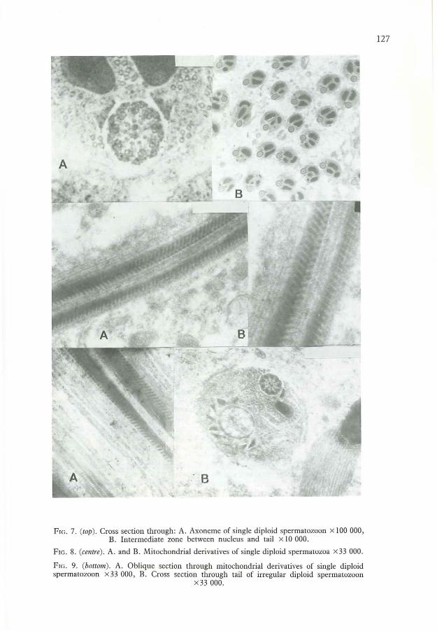
FIG. 7. (top). Cross section through: A. Axoneme of single diploid spermatozoon x 100 000,B. Intermediate zone between nucleus and tail x 10 000.
FIG. 8. (cemre). A. and B. Mitochondrial derivatives of single diploid spermatozoa x33 000.
FIG. 9. (bottom). A. Oblique section through mitochondrial derivatives of single diploidspermatozoon x 33 000, B. Cross section through tail of irregular diploid spermatozoon
x33000.
127

128
spermatozoon, forming the so-called cristae (Figs 8-9A). The grooves are 0'03 ~lm deep andare 0'045 ~tm apart. Their depth and spacing are similar to those found in this investigation forhaploid spermatozoa.
Cruz-Hafling et al. (1970) and Lensky et al. (1979) say that in a haploid spermatozoon thehelices are in opposite directions in the two derivatives. In diploid spermatozoa, however, aline can be found, within a single derivative, from which the grooves start in oppositedirections (Fig. 8). As a result, in longitudinal or oblique sections, grooves can be seen thatrun over both derivatives in the same or opposite directions. Mostly, however, fissuresrunning in the same directions are visible in both mitochondrial derivatives (Fig. 9A).
Triangular rodsTwo triangular rods lie parallel to the axoneme (Figs 4-7A). They are located between theaxoneme and the two mitochondrial derivatives. The bases face the axoneme and the apicespoint outwards. The base of the triangle measures about 0'125 ~lm and the height about0.055 ~tm.
MicrotubuliMicrotubuli are present in spermatozoa of drones (Figs. 4-7A), though not in those of maturedrones (Hoage & Kessel, 1968). The acrosome as well as the nucleus are covered with a sheathof longitudinal microtubuli. In the tail, the microtubuli are present between the differentstructures as well as in the peripheral zone, but they were never found between the axonemeand the triangular rods.
Irregular diploid spermatozoaSometimes vacuoles are present in the tails of diploid spermatozoa. Fig. 9B shows a sectionthrough a tail with one axoneme, one mitochondrial derivative and one vacuole. Only onetriangular rod is visible between the axoneme and the mitochondrial derivative, but eight ornine of them are present around the vacuole.
Double diploid spermatozoaCross sections through tails of some diploid spermatozoa show two axonemes, fourmitochondrial derivatives and four triangular rods (Figs. 10-12). Such spermatozoa are called'double'. The diameter of the tail is 1'235 ~tm, which is 164% of that of a single diploidspermatozoon, and 190% of that of a haploid one.
The diameter of the axonemes is 0'235 ~tm, which is the same as in single diploidspermatozoa. The disposition of the axonemes inside the tail varies. Sometimes they arelocated centrally (Fig. lOA). In other cases, one axoneme is in the centre and the other inperipheral zone (Fig. lOB). In still others, both are located peripherally, and can be closetogether (Fig. 11A), or diametrically opposed (Fig. lIB, Fig. 12A). Inside each axoneme, nineperipheral fibrils, nine inner double, and two central ones are visible. The scheme is thus9 + 9 + 2, just as in single diploid and haploid spermatozoa.
Usually two triangular rods lie alongside each axoneme, but sometimes an axoneme has onlyone rod or none at all. The triangular rods do not always face the mitochondrial derivatives asthey do in single spermatozoa. Sometimes one triangular rod is on the opposite side of theaxoneme from any mitochondrial derivative (Fig. 11A).
Cross sections of mitochondrial derivatives are often more irregularly shaped in doublespermatozoa than in single ones (Fig. 11A). Sometimes two mitochondrial derivatives unitecreating one large and two smaller ones (Fig. 12A). The disposition of the four mitochondrialderivatives inside the tail also varies considerably. Regular association of two mitochondrialderivatives with each axoneme can be seen only exceptionally. Fig. 12B shows a section wherethere is a pair of derivatives associated with one axoneme, but one derivative of the other pairis under the influence of one axoneme and the other under the influence of the other axoneme.More often, however, there is no association of a pair of derivatives with a particular axoneme(Figs 10-11). Inside some derivatives a spongy substance is seen, although more peripherallythe normal amorphic, electron-dense material is present and part of the periphery is coveredwith the usual layer of lower electron-density material. This material is disposed moreirregularly in double spermatozoa than in single ones. Sometimes it covers opposite sides of a

FIG. 10. (top). Cross sections through tails of double diploid spermatozoa x26 000, A. Bothaxonemes in the centre, B. One axoneme in the centre and the other on the periphery.
FIG. 11. (centre). Cross sections through tails of double diploid spermatozoa x 26 000, A. Bothaxonemes on the periphery, B. Axonemes diametrically opposed.
FIG. 12. (bottom). Cross sections through tails of double diploid spermatozoa, A. Twomitochondrial derivatives fused x26 000, B. One pair of mitochondrial derivatives associated
with, one axoneme x20000.
129

130
FIG. 13. (top). Identical directions of grooves on three mitochondrial derivatives of diploidmultiple spermatozoa, A. Oblique section x26 000, B. Longitudinal section x33000.
FIG. 14. (bottom). Cross sections through tails of irregular double diploid sp"ermatozoa, A.Enlarged mitochondrial derivatives x20 000, B. Vacuoles inside the tail x26000.
derivative (Fig. lIA). Helical grooves are present in this mass. Oblique sections (Fig. 13A), orlongitudinal ones (Fig. 13B), show that the directions of the helices are identical on all threemitochondrial derivatives.
Various irregularities may be found in the tails of some double spermatozoa. Onemitochondrial derivative may be enormously enlarged and filled with spongy material inside(Fig. 14A). Vacuoles with electron-dense inclusions may be seen in some others (Fig. 14B).Probably the vacuoles are transformed mitochondria, but no proof of this is presented.
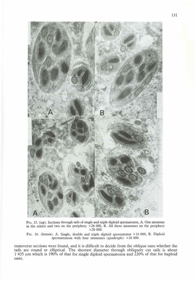
Triple diploid spermatozoaTransverse or oblique sections through tails of some spermatozoa show three axonemes and sixmitochondrial derivatives (Fig. 15, Fig. 16A). Such spermatozoa are called 'triple'. No exactly
131
FIG. IS. (top). Sections through tails of single and triple diploid spermatozoa, A. One axonemein the centre and two on the periphery x26 000, B. All three axonemes on the periphery
x20000.FIG. 16. (boltol/l). A. Single, double and triple diploid spermatozoa x 16 000, B. Diploid
spermatozoon with four axonemes (quadruple) x26 000.
transverse sections were found, and it is difficult to decide from the oblique ones whether thetails are round or elliptical. The shortest diameter through obliquely cut tails is about1'435 ~lm which is 190% of that for. single diploid spermatozoa and 220% of that for haploidones.

132
The diameter of each axoneme in triple spermatozoa is 0'235 ~lm, which is the same as it isin single diploid spermatozoa. The disposition ofaxonemes in the tail varies. One axonememay be located in the centre and two others in the periphery (Fig. 15A), or all three may be inthe periphery (Fig. 15B). The scheme of the internal structure is 9 + 9 + 2 fibrils, just as inhaploid or single diploid spermatozoa.
In oblique sections, mitochondrial derivatives of triple spermatozoa are mostly elliptical.Association of a pair of them with each axoneme cannot be seen. The internal structure issimilar to that found in single or double spermatozoa. Direction of helical grooves is the samein all six of the mitochondrial derivatives visible on Fig. 15 and Fig. 16A.
Quadruple diploid spermatozoaFig. 16B shows a section through a tail with four axonemes and three mitochondrialderivatives. The diameter of the tail is about 1'0 flm, which is larger than that of a singlediploid spermatozoon, but smaller than those of double and triple spermatozoa. Thus thesection was probably made through the tail of a quadruple spermatozoon near the posteriorend where some mitochondrial derivatives had already terminated, although the possibilitycannot be excluded that this is simply the tail of a spermatozoon with four axonemes but onlythree mitochondrial derivatives.
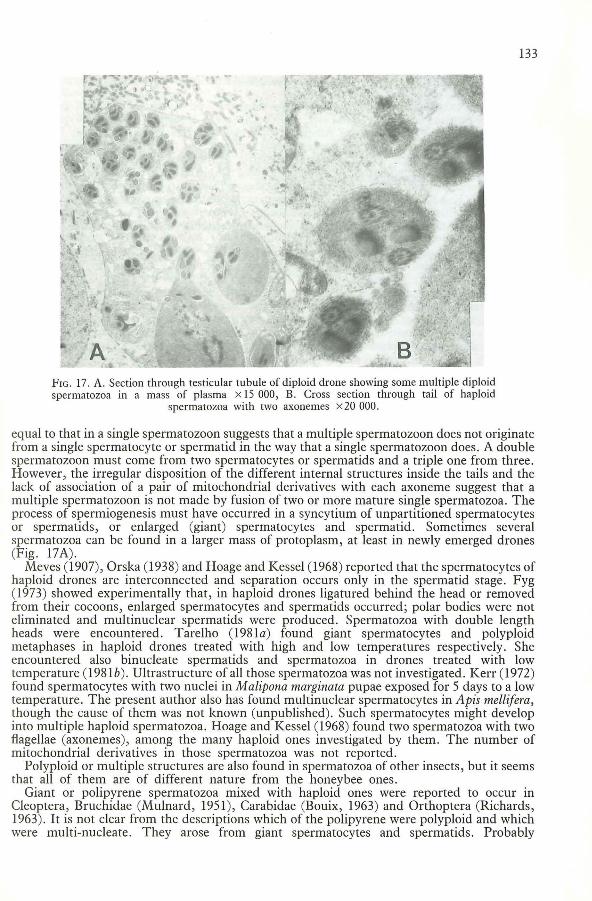
Although diploid spermatozoa were quite commonly found to be multiple in the waydescribed above, multiple haploid spermatozoa seem to be rare. The only examples foundwere from one haploid drone whose spermatozoa each had two axonemes but only twomitochondrial derivatives (Fig. 17B).
DiscussionAccording to Woyke (1983) the lengths of entire diploid spermatozoa (313 ~tm) are 129% ofthose of haploid ones (242 ~tm). Various structures of haploid spermatozoa were measured byCruz-Hafting et al. (1970) and Lensky et al. (1979). Their measurements mostly agree withthose given here for the haploids, or differ only slightly.
Compared with those of spermatozoa treated with Feuglen reaction (Woyke, 1983) thediploid nuclei now described are slightly shorter and haploid nuclei are slightly longer, so theratio of diploid to haploid lengths is slightly less.
The depth of the helical grooves (cristae) on mitochondrial derivatives found by the aboveauthors was 0'01 ~tm, whereas 0'03 ~tm was found in the present investigation for both haploidand diploid spermatozoa. The distance between grooves (0'045 ~lm) now found for bothhaploid and diploid spermatozoa agrees with the 0'04 ~tm given by Lensky et al. (1979) but islarger than the 0'025 flm given by Cruz-Hafting et al. (1970). The latter author reports 30° asthe angle between the direction of the oblique grooves on the mitochondrial derivatives andthe longitudinal axis of the spermatozoon, whereas Lensky et al. (1979) give 60°. The anglenow found is 60° for both haploid and diploid spermatozoa. Both the above groups of authorsagree that the directions of the helical grooves are opposite in the two mitochondrialderivatives of haploid spermatozoa, but in diploid spermatozoa it now appears that thedirections of the grooves are usually the same in both.
Since the linear dimensions of single diploid spermatozoa have been found to be 11 5-1 51%of those of haploid spermatozoa, the volume of diploid spermatozoa could be roughly twicethat of haploid ones.
The best description of spermiogenesis of haploid spermatozoa is presented by Orska(1938). She describes the retarded development of one mitochondrial derivative, which resultsin its smaller size. She suspects that this is caused by the haploidy of the drone, and the lack ofequal division of the spermatocytes. Woyke and Skowronek (1974) found that duringspermatogenesis in drones, polar bodies are created and equal division of spermatocytes doesnot occur. The smaller size of one mitochondrial derivatives in diploid spermatozoa suggestthat the same process of spermiogenesis occurs in diploid drones, where it cannot be caused byhaploidy, but might be caused by the aborative division of the spermatocytes. The internalstructures of mUltiple spermatozoa that were observed on oblique or longitudinal sections layparallel to each other throughout the sections. This demonstrates that the sections showintergrated cells and not locally united spermatozoa. The larger diameter of tails of multiplespermatozoa, and the fact that the diameter of each axoneme in a multiple spermatozoon is
133
FIG. 17. A. Section through testicular tubule of diploid drone showing some multiple diploidspermatozoa in a mass of plasma x 15 000, B. Cross section through tail of haploid
spermatozoa with two axonemes x20000.
equal to that in a single spermatozoon suggests that a multiple spermatozoon does not originatefrom a single spermatocyte or spermatid in the way that a single spermatozoon does. A doublespermatozoon must come from two spermatocytes or spermatids and a triple one from three.However, the irregular disposition of the different internal structures inside the tails and thelack of association of a pair of mitochondrial derivatives with each axoneme suggest that amultiple spermatozoon is not made by fusion of two or more mature single spermatozoa. Theprocess of spermiogenesis must have occurred in a syncytium of unpartitioned spermatocytesor spermatids, or enlarged (giant) spermatocytes and spermatid. Sometimes severalspermatozoa can be found in a larger mass of protoplasm, at least in newly emerged drones(Fig. 17A).
Meves (1907), Orska (1938) and Hoage and Kessel (1968) reported that the spermatocytes ofhaploid drones are interconnected and separation occurs only in the spermatid stage. Fyg(1973) showed experimentally that, in haploid drones ligatured behind the head or removedfrom their cocoons, enlarged spermatocytes and spermatids occurred; polar bodies were noteliminated and multinuclear spermatids were produced. Spermatozoa with double lengthheads were encountered. Tarelho (198la) found giant spermatocytes and polyploidmetaphases in haploid drones treated with high and low temperatures respectively. Sheencountered also binucleate spermatids and spermatozoa in drones treated with lowtemperature (1981 b). Ultrastructure of all those spermatozoa was not investigated. Kerr (1972)found spermatocytes with two nuclei in Malipona marginala pupae exposed for 5 days to a lowtemperature. The present author also has found multinuclear spermatocytes in Apis melli/era,though the cause of them was not known (unpublished). Such spermatocytes might developinto mUltiple haploid spermatozoa. Hoage and Kessel (1968) found two spermatozoa with twoflagellae (axonemes), among the many haploid ones investigated by them. The number ofmitochondrial derivatives in those spermatozoa was not reported.
Polyploid or multiple structures are also found in spermatozoa of other insects, but it seemsthat all of them are of different nature from the honeybee ones.
Giant or polipyrene spermatozoa mixed with haploid ones were reported to occur inCleoptera, Bruchidae (Mulnard, 1951), Carabidae (Bouix, 1963) and Orthoptera (Richards,1963). It is not clear from the descriptions which of the polipyrene were polyploid and whichwere multi-nucleate. They arose from giant spermatocytes and spermatids. Probably

134
endomitosis occurs here. Some of the giants were multinucleate. The ultrastructure of thesespermatozoa was not described.
Two spermatozoa may be connected by the head only or by part of the tail, as in thecoleopteran Dytiscus marginalis (Ballowitz, 1895), Thermobia domestica (Bawa, 1964), Lepismasacchanna (Werner, 1964), or the American opossum (Biggers & Creed, 1962; Phillips,1970a). The individuality of the two spermatozoa remains, and the plasma membrane of thetwo units remain separate (Bawa, 1964). The matrix of the spermatozoon of the silkwormBombyx mon is surrounded in the ejaculatory duct by a cylindrical sleeve. Most sleeves containonly one spermatozoon, but a small number of exceptional sleeves contain two or even threespermatozoa (Friedlander & Gitay, 1972). The spermatozoa are not integrated and associationof two mitochondrial derivatives with each axoneme is clearly visible. This is contrary to thelack of association of two mitochondrial derivatives with each axoneme in honeybee multiplespermatozoa.
Two or three axonemes were found in spermatozoa of a sea urchin (Afzelius, 1959), thelouse Pediculus humanus cO/pons (Ito, 1966) and the whole group of Rhynchotoids: Psocoptera,Mallophaga, Anoplura, Thysanoptera and Rynchota (Baccetti et aI., 1969). The doublinginvolved only the axonemes. There were only two mitochondrial derivatives there or even onlyone, whereas in diploid multiple spermatozoa of the honeybee, the numbers of mitochondrialderivatives as well as ofaxonemes, are multiplied. Spermatozoa with two axonemes werefound only in the primitive insect orders and not in the most evolved orders.
In the fungus gnat Sciara coprophila the axoneme consists of 70 doublets, instead of theusual 9 +9 + 2 patters of tubules (Phillips, 1966).
Spermatozoa of three species of hopper (Membracidae) branch into four tails with 2 + 2 or3 + 3 tubules in the axoneme (Phillips, 1969). Spermatozoa of the termite Mastotermesdatwiniensis have about 100 flagellae.
Abnormal tails are also associated with absence of one sex chromosome or presence ofsupernumerary chromosomes. In Drosophila melanogaster males that have XlO sex chromosomes instead of the normal set of XlY, abnormalities occur during development ofspermatozoa. Attachment between axonemes and mitochondria is disorganized. Some casesare found where two axonemes appear to be attached to the same mitochondrion whereas otheraxonemes are not closely associated with any mitochondria. The character is thus similar towhat is found in haploid honeybee spermatozoa, but in contrast to the honeybee no fullymature sperms are present in the testes of XlO Drosophila males (Kiefer, 1966). On the otherhand XlY/Y Drosophila males produce sperms twice as long as those of XlY males. Thusdoubling of sex chromosomes results in larger spermatozoa, as in the honeybee.
Thus some of the characteristics of diploid and multiple honeybee spermatozoa may befound separately in other groups of insects.
Perhaps diploid honeybee drones may be considered more primitive than haploid ones.
ConclusionsThe main difference between diploid and haploid spermatozoa is that the diploids, as well asmost of their parts, are bigger. Internally, they are very similar except for the direction of thegrooves on the mitochondrial derivatives.
Diploid drones normally produce multiple as well as single spermatozoa. These are largerthan the single ones. The number ofaxonemes and mitochondrial derivatives is multiplied inmultiple spermatozoa, but attachment of two derivatives to each axoneme does not eXIst III
them. Multiple spermatozoa are not formed by fusion of separate sperms and probablydevelop from unpartitioned spermatocytes.
ReferencesAFZELIUS, B. (1959) Electron microscopy of sperm tail. Results obtained with a new fixative. J. biophys.
biochem. CylOl. 5(2): 269-277BACCETTI, B. (1972) Insect sperm cells. Adv. illsect Physiol. 9 : 315-397BACCETTI, B.; DALLAl. R. (1978) The spermatozoon of Arthropoda. XXX. The multiflagellate spermatozoon
in the termite Mastotemles darwilliellSis. J. Cell Bioi. 76(3) : 569-576BACCETTI, B.; DALLAl, R.; ROSATI, F. (1969) The spermatozoon of Arthropoda. IV. Corrodentia, Mallophaga
and Thysanoptera. J. Microscopie 8 : 249-262BALLOWITZ, E. (1895) Die Doppelspermatozoon der Dytisciden. Z. wiss. Zool. 60(3): 458-499

135
BAWA, S. R. (1964) Electron microscope study of spermiogenesis in fire-brat insect, ThemlObia domestica Pack.I. Mature spermatozoon. J. Cell Bioi. 23(3): 431-446
BIGGERS, ]. D.; CREED, R. F. S. (1962) Conjugate spermatozoa of the North American opossum. Nature,Lond. 196(4859): 1112-1113
BOUIX, M. G. (1963) Sur la spermatogenese des Carabes (Col. Car.): modalite et frequence de la spermiogeneseatypique C. r. hebd. Seanc. Acad. Sci., Paris 256(12) : 2698-2701
CRuz-HoFLING, M. A. DA; CRuz-LAN DIM, C. DA; KITAJIMA, E. W. (1970) The fine structure of spermatozoafrom the honeybee. Anais Acad. bras. Cienc. 42 : 69-78
FRIEDLANDER, M.; GITAY, H. (1972) The fate of the normal-anucleated spermatozoa in inseminated femalesof the silkworm Bombyx mori. J. Morph. 138(1): 121-130
FYG, W. (1973) Uber den Einfluss von Metamorphosehautungsstiirungen und ligaturen auf die Spermatogenese der Honigbiene (Apis lIIellifera L.) Apidologie 4(3) : 227-265
HOAGE, T. R.; KESSEL, R. G. (1968) An electron microscope study of the process of differentiation duringspermatogenesis in the drone honeybee (Apis lIIellifera L.) with special reference to centriole replicationand elimination. J. Ultrastnlct. Res. 24 : 6-32
!To, S. (1966) Movement and structure of louse spermatozoa. J. Cell BioI. 31(2) : 128KERR, W. E. (1972) Effect of low temperature on male meiosis in Melipona lIIarginata. J. apic. Res.
11(2) : 95-99KEIFER, B. ]. (1966) Ultrastructural abnormalities in developing sperm of X/O Drosophila melanogaster.
Genetics 54(6) : 1441-1452LENSKY, Y.; BEN-DAVID, E.; SCHINDLER, H. (1979) Ultrastructure of the spermatozoon of the mature drone
honeybee. J. apic. Res. 18(4): 264-271MAYER, G. F. (1968) Spermiogenese in normalen und Y-deficienten Manchen von Drosphila melanogaster
und D. hydei Z. Zellforsch. mikrosk. Anat. 84(4): 141-175 •MEVES, F. (1907) Die Spermatocytenteilungen bei der Honigbiene (Apis lIIellifica L.) nebst Bemerkungen tiber
Chromatinreduktion. Arch. microsc. Anat. u. Entwgesch. 70(3) : 414-419MULNARD, ]. (1951) La spermatogenese double d'Acanthoscelides abteetus Say (Caleoptere, Bruchide). Annis
Soc. r. zool. Belg. 82(2) : 399-445ORSKA, ]. (1938) Badania cytologiczne nad spermatogeneneza pszczoly domowej (Apis mellifera L.)
[Cytological investigation on spermatogenesis of honeybee]. ArchwlII Tow. nauk Lw6w. III, 10(1) : 1-82PHILLIPS, D. M. (1966) Fine structure of Sciara coprophila sperm. J. Cell Bioi. 30(3): 499-517---(1969) Exception to the prevailing pattern of tubules (9 + 9 + 2) in the sperm flagella of certain insect
species J. Cell Bioi. 40(1) : 28-43---(1970a) Ultrastructure of spermatozoa of the woolly opossum Caluromys philander. J. Ultrastnlct. Res.
33(3-4) : 381-397---(1970b) Insect sperm: their structure and morphogenes J. Cell Bioi. 44(2) : 243-277RICHARDS, A. G. (1963) Giant sperm in cockroach, Periplaneta americana. Ent. News 74(3) : 57-60ROTHSCHILD, L. (1955) The spermatozoa of the honey bee. Trans. R. en!. Soc. Lond. 107: 289-294TARELHO, Z. V. S. (1981a) Effects of low and high temperatures on the spermatogenesis of Apis mellifera
Linne, 1754. I. Effects of temperature on metaphase, survival and development. Revta bras. Genet.4(2) : 193-212
---(1981b) Effects of low and high temperatures on the spermatogenesis of Apis mellifera Linne, 1754. II.Effects of temperature on anaphase, telpophase and spermiogenesis. Revta bras. Genet. 4(3) : 383-397
WERNER, G. (1964) Untersuchungen tiber die Spermiogenese beim Silberfischchen, Lepisma saccharina L. Z.Zellforsch. mikrosk. Anat. 63(7) : 880-912
WOYKE, ]. (1963) Drone larvae from fertilized eggs of the honeybee. J. apic. Res. 2(2): 73-75---(1969) A method of rearing diploid drones in a honeybee colony. J. apic. Res. 8(2): 65-71---(1975) DNA content of spermatids and spermatozoa of haploid and diploid drone honeybees. J. apic.
Res. 14(1) : 3-8---(1983) Lengths of haploid and diploid spermatozoa of the honeybee and the question of the production
of triploid workers. J. apic. Res. 22(3): 146-149WOYKE, ].; SKOWRONEK, W. (1974) Spermatogenesis in diploid drones of the honeybee. J. apic. Res.
13(3) : 183-190
183