ivus guided pci - intech - opencdn.intechopen.com/pdfs/27058/intech-ivus_guided_pci.pdfivus guided...
TRANSCRIPT
8
IVUS Guided PCI
T. Kovarnik and J. Horak 2nd Department of Medicine - Department of Cardiovascular Medicine,
First Faculty of Medicine, Charles University in Prague and General University Hospital in Prague,
Czech Republic
1. Introduction
Intravascular ultrasound is a clinical tool that has been used in a complimentary manner to
contrast angiography in order to enhance the procedure success rate and patient outcomes.
Particularly, there are some specific situations where IVUS can be very useful and information
from IVUS overcome angiography limitations. These situations can be divided into three parts:
- pre-interventional - during intervention - post-interventional Pre-interventional use of IVUS can help with assessment of hemodynamic significance of stenosis (in this place must be emphasized, that IVUS can estimate hemodynamic significance only non directly, but with acceptable correlation with fractional flow reserve and nuclear stress test), precise anatomic analysis (type of bifurcation, plaque burden in ostial part of side branch or left main) and can help with device selection: precise measurement of diameters in reference segments for better sizing of balloon or stent, type of lesion preparation such as dilatation with cutting balloon or atherectomy devices in bulky lesions, drug eluting stent for lesions with high probability of small in stent area achievement. IVUS can be used during intervention for assessment of lesion preparation (proper position of wire; especially it’s relation to stent struts, effect of predilatation, assessment of side branch ostium) for guiding in more complicated intervention (left main stenting, navigation of wires during PCI of chronic total occlusion, trifurcation PCI). Post-interventional use of IVUS can answer questions about result adequacy (stent
expansion, stent apposition), angiographic filling defects (“hazy lesions”) after PCI (edge
dissection, thrombus formation, inadequate stent expansion, prominent calcification) and
diagnosis of complications (dissection, geographic mismatch, plaque protrusion inside the
stent, inadequate stent expansion or apposition). IVUS can in most cases also reveal the
reasons behind development of complications after PCI such is in stent thrombosis (mainly
inadequate stent expansion or edge troubles such as dissection, or uncovered lesion) or
unexpected early in stent restenosis (inadequate stent expansion).
IVUS is a mandatory tool in the cardiac catheterization laboratory today. Like all medical
equipments it should be used by an experienced investigator for better understanding of
PCI mechanisms, prevention and solution of complications. Nowadays it is possible to use
many semi-automatic softwares for border detection, but all of them must be corrected
www.intechopen.com
Intravascular Ultrasound
108
manually to avoid serious mistakes and misunderstanding. The best way how to do that is
just routine use of IVUS.
1.1 Limitation of angiography Angiography is a gold standard for assessment of atherosclerotic impairment of coronary
arteries and for guiding of coronary intervention. On the other hand it has many limitations.
Widely accepted decision making point for diagnosis of flow-limiting lesion is lumen
diameter less than 50% of the reference segment. However, it has been proven that there is
no correlation between angiography assessment and functional measurement by fractional
flow reserve (FFR) in intermediate lesions between 40-70%1.
Based on autopsy studies we know, that atherosclerosis is more often diffuse than focal process affecting coronary arteries. These findings are in good correlation with intravascular ultrasound. The reference segment, which is angiographically normal often contains atherosclerotic plaques visible on IVUS. From this point of view we are comparing less affected segments with more affected ones during angiography and thus underestimate stenosis significance2,3. Other challenging issues are assessment of ostial lesions and bifurcations.
2. Quantitative lesion assessment
2.1 Technique The most important parameter for quantitative lesion assessment is minimal lumen area
(MLA), which has the best correlation with presence of ischemia (see below).Common
misunderstanding comes from the assessment of relative severity of stenosis in
percentage of “normal” reference diameter. Angiographic stenosis assessment is based on
comparison of lumen diameter in reference segment and lumen diameter in the lesion.
The most corresponding IVUS parameter is lumen area stenosis (LAS), which is calculated
as minimal lumen area in reference segment – minimal lumen area in lesion / minimal
lumen are in reference segment. More frequently used description of stenosis is plaque
burden (PB), computed as external elastic membrane area – lumen area / external elastic
membrane area4. We must interrogate each frame and look for the smallest lumen area,
use routinely nitrates before IVUS probe insertion for avoiding spasms. In case of
uncertainty in lumen measurement, especially in present of soft plaque it is recommended
to flush guiding catheter by saline for clearing of picture. Using of automatic pullback
devices is not necessary for quantitative lesion assessment, but allows us to perform
longitudinal diameters or volumetric analysis. Manual pullback is better for analysis of a
precise part of plaque, where we can stop IVUS probe to obtain more frames from the
region of interest. For imaging of aorto-ostial lesions it is necessary to retract the guiding
catheter back into the aorta and verify that the path of the IVUS probe is still coaxial with
the ostium of the vessel5.
2.2 Indication for coronary intervention The main indication for coronary intervention is lesion producing myocardial ischemia, which can be detect either non-invasively by stress myocardial perfusion imaging (SPECT) or invasively by measurement of pressure gradient across stenosis (fraction flow reserve, FFR). IVUS measurement correlates with both of them (table one).
www.intechopen.com

IVUS Guided PCI
109
Author Description Methods No of patients
Results
Briguori6 angiographic stenosis 40-70%
IVUS vs. FFR
43 FFR < 0.75 correlates with:- MLA ≤ 4 mm2
- MLD ≤ 1.8 mm - PB > 70% - Lesion length > 10 mm
Abizaid7 patients indicated for PCI
IVUS vs. CFR
73 CFR ≥ 2 correlates with: - MLA ≥ 4 mm2
- MLD ≥ 2 mm
Nishioka8 consecutive IVUS examinations
IVUS vs. myocardial SPECT
79 Positive scan correlates with:
- MLA ≤ 4 mm2 - PB > 73% - LAS >59%
Takagi9 consecutive IVUS examinations
IVUS vs. FFR
42 FFR <0.75 correlates with:- MLA ≤ 3 mm2
- LAS > 60%
Lee10 vessels smaller than 3 mm
IVUS vs. FFR
94 FFR < 0.75 correlates with:
- MLA ≤ 2.0 mm2
- PB ≥ 80%
- Lesion lenght ≥ 20 mm
Abizaid11 IVUS deferred PCI IVUS 300 Safe deferral of PCI correlates with:
- MLA ≥ 4 mm2 - MLD ≥ 2 mm
Jasti12 ambiguous left main stenosis
IVUS vs. FFR
55 FFR < 0.75 correlates with:- MLA ≤ 5.9 mm2
- MLD ≤ 2.8 mm
Abizaid13 patients with borderline left main stenosis
IVUS 122 MACE predictor :- MLD ≤ 3.0 mm
Kang 14 Consecutive patients IVUS vs. FFR
201 FFR <0.80 correlates with:- MLA 2.4 mm2
- PB ≥79%
Ben Dor15 patients with intermediate lesion during QCA
IVUS vs. FFR
84 FFR < 0.8 correlates with: - MLA 2.4 mm2 mm for vessels
2.5-3.0 mm - MLA 2.7 mm2 for vessels 3.0-3.5 mm - MLA 3.6 mm2 for vessels >3.5 mm
Ahn16 consecutive patients with SPECT and IVUS
IVUS vs. SPECT
150 Positive scan correlates with: - MLA 2.1 mm2
PROSPECT17 clinical follow-up after ACS
IVUS and MACE
700 Predictors for MACE:- PB > 70%
- MLA < 4 mm2 - TCFA
Abbreviations: CFR - coronary flow reserve, MACE – major cardiac adverse event, MLA – minimal lumen area, MLD – minimal lumen diameter, PB - plaque burden, PCI- percutaneous coronary interventions, QCA – quantitative coronary angiography, SPECT – single photon emission computed tomography, TCFA thin cap fibroatheroma
Table 1. Studies with IVUS evaluation of hemodynamic significance
www.intechopen.com
Intravascular Ultrasound
110
Based on older studies the following recommendations for IVUS detection of significant stenosis were established: Main epicardial artery: - MLA < 4 mm2 - MLD < 2 mm Left main: - MLA < 6 mm2 - MLD 3 mm However, recently published studies do not support these cut-off values. Instead of clear
cut-off points there are number of different recommended values, which do not seem to be
useful for routine practice. Based on these findings we must conclude that IVUS is not
suitable for assessment of haemodynamic significance of intermediate lesions. IVUS can be
only used for exclusion of haemodynamicly significant lesion with MLA more than 4 mm2
for main epicardial arteries and more than 6 mm2 for left main. The haemodynamic
significance of each lesion is caused not only by lumen area, but also by amount of viable
myocardium supplied by this vessel and by presence or absence of collaterales. These facts
simply cannot be examined solely by a morphologic modality like IVUS. On the other hand
the PROSPECT trial17 with clinical endpoints confirmed, that large plaque compromising
lumen to 4 mm2 and less, especially together with higher content of necrotic tissue is a risk
factor for future events. These issues will be matter of further studies.
There are no data about IVUS criteria for hemodynamic significance in saphenous vein graft, but it is recommend to use a cut-off point MLA 4 mm2 for graft supplying one coronary artery and MLA 6 mm2 for graft supplying two arteries. The precise assessment of hemodynamic significance is a crucial point, because the rate of
ischemic events is 5-10% / year in significant lesion and less than 1%/ year in non-ischemic
lesion. Performing PCI in non-ischemic lesion increase risk of event to 2-3% / year18 and
furthermore increases risk of periprocedural ischemia or myocardial necrosis during PCI.
Very old IVUS-skeptic sentence “If you want to stent, do IVUS” was quite recently
supported by the work of Nam et al.19. Authors randomized 167 patients with intermediate
coronary lesion between FFR guided (cut-off for PCI was FFR <0.8) and IVUS guided (cut-
off MLA< 4.0 mm2) coronary intervention. PCI was performed in 33.7% lesion in FFR arm
and in 91.5% in IVUS arm. This difference was highly statisticaly significant (p<0.001). On
the other hand we ourselves have a different experience with IVUS assessment in borderline
lesions. We estimate that we are performing PCI in 40-50% of borderline lesions based on
IVUS criteria, which is closer to FRR guided arm in this study. The finding of 91.5%
frequency of MLA < 4 mm2 in lesion between 40-70% is in our eyes unrealistic.
2.3 Assessment of left main (LM) Left main stenosis is a very important predictor for future cardiac events20. Angiographic
assessment of LM is often complicated for overlapping branches and short or no reference
segment and can lead to inappropriate estimates of lesion severity21. IVUS is more sensitive
for left main atherosclerosis than angiography22. Suter et al.23 found that in half of the
patients with an inconclusive angiogram IVUS detects a significant stenosis. There is no
difference for left main assessment during pullback from left anterior descending artery
(LAD) or left circumflex artery (LCX)24. On the other hand for accurate assessment of ostial
part of LAD and LCX is necessary to perform two pullbacks from both daughter vessels,
www.intechopen.com
IVUS Guided PCI
111
because oblique view can overestimates lumen area25. The main target in left main
bifurcation (like in other bifurcation) is to make a decision between one or two stent
strategy. Pullback from just one daughter artery can answer the question whether the
ostium of second branch is affected or not. For a more precise evaluation of minimal lumen
area it is necessary to perform pullback from both daughter branches.
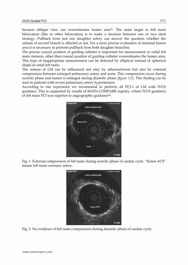
The precise coaxial position of guiding catheter is important for measurement or ostial left main stenosis, other than coaxial position of guiding catheter overestimates the lumen area. This type of inappropriate measurement can be detected by elliptical instead of spherical shape of ostial left main. The ostium of LM can be influenced not only by atherosclerosis but also by external compression between enlarged pulmonary artery and aorta. This compression occur during systolic phase and lumen is enlarged during diastolic phase (figure 1,2). This finding can be seen in patients with severe pulmonary artery hypertension According to our experience we recommend to perform all PCI´s of LM with IVUS guidance. This is supported by results of MAIN-COMPARE registry, where IVUS guidance of left main PCI was superior to angiographic guidance26.
Fig. 1. External compression of left main during systolic phase of cardiac cycle. “Kmen ACS” means left main coronary artery.
Fig. 2. No evidence of left main compression during diastolic phase of cardiac cycle.
www.intechopen.com
Intravascular Ultrasound
112
3. Lesion morphology
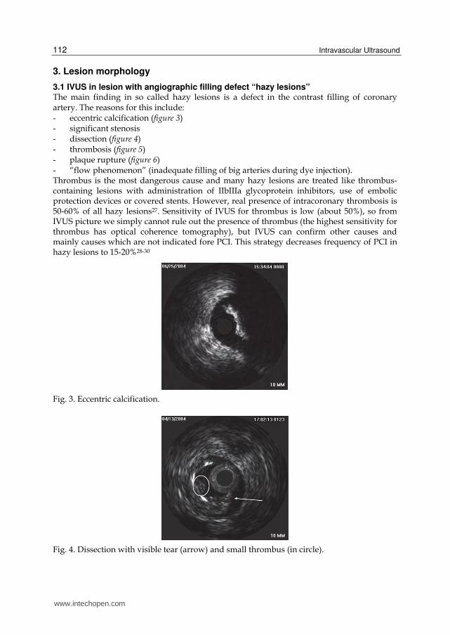
3.1 IVUS in lesion with angiographic filling defect “hazy lesions” The main finding in so called hazy lesions is a defect in the contrast filling of coronary artery. The reasons for this include: - eccentric calcification (figure 3) - significant stenosis - dissection (figure 4) - thrombosis (figure 5) - plaque rupture (figure 6) - “flow phenomenon” (inadequate filling of big arteries during dye injection). Thrombus is the most dangerous cause and many hazy lesions are treated like thrombus-containing lesions with administration of IIbIIIa glycoprotein inhibitors, use of embolic protection devices or covered stents. However, real presence of intracoronary thrombosis is 50-60% of all hazy lesions27. Sensitivity of IVUS for thrombus is low (about 50%), so from IVUS picture we simply cannot rule out the presence of thrombus (the highest sensitivity for thrombus has optical coherence tomography), but IVUS can confirm other causes and mainly causes which are not indicated fore PCI. This strategy decreases frequency of PCI in hazy lesions to 15-20%28-30
Fig. 3. Eccentric calcification.
Fig. 4. Dissection with visible tear (arrow) and small thrombus (in circle).
www.intechopen.com

IVUS Guided PCI
113
Fig. 5. Huge thrombus located on very small plaque causing acute coronary syndrome. Left side is cross sectional view, right side is longitudinal view (thrombus is located in the circle).
Fig. 6. Plague rupture with visible cavity after embolization of plaque mass. Patient with acute myocardial infarction.
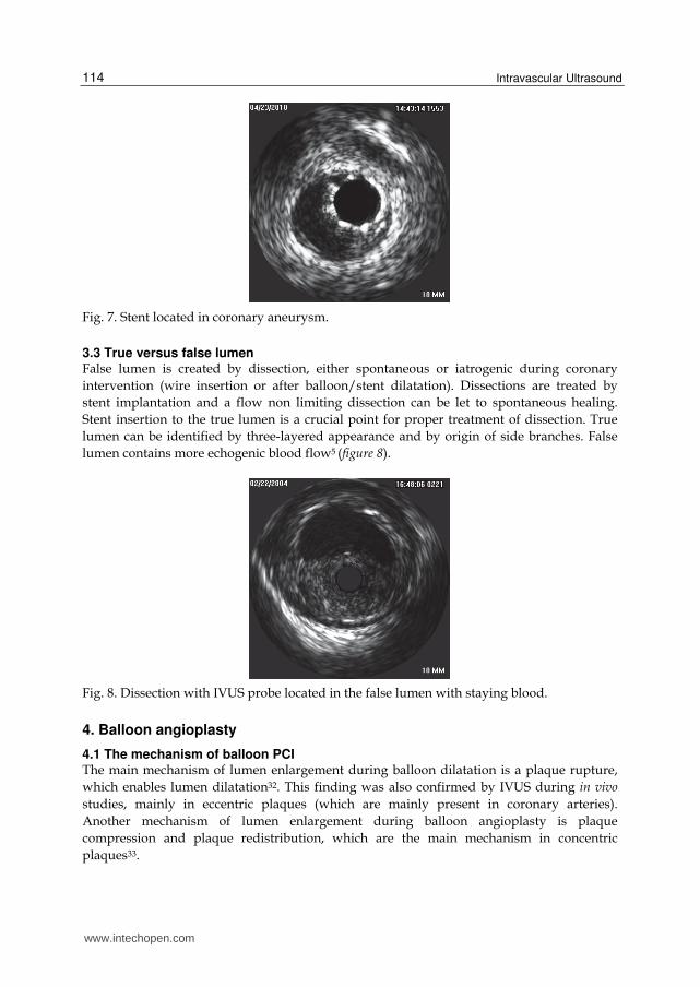
3.2 Aneurysms True aneurysm is defined as both an external elastic membrane (EEM) and lumen area 50% larger than the proximal reference segment with intact three layers of vessel wall (figure 7). Lesions which seem to be the aneurysms from angiographic assessment are an true aneurysm in 37% of such findings. The rest are normal segments adjacent to plaque (53%), complex atherosclerotic plaque (16%) and pseudoaneurysm (4%)31. Coronary pseudoaneurysm is due to rupture of vessel wall and in IVUS picture the three layers of vessel are not present, the shape of pseudoaneurysm is often irregular. From a practical point of view, pseudoaneurysms are seen only after coronary interventions causing trauma of vessel wall.
www.intechopen.com
Intravascular Ultrasound
114
Fig. 7. Stent located in coronary aneurysm.
3.3 True versus false lumen False lumen is created by dissection, either spontaneous or iatrogenic during coronary
intervention (wire insertion or after balloon/stent dilatation). Dissections are treated by
stent implantation and a flow non limiting dissection can be let to spontaneous healing.
Stent insertion to the true lumen is a crucial point for proper treatment of dissection. True
lumen can be identified by three-layered appearance and by origin of side branches. False
lumen contains more echogenic blood flow5 (figure 8).
Fig. 8. Dissection with IVUS probe located in the false lumen with staying blood.
4. Balloon angioplasty
4.1 The mechanism of balloon PCI The main mechanism of lumen enlargement during balloon dilatation is a plaque rupture,
which enables lumen dilatation32. This finding was also confirmed by IVUS during in vivo
studies, mainly in eccentric plaques (which are mainly present in coronary arteries).
Another mechanism of lumen enlargement during balloon angioplasty is plaque
compression and plaque redistribution, which are the main mechanism in concentric
plaques33.
www.intechopen.com
IVUS Guided PCI
115
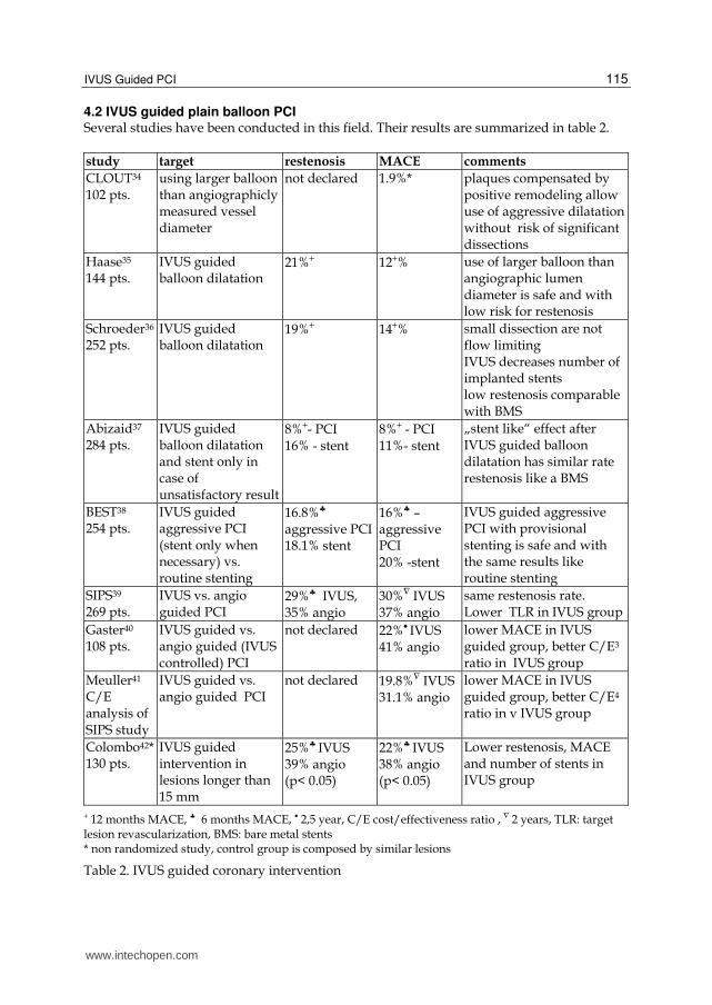
4.2 IVUS guided plain balloon PCI Several studies have been conducted in this field. Their results are summarized in table 2.
study target restenosis MACE comments
CLOUT34 102 pts.
using larger balloon than angiographicly measured vessel diameter
not declared 1.9%*
plaques compensated by positive remodeling allow use of aggressive dilatation without risk of significant dissections
Haase35 144 pts.
IVUS guided balloon dilatation
21%+ 12+% use of larger balloon than angiographic lumen diameter is safe and with low risk for restenosis
Schroeder36 252 pts.
IVUS guided balloon dilatation
19%+ 14+% small dissection are not flow limiting IVUS decreases number of implanted stents low restenosis comparable with BMS
Abizaid37 284 pts.
IVUS guided balloon dilatation and stent only in case of unsatisfactory result
8%+- PCI 16% - stent
8%+ - PCI 11%- stent
„stent like“ effect after IVUS guided balloon dilatation has similar rate restenosis like a BMS
BEST38 254 pts.
IVUS guided aggressive PCI (stent only when necessary) vs. routine stenting
16.8%♣
aggressive PCI18.1% stent
16%♣ – aggressive PCI 20% -stent
IVUS guided aggressive PCI with provisional stenting is safe and with the same results like routine stenting
SIPS39 269 pts.
IVUS vs. angio guided PCI
29%♣ IVUS, 35% angio
30%∇ IVUS 37% angio
same restenosis rate. Lower TLR in IVUS group
Gaster40
108 pts. IVUS guided vs. angio guided (IVUS controlled) PCI
not declared 22%• IVUS 41% angio
lower MACE in IVUS guided group, better C/E3 ratio in IVUS group
Meuller41 C/E analysis of SIPS study
IVUS guided vs. angio guided PCI
not declared 19.8%∇ IVUS31.1% angio
lower MACE in IVUS guided group, better C/E4 ratio in v IVUS group
Colombo42* 130 pts.
IVUS guided intervention in lesions longer than 15 mm
25%♣ IVUS 39% angio (p< 0.05)
22%♣ IVUS 38% angio (p< 0.05)
Lower restenosis, MACE and number of stents in IVUS group
+ 12 months MACE, ♣ 6 months MACE, • 2,5 year, C/E cost/effectiveness ratio , ∇ 2 years, TLR: target lesion revascularization, BMS: bare metal stents * non randomized study, control group is composed by similar lesions
Table 2. IVUS guided coronary intervention
www.intechopen.com
Intravascular Ultrasound
116
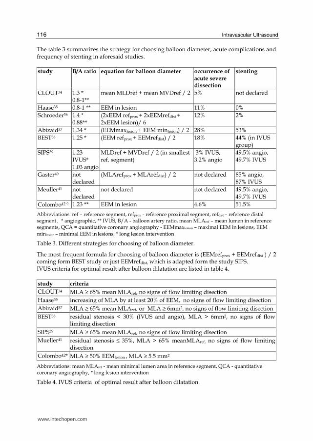
The table 3 summarizes the strategy for choosing balloon diameter, acute complications and frequency of stenting in aforesaid studies.
study B/A ratio
equation for balloon diameter occurrence of acute severe dissection
stenting
CLOUT34 1.3 * 0.8-1**
mean MLDref + mean MVDref / 2 5% not declared
Haase35 0.8-1 ** EEM in lesion 11% 0%
Schroeder36 1.4 * 0.88**
(2xEEM refprox + 2xEEMrefdist + 2xEEM lesion)/ 6
12% 2%
Abizaid37 1.34 * (EEMmaxlesion + EEM minlesion) / 2 28% 53%
BEST38 1.25 * (EEM refprox + EEMrefdist) / 2 18% 44% (in IVUS group)
SIPS39 1.23 IVUS* 1.03 angio
MLDref + MVDref / 2 (in smallest ref. segment)
3% IVUS, 3.2% angio
49.5% angio, 49.7% IVUS
Gaster40 not declared
(MLArefprox + MLArefdist) / 2 not declared 85% angio, 87% IVUS
Meuller41 not declared
not declared not declared 49.5% angio, 49.7% IVUS
Colombo42 ° 1.23 ** EEM in lesion 4.6% 51.5%
Abbreviations: ref – reference segment, refprox - reference proximal segment, refdist – reference distal segment , * angiographic, ** IVUS, B/A - balloon artery ratio, mean MLAref – mean lumen in reference segments, QCA = quantitative coronary angiography - EEMmaxlesion – maximal EEM in lesions, EEM
minlesion – minimal EEM in lesions, ° long lesion intervention
Table 3. Different strategies for choosing of balloon diameter.
The most frequent formula for choosing of balloon diameter is (EEMrefprox + EEMrefdist ) / 2 coming form BEST study or just EEMrefdist, which is adapted form the study SIPS. IVUS criteria for optimal result after balloon dilatation are listed in table 4.
study criteria
CLOUT34 MLA ≥ 65% mean MLAref, no signs of flow limiting disection
Haase35 increasing of MLA by at least 20% of EEM, no signs of flow limiting disection
Abizaid37 MLA ≥ 65% mean MLAref, or MLA ≥ 6mm2, no signs of flow limiting disection
BEST38 residual stenosis < 30% (IVUS and angio), MLA > 6mm2, no signs of flow limiting disection
SIPS39 MLA ≥ 65% mean MLAref, no signs of flow limiting disection
Mueller41 residual stenosis ≤ 35%, MLA > 65% meanMLAref, no signs of flow limiting disection
Colombo42* MLA ≥ 50% EEMlesion , MLA ≥ 5.5 mm2
Abbreviations: mean MLAref - mean minimal lumen area in reference segment, QCA - quantitative coronary angiography, * long lesion intervention
Table 4. IVUS criteria of optimal result after balloon dilatation.
www.intechopen.com

IVUS Guided PCI
117
Based on these studies with IVUS guided balloon dilatation we can summarize:
1. Using of larger balloons (balloon/artery ratio more than 1 according to angiographic
assessment) is safe and without increased risk for acute severe complications. In
hospital MACE were 1.4-3.9%. Occurrence of significant dissection varies in large
range, probably for different definition of this kind of complication. However, all
authors declare low risk for sever acute dissection.
2. The rate of in stent restenosis is consistently low in all studies. It means that it is safe to avoid stent implantation after fulfilling of IVUS criteria for adequate results after balloon dilation. Surprisingly, higher in stent restenosis and higher MACE were found in some studies in patients with stent implantation compared to plain balloon dilatation. The reason for this finding is probably for bias; according to the study design stents were implanted to lesions with non satisfactory results after balloon dilatation or for treatment of complications after balloon dilatation. Higher occurrence of in stent restenosis and MACE is expected in these types of lesions.
3. The dark side of IVUS guided balloon angioplasty is a prolonged procedural time (5-13 minutes), increased X-ray time (2-3.6 minutes) and higher amount of contrast dye (12-34 ml)38-41
4. Routine IVUS guided intervention can be beneficial from financial point of view. Optimal decision making decreases number of implanted stent and frequency of in stent restenosis and avoids peri-procedural complications from non-indicated coronary interventions40,41.
5. IVUS guided intervention should be used for avoiding stent implantation in patients
unsuitable for dual antiplatelet therapy.
5. Bare-metal stent implantation
5.1 Mechanism of lumen enlargement during stent implantation The most important mechanisms are plaque redistribution (inside stent) and plaque
extrusion (outside stent) to the reference segments (more frequently to the distal one)43.
Plaque redistribution and extrusion play role during restenosis in edge segment of stent44.
Less important factors are plaque compression, plaque embolization and vessel enlargement
(more in vessel with negative remodeling before PCI)43.
Final stent diameter is a result of interplay between pressure during implantation and vessel
wall resistance. Declared stent diameters for different pressures during dilatation do not
correlate with real stent diameter after implantation. These numbers for stent diameter come
from in vitro tests in water and do not reflect real situation in the vessel. Costa et al.45
compared 200 drug eluting stent diameters from IVUS measurement immediately after
implantation and declared stent diameter for nominal pressure. At least 90% of minimal
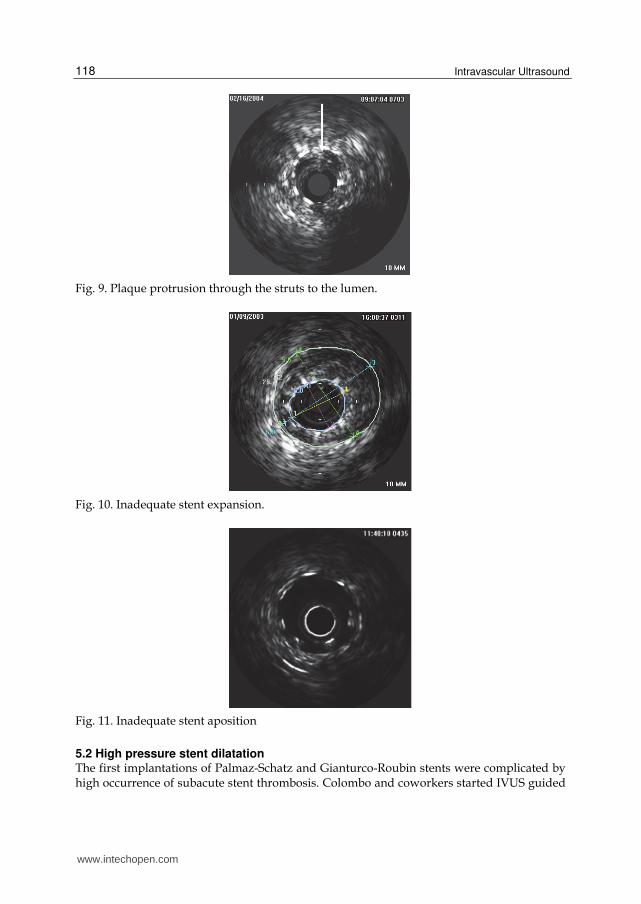
lumen diameter for nominal pressure was reached only in 4% of stents. In stent area can be
decreased immediately after implantation also by protrusion of plaque material through the
struts46 (figure9).
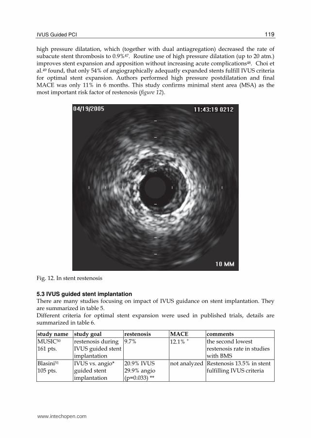
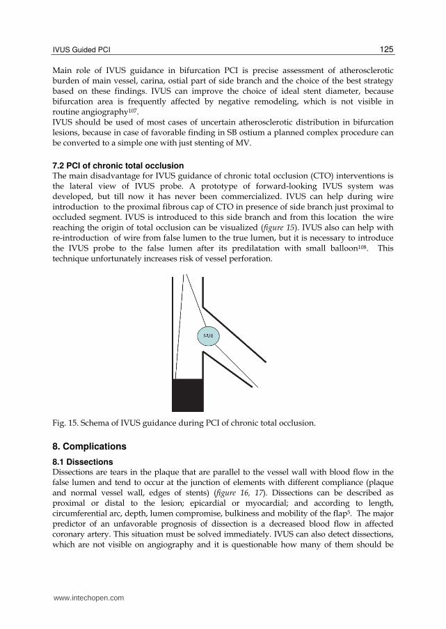
It is important to distinguish between stent expansion and stent apposition. Expansion
means ratio between minimal stent area (MSA) and lumen area in reference segment.
Inadequate expansion (figure 10) can be improved by high pressure postdilatation.
Apposition reflects contact between stent struts and vessel wall. Inadequate apposition
(figure 11) can be solved by low pressure dilatation with a bigger balloon.
www.intechopen.com
Intravascular Ultrasound
118
Fig. 9. Plaque protrusion through the struts to the lumen.
Fig. 10. Inadequate stent expansion.
Fig. 11. Inadequate stent aposition
5.2 High pressure stent dilatation The first implantations of Palmaz-Schatz and Gianturco-Roubin stents were complicated by high occurrence of subacute stent thrombosis. Colombo and coworkers started IVUS guided
www.intechopen.com
IVUS Guided PCI
119
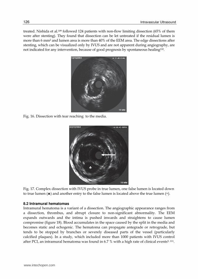
high pressure dilatation, which (together with dual antiagregation) decreased the rate of subacute stent thrombosis to 0.9%47. Routine use of high pressure dilatation (up to 20 atm.) improves stent expansion and apposition without increasing acute complications48. Choi et al.49 found, that only 54% of angiographically adequatly expanded stents fulfill IVUS criteria for optimal stent expansion. Authors performed high pressure postdilatation and final MACE was only 11% in 6 months. This study confirms minimal stent area (MSA) as the most important risk factor of restenosis (figure 12).
Fig. 12. In stent restenosis
5.3 IVUS guided stent implantation There are many studies focusing on impact of IVUS guidance on stent implantation. They are summarized in table 5. Different criteria for optimal stent expansion were used in published trials, details are summarized in table 6.
study name study goal restenosis MACE comments
MUSIC50 161 pts.
restenosis during IVUS guided stent implantation
9.7% 12.1% + the second lowest restenosis rate in studies with BMS
Blasini51 105 pts.
IVUS vs. angio* guided stent implantation
20.9% IVUS 29.9% angio (p=0.033) **
not analyzed Restenosis 13.5% in stent fulfilling IVUS criteria
www.intechopen.com

Intravascular Ultrasound
120
study name study goal restenosis MACE comments
RESIST52
study
155 pts.
IVUS vs. angio
guided (IVUS
controlled) stent
implantation
22.5% IVUS
28.8% angio
(p=0.25)
not declared IVUS guidance does not
decrease rate of in stent
restenosis
OPTICUS53
550 pts.
IVUS vs. angio
guided stent
implantation
24.5% IVUS
22.5% angio
(p=0.68)
17.9%♣ IVUS
15.3% angio
(p=0.4)
IVUS guiding do not
decrease neither rate of in
stent restenosis nor
MACE
CRUISE54
499 pts.
IVUS vs. angio
guided (IVUS
controlled) stent
implantation
not declared TVR
8.5%• in
IVUS group
15.3% in
angio group
(p=0.019)
IVUS guided stenting
leads to lower TVR
Choi49
278 pts.
IVUS vs. angio
guided stent
implantation
not declared 12%+IVUS
19% angio
(p=0,11)
IVUS leads to lower
periprocedural
complications with a
trend for lower TVR in
IVUS group(p=0.08)
AVID55
800 pts.
IVUS vs. angio
guided stent
implantation
not declared 18% IVUS
19% angio
IVUS leads to larger
lumen, but without any
effect on 30 days and 12
months MACE
TULIP56
150 pts.
IVUS vs. angio
guided stenting of
long lesions
23%+ IVUS
46% angio
(p=0.008)
6%+ IVUS
20% angio
(p=0.01)
Better clinical and
angiographic results in
IVUS group despite more
implanted stents
PRESTO57
796 pts with
IVUS vs.
8274 with
angio
IVUS vs. angio
guided stenting
not declared TVR 13.8%
IVUS 12.2%
angio (p=0.9)
Larger MLD in IVUS
group, but without
difference in MACE.
Criteria for adequate
IVUS stent expansion
was let only on operators
discretion
DIPOL58 163
pts.
IVUS vs. angio
guided stenting
10% IVUS 27%
angio
7.3% IVUS
16% angio
Best results for IVUS
guided in randomized
trial
Gaster55,60
108 pts
IVUS vs. angio
guided stenting
16% IVUS
25% angio
22% IVUS
41% angio
IVUS guiding is cost
saving
* non randomized trial, angio group is a historic control, ** IVUS criteria was fulfilled in 49.5% patients
and in these ones was restenosis 13.5%, + 6 months, ♣ 12 months, • 9 months, TVR - target vessel
revascularization
Table 5. Trials assessing IVUS guided stent implantation.
www.intechopen.com

IVUS Guided PCI
121
study criteria for optimal expanded stent
Katritsis61 full stent apposition, MSA ≥ 90% meanMLAref, symmetrical expansion (min SD/maxSD > 0.7) (correlation with FFR 0.94)
Fearon62 MSA 7 mm2 (correlates with FFR 0.96)
Hanekamp63 full stent apposition, MSA ≥ 90% meanMLAref, or MSA ≥ 100% minMLAref, symetrical expansion (minSD/maxSD > 0.7), (correlation with FFR 0.94)
Gorge48 symmetrical stent expansion, MSD ≥ 3 mm, full apposition
Choi49 complete stent apposition, MSA ≥ 80% distalMLAref , symmetrical expansion (minSD/maxSD > 0.8)
Colombo47 full stent apposition , MSA ≥ minMLAref, MLA > 60% MLAref in edge regions
Ahmed44 complete stent apposition, MSA ≥ 80% meanMLAref or MSA 7.5 mm2
MUSIC50*
- complete stent apposition, MSA ≥ 90 meanMLAref or ≥ 100 minMLAref - in case of > 9 mm2 :
MSA ≥ 80% meanMLAref or ≥ 90% minMLAref
MSA in proximal part of stent ≥ 90% proxMLAref symmetrical expansion (minSD/maxSD > 0.7)
Blasini51 full stent apposition, MSA > 8 mm2, or MSA ≥ 90% meanMLAref, complete coverage of dissection
RESIST52 MSA ≥ 80% meanMLAref
OPTICUS53 MUSIC criteria, residual stenosis les than 10% (angiographically)
CRUISE54 residual stenosis < 10% (angiographically)
AVID55 full stent apposition, residual stenosis < 10%, no signs of dissection
TULIP56 complete stent apposition, MSD ≥ 80% meanMLDref , MSA ≥ 90% distalMLAref
Abbreviations: SD - stent diameter, meanMLAref - mean minimal lumen area in reference segments,
minMLAref minimal - minimal lumen area i reference segments, MSD – minimal stent diameter, MSA –
minimal stent area * all criteria were fulfilled in 81% patients and these patients were treated only with acetylosalycilic acid and rate of subacute stent thrombosis was 1.3%
Table 6. Different criteria for optimal stent expansion.
The main goal for IVUS guidance of stent implantation is a larger minimal stent diameter and minimal stent area. Further improvement is seen in strut apposition. Minimal stent area is the most important risk factor for development of in stent restenosis (ISR). The cut-off point for
risk of ISR is 8mm2 in vessel ≥ 3 mm and 6 mm2 in vessel < 3mm64. The rate of in stent restenosis according to achievement of different IVUS parameters is summarized in table 7.
criteria restenosis rate
MSA > 9 mm2 11%
MSA > 9 mm2 a MSA ≥ 80% meanMLAref 12.5%
MSA ≥ 55% meanEEMref 17%
MSA ≥ 90% meanMLAref 21%
MSA ≥ 90% distal MLAref 22%
Abbrevation: MSA – minimal stent area
Table 7. Risk of ISR and achievement of different IVUS parameters.
www.intechopen.com

Intravascular Ultrasound
122
The lowest rate of ISR is in stents with MSA > 9mm2. However, for achievement of this MSA it is necessary to use a 3.5mm stent (with ideal stent area 9.6mm2), because 3mm stent has an ideal stent area 7.1mm2. MSA 8 mm2 as a sufficient post-stenting area was confirmed by Hoffmann et al65. They found a mean MSA in stent with ISR 7.1mm2 and 8.1mm2 in stents without development of ISR. This cut-off MSA is also a predictor for development of ISR in long stents66. Further predictors for ISR are: - length of stents67 - strong calcification68 - bulky plaque compensated by positive vessel remodelation69-72 (figure 13) - plaque protrusion through the stent struts73 MSA is the most important factor for development of in stent thrombosis (IST)74. The incidence of this feared complication is less than 0.5%74. Parise et al.75 published recently a meta-analysis of all randomized studies comparing IVUS
guided vs. angio guided stenting in pre-drug-eluting stent era. They concluded that IVUS
guided stenting significantly lowered the 6-month angiographic restenosis rates, 12-month
revascularization and MACE rate. They did not find any effect of IVUS guidance on death
or myocardial infarction.
Fig. 13. Large plaque compensated by positive vessel remodeling
6. Drug-eluting stent implantation
Drug eluting stents (DES) significantly reduce the risk of in stent restenosis compared to
BMS76-78. IVUS guidance seemed to be needless for DES excellent results. However, several
issues are still a problem even with the use of DES and some of them, like in stent
thrombosis, seem to be more important than in BMS .
In stent restenosis still exists and it´s incidence is 5-10% in selected populations79-80. It seems to
be more pronounced in paclitaxel eluting stent than sirolimus eluting stents81. Minimum stent
area that best separates restenosis from no restenosis in DES is between 5.0-6.0 mm2 82,83.
The occurrence of in stent thrombosis (IST) in DES is 1-1.5%84. The main cause of IST is stent
under expansion. DES with further IST development showed significantly lower MSA (4.3-
4.6 mm2). Further risk factor for IST development is residual edge stenosis, defined as a
MLA < 4 mm2 and a plaque burden > 70%85,74. These risk factors remain the same like in
BMS, where the presence of dissection, thrombus or tissue prolaps into the stent were
www.intechopen.com

IVUS Guided PCI
123
recognized as further risk factors for IST development86,87. The occurrence of IST in BMS is
estimated at 0.9%88.
The risk factors for IST development can be revealed by IVUS control after stent
implantation. Roy et al.89 published study, where IVUS guidance of DES implantation
reduced development of IST in 30 days (0.5% in IVUS vs. 1.5% in angio guided group) and
12 month (0.7% in IVUS vs. 2.0% in angio guided group), as well as decreased need for
revascularization and MACE in 30 day. Unfortunately, there were no clear criteria for
adequate stent deployment in this study. Claessen et al.90 published a study comparing
angio and IVUS guidance in 1504 sirolimus eluting stent implantation in the MATRIX
(Comprehensive Assessment of Sirolimus-Eluting Stents in Complex Lesions) registry. They
found significantly lower occurrence of myocardial infarction and combined endpoint of
myocardial infarction and death in IVUS guided group. Hur et al.91 published their
enormous registry of angio (3744 patients) versus IVUS (4627 patients) guided stent
implantation. The main finding of this study is lower mortality in IVUS guided group. This
result was seen mainly in DES implantation, because mortality in BMS group was the same.
However, the explanation of this interesting finding is not clear, because the occurrence of
myocardial infarction, target vessel revascularization and in stent thrombosis was without
significance differences between angio and IVUS group. Based on aforesaid trials we can
conclude, that IVUS guidance of DES implantation probably improves patient’s outcome,
but further trials are needed for better understanding of this phenomenon.
Important phenomenon in DES implantation is incomplete stent apposition (ISA), which
means, that at least one strut is not adequately apposed to vessel wall. One should
distinguish between acute stent malapposition, which is present immediately after stent
implantation, late stent malapposition (LSM), which can be persistent , it means that acute
malapposition is not healed and late-acquired stent malapposition (LASM), which develops
despite normal finding after stent implantation. The mechanism of LASM development is
not known, several mechanisms are thought:
- positive vessel remodeling leading to vessel enlargement
- decrease of plaque volume behind the stent caused by antiproliferative effect of DES
- thrombus dissolution after PCI of thrombus containing lesions
The frequency of LSM is not clear, a quite broad range for this phenomenon was published
(4-21%)92,93. LSM is not found only in DES, but also in BMS, where it’s incidence is 4.4-
5.4%94,95. Empty space behind stent struts can lead to decrease of blood flow in this region
and cause development of thrombosis. However, this theoretic concept was not proved in
any study following the natural course of patients with LSM. Hong et al.96,97 did not find any
clinical adverse event in patient with LSM during 10 months follow up. On the other hand
Cook et al.98 published correlation between LSM and very late IST. Moreover, Hassan et al.99
published meta-analysis of 17 randomized trials focusing on LSM in BMS as well as DES.
They found four times higher risk of LASM in patients with DES compared to BMS and
LSM (acquired or persistent) increased significantly risk for (very) late in stent thrombosis
(OR 6,51).
A rare complication (1.25%) of DES implantation is development of coronary aneurysm. The
definition of aneurysm is focal enlargement of vessel lumen, which is 50% larger than
adjacent reference vessel. Coronary aneurysm is not a benign finding as up to 40% of
patients needed revascularization in a study done by Alfonso et al.100.
www.intechopen.com

Intravascular Ultrasound
124
7. Ivus guided pci in specific situation
7.1 PCI of bifurcation lesions PCI in bifurcation is a more challenging procedure with a risk of compromising flow in side branch (SB) and with higher rate of restenosis. The “classical“ mechanisms of worsening flow in side branch were thought 101 - plaque compression in ostial part of SB during dilatation in main vessel (MV). The
presence of diffuse ostial plaque (figure 14) has higher risk for TIMI 2 flow after stenting than presence of eccentric plaque102
- plaque shifting from the MV to SB during dilatation in MV (“snow-plow” phenomenon)
- interposition of stent struts across the ostium of SB
Fig. 14. Bifurcation with diffuse impairment of side branch ostium.
Plaque in bifurcation is mainly localized in counter carina103. However, Medina et al.104 proved the presence of plaque also in carina. They found plaque in 32% of bifurcations and moreover, in 16% of bifurcations the plaque in the carina was larger than in counter carina area. Furthermore, authors showed, that the mechanism of ostial SB damage after stent implantation in the MV was always due to displacement of the carina and no cases of plaque shifting were found. Further interesting finding of this study is that plaque in the carina was greater in the bifurcations that had maximum stenosis located distal to the carina in the MV. Authors explain this finding by hypothesis of different flow velocity patterns in these lesions. Atheroprotective high shear stress can be transformed into an atero-prone low shear stress. Authors describe less damage of SB ostium after stenting of lesions with plaque at the carina. Plaque probably makes carina more resistant and does not allow carina shifting, which is now thought to be a dominant mechanism causing damage to the SB ostium when a stent is implanted into the MV. Important contribution for planning of PCI in bifurcations is a study done by Costa et al105.
They found that the part of MV just behind the origin of SB (so called “lower diamond” or
“polygon of confluence”) is more prone for small minimal stent area after dilatation of SB
ostium, which is not reverted even after kissing balloon dilatation. Kang et al.106 published a
study assessing the IVUS predictors for side branch compromise after single-stent crossover
technique and found two predictors for post stenting FFR<0.8: MLA 2.4mm2 and PB > 51%
in SB ostium.
www.intechopen.com
IVUS Guided PCI
125
Main role of IVUS guidance in bifurcation PCI is precise assessment of atherosclerotic burden of main vessel, carina, ostial part of side branch and the choice of the best strategy based on these findings. IVUS can improve the choice of ideal stent diameter, because bifurcation area is frequently affected by negative remodeling, which is not visible in routine angiography107. IVUS should be used of most cases of uncertain atherosclerotic distribution in bifurcation lesions, because in case of favorable finding in SB ostium a planned complex procedure can be converted to a simple one with just stenting of MV.
7.2 PCI of chronic total occlusion The main disadvantage for IVUS guidance of chronic total occlusion (CTO) interventions is the lateral view of IVUS probe. A prototype of forward-looking IVUS system was developed, but till now it has never been commercialized. IVUS can help during wire introduction to the proximal fibrous cap of CTO in presence of side branch just proximal to occluded segment. IVUS is introduced to this side branch and from this location the wire reaching the origin of total occlusion can be visualized (figure 15). IVUS also can help with re-introduction of wire from false lumen to the true lumen, but it is necessary to introduce the IVUS probe to the false lumen after its predilatation with small balloon108. This technique unfortunately increases risk of vessel perforation.
Fig. 15. Schema of IVUS guidance during PCI of chronic total occlusion.
8. Complications
8.1 Dissections Dissections are tears in the plaque that are parallel to the vessel wall with blood flow in the false lumen and tend to occur at the junction of elements with different compliance (plaque and normal vessel wall, edges of stents) (figure 16, 17). Dissections can be described as proximal or distal to the lesion; epicardial or myocardial; and according to length, circumferential arc, depth, lumen compromise, bulkiness and mobility of the flap5. The major predictor of an unfavorable prognosis of dissection is a decreased blood flow in affected coronary artery. This situation must be solved immediately. IVUS can also detect dissections, which are not visible on angiography and it is questionable how many of them should be
www.intechopen.com
Intravascular Ultrasound
126
treated. Nishida et al.109 followed 124 patients with non-flow limiting dissection (65% of them were after stenting). They found that dissection can be let untreated if the residual lumen is more than 6 mm2 and lumen area is more than 40% of the EEM area. The edge dissections after stenting, which can be visualized only by IVUS and are not apparent during angiography, are not indicated for any intervention, because of good prognosis by spontaneous healing110.
Fig. 16. Dissection with tear reaching to the media.
Fig. 17. Complex dissection with IVUS probe in true lumen, one false lumen is located down
to true lumen (♣) and another entry to the false lumen is located above the true lumen (∗).
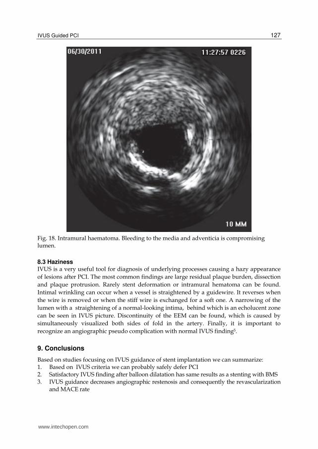
8.2 Intramural hematomas Intramural hematoma is a variant of a dissection. The angiographic appearance ranges from
a dissection, thrombus, and abrupt closure to non-significant abnormality. The EEM
expands outwards and the intima is pushed inwards and straightens to cause lumen
compromise (figure 18). Blood accumulates in the space caused by the split in the media and
becomes static and echogenic. The hematoma can propagate antegrade or retrograde, but
tends to be stopped by branches or severely diseased parts of the vessel (particularly
calcified plaques). In a study, which included more than 1000 patients with IVUS control
after PCI, an intramural hematoma was found in 6.7 % with a high rate of clinical events5, 111.
www.intechopen.com
IVUS Guided PCI
127
Fig. 18. Intramural haematoma. Bleeding to the media and adventicia is compromising lumen.
8.3 Haziness IVUS is a very useful tool for diagnosis of underlying processes causing a hazy appearance
of lesions after PCI. The most common findings are large residual plaque burden, dissection
and plaque protrusion. Rarely stent deformation or intramural hematoma can be found.
Intimal wrinkling can occur when a vessel is straightened by a guidewire. It reverses when
the wire is removed or when the stiff wire is exchanged for a soft one. A narrowing of the
lumen with a straightening of a normal-looking intima, behind which is an echolucent zone
can be seen in IVUS picture. Discontinuity of the EEM can be found, which is caused by
simultaneously visualized both sides of fold in the artery. Finally, it is important to
recognize an angiographic pseudo complication with normal IVUS finding5.
9. Conclusions
Based on studies focusing on IVUS guidance of stent implantation we can summarize: 1. Based on IVUS criteria we can probably safely defer PCI 2. Satisfactory IVUS finding after balloon dilatation has same results as a stenting with BMS 3. IVUS guidance decreases angiographic restenosis and consequently the revascularization and MACE rate
www.intechopen.com

Intravascular Ultrasound
128
4. IVUS guidance decreases rate of in stent thrombosis 5. Contraindication for IVUS guidance are small vessels, tortuous vessels and degenerated vein grafts This summary raises the question why IVUS is not used routinely during all PCI procedures. The answers can be divided into two groups. First there are the lessons we have learned from IVUS and this phenomenon is known as an “IVUS eye”. We know that vessels are bigger than they look on angio and we are using bigger stents than before IVUS studies. We know, that without high pressure post dilatation we are not able to achieve sufficient stent diameter despite information derived from manufacturer’s compliance charts. These factors dramatically contributed to lowering of in stent restenosis and therefore it is difficult to find statistical significant difference between IVUS and angio guided interventions, like it was described in OPTICUS trial. However, IVUS is still irreplaceable during investigation of unclear finding during angiography (like hazy lesions) or after stenting. IVUS is especially recommended during PCI of left main, the last remaining coronary artery and PCI in patients who are contraindicated for dual antiplatelet therapy for avoiding stent implantation. Furthermore, precise assessment of bifurcation can change the strategy from more complex to simple one in case of low risk profile of ostial part of side branch The group of arguments against routine use of IVUS during PCI is cost of IVUS catheter, longer procedural time and higher contrast dye consumption. Moreover IVUS guided PCI are more complex and need more skills than angio guided ones. However, in indicated cases IVUS guidance undoubtedly improves short as well as long term of coronary interventions.
10. References
[1] Pijls NH, de Bruyne B, Peels K et al, Measurement of Fractional Flow Reserve to Assess the Functional Severity of Coronary-Artery Stenoses. NEJM 1996; 334: 1703–1708
[2] Topol EJ, Nissen SE.Our preoccupation with coronary luminology. The dissociation between clinical and angiographic findings in ischemic heart disease. Circulation 1995;92:2333-2342
[3] Vlodaver Z, Frech R, Van Tassel RA, et al. Correlation of the antemortem coronary angiogram and the postmortem specimen. Circulation 1973;47:162-169
[4] Mintz GS, Nissen SE, Anderson WD, Erbel R, Fitzgerald PJ, Pinto FJ, Rosenfiels K, Siegel K, Tuzcu EM, Yock PG. American College of Cardiology Clinical Expert Consensus Document on Standards for Acquisition, Measurement and Reporting of Intravascular Ultrasound Studies (IVUS) A Report of the American College of Cardiology Task Force on Clinical Expert Consensus Documents. JACC 2001;37:1478-1492
[5] Mintz GS. Intracoronary ultrasound. Taylor & Francis 2005 [6] Briguori C, Anzuini A, Airoldi F, et al. Intravascular Ultrasound Criteria for the
Assessment of the Functional Significance of Intermediate Coronary Artery Stenoses and Comparison with Fractional Flow Reserve. Am J Cardiol 2001;87:136-141
[7] Abizaid A, Mintz GS, Pichard A, et al. Clinical, Intravascular Ultrasound, and Quantitative Angiographic Determinants of the Coronary Flow Reserve Before and After Percutaneous Transluminal Coronary Angioplasty. Am J Cardiol 1998;82:423-428
[8] Nishioka T, Amanullah A, Luo H, et al. Clinical Validation of Intravascular Ultrasound Imaging for Assessment of Coronary Stenosis Severity. Comparison With Stress Myocardial Perfusion Imaging. J Am Coll Cardiol 1999;33:1870-1878
www.intechopen.com
IVUS Guided PCI
129
[9] Takagi A, Tsurumi Y, Suzuki K, et al. Clinical Potential of Intravascular Ultrasound for Physiological Asessment of Coronary Stenosis. Circulation 1999;100:250-255
[10] Lee Ch, Tai B, Soon Ch, Low A, Poh K, Yeo T, Lim G, Yip J, Omar A, Teo S, Tan H. New set of intravascular ultrasound-derived anatomic criteria for defining functionally significant stenoses in small coronary arteries (results from intravascular diagnostic evaluation of atherosclerosis in Singapore [IDEAS] Study). Am J Cardiol 2010;105:1378-1384
[11] Abizaid A, Mintz GS, Mehran R, et al. Long Term Follow-up After Percutaneous Transluminal Coronary Angioplasty Was Not Performed Based on Intravascular Ultrasound Findings. Circulation 1999;100:256-261
[12] Jasti V, Ivan E, Yalamanchili V, et al. Correlation between fractional flow reserve and intravascular ultrasound in patients with an ambiguous left main coronary artery stenosis. Circulation 2004;110:2831-2836).
[13] Abizaid A, Mintz GS, A.Abizaid, et at. One-Year Follow-up After Intravascular Ultrasound Assessment of Moderate Left Main Coronary Artery Disease in Patients With Ambiquous Angiograms. J Am Coll Cardiol 1999;34:707-715
[14] Kang SJ, Lee JY, Ahn JM, Mintz GS, Kim WJ, Park DW, Yun SCh, Lee SW, Kim YH, Lee ChW, Park SW, Park SJ. Validation of Intravascular Ultrasound–Derived Parameters With Fractional Flow Reserve for Assessment of Coronary Stenosis Severity. Circulation: Cardiovascular Interventions 2011; 4: 65-71
[15] Ben-Dor I, Torguson R, Gaglia MA, Gonzales MA, Maluenda G, Bui AB, Xue Z, Satle LF, Suddath WO, Lindsay J, Pichard AD, Waksman R. Correlation between fractional flow reserve and intravascular ultrasound lumen area in intermediate coronary artery stenosis. Eurointervention 2011;7:225-233
[16] Ahn JM. Kang SJ, Mintz GS, Oh JH, Kim WJ, Lee JY, Park DW, Lee SW, Kim YH, Lee ChW, Park SW, Moon DH, Park SJ. Validation of minimal lumen area measured by intravascular ultrasound for assessment of functionally significant coronary stenoses. JACC Cardiovasc Interv 2011;4:665-671
[17] Stone GW, Maehara A, Lansky A, de Bruyne B, Cristea E, Minth GS, Mehran R, McPherson J, Farhat N, Marso SP, Parise H, Templin B, White R, Zhankg Z,Serruys PW. A prospective natural-history study of coronary atherosclerosis.NEJM 2011;364:226-235
[18] Fearon WF, Bornschein B, Tonino PA, Gothe RM, Bruyne BD, Pijls NH, Siebert U; Fractional Flow Reserve Versus Angiography for Multivessel Evaluation (FAME) Study Investigators. Economic evaluation of fractional flow reserve-guided percutaneous coronary intervention in patients with multivessel disease.Circulation 2010;122:2545-50
[19] Nam ChW, Yoon HJ, ChoYK, Park HS, Kim H, Hur SH, Kim YN, Chung IS, Koo BK, Tahk SJ, Fearon WF, Kim KB. Outcomes of percutaneous coronary intervention in intermediate coronary artery disease. Fractional flow reserve-guided versus intravascular ultrasound-guided. JACC Cardiol Intv 2010;3:812-817
[20] Ricciardi M, Meyers S, Choi K, et al. Angiographically silent left main disease detected by intravascular ultrasound: A marker for future adverse cardiac events. Am Heart J 2003;146:507-512
[21] Hermiller JB, Buller CE, Tenaglia AN, et al. Unrecognized left main artery disease in patients undergoing interventional procedures. Am J Cardiol 1993;71:173-176
www.intechopen.com

Intravascular Ultrasound
130
[22] Ge J, Liu F, Görge G, et al. Angiographically „silent“ plaque in the left main coronary artery detected by intravascular ultrasound. Coronary Artery disease 1995;6:805-810
[23] Russo RJ, Wong SC, Marchant D, et al. Intravascular ultrasound-directed clinical decision making in the setting of an inconclusive left main coronary angiogram: final results from the Left Main IVUS Registry. Circulation 2004;108:IV-462
[24] Suter Y, Schoenenberger AW, Toggweiler S, Jamshidi P, Resink T, Erne P. Intravascular ultrasound-based left main coronary artery assessment: comparison between pullback from left anterior descending and circumflex arteries. J Invasive Cardiol 2009;21:457-460
[25] Oviedo C, Maehara A, Mintz GS, Tsujita K, Kubo T, Doi H, Castellanos C, Lansky AJ, Mehran R, Dangas G, Leon MB, Stone G, Templin B, Araki H, Ochiai M, Moses JW. Is accurate intravascular ultrasound evaluation of the left circumflex ostium from a left anterior descending to left main pullback possible? Am J Cardiol 2010;105:948-95
[26] Park SJ, Kim YH, Park DW, Lee SW, Kim WJ, Suh J, Yun SCh, Lee CHW, Hong MK, Lee JH, Park SW; MAIN-COMPARE Investigators. Impact of intravascular guiaxdance on Long-term mortality in stenting for unprotected left main coronary artery stenosis. Circ Cardiovasc Interv 2009;2:167-177
[27] Kotani J, Mintz GS, Rai P, et al. Intravascular ultrasound assessment of angiographic filling defects in native coronary arteries: Do they always contain thrombi ? JACC 2004;44:2087-2089
[28] Kobayashi Y, De Gregorio J, Kobayashi N, et al. Stented segments length as an independent predictor of restenosis. J Am Coll Cardiol 1999:34:651-659
[29] Grewal J, Ganz P, Selwyn A, et al. Usefulness of intravascular ultrasound in preventing stenting of hazy areas adjacent to coronary stents and its support of support spot-stenting. Am J Cardiol 2001;87:1246-1249
[30] Ziada KM, Tuzcu EM, De Franco AC, et al. Intravascular ultrasound assessment of the prevalence and cause of angiographic „haziness“ following high-pressure coronary stenting. Am J Cardiol 1997;80:116-121
[31] Maehara A, Mintz GS, Ahmed JM, et al. An intravascular ultrasound classification of angiographic coronary artery aneurysms. Am J Cardiol 2001;88:365-370
[32] Mizuno K, Kurita A, Imazeki N. Pathological findings after percutaneous transluminal coronary angioplasty. Br Heart J 1984;52:588-590
[33] Honey J, Mahon D, Jain A, et al. Morphological effects of coronary balloon angioplasty in vivo assessed by intravascular ultrasound imaging. Circulation 1992;85:1012-1025
[34] Stone G, Hodgson J, Goar F, et al. Improved procedural results of coronary angioplasty with intravascular ultrasound-guided balloon sizing. The CLOUT Pilot Trial. Circulation 1997;95:2044-2052
[35] Haase K, Athanasiadis A, Mahrholdt H, et al. Acute and one year follow-up results after vessel size adapted PTCA using intravascular ultrasound. Eur Heart J 1998;19:263-272
[36] Schroeder S, Baumbach A, Haase K, et al. Reduction of restenosis by vessel size adapted percutaneous transluminal coronary angioplasty using intravascular ultrasound. Am J Cardiol 1999;83:875-879
www.intechopen.com

IVUS Guided PCI
131
[37] Abizaid A, Pichard A, Mintz GS, et al. Acute and long-term results of an intravascular ultrasound-guided percutaneous transluminal coronary angioplasty/provisional stent implantation strategy. Am J Cardiol 1999;84:1298-1303
[38] Schiele F, Meneveau N, Gilard M, et al. Intravascular ultrasound-guided balloon angioplasty compared with stent. Immediate and 6-month results of the multicenter, randomized balloon equivalent to stent study (BEST). Circulation 2003;107:545-551
[39] Frey A, Hodgson J, Muller Ch, et al. Ultrasound-guided strategy for provisional stenting with focal balloon combination catheter. Results from the randomized strategy for intracoronary ultrasound-guided PTCA and stenting (SIPS) trial. Circulation 2000;102:2497-2502
[40] Gaster A, Skjoldborg S, Larsen J, et al. Continued improvement of clinical outcome and cost effectiveness following intravascular ultrasound guided PCI: insight from a prospective, randomized study. Heart 2003;89:1043-1049
[41] Mueller Ch, Hodgson J, Schindler Ch, et al. Cost-effectiveness of intracoronary ultrasound for percutaneous coronary interventions. Am J Cardiol 2003;91:143-147
[42] Colombo A, De Gregorio J, Moussa I, et al. Intravascular ultrasound-guided percutaneous transluminal coronary angioplasty with provisional spot stenting for treatment of long coronary lesion. J Am Coll Cardiol 2001;38:1427-1433
[43] von Birgelen, Mintz GS, Eggebrecht H, et al. Preintervention arterial remodeling affects vessel stretch and plaque extrusion during coronary stent deployment as demonstrated by three-dimensional intravascular ultrasound. Am J Cardiol 2003;92:130-135
[44] Ahmed JM, Mintz GS, Weissman NJ, et al. Mechanism of lumen enlargement during intracoronary stent implantation. An intravascular ultrasound study. Circulation 2000;102:7-10
[45] Costa JR, Mintz GS, Carlier SG,Costa RA, Fujii K,Sano K, Kimura M, Lui J, Weisz G, Moussa I, Dangas G, Mehran R, Lansky AJ, Kreps EM, Collins M, Stone GW, Moses JW, MD, Leon MB. Intravascular Ultrasonic Assessment of Stent Diameters Derived from Manufacturer’s Compliance Charts. Am J Cardiol 2005;96:74 –78
[46] Ponde CK, Aroney CN, McEniery PT, et al. Plaque prolapse between struts of the intracoronary Palmaz-Schatz stent: report of two cases with a novel treatment of this unusual problem. Cathet Cardiovasc Diagn 1997;40:353-357
[47] Colombo A, Hall P, Nakamura S, et al. Intracoronary stenting without anticoagulation accomplished with intravascular ultrasound guidance. Circulation 1995;91:1676-1688
[48] Gorge G, Haude M, Ge J, et al. Intravascular Ultrasound After low and high inflation pressure coronary artery stent implantation. J Am Coll Cardiol 1995;26:725-730
[49] Choi JW, Vardi GM, Meyers SH, et al. Role of intracoronary ultrasound after high-pressure stent implantation. Am Heart J 2000;139:643-648
[50] de Jaegere P, Mudra H, Figulla H, et al. Intravascular ultrasound-guided optimized stent deployment. Immediate and 6 months clinical and angiographic results from the Multicenter Ultrasound Stenting in Coronaries Study (MUSIC Study). Eur Herat J 1998;19:1214-1223
[51] Blasini R, Neumann FJ, Schmitt C, et al. Restenosis rate after intravascular ultrasound-guided coronary stent implantation. Cathet Cardiovasc Diagn 1998;44:380-386
www.intechopen.com

Intravascular Ultrasound
132
[52] Schiele F, Meneveau N, Vuillemenot A, et al. Impact of intravascular ultrasound guidance in stent deployment on 6 month restenosis rate: a multicenter, randomized study comparing two strategies-with and without intravascular ultrasound guidance. J Am Coll Cardiol 1998;32:320-328
[53] Mudra H, di Mario C, de Jaegere P, et al. Randomized comparison of coronary stent implantation under ultrasound or angiographic guidance to reduce stent restenosis (OPTICUS study). Circulation 2001;104:1343-1349
[54] Fitzgerald PJ, Oshima A, Hayase M, et al. Final Results of the Can Routine Ultrasound Influence Stent Expansion (CRUISE) Study. Circulation 2000;102:523-530
[55] Russo RJ, Silva PD, Teirstein PS, Attubato MJ, Davidson CJ, De-Franco AC, Fitzgerald PJ, Goldberg SL, Hermiller JB, Leon MB, Ling FS, Lucisano JE, Schatz RA, Wong SC, Weissman NJ, Zientek DM;AVID Investigators. A randomized controlled triual of angiography versus intravascular ultrasound-directed bare-metal coronary stnet placement (the AVID trial). Circ Cardiovasc Interv 2009;2:113-123
[56] Oemrawsingh PV, Mintz GS, Schalij M, et al. Intravascular ultrasound guidance improves angiographic and clinical outcome of stent implantation for long coronary artery stenoses. Final results of a Randomized Comparison With Angiographic Guidance (TULIP Study). Circulation 2003;107:62-67
[57] Orford J, Denktas A, Williams B, et al. Routine intravascular ultrasound scanning guidance of coronary stenting is not associated with improved clinical outcomes. Am Heart J 2004;148:501-6
[58] Gil RJ, Pawlowski T, Dudek D, Horszczaruk G, Zmudka K, Lesiak M, Witkowski A, Ochala A, Kubica J; Investigators of Direct Stenting vs. Optimal Angioplasty trial (DIPOL). Comparison of angiographically guided direct stenting technique with direct stenting and optimal balloon angioplasty guided with intravascular ultrasound: the multicenter, randomized trial results. Am Heart J 2007;154:669-675
[59] Gaster AL, Slothuus U, Larsen J, Thayssen P, Hagfelt TH. Cost-effectiveness analysis of intravascular ultrasound guided percutaneous coronary intervention versus conventional percutaneous coronary intervention. Scand Cardiovasc J 2001;35:80-85.
[60] Gaster AL, Slothuus Skjoldborg U, Larsen J, Korsholm L, von Birgelen C, Jensen S, Thayssen P, Pedersen KE, Hagfelt TH. Continued improvement of clinical outcome and cost effectiveness following intravascular ultrasound guided PCI: insight from a prospective, randomized study. Heart 2003;89:1043-1049
[61] Katritsis D, Ioannidis J Korovessis S, et al. Comparison of myocardial fractional flow reserve and intravascular ultrasound for the assessment of slotted-tube stents. Cathet Cardiovasc Intervent 2001;52:322-326
[62] Fearon W, Luna J, Samady H, et al. Fractional flow reserve compared with intravascular ultrasound guidance for optimizing stent deployment. Circulation 2001;104:1917-1922
[63] Hanekamp CE, Koolen JJ, Pijls NH, Michels HR, Bonnier HJ. Comparison of quantitative coronary angiography, intravascular ultrasound, and coronary pressure measurement to assess optimum stent deployment. Circulation 1999;99:1015-21
[64] Moussa I, Moses J, di Mario C, et al. Does the specific intravascular ultrasound criterion used to optimize stent expansion have an impact on the probability of stent restenosis ? Am J Cardiol 1999;83:1012-1017
www.intechopen.com
IVUS Guided PCI
133
[65] Hoffmann R, Mintz GS, Mehran R, et al. Intravascular ultrasound predictors of angiographic restenosis in lesions treated with Palmaz-Schatz stents. J Am Coll Cardiol 1998;31:43-49
[66] Hong MK, Park SW, Mintz GS, et al. Intravascular ultrasonic predictors of angiographic restenosis after long coronary stenting. Am J Cardiol 2000;85:441-445
[67] de Feyter PJ, Kay P, Disco C, et al. Reference chart derived from post-stent-implantation intravascular ultrasound predictors of 6-moth expected restenosis on quantitative coronary angiography. Circulation 1999;100:1777-1783
[68] von Birgelen C, Mintz GS, Bose D. et al. Impact of moderate lesion calcium on mechanism of coronary stenting as assessed with three-dimensional intravascular ultrasound in vivo. Am J Cardiol 2003;92:5-10
[69] Hong MK, Park SW, Lee CW, et al. Preintervention arterial remodeling as a predictor of intimal hyperplasia after intracoronary stenting: a serial intravascular ultrasound study. Clin Cardiol 2002;25:11-15
[70] Endo A, Hirayama H, Yoshida O, et al. Arterial remodeling influences the development of intimal hyperplasia after stent implantation. JACC 2001;37:70-75
[71] Shiran A, Weissman NJ, Leiboff B, et al. Effect of preintervention plaque burden on subsequent intimal hyperplasia in stented coronary lesions. Am J Cardiol 2000;86:1318-1321, 69/ Alfonso F, Garcia P, Pimental G, et al. Predictors and implications of residual plaque burden after coronary stenting: an intravascular ultrasound study. Am Heart J 2003;145:254-261
[72] Shiran A, Mintz GS, Waksman R et al. Early lumen loss after treatment of in-stent restenosis: an intravascular ultrasound study. Circulation 1998;98:200-203
[73] Okabe T, Mintz GS, Buch A, et al. Intravascular ultrasound parameters associated with stent thrombosis after drug-eluting stent deployment. Am J Cardiol 2007;4:615-620
[74] Cheneau E, Leborgne L, Mintz GS, et al. Predictors of subacute stent thrombosis. Results of a systematic intravascular ultrasound study. Circulation 2003;108:43-47
[75] Parise H, Maehara A, Stone GW, Leon MB, Mintz GS. Meta-analysis of randomized studies comparing intravascular ultrasound versus angiographic guidance of percutaneous coronary intervention in pre-drug-eluting stent era. Am J Cardiol 2011;107:374-382
[76] Sousa JE, Costa MA, Abizaid A , et al. Lack of neointimal proliferation after implantation of sirolimus-coated stents in human coronary arteries: a quantitative coronary angiography and three-dimensional intravascular ultrasound study. Circulation 2001;103:192-195
[77] Holmes DR Jr, Leon MB, Moses JW, Popma JJ, Analysis of 1-year clinical outcomes in the SIRIUS trial: a randomized trial of a sirolimus-eluting stent versus a standard stent in patients at high risk for coronary restenosis. Circulation 2004;109:634-40
[78] Hong MK, Minz GS, Lee ChW, et al. Paclitaxel coating reduces in-stent intimal hyperplasia in human coronary arteries. A serial volumetric intravascular ultrasound analysis from the aSian Paclitaxel-Eluting Stent Clinical Trial (ASPECT). Circulation 2003;107:517-520
[79] Moses JW, Leon MB, Popma JJ, Fitzgerald PJ, Holmes DR, O’Shaughnessy C, Caputo RP, Kereiakes DJ, Williams DO, Teirstein PS, Jaeger JL, Kuntz RE. Sirolimus-eluting stents versus standad stents in patients with stenosis in a native coronary artery. N Engl J Med. 2003; 349:1315–1323
www.intechopen.com

Intravascular Ultrasound
134
[80] Stone GW, Ellis SG, Cox DA, Hermiller J, O’Shaughnessy C, Mann JT,Turco M, Caputo R, Bergin P, Greenberg J, Popma JJ, Russel ME. Apolymer-based, paclitaxel-eluting stent in patients with coronary artery disease. N Engl J Med. 2004;350:221–230
[81] Cervinka P, Costa MA, Angiolillo DJ, Spacek R, Bystron M, Kvasnak M, Veselka J, Nadna H, Futamatsu K. Head-to-Head Comparison Between Sirolimus-Elutingand Paclitaxel-Eluting Stents in Patients With Complex Coronary Artery Disease: An Intravascular Ultrasound Study. Catheterization and Cardiovascular Interventions 2006;67:846–851
[82] Sonoda S, Morino Y, Jako J, et al. Impact of final stent dimensions on long-term results following sirolimus-eluting stent implantation. Serial intravascular ultrasound analysis from the SIRIUS trial. J Am Coll Cardiol 2004;43:1959-1963,
[83] Moses JW, Dangas G, Mehran R, Mintz GS. Drug-eluting stents in real world: how intravascular ultrasound can improve clinical outcome. Am J Cardiol 2008;102[suppl]:24J-28J
[84] Jeremias A, Sylvia B, Bridges J, et al. Stent thrombosis after successful sirolimus-eluting stent implantation. Circulation 2004;109:1930 –2
[85] Fuji K, Carlier SG, Mintz GS, et al. Stent under expansion and residual reference segment stenos are related to stent thrombosis after sirolimus- eluting stent implantation: an intravascular ultrasound study. JACC 2005;45:995-998
[86] Uren NG, Schwarzacher P, Metz JA, et al. Predictors and outcomes of stent thrombosis. An intravascular ultrasound registry. Eur Heart J 2002;23:124-132
[87] Cheneau E, Leborgne L, Mintz GS, et al. Predictors of subacute stent thrombosis. Results of a systematic intravascular ultrasound study. Circulation 2003;108:43-47
[88] Cutlip DE, Baim DS, Ho KK, Popma JJ, Lansky AJ, Cohen DJ, Carrozza JP Jr, Chauhan MS, Rodriguez O, Kuntz RE. Stent thrombosis in the modern era: a pooled analysis of multicenter coronary stent clinical trials. Circulation 2001;103: 1967–1971.
[89] Roy R, Steinberg DH, Sushinsky SJ, Okabe T, Slottow TP, Kaneshige K, Xue Z, Satler LF, Kent KM, Suddath WO, Pichard AD, Weissman NJ, Lindsay J, Waksman R. The potencial clinical utility of intravascular ultrasound guidance in patients undergoing percutaneous coronary intervention with drug-eluting stents. European Heart J 2008;29:1851-1857
[90] Claessen BE, Mehran R, Mintz GS, Weisz G, Leon MB, Dogan O, de Ribamar Costa J, Stone GW, Apostolidou I, Morales A, Chantziara V, Syros G, Sanidas E, Xu K, Tijssen JG, Henriques JP, Piek JJ, Moses JW, Meahara A, Dangas DG. Impact of intravascular ultrasound imaging on early and late clinical outcomes following percutaneous coronary intervention with drug-eluting stents. JACC Cardiovasc Interv. 2011;4:974-981
[91] Hur SH, Kang SJ, Kim YH, Ahn JM, Park DW, Lee SW, Yun SC, Lee CW, Park SW, Park SJ. Impact of intravascular ultrasound-guided percutaneous coronary intervention on long-term clinical outcomes in a real world population. Catheter Cardiovasc Interv 2011; ahead of print
[92] Degertekin M, Serruys PW, Tanabe K, et al. Long-term follow-up of incomplete stent apposition in patients who received sirolimus-eluting stent for de novo coronary lesions. An intravascular ultrasound analysis. Circulation 2003;108:2747-2750
[93] Serruys PW, Degertekin M, Tanabe K, et al. Intravascular ultrasound findings in the multicenter, randomized, double-blind RAVEL (RAndomized study with the
www.intechopen.com
IVUS Guided PCI
135
sirolimus-eluting VElocity balloon-expandable stent in the treatment of patients with de novo native coronary artery Lesions) trial. Circulation. 2002;106:798–803
[94] Shah VM, Mintz GS, Apple S, et al. Background incidence of late malapposition after bare-metal stent implantation. Circulation 2002;106:1753-175
[95] Tanabe K, Serruys P, Degertekin M, et al. Incomplete stent apposition after implantation of paclitaxel-eluting stents or bare metal stents. Insights from the randomized TAXUS II trial. Circulation 2005;111:900-90
[96] Hong MK, Mintz GS, Lee ChW, Park DW, Park KM, Lee BK, Kim YH, Song JM, Han KH, Kang DH, Cheong SS, Song JK, Kim JJ, Park SW, Park SJ. Late Stent Malapposition After Drug-Eluting Stent Implantation An Intravascular Ultrasound Analysis With Long-Term Follow-Up. Circulation 2006; 113:414-419
[97] Kimura M, Mintz GS, Carlier S, Takebayashi H, Fujii K, Sano K, Yasuda T, Costa RA, Costa JR, Quen J, et al. Outcome after acute incomplete sirolimus-eluting stent apposition as assessed by serial intravascular ultrasound. Am J Cardiol 2006;98: 436-442
[98] Cook S, Wenaweser P, Togni M, et al. Incomplete stent apposition and very late stent thrombosis after drug-eluting stent implantation. Circulation 2007;115:2426 –36
[99] Hassan AK, Bergheanu SC, Stijnen T, van der Hoeven BL, Snoep JD, Plevier JW, Schalij MJ, Wouter Jukema J.Late stent malapposition risk is higher after drug-eluting stent compared with bare-metal stent implantation and associates with late stent thrombosis. European Heart Journal 2010;31:1172-1180
[100] Alfonso F, Pérez-Vizcayno MJ, Ruiz M, Suarez S, Cazares M,, Hernandez R, Escaned J, Bañuelos C, Jiménez-Quevedo P, Macaya C. Coronary Aneurysms After Drug-Eluting Stent Implantation Clinical, Angiographic, and Intravascular Ultrasound Findings. J Am Coll Cardiol 2009;53:2053–60
[101] Aliabadi D, Tilli FV, Bowers TR, Benzuly KH, Safian RD, Goldstein JA, et al. Incidence and angiographic predictors of side branch occlusion following high-pressure intracoronary stenting. Am J Cardiol 1997;:80:994-997
[102] Furukawa E, Hibi K, Kosuge M, et al. Intravascular Ultrasound Predictors of side branch occlusion in bifurcation lesions after percutaneous coronary intervention. Circulation Journal 2005;69:325-330
[103] Stankovic G, Darremont O, Ferenc M, Hildick-Smith D, Louvard Y, Albiero R,e t al. Percutaneous coronary intervention for bifurcation lesions: 2008 consensus document from the fourth meeting of the European Bifurcation Club. Eurointervention 2009;5:39-49
[104] Medina A, Martin P, de Lezo JS, Novoa J, Melian F, Hernandez E, de Lezo JS, Pan M, Burgos L, Amador C, Morera O, Garcia A. Ultrasound study of the prevalence of plaque at the carina in lesions that affect the coronary bifurcation: implications for treatment with provisional stent. Rev Esp Cardiol 2011;64:43-50
[105] De Lezo JS, Medina A, Martin P, et al. Ultrasound findings during percutaneous treatment of bifurcated coronary lesions. Rev Esp. Cardiol. 2008;61:930-935
[106] Kang SJ, Mintz GS, Kim WJ, Lee JY, Park DW, Lee SW, Kim YH, Lee ChW, Park SW, Park SJ. Preintervention angiographic and intravascular ultrasound predictors for side branch compromise after a single-stent crossover technique. Am J Cardiol 2011 ahead of prints
www.intechopen.com

Intravascular Ultrasound
136
[107] Fujii K, Kobayashi Y, Mintz GS, et al. Dominant contribution of negative remodeling to development of significant coronary bifurcation narrowing. Am J Cardiol 2003;92: 59-61
[108] Ito S, Suzuki T, Ito T, Katoh O, Ojio S, Sato H, Ehara M, Suzuki T,Kawase Y, Myoishi M, Kurokawa R, Ishihara Y, Suzuki Y, Sato K, Toyama S, Fukutomi T, Itoh M. Novel Technique Using Intravascular Ultrasound-Guided Guidewire Cross in Coronary Intervention for Uncrossable Chronic Total Occlusions. Circ J 2004; 68: 1088 –1092
[109] Nishida T, Colombo A, Briguori C, et al. Outcome of nonobstructive residual dissections detected by intravascular ultrasound following percutaneous coronary intervention. Am J Cardiol 2002;89:1257-1262
[110] Sheris SJ, Canos MP, Weissman NJ, et al. Natural history of intravascular ultrasound-detected edge dissections from coronary stent deployment. Am Heart J 2000;139:59-63
[111] Maehara A, Mintz GS, Bui AB, et al. Incidence, morphology, angiographic findings, and outcomes of intramural hematomas after percutaneous coronary interventions: an intravascular ultrasound study. Circulation 2002;105:2037-2042
www.intechopen.com

Intravascular UltrasoundEdited by Dr. Yasuhiro Honda
ISBN 978-953-307-900-4Hard cover, 207 pagesPublisher InTechPublished online 01, February, 2012Published in print edition February, 2012
InTech EuropeUniversity Campus STeP Ri Slavka Krautzeka 83/A 51000 Rijeka, Croatia Phone: +385 (51) 770 447 Fax: +385 (51) 686 166www.intechopen.com
InTech ChinaUnit 405, Office Block, Hotel Equatorial Shanghai No.65, Yan An Road (West), Shanghai, 200040, China
Phone: +86-21-62489820 Fax: +86-21-62489821
Intravascular ultrasound (IVUS) is a cardiovascular imaging technology using a specially designed catheterwith a miniaturized ultrasound probe for the assessment of vascular anatomy with detailed visualization ofarterial layers. Over the past two decades, this technology has developed into an indispensable tool forresearch and clinical practice in cardiovascular medicine, offering the opportunity to gather diagnosticinformation about the process of atherosclerosis in vivo, and to directly observe the effects of variousinterventions on the plaque and arterial wall. This book aims to give a comprehensive overview of this rapidlyevolving technique from basic principles and instrumentation to research and clinical applications with futureperspectives.
How to referenceIn order to correctly reference this scholarly work, feel free to copy and paste the following:
T. Kovarnik and J. Horak (2012). IVUS Guided PCI, Intravascular Ultrasound, Dr. Yasuhiro Honda (Ed.), ISBN:978-953-307-900-4, InTech, Available from: http://www.intechopen.com/books/intravascular-ultrasound/ivus-guided-pci

© 2012 The Author(s). Licensee IntechOpen. This is an open access articledistributed under the terms of the Creative Commons Attribution 3.0License, which permits unrestricted use, distribution, and reproduction inany medium, provided the original work is properly cited.