issn 2073-9990 east cent. afr. j. surg. (online) · issn 2073-9990 east cent. afr. j. surg. ......
TRANSCRIPT

135
ISSN 2073-9990 East Cent. Afr. J. surg. (Online)
COSECSA/ASEA Publication -East and Central African Journal of Surgery. March/April 2013; Vol 18 No 1
Pleuropulmonary Blastoma: Case Report
A.Tadesse, MD1, Philipos Kidane MD1, Birhanu Nega, MD1, Jakob Schneider, MD2, 1Department of Surgery, School of medicine, College of Heath Sciences, Addis Ababa University
2Department of Pathology, School of Medicine, College of Health Sciences, Addis Ababa University
Pleuropulmonary blastoma (PPB) is a rare and aggressive tumor that is emerging as a
distinct entity of early childhood disease. It is characterized by mesenchymal elements (including undifferentiated blastoma and often cartilaginous, rhabdomyoblastic, or
fibroblastic differentiation) and epithelium-lined spaces. The tumor arises in the lung and pleura and is regarded as a pulmonary dysontogenetic or embryonic neoplasm. It is the
pulmonary analog of other tumors of childhood including Wilms` tumor, Neuroblastoma, Hepatoblastoma, Pancreatoblastoma and Retinoblastoma. Due to their protean
presentation it is often difficult to make a preoperative diagnosis. A high index of suspicion therefore is needed. As a result these are diagnosed late, and these, along with other factors, affect the eventual outcome. We report a case of Pleuropulmonary blastoma diagnosed after the child was operated as a case of massive left hemothorax following blunt trauma.
Introduction
Pleuropulmonary blastoma (PPB) is a rare and highly aggressive intrathoracic malignancy in
childhood and less than 100 cases have been reported in the literature. In 1961, Spencer first
used the term and suggested that PPB arose from mesodermal blastoma because of its
similarities to nephroblastoma. In the year1988, Manivel et al. described PPB in children as an
entity that was distinct from the biphasic epithelial stromal morphology of the classic adult
type. Unlike pulmonary blastoma, PPB lacks the malignant epithelial component and entirely
consists of primitive blastoma showing varying levels of sarcomatous differentiation (1,2).We
present here a case of this rare tumor.
Case Report
A three and a half years old female child, the 3rd child and a twin, was presented to our hospital
with the complaint of left sided chest pain and shortness of breath of 3 weeks duration
following a fall accident while playing and was treated in one of the rural hospitals as a case of
community acquired pneumonia and later on chest tube insertion revealed hemorrhagic
effusion and was subsequently referred to our hospital. On presentation she was in respiratory
distress with RR of 55/min, PR of 144/min, Spo2 of 68% with Atm o2 and standard weight and
height for her age. Chest examination revealed respiratory distress with subcostal and
intercostal retractions, tracheal shift to the right side, and absent air entry with dullness on the
left side. Initial labs revealed WBC of 14,100 (71.7% neut), HCT of 22.8% and Hgb. of 8.8 gm/dl,
ESR of 85 mm/hr, RBS of 108 gm/dl and normal RFT. Pleural tap revealed frank blood and chest
x-ray (fig.1)revealed massive left pleural effusion with shift of the mediastinum to the right and
with the assessment of left massive haemothorax and possibly empyema she was admitted, put
on IV antibiotics and left chest tube inserted and clotted blood came out, transfused with whole
blood and HCT rose to 36%. The patient was operated through left thoracotomy and severely
lacerated left lung with a solid tumor between the upper and lower lobes with massive clotted
haemothorax found and hematoma evacuated, left lung removed en block with the tumor, chest
tube placed and chest cavity closed. The patient was subsequently discharged improved on her
136
ISSN 2073-9990 East Cent. Afr. J. surg. (Online)
COSECSA/ASEA Publication -East and Central African Journal of Surgery. March/April 2013; Vol 18 No 1
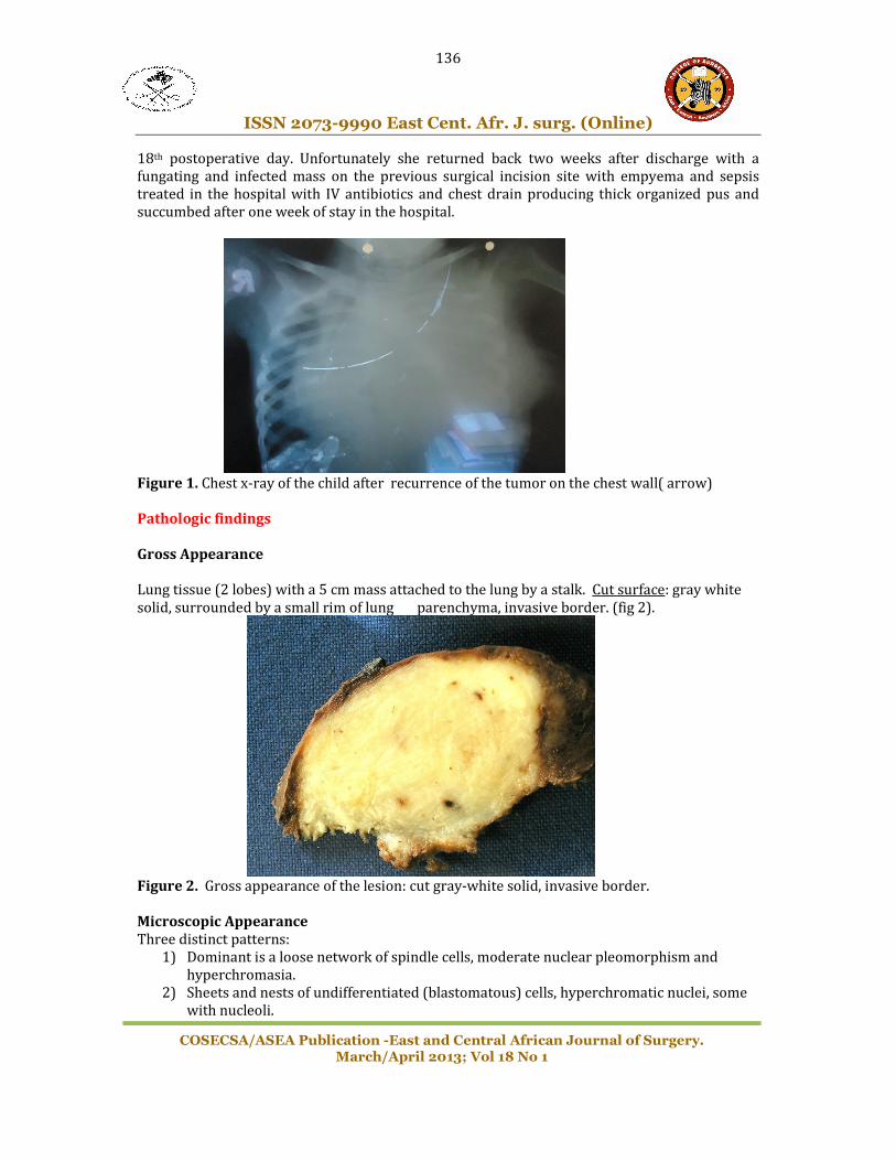
18th postoperative day. Unfortunately she returned back two weeks after discharge with a
fungating and infected mass on the previous surgical incision site with empyema and sepsis
treated in the hospital with IV antibiotics and chest drain producing thick organized pus and
succumbed after one week of stay in the hospital.
Figure 1. Chest x-ray of the child after recurrence of the tumor on the chest wall( arrow)
Pathologic findings
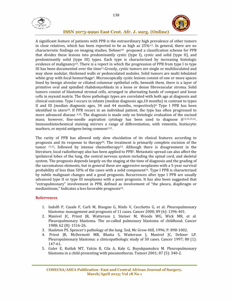
Gross Appearance
Lung tissue (2 lobes) with a 5 cm mass attached to the lung by a stalk. Cut surface: gray white
solid, surrounded by a small rim of lung parenchyma, invasive border. (fig 2).
Figure 2. Gross appearance of the lesion: cut gray-white solid, invasive border.
Microscopic Appearance
Three distinct patterns:
1) Dominant is a loose network of spindle cells, moderate nuclear pleomorphism and
hyperchromasia.
2) Sheets and nests of undifferentiated (blastomatous) cells, hyperchromatic nuclei, some
with nucleoli.

137
ISSN 2073-9990 East Cent. Afr. J. surg. (Online)
COSECSA/ASEA Publication -East and Central African Journal of Surgery. March/April 2013; Vol 18 No 1
3) Scattered glandular structures, cuboidal to columnar epithelium. (fig.3 A-D)
Figure 3. A. loose network of spindle cells, B. Sheets of blastomatous cells with hyperchromatic
nuclei, C. Scattered glandular elements, D. entrapped (non-neoplastic?) bronchus-like
structures. (all microscopic pictures: hematoxylin-eosin stains, initial magnification 400x.0
Discussion
Pleuropulmonary blastoma is a primary intrathoracic malignancy that occurs mainly in early
childhood. This dysontogenetic neoplasm, an analog to the other unique childhood tumors like
Wilms’ tumor and neuroblastoma, is classified with the “mesenchymal neoplasms” in the WHO
Classification of Lung Tumors17. There it is composed of immature mesenchyme, often
differentiating toward skeletal muscle, cartilage, fibrous tissue, and sometimes fat, and most
often includes epithelium. The mesenchymal elements are regarded as malignant.
Since PPB was recognized as clinicopathologic entity distinct from adult pulmonary blastoma,
which is characterized by malignant glands and malignant stroma, the epithelial elements in
PPB have been described as benign2. In the past, they have been termed pulmonary sarcoma
arising in mesenchymal cystic hamartoma, embryonal sarcoma, or rhabdomyosarcoma arising
in congenital cystic adenomatoid malformation or bronchogenic cysts3. The age onset of
presentation was between two weeks to 96 months4. Although respiratory difficulty with or
without fever is the most common clinical symptom, PPB can present with spontaneous
pneumothorax5, 6, or empyema7,16. There are few reports of bilateral PPB8, 9.
A
B
C
D
138
ISSN 2073-9990 East Cent. Afr. J. surg. (Online)
COSECSA/ASEA Publication -East and Central African Journal of Surgery. March/April 2013; Vol 18 No 1
A significant feature of patients with PPB is the extraordinary high prevalence of other tumors
in close relatives, which has been reported to be as high as 25%1,4. In general, there are no
characteristic findings on imaging studies. Dehner10 proposed a classification scheme for PPB
that divides these lesions into predominantly cystic (type I), cystic and solid (type II), and
predominantly solid (type III) types. Each type is characterized by increasing histologic
evidence of malignancy10. There is a report in which the progression of PPB from type I to type
III has been documented over the time11.Grossly, cystic tumors are single or multiloculated and
may show nodular, thickened walls or pedunculated nodules. Solid tumors are multi lobulated
white-gray with focal hemorrhage3. Microscopically cystic lesions consist of one or more spaces
lined by benign alveolar or ciliated columnar epithelial cells, beneath them, there is a layer of
primitive oval and spindled rhabdomyoblasts in a loose or dense fibrovascular stroma. Solid
tumors consist of blastomal stromal cells, arranged in alternating bands of compact and loose
cells in myxoid matrix. The three pathologic types are correlated with both age at diagnosis and
clinical outcome. Type I occurs in infants (median diagnosis age,10 months) in contrast to types
II and III (median diagnosis ages, 34 and 44 months, respectively)4 Type I PPB has been
identified in utero18. If PPB recurs in an individual patient, the type has often progressed to
more advanced disease 4,10. The diagnosis is made only on histologic evaluation of the excised
mass; however, fine-needle aspiration cytology has been used to diagnose it13,14,15,16.
Immunohistochemical staining mirrors a range of differentiation, with vimentin, histiocytic
markers, or myoid antigens being common3,12.
The rarity of PPB has allowed only slow elucidation of its clinical features according to
prognosis and its response to therapy20. The treatment is primarily complete excision of the
tumor 21,22, followed by intense chemotherapy23. Although there is disagreement in the
literature, local radiotherapy also has been applied to PPB1. Metastatic spread can also affect the
ipsilateral lobes of the lung, the central nervous system including the spinal cord, and skeletal
system. The prognosis depends largely on the staging at the time of diagnosis and the grading of
the sarcomatous elements, but in general these are aggressive neoplasms with a 5-year survival
probability of less than 50% of the cases with a solid component24. Type I PPB is characterized
by subtle malignant changes and a good prognosis. Recurrences after type I PPB are usually
advanced type II or type III neoplasms with a poor prognosis. It has also been suggested that
“extrapulmonary” involvement in PPB, defined as involvement of “the pleura, diaphragm or
mediastinum,” Indicates a less favorable prognosis19.
Referrences
1. Indolfi P, Casale F, Carli M, Bisogno G, Ninfo V, Cecchetto G, et al. Pleuropulmonary
blastoma: management and prognosis of 11 cases. Cancer 2000; 89 (6): 1396-401.
2. Manivel JC, Priest JR, Watterson J, Steiner M, Woods WG, Wick MR, et al.
Pleuropulmonary blastoma. The so-called pulmonary blastoma of childhood. Cancer
1988; 62 (8): 1516-26.
3. Hasleton PS. Spencer’s pathology of the lung. 5ed, Mc Grow-Hill, 1996; P: 898-1002.
4. Priest JR, McDermott MB, Bhatia S, Watterson J, Manivel JC, Dehner LP.
Pleuropulmonary blastoma: a clinicopathologic study of 50 cases. Cancer 1997; 80 (1):
147-61.
5. Guler E, Kutluk MT, Yalcin B, Cila A, Kale G, Buyukpamukcu M. Pleuropulmonary
blastoma in a child presenting with pneumothorax. Tumori 2001; 87 (5): 340-2.

139
ISSN 2073-9990 East Cent. Afr. J. surg. (Online)
COSECSA/ASEA Publication -East and Central African Journal of Surgery. March/April 2013; Vol 18 No 1
6. Kuzucu A, Soysal O, Yakinci C, Aydin NE. Pleuropulmonary blastoma: report of a case
presenting with spontaneous pneumothorax. Eur J Cardiothorac Surg 2001; 19 (2): 229-
30.
7. Katz DS, Scalzetti EM, Groskin SA, Kohman LJ, Patel LS, Landas S. Pleuropulmonary
blastoma simulating an empyema in a young child. J Thorac Imaging 1995:10 (2): 112-6.
8. Picaud JC, Levrey H, Bouvier R, Chappuis JP, Louis D, Frappaz D, et al. Bilateral cystic
pleuropulmonary blastoma in early infancy. J Pediatr 2000;136 (6): 834-6.
9. Mott BD, Canver CC, Nazeer T, Buchan A, Ilves R. Staged resection of bilateral
pleuropulmonary blastoma in a twomonth old girl. J Cardiovasc Surg (Torino) 2001; 42
(1): 135-7.
10. Dehner LP. Pleuropulmonary blastoma is THE pulmonary blastoma of childhood. Semin
Diagn Pathol 1994; 11 (2):144-51.
11. Wright JR Jr. Pleuropulmonary blastoma: A case report documenting transition from
type I (cystic) to type III (solid). Cancer 2000; 88 (12): 2853-8.
12. Hachitanda Y, Aoyama C, Sato JK, Shimada H. Pleuropulmonary blastoma in childhood. A
tumor of divergent differentiation. Am J Surg Pathol 1993; 17 (4):382-91.
13. Nicol KK, Geisinger KR. The cytomorphology of pleuropulmonary blastoma. Arch Pathol
Lab Med 2000;124 (3): 416-8.
14. Drut R, Pollono D. Pleuropulmonary blastoma: diagnosis by fine-needle aspiration
cytology: a case report. Diagn Cytopathol 1998; 19 (4): 303-5.
15. Gelven PL, Hopkins MA, Green CA, Harley RA, Wilson MM. Fine-needle aspiration
cytology of pleuropulmonary blastoma: case report and review of the literature. Diagn
Cytopathol 1997; 16 (4): 336-40.
16. Merriman TE, Beasley SW, Chow CW, Smith PJ, Robertson CF; Pediatric
Pulmonology[1996, 22(6):408-411]
17. Dehner LP, Tazelaar HD, Manabe T: Pathology and genetics of tumours of the lung,
pleura, thymus and heart, in Travis WD, Brambilla E, Muller-Hermelink HK, et al (eds):
World Health Organization Classification of Tumours. Lyon, France, IARC Press, 2004
18. Miniati DN, Chintagumpala M, Langston C, et al: Prenatal presentation and outcome of
children with pleuropulmonary blastoma. J Pediatr Surg 41: 66-71, 2006
19. Indolfi P, Casale F, Carli M, et al: Pleuropulmonary blastoma: Management and
prognosis of 11 cases. Cancer 89:1396-1401, 2000
20. Romeo C, Impellizzeri P, Grosso M, Vitarelli E, Gentile C. Pleuropulmonary blastoma:
long-term survival and literature review. Med Pediatr Oncol 1999; 33 (4): 372-6.
21. Tagge EP, Mulvihill D, Chandler JC, Richardson M, Uflacker R, Othersen HD. Childhood
pleuropulmonary blastoma: caution against nonoperative management of congenital
lung cysts. J Pediatr Surg 1996; 31 (1): 187-9; discussion 190.
22. Granata C, Gambini C, Carlini C, Repetto P, Torre M, Mazzola C, et al. Pleuropulmonary
blastoma. Eur J Pediatr Surg 2001; 11 (4): 271-3.
23. Parsons SK, Fishman SJ, Hoorntje LE, Jaramillo D, Marcus KC, Perez-Atayde AR, et al.
Aggressive multimodal treatment of pleuropulmonary blastoma. Ann Thorac Surg 2001;
72 (3): 939-42.
24. Christopher D.M. Fletcher. Diagnostic histopathology of tumors of the lung and pleura.
Moran C.A. .Suster S, Chapter 5, Churchill Livingstone. 2000; Vol. 1, p: 171- 208.