isrn dentistry, 2013(article id 296727) kou, w.,...
TRANSCRIPT

http://www.diva-portal.org
This is the published version of a paper published in ISRN Dentistry.
Citation for the original published paper (version of record):
Kou, W., Tsukasa, A., Watari, F., Sjögren, G. (2013)
An in vitro evaluation of the biological effects of carbon nanotube-coated dental zirconia.
ISRN Dentistry, 2013(Article ID 296727)
http://dx.doi.org/10.1155/2013/296727
Access to the published version may require subscription.
N.B. When citing this work, cite the original published paper.
Permanent link to this version:http://urn.kb.se/resolve?urn=urn:nbn:se:umu:diva-78252
Hindawi Publishing CorporationISRN DentistryVolume 2013, Article ID 296727, 6 pageshttp://dx.doi.org/10.1155/2013/296727
Research ArticleAn In Vitro Evaluation of the Biological Effects ofCarbon Nanotube-Coated Dental Zirconia
Wen Kou,1 Tsukasa Akasaka,2 Fumio Watari,2 and Göran Sjögren1
1 Dental Materials Science, Department of Odontology, Faculty of Medicine, Umea University, 90781 Umea, Sweden2Department of Biomedical Materials and Engineering, Graduate School of Dental Medicine, Hokkaido University,Sapporo 060-8586, Japan
Correspondence should be addressed to Wen Kou; [email protected]
Received 17 June 2013; Accepted 17 July 2013
Academic Editors: H. S. Cardash, G. H. Sperber, and A. Vissink
Copyright © 2013 Wen Kou et al.This is an open access article distributed under the Creative CommonsAttribution License, whichpermits unrestricted use, distribution, and reproduction in any medium, provided the original work is properly cited.
The purpose of this study is to evaluate functionalized multiwalled carbon nanotubes (fMWCNTs) as a potential coating materialfor dental zirconia from a biological perspective: its effect on cell proliferation, viability, morphology, and the attachment of anosteoblast-like cell. Osteoblast-like (Saos-2) cells were seeded onuncoated and fMWCNT-coated zirconia discs and in culture dishesthat served as controls.The seeding density was 104 cells/cm2, and the cells were cultured for 6 days. Cell viability, proliferation andattachment of the Saos-2 cells were studied. The results showed that Saos-2 cells were well attached to both the uncoated and thefMWCNT-coated zirconia discs. Cell viability and proliferation on the fMWCNT-coated zirconia discs were almost the same asfor the control discs. Better cell attachment was seen on the fMWCNT-coated than on the uncoated zirconia discs. In conclusion,fMWCNTs seem to be a promising coating material for zirconia-based ceramic surfaces to increase the roughness and therebyenhance the osseointegration of zirconia implants.
1. Introduction
During the last few years, the popularity of dental zirconiaimplants has increased because they are tooth colored, bio-compatible and have an osseointegration ability comparableto dental titanium implants [1, 2]. A fractography studyby Gahlert et al. (2012) [1], however, indicates that thefracture initiation site of dental zirconia implants is oftenlocated to the stress concentration area in the thread; thegrooves on the implant surface created by sandblasting oftenlead to stress concentration due to their notch effect. Thepurpose of sandblasting is to increase the surface area androughness of the dental zirconia implant and thus improveosseointegration [3]. Sandblasting can, however, introducedefects on the surfaces of zirconia implant, which will act aspotential fracture initiation sites [1]. The survival of dentalzirconia implants should, therefore, improve ifmethods otherthan sandblasting could be used.
In 1991 carbon nanotubes (CNTs) were discovered byIijima [4]. This material has been shown to have a largesurface area, good mechanical strength, ultra-light weight,
and excellent chemical and thermal stability [5]. The nan-otubes are structures of single or multiple sheets of graphenerolled up to form single-walled carbon nanotubes (SWCNTs)and multiwalled carbon nanotubes (MWCNTs). Since theirdiscovery CNTs have been used in many fields, such as inelectrical and mechanical applications and for biological andmedical purposes [6].The lack of solubility in aqueousmediahas been a major technical barrier in biological and biomed-ical applications, but the recent development of methodsto chemically modify and functionalize CNTs has made itpossible to dissolve and disperse CNTs inwater, making themmore suitable materials for biological applications [7].
The disadvantage of sandblasting mentioned previouslymakes it of interest to investigate other techniques to increasethe surface roughness of zirconia implants. One conceivablemethod could be the application of CNTs as a surface coatingon the implants. In a survey of the literature, no paperwas found addressing evaluation of the biological effectsof the application of MWCNT coating to dental zirconia.The aim of the present study, therefore, was to evaluatein vitro functionalized MWCNTs as a coating material for
2 ISRN Dentistry
(a) (b)
(c)
MWCNT-COOH
(d)
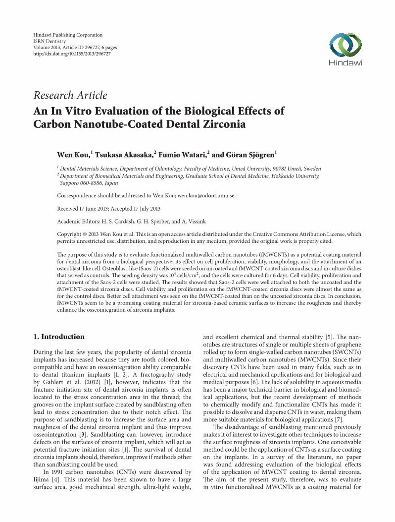
Figure 1: Uncoated (a, b) and MWCNT-COOH-coated (c, d) zirconia disc samples. (a, c) Macroview. (b, d) SEM images. A good coveringof MWCNT-COOH can be observed on the coated zirconia discs (d).
dental zirconia from a biological perspective: its effect on cellproliferation, viability, morphology, and the attachment of anosteoblast-like cell.
2. Materials and Methods
2.1. Preparation of MWCNTs. MWCNTs used in this studywere obtained fromNanoLab (Brighton, MA, USA). Accord-ing to the manufacturer, the MWCNTs were about 15 ± 5 nmin diameter and 1–5 𝜇m in length and produced by chemicalvapor deposition method. The purity of the MWCNTs wasabout 95wt%, and they were purified according to themethod devised by Sato et al. [8]. The MWCNTs werelater functionalized by carboxylation (MWCNT-COOH) toimprove their dispersion in aqueous solution, according toPeng et al. [9].
2.2. Specimen Preparation. Sixteen hot isostatic pressed(HIPed) yttria stabilized zirconia polycrystal (Y-TZP) discs(⌀13mm, thickness: 2mm) were prepared (Figure 1) bycomputer-aided design/computer-aided manufacturing(CAD/CAM) technique (CAD.esthetics AB, Skelleftea,Sweden).The HIPed Y-TZP discs were cleaned ultrasonically(Elma, Transonic 460/H, Singen, Germany) in deionizedwater for 30min. The MWCNT-COOH were dispersedin 99.5% ethanol to a final concentration of 100 ppm withsonification (Sonicator Vibracell VC 130, 130W, 20 kHzand amplitude 60% for 10min). The MWCNT-COOHsuspension obtained (1mL/dish) was poured onto thezirconia discs and kept at room temperature inside a fumehood for 1 h. After the ethanol had evaporated, the discs were
rinsed with deionized water and dried in a heated oven at65∘C for 20min. Thereafter, the specimens were individuallypackaged in sterilization bags and sterilized in a steamautoclave (Tomy, BS-235, Tokyo, Japan) at 121∘C for 15minand dried in a heated oven at 65∘C for 1 hour.
2.3. Cell Culture. Saos-2, a human osteosarcoma cell line,was obtained from Riken cell bank (Tsukuba, Japan). Thesehuman osteoblast-like cells (Saos-2) have been widely usedas a model system for human osteoblastic cells in biomaterialstudies [10, 11]. The cells were seeded on uncoated (𝑛 = 8)and MWCNT-COOH-coated zirconia discs (𝑛 = 8) andin culture dishes (𝑛 = 8) which served as controls, where𝑛 indicates the number of samples. The seeding densitywas 104 cells/cm2, and they were grown at 37∘C in 5% CO
2
and 95% air environment in Dulbecco’s Modified Eagle’sMedium (SigmaAldrich, St. Louise,Mo, USA) supplementedwith 10% FBS (MP Biomedical, LLC, Tokyo, Japan) and 1%penicillin-streptomycin antibiotic mixture (MP Biomedical,LLC, Tokyo, Japan). The cell viability, proliferation, andmorphology were evaluated after the cells were cultured for0 (6 hours after seeding), 2, 4 and 6 days.
2.4. Proliferation Test. The proliferation of the cells wasevaluated using a Cell Counting Kit-8 (CCK-8) (Sigma-Aldrich Chemie GmbH, Buchs, Switzerland), which is ahighly water-soluble tetrazolium salt WST-8 assay; 50𝜇LCCK-8 was added in the cell medium per well and incubatedfor 1 h at 37∘C. Afterwards, 100𝜇L of the supernatant of eachwell was removed into a 96-well plate, and the absorbance

ISRN Dentistry 3
was measured at 450 nm using a microplate reader (Bio-Rad,Hercules, CA, USA).
2.5. Live or Dead Viability Test. The LIVE/DEAD viabil-ity/cytotoxicity kit for mammalian cells (Molecular Probes,Eugene, OR, USA) was used to test the cell viability. 150𝜇Lof the LIVE/DEAD assay reagent solution was added to thecultured surface of the disc samples. After 30min incubationin room temperature, 10 𝜇L of the LIVE/DEAD assay reagentsolution was added to a clean dish. The zirconia disc wasmounted on the dish, and the cells were observed in afluorescence microscope (Olympus, IX 81, Tokyo, Japan)immediately, and photographs were taken during the exami-nation.
2.6. Detachment Test. As trypsin-EDTA solution is generallyused to detach cells, the cell adhesion was estimated bytreatment with this solution. On day 6, the Saos-2 cellscultured on theMWCNT-coated and uncoated zirconia discswere treated with 0.02% trypsin-EDTA solution for 10min.After detachment, the discs were observed using scanningelectron microscopy (SEM: S-4800, Hitachi, Tokyo, Japan).
2.7. SEM Evaluation. The samples were rinsed with phos-phate buffered saline (PBS) to remove nonadherent cells andchemically fixated in a solution of 2.5% glutaraldehyde for24 h at 4∘C.The disc samples were then dehydrated in a seriesof solutions with increasing ethanol concentrations (50%,70%, 80%, 90%, 95%, and 100%) followed by critical-pointdrying at 37∘C. Finally, the samples were coated with Pt-Pdalloys in an ion sputter (E-1030, Hitachi, Tokyo, Japan) andthe morphology of the cells was examined by SEM (S-4800,Hitachi, Tokyo, Japan).
3. Results
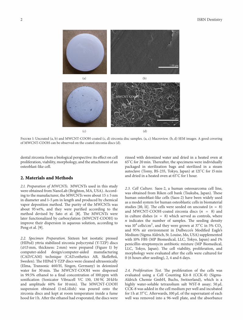
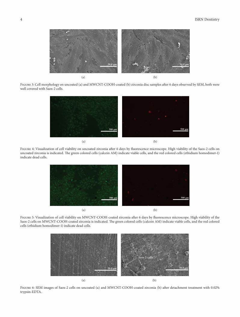
A good covering of MWCNT-COOH could be observedon the coated zirconia discs (Figure 1). The cells were wellattached both to the uncoated and MWCNT-COOH-coatedzirconia samples. The proliferation curve for the control,uncoated and MWCNT-COOH-coated zirconia is similar inappearance (Figure 2). SEM analysis of the cell morphologyshowed that the Saos-2 cells were flattened and well attachedto the surface of both the uncoated and MWCNT-COOH-coated zirconia with numerous filopodia andmicrovillosities,which are parts of the normal morphology of this kind ofcell (Figure 3). High viability of Saos-2 cells was seen bothon uncoated and MWCNT-COOH-coated zirconia (Figures4 and 5). After application of trypsin-EDTA to measure celldetachment, a number of Saos-2 cells could still be observedon MWCNT-COOH-coated zirconia, whereas no cells wereseen on the uncoated zirconia (Figure 6). In Figure 7 exampleof a Saos-2 cell attached to the MWCNT-COOH-coatedzirconia is shown, and the filopodia seem to be well attachedand elongated towards CNTs. In Figure 8 a and b example ofa Saos-2 cell on aMWCNT-COOH-coated zirconia disc aftertreated with trypsin-EDTA is presented. One filopodium
600
500
400
300
200
100
00 2 4 6
Times (days)
Cel
l pro
lifer
atio
n (%
)
ControlUncoated zirconiaMWCNT-COOH-coated zirconia
Figure 2: Proliferation curve for Saos-2 cells in culture dish (con-trol), on uncoated zirconia and MWCNT-COOH-coated zirconiadisc samples.
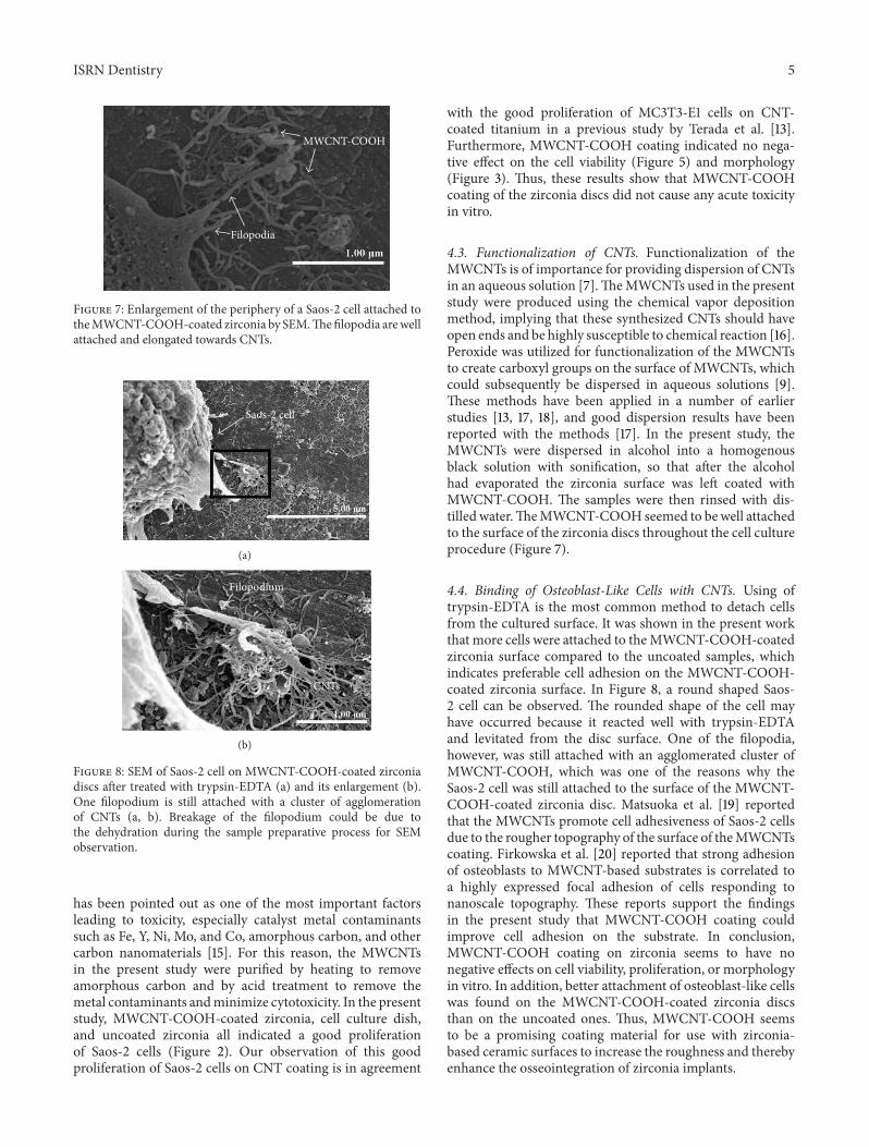
is still attached with a cluster of agglomeration of CNTs(Figure 8).
4. Discussion
4.1. Osseointegration of Zirconia Implants. Fracture and/orloss of dental implants cause patient discomfort and oftenclinical bone loss. It is therefore important that dentalimplants possess good mechanical properties and osseoin-tegration capability. As mentioned earlier, sandblasting isoften performed on zirconia implant surfaces to increase thesurface area in order to improve their osseointegration ability[3]. However, sandblasting can also introduce defects on theimplant surface, which act as stress concentration sites andsubsequently cause fracture of the implants [1]. In addition,sandblasting could have a negative impact on other materialproperties in zirconia ceramics such as phase transformationand Weibull modulus [12].
4.2. CNTs. Application of CNTs could conceivably be analternative to sandblasting achieving sufficient surface rough-ness on the implants. In a previous study by Terada et al.[13], 100 ppm MWCNT-COOH was used to coat titanium,which led to an increase in surface roughness from Ra =0.05 ± 0.01 𝜇m to Ra = 0.13 ± 0.01 𝜇m. A concentration of100 ppm was, therefore, also used in the present study to coatthe zirconia ceramic discs.
Application of CNTs as a biomaterial has previously beendebated [6], mostly because CNTs are very small in sizeand thus capable of entering the human body by inhalation,ingestion, and/or skin penetration and of interacting withintracellular structures [14]. Moreover, impurity in CNTs
4 ISRN Dentistry
(a) (b)
Figure 3: Cell morphology on uncoated (a) andMWCNT-COOH-coated (b) zirconia disc samples after 6 days observed by SEM, both werewell covered with Saos-2 cells.
(a) (b)
Figure 4: Visualization of cell viability on uncoated zirconia after 6 days by fluorescence microscope. High viability of the Saos-2 cells onuncoated zirconia is indicated. The green colored cells (calcein AM) indicate viable cells, and the red colored cells (ethidium homodimer-1)indicate dead cells.
(a) (b)
Figure 5: Visualization of cell viability on MWCNT-COOH-coated zirconia after 6 days by fluorescence microscope. High viability of theSaos-2 cells on MWCNT-COOH-coated zirconia is indicated. The green colored cells (calcein AM) indicate viable cells, and the red coloredcells (ethidium homodimer-1) indicate dead cells.
(a)
Saos 2-cells
(b)
Figure 6: SEM images of Saos-2 cells on uncoated (a) and MWCNT-COOH-coated zirconia (b) after detachment treatment with 0.02%trypsin-EDTA.
ISRN Dentistry 5
Filopodia
MWCNT-COOH
Figure 7: Enlargement of the periphery of a Saos-2 cell attached totheMWCNT-COOH-coated zirconia by SEM.Thefilopodia arewellattached and elongated towards CNTs.
Saos-2 cell
(a)
Filopodium
CNTs
(b)
Figure 8: SEM of Saos-2 cell on MWCNT-COOH-coated zirconiadiscs after treated with trypsin-EDTA (a) and its enlargement (b).One filopodium is still attached with a cluster of agglomerationof CNTs (a, b). Breakage of the filopodium could be due tothe dehydration during the sample preparative process for SEMobservation.
has been pointed out as one of the most important factorsleading to toxicity, especially catalyst metal contaminantssuch as Fe, Y, Ni, Mo, and Co, amorphous carbon, and othercarbon nanomaterials [15]. For this reason, the MWCNTsin the present study were purified by heating to removeamorphous carbon and by acid treatment to remove themetal contaminants andminimize cytotoxicity. In the presentstudy, MWCNT-COOH-coated zirconia, cell culture dish,and uncoated zirconia all indicated a good proliferationof Saos-2 cells (Figure 2). Our observation of this goodproliferation of Saos-2 cells on CNT coating is in agreement
with the good proliferation of MC3T3-E1 cells on CNT-coated titanium in a previous study by Terada et al. [13].Furthermore, MWCNT-COOH coating indicated no nega-tive effect on the cell viability (Figure 5) and morphology(Figure 3). Thus, these results show that MWCNT-COOHcoating of the zirconia discs did not cause any acute toxicityin vitro.
4.3. Functionalization of CNTs. Functionalization of theMWCNTs is of importance for providing dispersion of CNTsin an aqueous solution [7].TheMWCNTs used in the presentstudy were produced using the chemical vapor depositionmethod, implying that these synthesized CNTs should haveopen ends and be highly susceptible to chemical reaction [16].Peroxide was utilized for functionalization of the MWCNTsto create carboxyl groups on the surface of MWCNTs, whichcould subsequently be dispersed in aqueous solutions [9].These methods have been applied in a number of earlierstudies [13, 17, 18], and good dispersion results have beenreported with the methods [17]. In the present study, theMWCNTs were dispersed in alcohol into a homogenousblack solution with sonification, so that after the alcoholhad evaporated the zirconia surface was left coated withMWCNT-COOH. The samples were then rinsed with dis-tilled water.TheMWCNT-COOH seemed to bewell attachedto the surface of the zirconia discs throughout the cell cultureprocedure (Figure 7).
4.4. Binding of Osteoblast-Like Cells with CNTs. Using oftrypsin-EDTA is the most common method to detach cellsfrom the cultured surface. It was shown in the present workthat more cells were attached to theMWCNT-COOH-coatedzirconia surface compared to the uncoated samples, whichindicates preferable cell adhesion on the MWCNT-COOH-coated zirconia surface. In Figure 8, a round shaped Saos-2 cell can be observed. The rounded shape of the cell mayhave occurred because it reacted well with trypsin-EDTAand levitated from the disc surface. One of the filopodia,however, was still attached with an agglomerated cluster ofMWCNT-COOH, which was one of the reasons why theSaos-2 cell was still attached to the surface of the MWCNT-COOH-coated zirconia disc. Matsuoka et al. [19] reportedthat the MWCNTs promote cell adhesiveness of Saos-2 cellsdue to the rougher topography of the surface of theMWCNTscoating. Firkowska et al. [20] reported that strong adhesionof osteoblasts to MWCNT-based substrates is correlated toa highly expressed focal adhesion of cells responding tonanoscale topography. These reports support the findingsin the present study that MWCNT-COOH coating couldimprove cell adhesion on the substrate. In conclusion,MWCNT-COOH coating on zirconia seems to have nonegative effects on cell viability, proliferation, or morphologyin vitro. In addition, better attachment of osteoblast-like cellswas found on the MWCNT-COOH-coated zirconia discsthan on the uncoated ones. Thus, MWCNT-COOH seemsto be a promising coating material for use with zirconia-based ceramic surfaces to increase the roughness and therebyenhance the osseointegration of zirconia implants.
6 ISRN Dentistry
Acknowledgments
The authors acknowledge Dr. S. Abe, Dr. X. Chen, Dr.E. Hirata, and the graduate students in Department ofBiomedical Materials and Engineering, Graduate School ofDental Medicine, Hokkaido University, Japan, for their kindhelp. Many thanks go to Dr. P. Shrimpton for improvingthe English language. This work was supported by Facultyof Medicine, Umea University, Umea, Sweden, and JSPSPostdoctoral Fellowship for North American and EuropeanResearchers with Grant no.: PE11546. The authors declare noconflict of interests.
References
[1] M. Gahlert, D. Burtscher, I. Grunert, H. Kniha, and E. Stein-hauser, “Failure analysis of fractured dental zirconia implants,”Clinical Oral Implants Research, vol. 23, no. 3, pp. 287–293, 2012.
[2] M. Andreiotelli, H. J. Wenz, and R. J. Kohal, “Are ceramicimplants a viable alternative to titanium implants? A systematicliterature review,” Clinical Oral Implants Research, vol. 20, no. 4,pp. 32–47, 2009.
[3] A. Wennerberg and T. Albrektsson, “Current challenges insuccessful rehabilitation with oral implants,” Journal of OralRehabilitation, vol. 38, no. 4, pp. 286–294, 2011.
[4] S. Iijima, “Helicalmicrotubules of graphitic carbon,”Nature, vol.354, no. 6348, pp. 56–58, 1991.
[5] P. M. Ajayan, “Nanotubes from carbon,” Chemical Reviews, vol.99, no. 7, pp. 1787–1799, 1999.
[6] X. Li, Y. Fan, and F. Watari, “Current investigations into carbonnanotubes for biomedical application,” Biomedical Materials,vol. 5, no. 2, Article ID 022001, 2010.
[7] S. Vardharajula, S. Z. Ali, P.M. Tiwari et al., “Functionalized car-bon nanotubes: biomedical applications,” International Journalof Nanomedicine, vol. 7, pp. 5361–5374, 2012.
[8] Y. Sato, A. Yokoyama, K. Shibata et al., “Influence of length oncytotoxicity of multi-walled carbon nanotubes against humanacute monocytic leukemia cell line THP-1 in vitro and subcuta-neous tissue of rats in vivo,”Molecular BioSystems, vol. 1, no. 2,pp. 176–182, 2005.
[9] H. Peng, L. B. Alemany, J. L. Margrave, and V. N. Khabashesku,“Sidewall carboxylic acid functionalization of single-walledcarbon nanotubes,” Journal of the American Chemical Society,vol. 125, no. 49, pp. 15174–15182, 2003.
[10] S. Sofia, M. B. McCarthy, G. Gronowicz, and D. L. Kaplan,“Functionalized silk-based biomaterials for bone formation,”Journal of Biomedical Materials Research, vol. 54, no. 1, pp. 139–148, 2001.
[11] T. Akasaka, A. Yokoyama, M. Matsuoka et al., “Adhesion ofhumanosteoblast-like cells (Saos-2) to carbonnanotube sheets,”Bio-Medical Materials and Engineering, vol. 19, no. 2-3, pp. 147–153, 2009.
[12] S. Bhargava, H. Doi, R. Kondo, H. Aoki, T. Hanawa, and S.Kasugai, “Effect of sandblasting on the mechanical propertiesof Y-TZP zirconia,” Bio-Medical Materials and Engineering, vol.22, no. 6, pp. 383–398, 2012.
[13] M. Terada, S. Abe, T. Akasaka, M. Uo, Y. Kitagawa, and F.Watari, “Multiwalled carbon nanotube coating on titanium,”Bio-Medical Materials and Engineering, vol. 19, no. 1, pp. 45–52,2009.
[14] S. Sharifi, S. Behzadi, S. Laurent, M. L. Forrest, P. Stroeve, andM. Mahmoudi, “Toxicity of nanomaterials,” Chemical SocietyReviews, vol. 41, no. 6, pp. 2323–2343, 2012.
[15] Y. Liu, Y. Zhao, B. Sun, andC.Chen, “Understanding the toxicityof carbon nanotubes,” Accounts of Chemical Research, vol. 46,no. 3, pp. 702–713, 2012.
[16] C. P. Firme III and P. R. Bandaru, “Toxicity issues in the applica-tion of carbon nanotubes to biological systems,”Nanomedicine:Nanotechnology, Biology, and Medicine, vol. 6, no. 2, pp. 245–256, 2010.
[17] S. Abe, D. Hayashi, T. Akasaka et al., “Synthesis and characteri-zation of a water-soluble multi-walled carbon nanotube and itsbiodistribution inmice,”NanoBiomedicine, vol. 1, no. 2, pp. 143–150, 2009.
[18] E. Hirata, M. Uo, H. Takita, T. Akasaka, F. Watari, and A.Yokoyama, “Development of a 3D collagen scaffold coated withmultiwalled carbon nanotubes,” Journal of Biomedical MaterialsResearch B, vol. 90, no. 2, pp. 629–634, 2009.
[19] M. Matsuoka, T. Akasaka, Y. Totsuka, and F. Watari, “Strongadhesion of Saos-2 cells to multi-walled carbon nanotubes,”Materials Science and Engineering B, vol. 173, no. 1–3, pp. 182–186, 2010.
[20] I. Firkowska, E. Godehardt, and M. Giersig, “Interactionbetween human osteoblast cells and inorganic two-dimensionalscaffolds based onmultiwalled carbon nanotubes: a quantitativeAFM study,” Advanced Functional Materials, vol. 18, no. 23, pp.3765–3771, 2008.

Submit your manuscripts athttp://www.hindawi.com
Hindawi Publishing Corporationhttp://www.hindawi.com Volume 2013
Computational and Mathematical Methods in Medicine
International Journal of
BiomaterialsHindawi Publishing Corporationhttp://www.hindawi.com Volume 2013
ScientificaHindawi Publishing Corporationhttp://www.hindawi.com Volume 2013
Preventive MedicineAdvances in
Hindawi Publishing Corporationhttp://www.hindawi.com Volume 2013
Drug DeliveryJournal of
Hindawi Publishing Corporationhttp://www.hindawi.com Volume 2013
Hindawi Publishing Corporationhttp://www.hindawi.com Volume 2013
Case Reports in Dentistry
Hindawi Publishing Corporationhttp://www.hindawi.com Volume 2013
Radiology Research and Practice
BioMed Research International
Hindawi Publishing Corporationhttp://www.hindawi.com Volume 2013
ISRN Dentistry
Hindawi Publishing Corporationhttp://www.hindawi.com Volume 2013
Hindawi Publishing Corporationhttp://www.hindawi.com Volume 2013
Oral OncologyJournal of
Environmental and Public Health
Journal of
Hindawi Publishing Corporationhttp://www.hindawi.com Volume 2013
Hindawi Publishing Corporationhttp://www.hindawi.com Volume 2013
Oral DiseasesJournal of
Hindawi Publishing Corporationhttp://www.hindawi.com Volume 2013
OrthopedicsAdvances in
Hindawi Publishing Corporationhttp://www.hindawi.com Volume 2013
Dental SurgeryJournal of
Hindawi Publishing Corporationhttp://www.hindawi.com Volume 2013
Oral ImplantsJournal of
DentistryInternational Journal of
Hindawi Publishing Corporationhttp://www.hindawi.com Volume 2013
International Journal of
EndocrinologyHindawi Publishing Corporationhttp://www.hindawi.com
Volume 2013
Hindawi Publishing Corporation http://www.hindawi.com Volume 2013Hindawi Publishing Corporation http://www.hindawi.com Volume 2013
The Scientific World Journal
Hindawi Publishing Corporationhttp://www.hindawi.com Volume 2013
AnesthesiologyResearch and Practice