isolation of protease producing microorganism … · isolation of protease producing microorganism...
TRANSCRIPT
2015 November Edition |www.jbino.com | Innovative Association
J.Bio.Innov 4(6),pp: 322-346,2015|ISSN 2277-8330 (Electronic)
Yasser Hussein
ISOLATION OF PROTEASE PRODUCING MICROORGANISM FROM FOOD WASTE
Yasser Hussein Issa Mohammed
Department Of Biol-chemistry, Hajja University, Hajja, Yemen
Email: [email protected]
(Received on Date: 6th September 2015 Date of Acceptance: 14th October 2015)
ABSTRACT
Proteases are the class of enzymes, which occupy key position with respect to
their applications in both physiological and commercial fields. Proteases are
also known as peptidyl –peptide hydrolases and are industrially useful enzymes
which catalyze the hydrolysis of peptide bond from protein molecule. Proteases
constitute 0-6%of the global industrial enzyme market, most of which are alkaline
protease. They are commercially important and isolated from various living
sources such as plants, animals, bacteria and fungi. Isolation and screening of
protease producing bacteria five strains were isolated from food waste samples
collected mess yard, hotel and canteen. strains were selected on the basis of
zone of hydrolysis they exhibited on the agar plates and agar plate The selected
strains were inoculated on different sterilized nutrient media and incubated at
37ºC to get a standard colony. Substrate two types of wastes to be used as
substrate food waste samples were collected from different habitats food waste
areas. One gram of food waste sample was added to a glass tube containing
10 mL sterilized distilled water. The sample was serially diluted and streaked on
agar plates. The plates were incubated for 24 h at 37°C and enzyme activity
was observed. Positive colonies those degraded skim milk was studied for
protease production. Isolation and Screening: 1 gm of waste was suspended in
10 ml of sterile water and serialdilution10-7 was selected and streaked on
nutrient agar plate and incubated for 48 hrs at 37°C. Colonies were randomly
selected and streaked on Skimmed Milk Agar plate to isolate protease
producing bacteria. Two colonies produced clear zone on SMA plate and
colony having higher zone was selected for experiment. Two screening broth
such as: casein-yeast extract-lactose-mineral salt broth (casein-1.2%, yeast
extract-0. %, K2HPO4-0.0%,CaCl2-0.0% , MgSO4-0.0% ,Lactose- 0.0%) and
tryptone-yeast extract-dextrose broth (tryptone-1%, dextrose-0.1%, yeast extract-
0.%) were used for final selection.
Keyword: Proteases, food waste, casein-yeast, metalloproteases, Zymography.
2015 November Edition |www.jbino.com | Innovative Association
J.Bio.Innov 4(6),pp: 322-346,2015|ISSN 2277-8330 (Electronic)
Yasser Hussein
INTRODUCTION
Enzymes are biological catalysts that allow
chemical reactions to occur in living
organisms at ambient conditions. One
group of the enzymes that has been
studied extensively is proteases or
proteolytic enzymes due to their wide
variety of application in various fields.
Protease (peptidase or proteinase) is an
enzyme that hydrolyses the peptide bonds
that link amino acids together in the
polypeptide chain forming the protein.
Proteases differ in their ability to hydrolyze
various peptide bonds. Hence, specificity
associated with each enzyme differs
based on catalytic site. Proteases are
widely distributed in all living organisms: in
plants (papaya,pineapple)in animals and
mainly in microbes(bacteria, fungi and
viruses). Proteases are involved in
regulating practically every aspect of
plant life cycles. Plant proteases are
involved in seed germination, recycling
damaged plant proteins, regulating aging
processes in plants and modifying proteins
to perform specific purposes in plant cells.
Animal proteases play a very vital role in
the metabolism of the organism.
Microorganisms represent an excellent
source of enzymes owing to their broad
biochemical diversity and their
susceptibility to genetic manipulation.
Protease production is an inherent
capacity of all microorganisms and a large
number of microbes belonging to
bacteria, fungi, yeast and actinomycete
are known to produce proteases. Due to
processing almost all the characteristics
desired for their biotechnological
applications, microbial proteases are
preferred to the plant. Numerous
proteinases are produced by
microorganisms depending on the species
of the producers or the strains even
belonging to the same species. Several
proteinases are also produced by the
same strain under various cultural
conditions. From economical point of view
microbial proteases are the commercial
enzymes. Microbial proteases account for
approximately 40% of the total worldwide
sales of enzymes. Proteases constitute one
of the most important groups of industrial
enzymes, accounting for more than 65% of
the industrial enzyme market [1,2,7] of
these; alkaline protease accounts for 30%
of the world’s total enzyme production
and applications in detergent formulations
alone make up to 89% of total sales.
Microorganisms elaborate a large array of
proteases, which are intracellular and/or
extracellular. Intracellular proteases are
important for various cellular and
metabolic processes, such as sporulation
and differentiation, protein turnover,
maturation of enzymes and hormones and
maintenance of the cellular protein pool.
Extracellular proteases are important for
the hydrolysis of proteins in cell-free
environments and enable the cell to
absorb and utilize hydrolytic products. At
the same time, these extracellular
proteases have also been commercially
exploited to assist protein degradation in
various industrial processes. The vast
diversity of proteases, in contrast to the
specificity of their action has attracted
2015 November Edition |www.jbino.com | Innovative Association
J.Bio.Innov 4(6),pp: 322-346,2015|ISSN 2277-8330 (Electronic)
Yasser Hussein
worldwide attention focused on exploiting
their physiological and biotechnological
applications [2,12,18]. Proteases are also
having extensive applications in the
development of environmental friendly
technologies as well as in several
bioremediation processes. Several
classification systems currently available,
provides rich and vast information about
each and every identified protease. These
schemes can be categorized under 4
major categories based on the
characteristic features like: pH
(acidic/neutral/ alkaline), peptide bond
specificity (endo/exo peptidases) and
functional group present at active site
(serine / cysteine / aspartic /
metalloproteases) of proteolytic activity
[3,14]. Most commercially available
proteases belong to the class serine
proteases, produced by organisms
belonging to the genus Bacillus, Aspergillus
and Streptomyces. Alkaline proteases are
more preferable at industrial scale
compared other acidic proteases.
Metalloproteases are those enzymes
whose catalytic mechanism involves a
metal which plays an important role in
pathogenesis hence have advantage in
health care sector. A Metallo protease
These are proteolytic enzymes whose
catalytic mechanism involves a metal. The
enzymes typically require an essential
metal ion (Ni 2+, Mg2+, Mn2+, Ca2+, Zn2+ and
Co2+) for functional activity [4]. Most
metalloproteases are zinc-dependent, but
some use cobalt. The metal ion is
coordinated to the protein via three
ligands. The ligands coordinating the metal
ion can vary with histidine, glutamate,
aspartate, lysine and arginine. The fourth
coordination position is taken up by a
labile water molecule. The introduction of
metal-binding sites into proteins could
induce specific and predictable
conformational changes as well as allow
the regulation of enzymatic activity.
The first "consensus sequence" for members
of the metalloprotease family was based
on homology found within the human
fibroblast collagenase and the 11 amino
acids flanking the zinc-binding site of the
serratia protease, a bacterial
metalloprotease that also shares strong
homology with thermolysin at this site.
Later, the primary sequence motif HEXXH
was found in many zinc-containing
proteases, including several eukaryotic zinc
metalloproteases, and was suggested to
be indicative of membership in the family
[5,8,15].
Table 1: Examples of metalloproteases using specific metal ions
Metal ion Example of metalloprotease
Zn2+
Glutamate carboxypeptidase and
glutamylaminopeptidase, Collagenase and
2015 November Edition |www.jbino.com | Innovative Association
J.Bio.Innov 4(6),pp: 322-346,2015|ISSN 2277-8330 (Electronic)
Yasser Hussein
Endorphins
Mg2+
Inorganic
diphosphatase,Phosophoadenylylsulphatase
Co2+
Methylmalonyl coenzyme A mutase,methionine
aminopeptidase,nitrile hydratase
Ni2+
Urease
Ca2+
Lactonase
Table 2: Examples of organisms with metalloproteases produced by them:
Metalloprotease Name Organism
Thermolysin Bacillus thermoproteolyticus
Elastase Pseudomonas aeruginosa
Lysostaphin Staphylococcus sp
Acidolysin Clostridium acetobutylicum
Neutral metalloprotease Bacillus sp
Vimelysin Vibrio sp
Collagenase Clostridium histolyticum
Immunoglobulin A (IgA) protease Streptococcus sanguis
Metalloproteases A,B and C Erwinia chrysanthemi
MATERIALS AND METHODS
Seed medium
25 ml of Nutrient broth was prepared in
conical flask. Culture was taken from the
slant and inoculated. The medium was
incubated at 30oC for 18-24 h till the
optical density of the medium reaches 0.8
2015 November Edition |www.jbino.com | Innovative Association
J.Bio.Innov 4(6),pp: 322-346,2015|ISSN 2277-8330 (Electronic)
Yasser Hussein
at 600 nm. This culture was used for
inoculation of the production medium.
Production medium and culture conditions
The medium used for enzyme production
was YPD medium consisting (%, g/100ml) of
Yeast extract – 1.0, Peptone – 1.0, Dextrose
–1.0, MgSO4 - 0.02, KH2PO4 - 0.05, NaCl -
0.25, pH 7.0 was used. Seed medium was
used as the inoculum .1% inoculum was
added to the production medium and
incubated at 30oC for 2 days. The sample
was collected, centrifuged at 10000 rpm
for 10 minutes and the supernatant was
used for the protease assay.
Analytical Methods:
Protease assay
Protease activity was assayed according
to Anson method and was slightly modified
(24). The reaction mixture contained 2.5 ml
of 0.65% Hammerstein casein and 0.5 ml of
appropriately diluted enzyme. Enzyme was
diluted in 50 mM Glycine NaOH buffer pH
9.0. The reactants were incubated at 37°C
for 10 min and the reaction was stopped
by adding 2.5 ml of 110 mM trichloroacetic
acid (TCA). A suitable blank was run
simultaneously, in which 2.5 ml of TCA was
added to the 0.5ml of diluted enzyme
solution, followed by Casein substrate
addition after incubation for 10 min. After
incubation at room temperature for 30min
both test and blank solutions were
centrifuged at 10,000g for 10min. To the 0.4
ml supernatant, 1.0ml 50mM Na2CO3 and
0.2ml Folin-ciocalteau’s reagent was
added, the reaction mixture was
incubated at room temperature for 30 min
and the absorbance was measured at
660nm [2,6,21].
Protein Determination
The total protein content of the samples
was determined according to the method
described by Lowry et al., (1951). The
protein assay mixture consisted of 200 μl of
diluted extract of the enzyme, 1 ml of
alkaline copper solution(2%Na2CO3 in 0.1 N
NaOH:1.56% CuSO4:2.37% Potassium
sodium tartarate in ratio 100:1:1).The
contents in tube were vortexed and
incubated for 10 min. Then 200μl of freshly
prepared diluted Folin-ciocalteau reagent
(1:2) was added and mixed and kept in
dark at room temperature for 30 min. Blue
colour was developed and OD was
measured at 600nm.Simultaneously a blank
was set with 200μl distilled water,1ml
alkaline copper solution and 100μl of
Folinciocalteau reagent. The protein
concentration in the reaction was
determined based on the standard curve
obtained with bovine serum albumin as
standard
Dry cell weight
For the calculation of dry cell weight: Initial
weight of the eppendorf was measured.
One ml of culture was taken in the
eppendorf and culture was centrifuged at
10,000rpm for 10 min at 4oC.The
supernatant was discarded leaving the
pellet. The pellet was then dried until there
is no more moisture left. The weight of the
2015 November Edition |www.jbino.com | Innovative Association
J.Bio.Innov 4(6),pp: 322-346,2015|ISSN 2277-8330 (Electronic)
Yasser Hussein
eppendorf with pellet was noted. The initial
weight of the eppendorf was subtracted
from the weight obtained to get dry cell
weight.
Unit definition
One unit (U) of proteolytic enzyme activity
was defined as the amount of enzyme that
liberated 1μg tyrosine per ml per minute
from casein under specified assay
conditions. Enzyme units were measured
using slope obtained from tyrosine (0–1000
µg/ml) as standard.
Optimization of nutritional parameters for
the bacterial growth and the protease
production by isolated strain:
Effect of Carbon sources
The medium was prepared with
composition same as YPD but the carbon
source is replaced by different other
carbon sources. The composition consists
of Yeast extract – 1.0%, Peptone – 1.0%,
MgSO4 - 0.02%, KH2PO4 - 0.05%, NaCl -0.25%
and carbon source-1%. The various carbon
sources used include dextrose, fructose,
galactose and maltose. The medium was
prepared autoclaved and inoculated with
seed medium containing the culture. The
flasks were incubated at 30oC and
150rpm.The samples were collected for
every 3h upto 72h.The optical density of
the samples was measured at 600nm.The
samples were then centrifuged at
10000rpm for 10 min and the supernatant
was used as sample for protease assay to
calculate the units of enzyme produced.
The results were tabulated and graph was
plotted.
Effect of Nitrogen sources
The medium was prepared with the
composition: Nitrogen sources-2.0%, MgSO4
- 0.02%, KH2PO4 - 0.05%, NaCl -0.25% and
Dextrose-1%. Different nitrogen sources as
Beef extract, chitin, casitone, malt extract,
meat extract, peptone, tryptone, yeast
extract, casein, skim milk, soyabean meal
were used. The medium was autoclaved
and inoculated with 1% inoculum (seed
medium containing the culture). The flasks
were incubated at 30oC and 150rpm. The
samples were collected for every 24h upto
72h. The optical density of the samples was
measured at 600 nm. The samples were
centrifuged at 10000 rpm for 10 min and
supernatant was used as sample for
protease assay. The results were noted and
observed. The four nitrogen sources that
helped in producing more number of
enzyme units were selected. The
experiment was repeated those 4 Nitrogen
sources but the samples were collected for
every 3 h upto 72h. The protease assays
were carried out with the supernatant of
the samples. The results were tabulated
and graph was plotted.
Purification of protease produced from
Serratia sp
Enzyme production for purification
The production of protease was carried in
the medium that has been statistically
optimized. The medium composition is
2015 November Edition |www.jbino.com | Innovative Association
J.Bio.Innov 4(6),pp: 322-346,2015|ISSN 2277-8330 (Electronic)
Yasser Hussein
Dextrose -0.5%, Casein-2%, KH2PO4-0.06%,
CaCl2-0.004%, MgSO4-0.02%, NaCl-0.25%. A
1% inoculum was added from the seed
medium. The flasks were incubated in a
shaking incubator at 300C and 150 rpm.
After 48hrs of fermentation the cells were
separated from culture broth by
centrifugation at 10000rpm for 10 min at
40C.
Ammonium Sulphate precipitation and
Dialysis
The supernatant obtained after the
centrifugation was collected in a flask and
finely powdered Ammonium sulphate (20-
80%) was added slowly to the clear
supernatant with constant stirring for 2 hrs.
The solution was centrifuged at 10000 rpm
for 10 min at 40C and the supernatant
and pellet was collected. The pellet was
dissolved in 10ml of Glycine NaOH buffer
pH 7.2. Then the supernatant and the
pellet dissolved in buffer were used as
samples for protease and protein
estimations. The pellet dissolved in the
Glycine NaOH buffer was put in dialysis
membrane and packed with clips at the
two ends and then dialyzed against the
same buffer. The protease activity and the
protein content were estimated for the
dialyzed sample. The dialyzed sample was
used for the further purification.
Gel filtration chromatography
Gel filtration was performed with water-
cooled (5° C) columns of Sephadex G-75
equilibrated with 0.1 M phosphate buffer
(pH 7.2). The dialyzed enzyme preparation
was applied to the columns and eluted, in
the upward flow mode, at a flow rate of
30ml per hour with the equilibrating buffer.
Fractions (2 ml.) were collected and
assayed for protease activity and protein
was estimated using lowrys method at 600
nm. The unadsorbed protein fraction was
eluted with the same buffer. The active
fractions were collected and used for
further purification.
SDS-PAGE
Sodium dodecyl sulfate (SDS)-
polyacrylamide gel electrophoresis was
performed for determination of purity and
molecular weight of the enzyme. SDS-
PAGE was performed by using a BioRad
Mini-Protean II gel apparatus. Samples of
protein preparations and molecular mass
standards (BioRad, Promega) were
combined with sample buffer containing
1ml of 0.5M TRIS/HCl pH 6.8, 0.8ml glycerol
(v/v), 1.6ml 10% (w/v) SDS, 0.4ml ß-
mercaptoethanol (v/v), and 0.2ml 0.5%
(w/v) bromophenol blue and heated at
100 °C for 5 min. The samples were cooled
to 25 °C and loaded onto 10% (w/v), 0.75-
mm-thick gels and electrophoresed. The
molecular mass of purified protein samples
was determined by comparing its mobility
with those of various protein standards.
Zymography with casein
Casein zymography was performed using
10% polyacrylamide slab gels containing
SDS and casein receipe (15mg/ml ) in
separating gel. The protease containing
sample was applied to the gel in standard
2015 November Edition |www.jbino.com | Innovative Association
J.Bio.Innov 4(6),pp: 322-346,2015|ISSN 2277-8330 (Electronic)
Yasser Hussein
SDS loading buffer containing 0.1% SDS but
lacking 2-mercaptoethanol; it was not
boiled before loading. After
electrophoresis, the gels were soaked
thrice for 20 min in 2.5% (v/v) Triton X-100 at
room temperature to remove the SDS. The
gels were stained with 0.1% Coomassie
brilliant blue R-250 in methanol–glacial
acetic acid–water (40:10:60) followed by
destaining with methanol–glacial acetic
acid–water (40:10:60). Enzyme activity was
visualized by incubating the gel for 12 h in
50 mM Glycine–NaOH buffer at pH 11.0 at
room temperature.
Characterization of protease enzyme
The properties of extra cellular protease
produced by Serratia sp RSPB11 referred as
protease were studied and the enzyme
was characterized. All the experiments
were done in triplicate.
Effect of pH on enzyme activity
To study the effect of pH on enzyme
activity the caseinolysis in the buffers of
different pHs was studied. The buffers used
were Citrate buffer (pH 4.0-6.0), Sodium
Phosphate buffer (7.0-8.0) Glycine - NaOH
(pH 9.0-12.0).0.65% casein was dissolved in
buffers with different pH ranging from 4.0-
12.0.
Effect of pH on enzyme stability
For knowing the pH stability, the casein
and enzyme containing buffers were
incubated at 37oC for 1 h, 2 h and 3 h. The
residual enzyme activity was determined
using standard assay conditions. The
enzyme activity of the sample was
calculated before the incubation and
considered as control.
Effect of Temperature on enzyme activity
The effect of temperature on the enzyme
activity was studied by dissolving 0.65%
casein in Glycine NaOH buffer pH 9.0.This
casein solution is distributed into different
tubes. For studying the effect of
temperature on the enzyme, tubes were
incubated at various temperatures ranging
from 28 – 70°C for 10 minutes time intervals.
The residual activity of the enzyme was
determined and the tube that was
incubated at 37oC was considered as
control and relative activity is calculated
with respect to it i.e., considering the
activity at 37oC to be 100%.
Thermo stability of the enzyme
The reaction mixture with 0.65% casein
dissolved in Glycine NaOH buffer, pH 9.0
and enzyme are incubated at different
temperatures for period of 15 min, 30 min
and 60 min. Then the assay was performed
using the standard assay conditions to
calculate the residual enzyme activity. The
enzyme activity was also calculated
before the incubation and that is taken as
control.
RESULTS AND DISCUSSIONS
Isolation and screening of protease
producing strain:
2015 November Edition |www.jbino.com | Innovative Association
J.Bio.Innov 4(6),pp: 322-346,2015|ISSN 2277-8330 (Electronic)
Yasser Hussein
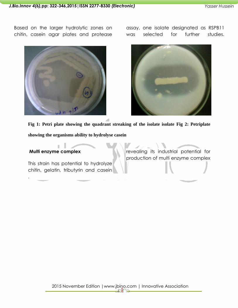
Based on the larger hydrolytic zones on
chitin, casein agar plates and protease
assay, one isolate designated as RSPB11
was selected for further studies.
Fig 1: Petri plate showing the quadrant streaking of the isolate isolate Fig 2: Petriplate
showing the organisms ability to hydrolyse casein
Multi enzyme complex
This strain has potential to hydrolyze
chitin, gelatin, tributyrin and casein
revealing its industrial potential for
production of multi enzyme complex
.
2015 November Edition |www.jbino.com | Innovative Association
J.Bio.Innov 4(6),pp: 322-346,2015|ISSN 2277-8330 (Electronic)
Yasser Hussein
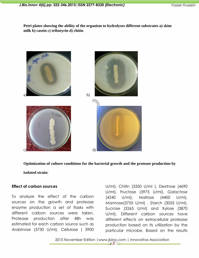
Petri plates showing the ability of the organism to hydrolyses different substrates a) skim
milk b) casein c) tributyrin d) chitin
a) b)
c) d)
Optimization of culture conditions for the bacterial growth and the protease production by
isolated strain:
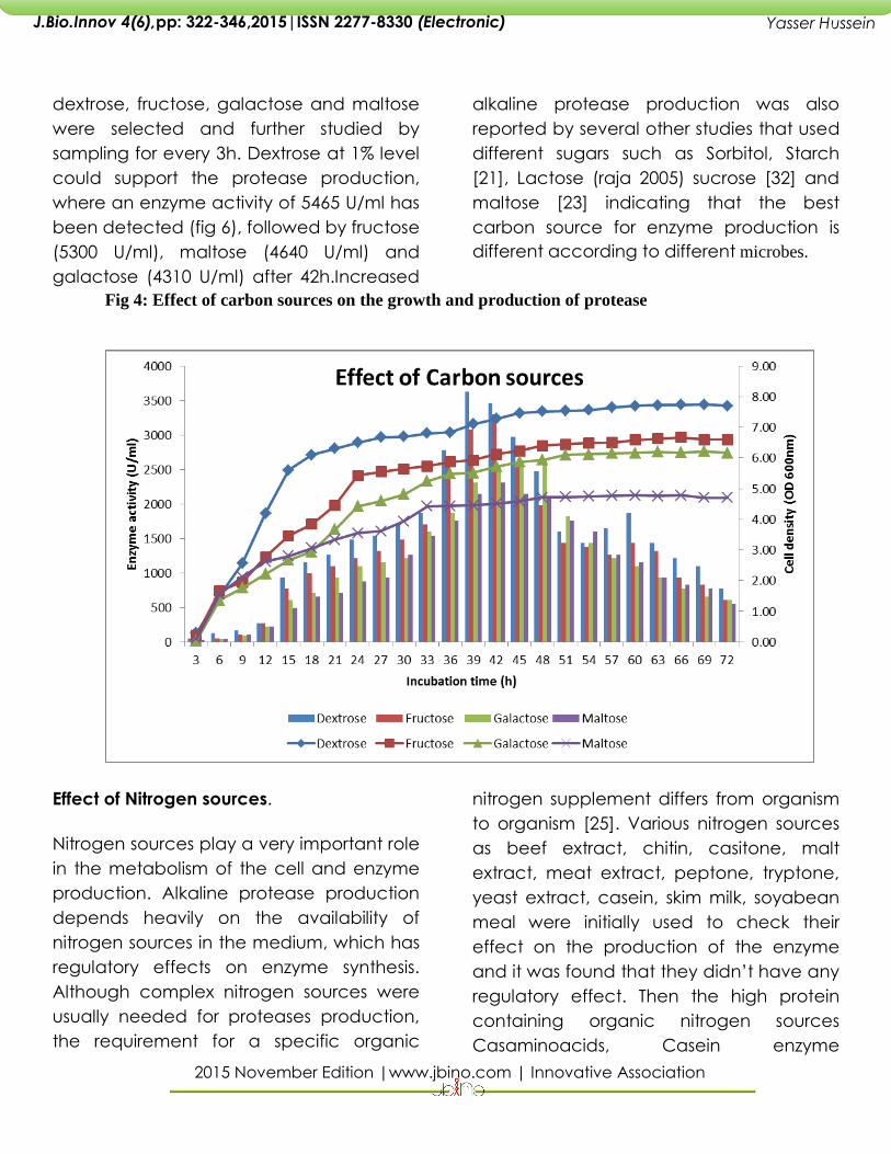
Effect of carbon sources
To analyze the effect of the carbon
sources on the growth and protease
enzyme production a set of flasks with
different carbon sources were taken.
Protease production after 48h was
estimated for each carbon source such as
Arabinose (3730 U/ml), Cellulose ( 3900
U/ml), Chitin (3350 U/ml ), Dextrose (4690
U/ml), Fructose (3975 U/ml), Galactose
(4240 U/ml), Maltose (4400 U/ml),
Mannose(3755 U/ml) , Starch (3055 U/ml),
Sucrose (3265 U/ml) and Xylose (3870
U/ml). Different carbon sources have
different effects on extracellular protease
production based on its utilization by the
particular microbe. Based on the results
2015 November Edition |www.jbino.com | Innovative Association
J.Bio.Innov 4(6),pp: 322-346,2015|ISSN 2277-8330 (Electronic)
Yasser Hussein
dextrose, fructose, galactose and maltose
were selected and further studied by
sampling for every 3h. Dextrose at 1% level
could support the protease production,
where an enzyme activity of 5465 U/ml has
been detected (fig 6), followed by fructose
(5300 U/ml), maltose (4640 U/ml) and
galactose (4310 U/ml) after 42h.Increased
alkaline protease production was also
reported by several other studies that used
different sugars such as Sorbitol, Starch
[21], Lactose (raja 2005) sucrose [32] and
maltose [23] indicating that the best
carbon source for enzyme production is
different according to different microbes.
Fig 4: Effect of carbon sources on the growth and production of protease
Effect of Nitrogen sources.
Nitrogen sources play a very important role
in the metabolism of the cell and enzyme
production. Alkaline protease production
depends heavily on the availability of
nitrogen sources in the medium, which has
regulatory effects on enzyme synthesis.
Although complex nitrogen sources were
usually needed for proteases production,
the requirement for a specific organic
nitrogen supplement differs from organism
to organism [25]. Various nitrogen sources
as beef extract, chitin, casitone, malt
extract, meat extract, peptone, tryptone,
yeast extract, casein, skim milk, soyabean
meal were initially used to check their
effect on the production of the enzyme
and it was found that they didn’t have any
regulatory effect. Then the high protein
containing organic nitrogen sources
Casaminoacids, Casein enzyme
2015 November Edition |www.jbino.com | Innovative Association
J.Bio.Innov 4(6),pp: 322-346,2015|ISSN 2277-8330 (Electronic)
Yasser Hussein
hydrolysate, Casitone and Tryptone were
selected for the optimization. It was
observed from the study that there was no
correlation between the growth of the
organism and protease enzyme
production indicating that protease
enzyme production is non-growth
associated. Similar effect was shown for
Bacillus sp.[31] and other marine isolate [7]
where growth was best supported by a
combination of peptone and yeast
extract, while the optimum protease
production was with casaminoacid. The
biomass yield was high but the enzyme
production was low when a combination
of yeast extract and peptone was used.
Tryptone gave a maximum production of
9845 U/ml followed by Casaminoacids
(9320 U/ml), Casein enzyme hydrolysate
(8910 U/ml), Casitone (6710 U/ml) and
Yeast extract-Peptone (5005 U/ml) after
48h.Tryptone was the best nitrogen source
for protease production by Serratia sp. [35].
Fig 5: Effect of nitrogen sources on the growth and
productionprotease
Purification of the protease enzyme
2015 November Edition |www.jbino.com | Innovative Association
J.Bio.Innov 4(6),pp: 322-346,2015|ISSN 2277-8330 (Electronic)
Yasser Hussein
Ammonium sulphate precipitation method
and Dialysis
Initial step of purification was the
concentration of proteins by precipitation
using ammonium sulphate. The
precipitation of protease was performed
with 20%-80% of ammonium sulphate
saturation. The precipitate was collected
by centrifugation at 10,000 rpm for 15 min,
The supernatant and pellet were assayed
after each fraction .The protein and the
protease in each fraction was determined.
There was a maximum enzyme activity at
80%.
Precipitation using 80% ammonium
sulphate yielded a recovery of 37.61%. This
treatment resulted in increase of specific
activity to 6833.33 mg compared to crude
supernatant. So crude extracellular protein
was precipitated by addition of 80%
ammonium sulphate for further studies. The
precipitated enzyme was dissolved in
Phosphate buffer pH 7.0 and the sample
was dialyzed. The protease activity and
the protein were estimated for the dialyzed
sample. The specific activity The specific
activity has doubled when compared to
that obtained by ammonium sulphate
precipitation. . Dialysis using the obtained
solution has yielded a recovery of 39.26%
.The dialyzed sample was used for the
further purification.
Gel filtration chromatography
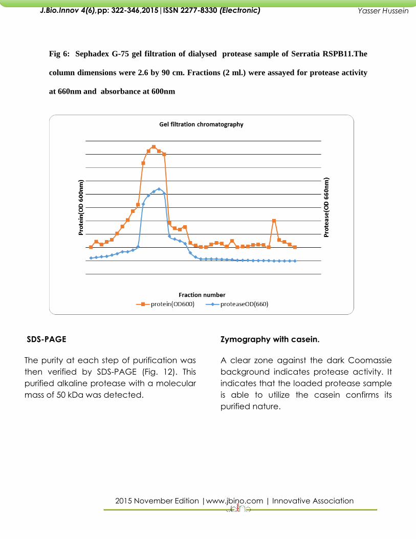
A symmetrical peak of protease activity,
accounting for approximately 95 per cent
of the applied activity, was observed.
During this method, 40 fractions were
collected and the protein and protease in
each fraction was determined. Maximum
protease activity was observed in active
fraction number 14 with 220501U/ml. The
peak was well separated from small
amounts of non proteolytic 660 nm.-
absorbing material. The specific activity
18,375.15 (U/mg). This step resulted in 13.57-
fold purification of the enzyme with 36.7%
recovery. Active fractions were pooled
and dialysed for further purification.
2015 November Edition |www.jbino.com | Innovative Association
J.Bio.Innov 4(6),pp: 322-346,2015|ISSN 2277-8330 (Electronic)
Yasser Hussein
Fig 6: Sephadex G-75 gel filtration of dialysed protease sample of Serratia RSPB11.The
column dimensions were 2.6 by 90 cm. Fractions (2 ml.) were assayed for protease activity
at 660nm and absorbance at 600nm
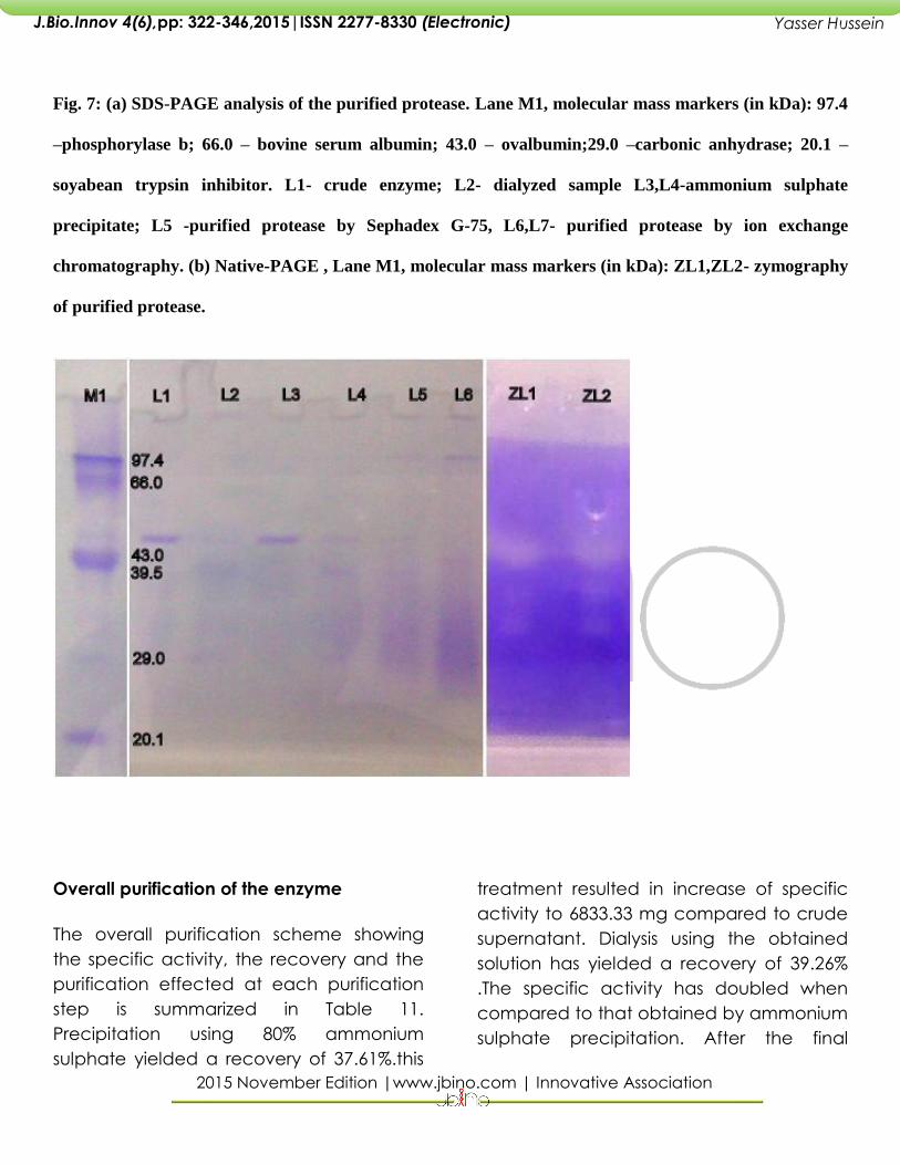
SDS-PAGE
The purity at each step of purification was
then verified by SDS-PAGE (Fig. 12). This
purified alkaline protease with a molecular
mass of 50 kDa was detected.
Zymography with casein.
A clear zone against the dark Coomassie
background indicates protease activity. It
indicates that the loaded protease sample
is able to utilize the casein confirms its
purified nature.
2015 November Edition |www.jbino.com | Innovative Association
J.Bio.Innov 4(6),pp: 322-346,2015|ISSN 2277-8330 (Electronic)
Yasser Hussein
Fig. 7: (a) SDS-PAGE analysis of the purified protease. Lane M1, molecular mass markers (in kDa): 97.4
–phosphorylase b; 66.0 – bovine serum albumin; 43.0 – ovalbumin;29.0 –carbonic anhydrase; 20.1 –
soyabean trypsin inhibitor. L1- crude enzyme; L2- dialyzed sample L3,L4-ammonium sulphate
precipitate; L5 -purified protease by Sephadex G-75, L6,L7- purified protease by ion exchange
chromatography. (b) Native-PAGE , Lane M1, molecular mass markers (in kDa): ZL1,ZL2- zymography
of purified protease.
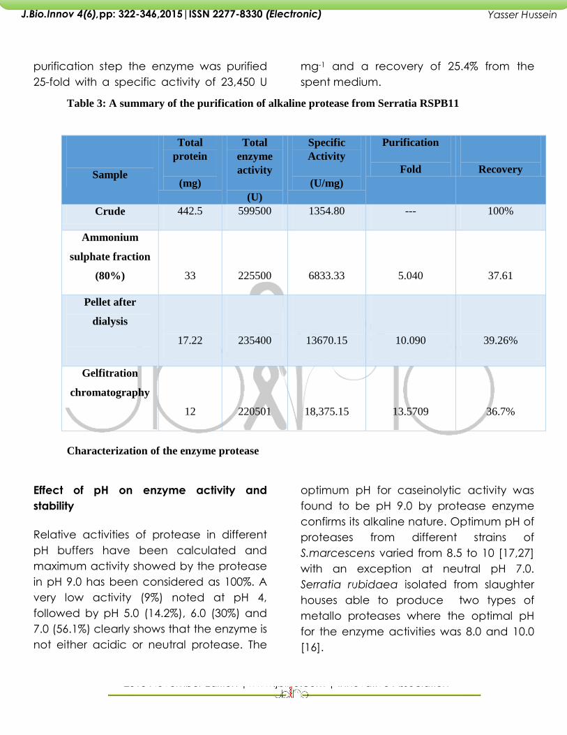
Overall purification of the enzyme
The overall purification scheme showing
the specific activity, the recovery and the
purification effected at each purification
step is summarized in Table 11.
Precipitation using 80% ammonium
sulphate yielded a recovery of 37.61%.this
treatment resulted in increase of specific
activity to 6833.33 mg compared to crude
supernatant. Dialysis using the obtained
solution has yielded a recovery of 39.26%
.The specific activity has doubled when
compared to that obtained by ammonium
sulphate precipitation. After the final
2015 November Edition |www.jbino.com | Innovative Association
J.Bio.Innov 4(6),pp: 322-346,2015|ISSN 2277-8330 (Electronic)
Yasser Hussein
purification step the enzyme was purified
25-fold with a specific activity of 23,450 U
mg-1 and a recovery of 25.4% from the
spent medium.
Table 3: A summary of the purification of alkaline protease from Serratia RSPB11
Sample
Total
protein
(mg)
Total
enzyme
activity
(U)
Specific
Activity
(U/mg)
Purification
Fold
Recovery
Crude 442.5 599500 1354.80 --- 100%
Ammonium
sulphate fraction
(80%)
33
225500
6833.33
5.040
37.61
Pellet after
dialysis
17.22
235400
13670.15
10.090
39.26%
Gelfitration
chromatography
12
220501
18,375.15
13.5709
36.7%
Characterization of the enzyme protease
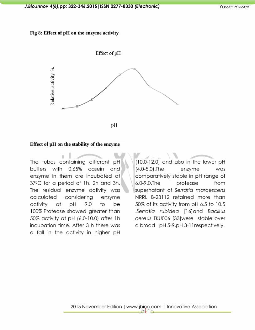
Effect of pH on enzyme activity and
stability
Relative activities of protease in different
pH buffers have been calculated and
maximum activity showed by the protease
in pH 9.0 has been considered as 100%. A
very low activity (9%) noted at pH 4,
followed by pH 5.0 (14.2%), 6.0 (30%) and
7.0 (56.1%) clearly shows that the enzyme is
not either acidic or neutral protease. The
optimum pH for caseinolytic activity was
found to be pH 9.0 by protease enzyme
confirms its alkaline nature. Optimum pH of
proteases from different strains of
S.marcescens varied from 8.5 to 10 [17,27]
with an exception at neutral pH 7.0.
Serratia rubidaea isolated from slaughter
houses able to produce two types of
metallo proteases where the optimal pH
for the enzyme activities was 8.0 and 10.0
[16].
2015 November Edition |www.jbino.com | Innovative Association
J.Bio.Innov 4(6),pp: 322-346,2015|ISSN 2277-8330 (Electronic)
Yasser Hussein
Fig 8: Effect of pH on the enzyme activity
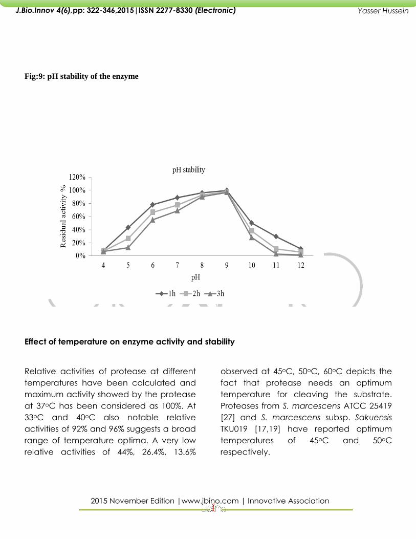
Effect of pH on the stability of the enzyme
The tubes containing different pH
buffers with 0.65% casein and
enzyme in them are incubated at
370C for a period of 1h, 2h and 3h.
The residual enzyme activity was
calculated considering enzyme
activity at pH 9.0 to be
100%.Protease showed greater than
50% activity at pH (6.0-10.0) after 1h
incubation time. After 3 h there was
a fall in the activity in higher pH
(10.0-12.0) and also in the lower pH
(4.0-5.0).The enzyme was
comparatively stable in pH range of
6.0-9.0.The protease from
supernatant of Serratia marcescens
NRRL B-23112 retained more than
50% of its activity from pH 6.5 to 10.5
.Serratia rubidea [16]and Bacillus
cereus TKU006 [33]were stable over
a broad pH 5-9,pH 3-11respectively.
2015 November Edition |www.jbino.com | Innovative Association
J.Bio.Innov 4(6),pp: 322-346,2015|ISSN 2277-8330 (Electronic)
Yasser Hussein
Fig:9: pH stability of the enzyme
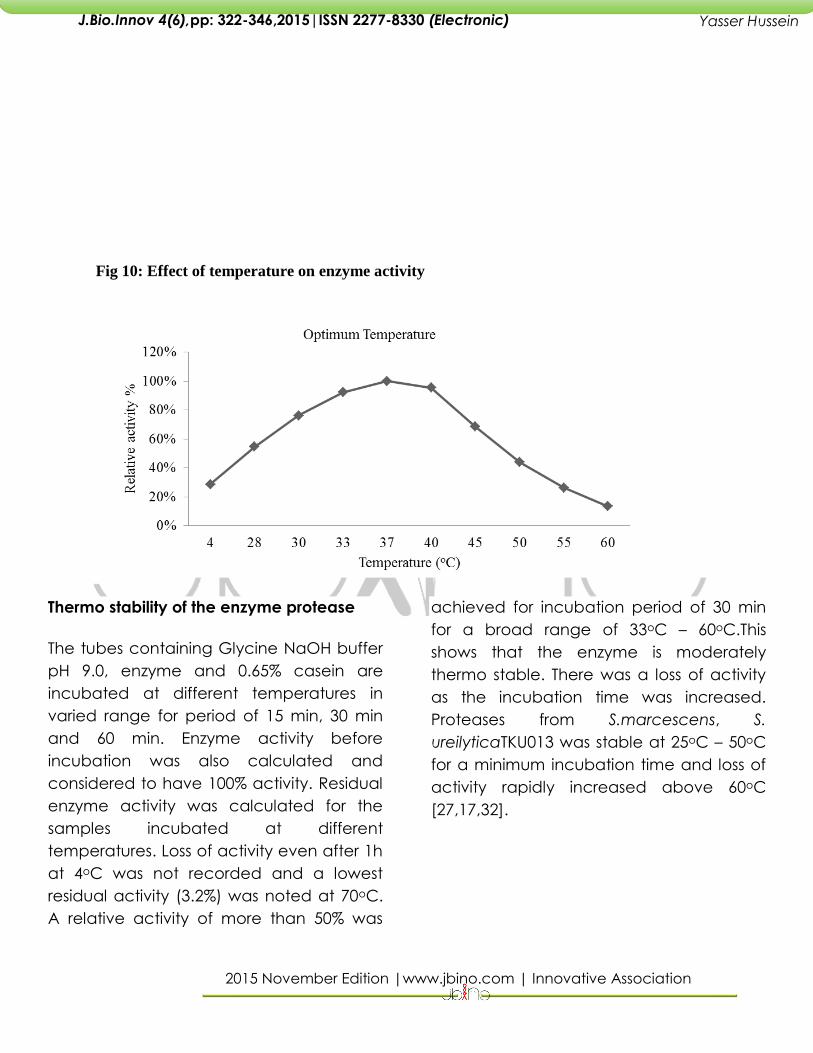
Effect of temperature on enzyme activity and stability
Relative activities of protease at different
temperatures have been calculated and
maximum activity showed by the protease
at 37oC has been considered as 100%. At
33oC and 40oC also notable relative
activities of 92% and 96% suggests a broad
range of temperature optima. A very low
relative activities of 44%, 26.4%, 13.6%
observed at 45oC, 50oC, 60oC depicts the
fact that protease needs an optimum
temperature for cleaving the substrate.
Proteases from S. marcescens ATCC 25419
[27] and S. marcescens subsp. Sakuensis
TKU019 [17,19] have reported optimum
temperatures of 45oC and 50oC
respectively.
2015 November Edition |www.jbino.com | Innovative Association
J.Bio.Innov 4(6),pp: 322-346,2015|ISSN 2277-8330 (Electronic)
Yasser Hussein
Fig 10: Effect of temperature on enzyme activity
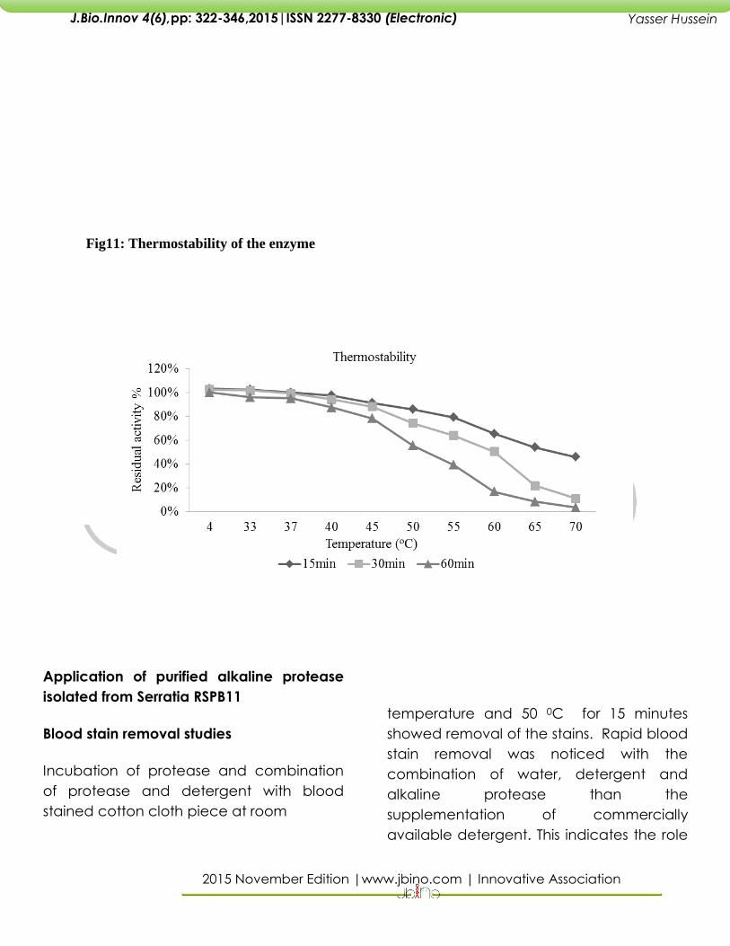
Thermo stability of the enzyme protease
The tubes containing Glycine NaOH buffer
pH 9.0, enzyme and 0.65% casein are
incubated at different temperatures in
varied range for period of 15 min, 30 min
and 60 min. Enzyme activity before
incubation was also calculated and
considered to have 100% activity. Residual
enzyme activity was calculated for the
samples incubated at different
temperatures. Loss of activity even after 1h
at 4oC was not recorded and a lowest
residual activity (3.2%) was noted at 70oC.
A relative activity of more than 50% was
achieved for incubation period of 30 min
for a broad range of 33oC – 60oC.This
shows that the enzyme is moderately
thermo stable. There was a loss of activity
as the incubation time was increased.
Proteases from S.marcescens, S.
ureilyticaTKU013 was stable at 25oC – 50oC
for a minimum incubation time and loss of
activity rapidly increased above 60oC
[27,17,32].
2015 November Edition |www.jbino.com | Innovative Association
J.Bio.Innov 4(6),pp: 322-346,2015|ISSN 2277-8330 (Electronic)
Yasser Hussein
Fig11: Thermostability of the enzyme
Application of purified alkaline protease
isolated from Serratia RSPB11
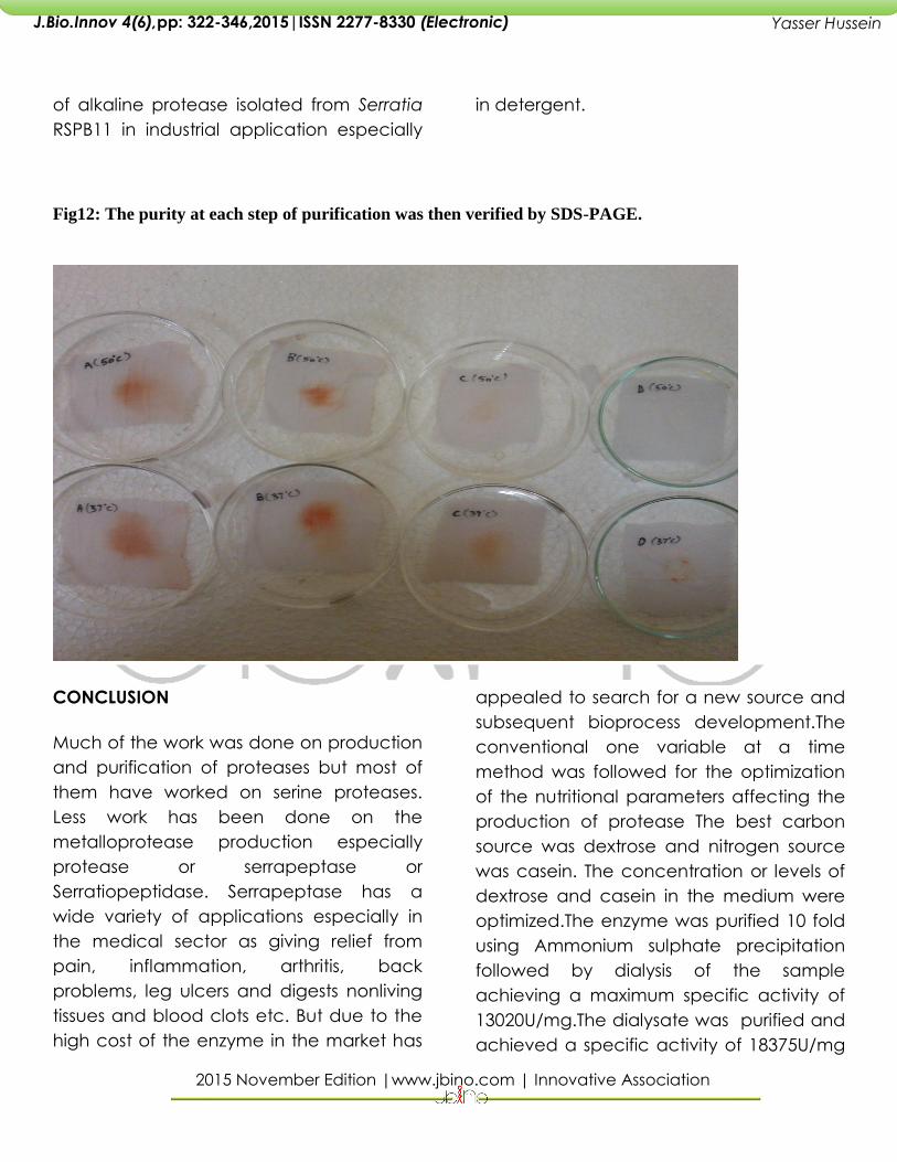
Blood stain removal studies
Incubation of protease and combination
of protease and detergent with blood
stained cotton cloth piece at room
temperature and 50 0C for 15 minutes
showed removal of the stains. Rapid blood
stain removal was noticed with the
combination of water, detergent and
alkaline protease than the
supplementation of commercially
available detergent. This indicates the role
2015 November Edition |www.jbino.com | Innovative Association
J.Bio.Innov 4(6),pp: 322-346,2015|ISSN 2277-8330 (Electronic)
Yasser Hussein
of alkaline protease isolated from Serratia
RSPB11 in industrial application especially
in detergent.
Fig12: The purity at each step of purification was then verified by SDS-PAGE.
CONCLUSION
Much of the work was done on production
and purification of proteases but most of
them have worked on serine proteases.
Less work has been done on the
metalloprotease production especially
protease or serrapeptase or
Serratiopeptidase. Serrapeptase has a
wide variety of applications especially in
the medical sector as giving relief from
pain, inflammation, arthritis, back
problems, leg ulcers and digests nonliving
tissues and blood clots etc. But due to the
high cost of the enzyme in the market has
appealed to search for a new source and
subsequent bioprocess development.The
conventional one variable at a time
method was followed for the optimization
of the nutritional parameters affecting the
production of protease The best carbon
source was dextrose and nitrogen source
was casein. The concentration or levels of
dextrose and casein in the medium were
optimized.The enzyme was purified 10 fold
using Ammonium sulphate precipitation
followed by dialysis of the sample
achieving a maximum specific activity of
13020U/mg.The dialysate was purified and
achieved a specific activity of 18375U/mg
2015 November Edition |www.jbino.com | Innovative Association
J.Bio.Innov 4(6),pp: 322-346,2015|ISSN 2277-8330 (Electronic)
Yasser Hussein
and 36.7% recovery by gel filtration
chromatography. The active fractions
were pooled and dialysed for further
purification.Previously purified protease
sample by gel filtration is subjected to ion
exchange chromatography and attained
a maximum specific activity of 23,450
u/mg. The sample was 17 fold purified and
was further characterized. The
characterization of the enzyme was done.
The optimum pH for caseinolytic activity
was found to be pH 9.0 by protease
enzyme confirms its alkaline nature. The
enzyme was stable in pH range of 6.0-
9.0.The enzyme protease was stable at pH
of 9.0 and there was 50% activity after
incubation for 1 hr. The optimum
temperature required for maximum activity
of protease was found to be 37 oC. There
was a broad range of temperature
optimum ranging from 330C to 400C.The
enzyme was stable at this temperature for
an incubation period of 30 min. The
purified alkaline protease has shown its
proteolytic activity for the removal of
blood stains proved its importance in
detergent industries.
ACKNOWLEDGEMENTS
Yasser Eissa wish to acknowledge the
financial assistance provide by University of
Hajja, Yemen.
REFERENCES
Banik RM, Prakash M (2004) Laundry
detergent compatibility of the alkaline
protease from Bacillus cereus .Microbiol
Res 159:135-140
Prakasham, R.S., SubbaRao, Ch., Rao, R.S.,
Lakshmi, G.S. and Sarma, P.N. (2007).L-
Asparaginase production by isolated
Staphylococcus sp. – 6A: design of
experiment considering interaction effect
for process parameter optimization.
Journal of Applied Microbiology, 102:1382–
1391.
Pansuriya, R.C., and Rekha, S. S., (2010).
Evolutionary Operation (EVOP) to Optimize
Whey-Independent Serratiopeptidase
Production from Serratia marcescens NRRL
B-23112, Journal of Microbiology and
Biotechnology, 20: 950–957
Benjamin, K. Simpson ,Food Biochemistry
and Food Processing.
Claudia c. hase and Richard a. finkelstein,
Bacterial Extracellular Zinc-Containing
Metalloproteases, microbiological reviews,
Dec. 2003, p. 823-837
Claudia c. hase and Richard a. finkelstein,
Bacterial Extracellular Zinc-Containing
Metalloproteases, microbiological reviews,
Dec. 2003, p. 823-837
Roychoudhury S, Parulekar SJ, Weigand
WA. Cell Growth and a-Amylase
Production Characteristics of Bacillus
amyloliquefaciens. Biotechnol Bioeng.
1988;33:197–206. doi: 10.1002/bit.260330209
2015 November Edition |www.jbino.com | Innovative Association
J.Bio.Innov 4(6),pp: 322-346,2015|ISSN 2277-8330 (Electronic)
Yasser Hussein
R. B. Raja, D. J. Mukesh Kumar, M. D.
Balakumaran, P. T. Kalaichelvan, A.
Pandey, A. Singh (2011). Isolation,
Production & Application of Extracellular
Phytase By Serratia marcescens. Asian J.
Exp. Biol. Sci.Vol 2(4): 663 – 666.
Romero, F., Garcia, L.A., Salas, J., Diaz, M.
and Quiros, L. (2001). Production,
purification and partial characterization of
two extracellular proteases from Serratia
marcescens grown in whey.
ProcessBiochemistry, 36: 501-515.
Rahman, R.N.Z.R., Geok, L.P., Basri M,
Salleh, A.B., (2005). Physical factors
affecting the production of organic
solvent-tolerant protease by 30
Pseudomonas aeruginosa strain K.
Resource Technology, 96: 429-436.
Nguyen, T.T., Quyen, D.T., (2011). Over
production of an extracellular protease
from Serratia sp.DT3 just using soybean
powder, World journal of agricultural
sciences 7(1):29-36.
P. Lakshmi Bhargavi and R. S. Prakasham
(2012) Proteolytic Enzyme Production by
Isolated Serratia sp RSPB11: Role of
Environmental Parameters. Current Trends
in Biotechnology and Pharmacy Vol. 6 (1)
55-65.
Lakshmi Narasu Mangamoori, Kiran Kumar
Doddapaneni, Radhika Tatineni, Ravi
Nagaraj Vellanki, Bharat Gandu, Nagender
Reddy Panyala, Balumaddileti
Chakali(2007), Purification and
characterization of two novel extra cellular
proteases from Serratia rubidaea. Process
Biochemistry 42:1229–1236.
Jiang Jiaxin, Huang Guangrong, Dai Dehui
and Hu Weilian (2008) Optimization of
medium composition for thermostable
protease production by Bacillus sp. HS08
with astatistical method. African Journal of
Biotechnology Vol. 7 (8), pp. 1115-1122
Vsenil, C.K., Lakshmanperumalsamy, P.,
(2009). Application of response surface
methodology in medium optimization for
protease production by the new strain of
Serratia marcescens sb08, Polish journal of
microbiology 58:117-124.
C. M. Cabral, A. Cherqui,† A. Pereira, and
N. Simo˜es* .,(2004). Purification and
Characterization of Two Distinct
Metalloproteases Secreted by the
Entomopathogenic Bacterium
Photorhabdus sp. Strain Az29.Applied and
environmental microbiology, July 2004, p.
3831–3838 Vol. 70.
P. Charles, V. Devanathan, Periasamy
Anbu, M. N. Ponnuswamy, P. T.
Kalaichelvan and Byung-Ki Hur., (2008).
Purification, characterization and
crystallization of an extracellular alkaline
protease from Aspergillus nidulans HA-10.
Journal of Basic Microbiology 2008, 48,
347–352.
Arnold S. Kreger and Olwcn K. Griffin.,
(1975). Cornea-damaging proteases of
2015 November Edition |www.jbino.com | Innovative Association
J.Bio.Innov 4(6),pp: 322-346,2015|ISSN 2277-8330 (Electronic)
Yasser Hussein
Serratia marcescens* Volume 14 Niimhur
Investigative Ophthalmology.
A Bayoudh, N Gharsallah, M Chamkha, A
Dhouib, S Ammar and M Nasri.,(2000).
Purification and characterization of an
alkaline protease from Pseudomonas
aeruginosa MN1. Journal of Industrial
Microbiology & Biotechnology 24, 291–
295.
Ishtiaq Ahmed, Muhammad Anjum Zia and
Hafiz Muhammad Nasir Iqbal., (2011).
Purification and Kinetic parameters
characterization of an Alkaline Protease
from Bacillus subtilis through Submerged
Fermentation Technique. Pakistan World
Applied Sciences Journal 12 (6): 751-757.
M. Kalpana Devi, A. Rasheedha Banu, G.R.
Gnanaprabhal, B.V. Pradeep and M.
Palaniswamy., (2008). Purification,
characterization of alkaline protease
enzyme from native isolate Aspergillus
Niger and its compatibility with commercial
detergents. Indian Journal of Science and
Technology http://www.indjst.org Vol.1 No
7 “Fungal alkaline protease”
Malay Mandal, Sudip Das, Tapati
Chakraborti, Amritlal Mandal and Sajal
Chakraborti., (2003). Identification,
purification and partial characterization of
tissue inhibitor of matrix metalloproteinase-
1 (TIMP-1) in bovine pulmonary artery
smooth muscle. Molecular and Cellular
Biochemistry 254: 145–155.
Ch. Subba Rao, T. Sathish, P. Ravichandra,
R.S. Prakasham *. , (2009). Characterization
of thermo- and detergent stable serine
protease from isolated Bacillus circulans
and evaluation of eco-friendly
applications. Process Biochemistry 44 .,
262–268.
Wang, S.L., Chao, C.H., Liang, T.Z. and
Chen, C.C. (2009). Purification and
Characterization of Protease and Chitinase
from Bacillus cereus TKU006 and
Conversion of Marine Wastes by These
Enzymes, Marine biotechnology11:334-344
Charles Gerday, Jean-Pierre Chessa, Ioan
Petrescu, Mostafa Bentahira, Jozef Van
Beeumen (2000). Purification, physico-
chemical characterization and sequence
of a heat labile alkaline metalloprotease
isolated from a psychrophilic
Pseudomonas species. Biochimicaet
BiophysicaActa 1479:265-274.
Egwin, Evans C (PhD). (2011). Partial
Characterization of Protease activity from
Rhynchophorus palmarum (Palm Weevil)
African Journal of Food Science and
Technology (ISSN: 2141-5455) Vol. 2(6) pp.
140-145.
Anson, M.L. (1938). Estimation of Pepsin,
Papain and Cathepsin with haemogloblin.
Journal of General Physiology, 22: 79-89.
Lowry, O. H., Rosebrough, N. J. and
Randall, R. J. (1951). Protein measurement
with the Folin phenol reagent. Journal of
Biological Chemistry, 193: 265-275.
2015 November Edition |www.jbino.com | Innovative Association
J.Bio.Innov 4(6),pp: 322-346,2015|ISSN 2277-8330 (Electronic)
Yasser Hussein
Wang SL, Kao TY, Wang CL, Yen YH, Chern
MK, Chen YH. A solvent stable
metalloprotease produced by Bacillus sp.
TKU004 and its application in the
deproteinization of squid pen for β-chitin
preparation. Enzyme Microb Tech. 2006;39:
724–731. doi:
10.1016/j.enzmictec.2005.12.007
Daatsellar MCC, Harder W. Some aspects
of the regulation of the production of
extracellular proteolytic enzyme by a
marine bacterium. Arch Microbiol
1974;101:21–34
Li-Qun Jin, Zhi-Qiang Liu, Yu-GuoZheng
and Yin-Chu Shen , Identification and
characterization of Serratia marcescens
ZJB-09104, a nitrile-converting bacterium,
World Journal of Microbiology and
Biotechnology Volume 26, Number 5
(2010), 817-823, DOI: 10.1007/s11274-009-
0238-5
Wang, S.L., Chao, C.H., Liang, T.Z. and
Chen, C.C. (2009). Purification and
Characterization of Protease and Chitinase
from Bacillus cereus TKU006 and
Conversion of Marine Wastes by These
Enzymes, Marine Biotechnology, 11:334–
344.