isolation of fungal pathogen fusarium sp. from maize ... · isolation of fungal pathogen fusarium...
TRANSCRIPT
www.scholarsresearchlibrary.comt Available online a
Scholars Research Library
Der Pharmacia Lettre, 2016, 8 (19):57-69
(http://scholarsresearchlibrary.com/archive.html)
ISSN 0975-5071
USA CODEN: DPLEB4
57 Scholar Research Library
Isolation of fungal pathogen Fusarium sp. from maize kernel and its inhibition by Pseudomonas containing liquid Bioformulation
Malvika Rajnandini* 1, Rajneesh Kumar Gautam2, Islamuddin3, Nandkishor More4
and Aakrati Sharma5
1Department of Environmental Microbiology, Babasaheb Bhimrao Ambedkar Central University, Lucknow, -226025, India
2,3Department of Civil Engeering, Babasaheb Bhimrao Ambedkar Central University, Lucknow, -226025, India 4Department of Environmental Sciences, Babasaheb Bhimrao Ambedkar Central University, Lucknow, -226025,
India 5Banasthali University, Banasthali -304022, Rajasthan, India
_____________________________________________________________________________________________ ABSTRACT Maize (Zea mays L.) is one of the most versatile emerging crops having wider adaptability under varied agro - climatic conditions. Maize has the highest genetic yield potential among the cereals. In India, about 28% of maize produced is used for food purpose , about 11% as livestock feed , 48% as poultry feed , 12% in wet milling industry (for example starch and oil production ) and 1% as seed (AICRP , 2007). Pseudomonas fluorescence, a major constituent of rhizospheric bacteria, not only encourages the plant growth through their diverse mechanisms and but also show a wider potential for biological control of different phytopathogens. This paper presents the study on the isolation and identification of fluorescent Pseudomonas and to check their bio-control potential against phytopathogen F. moniliforme and on the basis of various plant growth promoting and biocontrol attribute select the elite strain in designing of liquid based bio-formulation. Apart from this application of this Bio-formulation for its efficacy in maize growth and disease suppression is the main objective of the study. Key words: Maize, Genetic yield potential, Pseudomonas, Bio-formulation, F.moniliforme _____________________________________________________________________________________________
INTRODUCTION In developed countries, maize is consumed mainly as second – cycle produce, in the form of meat, eggs and dairy products. In developing countries, maize is consumed directly and serves as staple diet. In India, maize is the third most important food crops after rice and wheat (http://faostat3.fao.org). Global maize production has grown at a calculated annual growth rate (CAGR) of 3.4 per cent over the last ten years from 716 million metric tons (MnMT) / year in 2004-05 to 967 million metric tons / year in 2013-14. USA is the largest producer of maize in the world, followed by China and Brazil (Ranum , 2014 ). USA is also the largest exporter of maize. Production of maize in India has increased at a CAGR of 5.5 per cent from 14 MnMT in 2004-05 to 23 MnMT in 2013-14 and the areas where it is more often cultivated are Andhra Pradesh and Karnataka which contributes to ~38 per cent of the total production. India’s yield at 2.5 MT/hectare is less than half the global average of 5.5 MT/hectare. Maize consumption has increased at a CAGR of 3.6 per cent over the last five years; poultry feed accounts for ~50 per cent of maize consumption. (IMS, 2014). In India, maize is grown in a wide range of environments, extending from extreme semi-arid to sub-humid and humid regions. The crop is also very popular in the low- and mid -hill areas of

Malvika Rajnandini et al Der Pharmacia Lettre, 2016, 8 (19):57-69 ______________________________________________________________________________
58 Scholar Research Library
the western and north eastern regions. Broadly, maize cultivation can be classified into two production environments, (1) traditional maize growing areas, including Bihar, Madhya Pradesh, Rajasthan, and Uttar Pradesh , and (2) non-traditional maize areas, including Karnataka and Andhra Pradesh. (Singh, 2005.) . Maize is often prone to several fungal pathogens like Aspergillus flavus (Aspergillus ear and kernel rot) , A. glaucus (minor ear rots) , A. niger (minor ear rots) , Rhizopus stolonifer, Lasiodiplodia theobromae (black kernel rot) Macrophomina phaseolina (seed rot seedling blight) , Penicillium spp (Penicillium ear rot ,blue eye, blue mold) , Fusarium subglutinans (Fusarium ear and stalk rot) , F. monoliforme (Fusarium ear and stalk rot , Seed rot seedling blight) , F. acuminatum (Root rots , minor) F. equiseti (root rots , minor) , F. oxysporum (minor root rots , minor stalk rots), F. roseum (Minor Root rots). Among them Fusarium ear rot is the most damaging disease of corn ( Davis , 1989) and various Fusarium spp. causes many diseases to corn such as Fusarium ear rot(F. moniliforme, F. verticillioides and F. proliferatum), Gibberella ear and stalk rot (F. graminearum), Fusarium stalk rot (F. verticillioides), and Fusarium root rot (F. graminearum and F.verticillioides) and seedling blight (F. graminearum and F. verticillioides) (Munkvold and O'Mara, 2002; Rodriguez et al., 2010; Reyes et al., 2011). P. fluorescence are Gram-negative rod shaped bacteria that inhabit soil, plants, and water surfaces. (Wook et. al, 2009). The optimum growth temperature is between 25-30 degrees Celsius considered to be the most assuring group of plant growth promoting rhizobacteria involved in biocontrol of plant diseases (Gardner et al., 1984; Moeinzadeh et al., 2010). They produce secondary metabolites such as antibiotics (Keel et al., 1992), phytohormones (Keel et al., 1992), volatile compound hydrogen cyanide (HCN) and ammonia (Thomashow and Weller 1988; Voisard et al. 1989; Defago and Haas 1990; Thara and Gnanamanickam 1994), lytic enzyme such as chitinase and iron chelating compound siderophores (Neiland, 1995) to antagonise pathogens. Study reveals that ability of P. fluorescence to convert insoluble form of phosphate in to soluble form increases growth in maize, lettuce, soyabean and various other crop (Gupta et al., 2002; Deshwal et al. 2011).. Active ingredient is mostly a viable organism, it may be live microbes or spore and its survival during storage is very essential for successful formulation development (Auld et al. 2003; Hynes and Boyetchko 2006). Broadly two types of bio-formulations are available, liquid and solid (Rhodes, 1993; Burges 1998). Liquid formulation is also known as flow able or aqueous suspensions and consists of biomass suspensions in water, oil or combinations of both (emulsions) (Schisler et al., 2004). A typical liquid formulation contains 10-40 % microorganism , 1-3 % suspender ingredient , 1-5 % dispersant, 3-8 % surfactant, and 35-65% carrier liquid (oil or water ) (Brar et al. 2006). 1.1 Main Objectives Of Current research: The main objectives involved in the current research are a) Isolation and screening of various fluorescent Pseudomonad’s. b) Isolation and morphological characterization of phytopathogen fungus from diseased maize plant. c) To assess PGP and bio-control activities of isolated strains against phytopathogen fungus. d) Biochemical characterization of isolated fluorescent Pseudomonad’s. e) Designing of liquid bio-formulation. f) To perform pot experiments to check the efficacy of liquid bio-formulation.
MATERIALS AND METHODS
2.1 Step by step experimental design a) Collection of soil sample The soil sample was collected from the residential area of Customs and Central Excise colony, Gujaini, Kanpur from the rhizospheric area of the tomato plant, lemon grass, weed plant and garden soil. Garden soil was collected from 15-20 cm depth and were aseptically placed in polybags respectively and transported to the laboratory within 2 hrs. for analys. b) Isolation of Fluorescent Pseudomonads (King et al., 1954) King’s B medium was sterilized at 121°C and 15 lb pressure for 15 minutes. After autoclaving the medium was allowed to cool down and then poured in petri plates in triplicates. Soil sample was serially diluted. For this one gram (1g) of soil sample was taken in 9 ml of autoclaved distilled water and was thoroughly shaken. 1 ml of the above solution was again transferred to 9 ml of sterile distilled water to form 10-2 dilution. Similarly 10-3, 10-4, 10-5, 10-6 and10-7 dilution also were prepared for soil sample. 0.1ml of 10-7 dilution for each of the soil was spread on King’s B medium and after spreading petriplates were incubated at 27 - 30°C for 3 days. After incubation colonies formed on plates were observed. Slimy colonies with surrounding fluorescein were picked and further streaked on
Malvika Rajnandini et al Der Pharmacia Lettre, 2016, 8 (19):57-69 ______________________________________________________________________________
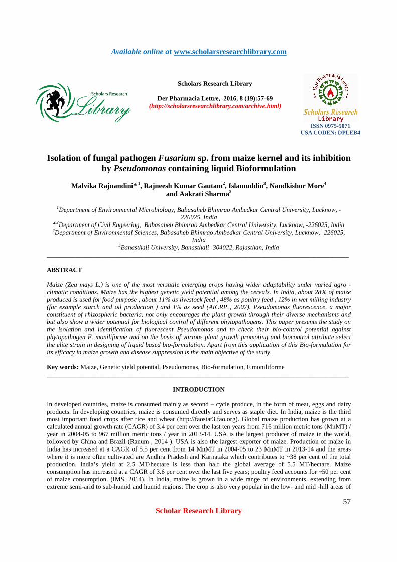
59 Scholar Research Library
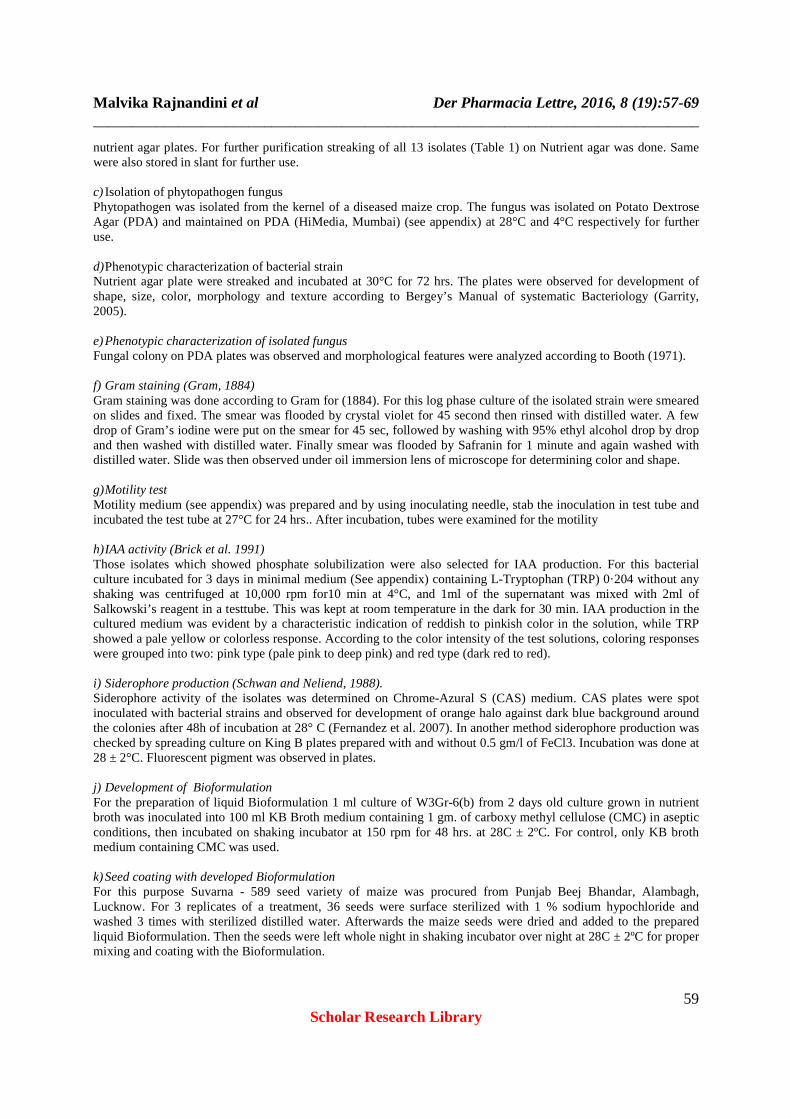
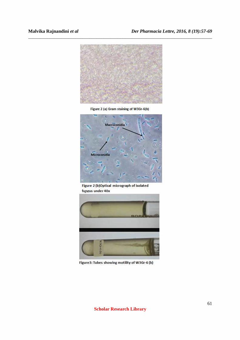
nutrient agar plates. For further purification streaking of all 13 isolates (Table 1) on Nutrient agar was done. Same were also stored in slant for further use. c) Isolation of phytopathogen fungus Phytopathogen was isolated from the kernel of a diseased maize crop. The fungus was isolated on Potato Dextrose Agar (PDA) and maintained on PDA (HiMedia, Mumbai) (see appendix) at 28°C and 4°C respectively for further use. d) Phenotypic characterization of bacterial strain Nutrient agar plate were streaked and incubated at 30°C for 72 hrs. The plates were observed for development of shape, size, color, morphology and texture according to Bergey’s Manual of systematic Bacteriology (Garrity, 2005). e) Phenotypic characterization of isolated fungus Fungal colony on PDA plates was observed and morphological features were analyzed according to Booth (1971). f) Gram staining (Gram, 1884) Gram staining was done according to Gram for (1884). For this log phase culture of the isolated strain were smeared on slides and fixed. The smear was flooded by crystal violet for 45 second then rinsed with distilled water. A few drop of Gram’s iodine were put on the smear for 45 sec, followed by washing with 95% ethyl alcohol drop by drop and then washed with distilled water. Finally smear was flooded by Safranin for 1 minute and again washed with distilled water. Slide was then observed under oil immersion lens of microscope for determining color and shape. g) Motility test Motility medium (see appendix) was prepared and by using inoculating needle, stab the inoculation in test tube and incubated the test tube at 27°C for 24 hrs.. After incubation, tubes were examined for the motility h) IAA activity (Brick et al. 1991) Those isolates which showed phosphate solubilization were also selected for IAA production. For this bacterial culture incubated for 3 days in minimal medium (See appendix) containing L-Tryptophan (TRP) 0·204 without any shaking was centrifuged at 10,000 rpm for10 min at 4°C, and 1ml of the supernatant was mixed with 2ml of Salkowski’s reagent in a testtube. This was kept at room temperature in the dark for 30 min. IAA production in the cultured medium was evident by a characteristic indication of reddish to pinkish color in the solution, while TRP showed a pale yellow or colorless response. According to the color intensity of the test solutions, coloring responses were grouped into two: pink type (pale pink to deep pink) and red type (dark red to red). i) Siderophore production (Schwan and Neliend, 1988). Siderophore activity of the isolates was determined on Chrome-Azural S (CAS) medium. CAS plates were spot inoculated with bacterial strains and observed for development of orange halo against dark blue background around the colonies after 48h of incubation at 28° C (Fernandez et al. 2007). In another method siderophore production was checked by spreading culture on King B plates prepared with and without 0.5 gm/l of FeCl3. Incubation was done at 28 ± 2°C. Fluorescent pigment was observed in plates. j) Development of Bioformulation For the preparation of liquid Bioformulation 1 ml culture of W3Gr-6(b) from 2 days old culture grown in nutrient broth was inoculated into 100 ml KB Broth medium containing 1 gm. of carboxy methyl cellulose (CMC) in aseptic conditions, then incubated on shaking incubator at 150 rpm for 48 hrs. at 28C ± 2ºC. For control, only KB broth medium containing CMC was used. k) Seed coating with developed Bioformulation For this purpose Suvarna - 589 seed variety of maize was procured from Punjab Beej Bhandar, Alambagh, Lucknow. For 3 replicates of a treatment, 36 seeds were surface sterilized with 1 % sodium hypochloride and washed 3 times with sterilized distilled water. Afterwards the maize seeds were dried and added to the prepared liquid Bioformulation. Then the seeds were left whole night in shaking incubator over night at 28C ± 2ºC for proper mixing and coating with the Bioformulation.

Malvika Rajnandini et al Der Pharmacia Lettre, 2016, 8 (19):57-69 ______________________________________________________________________________
60 Scholar Research Library
l) Preparation of fungal inoculum For this a 7 day old, 8 mm disc of fungal pathogen (isolated from maize kernel) actively grown on a PDA plate were transferred into PDB aseptically and incubated on a shaking incubator for 5 days at 28ºC 2.2 Pot experiment Pot experiments were conducted in Basaheb Bhimrao Ambedkar University campus, Lucknow, India during the month of April-May. The soil was collected from campus field then autoclaved consecutively 3 times at 121 C for 30 min at 15 psi. Each earthen pot of (30 x 24 x 16 cm) was filled with 5 kg of sterilized soil. Pots were kept in the open with average day and night temperature of 36ºC and 28ºC, respectively. Enough moisture was maintained by watering the pots regularly. The treatment scheme is as follow (in triplicates): 1. Seed control : each pot were sown equidistance of 15 cm with 3 seeds coated only with KB broth and CMC 2. Pathogen control: For this, pot was filled in 3 layer of soil lower, middle and top by proper mixing of 100 ml of fungal pathogen. After mixing of fungal inoculum on the top layer 3 control seed were sown at equidistance of 15 cm. 3. Seed + Bacteria: 3 seeds coated with liquid Bioformulation were sown equidistantly at 15 cm 4. Pathogen + Bacteria : For this, pot was filled in 3 layer of soil lower , middle and top by proper mixing of 100 ml of fungal pathogen .after mixing of fungal inoculum on the top layer 3 seeds coated with Bioformulation were sown at equidistance of 15 cm. After 30 days maize plants were observed for appearance and characteristic of disease caused by F. moniliforme and growth enhancement by W3Gr-6(b) strain by measuring root length, shoot length, root weight, shoot weight and number of leaves were also counted.
RESULTS
i) Phenotypic and biochemical characterization of bacterial strains Fluorescent Pseudomonads isolates which showed PGP activity were assessed for phenotypic and biochemical characterization and following results were obtained. (Table no.1 & 6) ii) Phenotypic characterization of bacterial isolates Slimy mucous colony with elevated margin was observed. iii) Phenotypic characterization of isolated fungus (Booth, 1971) Cotton blue mount of isolated fungus was observed under microscope by 10x, 40x and 100x (name of microscope) for hyaline, simple or branched conidiophores, bearing conidia in chains. Two kinds of conidia were observed: macroconidia for its boat-shaped, slightly curved apical cells, hooked foot cells, and 2 central cylindrical cells, mainly 4 to 5 celled, and microconidia hyaline, ellipsoidal or ovate, apiculate at one end. Chlamydospores were absent. (figure 2.b )
Table 1 Phenotypic Characteristics and motility
Name of strain Gram staining Shape Motility GS-4 - Curved rod - GS-5 - Curved rod + TS-4 - Curved rod - TS-5 - Curved rod + TS-6 - Curved rod + TS-41 - Curved rod + TS-42 - Curved rod + TS-43 - Curved rod + TS-44 - Curved rod +
W3Gr-4 - Curved rod - W3Gr-6(a) - Curved rod + W3Gr-6(b) - Curved rod +
W4H5 - Curved rod +
Malvika Rajnandini et al Der Pharmacia Lettre, 2016, 8 (19):57-69 ______________________________________________________________________________
61 Scholar Research Library
Malvika Rajnandini et al Der Pharmacia Lettre, 2016, 8 (19):57-69 ______________________________________________________________________________
62 Scholar Research Library
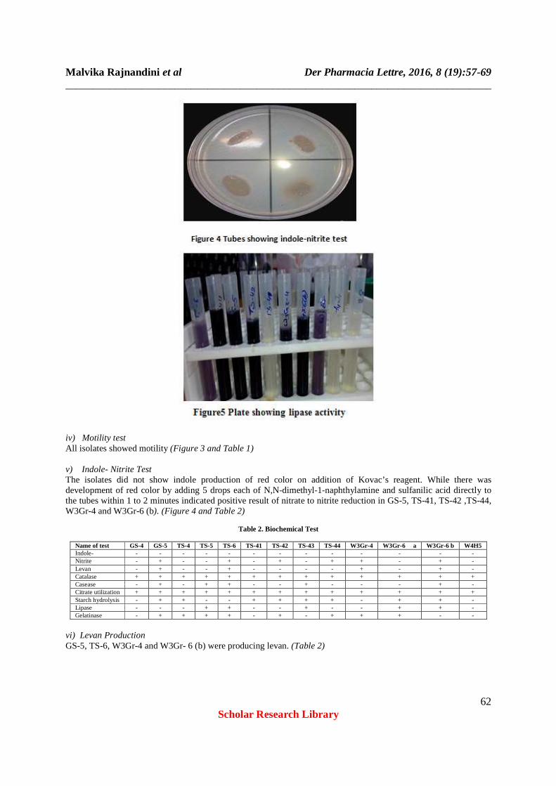
iv) Motility test All isolates showed motility (Figure 3 and Table 1) v) Indole- Nitrite Test The isolates did not show indole production of red color on addition of Kovac’s reagent. While there was development of red color by adding 5 drops each of N,N-dimethyl-1-naphthylamine and sulfanilic acid directly to the tubes within 1 to 2 minutes indicated positive result of nitrate to nitrite reduction in GS-5, TS-41, TS-42 ,TS-44, W3Gr-4 and W3Gr-6 (b). (Figure 4 and Table 2)
Table 2. Biochemical Test
Name of test GS-4 GS-5 TS-4 TS-5 TS-6 TS-41 TS-42 TS-43 TS-44 W3Gr-4 W3Gr-6 a W3Gr-6 b W4H5 Indole- - - - - - - - - - - - - - Nitrite - + - - + - + - + + - + - Levan - + - - + - - - - + - + - Catalase + + + + + + + + + + + + + Casease - + - + + - - + - - - + - Citrate utilization + + + + + + + + + + + + + Starch hydrolysis - + + - - + + + + - + + - Lipase - - - + + - - + - - + + - Gelatinase - + + + + - + - + + + - -
vi) Levan Production GS-5, TS-6, W3Gr-4 and W3Gr- 6 (b) were producing levan. (Table 2)

Malvika Rajnandini et al Der Pharmacia Lettre, 2016, 8 (19):57-69 ______________________________________________________________________________
63 Scholar Research Library
vii) Catalase activity All PGP bacteria were found catalase positive as confirmed by liberation of effervescence of O2 around the bacterial colonies when H2O2 was added. (figure 5 table 2)
viii) Casease test In all PGP isolates W3Gr-6(b), GS-5, TS-5, TS-6, TS-42 and TS- 44 were showing clearing of skim milk agar. (figure 6) (table 2) ix) Starch hydrolysis Among all the isolates GS-5, TS-4, TS-41, TS-42, TS-43 , TS-44 , W3Gr-6 , W3Gr-6a , and W3Gr-6(b) showed positive result as clear zone was observed around colonies when iodine was flooded on colonies (Figure 7) (Table 2).
x) Lipase test Only W3Gr-6(a), W3Gr-6(b) , TS-5 , TS-6 and TS-43 showed lipolytic activity as the well visible halo which is due to crystals of the calcium salt of the fatty acid liberated by lipolysis were observed (Figure 5, Table 2). xi) Gelatinase Among all the isolated bacteria GS-5, TS-4, TS-5, TS-6, TS-42, TS-44, W3Gr-4 and W3Gr-6(a) showed gelatin liquefaction (Figure 8) (Table 2).
Malvika Rajnandini et al Der Pharmacia Lettre, 2016, 8 (19):57-69 ______________________________________________________________________________
64 Scholar Research Library
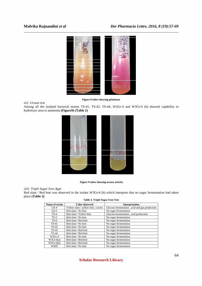
Figure 8 tubes showing gelatinase xii) Urease test Among all the isolated bacterial strains TS-41, TS-42, TS-44, W3Gr-4 and W3Gr-6 (b) showed capability to hydrolyze urea to ammonia (Figure9) (Table 2)
Figure 9 tubes showing urease activity
xiii) Triple Sugar Iron Agar Red slant / Red butt was observed in the isolate W3Gr-6 (b) which interprets that no sugar fermentation had taken place (Table 3)
Table 3. Triple Sugar Iron Test
Name of strain Color observed Interpretation GS-4 Yellow slant / yellow butt ; cracks Glucose fermentation , acid and gas production GS-5 Red slant / No butt No sugar fermentation TS-4 Red slant / Yellow butt Glucose fermentation , acid production TS-5 Red slant / No butt No sugar fermentation TS-6 Red slant / Red butt No sugar fermentation TS-41 Red slant / No butt No sugar fermentation TS-42 Red slant / No butt No sugar fermentation TS-43 Red slant / Red butt No sugar fermentation TS-44 Red slant / Red butt No sugar fermentation
W3Gr-4 Red slant / No butt No sugar fermentation W3Gr-6(a) Red slant / Red butt No sugar fermentation W3Gr-6(b) Red slant / Red butt No sugar fermentation
W4H5 Red slant / No butt No sugar fermentation
Malvika Rajnandini et al Der Pharmacia Lettre, 2016, 8 (19):57-69 ______________________________________________________________________________
65 Scholar Research Library
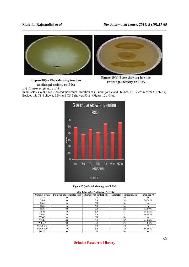
xiv) In vitro antifungal activity In all isolates W3Gr-6(b) showed maximum inhibition of F. moniliforme and 56.66 % PRIG was recorded (Table 4). Besides this TS-6 showed 55% and GS-5 showed 50% (Figure 10 a & b).
Figure 10 (b) Graph showing % of PRIG
Table 4. In- vitro Antifungal Activity Name of strain Diameter of petriplates (cm) Diameter of control(cm) Diameter of inhibition(cm) Inhibition %
GS-4 Nd Nd Nd Nd GS-5 8.0 6.0 3.0 50.00 % TS-4 Nd Nd Nd Nd TS-5 Nd Nd Nd Nd TS-6 8.0 6.0 2.5 55.00% TS-41 8.0 6.0 3.7 38.33 % TS-42 8.0 6.0 3.2 46.66 % TS-43 Nd Nd Nd Nd TS-44 8.0 6.0 3.5 41.66%
W3Gr-4 8.0 6.0 2.5 41.66% W3Gr-6(a) Nd Nd Nd Nd W3Gr-6(b) 8.0 6.0 2.6 56.66 %
W4H5 Nd Nd Nd Nd

Malvika Rajnandini et al Der Pharmacia Lettre, 2016, 8 (19):57-69 ______________________________________________________________________________
66 Scholar Research Library
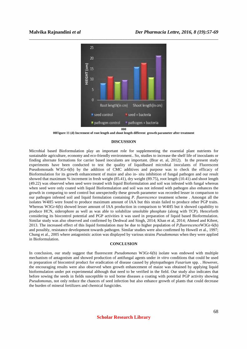
xvii) Pot experiment The scheme of pot experiment is shown in (figure 11.a) For the effect of liquid Bioformulation on the growth of maize and disease suppression by M. moniliforme on complementation of one month, crop was analyzed for shoot length, root length fresh weight, dry weight and number of leaves. Result showed that maximum % increment in fresh weight (85.41), dry weight (89.75), root length (10.41) and shoot length (49.22) was observed when seed were treated with liquid Bioformulation (Figure 11 a, b, c, d & e).
Figure 11 (a) Scheme of Pot experiment

Malvika Rajnandini et al Der Pharmacia Lettre, 2016, 8 (19):57-69 ______________________________________________________________________________
67 Scholar Research Library
Figure 11 (b) Plant Showing increment of different growth parameter after treatment
Figure 11 (c) graph Showing increment of different growth parameter after treatment
Malvika Rajnandini et al Der Pharmacia Lettre, 2016, 8 (19):57-69 ______________________________________________________________________________
68 Scholar Research Library
000
00Figure 11 (d) Increment of root length and shoot length different growth parameter after treatment
DISCUSSION
Microbial based Bioformulation play an important role for supplementing the essential plant nutrients for sustainable agriculture, economy and eco-friendly environment.. So, studies to increase the shelf life of inoculants or finding alternate formations for carrier based inoculants are important. (Brar et. al, 2012). In the present study experiments have been conducted to test the quality of liquidbased microbial inoculants of Fluorescent Pseudomonads W3Gr-6(b) by the addition of CMC additives and purpose was to check the efficacy of Bioformulation for its growth enhancement of maize and also in- situ inhibition of fungal pathogen and our result showed that maximum % increment in fresh weight (85.41), dry weight (89.75), root length (10.41) and shoot length (49.22) was observed when seed were treated with liquid Bioformulation and soil was infested with fungal whereas when seed were only coated with liquid Bioformulation and soil was not infested with pathogen also enhances the growth in comparing to seed control but unexpectedly these growth parameter was recorded lesser in comparison to our pathogen infested soil and liquid formulation containing P. fluorescence treatment scheme . Amongst all the isolates W4H5 were found to produce maximum amount of IAA but this strain failed to produce other PGP traits. Whereas W3Gr-6(b) showed lesser amount of IAA production in comparison to W4H5 but it showed capability to produce HCN, siderophore as well as was able to solubilize unsoluble phosphate (along with TCP). Henceforth considering its biocontrol potential and PGP activities it was used in preparation of liquid based Bioformulation. Similar study was also observed and confirmed by Deshwal and Singh, 2014; Khan et al, 2014; Ahmed and Kibret, 2013. The increased effect of this liquid formulation may be due to higher population of P.fluorescenceW3Gr-6(b) and possibly, resistance development towards pathogen. Similar studies were also confirmed by Howell et al., 1997; Chung et al., 2005 where antagonistic action was displayed by various strains Pseudomonas when they were applied in Bioformulation.
CONCLUSION
In conclusion, our study suggest that fluorescent Pseudomonas W3Gr-6(b) isolate was endowed with multiple mechanism of antagonism and showed production of antifungal agents under in vitro conditions that could be used in preparation of biocontrol product for eradication of disease caused by phytopathogen Fusarium spp. . However, the encouraging results were also observed when growth enhancement of maize was obtained by applying liquid bioformulation under pot experimental although that need to be verified in the field. Our study also indicates that before sowing the seeds in fields susceptible to soil borne diseases a coating with potential PGP activity showing Pseudomonas, not only reduce the chances of seed infection but also enhance growth of plants that could decrease the burden of mineral fertilizers and chemical fungicides.

Malvika Rajnandini et al Der Pharmacia Lettre, 2016, 8 (19):57-69 ______________________________________________________________________________
69 Scholar Research Library
Acknowledgements This research was conducted at Department of Evironmental Microbiology of Babasaheb Bhimrao Ambedkar University Lucknow .I am truly thankful to Dr. Nandkishor More, Dr. Rajesh Kumar, laboratory staff and my colleagues of school of Environmental Sciences, Babasaheb Bhimrao Ambedkar University Lucknow . for consistently guiding and encouraging me.
REFERENCES
[1] AhemadMunees and KibretMuluget (2013) 1018-3647 a. Mechanisms and applications of plant growth promoting rhizobacteria: Current perspective. In Production and hosting by Elsevier B.V.on behalf of King Saud University. [2] AICRP (All India Coordinated Research Project) on Maize(2007) New Delhi. [3] Ardakani SS, Heydari A, Khorasani N, Arjmandi R (2010). J. Plant Pathol. 92:83-88. [4] Arora N K and Khare E(2010). In MaheshwariD K (ed.), Plant Growth and Health Promoting Bacteria, Microbiology Monographs 18, # Springer-Verlag Berlin Heidelberg. [5] Ashrafuzzaman M, Hossen FA, Ismail MR, Hoque MA, Islam MZ, Shahidullah SM, Meon S (2009). Afr.J. Biotechnol., 8: 1247-1252. [6] Auld B A, Hertherington S D, Smith H E (2003). Weed Biology and Management 3, 61–67. [7] BakthavatchaluSasirekha, SrividyaShivakumar, ShankerBhatSullia (2012). ActaBiologicaIndica, 1(1):61-67 [8] Bakker A W, Schippers B (1987). Microbial cyanide production in the rhizosphere in relation to potato yield reduction and Pseudomonas spp.- mediated plant growth stimulation. Soil Biol. Biochem. [9] Beishir L (1991). Microbiology in Practice: A Self-Instructional Laboratory Course, Fifth Edition. (Harper Collins: New York). [10] Berg G, Seech A G, Lee H &Trevors J T (1990). Journal of Environmental Science and Health A25, 753-764. [11] Bhattacharyya PN and Jha D K (2012) World J. Microbiol. Biotechnol., 28 (2012), pp. 1327–1350 [12] Blackburn T H (1968). Protease production by Bacteroidesarnylophilusstrain ~ 1 8J..gefiMicrobiol. 53, 27 [13] Booth C (1971). The genus Fusarium.Commonw. Mycol.Inst., Kew. 237 pp. [14] Bottalico A (1998). J. Plant Pathol., 80: 85–103 [15] Burges H D (1998). Formulation of mycoinsecticides. p. 131–185.In:“ Formulation of Microbial Biopesticides” (H.D. Burges,ed.). Kluwer Academic Publishes, Dordercht. [16] Boyette C D (2006). Biocontrol Science and Technology 16, 1057–1066. [17] Brar S K, Verma M, Tyagi R D, Valéro J R and Surampalli R Y (2006a). Water Res., 40, 1310-1320. [18] Brar SK, Sarma SJ, Chaabouni E (2012) J BiofertilBiopestici 3:e109. doi:10.4172/2155-6202.1000e109