isolation and purification of glutathione-s-transferase from the bulb mite rhizoglyphus robini
TRANSCRIPT
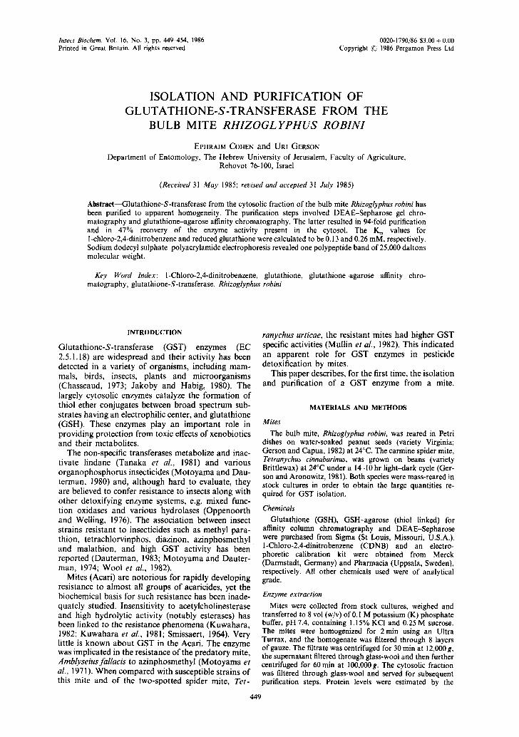
Insect Biochem. Vol. 16, No. 3, pp. 449-454, 1986 0020-1790/86 $3.00 +0.00 Printed in Great Britain. All rights reserved Copyright ~-~ 1986 Pergamon Press Ltd
ISOLATION AND PURIFICATION OF GLUTATHIONE-S-TRANSFERASE FROM THE
BULB MITE RHIZOGLYPHUS ROBINI
EPHRAIM COHEN and UR~ GERSON Department of Entomology, The Hebrew University of Jerusalem, Faculty of Agriculture,
Rehovot 76-100, Israel
(Received 31 May 1985; revised and accepted 31 July 1985)
Abstract--Glutathione-S-transferase from the cytosolic fraction of the bulb mite Rhizoglyphus robini has been purified to apparent homogeneity. The purification steps involved DEAE-Sepharose gel chro- matography and glutathione-agarose affinity chromatography. The latter resulted in 94-fold purification and in 47% recovery of the enzyme activity present in the cytosol. The K~ values for 1-chloro-2,4-dinitrobenzene and reduced glutathione were calculated to be 0.13 and 0.26 mM, respectively. Sodium dodecyl sulphate-polyacrylamide electrophoresis revealed one polypeptide band of 25,000 daltons molecular weight.
Key Word Index: l-Chloro-2,4-dinitrobenzene, glutathione, glutathione-agarose affinity chro- matography, glutathione-S-transferase, Rhizoglyphus robini
INTRODUCTION
Glutathione-S-transferase (GST) enzymes (EC 2.5.1.18) are widespread and their activity has been detected in a variety of organisms, including mam- mals, birds, insects, plants and microorganisms (Chasseaud, 1973; Jakoby and Habig, 1980). The largely cytosolic enzymes catalyze the formation of thiol ether conjugates between broad spectrum sub- strates having an electrophilic center, and glutathione (GSH). These enzymes play an important role in providing protection from toxic effects of xenobiotics and their metabolites.
The non-specific transferases metabolize and inac- tivate lindane (Tanaka et al., 1981) and various organophosphorus insecticides (Motoyama and Dau- terman, 1980) and, although hard to evaluate, they are believed to confer resistance to insects along with other detoxifying enzyme systems, e.g. mixed func- tion oxidases and various hydrolases (Oppenoorth and Welling, 1976). The association between insect strains resistant to insecticides such as methyl para- thion, tetrachlorvinphos, diazinon, azinphosmethyl and malathion, and high GST activity has been reported (Dauterman, 1983; Motoyama and Dauter- man, 1974; Wool et al., 1982).
Mites (Acari) are notorious for rapidly developing resistance to almost all groups of acaricides, yet the biochemical basis for such resistance has been inade- quately studied. Insensitivity to acetylcholinesterase and high hydrolytic activity (notably esterases) has been linked to the resistance phenomena (Kuwahara, 1982; Kuwahara et al., 1981; Smissaert, 1964). Very little is known about GST in the Acari. The enzyme was implicated in the resistance of the predatory mite, Amblyseiusfallacis to azinphosmethyl (Motoyama et aL, 1971). When compared with susceptible strains of this mite and of the two-spotted spider mite, Tet-
ranychus urticae, the resistant mites had higher GST specific activities (Mullin et al., 1982). This indicated an apparent role for GST enzymes in pesticide detoxification by mites.
This paper describes, for the first time, the isolation and purification of a GST enzyme from a mite.
MATERIALS AND METHODS
Mites
The bulb mite, Rhizoglyphus robini, was reared in Petri dishes on water-soaked peanut seeds (variety Virginia; Gerson and Capua, 1982) at 24°C. The carmine spider mite, Tetranychus cinnabarinus, was grown on beans (variety Brittlewax) at 24°C under a 14-10 hr light-dark cycle (Ger- son and Aronowitz, 1981). Both species were mass-reared in stock cultures in order to obtain the large quantities re- quired for GST isolation.
Chemicals
Glutathione (GSH), GSH-agarose (thiol linked) for affinity column chromatography and DEAE-Sepharose were purchased from Sigma (St Louis, Missouri, U.S.A.). 1-Chloro-2,4-dinitrobenzene (CDNB) and an electro- phoretic calibration kit were obtained from Merck (Darmstadt, Germany) and Pharmacia (Uppsala, Sweden), respectively. All other chemicals used were of analytical grade.
Enzyme extraction
Mites were collected from stock cultures, weighed and transferred to 8 vol (w/v) of 0.1 M potassium (K) phosphate buffer, pH 7.4, containing 1.15% KCI and 0.25 M sucrose. The mites were homogenized for 2 rain using an Ultra Turrax, and the homogenate was filtered through 8 layers of gauze. The filtrate was centrifuged for 30 rain at 12,000 g, the supernatant filtered through glass-wool and then further centrifuged for 60 min at 100,000g. The cytosolic fraction was filtered through glass-wool and served for subsequent purification steps. Protein levels were estimated by the
449

45(1 EPHRAIM COHEN and U}tl GERSON
Bradford method (Bradford, 1976), using bovine serum albumin as a standard.
GS T assay
GST activity in the mite preparations was determined by essentially following the method of Habig et al. (1974), using CDNB as the electrophilic substrate, The reaction mixture (in ! ml volume) consisted of 50 mM K-phosphate buffer (pH 6.5) containing 0.58% KC1, 1 mM CDNB and 5 mM GSH. The enzymatic reaction, carried out at 25 C, was initiated by adding the enzyme preparation. Absorbance of the GSH CDNB conjugate was continuously monitored at 340nm, using a Uvikon 810 double-beam spec- trophotometer. The GST activity was calculated using the extinction coefficient (9,6mM kcm L) of the S-(2.4- dinitrophenyl)-glutathione conjugate.
GST pur(fication
In the case of Rhizoglyphus, the 100,000g filtered super- natant was loaded on DEAE-Sepharose (6B) column ( l .Sx25cm) previously equilibrated with 10raM K- phosphate buffer, pH 7.4, containing 100mM KC1. The column was eluted with the above buffer and 6 ml fractions were continuously collected. Absorbance at 280 nm (pro- tein) was monitored for each fraction and GST activity, expressed as/~mol CDNB conjugated per min per ml was determined. Fractions containing GST activity were pooled and applied to a GSH-agarose affinity column (1 x 10 cm) which had been equilibrated with 22raM K-phosphate buffer (pH 7.0). When Tetranychus enzyme was purified, the DEAE Sepharose gel filtration step was omitted, and the 100,000g cytosolic fraction applied directly to the affinity column. The column was eluted with the above K- phosphate buffer and protein (absorbance at 280 nm), and GST activity were measured in the collected fractions. After removal of the bulk proteins, the GST bound enzyme was released by elution with 50 mM Tris-HCl buffer (pH 9.6) containing 5 mM GSH, and the eluents containing GST activity were pooled and lyophilized. Using the purified enzyme, the K m values were estimated from Lineweaver Burk double-reciprocal plots. At fixed level of GSH (5 raM) and CDNB (1 mM), the corresponding co- substrate concentrations varied between 0.04 and 2 mM and 0.05 and 5 mM.
SDS~polyacrylamide electrophoresis
The lyophilized Rhizoglvphus enzyme was treated with SDS /~'-mercaptoethanol for 5 min in boiling water. A mix- ture containing approx. 101tg protein was subjected to polyacrylamide electrophoresis using 15% polyacrylamide slab gels containing 0.1 SDS (Weber and Osborn, 1969). After about 8 hr of electrophoresis at 180 mA, the gels were fixed in 10% TCA for 15 rain, stained with 50% methanol containing 10% TCA and 0.25% Coomassie brilliant blue. and destained in 5% acetic acid. Proteins from a calibration kit were simultaneously electrophoresed as described above, in order to determine the molecular weight of the mite GST.
R E S U L T S A N D D I S C U S S I O N
Appreciable levels of GST specific activities were detected in 100,000g supernatants prepared from whole bodies of R. robini and T. cinnabarinus (130 and 62 nmol /min per mg protein, respectively). The former mite was chosen for isolation and purification of the transferase because of its relatively larger size and ease in rapidly obtaining vast numbers. The 100,000g filtered supernatant was subjected to two steps of gel chromatography. In the first, the filtrate was passed through a DEAE-Sepharose gel filtration column (Fig. 1). GST activity detected in the first 10 fractions appeared as a sharp peak coinciding with bulk proteins eluted in the void volume. The remain- ing proteins were collected in 10 additional fractions. Enzyme recovery was 63°/; and only a slight (1.6-fold) purification was attained (Table 1). In the case of T. cinnabarinus where the amount of biolog- ical material was limited, the DEAE-Sepharose gel filtration step was omitted. In the second purification step, the GST-containing pooled fractions were sub- jected to a GSH-agarose affinity column (Fig. 2). The majority of proteins were removed at the begin- ning of the elution procedure and no GST activity was measurable in the corresponding fractions. When no protein could be detected, the enzyme was released from its binding sites in the affinity column by a Tris
(O)
4
3
1
~_ I I I I
10 20 30 40 50 FRACTION NUMBER (6,Oral)
- 0 . 8
E - 0 . 6 o
E
0.4 N
k.)
~A 0.2~
N :Z kd
0
Fig. 1. Elution pattern of Rhizoglyphus robini cytosolic fractions using DEAE-Sepharose column chromatography. The 100,000 g fraction was loaded on the column and eluted with K-phosphate buffer.
The GST activity was measured spectrophotometrically using CDNB as the electrophilic substrate.

IOOLc~O I I I I
I I I I 100 0.2 O.Z. 0.6 O.B
RELATIVE MOBILITY
I 1.0
Fig. 3. Molecular weight determination of the purified Rhizoglyphus robini glutathione-S-transferase (GST) using sodium dodecyl sulphate-polyacrylamide gel electrophoresis. The GST enzyme (B) was run simultaneously with standard proteins (A) having the following molecular weights: a--phosphorylase b (94,000); b--bovine serum albumin (67,000); c--ovalbumin (43,000); d---carbonic anhydrase (30,000); e---soybean trypsin inhibitor (20,000); f--a-lactalbumin (14,000). The Rhizoglyphus GST enzyme has an
apparent molecular weight of 25,000 daltons.
451


Purification of Rhizoglyphus robini glutathione-S-transferase 453
4 (O)
E t -
O
20 40 60
1 I 0.~
(ix)
O.3 E
- - 0 . 2 E ~
t lJ
> - N Z
..O--dO 0 " ' 80 100 120
EFFLUENT VOLUME (ml)
Fig. 2. Elution profile of Rhizoglyphus robini glutathione-S-transferase (GST) using glutathione (GSH)-agarose affinity chromatography. The GST-containing fractions from the DEAE-Sepharose gel filtration were subjected to the GSH-agarose affinity column and eluted with K-phosphate buffer. The arrow indicates the point at which elution of the bound GST with GSH-containing Tris-HC1 buffer was initiated. GST activity was measured spectrophotometrically using CDNB as the electrophilic substrate.
buffer containing high concentration of GSH. The GST activity appeared as a sharp peak whereas in the coinciding fractions only very low protein levels were measured, indicating considerable enzyme purification. Indeed, a 94-fold purification was ob- tained and 47% of the original GST in the 100,000g supernatant were recovered (Table 1). The combined fractions were freeze-dried and subjected to kinetic studies and SDS-polyacrylamide electrophoresis. The specific activities of the purified R. robini and T. cinnabarinus GST enzymes (12.2 and 3.8 pmol/min per mg protein, respectively; Table 1), resemble those reported for purified enzymes from various insect species (Clark and Dauterman, 1982; Clark and Drake, 1984; Yawetz and Koren, 1984). The K m values of the Rhizoglyphus purified GST determined from double-reciprocal plots of the initial velocities at 25°C, were 0.13 and 0.26 mM for CDNB and GSH, respectively (Table 1, footnote). The above values are also similar to those obtained with various transfer- ases of insect origin. Only one polypeptide band, which appears to be the major GST protein in Rhizoglyphus, was detected following SDS- polyacrylamide electrophoresis. Nevertheless, the possibility that other GST forms exist in various strains of the mite cannot be excluded. Compared with migration distances of standard proteins, an apparent molecular weight of 25,000 daltons was thus estimated (Fig. 3). It is noteworthy that molecular
weights of GST subunits from different insect species range between 21,000 and 26,000 daltons (Chang et al., 1981; Clark and Drake, 1984; Dauterman, 1983; Motoyama and Dauterman, 1978; Yawetz and Koren, 1984).
GST enzymes which were found to metabolize and biotransform various organophosphorus insecticides in insects (Yang, 1976) have been implicated in conferring resistance to these compounds (Op- penoorth and Welling, 1976). Transferases were iso- lated and purified from various insects, including Galleria mellonella (Chang et al., 1981), Wiseana cervinata (Clark and Drake, 1984), Musca domestica (Motoyama and Dauterman, 1978) and Ceratitis capitata (Yawetz and Koren, 1984). However, infor- mation on GST and its biochemical role in mites is meagre. Motoyama et al. (1971) showed that the predaceous mite Amblyseiusfallacis, resistant to azin- phosmethyl, desmethylated the pesticide at a higher rate than a sensitive line. This reaction proceeded faster in the presence of GSH and was restricted to the soluble fraction, indicating the involvement of a GST enzyme. Increased GST activity in pesticide- resistant strains of the predaceous mites A. fallacis and Phytoseiulus persimilis, and in the phytophagous T. urticae was recently reported (Helle, 1984; Mullin et al., 1982). It is noteworthy that considerably higher GST activity was detected in the predatory mite as compared with the phytophagous species (Mullin et
Table 1. Steps in purification of Rhizoglyphus robini glutathione-S-transferase (GST)
Volume Protein* Activityf Total activity Yield Specific activity Purification Purification step (ml) (rag) (#mol/min per ml) (#mol/min) (%) (gmol/min per mg) (fold)
100,000 g~ supernatant 10 90 1.2 12 100 0.13 I DEAE-Sepharose
(peak pool) 26 34 0.29 7.5 63 0.22 1.6 GSH-affinity column§
(peak pool) 35 0.46 0.16 5.6 47 12.2 93.9
*Protein levels after each step was measured by the Bradford Method. fThe GST activity was determined spectrophotometrically at 340 nm using CDNB as the electrophilic substrate. ~Two grams of mites were initially homogenized to prepare the cytosolic fraction. §The purified and lyophilized Rhizoglyphus GST has K,. values of 1.25 × 10 4 and 2.6 x 10 4M for CDNB and glutathione (GSH), respectively, The GST specific activities in Tetranychus cinnabarinus 100,000g supernatant and in the lyophilized fractions after GSH-agarose atfnity chromatography were 0.062 and 3.80,umol conjugated CDNB/min per mg protein, respectively.

454 EPHRAIM COHEN and URI GERSON
al., 1982). The values of G ST specific activities ob- served for A.J~tllacis and T. urticae were considerably lower than those measured in the 100,000g super- na tan t s of R. robini and T. cinnabarinus (Table 1). Such compar isons suffer f rom lack of details regard- ing the condi t ions of the enzymatic reaction and of the type of electrophilic subst ra te used by Mull in et al. (1982).
The relatively high levels of G ST specific activities detected in R. robini and T. cinnabarinus suggest a significant physiological role for this enzyme system in acarine detoxification processes.
Acknowledgements--The skilful technical assistance of Batia Kaminsky is greatly appreciated. This research was supported by Grant no. 1-140-79 from the United States Israel (Binational) Agricultural Research and Devel- opment Fund (BARD).
REFERENCES
Bradford M. M. (1976) A rapid and sensitive method for the quantitation of microgram quantities of protein, utilizing the principle of protein-dye binding. Analyt. Biochem. 72, 248-254.
Chang C. K., Clark A. G., Fieldes A. and Pound S. (1981) Some properties of a glutathione S-transferase from the larvae of Galleria mellonella. Insect Biochem. 11, 179 - 186.
Chasseaud L. F. (1973) The nature and distribution of enzymes catalyzing the conjugation of glutathione and foreign compounds. Drug Metab. Rev. 2, 185-220.
Clark A. G. and Dauterman W. C. (1982) The character- ization by affinity chromatography of glutathione S- transferase from different strains of house fly. Pestic. Biochem. Physiol. 17, 307 314.
Clark A. G. and Drake B. (1984) Purification and properties of glutathione-S-transferase from larvae of Wiseana cer- vinata. Biochem. J. 217, 41 50.
Dauterman W. C. (1983) Role of hydrolases and glutathione-S-transferase in insecticide resistance. In Pest Resistance to Pesticides (Edited by Georghiou G. P. and Saito T.), pp. 229 247. Plenum Press, New York.
Gerson U. and Aronowitz A. (1981) Spider mite webbing. Part IV: The effect of acaricides on spinning by the carmine spider mite Tetranychus cinnabarinus (Bois- duval). Pestic. Sei. 12, 211 214.
Gerson U. and Capua S. (1982) Allometric variation in Rhizoglyphus robini Clapar+de (Acari: Astigmata: Acar- idae). Israel J. Ent. 16, 69-72.
Habig W. H., Pabst M. J. and Jakoby W. B. (1974) Glutathione-S-transferase, the first enzymatic step in mercapturic acid formation. J. biol. Chem. 249, 7130-7139.
Helle W. (1984) Aspects of pesticide resistance in mites. In Aearology VI (Edited by Griffiths D. A. and Bowman C. E.), Vol. 1, pp. 122-131. Ellis Horwood, Chichester.
Jakoby W. B. and Habig W. H. (1980) Glutathione transfer-
ase. In Enzymatic Basis ~I Detoxilication (Edited b', Jakoby W. B.), Vol. I1, pp. 63 94. Academic Press. New York.
Kuwahara M. (1982) Insensitivity of the acetylcholines- terase from the organophosphate-resistant Kanzawa spider mite, Tetranychus kanzawai Kishida (Acar- ina:Tetranychidae) to organophosphorus and carbamatc insecticides. Appl. ent. Zool. 17, 486 493.
Kuwahara M., Miyata T., Saito T. and Eto M. (1981) Relationship between high esterase activity and in ~'itro degradation of ~4C-malathion by organophosphate- resistant and susceptible strains of Kanzawa spider mite, Tetranychus kanzawai Kishida (Acarina:Tetranychidac). and their inhibition with specific synergists. Appl. ent. Zool. 16, 297 305.
Motoyama N. and Dauterman W. C. (1974) The role o1 nonoxidative metabolism in organophosphorus re- sistance. J. agric. Fd Chem. 22, 350 356.
Motoyama N. and Dauterman W. C. (1978) Molecular weight, subunits and multiple forms of glutathione-S- transferase from the house fly. Insect Biochem. 8, 337 348.
Motoyama N. and Dauterman W. C. (1980) Glutathione-S- transferases: Their role in metabolism of organo- phosphorus insecticides. Rer. Biochem. Toxic. I1, 49 69.
Motoyama N., Rock G. C. and Dauterman W. C. (19711 Studies on the mechanism of azinphosmethyl resistance in the predaceous mite, Neoseiulus (T.)/'allacis (family Phy- toseiidae). Pestic, Biochem. Physiol. I, 205 215.
Mullin C. A., Croft B. A., Strickler K., Matsumura F. and Miller J. R. (1982) Detoxification enzyme differences between a herbivorous and predatory mite. Science 217, 1270-1272.
Oppenoorth F. J. and Welling W. (1976) Biochemistry and physiology of resistance. In Insecticide Biochemistry and Physiology (Edited by Wilkinson C. F.), pp. 507 551. Plenum Press, New York.
Smissaert H. R. (1964) Cholinesterase inhibition in spider mites susceptible and resistant to organophosphate. Sci- ence 143, 129 131.
Tanaka K., Nakajima M. and Kurihara N. (1981) The mechanism of resistance to lindane and hexadeuterated lindane in the third Yumenoshima strain of house fly. Pestic. Biochem. Physiol. 16, 149 157.
Weber K. and Osborn M. (1969) The reliability of molecular weight determination by dodecyl-sulphate polyacrylamide gel electrophoresis. J. biol. Chem. 244, 4406 4412.
Wool D., Noiman S., Manheim O. and Cohen E. 11982) Malathion resistance in Tribolium strains and their hy- brids: Inheritance patterns and possible enzymatic mech- anisms (Coleoptera, Tenebrionidae). Biochem. Genet. 20, 621-636.
Yang R. S. (1976) Enzymatic conjugation and insecticide metabolism. In Insecticide Biochemistry and Physiologl' (Edited by Wilkinson C. F.), pp. 177 225. Plenum Press, New York.
Yawetz A. and Koren B. (1984) Purification and properties of the Mediterranean fruit fly Ceratitis capitata W. glutathione-S-transferase. Insect Biochem. 14, 663 670.