isolation and characterization of two proteins …jvi.asm.org/content/6/4/445.full.pdf · isolation...
TRANSCRIPT

JOURNAL OF VIROLOGY, Oct. 1970, p. 445-454Copyright © 1970 American Society for Microbiology
Vol. 6, No. 4Printed in U.S.A.
Isolation and Characterization of Two Basic InternalProteins from the T-Even Bacteriophages1
KENNETH R. STONE AND DONALD J. CUMMINGS
Department of Microbiology, University of Colorado Medical Center, Denver, Colorado 80220
Received for publication 9 June 1970
Two species of basic internal proteins were found in osmotic shock supernatantsolutions of bacteriophages T4B, T4D, T2H, T2L, and T6. The major species ofprotein isolated had a molecular weight of approximately 21,000 daltons, whereasthe minor protein molecular weight was near 9,500 daltons. The two protein speciesexhibited unique isoelectric points and amino acid compositions. The 21,000-dalton protein of T2L showed major electrophoretic and compositional differencesfrom the other 21,000-dalton proteins isolated. Similarities between the 21,000-dalton proteins and phage lysozyme are discussed.
Studies on the role of deoxyribonucleic acid(DNA) in the infective process of the T-evenbacteriophages led to the discovery of certainsubstances which appeared to be associated withthe viral nucleic acid and which were detectedonly after rupture of the head membrane byosmotic shock (23, 24). These substances in-cluded: (i) an acid-soluble fraction which derivedmost of its carbon from arginine and yet yieldedno arginine upon acid hydrolysis; (ii) an acid-soluble peptide which yielded predominantlylysine, glutamic acid, and aspartic acid upon acidhydrolysis; and (iii) an acid-insoluble proteinfraction which could be distinguished from ghostsand whole phages by immunological means. Thefirst two substances have now been well charac-terized. Ames and co-workers have demonstratedthe presence of the polyamines, putrescine andspermidine, in T4 phage (3), whereas Champeet al. have found two acid-soluble peptides inT2H, T4D, and T6 phage (10, 18, 41). The acid-insoluble fraction, however, has not been as wellcharacterized. Levine and co-workers reportedthe presence of an internal antigen in T2 and T4phage (31, 35) which they called the internalprotein. Minagawa (34) attempted to correlatethis internal antigen with Hershey's (23) acid-insoluble material.The internal protein has been estimated to
account for 3 to 7% of the total phage protein(23, 31, 34) and its function remains unknown.Several laboratories have attempted isolations ofthe internal proteins from T-even bacteriophages(7, 8, 11, 29, 31, 34; M. L. Coval, V. M0ller, and
I Presented by the senior author in partial fulfillment of therequirements for the Ph.D. degree to the Department of Micro-biology, University of Colorado Medical Center.
H. Van Vunakis, Fed. Proc. 19:253, 1960).Coval's group reported a partial amino acidcomposition in their abstract (M. L. Coval et al.,Fed. Proc. 19:253, 1960 ). The protein from T2was high in lysine and histidine, and no cysteinewas detected. Minagawa (34), however, sug-gested the presence of more than one internalprotein in T2. The recent reports of Bachrachet al. (8) and Kokurina and Tikhonenko (29)would tend to confirm this finding.The object of the present study was to isolate
and characterize the internal proteins Evidenceis presented which demonstrates the presence, inall of the T-even phage examined, of two speciesof basic internal proteins which can be distin-guished by a number of such parameters asmolecular weight, electrophoretic characteristics,and amino acid composition.
MATERIALS AND METHODSBacteriophage growth and purification. Bacterio-
phages T4B, T4BO1, T4D, T2H, T2L, and T6 wereprepared and purified under standard conditions (1).(All of these bacteriophages with the exception of T6have been used in our laboratory for several years;T6 was obtained from M. Jesaitis.) The mu-tant phage AmN85 (G48) and AmH21 (G54)were obtained from R. S. Edgar. Escherichia coli Bwas grown in 70-liter volumes of pH 7.3 CasaminoAcids-glycerol medium similar to that of Kozloff andLute (30), which contained per liter: 1.2 g of NH4CI,1.0 g of NaCl, 0.5 g of KCI, 2.4 g of tris(hydroxy-methyl)aminomethane, 0.1 g of gelatin, 24 ml ofglycerol, 1.0 ml of 37% HCl, 1.0 g of MgSO4, and23.5 mg of CaCl2. Growth of bacteriophage T4Brequired the addition of 0.2 g of L-tryptophan perliter for adsorption (4, 5). The medium was supple-mented with 30 mg of thymine per liter 10 min priorto bacteriophage infection; in some cases, especially
445
on June 18, 2018 by guesthttp://jvi.asm
.org/D
ownloaded from
STONE AND CUMMINGS
with T6, better yields of bacteriophage were obtainedin the presence of thymine. When the bacterial celldensity was 2 X 10⁸ to 3 X 10⁸ cells/ml, the bacteriawere infected with phage, prepared 2 days prior to use,at a multiplicity of three phage per bacterium. Thephage growth was then allowed to proceed withaeration for another 5 hr.
At the end of the bacteriophage growth period, thephage were concentrated by the polyethylene glycol-dextran two-phase system (2) as modified by S.Ward (personal communication). Chloroform (1 ml/liter) and a small quantity of deoxyribonuclease andribonuclease were added to the lysate. The followingmaterials were then added, in order: 17 g of sodiumchloride per liter, 2.3 g of sodium dextran sulfate 500(Pharmacia Fine Chemicals) per liter, and 71 g ofpolyethylene glycol 6000 (Carbowax, Union Carbide)per liter. Each compound was dissolved completelybefore the next was added. The lysate was then placedat 4 C for 1 to 2 days to allow phase separation. Thebulk of the upper polyethylene glycol phase wasremoved by suction and discarded. The lower dextranphase was poured into a 4-liter beaker and allowed toseparate again overnight at 4 C. The remaining poly-ethylene glycol was removed, and the dextran phasewas diluted threefold with saline stock solution (0.15M NaCl, 1 mM MgSO4, and 1 mM P04, pH 7.5). Thedextran was then precipitated by the addition of 0.2volumes of 3 M KCI with stirring. The bacteriophagewere purified by two successive cycles of differentialcentrifugation (2,000 X g for 10 min, followed by15,000 X g for 1.5 hr) and finally resuspended inminimal volumes of saline stock solution. Bacterio-phage yields were in the order of 1015 to 2 X 10¹⁸particles per 70 liters.
Glycerol osmotic shock procedure. Osmotic shockof the phage particles by using high salt concentra-tions (22) was found to liberate moderate amounts ofphage structural proteins. Consequently, osmoticshock was induced by rapid dilution of phage inglycerol. Purified phage, resuspended from pellets inminimal volumes of saline stock solution at 3 X 10'sto 5 X 10¹³ phage per ml were mixed with 0.43 volumesof glycerol (4 M final concentration). After 1 hr ofequilibration in the presence of a small amount ofdeoxyribonuclease, the phage were osmoticallyruptured by rapid dilution with 18 volumes of waterat 25 C containing the deoxyribonuclease medium ofCummings (12). After 2 hr of deoxyribonucleasedigestion, the phage capsids [or "ghosts" (22)] wereremoved by sedimentation at 40,000 X g for 3 hr.The supernatant solution was again centrifuged at40,000 X g for 15 hr to remove any remaining phagesubstructures (14). This final supernatant solution,which contained the internal proteins, was thenanalyzed by chromatography on carboxymethylcellulose (CM-cellulose).
Chromatography on CM-cellulose. CM-cellulose(CM23, Whatman) was equilibrated with ammoniumacetate buffer (0.1 M acetate, pH 5.0) and dried bysuction. The supernatant solution containing theinternal proteins was adjusted to pH 4.8 with aceticacid and mixed with sufficient CM-cellulose to makea column 2.5 by 10 cm (39). The mixture was gently
stirred for 30 min and poured into the column. Thecolumn was washed with starting buffer (0.1 M acetate,pH 5.0) until all digested DNA and unbound materialswere eluted. All buffers were 0.1 M in acetate andadjusted to the desired pH with concentrated am-monium hydroxide. Two linear gradients were thenused in sequence to elute basic proteins (buffer atpH 5 to buffer at pH 10; followed by buffer at pH 10to the same buffer containing 0.2 M Na2CO3). Theflow rate was 2.5 ml/min, and 5 ml fractions werecollected. Protein peaks were monitored by fluore-scence with an Aminco-Bowman Spectrophotofluor-ometer (excitation, 280 nm; emission, 340 nm). Frac-tions were pooled in each peak and precipitated with5% trichloroacetic acid (final concentration). ThepH 5 to 10 gradient was very steep across the pH 6 to8 region since it buffered very poorly in this pH range.
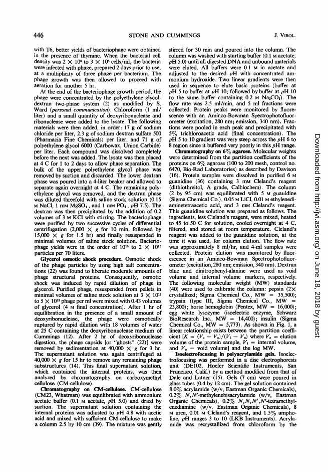
Chromatography on 6% agarose. Molecular weightswere determined from the partition coefficients of theproteins on 6% agarose (100 to 200 mesh, control no.6470, Bio-Rad Laboratories) as described by Davison(16). Protein samples were dissolved in purified 6 Mguanidine (36) containing 3 mm Cleland's reagent(dithiothreitol, A grade, Calbiochem). The column(2 by 95 cm) was equilibrated with 5 M guaiiidine(Sigma Chemical Co.), 0.05 M LiCl, 0.01 M ethylenedi-aminetetraacetic acid, and 3 mm Cleland's reagent.This guanidine solution was prepared as follows. Theingredients, less Cleland's reagent, were mixed, heatedto 45 to 50 C for solution, cooled overnight at 4 C,filtered, and stored at room temperature. Cleland'sreagent was added to the guanidine solution, at thetime it was used, for column elution. The flow ratewas approximately 8 ml/hr, and 4-ml samples werecollected. Protein elution was monitored by fluor-escence in an Aminco-Bowman Spectrophotofluor-ometer (excitation, 280 nm; emission, 340 nm). Dextranblue and dinitrophenyl-alanine were used as voidvolume and internal volume markers, respectively.The following molecular weight (MW) standards(40) were used to calibrate the column: pepsin (2Xcrystallized; Sigma Chemical Co., MW = 35,500);trypsin (type III, Sigma Chemical Co., MW =
23,800); horse hemoglobin (Pentex, MW = 16,000);egg white lysozyme (isoelectric enzyme, SchwarzBioResearch Inc., MW = 14,400); insulin (SigmaChemical Co., MW = 5,773). As shown in Fig. 1, alinear relationship exists between the partition coeffi-cient [K = (Ve - Vo)/(Vi - V.) where Ve = elutionvolume of the protein sample, Vi = internal volume,and V. = void volume] and the log MW.
Isoelectrofocusing in polyacrylamide gels. Isoelec-trofocusing was performed in a disc electrophoresisunit (DE102, Hoefer Scientific Instruments, SanFrancisco, Calif.) by a method modified from that ofDale and Latner (15). Gels (7 cm) were poured inglass tubes (0.4 by 12 cm). The gel solution contained8.0% acrylamide (w/v, Eastman Organic Chemicals),0.2% N,N'-methylenebisacrylamide (w/v, EastmanOrganic Chemicals), 0.2% N,N,N',N'-tetramethyl-enediamine (w/v, Eastman Organic Chemicals), 8M urea, 0.01 M Cleland's reagent, and 1.5% ampho-line, pH ranges 3 to 10 (LKB Instruments). Acryla-mide was recrystallized from chloroform by the
446 J. VIROL.
on June 18, 2018 by guesthttp://jvi.asm
.org/D
ownloaded from

T-EVEN BACTERIOPHAGE PROTEINS
to 30 \30
I- \TRYPSIN20
HEMOGLOBINLYSOZYME
U \
10\
INSULIN
.1 .2 .3 .4 .5 .6 .7K
FiG. 1. Chromatography ofMW standards on a 6%agarose column. Pepsin (35,500 daltons), trypsin(23,800 daltons), horse hemoglobin (16,000 daltons),egg white lysozyme (14,400 daltons), and insulin(5,773 daltons) (40) were dissolved in 6 M guanidinehydrochloride containing 3 mM Cleland's reagent andwere chromatographed on a 6% agarose column.
procedure of Bishop et al. (9). No difference in theelectrophoretic profile of the proteins was observedby using purified acrylamide. Protein samples dis-solved in not more than 0.05 ml of 10 M urea plus0.1 M Cleland's reagent were placed in the bottom ofgel tubes. Ammonium persultate (E-C ApparatusCorp.) was added at 1.4 mg/10 ml of gel solution ascatalyst. The gels were quickly poured with thoroughmixing of the protein sample throughout the gel.Usually six tubes could be poured with 10 ml of gelbefore polymerization began. The gels were given 15min to set properly and were placed in the disc elec-trophoresis unit so that the 7-cm gels were completelyimmersed in the lower water-jacketed chamber. Thebottom cathode solution was 1% ethylenediamine inwater, and the upper anode solution was 1.4% ortho-phosphoric acid. Electrophoresis was for 6 hr withan initial current of 5 mamp per tube.The gels were stained with bromphenol blue by the
method ofAwdeh (6). Minor bands were more evidentif the gels were placed in distilled water for a fewhours after destaining by the Awdeh method. Also,the bands were more stable when stored in waterthan in the destaining solution.Numerous attempts were made to calibrate the
isoelectrofocusing gels for accurate determination ofisoelectric points. This was not possible for tworeasons. First, the ampholine carrier ampholytes didnot give a perfectly linear gradient and there wereminor variations between different batches of ampho-line. Second, there were not enough proteins availablewith defined isoelectric points to span the gradient.At best, only an approximation could be made forisoelectric pH values by using the following proteinsas standards: lysozyme (pl - 10.5 to 11.0), chymo-
trypsinogen A (pI - 9.2), trypsin (pI - 10.0),bovine serum albumin (pl -. 5.1), and horse hemo-globin (pI - 6.9). The pH gradient was calibrated bya linear plot of pH versus the ratio of the position towhich a protein species migrated from the anode endof the gel over the total length of the gel. This ratiofor a given protein was reproducible, but the isoelec-tric points of the standard proteins were not definedunder the conditions used here. Thus, the accuracy ofthe estimated pH gradient with these standards wasprobably no greater than i 0.5 pH units.
Isoelectrofocusing in a sucrose gradient. The LKBelectrofocusing column (no. 8101) was used formeasurement of the isoelectric points of the basicinternal proteins. Protein samples were dissolved in10 M urea and applied in the middle of the sucrose stepgradient of the column. Ampholine carrier ampholyteswith a pH range of 7 to 10 were used. The experimentswere performed in the normal manner with the excep-tion that 8 M urea was used in the suspending medium.Electrophoresis was for 24 hr at 400 v with thecathode as the lower electrode. At the completion ofthe electrofocusing experiment, 2-ml fractions werecollected and analyzed for pH and for fluorescence inan Aminco-Bowman Spectrophotofluorometer (exci-tation, 280 nm; emission, 340 nm).
1251I labeling of proteins. Proteins were labeled with125I by the method of McConahey and Dixon (33).Amounts (mg) of the protein samples were diluted in1 ml of 0.05 M phosphate buffer (pH 7.0) in a 10-mlbeaker and stirred gently in an ice bath; 0.05 mCi of1251 in 0.05 M phosphate buffer was added. Labelingwas then initiated by the addition of 50 ,ug of chlor-amineT in phosphate buffer. The reaction was allowedto progress for 10 min and was then stopped by theaddition of 50 ,ug of sodium metabisulfite in phos-phate buffer. Guanidine (1.0 g/ml) and a smallamount of Cleland's reagent were used to dissolve thelabeled proteins, which were then fractionated by gelfiltration on the 6% agarose column. The free 1251was eluted with the internal volume (Vi) of thecolumn. Portions (1 ml) of eluted fractions werecounted in an Automatic Well Gamma Counter(Nuclear-Chicago Corp.).Amino acid analyses. Amino acid analyses were per-
formed in a Beckman-Spinco model 120 Amino AcidAnalyzer equipped with sensitive cuvettes and amodel CRS-110A Automatic Digital Integrator(Infotronics Corp., Houston).
Quantities (mg) of the protein samples werehydrolyzed in vacuo at 110 C for 24 hr in 2-ml volumesof three times distilled 6 N HCI containing 2-mercapto-ethanol and phenol (10 ml of 6 N HCI plus 5 ,literseach of 2-mercaptoethanol and liquified phenol) to re-tard destruction of tryptophan (J. M. Stewart, personalcommunication). By using egg-white lysozyme as astandard, approximately 75 to 85% recovery oftryptophan was achieved by this method. Half-cystine was determined as cysteic acid by the per-formic oxidation method of Hirs (25).
RESULTSIsolation of the basic internal proteins. The non-
sedimentable proteins released from the phage by
447VOL. 6, 1970
on June 18, 2018 by guesthttp://jvi.asm
.org/D
ownloaded from

STONE AND CUMMINGS
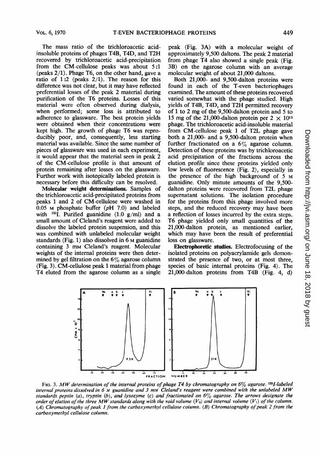
osmotic shock were fractionated on carboxy-methyl cellulose. Elution profiles for these pro-teins from phages T4, T2H, T6, and T2L areshown in Fig. 2. All phage supernatant solutionsexamined, with the exception of T2L, resulted intwo peaks of proteins which differed in pH andionic strength required for elution from thisresin. Bacteriophage T2L contained both com-ponents found in the other viruses, but theyeluted too closely together to be separated by themethod used here (Fig. 2D). The material fromthe T2L peak was further fractionated by gelfiltration on 6% agarose.
Fractions across peaks 1 and 2 were separatelypooled, and the proteins were precipitated with5% trichloroacetic acid. The proteins in thesepeaks appeared to contain all of the internalproteins released by osmotic shock. No othertrichloroacetic acid-insoluble material was de-tected across the elution profile other than occa-
A
120
2100
00
20
1.)
zui 20 4, 600 80 00o 20
n
O24
sional trailing from one of these peaks. In allcases examined, the material from peak trails wasidentical to that of the preceding peak. Nofurther trichloroacetic acid-insoluble materialwas released from the CM-cellulose upon theaddition of 0.5 N NaOH solution containing 0.5M NaCi. The void volume of the column con-tained only minor amounts of phage structuralproteins and small amounts of peak 1 material.Thus, no acid-insoluble acidic internal proteinswere found in any of the phage examined.As will become evident in the electrophoretic
studies, the CM-cellulose column did not alwaysfractionate the peaks cleanly. As a result, smallamounts of the peak 2 proteins could be demon-strated in peak 1 and very minor amounts of thepeak 1 material were recovered from the voidvolume. Somewhat better separations occurredby using step gradients, but splitting of each ofthe two peaks was observed with this method.
FRACTION NUMBER
FIG. 2. Chromatography ofthe internal proteins on carboxymethyl cellulose. Supernatant solutions from osmoticshock ofthe bacteriophages were equilibrated with sufficient carboxymethyl cellulose to pour a column 2.5 by 10 cm.The column was washed with starting buffer (0.1 M acetate, pH 5.0) until all unbound materials and digested DNAwere eluted. Separation was then achieved with two linear gradients. The first gradient (buffer at pH S to bufferat pH JO) covered approximately fractions I to 60, followed by the second gradient (buffer at pH 10 to the samebuffer containing 0.2 Af Na2CO3). Variations occurred in the gradient volumes, resulting in displacement ofpeaksin different experiments.
448 J. VIROL.
on June 18, 2018 by guesthttp://jvi.asm
.org/D
ownloaded from
T-EVEN BACTERIOPHAGE PROTEINS
The mass ratio of the trichloroacetic acid-insoluble proteins of phages T4B, T4D, and T2Hrecovered by trichloroacetic acid-precipitationfrom the CM-cellulose peaks was about 5:1(peaks 2/1). Phage T6, on the other hand, gave aratio of 1:2 (peaks 2/1). The reason for thisdifference was not clear, but it may have reflectedpreferential losses of the peak 2 material duringpurification of the T6 proteins. Losses of thismaterial were often observed during dialysis,when performed; some loss is attributed toadherence to glassware. The best protein yieldswere obtained when their concentrations werekept high. The growth of phage T6 was repro-ducibly poor, and, consequently, less startingmaterial was available. Since the same number ofpieces of glassware was used in each experiment,it would appear that the material seen in peak 2of the CM-cellulose profile is that amount ofprotein remaining after losses on the glassware.Further work with isotopically labeled protein isnecessary before this difficulty can be resolved.Molecular weight determinations. Samples of
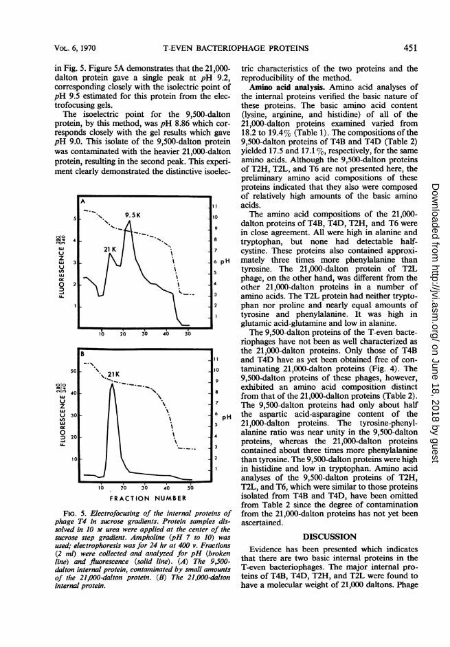
the trichloroacetic acid-precipitated proteins frompeaks 1 and 2 of CM-cellulose were washed in0.05 M phosphate buffer (pH 7.0) and labeledwith "2I. Purified guanidine (1.0 g/ml) and asmall amount of Cleland's reagent were added todissolve the labeled protein suspension, and thiswas combined with unlabeled molecular weightstandards (Fig. 1) also dissolved in 6 M guanidinecontaining 3 mm Cleland's reagent. Molecularweights of the internal proteins were then deter-mined by gel filtration on the 6% agarose column(Fig. 3). CM-cellulose peak 1 material from phageT4 eluted from the agarose column as a single
peak (Fig. 3A) with a molecular weight ofapproximately 9,500 daltons. The peak 2 materialfrom phage T4 also showed a single peak (Fig.3B) on the agarose column with an averagemolecular weight of about 21,000 daltons.Both 21,000- and 9,500-dalton proteins were
found in each of the T-even bacteriophagesexamined. The amount of these proteins recoveredvaried somewhat with the phage studied. Highyields of T4B, T4D, and T2H permitted recoveryof 1 to 2 mg of the 9,500-dalton protein and 5 to15 mg of the 21,000-dalton protein per 2 x 10¹⁵phage. The trichloroacetic acid-insoluble materialfrom CM-cellulose peak 1 of T2L phage gaveboth a 21,000- and a 9,500-dalton protein whenfurther fractionated on a 6% agarose column.Detection of these proteins was by trichloroaceticacid precipitation of the fractions across theelution profile since these proteins yielded onlylow levels of fluorescence (Fig. 2), especially inthe presence of the high background of 5 Mguanidine. Only minute amounts of the 9,500-dalton proteins were recovered from T2L phagesupernatant solutions. The isolation procedurefor the proteins from this phage involved moresteps, and the reduced recovery may have beena reflection of losses incurred by the extra steps.T6 phage yielded only small quantities of the21,000-dalton protein, as mentioned earlier,which may have been the result of preferentialloss on glassware.
Electrophoretic studies. Electrofocusing of theisolated proteins on polyacrylamide gels demon-strated the presence of two, or at most three,species of basic internal proteins (Fig. 4). The21,000-dalton proteins from T4B (Fig. 4, d)
FRACTION NUMBER
FIG. 3. MW derermination of the internal proteins ofphage T4 by chromatography on 6% agarose. 125I-labeledinternal proteins dissolved in 6 M guanidine and 3 mm Cleland's reagent were combined with the unlabeled MWstandards pepsin (a), trypsin (b), and lysozyme (c) and fractionated otn 6% agarose. The arrows designate theorder ofelution of the three MW standards along with the void volume (Vo) and internal volume (Vi) of the column.(A) Chromatography ofpeak I from the carboxymethyl cellulose column. (B) Chromatography ofpeak 2 from thecarboxymethyl cellulose column.
VOL. 6, 1970 449
on June 18, 2018 by guesthttp://jvi.asm
.org/D
ownloaded from

STONE AND CUMMINGS
Electrofocusing Gels of the internal Proteins
21K r 9.5K
pH 9.5
FIG. 4. Electrofocusing of the internal proteins onpolyacrylamide gels. Electrophoresis of the proteinsamples mixed throughout the gels was for 6 hr at 400v with pH 3 to 10 range ampholine. Cathode (-) was
at the top. Gels of the 21,000-dalton proteins are (a)T2H, (b) T2L, (c) T4BO0, (d) T4B, and (e) T4 lysozyme.Gels of the 9,500-daltons proteins read (f) T2H, (g)T6, and (h) T4D. Variations in gel lengths occurredresulting in displacement of some minor bands. Thegels were aligned by isoelectric points of the majorbands.
and T2H (Fig. 4, a) gave single bands by thismethod at approximately pH 9.5. The 21,000-dalton protein from T2L phage (Fig. 4, b),however, exhibited a minor band at this pH anda major band at about pH 9.0. As will be dis-cussed later, major differences in the amino acidcomposition were also noted between the 21,000-dalton protein of T2L and those of the otherphages. The 21,000-dalton protein of T6 phagewas not examined on gels since the small amountisolated was used for amino acid analysis.
Included in Fig. 4 is a gel of the 21,000-daltonprotein from phage T4B01 (Fig. 4, c). Thisisolate was prepared by direct trichloroaceticacid precipitation of the phage supernatant solu-tion after sedimentation of the phage capsids. Itwas thus contaminated by small amounts of themore acidic 18,000-dalton structural proteins ofthe phage head (21; G. Forrest and D. Cum-mings, unpublished data), illustrating the necessityfor identification of other minor proteins releasedby osmotic shock.
Finally, an electrofocusing gel of T4 lysozyme(lot 802025, Calbiochem) has been included(Fig. 4, e). It is of interest that this protein with amolecular weight similar to the 21,000-daltonprotein should also have the same isoelectricpoint. This similarity will be considered ingreater detail later.
Figure 4 also shows representative electro-focusing gels of the 9,500-dalton internal pro-
teins. It is clear that in many of the cases ex-amined, the 9,500-dalton proteins exhibited botha major and several minor bands. The majorband of all of the 9,500-dalton proteins (Fig. 4;f, g, h) was at approximately pH 9.0, whereas theminor bands of T2H and T6 were in the region ofthe 21,000-dalton protein. By "2I labeling and gelfiltration on 6% agarose, these minor bands havebeen shown to be the result of contamination ofthe CM-cellulose peak 1 with the 21,000-daltonmaterial from peak 2. The 21,000-dalton proteinof T2L (Fig. 4, b), on the other hand, was at thesame position as the 9,500-dalton proteins. Thiswould be expected from its behavior on the CM-cellulose column.The electrofocusing gel of the 9,500-dalton
protein from T6 phage (Fig. 4, g) illustratesagain how minor contaminants can be identifiedby this method. In this case, several of the struc-tural capsid proteins eluted in CM-cellulose peak1 (13, 14, 21; G. Forrest and D. Cummings,unpublished data). Only the head structural11,000-dalton protein had an isoelectric pointnear the internal proteins (21; G. Forrest and D.Cummings, unpublished data). It gave an iso-electric point of approximately pH 7, near themiddle of the gel.
Structural proteins isolated from the heads ofthe T4 amber mutants AmN85 (G48) andAmH21(G54) were also examined on isoelectrofocusinggels. Both of these mutant phages produced freeheads and tail plates when grown under restrictiveconditions (R. S. Edgar, personal communication).Free heads devoid of DNA were separated fromtail plates and isolated as described previously(13, 14, 21) and were examined without osmoticshock treatment. Both the 9,500- and 21,000-dalton internal proteins were present in theseheads. It is of interest that these empty headscontained the internal proteins and may indicateeither that the presence of DNA is not necessaryfor their occurrence or that the heads originallycontained DNA which was digested by deoxy-ribonuclease treatment during purification. Themajor point to be made is that these internalproteins could not have originated from tailsubstructures released during osmotic shock.The internal proteins were also electrofocused
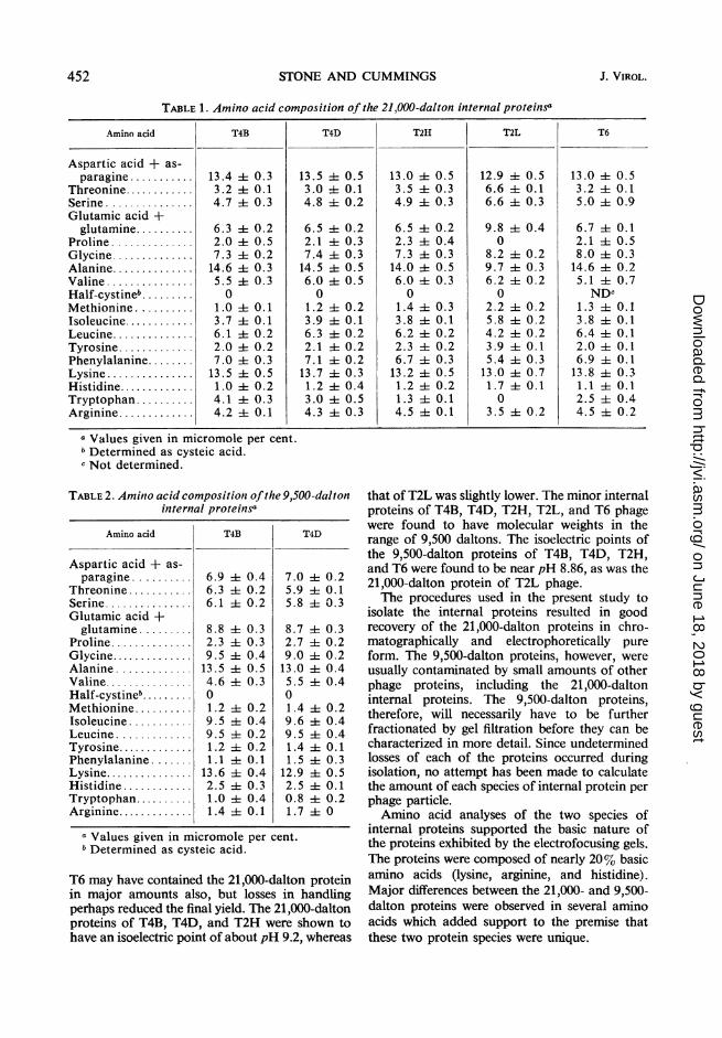
in sucrose gradients by using the LKB electro-focusing column to determine more preciselytheir isolectric points. This instrument alloweddirect measurement of the pH across the ampho-line gradient, whereas the pH gradient of the gelswas estimated from positions to which proteinsof known isoelectric points migrated. The resultsobtained for the 21,000- and 9,500-dalton pro-teins of T4B from the electrofocusing column byusing pH 7 to 10 range ampholine are presented
450 J. VIROL.
on June 18, 2018 by guesthttp://jvi.asm
.org/D
ownloaded from
VOL.6,1970T-EVENBACTERIOPHAGE PROTEINS45
in Fig. 5. Figure 5A demonstrates that the 21,000--dalton protein gave a single peak at pH 9.2,corresponding closely with the isolectric point ofpH 9.5 estimated for this protein from the elec-trofocusing gels.The isoelectric point for the 9,500-dalton
protein, by this method, was pH 8.86 which cor-responds closely with the gel results which gavepH 9.0. This isolate of the 9,500-dalton proteinwas contaminated with the heavier 21,000-daltonprotein, resulting in the second peak. This experi-ment clearly demonstrated the distinctive isoelec-
A
9.5K 10
9
4 ~~~~~~~~~~~~~~8ui ~~21 K 7
U.' 3 6p
0 2 4
2
10 20 30 40 50
B
50 I10\..21K
u 7
LU ~~~~~~6pU30pH4,,ui ~~~~~~~~~~~~~~~5
0 4D 20
U. 3
10 2
A02 30 4~0 50
FRACTION NUMBER
FiG. 5. Electrofocusing of the internal proteins ofphage T4 in sucrose gradients. Protein samples dis-solved in 10 m urea were applied at the center of thesucrose step gradient. Ampholine (pH 7 to 10) wasused; electrophoresis was for 24 hr at 400 v. Fractions(2 ml) were collected and analyzed for pH (brokenline) and fluorescence (solid line). (A) The 9,500-dalton internal protein, contamninated by small amountsof the 21,000-dalton protein. (B) The 21,000-datoninternal protein.
tric characteristics of the two proteins and thereproducibility of the method.Amino acid analysis. Amino acid analyses of
the internal proteins verified the basic nature ofthese proteins. The basic amino acid content(lysine, arginine, and histidine) of all of the21,000-dalton proteins examined varied from18.2 to 19.4% (Table 1). The compositions of the9,500-dalton proteins of T4B3 and T4D3 (Table 2)yielded 17.5 and 17.1%, respectively, for the sameamino acids. Although the 9,500-dalton proteinsof T2H, T2L, and T6 are not presented here, thepreliminary amino acid compositions of theseproteins indicated that they also were composedof relatively high amounts of the basic aminoacids.The amino acid compositions of the 21,000-
dalton proteins of T4B, T41D, T2H4, and T6 werein close agreement. All were high in alanine andtryptophan, but none had detectable half-cystine. These proteins also contained approxi-mately three times more phenylalanine thantyrosine. The 21,000-dalton protein of T2Lphage, on the other hand, was different from theother 21,000-dalton proteins in a number ofam-ino acids. The T2L, protein had neither trypto-phan nor proline and nearly equal amounts oftyrosine and phenylalanine. It was high inglutamic acid-glutamine and low in alanine.The 9,500-dalton proteins of the T-even bacte-
riophages have not been as well characterized asthe 21,000-dalton proteins. Only those of T4B3and T4D3 have as yet been obtained free of con-taminating 21,000-dalton proteins (Fig. 4). The9,500-dalton proteins of these phages, however,exhibited an amino acid composition distinctfrom that of the 21,000-dalton proteins (Table 2).T'he 9,500-dalton proteins had only about halfthe aspartic acid-asparagine content of the21,000-dalton proteins. The tyrosine-phenyl-alanine ratio was near unity in the 9,500-daltonproteins, whereas the 21,000-dalton proteinscontained about three times more phenylalaninethan tyrosine. The 9,500-dalton proteins were highin histidine and low in tryptophan. Amino acidanalyses of the 9,500-dalton proteins of T2H4,T2L, and T6, which were similar to those proteinsisolated from T413 and T41D, have been omittedfrom Table 2 since the degree of contaminationfrom the 21,000-dalton proteins has not yet beenascertained.
DISCUSSIONEvidence has been presented which indicates
that there are two basic internal proteins in theT-even bacteriophages. The major internal pro-teins of T4B3, T4D3, T2H, and T2L, were found tohave a molecular weight of 21,000 daltons. Phage
VOL. 6, 1970 451
on June 18, 2018 by guesthttp://jvi.asm
.org/D
ownloaded from
STONE AND CUMMINGS
TABLE 1. Amino acid composition of the 21,000-dalton internal proteinsa
Amino acid T4B T4D T2H T2L T6
Aspartic acid + as-paragine ........... 13.4 it0.3 13.5 4- 0.5 13.0 it 0.5 12.9 it0.5 13.0 i0.5
Threonine............ 3.2 4t 0.1 3.0 1-- 0.1 3.5 ±- 0.3 6.6 it0.1 3.2 i0.1Serine............... 4.7 i0.3 4.8 -- 0.2 4.9 it0.3 6.6 -0.3 5.0 ± 0.9Glutamic acid +glutamine .......... 6.3 i 0.2 6.5 -- 0.2 6.5 it 0.2 9.8 i 0.4 6.7 ±4 0.1
Proline .............. 2.0 -0.5 2.1 -- 0.3 2.3 it0.4 0 2.1 it0.5Glycine.............. 7.3 i0.2 7.4 ±i 0.3 7.3 it 0.3 8.2 i0.2 8.0 ±t0.3Alanine .............. 14.6 i 0.3 14.5 ±t 0.5 14.0 ± 0.5 9.7 ±t 0.3 14.6 it 0.2Valine............... 5.5 -0.3 6.0 -- 0.5 6.0 i0.3 6.2 -- 0.2 5.1 -t 0.7Half-cystineb ......... 0 0 0 0 NDcMethionine .......... 1.0 4t 0.1 1.2 -- 0.2 1.4 it 0.3 2.2 it0.2 1.3 4- 0.1Isoleucine............ 3.7 4- 0.1 3.9 4t 0.1 3.8 ±t0.1 5.8 it0.2 3.8 -- 0.1Leucine.............. 6.1 it 0.2 6.3 -- 0.2 6.2 4t 0.2 4.2 -0.2 6.4 -0.1Tyrosine............. 2.0 4- 0.2 2.1 -- 0.2 2.3 it0.2 3.9 i0.1 2.0 it 0.1Phenylalanine........ 7.0 it 0.3 7.1 it0.2 6.7 ±t0.3 5.4 it0.3 6.9 -t 0.1Lysine..... 13.5 4- 0.5 13.7 ± 0.3 13.2 -- 0.5 13.0 ± 0.7 13.8 i- 0.3Histidine............. 1.0 it 0.2 1.2 it0.4 1.2 -0.2 1.7 -0.1 1.1 -0.1Tryptophan .......... 4.1 i 0.3 3.0 i 0.5 1.3 i 0.1 0 2.5 i 0.4Arginine............. 4.2 + 0.1 4.3 i 0.3 4.5 4 0.1 3.5 -h 0.2 4.5 ± 0.2
a Values given in micromole per cent.Determined as cysteic acid.
c Not determined.
TABLE 2. Amino acid composition ofthe 9,500-daltoninternal proteinsa
Amino acid T4B T4D
Aspartic acid + as-paragine .......... 6.9 ± 0.4 7.0 4 0.2
Threonine ........... 6.3 i 0.2 5.9 A1 0.1Serine............... 6.1 ± 0.2 5.8 i 0.3Glutamic acid +glutamine......... 8.8 i 0.3 8.7 i4 0.3
Proline.............. 2.3 ± 0.3 2.7 ± 0.2Glycine.............. 9.5 i 0.4 9.0 i 0.2Alanine ............. 13.5 i1 0.5 13.0 i: 0.4Valine............... 4.6 i0.3 5.5 i 0.4Half-cystineb......... 0 0Methionine.......... 1.2 i 0.2 1.4 4 0.2Isoleucine ........... 9.5 i 0.4 9.6 i 0.4Leucine ............. 9.5 ±t0.2 9.5 ± 0.4Tyrosine............. 1.2 ±t0.2 1.4 i 0.1Phenylalanine ....... 1.1 i4 0.1 1.5 ± 0.3Lysine............... 13.6 i 0.4 12.9 i 0.5Histidine ............ 2.5 4± 0.3 2.5 i 0.1Tryptophan.......... 1.0 ± 0.4 0.8 4 0.2Arginine............. 1.4 -t0.1 1.7 i 0
a Values given in micromole per cent.b Determined as cysteic acid.
T6 may have contained the 21,000-dalton proteinin major amounts also, but losses in handlingperhaps reduced the final yield. The 21,000-daltonproteins of T4B, T4D, and T2H were shown tohave an isoelectric point of about pH 9.2, whereas
that of T2L was slightly lower. The minor internalproteins of T4B, T4D, T2H, T2L, and T6 phagewere found to have molecular weights in therange of 9,500 daltons. The isoelectric points ofthe 9,500-dalton proteins of T4B, T4D, T2H,and T6 were found to be near pH 8.86, as was the21,000-dalton protein of T2L phage.The procedures used in the present study to
isolate the internal proteins resulted in goodrecovery of the 21,000-dalton proteins in chro-matographically and electrophoretically pureform. The 9,500-dalton proteins, however, wereusually contaminated by small amounts of otherphage proteins, including the 21,000-daltoninternal proteins. The 9,500-dalton proteins,therefore, will necessarily have to be furtherfractionated by gel filtration before they can becharacterized in more detail. Since undeterminedlosses of each of the proteins occurred duringisolation, no attempt has been made to calculatethe amount of each species of internal protein perphage particle.Amino acid analyses of the two species of
internal proteins supported the basic nature ofthe proteins exhibited by the electrofocusing gels.The proteins were composed of nearly 20% basicamino acids (lysine, arginine, and histidine).Major differences between the 21,000- and 9,500-dalton proteins were observed in several aminoacids which added support to the premise thatthese two protein species were unique.
452 J. VIROL.
on June 18, 2018 by guesthttp://jvi.asm
.org/D
ownloaded from

T-EVEN BACTERIOPHAGE PROTEINS
One of the major problems encountered in thepresent study has been identification of theinternal proteins. This is evident in Fig. 4, whichshows that the isolates of these proteins can becontaminated by a variety of phage structuralproteins. Simultaneous investigations of otherphage structures in this laboratory (13, 14, 21)have been invaluable in this respect. Thus, con-tamination of the internal protein preparations by18,000- and 11,000-dalton head structural pro-teins were easily monitored on the electrofocusinggels. Three procedures employed in the presentstudy have greatly reduced the level of contamina-tion with other phage proteins. Osmotic shockwith glycerol, instead of high salt, greatly de-creased the amount of head structural proteinsreleased. At the same time, osmotic shock inducedby the glycerol method resulted in ionic conditionssuitable for chromatography of the proteins onthe carboxymethyl cellulose. Use of this resinallowed recovery of the 21,000-dalton proteinvirtually free of contamination from the moreacidic 18,000-dalton head proteins. Finally,centrifugation of the osmotic shock supernatantsolution for 15 hr after removal of the capsidsresulted in quantitative removal of free tailcomponents and unsedimented capsids whichwould have accounted for a significant proportionof the proteins isolated.
Additional support for the belief that the twoproteins isolated were indeed internal proteinsof the phage head came from preliminary studiesof proteins isolated from two amber mutants ofT4. Electrofocusing gels of proteins isolated frompurified heads of AmN85 (G48) and AmH21(G54) clearly demonstrated the presence of thetwo internal proteins. It should also be pointedout that Black (L. Black, personal communication)found that there were two, and possibly three,internal proteins in T4D, having propertiessimilar to those properties reported here. Also,Champe (S. Champe, personal communication)has indicated that these internal proteins werephage specific and accumuilated during the time-course of infection.
Correlation of the previous findings with thoseof other laboratories has been difficult since it isnot known if the other groups isolated both ofthe internal proteins together or only one of thetwo proteins. Levine et al. (31) reported isolationof the internal antigen in an electrophoretically,chromatographically, and immunochemicallypure form. Coval et al. (Fed. Proc. 19:253, 1960)stated that this protein had an amino acid com-position which was high in lysine and histidineand contained no cysteine. Both the 9,500-daltonprotein isolated in the present study and the11,000-dalton head structural protein (G. Forrest
and D. Cummings, unpublished data) would fitthis brief description. The composition of the21,000-dalton internal protein, however, wasclearly not high in histidine.Four studies reported the possible existence of
multiple internal proteins. Levine (32) reportedthat the 31S-labeled nonsedimentable proteinsreleased by osmotic shock were separated bychromatography and yielded label in severalfractions, only one of which possessed antigenicactivity. Minagawa (34) also found that 20% ofthe 35S-labeled nonsedimentable proteins of T2were not antigenically active against antibodyproduced against ruptured phage. RecentlyBachrach et al. (8) found one major and twominor proteins could be isolated with phage DNAreleased by Sarkosyl rupture of the phage headmembrane. Kokurina and Tikhonenko (29)fractionated ruptured phage on Sephadex G-200and found four fractions which reacted to anti-body prepared against ruptured phage. Only twoof these four fractions lacked cross-reactivity tocapsid proteins.
Earlier in this report, the close similarities ofthe 21,000-dalton internal protein and T4 lyso-zyme were noted. The two proteins possess similarmolecular weights and identical isolectric points.The amino acid compositions of the two proteinsare quite different, however (27, 42). Severalearlier reports suggested the presence of a lyso-zyme-like activity in phage lysates and internalprotein preparations (26, 32, 34, 37, 38). Panijel(26, 37, 38) found a "prolysine" activity, capableof lysing acetone powders of bacteria, which wasreleased from purified phage by osmotic shock.Levine (32), however, reported that the lyticactivity of Panijel and the internal antigen wereseparable by chromatography. Minagawa (34)found that lysozyme activity was present ininternal protein preparations prepared by osmoticshock with high salt, but were absent whenglycerol was substituted. In the latter case, thelysozyme activity was found associated with thecapsids. Emrich and Streisinger (20) recentlyfound low levels of a new lytic activity in purifiedphage carrying deletions of the e gene. The phageused were es mutants [s = spackle; the spacklemutation allowed e gene mutants to lyse, releasingfree phage (19)]. The low level of lytic activityobserved was not blocked by antibody to the egene lysozyme, suggesting the presence of asecond species of lysozyme in purified phage.
Several other possible functions for the internalproteins have been proposed; these include a rolein organization of the phageDNA during matura-tion (28), possible involvement in regulation oftranscription ofthe phage DNA (11), and restora-tion of the membrane function after phage infec-
VOL. 6, 1970 453
on June 18, 2018 by guesthttp://jvi.asm
.org/D
ownloaded from

STONE AND CUMMINGS
tion (17). Several of these theories, and otherpossibilities, will be the subject of future study byusing the two proteins isolated here.
ACKNOWLEDGMENTS
We are indebted to Selina Janion for translation of the Koku-rina and Tikhonenko paper.
This investigation was supported by Public Health Servicegrants Al-06472 and AI-08265 from the National Institute ofAllergy and Infectious Diseases and GM-01379 from the Na-tional Institute of General Medical Sciences.
LITERATURE CITED
1. Adams, M. H. 1959. Bacteriophages. Interscience PublishersInc., New York.
2. Albertsson, P.-A. 1960. Partition of cell particles and macro-molecules. John Wiley and Sons, New York.
3. Ames, B. N., D. T. Dubin, and S. M. Rosenthal. 1958. Pres-ence of polyamines in certain bacterial viruses. Science(Washington) 127:814-816.
4. Anderson, T. F. 1945. The role of tryptophane in the adsorp-tion of two bacterial viruses on their host, E. coli. J. Cell.Comp. Physiol. 25:17-26.
5. Anderson, T. F. 1948. The activation of the bacterial virusT4 by L-tryptophan. J. Bacteriol. 55:637-649.
6. Awdeh, Z. L. 1969. Staining method for proteins after iso-electric focusing in polyacrylamide gel. Sci. Tools 16:42-43.
7. Bachrach, U., and A. Friedmann. 1967. Purification and somepossible functions of internal proteins from coliphage T2.Biochem. Biophys. Res. Commun. 26:596-601.
8. Bachrach, U., R. Levin, and A. Friedmann. 1970. Studies onphage internal proteins: isolation of protein-DNA com-plexes from T2 phages and from phage-infected bacteria.Virology 40:882-892.
9. Bishop, D. H. L., J. R. Claybrook, and S. Spiegelman. 1967.Electrophoretic separation of viral nucleic acids on poly-acrylamide gels. J. Mol. Biol. 26:373-387.
10. Champe, S. P., and H. L. Eddleman. 1967. Polypeptides asso-ciated with morphogenic defects in bacteriophage T4,p. 55-70. In J. S. Colter and W. Paranchych (ed.), Themolecular biology of viruses. Academic Press Inc., NewYork.
11. Chaproniere-Rickenberg, D. M., H. R. Mahler, and D.Fraser. 1964. The interaction of DNA and internal proteinfrom coliphage T2. Virology 23:96-102.
12. Cummings, D. J. 1963. Subunit basis of head configurationalchanges in T2 bacteriophage. Biochim. Biophys. Acta68:472-480.
13. Cummings, D. J., A. R. Kusy, V. A. Chapman, S. S. DeLong,and K. R. Stone. 1970. Characterization of T-even bac-teriophage substructures. I. Tail fibers and tail tubes. J.Virol. 6:534-544.
14. Cummings, D. J., V. A. Chapman, S. S. DeLong, A. R.Kusy, and K. R. Stone. 1970. Characterization of T-evenbacteriophage substructures. II. Tail plates. J. Virol. 6:545-554.
15. Dale, G., and A. L. Latner. 1968. Isoelectric focusing inpolyacrylamide gels. Lancet 1:847-848.
16. Davison, P. F. 1968. Proteins in denaturing solvents: gel ex-
clusion studies. Science 161:906-907.17. Duckworth, D. H. 1970. The metabolism of T4 phage ghost-
infected cells. I. Macromolecular synthesis and transportof nucleic acid and protein precursors. Virology 40:673-684.
18. Eddleman, H. L., and S. P. Champe. 1966. Components in
T4-infected cells associated with phage assembly. Virology30:471-481.
19. Emrich, J. 1968. Lysis of T4-infected bacteria in the absenceof lysozyme. Virology 35:158-165.
20. Emrich, J., and G. Streisinger. 1968. The role of phage lyso-zyme in the life cycle of phage T4. Virology 36:387-391.
21. Forrest, G. L., and D. J. Cummings. 1970. Head proteinsfrom T-even bacteriophage. I. Molecular weight charac-terization. J. Virol. 5:398-405.
22. Herriott, R. M., and J. L. Barlow. 1957. The protein coatsor 'ghosts' of coliphage T2. J. Gen. Physiol. 40:809-825.
23. Hershey, A. D. 1955. An upper limit to the protein content ofthe germinal substance of bacteriophage T2. Virology1:108-127.
24. Hershey, A. D. 1957. Some minor components of bacterio-phage T2 particles. Virology 4:237-264.
25. Hirs, C. H. W. 1956. The oxidation of ribonuclease withperformic acid. J. Biol. Chem. 219:611-621.
26. Huppert, J., and J. Panijel. 1956. Recherches sur les proly-sines. II. Le cas gen6ral de la synthese des prolysines. Ann.Inst. Pasteur 90:711-727.
27. Inouye, M., and A. Tsugita. 1968. Amino acid sequence ofT2 phage lysozyme. J. Mol. Biol. 37:213-223.
28. Kellenberger, E. 1961. Vegetative bacteriophage and thematuration of the virus particles. Advan. Virus Res. 8:1-61.
29. Kokurina, N. K., and T. 1. Tikhonenko. 1969. Isolation andfractionation of inner proteins of T2 and DDVI bacterio-phages. Vop. Virusol. 2:224-228.
30. Kozloff, L. M., and M. Lute. 1960. Calcium content of bac-teriophage T2. Biochim. Biophys. Acta 37:420-424.
31. Levine, L., J. L. Barlow, and H. Van Vunakis. 1958. An inter-nal protein in T2 and T4 bacteriophages. Virology 6:702-717.
32. Levine, L. 1961. Immunochemical nature of the internalmaterial in the T-even coliphages, p. 171-182. In M. Heidel-berger and 0. J. Plescia (ed.), Immunochemical approachesto problems in microbiology. The Rutgers UniversityPress, New Brunswick.
33. McConahey, P. J., and F. J. Dixon. 1966. A method of traceiodination of proteins for immunologic studies. Int. Arch.Allergy 29:185-189.
34. Minagawa, T. 1961. Some characteristics of the internal pro-tein phage T2. Virology 13:515-527.
35. Murakami, W. T., H. Van Vunakis, and L. Levine. 1959.Synthesis of T2 internal protein in infected Escherichia coli,strain B. Virology 9:624-635.
36. Nozaki, Y., and C. Tanford. 1967. Acid-base titrations inconcentrated guanidine hydrochloride. Dissociation con-stants of the guanidinium ion and of some amino acids.J. Amer. Chem. Soc. 89:736-742.
37. Panijel, J., and J. Huppert. 1956. Recherches sur les proly-sines. 1. La prolysine du phage Fcz. Ann. Inst. Pasteur90:619-636.
38. Panijel, J. 1959. Les activites enzymatiques liees aux phageset a la synthese phagioue. Ann. Inst. Pasteur 97:198-217.
39. Rhodes, M. B., P. R. Azari, and R. E. Feeney. 1958. Analysis,fractionation, and purification of egg white proteins withcellulose cation exchanger, J. Biol. Chem. 230:399-408.
40. Sober, H. A. 1968. Handbook of biochemistry. The ChemicalRubber Co., Cleveland.
41. Stemnberg, N., and S. P. Champe. 1969. Genetic determinantof an internal peptide of bacteriophage T4. J. Mol. Biol.46:377-392.
42. Tsugita, A., and M. Inouye. 1968. Complete primary struc-ture of phage lysozyme from Escherichia coli T4. J. Mol.Biol. 37:201-212.
454 J. VIROL.
on June 18, 2018 by guesthttp://jvi.asm
.org/D
ownloaded from