isolation and characterization of bacteriocinproducing...
TRANSCRIPT

International Journal of ChemTech Research CODEN (USA): IJCRGG ISSN : 0974-4290
Vol.6, No.4, pp 2507-2520, July-Aug 2014
Isolation and Characterization of BacteriocinProducing Lactic Acid Bacteria from some Syrian fermented foods
Iman Askoul1*, Saiah Abo Gorrah2, Lina Al-Amir1
1National Commission for Biotechnology- Syria.
2Food Science Department - Faculty of agriculture - Damascus University- Syria.
*Corres. author: [email protected]
Abstract: A total of 25 isolates of lactic acid bacteria from different Syrian dairy products, pickels and a dried plant samples were obtained and tested for their antibacterial activity against four pathogens one gram positive bacteria (Staphylococcus aureus) and three gram negative bacteria (Klebsiella pneumoniae, Escherichiacoli and Pseudomonas aeruginosa). The cell free supernatants of fifteen LAB isolates demonstrated significant antibacterial activity against the four tested pathogens. Results of the standard physiological and biochemical tests identified fourteen isolates as Lactobacillus plantarum and one isolate as Lactobacillus fermentum.All fifteen isolates were mesophilic and were able to grow in the presence of 4 % NaCl and in pH values ranging from 4-9.The antibacterial activity of the CFSsof the most active six isolates were significantly influenced after treatment with proteinase K and after neutralization, while no change occurred after heat treatment at 60°C and 80°C for 15 minutes.Two of the tested L. plantarumisolates (SH4 and CK56) had the structural genes for both bacteriocins plantaricin EF and plantaricin N, and two of them (CK57 and CP44) had the structural genes for plantaricin N only. Key words: Lactic acid bacteria, Plantaricin, Antibacterial effect.
Introduction
Lactic acid bacteria (LAB) are among the most important groups of microorganisms used in food fermentation where they play an essential role and a wide variety of strains are routinely employed as starter cultures in the manufacture of dairy, meat, vegetable and bakery products1,2. One of the most important contributions of these microorganisms is the extended shelf life of the fermented products. Growth of spoilage and pathogenic bacteria in these foods is inhibited due to competition for nutrients and the presence of starter-derived inhibitors such as lactic acid, hydrogen peroxide, diacetyl and bacteriocins1,3.
Bacteriocins are antimicrobial peptides or proteins produced by strains of diverse bacterial species. The antimicrobial activity of this group of natural substances against foodborne pathogens, as well as spoilage bacteria, has raised considerable interest for their application in food preservation1,4,5. In the past years, a lot of work has aimed to detect, purify and characterize bacteriocins, as well as their application in food preservation strategies. Application of bacteriocins may help reduce the use of chemical preservatives and/or the intensity of heat and other physical treatments, satisfying the demands of consumers for foods that are fresh tasting, ready to eat, and lightly preserved. In recent years, considerable effort has been made to develop food applications for many different bacteriocins using bacteriocinogenic strains1,3,5,6.
According to the importance of bacteriocins as food preservatives we will focus in this study on the isolation and characterization of bacteriocin producing local lactic acid bacteria isolates, beside the activity of these strains against several spoilage and pathogenic bacteria, choosing the best isolate which which has the best antibacterial activity.

Iman Askoul et al /Int.J. ChemTech Res.2014,6(4),pp 2507-2520.
2508
Materials and methods
Isolation and of lactic acid bacteria
Fifty one samples were collected from different sources of local foods including: Dairy products, pickles, dried plant. Samples were cultured using the dilution pour plate method, for this purpose, 2 grams of each sample were weighed aseptically and homogenized in 18 ml of sterile salt solution(0.1% NaCl) using the vortex. Then, sequential decimal dilutions of the homogenate were obtained. One ml of each aliquot dilution was used, for the isolation of LAB surface plated MRS agar and M17 agar were incubated anaerobically for 72 h at 37°C, plates with colony forming units (CFU) ranging from 30 and 300 were selected for enumeration7.The colonies were randomly picked from plate and purified by successive streaking on MRS agar media before being subjected to characterization.
Gram-positive and catalase-negative isolates were isolated and characterized by phenotypic criteria8. The isolates were stocked on MRS agar slant at4°C and sub-cultured monthly.
Test micro organisms
Pathogens used for testing antibacterial activity were gram positive bacteria (Staphylococcus aureus) and gram negative bacteria (Klebsiella pneumoniae, Escherichia coli and Pseudomonas aeruginosa) received from the collection of microbiology laboratory (National Commission for Biotechnology, Syria).
Antibacterial activity of isolated LAB
The pathogenic bacteria were inoculated on nutrient blood agar then in nutrient broth for 24 hours and incubated at 37°C. Lactic acid bacteria were inoculated in MRS broth for bacilli and M17 for cocci for 48 hours at 37°C. Five microliters of each tested isolate were spotted on MRS/M17 agar, plates were incubate for 48 hours anaerobically at37°C, a layer of Mueller Hinton agar was poured and left till it could well solidify, plates were then left in the refrigerator (4°C) for about two hours to allow the antibacterial substance to disperse on the Mueller Hinton agar layer, the indictor (target) isolates were then spread with cotton swap on the Mueller Hinton agar. The plates were then incubate 24 hours aerobically at 37°C, bacteriocin-positive cultures displayed a halo of clearing in the lawn around the original button of growth, bacteriocin-positive isolates were selected for further study9, 10.
Antibacterial Activity of the Cell Free Supernatant
Lactic acid bacteria were inoculated in MRS broth for 48 hours at 37°C. Cell free supernatants were obtained by centrifugation of the liquid culture (8000 rpm for 20 minutes at 4°C). For screening of the antibacterial activity of the cell free supernatants, they were tested for their antibacterial activity using the well diffusion method11with some modification. Five milliliters diameter wells were loaded with 250 µl of each of the cell free supernatants. Wells were prepared in the Mueller Hinton agar previously seeded with the test isolates. The plates were then incubated at 37°C for 24 h after which the diameter of inhibition zones was determined.
Physiological and biochemical characterizations of selectedLAB
All isolates active against tested pathogens were characterized by their carbohydrate fermentation pattern using the API 50 CHL strips according to the manufacturer’s instructions (API system, Bio-Merieux, France). For the identification of isolates into species, resulting patterns were analysed with API computer program (Bio Merieux) which discriminates between species on the basis of a pattern matching principle 12, 13.
The strains were further tested for salt tolerance incubation for 48h at 37°C in MRS broth supplemented with 4% and 6.5% NaCl14.
The Growth at different temperatures was observed in MRS broth after incubation for 7 days at 4 °C, 10 °C, 37°C, 40 °C and 45 °C. The determination of their fermentative type was also done in MRS broth with Durhambell14.
Partial characterization of inhibitory substances in supernatant
Selected LAB strains were grown in MRS broth for 48 h at 37°Cand the cell free supernatant was obtained by centrifugation of the liquid culture (8000 rpm for 20 minutes at 4°C). Some samples were neutralized with 3M NaOH to pH 7.0 to eliminate the action of acid, and were then used for the antimicrobial

Iman Askoul et al /Int.J. ChemTech Res.2014,6(4),pp 2507-2520.
2509
activity using the agar well diffusion method as described above, plates were then incubated at 37°C for 24 h after which the diameter of inhibition was determined15.
In order to test the sensitivity of CFSs inhibitory substances to proteinase K CFSs were incubated for 24 h with the enzyme at a final concentration of (1mg/ml) at 37°C. The treated CFS samples were then tested for their antibacterial activity using the well diffusion method as described above15.
The heat sensitivity was determined by heating aliquots of CFSs preparation (5ml) at 60, 80 and 100°C for 15 min prior to antibacterial activity evaluation15.
Detection of Plantaricin Genes by PCR
Genomic DNA Isolation
Genomic DNA was prepared using Cardinal et al (1997)16 procedure17. The method was modified by omitting the use of enzymes. Three ml overnight cultures were prepared in MB broth. Cells were harvested in a microfuge for 5 min at 6000 rpm. After that cells were resuspended in 500 µl TE buffer (pH 8) containing 10% SDS. The cell suspensions were then incubated for 2 h at 55 °C in a water bath. After incubation, chloroform extraction was performed twice using an equal volume of chloroform (chloroform/isoamyl alcohol:24/1), and samples were incubated for 0.5 h at -20° C then centrifuged for 10 min at 7000 rpm to eliminate cellular debris. The aqueous phase was transferred into a clean eppendorf tube and the genomic DNA was precipitated by the addition of one and half volume of ethanol 96% and incubated for 0.5 h at -20° C then centrifuged for 10 min at 7000 rpm. The precipitated DNA was washed once with 100 µl of 70% ethanol. DNA was pelleted by centrifugation for 10 min at 6000 rpm. Ethanol was removed and the pellets were dried for 30 min at 37°C. Dried DNA pellets were dissolved in 100 µl TE for 24 h at 37°C, after which they were stored at -20°C.
PCR amplification of plantaricin genes
For screening ofplantaricin structural genes, amplification by PCR was used to amplify genomic DNA of the six most active isolates of L. plantarum. Plantaricin genes were amplified in 25 µl volumes each containing 200 ng template DNA, 12.5 master mix and 25 pM of each forward and reverse primer. PCR amplification of the bacteriocin genes was carried out using the primers for the described plantaricin genes using the amplification conditions as shown in Table 1. The PCR reactions were performed with an initial denaturation step at 94 °C for 3 min, followed by 32 cycles of 94 °C for 1 min at a different primer annealing temperature for 1 min and 72 °C extension for 30 sec, followed by a final extension step at 72 °C for 6 min18.
Table 1: PCR primers used for amplification of plantaricin genes
Target Annealing temp (°C) Amplicon size (bp) Primer sequence
plnJK 56 306 F: ACG GGG TTG TTG GGG GAG GC R: TTA TAA TCC CTT GAA CCA CC
plnEF 60 365 F: GGT GGT TTT AAT CGG GGC GG R: ACT TGA TGG CTT GAA CTA TCC
plnW 58 387 F: CTA GTC GTC GTA AGA ATG CT R: CTT GGC ATT CAT GTG ACA AGG
plnNC8 56 344 F: CAA ATT GAG GGC GGA TCA GTC R: TAA TCA CAC TGA ACA TCT CTA A
pln1.25β 50 249 F: TTA GCA TTG ATT GAT GGA GGA R: GCA TCC TAT GTG AGG CTG CTG
plnS 54 460 F: ATG CTG TTA TCG GTG GGA A R: TCA TGC AAG GAG TGC CCA TGC
pln423 50 197 F: TAT GAT GAA AAA AAT TGA AAA AT R: CCA AAG ATA ATC CCC CCC CAT
plnN 50 160 F: GGG TTA GGT ATC GAA ATG G R: CTA ATA GCT GTT ATT TTT AAC C
Agarose gel-electrophoresis for PCR products
PCR products were separated by gel electrophoresis using a 2% (w/v) agarose gel. Two DNA ladders were used (50 bp and 100 bp) as molecular weight markers. Electrophoresis was performed at 60 V for 1.5 h. The stained gel was then documented using a UV transilluminator.

Iman Askoul et al /Int.J. ChemTech Res.2014,6(4),pp 2507-2520.
2510
Statistical analysis
Analysis of variances and differences between means were evaluated using SPSS version 17 referring to triplicates.
Results and Discussion
Isolation of Lactic Acid Bacteria
Ninety bacterial isolates were obtained from 51 food samples, 25 isolates of them shared characteristics of lactic acid bacteria which are gram positive, catalase negative, none motile, none sporulating and anaerobic bacteria. Twenty three of these isolates appeared under the microscope as bacilli, while only two were cocci (Table 2).
Antibacterial activity of isolated LAB
A total of twenty two isolates were shown to produce inhibition zones against the pathogenic bacteria used in this study. Seven isolates had weak antibacterial activity while fifteen showed good activity (Table 3).
Antibacterial Activity of the Cell Free Supernatant
All the CFSs were found to produce inhibition zones against pathogenic bacteria in this study,significant differences among the isolates appeared as can be observed by the data mentioned in Table 4 (Fig 2).
The CFSs of the selected isolates inhibited the growth of K. pneumoniae, S.aureus,E. coli and P. aeruginosa similar results were reported by4, 6, 19, 20.
Table 2: Morphology of isolated LAB isolates.
Isolates Sources Isolate Code Identification Shape of Cell Cheese C35 Lactobacillus ssp. Bacilli Cheese C2 Lactobacillus ssp. Bacilli Cheese C4 Lactobacillus ssp. Bacilli Cheese C12 Lactobacillus ssp. Bacilli Cheese C20 Lactobacillus ssp. Bacilli Cheese C25 Lactobacillus ssp. Bacilli Milk H2 Lactobacillus ssp. Bacilli Jamid J1 Lactobacillus ssp. Bacilli Kishk K3 Lactobacillus ssp. Bacilli
Shanglish SH1 Lactobacillus ssp. Bacilli Shanglish SH2 Lactobacillus ssp. Bacilli Shanglish SH4 Lactobacillus ssp. Bacilli
Cucumber Pickle CP1 Lactobacillus ssp. Bacilli Cucumber Pickle CP10 Lactobacillus ssp. Bacilli Cucumber Pickle CP11 Lactobacillus ssp. Bacilli Cucumber Pickle CP44 Lactobacillus ssp. Bacilli Cucumber Pickle CP46 Lactobacillus ssp. Bacilli Cucumber Pickle CP48 Lactobacillus ssp. Bacilli Cucumber Pickle CP50 Lactobacillus ssp. Bacilli Cucumber Pickle CP58 Lactobacillus ssp. Bacilli
Pepper Pickle PP33 Lactobacillus ssp. Bacilli Pepper Pickle PP34 - Cocci Pepper Pickle PP35 - Cocci
Karkadeh CK56 Lactobacillus ssp. Bacilli Karkadeh CK57 Lactobacillus ssp. Bacilli
Iman Askoul et al /Int.J. ChemTech Res.2014,6(4),pp 2507-2520.
2511
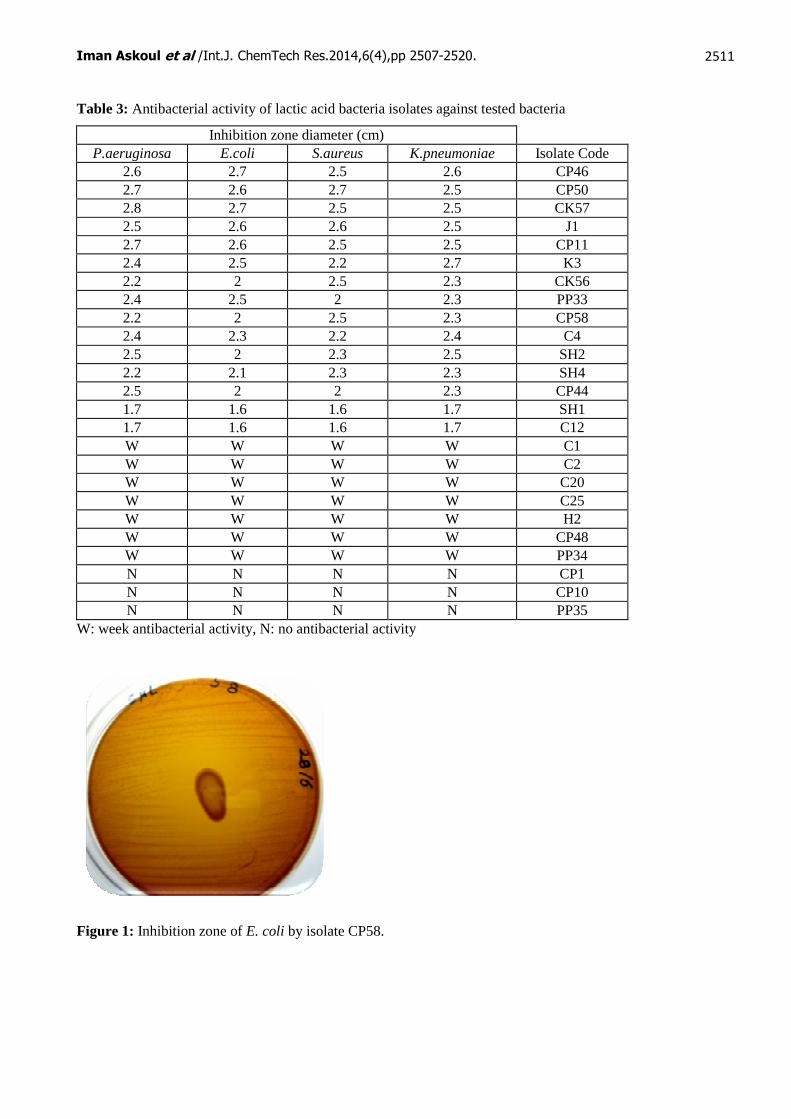
Table 3: Antibacterial activity of lactic acid bacteria isolates against tested bacteria
Inhibition zone diameter (cm) P.aeruginosa E.coli S.aureus K.pneumoniae Isolate Code
2.6 2.7 2.5 2.6 CP46 2.7 2.6 2.7 2.5 CP50 2.8 2.7 2.5 2.5 CK57 2.5 2.6 2.6 2.5 J1 2.7 2.6 2.5 2.5 CP11 2.4 2.5 2.2 2.7 K3 2.2 2 2.5 2.3 CK56 2.4 2.5 2 2.3 PP33 2.2 2 2.5 2.3 CP58 2.4 2.3 2.2 2.4 C4 2.5 2 2.3 2.5 SH2 2.2 2.1 2.3 2.3 SH4 2.5 2 2 2.3 CP44 1.7 1.6 1.6 1.7 SH1 1.7 1.6 1.6 1.7 C12 W W W W C1 W W W W C2 W W W W C20 W W W W C25 W W W W H2 W W W W CP48 W W W W PP34 N N N N CP1 N N N N CP10 N N N N PP35
W: week antibacterial activity, N: no antibacterial activity
Figure 1: Inhibition zone of E. coli by isolate CP58.

Iman Askoul et al /Int.J. ChemTech Res.2014,6(4),pp 2507-2520.
2512
Table 4: Antimicrobial activity of cell free supernatant against tested isolates
Values followed with different letters in superscript on the same column are significantly different (P < 0.05).
Figure 2: Inhibition zone formed by K3 L. plantarum CFS on the growth of E. coli
Physiological and biochemical characterizations of selected LAB
Identification of Selected Isolates
Positive sugar fermentation results indicated that 14 of the isolates were Lactobacillus plantarum and one isolatewasL.fermentum as shown in Table 5.The API 50 CHL classification system proved to be reliable because all of the Lactobacilli isolates were well classified.
Lactobacillus plantarum bacteria in this study were isolated from cheese, cucumber pickles, pepper pickles, jamid, Kishk, Shanglish and karkadeh. It has been isolated from different sources of food, from ”Jiaoke” a traditional fermented cream from China4, from Tenerife goats' cheese19, from fermented cucumber21,fromItalian ewe cheeses22, and from raw goats' milk23.The dominant isolated Bacillus genus from Koopeh Cheese was Lactobacillus plantarum (58% of lactobacilli population)as reported by2.
Inhibition zone diameter (cm)
P.aeruginosa E.coli S.aureus K.pneumoniae Isolate Code No.
1.9 ±00 a 2 ±00 a 1.9 ± 00 a 1.8 ± 0.10 a CK56 1
1.86 ±0.057 ab 1.86 ±0.057 b 1.83 ±0.057 a 1.8 ± 00 a SH4 2
1.8 ±00 b 1.86 ±0.057 b 1.73 ±0.057 b 1.8 ± 0.10 a CK57 3 1.66 ±0.057 c 1.66 ±0.057 c 1.63 ±0.057 c 1.4 ±0.10 bc CP50 4
1.66 ±0.057 c 1.6 ±00 c 1.66 ±0.057 bc 1.5 ±0.10 b CP58 5 1.66 ±0.057 c 1.63 ±0.057 c 1.6 ±00 c 1.46 ±0.057 b CP44 6
1.43 ±0.057 d 1.4 ±00 de 1.46 ±0.057 d 1.46 ±0.057 b CP11 7 1.33 ±0.057 ef 1.46 ±0.057 d 1.43 ±0.057 d 1.33 ±0.115 cd PP33 8
1.36 ±0.057 de 1.46 ±0.057 d 1.33 ±0.057 e 1.26 ±0.057 d C4 9 1.3 ±00 ef 1.33 ±0.057 e 1.23 ±0.057 f 1.4 ±00bc K3 10
1.26 ±0.057 f 1.2 ±00 f 1.13 ±0.057 g 1.4 ±0.10bc CP46 11 1.36 ±0.057 de 1.23 ±0.057 f 1.2 ±00 fg 1.3 ±00 cd J1 12
1.16 ±0.057 g 1.16 ±0.057 f 1.13 ±0.057 g 1.33 ±0.057 cd SH2 13 1.03 ±0.057 h 0.93 ±0.057 g 1.03 ±0.057 h 1.06 ±0.057 e SH1 14
0.63 ±0.057i 0.63 ±0.057h 0.83 ±0.057i 0.66 ±0.057 f C12 15

Iman Askoul et al /Int.J. ChemTech Res.2014,6(4),pp 2507-2520.
2513
Table 5: Biochemical Identification and Classification of Selected Isolates
API 50 CHL ID % Identification Isolates 99.9 L. plantarum C4 99.7 L .fermentum C12 99.9 L .plantarum J1 99.9 L .plantarum K3 99.9 L .plantarum SH1 99.9 L .plantarum SH2 99.9 L .plantarum SH4 99.9 L .plantarum CP11 99.9 L .plantarum CP44 99.9 L .plantarum CP46 99.9 L .plantarum CP50 99.9 L .plantarum CP58 99.9 L .plantarum PP33 99.9 L .plantarum CK56 99.8 L .plantarum CK57
Growth at Different Temperatures
Fifteen isolates were mesophilic as those isolated by20,23,24,25. It was also observed that all 15 isolates were able to grow at 10oC and 40oC, (Table 6), while two L. plantarum isolates isolated by24weren’t able to grow at the mentioned temperatures. Two isolates L. plantarumK3 and L. plantarumSH4 and L. fermentum C12 were able to grow at 45oC, while 12 isolates weren’t able to grow at the same temperature (Table 6). L. plantarum isolates isolated by20,24,25 weren’t able to grow at 45oC. None of the isolates were able to grow at 4oC (Table 4), the same result was reported by 20,23,24,25.
Growth at Different NaCl Concentrations
As shown in (Table 6) 15 isolates were able to grow in the presence of 4% NaCl, while they weren’t able to grow in the presence of 6.5% NaCl. The growth of two L. plantarum isolates isolated by24 showed very weak growth in the presence of4% NaCl.
Gas Production from Glucose
Three L. plantarum isolates (K3, SH2, CP58) and L. fermentum isolate C12 were heterofermentative as deduced by gas production, while 11 L. plantarum isolates were homofermentative since they didn’t produce CO2 as show in Table 6. All isolates of L.plantarum isolated by2,20,23,24,25 were homo-fermentative.
Table 6: Physiological characteristics of the fifteen local isolates
Gas
pH 9
pH 8
pH 7
pH 6
pH 5
pH 4
pH 3
pH 2
45°C
40°C
37°C
10°C
4°C
NaCl 6.5%
NaCl 4%
Isolate
Code _ + + + + + + _ _ _ + + + _ _ + C4
+ + + + + + + + _ + + + + _ _ + C12
_ + + + + + + _ _ _ + + + _ _ + J1
+ + + + + + + _ _ + + + + _ _ + K3
_ + + + + + + + _ _ + + + _ _ + SH1
+ + + + + + + _ _ _ + + + _ _ + SH2
_ + + + + + + _ _ + + + + _ _ + SH4
_ + + + + + + _ _ _ + + + _ _ + CP11
_ + + + + + + _ _ _ + + + _ _ + CP44
_ + + + + + + _ _ _ + + + _ _ + CP46

Iman Askoul et al /Int.J. ChemTech Res.2014,6(4),pp 2507-2520.
2514
_ + + + + + + + _ _ + + + _ _ + CP50
+ + + + + + + + _ _ + + + _ _ + CP58
_ + + + + + + _ _ _ + + + _ _ + PP33
_ + + + + + + _ _ _ + + + _ _ + CK56
_ + + + + + + _ _ _ + + + _ _ + Ck57
Each value is represented by the mean of triplicate
Growth at different pH values
All of the 15 studied isolates were able to grow at pH ranging from 4 to 9, but only 3 L. plantarum isolates (SH1, CP50 and CP58 ) and the L. fermentum isolate C12 were able to grow at pH 3 as shown in Table 6.The growth at pH 4.4 was weak24, while it was good at pH 3.9 - 4.820.
Partial characterization of inhibitory substances in supernatants
Antibacterial activities of selected LAB-CFS were significantly influenced by pH as shown in Figures (3-6). In this respect, it was observed that the activities at pH 4.5 were significantly higher than those at pH 7.0 suggesting an inhibition effect of acidity on the growth of S. aureus, K. pneumoniae, P.aeruginosa and E. coli. Most of LAB excrete acid that has been shown to inhibit growth of pathogens. These observations are in agreement with those reported by 15.On the other hand, it was observed a residual activity at pH 7.0 suggesting that compounds other than acids inhibit the growth of the tested pathogens. These observations are in agreement with those reported by18who showed that L. plantarum excreted other compounds such as bacteriocins that inhibited the growth of pathogens.
Figure 3: Effect of pH change on CFSs antibacterial activity against K. pneumoniae.
Figure 4: Effect of pH change on CFSs antibacterial activity against S. aureus.

Iman Askoul et al /Int.J. ChemTech Res.2014,6(4),pp 2507-2520.
2515
Figure 5: Effect of pH change on CFSs antibacterial activity against E.coli.
Figure 6: Effect of pH change on CFSs antibacterial activity against P. aeruginosa.
CFSs antibacterial activity after treatment with proteinase K
As shown in Figure (7-10) the treatment of cell free supernatants with the proteolytic enzyme, proteinase K, at pH 7 resulted in a significant reduction of the antibacterial against test pathogens. This result suggested that the antibacterial activity was associated with peptide inhibiting molecules, usually known as bacteriocin, these observations are in agreement with those reported by15, 19, 21, 26, 27.
Figure 7: Antibacterial activity before and after treatment with proteinase K againstK. pneumoniae.
Iman Askoul et al /Int.J. ChemTech Res.2014,6(4),pp 2507-2520.
2516
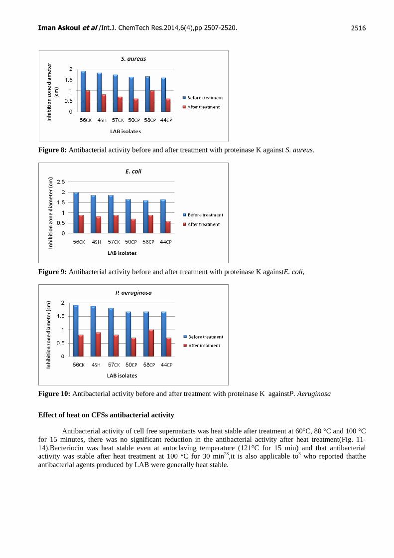
Figure 8: Antibacterial activity before and after treatment with proteinase K against S. aureus.
Figure 9: Antibacterial activity before and after treatment with proteinase K againstE. coli,
Figure 10: Antibacterial activity before and after treatment with proteinase K againstP. Aeruginosa
Effect of heat on CFSs antibacterial activity
Antibacterial activity of cell free supernatants was heat stable after treatment at 60°C, 80 °C and 100 °C for 15 minutes, there was no significant reduction in the antibacterial activity after heat treatment(Fig. 11-14).Bacteriocin was heat stable even at autoclaving temperature (121°C for 15 min) and that antibacterial activity was stable after heat treatment at 100 °C for 30 min28,it is also applicable to3 who reported thatthe antibacterial agents produced by LAB were generally heat stable.

Iman Askoul et al /Int.J. ChemTech Res.2014,6(4),pp 2507-2520.
2517
Figure 11: Effect of heat treatment on antibacterial activity of CFSs against K. pneumoniae.
Figure 12: Effect of heat treatment on antibacterial activity of CFSs against S. aureus
Figure 13: Effect of heat treatment on antibacterial activity of CFSs against E. coli
Figure 14: Effect of heat treatment on antibacterial activity of CFSs against P. aeruginosa

Iman Askoul et al /Int.J. ChemTech Res.2014,6(4),pp 2507-2520.
2518
PCR amplification of plantaricin genes
Two of the L. plantarum isolates SH4 and CK56 contained the structural genes for both plantaricin EF and plantaricin N, and two of the L. plantarum isolates CK57 and CP44 contained the structural genes for plantaricin N only(Fig 15, Fig 16).It wasmentioned thatL. plantarum strains BFE 5092 and 299V strains contained the structural genes for the plantaricin EF, plantaricin JK, and plantaricin N, while L. plantarum strain PC S20 contained the structural genes for plantaricin EF only18. Moghadamet al (2010)29 reported that combination of plantaricin EF and plantaricin W structural genes were successfully amplified in all six strains he studied, while Noonpakdeeet al (2009)28 mentioned that L. plantarum PMU33 contained the structural genes for plantaricin W only.
Primers used for amplification of plantaricins genes were selected according to their size since the longer the primer is the more specific the result will be. Amplicon sizes of the amplified sequences were close to those obtained by Cho et al (2010)18who reported that the amplicon size of the amplified sequence of pln N was 160 bp, while ofpln EF it was 360 bp.
Figure 15:Amplification products from the most active six isolates with primer plnEF. M1: Ruler DNA 50bp, M2: Ruler DNA 100bp. Agarose gel 2 % at 60 volt for 1.5 h.
Figure 16:Amplification products from the most active six isolates with primer plnN. M1: Ruler DNA 50bp, M2: Ruler DNA 100bp.Agarose gel 2 % at 60 volt for 1.5 h.

Iman Askoul et al /Int.J. ChemTech Res.2014,6(4),pp 2507-2520.
2519
References:
1. Noopur, M. S., Sucheta, N. P., and Aglave, B.A, 2010. Extraction of bacteriocin and study of its antagonistic assay. International Journal of Biotechnology and Biochemistry.ISSN0973-2691. Vol: 6. Num: 6. Pp: 865–870.
2. Hassanzadazar, H., and Ehsani, A, 2013. Phenotypic Characterization of Lactic Acid Bacteria Isolated from Traditional Koopeh Cheese. Global Veterinaria 10 (2): 148-152.
3. Noordiana, N., Fatimah, A. B., and Mun, A. S, 2013. Antibacterial agents produced by lactic acid bacteria isolated from Threadfin Salmon and Grass Shrimp. International Food Research Journal. 20(1): 117-124.
4. Gong, H.S., Meng, X.C., and Wang, H, 2010. Plantaricin MG active against Gram-negative bacteria produced by Lactobacillus plantarum KLDS1.0391 isolated from ‘‘Jiaoke”, a traditional fermented cream from China. Food Control. 21: 89–96.
5. Ana, A. Z, 2012. Antimicrobial Activities of Lactic Acid Bacteria Strains Isolated from Nile Tilapia Intestine (Oreochromisniloticus). Journal of Biology and Life Science. Vol: 4. No: 1. P. 164- 171.
6. Adenike, A. O., Mopelola, A., and Adeleye, J, 2007. In vitro antimicrobial characteristics of bacteriocin producing Lactobacillus strains from Nigerian indigenous fermented foods. African Journal of Biotechnology. Vol. 6 (4), pp. 445-453.
7. Bukola, C., Adebayo, T., and Abiodun, A, 2008. Screening of Lactic Acid Bacteria Strains Isolated from Some Nigerian Fermented Foods for EPS Production. World Applied Sciences Journal. 4 (5): 741-747.
8. Harrigan, W., McCance, M, 1990. Laboratory methods in Food and Dairy Microbiology. 8th Edition. Academic Press. London. UK.
9. Piddock, L .J. V, 1990. Techniques used for the determination of antimicrobial resistance and sensitivity in bacteria. J ApplBacteriol. 68: 307-18.
10. Parente, E., Riccardi, A, 1999. Production, recovery and purification of bacteriocins from lactic acid bacteria. ApplMicrobiolBiotechnol. 52: 628–638.
11. Todorov, S. D., van Reenen, C. A., and Dicks, L. M. T, 2004. Optimization of bacteriocin production by Lactobacillus plantarum ST13BR, a strain isolated from barley beer. J. Gen. Appl. Microbiol. 50: 149–157.
12. Conter, M., Muscariello ,T., Zanardi, E., Ghidini, S., Vergara, A., Campanini, G., and Ianieri, A, 2005. Characterization of Lactic Acid Bacteria Isolated from An Italian Dry Fermented Sausage. Ann. Fac. Medic. Vet. di Parma (Vol. XXV). p. 167 – 174.
13. Lengkey, H. A.W., Balia, R. L., Togoe, I., Taşbac, B. A., Ludong, M, 2009. ISOLATION AND IDENTIFICATION OF LACTIC ACID BACTERIA FROM RAW POULTRY MEAT. Biotechnology in Animal Husbandry. 25 (5-6), p 1071-1077.
14. Bulut, Ҫ, 2003. Isolation and Molecular Characterization of Lactic Acid Bacteria from Cheese. Thesis for Master Degree of Science. İzmir Institute of Technology.
15. Tatsadjieu, N. L., Njintang, Y. N., Sonfack, T. K., Daoudou, B., and Mbofung, C. M. F, 2009. Characterization of lactic acid bacteria producing bacteriocins against chicken Salmonella enterica and Escherichia coli. African Journal of Microbiology Research. Vol. 3 (5) pp. 220-227.
16. Cardinal, M. J., Meghrous, J., Lacroix, C., and Simard, R. E, 1997.Isolation of Lactococcus lactis strain producing inhibitory activity against Listeria. Food Biotechnology.11(2), 129-146.
17. John, F. T. S., and Alicia, L. Ragout de Spencer, 2001. Food Microbiology Protocols. Humana Press Inc.
18. Cho, G., Huch, M., Hanak, A., Holzapfel, W. H., Franz, C. M. A. P, 2010. Genetic analysis of the plantaricin EFI locus of Lactobacillus plantarum PCS20 reveals an unusual plantaricin E gene sequence as a result of mutation. International Journal of Food Microbiology. 141: S117 – S124.
19. Hernández, D., Cardell, E., and Zárate, V, 2005. Antimicrobial activity of lactic acid bacteria isolated from Tenerife cheese: initial characterization of plantaricin TF711, a bacteriocin-like substance produced by Lactobacillus plantarum. J ApplMicrobiol. TF711. 99(1):77-84.
20. Obadina, A.O., Oyewole, O.B., Sanni, L.O., and Tomlins, K.I, 2006. Bio-preservative activities of Lactobacillus plantarum strains in fermenting Casssava ‘fufu’. African Journal of Biotechnology. Vol. 5 (8), pp. 620-623.
21. Daeschel, M. A., Mc Kenney, M. C., and McDonald, L. C, 1990. Bacteriocidal activity of Lactobacillus plantarum C-11. Food Microbiology. 7. 91-98.

Iman Askoul et al /Int.J. ChemTech Res.2014,6(4),pp 2507-2520.
2520
22. De Angelis, M., Corsetti, A., Tosti, N., Rossi, J., Corbo, M. R., and Gobbetti, M, 2001. Characterization of Non-Starter Lactic Acid Bacteria from Italian Ewe Cheeses Based on and Cell Wall Protein Analyses. Applied and environmental microbiology. p. 2011–2020.
23. Guessas, B ., and Kihal, M, 2004. Characterization of lactic acid bacteria isolated from Algerian arid zone raw goats' milk. African Journal of Biotechnology. Vol. 3 (6). 339-342.
24. Vijai Pal, V., Jamuna, M., and Jeevaratnam, K, 2004. Isolation and Characterization of Bacteriocin Producing Lactic Acid Bacteria from A South Indian Special Dosa (Appam) Batter. Journal of Culture Collections. Vol 4. pp. 53-60.
25. Nair, P. S., and Surendran, P. K, 2005. Biochemical Characterization of Lactic Acid Bacteria Isolated from Fish and Prawn. Journal of Culture Collections. Vol 4. pp. 48-52.
26. vanReenen., L. M. T. Dicks and Chikindas, M. L, 1998. Isolation, purification and partial characterization of plantaricin 423, a bacteriocin produced by Lactobacillus plantarum. Journal of Applied Microbiology. 84, 1131–1137.
27. Savadogo, A., Ouattara, C. A. T., Bassole, I. H. N., Traore, A. S, 2004. Antimicrobial Activities of Lactic Acid Bacteria Strains Isolated from Burkina Faso Fermented Milk. Pakistan Journal of Nutrition. 3 (3): 174-179.
28. Noonpakdee, W., Jumriangrit, P., Wittayakom, K., Zendo, J., Nakayama, J., Sonomoto, K., and Panyim, S, 2009. Bacteriocin from Lactobacillus plantarum PMU33 strains Asia Pacific Journal of Molecular Biology and Biotechnology. Asia Pacific Journal of Molecular Biology and Biotechnology. Vol. 17(1): 19-25.
29. Moghadam, M. Sh., Foo, H. L., Leow, T. C., Rahim, R. A., and Loh, T. C, 2010. Novel bacterio cinogenic Lactobacillus plantarum strains and their differentiation by sequence analysis of 16S rDNA,16S-23S and 23S-5S intergenic spacer regions and randomly amplified polymorphic DNA analysis. Food Technol. Biotechnol. 48 (4): 476–483.
*****