ischemic colitis, the great imitator: a mass...
TRANSCRIPT

ACG CASE REPORTS JOURNAL
acgcasereports.gi.org ACG Case Reports Journal | Volume 1 | Issue 2 | January 2014100
CASE REPORT | COLON
Ischemic Colitis, the Great Imitator: A Mass Completely Resolved
Roshanak Rabbanifard, MD1 and Jeffrey A. Gill, MD2
1Department of Internal Medicine, University of South Florida, Tampa, FL 2Division of Digestive Disease and Nutrition, James A. Haley Veterans Affairs Hospital, Tampa, FL
AbstractIschemic colitis (IC) is the most common type of intestinal ischemia, with a vast clinical spectrum of injury rang-ing from mild and transient ischemia to acute fulminant colitis. The pattern of injury is usually segmental, but it is mainly dictated by individual anatomy, duration of ischemia, and degree of re-perfusion injury. Analysis of clinical presentation, early endoscopic evaluation, and biopsy are all essential for prevention of misdiagnosis. We pres-ent a unique case of IC with mass-like features on regular imaging, emphasizing the importance of endoscopy and biopsy for accurate diagnosis.
IntroductionIschemic colitis (IC) is a common disorder of the large bowel. It is frequently seen in older patients, and is the most common form of intestinal ischemic injury. It accounts for about 50–60% of all gastrointestinal ischemic episodes, most often in the absence of major vessel occlusion.1–4 IC is a result of inadequate blood flow to the colon, causing mucosal injury that is mediated by hypoxia, followed by reperfusion injury.4 The pattern of injury is usually segmental, mainly involving the “watershed” zones of the splenic flexure, descending colon, and the rectosigmoid junction; however, any part of the colon can be affected, including isolated right-sided colon, which carries a higher morbidity and mortality rate.2 Common clinical manifestations include sudden presentation of abdominal pain that is usually located over the affected area of the colon, followed by bright red blood per rec-tum.4 The severity of clinical symptoms varies, including fever, diarrhea, peritonitis, and septic shock, depending on the extent of colonic injury. We report a unique presentation of IC and stress the significance of endoscopy and biopsy for accurate diagnosis and therapy.
Case ReportA 76-year-old Caucasian male with a past medical history of tobacco use, hypertension, and chronic kidney disease stage III, not on dialysis, presented to the emergency room with acute right lower abdominal pain and non-bloody diarrhea. He reported a history of a previous colonoscopy with 1 polyp removed 2 years ago at an outside hospital. In the emergency department, he was febrile to 102°F and tachycardic. Physical exam revealed a well-developed and well-nourished male in no acute distress with a tender right lower quadrant with no rebound or guarding. Other systems were normal. Laboratory findings were remarkable for a white blood cell count of 12.1 x 10³/L and a creatinine of 1.9 mg/dL.
CT scan of the abdomen revealed annular thickening of the cecum, concerning for malignancy (Figure 1). The patient was admitted and treated supportively with intravenous fluids and pain management. Colonoscopy showed a large, necrotic-appearing mass encompassing the majority of the cecum (Figure 2). Pathology revealed fragments of necrotic colonic mucosa, fibrinopurulent exudates, and partial viable tissue and associated repara-
ACG Case Rep J 2014;1(2):100–102. doi:10.14309/crj.2014.14. Published online: January 10, 2014.
Correspondence: Jeffrey A. Gill, MD, James A. Haley VA Hospital, 13000 Bruce B. Downs Blvd, Tampa, FL, 33612 ([email protected]).
Copyright: © 2014 Rabbanifard and Gill. This is an open-access article distributed under the terms of the Creative Commons Attribution License, which permits unrestricted use, distribution, and reproduction in any medium, provided the original author and source are credited.

Ischemic Colitis, the Great Imitator
acgcasereports.gi.org ACG Case Reports Journal | Volume 1 | Issue 2 | January 2014
Rabbanifard and Gill
101
Figure 1. CT abdomen/pelvis showing segmental abnormal mucosal thick-ening in an annular fashion involving the cecum, highly suggestive of a colonic malignancy.
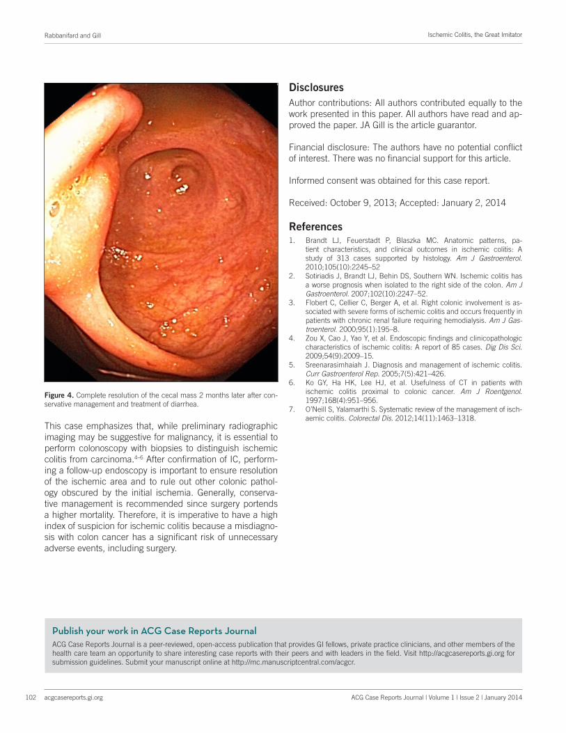
tive changes without evidence of malignant cells (Figure 3). IC was diagnosed when these findings were correlated with the clinical findings. The patient was treated conservatively with resolution of his symptoms. He was discharged 1 week later with outpatient gastroenterology follow-up. A repeat colonoscopy done 2 months later showed complete resolu-tion of the mass (Figure 4).
DiscussionIC is the most common manifestation of ischemic bowel dis-ease and is important to recognize, especially in the elderly population. Commonly seen risk factors among patients with IC include coronary artery disease, hypertension, chronic kidney disease, diabetes, and hyperlipidemia.1 Ischemia typically affects the colon in a segmental manner secondary to the pattern of vascular supply to the colon. Colon isch-emia has many causes, most often associated with an acute change in blood flow to the colon, usually in the absence of major vessel occlusion.1-2 Isolated right-sided colon isch-emia, as seen in this case, accounts for about 25% of cases. It is usually precipitated by reduction in circulating blood
volume, marked fluid shift, and hypotension, which could lead to superior mesenteric artery (SMA) vasoconstriction.2-3 Patients usually present with symptoms of non-specific ab-dominal pain but lack the bloody diarrhea seen in classic IC affecting the splenic flexure or “watershed area.” There is higher morbidity and mortality with right-sided colon involve-ment compared to the other segments of the colon. This is hypothesized to be due to insufficient collateralization and blood flow to the right side of the colon.2 Given the vague symptoms and higher morbidity and mortality, early recogni-tion and treatment is critical.
Endoscopic manifestations of IC are vast and range from mild erythema to pseudo-tumor, as reported in this case. Lymphocytic and neutrophilic infiltration along with edema and ulceration of the superficial mucosa are seen in mild forms of ischemia. In such cases, the glandular architecture of the deep mucosa is preserved. Gland degeneration and fibrinopurulent exudate are seen in more severe ischemia. Pseudo-tumor can be attributed to submucosal hemor-rhage, which can create a mass-like appearance when se-vere.4 Given that there is no widely accepted diagnostic cri-teria for IC, clear recognition and knowledge of the clinical, endoscopic, and pathologic characteristics is crucial in mak-ing an accurate diagnosis and providing timely treatment.
Figure 2. Colonoscopy showing a large cecal mass encompassing the ma-jority of the cecum.
Figure 3. H&E stain of colonic biopsy specimen at (A) 100x, (B) 200x, and (C) 400x, significant for fragments of necrotic colonic mucosa and fibri-nopurulent exudate with evidence of submucosal hemorrhage and crypt destruction. No evidence of malignancy.
A
C
B
Publish your work in ACG Case Reports JournalACG Case Reports Journal is a peer-reviewed, open-access publication that provides GI fellows, private practice clinicians, and other members of the health care team an opportunity to share interesting case reports with their peers and with leaders in the field. Visit http://acgcasereports.gi.org for submission guidelines. Submit your manuscript online at http://mc.manuscriptcentral.com/acgcr.
Rabbanifard and Gill
acgcasereports.gi.org
Ischemic Colitis, the Great Imitator
102 ACG Case Reports Journal | Volume 1 | Issue 2 | January 2014
This case emphasizes that, while preliminary radiographic imaging may be suggestive for malignancy, it is essential to perform colonoscopy with biopsies to distinguish ischemic colitis from carcinoma.4–6 After confirmation of IC, perform-ing a follow-up endoscopy is important to ensure resolution of the ischemic area and to rule out other colonic pathol-ogy obscured by the initial ischemia. Generally, conserva-tive management is recommended since surgery portends a higher mortality. Therefore, it is imperative to have a high index of suspicion for ischemic colitis because a misdiagno-sis with colon cancer has a significant risk of unnecessary adverse events, including surgery.
Disclosures
Author contributions: All authors contributed equally to the work presented in this paper. All authors have read and ap-proved the paper. JA Gill is the article guarantor.
Financial disclosure: The authors have no potential conflict of interest. There was no financial support for this article.
Informed consent was obtained for this case report.
Received: October 9, 2013; Accepted: January 2, 2014
References1. Brandt LJ, Feuerstadt P, Blaszka MC. Anatomic patterns, pa-
tient characteristics, and clinical outcomes in ischemic colitis: A study of 313 cases supported by histology. Am J Gastroenterol. 2010;105(10):2245–52
2. Sotiriadis J, Brandt LJ, Behin DS, Southern WN. Ischemic colitis has a worse prognosis when isolated to the right side of the colon. Am J Gastroenterol. 2007;102(10):2247–52.
3. Flobert C, Cellier C, Berger A, et al. Right colonic involvement is as-sociated with severe forms of ischemic colitis and occurs frequently in patients with chronic renal failure requiring hemodialysis. Am J Gas-troenterol. 2000;95(1):195–8.
4. Zou X, Cao J, Yao Y, et al. Endoscopic findings and clinicopathologic characteristics of ischemic colitis: A report of 85 cases. Dig Dis Sci. 2009;54(9):2009–15.
5. Sreenarasimhaiah J. Diagnosis and management of ischemic colitis. Curr Gastroenterol Rep. 2005;7(5):421–426.
6. Ko GY, Ha HK, Lee HJ, et al. Usefulness of CT in patients with ischemic colitis proximal to colonic cancer. Am J Roentgenol. 1997;168(4):951–956.
7. O’Neill S, Yalamarthi S. Systematic review of the management of isch-aemic colitis. Colorectal Dis. 2012;14(11):1463–1318.
Figure 4. Complete resolution of the cecal mass 2 months later after con-servative management and treatment of diarrhea.