iron oxide deposits associated with the ectosymbiotic...
TRANSCRIPT
Biogeosciences, 5, 1295–1310, 2008www.biogeosciences.net/5/1295/2008/© Author(s) 2008. This work is distributed underthe Creative Commons Attribution 3.0 License.
Biogeosciences
Ir on oxide deposits associated with the ectosymbiotic bacteria in thehydrothermal vent shrimp Rimicaris exoculata
L. Corbari 1, M.-A. Cambon-Bonavita2, G. J. Long3, F. Grandjean4, M. Zbinden5, F. Gaill5, and P. Compere1
1Universite de Liege, Laboratoire de Morphologie fonctionnelle et Evolutive, Unite de Morphologie ultrastructurale et Celluled’Appui Technologique en Microscopie (Catµ), allee de la chimie, 3, 4000 Liege, Belgium2Laboratoire de Microbiologie et Biotechnologie des Extremophiles, Ifremer, centre de Brest, BP 70, 29280 Plouzane, France3Department of Chemistry, Missouri University of Science and Technology, University of Missouri-Rolla, Rolla, Missouri65409-0010, USA4Department of Physics, B5, University of Liege, 4000 Sart-Tilman, Belgium5UMR CNRS 7138 “Systematique, Adaptation et Evolution”, Universite Pierre et Marie Curie, 7 Quai St Bernard, BatimentA, 75252 Paris Cedex 05, France
Received: 12 February 2008 – Published in Biogeosciences Discuss.: 24 April 2008Revised: 1 August 2008 – Accepted: 14 August 2008 – Published: 10 September 2008
Abstract. The Rimicaris exoculata shrimp is consideredas a primary consumer that dominates the fauna of mostMid-Atlantic Ridge (MAR) hydrothermal ecosystems. Theseshrimps harbour in their gill chambers an important ec-tosymbiotic community of chemoautotrophic bacteria asso-ciated with iron oxide deposits. The structure and elemen-tal composition of the mineral concretions associated withthese bacteria have been investigated by using LM, ESEM,TEM STEM and EDX microanalyses. The nature of theiron oxides in shrimps obtained from the Rainbow vent fieldhas also been determined by Mossbauer spectroscopy. Thismultidisciplinary approach has revealed that the three layersof mineral crust in theRimicaris exoculatashrimps consistof large concretions formed by aggregated nanoparticles oftwo-line ferrihydrite and include other minor elements as Si,Ca, Mg, S and P, probably present as silicates cations, sul-phates or phosphates respectively that may contribute to sta-bilise the ferrihydrite form of iron oxides. TEM-observationson the bacteria have revealed their close interactions withthese minerals. Abiotic and biotic precipitation could oc-cur within the gill chamber ofRimicaris exoculata, suggest-ing the biologically-mediated formation of the iron oxide de-posits. The difference of the bacterial density in the three-mineral crust layers could be correlated to the importance ofthe iron oxide concretions and suggest that the first mineralparticles precipitates on the lower layer which could be con-sidered as the most likely location of iron-oxidizing bacteria.
Correspondence to:L. Corbari([email protected])
1 Introduction
Rimicaris exoculata(Williams and Rona, 1986) is one ofthe most dominant species found at the Mid-Atlantic Ridge(MAR) hydrothermal vents. This endemic shrimp swarmson the chimney walls, exhibiting a patch-like distributionof up to several thousand per square meter (Segonzac etal., 1993). In extreme deep-sea environments, such highpopulation density levels require some specific adaptations.Many hydrothermal organisms derive their nutrition fromchemoautotrophic bacteria through symbioses relying mostoften on sulphide or methane as an energy source (Ca-vanaugh, 2006).R. exoculatapossess an original ectosymbi-otic bacterial community, housed in its expanded gill cham-bers and mouth parts (Van Dover et al., 1988; Casanova etal., 1993; Zbinden et al., 2004; Corbari et al., 2008). Eventhough numerous authors have suggested that, if really ec-tosymbiotic, the bacteria could be a direct or indirect foodsource for the shrimp (Segonzac et al., 1993; Rieley et al.,1999; Gebruk et al., 2000; Zbinden et al., 2004, 2008). Stillundetermined, however, is the origin of the nutritional car-bon of R. exoculataand the role of the bacterial ectosym-biosis play as a trophic resource (Pond et al., 1997; Polz etal., 1998; Zbinden and Cambon-Bonavita, 2003). The bac-terial community housed in the gill chamber ofR. exoculatahas been identified as chemoautotrophic bacteria (Wirsen etal., 1993) and phylogenetic analysis revealed that the bacte-ria could correspond to a single epsilon-proteobacteria phy-lotype (Polz and Cavanaugh, 1995). Some authors havealso hypothesized that the bacteria could acquire their en-ergy from sulphide oxidation (Gebruk et al., 1993; Polz and
Published by Copernicus Publications on behalf of the European Geosciences Union.

1296 L. Corbari et al.: Bacteriogenic iron oxides
Cavanaugh, 1995), but this hypothesis has never been con-firmed by any culture experiment. Recently, Zbinden etal. (2008) demonstrated that the bacterial community in theshrimp gill chamber is composed of more than one phylotypeand revealed from molecular analysis that three metabolicbacterial types (iron, sulfide and methane oxidation) may co-occur within the symbiotic community associated withRim-icaris exoculata.
In the absence of cultivated bacteria, studies have focussedon the description of both the bacteria and the mineral de-posits inR. exoculata. Zbinden et al. (2004) described threebacterial morphotypes, individual rods with an approximatesize of 0.5×1.5µm, and two types of multicellular filaments,i.e. thick filaments with 2 to 3µm diameters and thin fila-ments with 0.5 to 1µm diameters, found within the entire gillchamber. They mapped the location of these bacteria and di-vided their associated minerals into three functional compart-ments, that which were considered to represent distinct mi-croenvironments. One of these compartments, the upper pre-branchial chamber, houses the highest density of both bacte-ria and minerals. Recently, Corbari et al. (2008) focussed onthis compartment and delineated the shrimp-bacteria-mineralassociation throughout the shrimp moult cycle. This studyperformed on about 300 specimens from two vent sites, TAGand Rainbow, indicated that the bacterial community restartsafter each exuviation and gradually colonises the gill cham-ber in five moult stage-correlated steps. Moreover, the pres-ence of red-brown mineral deposits in the gill chamber, in-cluding the mouth parts and branchiostegites, of theR. exocu-lata has already been described (Gloter et al., 2004; Zbindenet al., 2004). These deposits have been identified as hy-drous iron oxide in the form of ferrihydrite (Gloter et al.,2004). The extent and density of iron oxide deposits withinthe gill chamber are both responsible for the external colourof the shrimp, a colour that may be macroscopically observedby transparency through the branchiostegites (Zbinden et al.,2004). The shrimp external colour ranges from white, indica-tive of no mineral deposits, to dark-red, indicative of a heav-ily mineralised crust; the colour appears to be highly corre-lated with the moult stages (Corbari et al., 2008). The fully-formed mineral crust is roughly organised in three step-levelsthat illustrate the time-related formation and growth of themineral particles (Corbari et al., 2008). The shrimp-bacteriaectosymbiosis is characterised by the presence of iron oxidedeposits suggesting that iron oxidation may represent the ma-jor energy-pathway for the bacterial community, especiallyin shrimps found at the Rainbow site (Gloter et al., 2004;Zbinden et al., 2004, 2008; Schmidt et al., 2008). Moreover,the simultaneous occurrence of the bacteria and the iron ox-ide deposits in the gill chamber ofR. exoculatamay be inter-preted as biologically-mediated or biogenic (Zbinden et al.,2004; Gloter et al., 2004, Anderson et al., 2008).
The definition of “biogenic iron oxides” (Fortin andChatellier, 2003) refers to iron oxides formed in the pres-ence of bacteria and includes iron oxides formed as a di-
rect result of microbial activities (i.e. from enzymatic re-actions) or of passive mechanisms whereby bacterial exu-dates trigger the formation and precipitation of iron oxidesminerals. Several studies have documented the formationand occurrence of iron oxides formed as a result of bioticpathways in natural environments (Fortin et al., 1998; Fortinand Chatellier, 2003; Fortin and Langley, 2005; Banfield etal., 2000; Kennedy et al., 2003, 2004). In this context, thebacteria-hydrous ferric oxide interactions have also been in-vestigated to determine the direct and/or indirect bacterial in-fluence on the hydrous ferric oxide formation (see reviews inFortin and Langley, 2005; Klapper and Straub, 2005). Someauthors (Mavrocordatos and Fortin, 2002; Rancourt et al.,2005) have indicated that bacteria, either iron-metabolizingor non-metabolizing, could influence the mineral deposition.They found evidence of the biogenic origin of the hydrousferric oxide, identified the presence of poorly crystallizediron oxides, and determined the typical Fe/O ratios and par-ticle size ranges. Natural biogenic iron oxides generally con-tain impurities, such as adsorbed or structural Si, and phos-phate, sulphate, and manganese and aluminium ions, impuri-ties that may influence the spatial organization and the mor-phology of the mineral particles (Fortin and Chatellier, 2003,Chatellier et al., 2004). The properties of the iron oxide de-posits and the influence of any impurities on these proper-ties have been studied in samples from natural environments(Fortin and Langley, 2005) but have never been investigatedin the case of a bacterial ectosymbiosis.
Because the iron oxide deposition could be actively orpassively promoted byR. exoculataectosymbiotic bacte-ria, the main goal of this study is to investigate in detailthe structure and the composition of the bacteria-associatedmineral particles by using various imaging techniques, suchas back-scattered electron imaging, transmission electronmicroscopy, energy dispersive EDX microanalysis, andMossbauer spectroscopy. These investigations have beenperformed on the fully formed mineral crust of premoultshrimps, a crust that is divided into three layers related withthe successive steps of formation and growth of the mineralparticles.
2 Materials and methods
2.1 Shrimp selection and samples treatment
Specimens ofRimicaris exoculatawere collected during theFrench cruise “EXOMAR” (August 2005) at the MAR hy-drothermal vent site Rainbow (36◦14′ N, 33◦54′ W, 2300 mdepth) by using the suction sampler of the ROV “Victor6000” operating from the RV “Atalante.” Immediately af-ter retrieval, entire living specimens were either frozen at−80◦C or dissected into body parts, branchiostegite and tail,and fixed in a 2.5% glutaraldehyde in seawater 7/10 at pH 7.2medium.
Biogeosciences, 5, 1295–1310, 2008 www.biogeosciences.net/5/1295/2008/

L. Corbari et al.: Bacteriogenic iron oxides 1297
Observations and analyses were performed on preecdysialspecimens, in moult stages D1”’ and D2, in agreementwith the moult-staging method of Drach and Tchernigovt-eff (1967), based on the development of setae matrices alongthe uropods borders. The six frozen and four glutaraldehyde-fixed specimens all exhibited an important red-mineral cruston the inner side of the gill chamber (Fig. 1a and b) in agree-ment with the colour categorisation by Corbari et al. (2008).All the observations were performed on the dorsal medianzone of the branchiostegite ofR. exoculata(Fig. 1a) becauseit exhibits a regular bacterial and mineral cover that linesthe antero-dorsal compartment of the gill chamber (Zbindenet al., 2004). The complete branchiostegite and some por-tions were photographed with an Olympus SZ40 stereo mi-croscope.
In order to determine the structure and elemental compo-sition of the bacteria-associated minerals, samples were pre-pared for study by transmission and scanning electron mi-croscopy, EDX microanalysis, and Mossbauer spectroscopy.During theses preparations, contact between air and the sam-ples was avoided to prevent alteration in the oxidation and/orhydration states of the iron oxide minerals. To avoid this air-contact, frozen specimens were used for compositional anal-yses and compared with glutaraldehyde-fixed specimens. Foranalytical electron microscopy measurements, the samples,dissected from frozen specimens, were directly dehydratedin absolute ethanol and embedded in epoxy resin (Epofix,Struers) through propylene oxide. Glutaraldehyde-fixedsamples, conserved in seawater with NaN3, were quicklyrinsed in distilled water and dehydrated through an ethanol-propylene oxide series of rinses before embedding in theEpoFix resin (Struers).
2.2 Scanning Electron Microscopy (SEM) and Energy-Dispersive X-ray (EDX) microanalysis
Polished thin slices of 20 to 50µm thickness were obtainedfor the branchiostegites of two glutaraldehyde-fixed and twofrozen specimens. The specimens were cut as vertical cross-sections through the mineral crust, i.e. perpendicular to thebranchiostegite cuticle. They were polished by abrasionon diamond disks and finally mirror polished with a non-aqueous 1µm diamond suspension (ESCIL, PS-1MIC). Thepolished-thin slices were surrounded with a conductive sil-ver paint to make contact on the surface, carbon-coated ina Balzers BAF-400 rotary evaporator, and then maintainedin desiccators to prevent air-contact before analysis. Struc-tural mineral observations and elemental energy-dispersiveX-ray microanalysis were rapidly performed within two daysof preparation in an environmental scanning electron micro-scope (FEI XL30 ESEM-FEG), operating at 15 to 20 kVand a working distance of 10 mm. A total of 15 polished-thin slices were imaged by back-scattered electrons (BSE)and analysed for the elemental composition of the mineralspresent.
1 cm
30 µm
a
b
Corbari et al., Figure 1
bg-2008-0022
Fig. 1. Rimicaris exoculata.(a) Inner side of the branchiostegite(left side) of premoult specimen in moult stage D1”’ exhibiting adense and uniform coating of mineral deposits. The dashed linesdelimit the observed area, the median zone.(b) Polished thin crosssection slices of the mineral crust observed under a light microscopeand exhibiting three different layers of mineral density. Note thatthe bacterial filaments are distinguishable on the upper level of thepicture.
Elemental analyses have been carried out on the sur-face of 2µm size mineral concretions. EDX microanaly-ses with an acquisition time of 60 s have been obtained forboth the glutaraldehyde-fixed and the frozen samples in or-der to determine whether any mineral transformation tookplace through chemical reactions during sample preparation.The elemental quantitative analysis used an automatic back-ground subtraction and a ZAF correction matrix has beenused to calculate the elemental composition in weight per-cents and atomic percents. For quantitative analysis of themineral concretions, the contributions of the C-coating andthe embedding resin containing C, O and trace of Cl weresubtracted from the quantitative data of each spectrum. Thecontribution of C-coating was evaluated to 25 At % of C froma pure mineral sample (hydroapatite) C-coated in the sameconditions. The remaining C was attributed to the resin and
www.biogeosciences.net/5/1295/2008/ Biogeosciences, 5, 1295–1310, 2008
1298 L. Corbari et al.: Bacteriogenic iron oxides
an amount of O (At %) was subtracted in the same propor-tion, deduced from reference spectra of pure resin (C/O ratio8:1).
2.3 Transmission Electron Microscopy (TEM)
Four glutaraldehyde-fixed specimens were post-fixed in os-mium 1%, dehydrated in an ethanol-propylene oxide seriesand then embedded in epoxy resin (SPI-PON 812). Ultra-thin sections were obtained with a Reichert-Jung Ultramicro-tome (Ultracut E) by using a diamond knife; uranium acetateand lead citrate were used as contrast agents. The specimenswere studied with a Jeol (JEM 100-SX) transmission electronmicroscope operating at 80 kV. In order to provide a three-dimensional view of the mineral crust organisation, verticalcross-sections were cut perpendicular to the branchiostegitecuticle and the mineral surface and horizontal sections werecut at the three levels of mineral crust as the previously de-fined.
2.4 Scanning Transmission Electron Microscopy (STEM)and Energy-Dispersive X-ray (EDX) microanalysis
Ultrathin sections of samples from two frozen shrimps werecut as previously described, placed on a formvar-coated ti-tanium grid and carbon-coated in a Balzers BAF-400 rotaryevaporator. They were then imaged without any additionalcontrast in a FEI Tecnai G2 Twin scanning-transmissionelectron microscope operating at 200 kV.
In order to determine the elemental composition of thestrata observed in the mineral concretions, scanning trans-mission electron imaging has been carried out in a both directbright-field and a high-angle annular dark-field (HAADF)imaging modes. The energy-dispersive EDX nanoanalyseswere performed with a nanoprobe spot size of 1nm in diam-eter. Profile spectra have been determined on 1 to 1.5µmlength of ten various mineral concretions.
2.5 Mossbauer spectroscopy
The Mossbauer spectra have been obtained on samples fromfrozen shrimps. Two different types of Mossbauer spectralabsorbers have been used. The first contained boron nitridemixed with 14 mg/cm2 of lyophilized powder of crust min-erals obtained by scrapings from three shrimps. The sec-ond consisted in the superimposed branchiostegites of oneshrimp. The spectra were measured between 4.2 and 295 Kon a constant-acceleration spectrometer that utilised a roomtemperature rhodium matrix cobalt-57 source and was cali-brated at 295 K withα-iron powder. The estimated relativeerrors are±0.005 mm/s for the isomer shifts,±0.01 mm/s forthe quadrupole splittings and line widths, and ca.±0.5 T forthe hyperfine field. The absolute errors are estimated to beapproximately twice as large.
2.6 Statistical analysis
The mineralogical compositions and quantifications are re-ported as mean values± standard deviation. Comparisonsof the mineral composition between glutaraldehyde-fixedand frozen samples have been evaluated by using a Mann–Whitney U-test, a two-tailed Student’st-test, a Fisher test,and/or analysis of the variance.P<0.05 was taken as thefiducial limit for statistical significance.
3 Results
3.1 Mineral crust ultrastructure
The formation of a thick mineral crust overlying the bacte-rial community in the medium zone of the branchiostegites(Fig. 1a) of dark-red shrimps has been previously described(Corbari et al., 2008). Three levels in the crust were arbi-trarily defined according to the aggregate density that gradu-ally increases from the cuticle towards the top surface of thecrust. All analysed specimens ofRimicaris exoculataexhibiton the inner side of their branchiostegites, a dense, compact,mineral coating with a thickness of up to 100µm, a min-eral coating that corresponds to the mineral crust (Fig. 1b).BSE images of the vertical sections obtained in polished thinslices show the gradual increase in aggregate density fromthe cuticle to the surface of the crust (Fig. 2a, c, and e). Incontrast, horizontal ultrathin sections obtained at each of thethree identified mineral levels reveal different bacterial den-sities.
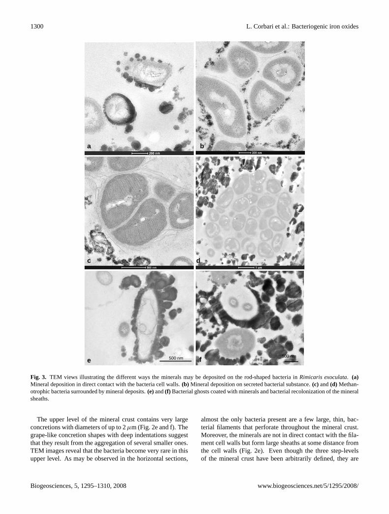
The lower level of the mineral crust (Fig. 2a and b) corre-sponds to the lower side of the mineral crust and is charac-terised by a heterogeneous distribution of mineral particles, adistribution that is very fine and seems to correspond to clus-ters of less than 500 nm size (Fig. 2a). Their morphologyseems directly related to the bacterial shape. TEM images(Fig. 2b) reveal that the lower level is characterised by a highdensity of rod-shaped bacteria; mineral precipitation occurson the cell walls of these bacteria (Fig. 3a). The mineralprecipitates as individual globular concretions of 10 to 30nm diameter, that tend to undergo agglomeration into largerconcretions. High resolution images (Fig. 3b and c) revealthat the minerals appear as diffuse precipitates on secretionsof the bacteria, i.e. exopolysaccharides. TEM observationsof vertical sections of all the analysed samples reveal thatthe bacteria form a dense community close to the cuticle,a location in which mineral particles are almost absent be-cause of the probable presence of bacterial secretion (Fig. 4).Interestingly, the methanotrophic bacteria, characterised bytheir stacks of intracytoplasmic membranes (Fig. 3c), are fre-quently observed at the lower level of the crust. Most oftenthey aggregate in isolated groups that remain free of any min-eral precipitates (Fig. 3d).
Biogeosciences, 5, 1295–1310, 2008 www.biogeosciences.net/5/1295/2008/

L. Corbari et al.: Bacteriogenic iron oxides 1299
5 µm
5 µm
2 µma b
c d
e f
Corbari et al., Figure 2bg-2008-0022
Fig. 2. The three levels of the mineral crust. Comparison between the electron back scattering images of the polished-thin sections (verticalsections) revealing the mineral densities and structures (left column) and the TEM micrographs of horizontal cross-sections exhibitingmineral associated with bacterial community (right column) at the lower level (aandb), the median level (candd) and the upper level (eandf).
The median level of the crust exhibits larger mineral con-cretions, globular in shape, (Fig. 2c and d). These globu-lar concretions often meet to form larger ones that exhibita botryoidal structure. In the horizontal cross-sections, theaggregate density appears rather heterogeneous and consistsof highly mineralised patches interspersed with bacteria richareas. The bacterial density in the median layer is alwayssmaller than in the lower layer; there are fewer rod-shapedbacteria. Moreover, ghosts of bacteria are also observed
(Fig. 3e and f) in TEM images. These ghosts have bacte-rial shapes that are completely enclosed in a heavy mineralsheath. Sometimes the bacteria are still present but appeareither to be damaged or as membrane remain (Fig. 3e and f).In other cases, the mineral sheath appears to be empty or tohave been recolonised by other rod-shaped bacteria. Theseobservations suggest that mineral formation may influencethe survival rate of the bacteria.
www.biogeosciences.net/5/1295/2008/ Biogeosciences, 5, 1295–1310,2008
1300 L. Corbari et al.: Bacteriogenic iron oxides
500 nm 500 nm
c
e
d
f
a b
Corbari et al., Figure 3bg-2008-0022
Fig. 3. TEM views illustrating the different ways the minerals may be deposited on the rod-shaped bacteria inRimicaris exoculata.(a)Mineral deposition in direct contact with the bacteria cell walls.(b) Mineral deposition on secreted bacterial substance.(c) and(d) Methan-otrophic bacteria surrounded by mineral deposits.(e)and(f) Bacterial ghosts coated with minerals and bacterial recolonization of the mineralsheaths.
The upper level of the mineral crust contains very largeconcretions with diameters of up to 2µm (Fig. 2e and f). Thegrape-like concretion shapes with deep indentations suggestthat they result from the aggregation of several smaller ones.TEM images reveal that the bacteria become very rare in thisupper level. As may be observed in the horizontal sections,
almost the only bacteria present are a few large, thin, bac-terial filaments that perforate throughout the mineral crust.Moreover, the minerals are not in direct contact with the fila-ment cell walls but form large sheaths at some distance fromthe cell walls (Fig. 2e). Even though the three step-levelsof the mineral crust have been arbitrarily defined, they are
Biogeosciences, 5, 1295–1310, 2008 www.biogeosciences.net/5/1295/2008/

L. Corbari et al.: Bacteriogenic iron oxides 1301
2 µm
Corbari et al., Figure 4bg-2008-0022
Fig. 4. Bacteria-mineral interactions inRimicaris exoculata. TEMview of the lower level (vertical cross-section) where rod-shape bac-teria are very abundant. Note that the bacteria seem to produce somesubstance which prevents any direct mineral deposition close to thislayer.
representative of the different phases of the mineral forma-tion and are characterised by an inverse correlation, fromlow to upper levels, between the amount of mineral depositspresent and the bacterial density.
3.2 Iron oxides identified by Mossbauer spectroscopy
Mossbauer spectroscopy has been used to identify both thenature and oxidation states of the iron oxides found in thenative minerals through the measurements of the iron-57 iso-mer shift and quadrupole splitting. The Mossbauer spec-tra, obtained at 85 and 295 K and between 4.2 and 60 K areshown in Figs. 5a and b, respectively. At 85 and 295 K thespectra consist of broadened quadrupole doublets, whereasbelow 60 K they consist of a superposition of broadened dou-blets and sextets. The observed temperature dependence ofthe Mossbauer spectra is typical of small superparamagneticparticles. The spectra have been fit with two symmetricquadruople doublets and one to three magnetic sextets; theaverage hyperfine parameters are given in Table 1.
The weighted average isomer shift,<δ>, is typical ofiron(III) (Shenoy et al., 1978) and spectral analysis indi-cates that at least 98 % of the iron in the mineral crustmust be present as iron(III); two percent by spectral areais the approximate detection limit for the presence of anyiron(II). The average hyperfine parameters observed at 295and 4.2 K are typical (Murad et al., 1987) of two-line fer-rihydrite. Two-line ferrihydrite, Fe5HO8·4H2O, is a poorlycrystalline mineral that forms spherical nanoparticles with
94
95
96
97
98
99
100
92
94
96
98
100
-4 -3 -2 -1 0 1 2 3 4
Perc
ent T
rans
mis
sion
Velocity, mm/s
295 K
85 K
98.0
98.5
99.0
99.5
100.0
-12 -10 -8 -6 -4 -2 0 2 4 6 8 10 12Velocity, mm/s
96.0
97.0
98.0
99.0
100.0
97.0
97.5
98.0
98.5
99.0
99.5100.0
Per
cent
Tra
nsm
issi
on
98.4
98.8
99.2
99.6
100.0
50 K
4.2 K
40 K
45 K
95.0
96.0
97.0
98.0
99.0
100.0
60 K
a
b
Corbari et al., Figure 5
bg-2008-0022
Fig. 5. (a)The 85 and 295 K iron-57 Mossbauer spectra of mineralsfrom the crust collected on the branchiostegites of three shrimps.(b) The iron-57 Mossbauer spectra of two superimposed bran-chiostegites parts of one shrimp obtained at the indicated temper-atures.
www.biogeosciences.net/5/1295/2008/ Biogeosciences, 5, 1295–1310, 2008

1302 L. Corbari et al.: Bacteriogenic iron oxides
Table 1.Mossbauer spectral parameters obtained for theRimicaris exoculatahydrothermal shrimp.
Compound T , <δ>, <1EQ> , <H>, <0>, Area, Abs. Area, AssignmentK mm/s∗ mm/s T mm/s % (%ε) (mm/s)
Rainbow 295 0.358 0.78 0 0.46 100 7.302 Superparamagnetic iron(III)225 0.403 0.78 0 0.38 100 10.106 Superparamagnetic iron(III)155 0.439 0.79 0 0.39 100 11.010 Superparamagnetic iron(III)85 0.465 0.80 0 0.42 100 11.064 Superparamagnetic iron(III)60 0.464 0.80 0 0.42 82 6.196 Superparamagnetic iron(III)
0.461 −0.04 39.6 1.13 18 1.314 Partially blocked iron(III)50 0.464 0.80 0 0.42 36 3.6935 Superparamagnetic iron(III)
0.463 −0.05 21.6 4.35 64 6.693 Partially blocked iron(III)45 0.464 0.80 0 0.42 23 2.332 Superparamagnetic iron(III)
0.463 −0.06 28.1 3.54 77 7.753 Partially blocked iron(III)40 0.464 0.80 0 0.42 8 0.901 Superparamagnetic iron(III)
0.463 −0.06 24.0 3.75 92 10.075 Partially blocked iron(III)4.2 0.481 −0.045 47.8 0.56 100 10.727 Blocked iron(III)
∗ Theisomer shifts are given relative to room temperatureα-iron powder.
Table 2.Typical elemental EDX microanalysis of the mineral crustof Rimicaris exoculata. Data are expressed as both weight andatomic percents.
Element Wt % At %
C K 37.5 57.3O K 25.5 29.2MgK 0.4 0.3SiK 1.7 1.1P K 0.6 0.3S K 0.3 0.2CIK 0.3 0.2CaK 0.6 0.3FeK 32.7 10.8
a diameter of between 2 and 7 nm, a diameter that dependsupon both the crystallinity of the material and the presenceof impurities. Also for the same reasons, the average hy-perfine field observed at 4.2 K may be reduced from 50 to46.5 T. In the specimens understudy, the average hyperfinefield of 47.8 T corresponds to a reasonably well crystallisedsample. The blocking temperature, i.e. the temperature atwhich the absorption areas of the doublets and sextets areequal, is 55±2 K. By using the anisotropy constant (Muradet al., 1987) of 4×10⁴ J/m3 for 5 nm Fe5HO8·4H2O particles,an average particle diameter of 5.6 nm is obtained.
3.3 Quantitative EDX microanalyses
BSE images of polished thin slices of frozen specimens pro-vide a detailed map of the mineral particles in the crust.EDX microanalyses (n=14) performed in ESEM give accu-rate qualitative and quantitative determinations of the ele-
8.0
C Ka
O Ka
Si KaP Ka
Ca Ka
Fe Ka
Fe Kb
Fe La
S KaCl KaMg Ka
2.0 4.0 6.00KeV
Corbari et al., Figure 6bg-2008-0022
Fig. 6. Elemental EDX microanalyses of the mineral crust ofRim-icaris exoculata.(a) A typical spectrum obtained on mineral parti-cles of up to 2µm diameter. The peaks are labelled with the EDXline of the corresponding element.
mental composition of the mineral deposits. These micro-analyses reveal the predominance of iron, with aKα peakat 6.400 keV and aKβ peak at 7.059 keV, and oxygen witha Kα peak at 0.5425 keV, in the mineral crust (Fig. 6). Mi-nor amounts of silicon with aKα peak at 1.740 keV, calciumwith a Kα peak at 3.690 keV and aKβ peak at 4.012 keV,phosphorus with aKα peak at 2.013 keV, magnesium witha Kα peak at 1.253 keV, and sulphur with aKα peak at2.307 keV, have also been detected. Elemental quantitativeanalyses yield the weight and atomic percentages of the ele-ments present, see Table 2. In order to determine the relativeamount of the iron oxides and other minerals in the concre-tions, we subtract the C of the C-coating and the C, O and Clof the embedding resin. It was also assumed that the minorelements such as Si, Ca, Mg, S and P are present as silicate
Biogeosciences, 5, 1295–1310, 2008 www.biogeosciences.net/5/1295/2008/

L. Corbari et al.: Bacteriogenic iron oxides 1303
Table 3. Mineralogical characterisation of the mineral crust inRimicaris exoculata. Data are expressed in atomic percentages and resultsfrom EDX quantitative analyses (n=14) of polished thin slices of minerals from two frozen specimens. Mean values have been calculatedafter removing the peripheral elements inherent to the resin and the carbon coating. The amount of available oxygen for each mineral orligands is calculated by assuming the presence of SO2−
4 and PO3−
4 anionic groups as well as silicate with at least two O atoms associatedto each Si atom. The percentages of mineral constituents and ligands have been calculated under the assumption of their most probableoccurrence. All values are mean± standard deviation.
Elements O Fe Si S P Ca Mg
Mean 67.8±1.4 27.3±0.7 2.6±0.4 0.5±0.1 0.5±0.2 0.7±0.1 0.5±0.3Available O − 58.5±3.1 5.3±0.8 2.1±0.4 2.0±0.9 − −
Minerals and FexOy(OH)z + vH2O Si (O2) SO2−
4 PO3−
4 Ca, MG TotalInorganic ligands 85.8±2.9 7.9±1.2 2.6±0.5 2.5±1.2 1.2±0.49 9.9±0.2
cationsand anions (sulphate and phosphate) respectively asthey appear as the most probable ligand forms, as suggestedby Chatellier et al. (2001, 2004) and Fortin and Langley(2005). The associated oxygen was calculated according tothe stoichiometric ratio in the forms of SO2−
4 and PO3−
4 andreported as available O in Table 3. For silicate, a minimumof two oxygen atoms arbitrary attributed to each Si atom thatappears as Si(O2) in Table 3. The remaining O was consid-ered as available for Fe and taken into account in the calcula-tion of the Fe/O ratio. The results of these calculations givethe relative contribution of all the mineral constituents of theconcretions (Table 3). They show that iron(III) oxides corre-spond to ca. 85 At % (ca. 90 Wt %) of the minerals presentin the crust, as confirmed by Mossbauer spectroscopy. Thisvalue does not include hydrogen atoms (not measurable) thatmay represent 25–50% of the atoms in the mineral, accord-ing to the general Ferrihydrite formula and hydration statebut does never exceed 2 Wt%. The contribution of the inor-ganic ligands in the concretions is approximately evaluated at8% for Si(O2), 2.5% for SO2−
4 and 2.5% of PO3−
4 and 1.2%for Ca and Mg. It is very probable that Ca2+ and Mg2+ arecombined in salts with the anions SO2−
4 and PO3−
4 becauseof the perfect accordance with the stoichiometric ratio in (Ca,Mg) SO4 and (Ca, Mg)3 (PO4)2. Ca, Mg salts could thus rep-resent approximately 6% in the concretions.
Comparative elemental EDX microanalyses carried out on12 polished thin slices of glutaraldehyde-fixed specimens re-veal very similar proportions of the minor ligands suggest-ing that they are not solubilised or removed by the aqueouspreparation procedure. However, these analyses differ fromthose of the frozen sample by the Fe/O ratio in the iron ox-ide, i.e. after the subtraction of the oxygen linked to minorelements, see Table 4. The Fe/O ratio is 0.47 in the frozenspecimens while it reaches 0.60 in the glutaraldehyde-fixedspecimens. These measured Fe/O ratios are statistically dif-ferent as is indicated by a pairedt-test which yieldst=7.1,d.f.=11, andP=0.00002. Hence, the atomic percentage ofoxygen is lower in the glutaraldehyde-fixed specimens, inwhich the glutaraldehyde could act as a reducing agent andmodify the iron oxidation state in the mineral particles.
Table 4. Comparison of the mineralogical composition be-tween frozen and glutaraldehyde-fixed specimens. EDX elemen-tal quantitative analysis of fourteen frozen samples and twelveglutaraldehyde-fixed samples. The data have been obtained withthe same experimental procedure. All values are mean± standarddeviation.
Frozensp. Glutharaldehyde-fixed sp.
Elements O Fe O Fe
Mean 67.8±1.4 27.3±0.7 64.5±1.4 28.3±1.3
Available O − 58.5±3.1 − 47.8±3.0
Ratio Fe/O 0.47±0.03 0.60±0.05a
a Significantlydifferent.
3.4 Structure and composition of the crust minerals
TEM images obtained on U/Pb contrasted ultra-thin verticalsections indicate that most of the mineral concretions, whichhave diameters ranging from 200 to 600 nm, exhibit layeredfeatures in the lower level of the crust (Fig. 7a and b). Mostof the specimens exhibit a multilayered pattern with a peri-odicity of a few nanometers, a pattern that suggests that theseconcretions are composed of ca. 5 to 10 successive strata. Allof these strata appear as concentric growth layers originatingfrom a unique nucleation centre, as multiglobular particlesthat change their shape and, for the outer particles, followthe outer particle border (Fig. 7b). Neighbouring concretionsalso exhibit layered patterns that may correspond to similarmineral deposition sequences (Fig. 7a). Mineral nucleationand deposition occur either close to the rod-shaped bacteriawalls or in their near-neighbour environment. Several nucle-ation centres are located close to the same bacteria and theaccumulation of strata leads to the aggregation of mineralparticles that, as a consequence, exhibit a grape-like shape(Fig. 7b). STEM-HAADF images of particles from frozenspecimens give an inverted mass contrast of the strata. Theseimages reveal the reality of the strata in terms of the changingaggregate density and/or the composition within the particles(Fig. 7c and d).
www.biogeosciences.net/5/1295/2008/ Biogeosciences, 5, 1295–1310,2008

1304 L. Corbari et al.: Bacteriogenic iron oxides
1 µm
a
c Bd
B
200 nm
b
Corbari et al., Figure7bg-2008-0022
Fig. 7. (a)and(b) TEM views of the mineral particles in the lower level of the crust. The occurrence of contrasted strata is clearly visible.(c) and (d) STEM images of ultrathin sections of non-contrasted mineral particles revealing the different nature of the mineral strata.B
indicates the bacteria.
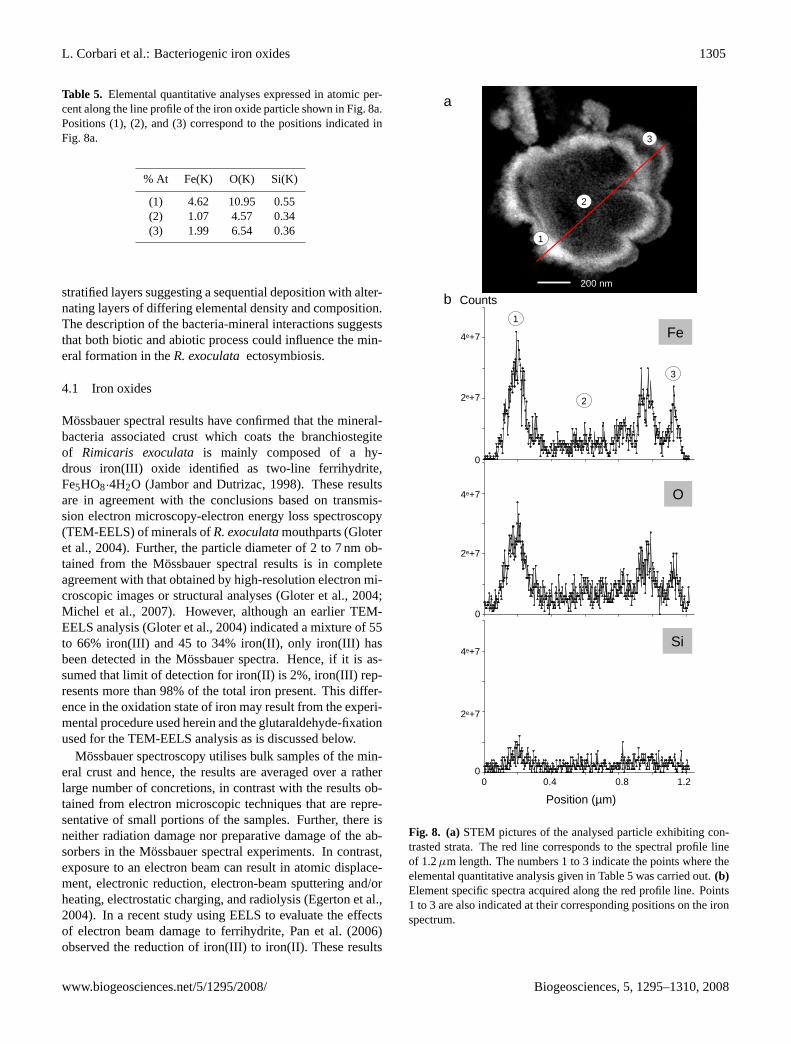
In order to determine the exact nature of these strata, EDXnanoanalyses have been carried out by STEM along a direc-tion perpendicular to the strata of some particles observedin the ultrathin sections. The experimental procedure forobtaining these line profiles of 1 to 1.5µm length is basedon the acquisition of a sequence of ca. 700 EDX spectraper line profile. Subsequent data analysis can differentiatedifferent elements through the number of counts that corre-spond to a given element found along the line. Figure 8 illus-trates a typical elemental profile for iron, oxygen, and siliconalong a 1.2µm line through a stratified mineral particle. Theclear lighter coloured strata in Fig. 8a correspond to a higheriron and oxygen content than is found in the dark strata, seeFig. 8b. In spite of a low number of counts, the silicon profileseems to correlate well with those of iron and oxygen. In or-der to more accurately characterise the nature of the mineralstrata, quantitative elemental analyses have been performedat specific points, i.e. points 1, 2, and 3 in Fig. 8a, at well
separated strata along the line profile. The results (Table 5)confirm that the iron and oxygen atomic percentages are highat the lighter strata, points 1 and 3, and very low in the darkstrata, position 2. The strata result thus rather from changesin the aggregation density than from real compositional dif-ferences.
4 Discussion
A multifacitated analysis carried out on the mineral crust ofR. exoculatareveals a mineral content of 85% iron(III) oxideand associated with 15 % of minor inorganic ligands, possi-bly present as silicate and (Ca, Mg) sulphate and phosphate.The Mossbauer spectral results indicate that the iron(III)oxide corresponds to two-line ferrihydrite, Fe5HO8·4H2O,which is present in nanometric particles of less than 5 nm di-ameter. A transmission and scanning electron microscopicstudy of the mineral crust reveals the concretions exhibit
Biogeosciences, 5, 1295–1310, 2008 www.biogeosciences.net/5/1295/2008/
L. Corbari et al.: Bacteriogenic iron oxides 1305
Table 5. Elemental quantitative analyses expressed in atomic per-cent along the line profile of the iron oxide particle shown in Fig. 8a.Positions (1), (2), and (3) correspond to the positions indicated inFig. 8a.
% At Fe(K) O(K) Si(K)
(1) 4.62 10.95 0.55(2) 1.07 4.57 0.34(3) 1.99 6.54 0.36
stratifiedlayers suggesting a sequential deposition with alter-nating layers of differing elemental density and composition.The description of the bacteria-mineral interactions suggeststhat both biotic and abiotic process could influence the min-eral formation in theR. exoculataectosymbiosis.
4.1 Iron oxides
Mossbauer spectral results have confirmed that the mineral-bacteria associated crust which coats the branchiostegiteof Rimicaris exoculata is mainly composed of a hy-drous iron(III) oxide identified as two-line ferrihydrite,Fe5HO8·4H2O (Jambor and Dutrizac, 1998). These resultsare in agreement with the conclusions based on transmis-sion electron microscopy-electron energy loss spectroscopy(TEM-EELS) of minerals ofR. exoculatamouthparts (Gloteret al., 2004). Further, the particle diameter of 2 to 7 nm ob-tained from the Mossbauer spectral results is in completeagreement with that obtained by high-resolution electron mi-croscopic images or structural analyses (Gloter et al., 2004;Michel et al., 2007). However, although an earlier TEM-EELS analysis (Gloter et al., 2004) indicated a mixture of 55to 66% iron(III) and 45 to 34% iron(II), only iron(III) hasbeen detected in the Mossbauer spectra. Hence, if it is as-sumed that limit of detection for iron(II) is 2%, iron(III) rep-resents more than 98% of the total iron present. This differ-ence in the oxidation state of iron may result from the experi-mental procedure used herein and the glutaraldehyde-fixationused for the TEM-EELS analysis as is discussed below.
Mossbauer spectroscopy utilises bulk samples of the min-eral crust and hence, the results are averaged over a ratherlarge number of concretions, in contrast with the results ob-tained from electron microscopic techniques that are repre-sentative of small portions of the samples. Further, there isneither radiation damage nor preparative damage of the ab-sorbers in the Mossbauer spectral experiments. In contrast,exposure to an electron beam can result in atomic displace-ment, electronic reduction, electron-beam sputtering and/orheating, electrostatic charging, and radiolysis (Egerton et al.,2004). In a recent study using EELS to evaluate the effectsof electron beam damage to ferrihydrite, Pan et al. (2006)observed the reduction of iron(III) to iron(II). These results
Counts
Position (µm)
0
2e+7
4e+7
0
2e+7
4e+7
0 0.4 0.8 1.2
O
Fe
0
2e+7
4e+7Si
1
2
1
2
200 nm
3
3
a
b
Corbari et al., Figure 8bg-2008-0022
Fig. 8. (a)STEM pictures of the analysed particle exhibiting con-trasted strata. The red line corresponds to the spectral profile lineof 1.2µm length. The numbers 1 to 3 indicate the points where theelemental quantitative analysis given in Table 5 was carried out.(b)Element specific spectra acquired along the red profile line. Points1 to 3 are also indicated at their corresponding positions on the ironspectrum.
www.biogeosciences.net/5/1295/2008/ Biogeosciences, 5, 1295–1310, 2008
1306 L. Corbari et al.: Bacteriogenic iron oxides
highlight how investigations carried out under high vacuumin a transmission electron microscope may cause substantialand perhaps unsuspected changes to a mineral sample.
Ferrihydrite is thus an essential mineral component of themineral-bacteria associated crust inR. exoculata. The mostcommon extracellular biogenic iron oxides include oxyhy-droxides, e.g. goethite, lepidocrocite, akaganeite, and poorlyordered phases, e.g. two-line and six-line ferrihydrite (Cor-nell and Schwertmann, 2003; Fortin and Langley, 2005).Furthermore, it is commonly accepted that the product of mi-crobial micro-aerobic iron(II) oxidation is often identified asa poorly crystalline ferrihydrite. The observation of two-lineferrihydrite thus supports the hypothesis of the presence ofiron-oxidisers among the ectosymbiotic bacterial communityof R. exoculata(Zbinden et al., 2004) and validates the firstobservations on bacterial cultures (Cambon-Bonavita, pers.com.). In hydrothermal environments, ferrihydrite has pre-viously been identified because it is commonly intermixedwith lithoautotrophic iron(II) oxidizing bacteria, that act as acausative agent in the formation of the ferrihydrite (Emer-son and Moyer, 2002; Kennedy et al., 2003, 2004; Littleet al., 2004). Very few vent animals have been discoveredto live in close association with iron oxide deposits. To thebest of our knowledge, only the scaly-foot gastropod foundin the hydrothermal vents at the Indian Ridge has been shownto exhibit scale-shaped structures, mineralised with iron sul-phides on its foot (Goffredi et al., 2003; Waren et al., 2003).These structures are associated with bacteria but an iron iso-topic analysis indicates that sulphur and iron in the scleritesoriginate from hydrothermal fluids rather than from bacteria(Suzuki et al., 2006). Herein, the presence of ferrihydritein close interaction with hydrothermal metazoan has beendiscovered for the first time, in the specific case of the ventshrimpR. exoculata.
Elemental quantitative analyses have been performed todetermine the Fe/O ratio. Because of the influence of thesample preparation for ultrastructural and elemental analy-ses, an alternative experimental procedure has been adoptedin this study to reduce damage to the samples containing bothbacteria and minerals. The samples for mineralogical anal-yses were frozen until used and were directly dehydrated byethanol to avoid both air-contact and any aqueous chemicalfixation. This procedure is based on the work of Mavro-cordatos and Fortin (2002), who investigated the influenceof sample preparation on poorly ordered biotic hydrous ironoxide. To assess the impact of sample fixation on the compo-sition of theR. exoculataminerals, Fe/O ratios determinedthrough identical procedures have been compared betweenfrozen and glutaraldehyde-fixed samples. The Fe/O ratioof the glutaraldehyde-fixed minerals is significantly differ-ent from that of the frozen minerals. Thus sample prepara-tion may modify the oxidation state of iron. The use of glu-taraldehyde, a reducing agent, in addition to electron beamdamage in TEM-EELS studies can explain the higher per-centage of iron(II) reported by Gloter et al. (2004) in their
mineral samples. The Fe/O ratio in frozen minerals has beenmeasured to be 0.47, after subtraction of the oxygen associ-ated with inorganic elements. This value cannot be comparedwith the Fe/O ratio of 0.33 obtained by Gloter et al. (2004)because both their analytical approach and mineralogical in-terpretations are quite different from those used herein. Incontrast, the calculated Fe/O ratio of 0.47 may be comparedwith the 0.417 ratio obtained for abiotic ferrihydrite (Mavro-cordatos and Fortin, 2002 and references therein). Moreover,the calculated Fe/O ratio of 0.47 in frozen minerals is similarto the Fe/O ratio of 0.48 (Mavrocordatos and Fortin, 2002).In this study, quantitative TEM-EELS analyses have beenperformed on ferrihydrite sample experimentally obtainedby the precipitation of hydrous ferric oxides in the presenceof bacteria exhibiting extracellular polymers. Despites ourslightly different methodologies, the similar Fe/O ratio de-termined in the presence of bacteria, suggest the influence ofthe bacterial ectosymbiosis on the iron oxide formation.
In conclusion, for future research onR. exoculatamin-erals, sample preparation and, more specifically, the dryingprocess and fixation of the biological samples must be ac-curately delineated because they both influence the surfaceproperties of ferrihydrite and hence, the iron oxidation stateand the Fe/O ratio.
4.2 Intrinsic inorganic constituents
Even though ferrihydrite represents the main component ofthe mineral crust inR. exoculata, elemental and quantitativeanalyses of the mineral particles have revealed the presenceof minor elements, such as Si, P, Ca, S, and Mg. These el-ements are not considered as impurities (Gloter et al., 2004)but rather to form intrinsic inorganic constituents or ligandsas suggested by Chatellier et al. (2004) because these au-thors found that their presence during the oxidation processcan affect the mineralogy as well as the size and structureof the iron oxide particles. Because of their large surfaceareas, small particles of natural biogenic iron oxides gen-erally contain adsorbed elements and ions, such as silicate,sulphate, phosphate, and manganese and aluminium cations(Fortin and Chatellier, 2003; Fortin and Langley, 2005). Forexample, nanoparticles of iron oxyhydroxide formed dur-ing the mineralisation process, can adsorb phosphate andsilicate ions (Gilbert and Banfield, 2005). The adsorptionof compounds on ferrihydrite from the surrounding aque-ous milieu can affect its subsequent mineral ordering pro-cesses. Specifically, adsorption of silicates has been foundto inhibit the conversion of ferrihydrite to more crystallineiron oxides, such as hematite and goethite (Kennedy et al.,2003; Chatellier et al., 2004). EDX nanoanalyses and ele-mental profiles performed on stratifiedR. exoculatamineralparticles have revealed that silicon is already present in theearly stages of mineral development. Thus, inR. exoculata,it is evident that inorganic ligands co-precipitate with ironoxide and are closely associated with it in the nanoparticles
Biogeosciences, 5, 1295–1310, 2008 www.biogeosciences.net/5/1295/2008/
L. Corbari et al.: Bacteriogenic iron oxides 1307
because the Si portion cannot be located separately from theiron oxide even on the nanometric scale reached in STEM. Inthe global characterisation of the mineral particles, we haveassumed that they correspond to separated compounds suchas silicate, Ca2+, Mg2+, SO2−
4 , and PO3−
4 , even though theauthors consider that silicate, sulphate, phosphate, and mag-nesium and calcium cations are substituted or adsorbed toferrihydrite. Their presence could also influence or stabilisethis poorly crystalline form of iron oxide. Considering thestoichiometric ratio of Ca, Mg and P, it is probable that SO2−
4and PO3−
4 are associated with Ca and Mg in (Ca, Mg) salts.In order to further assess the distribution of the intrinsic
inorganic constituents, EDX spectra acquired at line profileshave been performed on mineral particles exhibiting strati-fied features. At the atomic level, the results indicate thatboth iron and the other inorganic ligands are already associ-ated within nanosized mineral particles. These observationsreveal that iron, oxygen, and silicon proportions are constantbut that the stratification is mainly the consequence of paral-lel variations of the concentration of all the constituting ele-ments. Bacteria-associated mineral concretions appear to beidentical and stratification seems to follow the same sequencewithin neighbouring mineral particles. These observationssuggest that micro-environmental variations can occur in thenearby environment during the formation of the mineral par-ticles. Uniform patterns of stratification on neighbouringparticles can be explained by either environmental variationsfound at the shrimp growth level or by variations in the bac-terial metabolism or activities. Similar patterns of stratifica-tion have been previously described on sediment microfaciesand delineated as microstromatolite (Boulvain et al., 2001).The “microstromatolite” aspect of the mineral particles inR.exoculataprovides is in favour of the biologically-mediatedorigin of iron oxides.
4.3 Bacteria-mineral interactions
In R. exoculata, iron oxide is complexed with intrinsic inor-ganic ligands in the presence of an important bacterial com-munity. Both bacteria and the intrinsic inorganic ligands mayplay a role in mineral deposition. The term of biogenic ironoxides is commonly used to refer to iron oxide formed in thepresence of bacteria. It also includes iron oxides formed asa direct result of microbial metabolism, i.e. through enzymeactivities, or by passive mechanisms through which bacte-rial secretions trigger the formation and precipitation of ironoxides minerals (Fortin and Chatellier, 2003).
In R. exoculata, mineral deposition only occurs when thebacterial community is well-developed on the inner side ofthe branchiostegite (Corbari et al., 2008). This observationboth contradicts the idea that iron oxide deposition couldonly result from a passive chemical-induced precipitationand supports the idea that bacteria must participate in min-eral formation. Ferrihydrite formed in the absence of bac-teria may be metastable and, typically after few days, trans-
forms into a more structurally ordered iron oxide, such ashematite or goethite (Cornell and Schwertmann, 2003). Ex-periments performed on bacteriogenic ferrihydrite mineralsobtained from the hydrothermal vents in the Axial Volcano(Pacific Ridge) demonstrated that even if they were subjectedto heating of up to 80◦C, these minerals did not undergo aphase transition, and therefore suggested that the presence ofbacteria inhibited the ferrihydrite transformation (Kennedyet al., 2004). Hence, inside the gill chamber ofR. exoculata,ferrihydrite deposits appear to result from the presence of anabundant bacterial community and their formation is withoutany doubt biologically-mediated. If the moult cycle is usedas a time-scale, the first ferrihydrite deposits are only ob-served when the bacterial density reaches a maximum in theearly preecdysial individuals, i.e. the light-red or medium-redindividuals, in stages D0 to D1 (Corbari et al., 2008). Duringthe ten day moult cycle of the vent shrimp, the first mineralparticles appear as ferrihydrite (Corbari and Compere, un-published data) as early as the second post-moult day andcontinue to deposit until the tenth day, just before exuvia-tion. Hence, we conclude that the bacterial community couldcontribute to the stabilisation of the iron oxide in the formof ferrihydrite. However, the bacteria organic moieties canhinder its transformation into a more crystallized iron oxide(Kennedy et al., 2004). InR. exoculata, such bacteria or-ganic moieties can act together with the minor ligands and/orfavour their incorporation in the concretions.
TEM observations of the bacterial morphotypes andtheir mineral interactions help to elucidate the biologically-mediated origin of the ferrihydrite deposits inside the gillchamber ofR. exoculata. Different ways of mineral deposi-tion have been identified, based on the recurrent observationsof both bacterial morphotypes and mineral morphologies.Two association modes between minerals and rod shapedbacteria have been observed. In the first, iron oxide depo-sition occurs in close contact to the rod cell walls and, in thesecond, the iron oxide precipitates on polysaccharide or pro-teinaceous extracellular secretions at a significant distancefrom the bacterial cells. Such bacterial-mineral relationshipshave previously been described inR. exoculata, suggestingthat rods are mainly involved in iron oxide formation (An-derson et al., 2008). The precipitation of iron oxides on ornear the bacterial cell walls raises the question of the passiveor active implication of the bacteria in their deposition.
Passive production of biogenic iron oxide is related to thereactivity of the bacterial cell walls. Mineral formation onthe bacteria is generally not controlled by the organism but,rather results from the chemistry of the cell environment andthe physicochemistry of the bacterial surface, (Fortin andLangley, 2005). This implies the adsorption and/or nucle-ation of iron oxide particles on bacterial cell walls that sim-ply acts as a passive deposition template (Konhauser, 1997;Fortin and Chatellier, 2003; Klapper and Straub, 2005,).Whatever the type of surface structure the cell may have,the main charged chemical groups found at neutral pH are
www.biogeosciences.net/5/1295/2008/ Biogeosciences, 5, 1295–1310, 2008

1308 L. Corbari et al.: Bacteriogenic iron oxides
carboxyl, phosphoryl, and amino groups (Douglas and Bev-eridge, 1998). We thus suspect that similar mineral-bacteriaassociations take place inR. exoculataand that passive de-position of iron oxides could occur. However, the obser-vation of bacterial ghost surrounded by dense mineral de-posits suggest that the precipitation on iron (III) oxide inthe vicinity of the cell or at the cell surface is harmful forthe cells, probably by limiting substrate diffusion and up-take as assumed by Hallberg and Ferris (2004). Moreover,these bacteria encrusted in iron oxides are probably not iron-oxidisers because iron oxides seem to improve their sur-vival rates. Indeed, the most studied iron-oxidising bacteria,the neutrophilic aerobic iron(II) oxidisers fromGallionellaandLeptothrixgenus, are known to produce extracellular or-ganic polymers that nucleate iron(III) precipitates at somedistance of the bacterial envelope, avoiding encrustation thatcould cause cell death (Hallberg and Ferris, 2004; Kappleret al., 2005). Thus this active production of iron(III) ox-ide by metabolic oxidation of environmental iron(III) fol-lowed by bacterial-induced iron(III) oxide deposition on spe-cific bacterial secretions must be distinguished from passivebiologically-induced iron oxide precipitation. Hence, if iron-oxidizing bacteria are present among the ectosymbiotic com-munity in R. exoculata, they could use this strategy to keeptheir metabolism active and only those with extracellular se-cretion would be involved in iron oxide formation. The pres-ence of a mineral boundary between the dense rod popula-tion, located at the lower layer and the mineralised area sug-gests a strategy involving the production of an extracellularorganic material in order to prevent mineral deposition di-rectly on the bacterial cell walls. This could also be true forthe sheathed large filaments.
Interestingly, the appearance of methanotrophic bacte-ria clusters also support the above described mechanism inwhich bacteria exude organic substances to prevent any min-eral deposition directly on their cell walls. Further, the pres-ence of methanotrophic bacteria (Zbinden et al., 2008) sug-gests a more diversified bacterial community than previouslymentioned (Segonzac et al., 1993; Zbinden et al., 2004; Cor-bari et al., 2008). Herein, methanotrophic bacteria show in-tact internal structures, i.e. stalks, and are distributed in clus-ters that indicate an active metabolism.
The three step-levels in the mineral crust formation previ-ously described (Corbari et al., 2008) indicates that iron ox-ide particle growths are continuously initiated from the lowerlevel, in close association with growing bacteria and subse-quently grow into the median and upper levels. The min-eralisation within the gill chamber could be described as adynamic process in which particles increase in size and aresimultaneously pushed upward by the formation of new par-ticles. As has been illustrated herein, the lower level exhibitsthe highest bacterial density and is mainly composed of rodbacteria. This level may be considered as a bacterially ac-tive layer and its evolution in time, based on the moult cycle,shows continuous growth (Corbari et al., 2008). Neverthe-
less, the concretions in their final state may results from bi-otic as well as from abiotic iron oxide precipitation, owingthat, if present, iron-oxidising bacteria have to compete withabiotic oxidation of iron (Schmidt et al., 2008).
Finally, the weak mineral deposition, maximal rod-shapedbacterial density, and presence of extracellular secretionssuggest that iron-oxidising bacteria may be located in thislayer, a layer that may act as a potential reserve for activeectosymbiotic bacteria.
5 Conclusions
The multidisciplinary approach used in the present studyprovides new details about the iron oxide deposits associ-ated with ectosymbiotic bacteria inRimicaris exoculata. Themineral crust has been identified as a dense layer of two-lineferrihydrite nanoparticles associated with other intrinsic in-organic ligands. Ultrastructural observations and analyticaldata give evidences of the biologically induced depositionof iron oxides and support the role of bacterial morphotypesin active production of iron oxides. The process of miner-alisation in the gill chambers ofR. exoculataremains com-plex (co-occurrence of biotic and abiotic processes) becausethe combined effects of the intrinsic inorganic constituentsand the bacterial influence are difficult to disentangle. Butthe evolution of the bacterial density in the three levels ofthe mineral crust is closely related to the amount of iron de-posited and it is proposed that the lower level is the likely re-gion where the iron-oxidising bacteria could be located. Butthe presence of a more diversified bacterial community raisesthe question on the metabolic or genetic diversity of thesebacteria.
Because the main studies onR. exoculataectosymbiosishave been performed on shrimps from the vent site Rainbow,the influence of the chemical vent environment should bestudied in the future by comparing ectosymbiosis and its as-sociated minerals inR. exoculataspecimens collected at dif-ferent vent sites, for instance TAG, Logatchev, and Snake Pit.This indirect approach could be used to evaluate how repre-sentative is theR. exoculataectosymbiosis and, thus to deter-mine whether iron oxidation represents the most favourableenergetic-pathways for ectosymbiotic bacteria (Schmidt etal., 2008).
Acknowledgements.The authors thank A. Godfroy, the chiefscientist of the EXOMAR cruise, as well as the captain and crewof the RV “Atalante” and the ROV “Victor” team. The authors alsowish to express their appreciation to N. Decloux for her excellenttechnical assistance with transmission and scanning electronmicroscopy. This work was partly funded with the help of theMOMARNET program. The fellowship of L. Corbari and a partof this work were supported by the Belgian Fund for Joint BasicResearch FNRS (F.R.F.C Belgium, conventions no. 2.4594.07.F).The authors also thank the centre of Microscopy of Liege (CATµ;dir. R. Cloots) for giving access to high performance equipment
Biogeosciences, 5, 1295–1310, 2008 www.biogeosciences.net/5/1295/2008/

L. Corbari et al.: Bacteriogenic iron oxides 1309
EM, funded by F.R.F.C and FEDER. F. Grandjean acknowledges,with thanks, the financial support of the FNRS, Belgium, throughgrants 9.456595 and 1.5.064.05.
Edited by: K. Kusel
References
Anderson,L., Halary, S., Lechaire, J.-P., Boudier, T., Frebourg,G., Marco, S., Zbinden, M., and Gaill, F.: Tomography ofbacteria-mineral associations within the deep-sea hydrothermalvent shrimpRimicaris exoculata, CR Chim., 11, 268–280, 2008.
Banfield, J. F., Welch, S. A., Zhang, H., Ebert, T. T., and Penn, L.P.: Aggregation-based crystal growth and microstructure devel-opment in natural iron oxyhydroxide biomineralization products,Science 289, 751–754, 2000.
Boulvain, F., De Ridder, C., Mamet, B., Preat, A., and Gillan,D.: Iron microbial communities in belgian frasnian carbonatemounds, Facies, 44, 47–60, 2001.
Casanova, B., Brunet, M., and Segonzac, M.: L’impact d’uneepibiose bacterienne sur la morphologie fonctionnelle decrevettes associeesa l’hydrothermalisme medio-atlantique, Cah.Biol. Mar., 34, 573–588, 1993.
Cavanaugh, C., McKiness, Z., Newton, I., and Stewart, F.: Ma-rine chemosynthetic symbioses, in: The prokaryotes, edited by:Dworkin, M., Falkow, S., Rosenberg, E., Schleifer, K., andStackebrandt, E., Springer, New York, 475–507, 2006.
Chatellier, X., Fortin, D., West, M., Leppard, G., and Ferris, F.:Effect of the presence of bacterial surfaces during the synthesisof fe-oxides by oxidation of ferrous ions, Eur. J. Miner., 13, 705–714, 2001.
Chatellier, X., West, M. M., Rose, J., Fortin, D., Leppard, G. G.,and Ferris, F. G.: Characterization of iron-oxides formed by oxi-dation of ferrous ions in the presence of various bacterial speciesand inorganic ligands, Geomicrobiol. J., 21, 99–112, 2004.
Corbari, L., Zbinden, M., Cambon-Bonavita, M.-A., Gaill, F., andCompere, P.: Bacterial symbionts and mineral deposits in thebranchial chamber of the hydrothermal vent shrimpRimicarisexoculata: Relationship to moult cycle, Aquatic Biology, 1, 225–238, 2008.
Cornell, R. and Schwertmann, U.: The iron oxides – structure, prop-erties, occurrences and uses 2nde edition, Weinheim ed., Wiley-VCH Verlag, 664 pp., 2003.
Desbruyeres, D., Biscoito, M., Caprais, J.-C., Colaco, A., Comtet,T., Crassous, P., Fouquet, Y., Khripounoff, A., Le Bris, N., andOlu, K.: Variations in deep-sea hydrothermal vent communitieson the Mid-Atlantic Ridge near the Azores plateau, Deep-seaRes. PtI, 48, 1325–1346, 2001.
Douglas, S. and Beveridge, T. J.: Mineral formation by bacteria innatural microbial communities, FEMS Microbiol. Ecol., 26, 79–88, 1998.
Drach, P. and Tchernigovtzeff, C.: Sur la methode de determinationdes stades d’intermue et son application generale aux crustaces,Vie et Milieu, 18, 595–609, 1967.
Edwards, K. J., Bach, W., McCollom, T. M., and Rogers, D. R.:Neutrophilic iron-oxidizing bacteria in the ocean: Their habitats,diversity, and roles in mineral deposition, rock alteration, andbiomass production in the deep-sea, Geomicrobiol. J., 21, 393–404, 2004.
Egerton,R. F., Li, P., and Malac, M.: Radiation damage in the TEMand SEM, Micron, 35, 399–409, 2004.
Emerson,D. and Moyer, C. L.: Neutrophilic Fe-oxidizing bacteriaare abundant at the Loihi seamount hydrothermal vents and playa major role in Fe oxide deposition, Appl. Environ. Microbiol.,68, 3085–3093, 2002.
Fortin, D., Ferris, G., and Scott, S.: Formation of Fe-silicates andFe-oxides on bacterial surfaces in samples collected near hy-drothermal vents on the southern explorer ridge in the northeastPacific ocean, American Mineralogist, 83, 1399–1408, 1998.
Fortin, D. and Chatellier, X.: Biogenic iron-oxides, Recent Re-search Developments in Mineralogy, 3, 47–63, 2003.
Fortin, D. and Langley, S.: Formation and occurrence of biogeniciron-rich minerals, Earth-Sci. Rev., 72, 1–19, 2005.
Gebruk,A., Pimenov, N., and Savvichev, A.: Feeding specializationof bresiliid shrimps in the tag site hydrothermal community, Mar.Ecol.-Prog. Ser., 98, 247–253, 1993.
Gebruk,A. V., Southward, E. C., Kennedy, H., and Southward, A.J.: Food sources, behaviour, and distribution of hydrothermalvent shrimps at the mid-atlantic ridge, J. Mar. Biol. Ass. U.K,80, 485–499, 2000.
Gilbert,B. and Banfield, J. F.: Molecular-scale processes involvingnanoparticulate minerals in biogeochemical systems, Rev. Min-eral. Geochem., 59, 109-155, 2005.
Gloter, A., Zbinden, M., Guyot, F., Gaill, F., and Colliex, C.: TEM-EELS study of natural ferrihydrite from geological-biological in-teractions in hydrothermal systems, Earth Planet. Sci. Lett., 222,947–957, 2004.
Goffredi, S., Hurtado, L., Hallam, S., and Vrijenhoek, R.: Evolu-tionary relationships of deep-sea vent and cold seep clams (mol-lusca: Vesicomyidae) of the “Pacifica” Species complex, Mar.Biol., 142, 311–320, 2003.
Hallberg, R. and Ferris, F. G.: Biomineralization byGallionella,Geomicrobiol. J., 21, 325–330, 2004.
Jambor, J. L. and Dutrizac, J. E.: Occurrence and constitution ofnatural and synthetic ferrihydrite, a widespread iron oxyhydrox-ide, Chem. Rev., 98, 2549–2586, 1998.
Kappler, A., Schink, B., and Newman, D. K.: Fe(III) mineralformation and cell encrustation by the nitrate-dependent Fe(II)-oxidizer strain bofen1, Geobiology, 3, 235–245, 2005.
Kappler, A. and Straub, K. L.: Geomicrobiological cycling of iron,Rev. Mineral. Geochem., 59, 85–108, 2005.
Kennedy, C. B., Martinez, R. E., Scott, S., and Ferris, F. G.: Sur-face chemistry and reactivity of bacteriogenic iron oxidesfromaxial volcano, juan de fuca ridge, north-east pacific ocean, Geo-biology, 1, 59–69, 2003.
Kennedy, C. B., Scott, S. D., and Ferris, F. G.: Hydrothermal phasestabilization of 2-line ferrihydrite by bacteria, Chem. Geol., 212,269–277, 2004.
Konhauser, K. O.: Bacterial iron biomineralisation in nature, FEMSMicrobiol. Rev., 20, 315–326, 1997.
Little, C., Glynn, S., and Mills, R. A.: Four-hundred-and-ninety-million-year record of bacteriogenic iron oxide precipitation atsea-floor hydrothermal vents, Geomicrobiol. J., 21, 415–429,2004.
Mavrocordatos, D. and Fortin, D.: Quantitative characterization ofiron oxides formed on bacterial walls by tem-eels, Am. Miner.,87, 940–946, 2002.
www.biogeosciences.net/5/1295/2008/ Biogeosciences, 5, 1295–1310,2008
1310 L. Corbari et al.: Bacteriogenic iron oxides
Michel, F. M., Ehm, L., Antao, S. M., Lee, P. L., Chupas, P. J.,Liu, G., Strongin, D. R., Schoonen, M. A. A., Phillips, B. L.,and Parise, J. B.: The structure of ferrihydrite, a nanocrystallinematerial, Science, 316, 1726–1729, 2007.
Murad, E. and Johnston, J. H.: Mossbauer spectroscopy applied toinorganic chemistry, G. J. Long, Ed, Plenum Press New York,507 pp., 1987.
Murad, E. and Schwertmann, U.: Iron oxide mineralogy of somedeep-sea ferromanganese crusts, Am. Miner., 73, 1395–1400,1988.
Page, H. M., Fisher, C. R. and Childress, J.: The role of suspension-feeding in the nutritional biology of a deep-sea mussel withmethanotrophic symbionts, Mar. Biol., 104, 251–257, 1990.
Pan, Y., Brown, A., Brydson, R., Warley, A., Li, A., and Powell,J.: Electron beam damage studies of synthetic 6-line ferrihydriteand ferritin molecule cores within a human liver biopsy, Micron,37, 403–411, 2006.
Polz, M. F. and Cavanaugh, C. M.: Dominance of one bacterial phy-lotype at a Mid-Atlantic Ridge hydrothermal vent site, PNAS, 92,7232–7236, 1995.
Polz, M. F., Robinson, J. J., Cavanaugh, C. M. and Van Dover, C. L.:Trophic ecology of massive shrimp aggregations at a mid-atlanticridge hydrothermal vent site, Limnol. Oceanogr., 43, 1631–1638,1998.
Pond, D., Bell, M., Dixon, D., and Sargent, J.: Occurrence of16:2(n-4) and 18:2(n-4) fatty acids in the lipids of the hydrother-mal vent shrimpRimicaris exoculata: Nutritional and trophic im-plications, Mar. Ecol.-Prog. Ser., 156, 167–174, 1997.
Rieley, G., Dover, C. L. V., Hedrick, D. B., and Eglinton, G.:Trophic ecology ofRimicaris exoculata: A combined lipid abun-dance/stable isotope approach, Mar. Biol., 133, 495–499, 1999.
Schmidt, C., Vuillemin, R., Le Gall, C., Gaill, F., and Le Bris, N.:Geochemical energy sources for microbial primary production inthe environment of hydrothermal vent shrimps, Mar. Chem., 108,18–31, 2008.
Segonzac, M., de Saint-Laurent, M., and Casanova, B.: L’enigmedu comportement trophique des crevettes Alvinocarididae dessites hydrothermaux de la dorsale Medio-Atlantique, Cah. Biol.Mar., 34, 535–571, 1993.
Shenoy, G. K., Wagner, F. E., and Kalvius, G. M.: Mossbauer iso-mer shifts, edited by: Shenoy, G. K. and Wagner, F. E., North-Holland Amsterdam, 49 pp., 1978.
Suzuki,Y., Kopp, R. E., Kogure, T., Suga, A., Takai, K., Tsuchida,S., Ozaki, N., Endo, K., Hashimoto, J., Kato, Y., Mizota,C., Hirata, T., Chiba, H., Nealson, K. H., Horikoshi, K., andKirschvink, J. L.: Sclerite formation in the hydrothermal-vent“Scaly-foot” Gastropod-possible control of iron sulfide biomin-eralization by the animal, Earth Planet. Sci. Lett., 242, 39–50,2006.
Van Dover, C., Fry, B., Grassle, F., Humphris, S., and Rona, P. A.:Feeding biology of the shrimpRimicaris exoculataat hydrother-mal vents on the mid-atlantic ridge, Mar. Biol., 98, 209–216,1988.
Waren, A. S., Bengtson, S. K., Goffredi, S., and Van Dover, C. L.:A hydrothermal-vent gastropod with iron sulfide biomineralizeddermal sclerites, Science, 302, 1007, 2003.
Williams, A. and Rona, P. A.: Two new caridean shrimps (bresili-idae) from a hydrothermal field on the mid-Atlantic Ridge., J.Crustac. Biol., 6, 446–462, 1986.
Wirsen, C. O., Jannasch, H. W., and Molyneaux, S.: Chemosyn-thetic microbial activity at Mid-Atlantic Ridge hydrothermalvent sites, J. Geophys. Res., 98, 9693–9703, 1993.
Zbinden,M. and Cambon-Bonavita, M.-A.: Occurrence of defer-ribacterales and entomoplasmatales in the deep-sea alvinocaridshrimpRimicaris exoculatagut, FEMS Microbiol. Ecol., 46, 23–30, 2003.
Zbinden,M., Le Bris, N., Gaill, F., and Compere, P.: Distribu-tion of bacteria and associated minerals in the gill chamber ofthe vent shrimpRimicaris exoculataand related biogeochemicalprocesses, Mar. Ecol.-Prog. Ser., 284, 237–251, 2004.
Zbinden,M., Shillito, B., Le Bris, N., de Villardi de Montlaur, C.,Roussel, E., Guyot, F., Gaill, F., and Cambon-Bonavita, M.-A.:New insigths on the metabolic diversity among the epibiotic mi-crobial communitiy of the hydrothermal shrimp rimicaris exocu-lata, J. Exp. Mar. Biol. Ecol., 359, 131–140, 2008.
Biogeosciences, 5, 1295–1310, 2008 www.biogeosciences.net/5/1295/2008/