iron metabolism and anemia - sanquin.org · laboratorium-geneeskunde, nvkc) public transport....
TRANSCRIPT

Spring Seminars 2017
Iron metabolism and anemia
20 - 21 April 2017Amsterdam, The Netherlands
2

Dear delegates,
On behalf of Sanquin Blood Supply Foundation it is our great pleasure welcoming you to Amsterdam and this conference in particular.This year’s Spring Seminar is the sixth in a biennial series addressing different themes that are central to Sanquin’s activities. The program of this year’s theme ‘Iron metabolism and anemia’ covers a broad research area from donor to patient and from basic research to novel therapies. We are delighted that so many top-notch speakers, all experts in their field, were able to accept our invitation to present their work and innovative insights.
With the poster session also the younger generation has a platform to show and discuss their work. In six out of seven sessions an abstract was selected for an oral presentation.
We feel that the scientific and organizing committees have put together an exciting program, and we sincerely hope that you will enjoy the talks and will take the opportunity to discuss your work with other delegates during the breaks and the conference buffet.
Marian van Kraaij René van Lier Conference chair Sanquin Executive Board
Scientific committee
Marian van Kraaij MD PhD, conference presidentUnit director Donor affairs and Transfusion medicine, Sanquin Blood Bank, Amsterdam, The Netherlands
Prof Wim de Kort MD PhDManager Dept. Donor studies, Sanquin Research and professor of Donor health care, Academic Medical Center, University of Amsterdam, Amsterdam, The Netherlands
Robin van Bruggen PhDSenior researcher Dept. Blood cell research, Sanquin Research, Amsterdam, The Netherlands
Prof René van Lier MD PhDMember Sanquin Executive Board, Director Sanquin Research, Amsterdam and professor of Experimental immunology, Academic Medical Center, University of Amsterdam, Amsterdam, The Netherlands
Marieke von Lindern PhDManager Dept. Hematopoiesis, Sanquin Research, Amsterdam, The Netherlands
Prof Sacha Zeerleder MD PhDHematologist and professor of Translational immunohematology, Academic Medical Center, University of Amsterdam and senior researcher Dept Immunopathology, Sanquin Research, Amsterdam, The Netherlands
Organizing committee Conference supportMarian van Kraaij Martinet ConstantOdette van Dinteren Jan Willem Smeenk
3A WORD OF WELCOME
Marian van Kraaij René van Lier
COMMITTEES
4
A word of welcome 3
Committees 3
Table of contents 5
Practical information 6
The Jordan area tour 7
Program Thursday 20 April 2017 8
Thursday 20 April 2017 - Opening and introduction 9René van Lier | Marian van Kraaij
Thursday 20 April 2017 - Session I - Iron physiology 10- Iron homeostasis and its relationship to erythropoiesis and innate immunity Tomas Ganz 11- Pumping iron: When monocytes come to the rescue | Filip Swirski 12- Devil’s dance: heme, iron and hemopexin in hemolysis | Sacha Zeerleder 13
Thursday 20 April 2017 - Poster pitches 14
Thursday 20 April 2017 - Session II - Anemia of inflammation 15- Anemia on the ICU, how to fight an ancient foe | Lucas van Eijk 16- DARC epitope exposure explains Plasmodium vivax tropism | Francesca Aglialoro 17- Macrophages at the crossroads of anemia of inflammation Francesca Katherine C MacNamara 18
Thursday 20 April 2017 - Session III - Hemoglobinopathies 19- Genetic and epigenetic regulation of hemoglobin switching | Sjaak Philipsen 20- Dynamics of von Willebrand factor reactivity in sickle cell disease | Brenda Luken 21- Trojan horses in sickle cell disease | Karin Fijnvandraat 22
Program Friday 21 April 2017 23
5
Friday 21 April 2017 - Session IV - Molecular mechanisms of anemias 24- Consequences of DMT1 deficiency on erythropoiesis | Monika Horváthová 25- The effect of complement inhibition on erythrocyte destruction in AIHA Inge Baas 26- Clinical pathological entities and molecular phenotypes in myelodysplastic syndromes | Arjan van de Loosdrecht 27
Friday 21 April 2017 - Session V - Donors and iron 28- Monitoring Iron Status of Blood Donors | Alan Mast 29- 11% of Finnish blood donor have iron deficiency - sTFR enrolment data from Fin Donor 10 000 study | Pia Niittymäki 30- Using plasma hepcidin profiles to optimize iron supplementation schedules: short and long term studies in young women with depleted iron stores | Diego Moretti 31
Friday 21 April 2017 - Poster presentations 32
Friday 21 April 2017 - Session VI - Rare congenital anemias 33- Microcytic anemias due to genetic disorders of iron metabolism or heme synthesis Dorine W Swinkels 34- Iron overload in hereditary hemolytic anemia | Stephanie van Straaten 35- Molecular and clinical aspects of Diamond Blackfan Anemia, and related bone marrow failure syndromes | Marcin Wlodarski 36
Friday 21 April 2017 - Session VII - New treatments of (congenital) anemia and overload disorders 37- The mutual regulation of iron and erythropoiesis: implications for treatment of iron loading anemias | Clara Camaschella 38- Fetal hemoglobin expression in adult erythroid cultures is repressed by CD14+ cells | Steven Heshusius 39- Large scale culture and differentiation of erythroblasts from different sources including IPSC | Emile van den Akker 40
Sanquin Spring Seminar - posters 41-59
TABLE OF CONTENTS
Registration deskThe registration desk will be open at the following timesThursday 20 April 2017: 08:30-20:30 hrsFriday 21 April 2017: 08:00-17:30 hrs
Certificates of attendanceCertificates of attendance will be provide digitally in the week after the con-ference. The certificate will show the accrediting societies and accreditation points awarded.
Accreditation is awarded by:Dutch Society for Internal Medicine 12 points(Nederlandse Internisten Vereniging, NIV)
Dutch Society for Pediatrics 11 points(Nederlandse Vereniging voor Kindergeneeskunde, NVK)
Netherlands Association for Donor Medicine 12 points(Nederlandse Vereniging voor Donorgeneeskunde; Koepel Artsen Maatschappij en Gezondheid, KAMG, AbSG)
Dutch Society for Laboratory Medicine 12 points(Nederlandse Vereniging voor Klinische Chemie en Laboratorium-geneeskunde, NVKC)
Public transportAmsterdam has a compact city center. Most museums and attractions are within walking distance from the conference venue. Amsterdam has an extended public transport network. Cash payment is not accepted in busses, trams and metro. Tickets may be bought in advance at ticket centers or ticket machines, where most debit and credit cards are accepted.
ShopsMost shops in Amsterdam are open from 09:00 to 18:00 hrs. Evening shopping on Thursday until 21:00 hrs. Within the city center shops may be open longer.
TaxisNumerous taxi stands are located throughout Amsterdam. Licensed taxis mail be hailed on the street.Central taxi service: +31 20 777 77 77
WeatherWhile April may offer lovely spring weather, it can be quite unpredictable and it might be chilly in the evening. As showers might occur, we advise you to bring a raincoat or umbrella.
WifiFree WiFi is available at the conference venue. Network: Rode HoedPassword: rodehoed1987
6 PRACTICAL INFORMATION

History of the “ Rode Hoed”
Until 1629, a hat maker was established behind the Keizersgracht 102-106 premises. A plaque with a little red hat on the façade of number 104 is all that is left of the craft that lent its name to the Rode Hoed. Since 2010, the ring of canals, within which the Rode Hoed is located, has been inscribed in the UNESCO World Heritage List.
The venue is located at one of the canals in the heart of Amsterdam. The small red hat (rode hoed) on the facade remind us of the time a millinery was located at this address. It was there that in the 17th century a Remonstrant clandestine church was build in the backyard behind the facade of three sta-tely and age-old canal houses. This hidden church remained active until the 1950’s. In 1990 the not-for-profit foundation ‘De Rode Hoed’ was founded as a center for religion, philosophy, music and poetry. At the turn of the century more attention was given to contemporary societal issues in general.
The characteristic organ in the Oosterhuiszaal dates from 1719 and is built by the German Thomas Weidtmann. In 1862, the complete organ had been restored, but it gradually became dilapidated. Since the latest restoration in 2009, however, the organ is in great condition and is played during theregular Clandestine Church Sunday Afternoon Concert Series.
The Jordan Area TourThursday 20 april - 18.30 (till max 20.00)
The venue the Rode Hoed is located in the Jordan area, the liveliest neighborhood of Amsterdam with its many hidden courtyards, streets and picturesque houses! Curious about all the stories about the Jordaan? Discover this during our Jordan Area Tour!
We leave at 18.30 at the reception of the Rode Hoed and we will be back at 20.00. You may collect luggage when you return to the Rode Hoed.
If you want to join, register at the conference desk before the end of the lunch break.
7THE JORDAN AREA TOUR
08:30 - 20:30 Registration09:30 - 09:35 Welcome René van Lier | Director Sanquin Research & member executive board Sanquin09:35 - 09:40 Introduction Marian van Kraaij | Conference Chair09:40 SESSION I - Iron physiology09:40 - 10:30 Iron homeostasis and its relationship to erythropoiesis and innate immunity Tomas Ganz | David Geffen School of Medicine, Los Angeles, USA10:30 - 11:10 Pumping iron: When monocytes come to the rescue Filip Swirski | Massachusetts General Hospital, Boston, USA11.10 - 11.30 Break & posters11:30 - 12:10 Devil’s dance: heme, iron and hemopexin in hemolysis Sacha Zeerleder | Academic Medical Centre and Sanquin Research, Amsterdam, The Netherlands12:10 - 12:30 Poster pitches Moderator: 12:30 - 13:30 Lunch & posters13:30 SESSION II - Anemia of inflammation13:30 - 14:10 Anemia on the ICU, how to fight an ancient foe Lucas van Eijk | Radboud University Medical Center, Nijmegen, The Netherlands
14:10 - 14:25 DARC epitope exposure explains plasmodium vivax tropism Francesca Aglialoro | Sanquin Research, Dept Hematopoiesis and Landsteiner Laboratory, Academic Medical Center, Amsterdam, The Netherlands14:25 - 15:05 Interferon gamma and anemia of inflammation Katherine MacNamara | Albany Medical Center, Albany, USA15:05 - 15:30 Break & posters15:30 SESSION III - Hemoglobinopathies15:30 - 16:10 Genetic and epigenetic regulation of hemoglobin switching Sjaak Philipsen | Erasmus Medical Center, Rotterdam, The Netherlands16:10 - 16:25 Dynamics of von Willebrand factor reactivity in sickle cell disease Brenda Luken | Sanquin Research, dept Immunopathology and Landsteiner Laboratory, Academic Medical Center, Amsterdam, The Netherlands16:25 - 17:05 Trojan horses in sickle cell disease Karin Fijnvandraat | Academic Medical Center and Sanquin Blood bank & Research, Amsterdam, The Netherlands 17:05 - 19:00 Drinks & buffet18:25 - 20:00 The Jordan Area Tour
8 PROGRAM THURSDAY 20 APRIL 2017

09:30 - 09:35 WelcomeRené van Lier Director Sanquin Research & member executive board Sanquin
09:35 - 09:40 IntroductionMarian van Kraaij Unit director Donor affairs and Transfusion medicine, Sanquin Blood Bank, Amsterdam, The NetherlandsConference Chair
9THURSDAY 20 APRIL 2017 OPENING AND INTRODUCTION
ChairDorine SwinkelsRadboud University Medical Center, Nijmegen, The Netherlands
10 THURSDAY 20 APRIL 2017 - SESSION I - IRON PHYSIOLOGY
09:40 - 10:30 Iron homeostasis and its relationship to erythropoiesis and innate immunityTomas Ganz
David Geffen School of Medicine, Los Angeles, USA
The iron-homeostatic system provides sufficient iron for erythropoiesis and other organ requirements, while avoiding the tendency of iron to promote microbial infections and cause tissue injury. The peptide hormone hepcidin, secreted by hepatocytes, controls total body iron content and plasma iron concentrations by inducing the endocytosis of its receptor, the cellular iron exporter ferroportin, thereby suppressing the absorption of dietary iron and the release of recycled iron from splenic and other macrophages and from hepatic stores. Recent studies advanced the understanding of the structural basis of ferroportin function and its interaction with hepcidin.
In response to changing iron needs, hepcidin production is controlled by plas-ma iron-transferrin concentrations and iron stores in the liver, both of which exert a positive feedback on hepcidin transcription. The key pathway for this response involves the BMP receptor interacting with several iron sensors and adaptors. Genetic studies in mice and humans have led to the detection of the many components of the hepcidin-regulating pathways but the bioche-mistry of their interactions is complex and under active study.
In response to blood loss or other erythropoietic stimuli, plasma hepcidin concentrations decrease within hours, allowing the compensatory absorption of dietary iron and its release from stores. Here, hepcidin is regulated by erythroferrone, a member of the TNFα superfamily, secreted by erythropoie-
tin-stimulated erythroblasts. Release of erythroferrone mediates early hepci-din suppression after hemorrhage and contributes to the characteristic iron overload in β-thalassemia intermedia, which develops even in the absence of erythrocyte transfusions.
During infections, hepcidin is predominantly controlled by interleukin-6. Within hours after a systemic infection, hepcidin concentration rise and decrease iron concentrations in plasma, an important host defense mecha-nism most effective against siderophilic bacteria such as Vibrio vulnificus or Yersinia enterocolitica. In hereditary hemochromatosis hepcidin is deficient, making the patients susceptible to life-threatening infection with these mi-crobes. Recent studies implicate non-transferrin iron (NTBI) as the key form of iron promoting infections. It is likely that the host defense role of hepcidin is to limit the concentration of NTBI, and this may affect a broader spectrum of microbes than heretofore realized.
Increasing understanding of systemic iron homeostasis and its disorders is identifying biological targets for diagnostic and therapeutic applications.
11THURSDAY 20 APRIL 2017 - SESSION I - IRON PHYSIOLOGY
10:30 - 11:10 Pumping iron: When monocytes come to the rescueFilip SwirskiMassachusetts General Hospital, Boston, USA
Iron is an essential component of the erythrocyte protein hemoglobin and is crucial to oxygen transport in vertebrates. In the steady state, erythrocyte production is in equilibrium with erythrocyte removal. In various pathophysiological conditions, however, erythrocyte life span is severely compromised, which threatens the organism with anemia and iron toxicity. In this talk, I will discuss an on-demand mechanism that clears erythrocytes and recycles iron. I will show that Ly-6Chigh monocytes ingest stressed and senescent erythrocytes, accumulate in the liver via coordinated chemotactic cues, and differentiate to ferroportin 1 (FPN1)-expressing macrophages that can deliver iron to hepatocytes. Monocyte-derived FPN1+ Tim-4neg macrophages are transient, reside alongside embryonically-derived Tim-4high Kupffer cells, and depend on Csf1 and Nrf2. The spleen likewise recruits iron-loaded Ly-6Chigh monocytes, but these do not differentiate into iron-recycling macrophages due to the suppressive action of Csf2. Inhibiting monocyte recruitment to the liver leads to kidney and liver damage. These observations identify the liver as the primary organ supporting rapid erythrocyte removal and iron recycling and uncover a mechanism by which the body adapts to fluctuations in erythrocyte integrity.
12 THURSDAY 20 APRIL 2017 - SESSION I - IRON PHYSIOLOGY

11:30 - 12:10 Devil’s dance: heme, iron and hemopexin in hemolysisSacha ZeerlederAcademic Medical Centre and Sanquin Research, Amsterdam, The Netherlands
Hemolysis is a hallmark of a variety of diseases, such as sickle cell disease, autoimmune hemolytic anemia, paroxysomal nocturnal hemoglobinuria and thrombotic microangiopathies. Hemolytic diseases are characterized by systemic inflammation induced by cell-free heme released upon erythrocyte destruction. Heme toxicity is mediated through the formation of highly reactive oxygen species, which is dependent on TLR4 and induction of the NFkappaB pathway. In addition, heme induces activation of neutrophils and endothelial cells thereby perpetuating systemic inflammation. Heme released from cell-free hemolglobin and cell-free heme is effectively neutralized by complex formation with haptoglobin and hemopexin, respectively, and subsequent removal of these complexes by the reticulohistiocytic system. However, due to scavenger consumption these protective systems are quickly exhausted. Therapeutic administration of hemopexin and/or haptoglobin may therefore be beneficial to neutralize heme toxicity. As a cellular defense line inducible heme oxygenase -1 (HO-1) may in part compensate for the exhausted plasma scavangers by degrading heme into biliverdine, CO and iron, respectively. Upregulation of HO-1 turned out to be a very effective system to protect the host from the heme induced systemic inflammation. Interestingly, these protective effects are not restricted to hemolytic diseases but to other diseases characterized by systemic inflammation, e.g sepsis.
13THURSDAY 20 APRIL 2017 - SESSION I - IRON PHYSIOLOGY
12:10 - 12:30 Poster pitches Moderator: Robin van BruggenSenior researcher Dept. Blood cell research, Sanquin Research, Amsterdam, The Netherlands
Poster presentations | 13.00-13.30 | odd numbers1. Michael Wilson | Preoperative iron deficiency in colorectal cancer patients prevalence and treatment3. Eline Pronk | Identification of transcripts that are differentially translated upon phosphorylation of translation factor eIF2 in erythroblasts5. Margit Boshuizen | Iron metabolism in critically ill patients developing anemia of inflammation7. Kristof Van Avondt | Free iron contributes to neutrophil activation in sickle cell disease11. Eszter Varga | HUMAN Induced Pluripotent Stem Cell Differentiation to red blood cells13. Marlijn Hoeks | Bone marrow iron overload in transfused acute myeloid leukemia patients15. Djuna de Back | A method for the biotinylation of red blood cells for clinical research that complies with Good Manufacturing Practice regulation17. Tiffany Timmer | Associations between SNPs and erythrocyte traits including hemoglobin in humans: a systematic literature review and Donor InSight-III data collection19. Lisanne Huis in ‘t Veld | Risk dependent intervention on the prevalence of low hemoglobin deferral
14 THURSDAY 20 APRIL 2017 - POSTER PITCHES

ChairDiana WoutersSenior researcher Dept. Immunopathology, Sanquin Research Amsterdam, The Netherlands
15THURSDAY 20 APRIL 2017 - SESSION II - ANEMIA OF INFLAMMATION
13:30 - 14:10 Anemia on the ICU, how to fight an ancient foe Lucas van EijkRadboud University Medical Center, Nijmegen, The Netherlands
Anemia is a frequent complication of critical illness and is associated with poor outcomes, particularly in patients with ischemic heart disease. At the same time studies have also reported increased morbidity and mortality in patients who receive blood transfusions. In principle, increasing hemoglobin level is aimed at increasing oxygen delivery (DO2). However, transfusion of packed red blood cells may not have the desired effect on DO2. Moreover, the causes of anemia in critical care patients are numerous, further complicating the decision when and how to increase hemoglobin level. The pathophysiology of anemia in the ICU relates to anemia of inflammation, which is caused by 1) inflammation-induced hepcidin production resulting in iron-restricted erythropoiesis, 2) decreased erythropoietin concentrations and 3) a decreased response of erythroid progenitor cells to erythropoietin. New therapies are being developed, targeted at hepcidin to allow incre-ased iron absorption and mobilization from iron stores. Furthermore, as a significant part of ICU patients are elective surgery patients, pre-operative optimization of hemoglobin, e.g. by intravenous iron supplementation has received new attention. Finally, decisions to treat anemia must beindividualized, taking into account specific patient factors. This approach will ensure that anemia is treated when necessary while avoiding unnecessary exposure to red blood cells.
16THURSDAY 20 APRIL 2017 - SESSION II - ANEMIA OF INFLAMMATION
14:10 - 14:25 DARC epitope exposure explains Plasmodium vivax tropism Francesca AglialoroSanquin Research, Dept Hematopoiesis and Landsteiner Laboratory, Academic Medical Center, Amsterdam, The Netherlands
Aglialoro F1*, Ovchynnikova E1*, Bentlage AEH 2, Salinas N3, Von Lindern M1, Tolia N3, Van den AkkerE1
1 Sanquin Research, Dept. Hematopoiesis, Amsterdam, The Netherlands, and Landsteiner Laboratory, Academic Medical Centre, University of Amsterdam, Amsterdam, The Netherlands.2 Sanquin Research, Dept. Experimental immunohematology, Amsterdam, The Netherlands, and Landsteiner Laboratory, Academic Medical Centre, University of Amsterdam, Amsterdam, The Netherlands.3 Washington University, St. Louis, Missouri, United States of America*equal contribution
Background: Plasmodium vivax (P. vivax) is the second most prevalent parasite species causing malaria in humans and exclusively infects reticulocytes. P. vivax Duffy binding protein (DBP) association with Duffy antigen chemokine receptor, DARC is essential for entry. Importantly, DARC expression in erythrocytes and reticulocytes is unchanged and cannot explain P. vivax reticulocyte preference. Reticulocytes express CD71 and have residual RNA that can be detected by Thiazole orange (TO) staining, both markers are gradually lost during reticulocyte maturation.
Aim: We hypothesize a small population of immature reticulocytes may display increased association with P. vivax DBP potentially explaining the preference of P. vivax for aspecific reticulocyte population.
Methods: Reticulocytes were enriched from human peripheral blood by continuous percoll gradient. FACS was used to delineate reticulocyte populations. Western blotting was used to assess expression levels of DARC and other membrane proteins.
Results: CD71/TO double staining of peripheral blood reveals four distinct reticulocyte populations. These are with increasing maturity: CD71high/TOhigh, CD71low/TOhigh, CD71-/TOhigh, and CD71-/TOlow. Binding of Duffy antibodies recognizing the DBP binding pocket as well as DBP itself to CD71high/TOhigh reticulocytes was significantly higher compared to other reticulocyte populations. Interestingly, the expression of DARC did not change significantly during reticulocyte maturation.
Summary/Conclusion: The data suggests an increased epitope exposure of membrane proteins and in particular Duffy epitopes in immature reticulocytes which is probably a key to the preferential binding of DBP to immature reticulocytes and a potential mechanism underlying the preferential infection of reticulocyte subset by P. vivax.
17THURSDAY 20 APRIL 2017 - SESSION II - ANEMIA OF INFLAMMATION

14:25 - 15:05Macrophages at the crossroads of anemia of inflammation Francesca Katherine C MacNamaraAlbany Medical College, Albany, New York, USA
Anemia of inflammation is the second most common type of anemia and accompanies many chronic diseases, such as infection, cancer, and autoimmune disorders. Because low levels of hemoglobin are associated with increased mortality in a variety of chronic diseases, understanding the pathophysiology of anemia of inflammation is vital for treating chronically ill patients. In addition, normal aging is associated with an increase in inflammation and is also accompanied by anemia. In contrast to iron-deficient anemia, anemia of inflammation is characterized by inaccessible iron stores and is resistant to erythropoietin. Inflammation significantly impacts iron recycling. While inflammation-induced iron sequestration has evolved to support host defense by depriving invading microbes of this crucial metal, prolonged inflammation leads to iron-restricted anemia. Macrophages play a key role in recycling the majority of iron required for erythropoiesis and resident erythroblastic island macrophages support erythropoiesis in the bone marrow. Moreover, macrophages are targets of inflammatory factors and cytokines, as well as some pathogens. Recent data demonstrate a critical role for bone marrow resident macrophages in regulating hematopoiesis under acute inflammatory conditions and in driving hematopoietic dysfunction and anemia during aging. The impact of inflammatory cytokines on anemia of inflammation will be discussed with a particular focus on how interferon gamma and colony stimulating factors regulate bone marrow resident macrophages and, as a consequence, iron recycling and erythropoiesis.
18THURSDAY 20 APRIL 2017 - SESSION II - ANEMIA OF INFLAMMATION

ChairKarin FijnvandraatProfessor in Pediatric Hematology, Academic Medical Center, University of Amsterdam and Sanquin Blood bank & Research, Amsterdam, The Netherlands
19THURSDAY 20 APRIL 2017 - SESSION III – HEMOGLOBINOPATHIES

15:30 - 16:10Genetic and epigenetic regulation of hemoglobin switching Sjaak PhilipsenDept. Cell Biology, Erasmus Medical Center, Rotterdam, The Netherlands
Elevated expression of the fetal γ-globin genes greatly ameliorates the symptoms of β-thalassemia and sickle cell disease, as γ-globin efficiently replaces β-globin in adult erythroid cells. The molecular mechanism of hemoglobin switching involves developmental stage-specific changes in transcription factors and chromatin modifiers. A regulatory cascade involving MYB, KLF1 and BCL11A has been uncovered in recent years. Genome-wide association studies indicate that common SNPs in the BCL11A, HSB1L-MYB and HBB loci account for ~50% of the variation in HbF levels, suggesting that additional factors are involved. In agreement with this notion, LRF was recently reported as an independent repressor of γ-globin, and our lentiviral shRNA screen in human erythroid progenitor cells has yielded ten novel potential γ-globin repressors. Based on the latter results, our current work is focused on functional analysis of protein arginine methylation in hemoglobin switching, using mice as the model system. These animal studies are complemented by analysis of patients with rare variants of known γ-globin modulators, and by genetic and epigenetic analysis of the modulation of γ-globin expression in the HUDEP-2 cell line, a model for adult human erythropoiesis.
20THURSDAY 20 APRIL 2017 - SESSION III – HEMOGLOBINOPATHIES
16:10 - 16:25Dynamics of von Willebrand factor reactivity in sickle cell diseaseBrenda Luken Sanquin Research, and Landsteiner Laboratory, Academic Medical Center, University of Amsterdam, Amsterdam, The Netherlands
Luken BM3, Sins JWR1,2, Schimmel M1,3, Nur E1, Zeerleder SS1,3, Van Tuijn CFJ1,
Brandjes DPM4, Kopatz WF5, Urbanus RT6, Meijers JCM5,7, Biemond BJ1,
Fijnvandraat K2,7
1Dept. Hematology, Academic Medical Center, University of Amsterdam, Amster-
dam, The Netherlands2Dept. Pediatric Hematology, Emma Children’s Hospital, Academic Medical Center,
University of Amsterdam, Amsterdam, The Netherlands3Dept. Immunopathology, Sanquin Research, and Landsteiner Laboratory,
Academic Medical Center, University of Amsterdam, Amsterdam, The Netherlands4Dept. Internal Medicine, Slotervaart Hospital, Amsterdam, The Netherlands5Dept. Experimental Vascular Medicine, Academic Medical Center, University of
Amsterdam, Amsterdam, The Netherlands6Dept. Clinical Chemistry and Hematology, University Medical Center Utrecht,
Utrecht, The Netherlands7 Dept. Plasma Proteins, Sanquin Research, and Landsteiner Laboratory, Academic
Medical Center, University of Amsterdam, Amsterdam, The Netherlands
Background: Endothelial activation with von Willebrand factor (VWF) release plays a central role in the pathophysiology of vaso-occlusive crisis (VOC) in sickle cell disease (SCD), facilitating adhesive interactions with sickle red
blood cells, neutrophils and platelets. However, the precise role of VWF in the pathogenesis of VOC in SCD is unclear.
Aim: To assess the quantity and reactivity of VWF and its protease ADAMTS13 during VOC, and to determine correlations with damage associated molecular patterns (DAMPs) released as a consequence of hemolysis, inflammation, and neutrophil activation.
Methods: In this observational study we obtained sequential blood samples in adult SCD patients during VOC.
Results: VWF reactivity significantly increased during VOC (active VWF, VWF activity and high-molecular weight VWF multimers), whereas platelet count and ADAMTS13 antigen and ADAMTS13 activity concomitantly declined when compared to steady state. Levels of VWF antigen, VWF propeptide and ADAMTS13 specific activity did not change during VOC. VWF reactivity correlated strongly with DAMPs released during hemolysis, inflammation and neutrophil activation, and was inversely correlated with hemoglobin levels and platelet count. In patients that developed acute chest syndrome, levels of VWF were significantly higher, while the ADAMTS13 specific activity was lower than in patients without this complication.
Summary/conclusion: We show that VOC in SCD is associated with increased reactivity of VWF, without ADAMTS13 deficiency. This hyper-reactivity may be explained by resistance of VWF to proteolysis, secondary to processes such as hemolysis, inflammation and oxidative stress. Hyper-adhesive VWF, scavenging blood cells in the microcirculation, may thereby promote VOC in SCD.
21THURSDAY 20 APRIL 2017 - SESSION III – HEMOGLOBINOPATHIES

16:25 - 17:05Trojan horses in sickle cell diseaseKarin FijnvandraatProfessor in Pediatric Hematology, Academic Medical Center, University of Amsterdam and Sanquin Blood bank & Research, Amsterdam, The Netherlands
Worldwide 300.000 children are annually born with sickle cell disease. Sickle cell disease is a recessive hereditary disorder of hemoglobin. Homozygosity for the HbS allele is the most common genotype, but some patients are heterozygous for HbS and another hemoglobin mutation such as HbC (hemoglobin SC disease) or β thalassaemia (Sβ0 and Sβ+ diseases). The abnormal hemoglobin (hemoglobin S), polymerizes when deoxygenated, changing the normally biconcave shape of the red blood cells into a sickle shape. Sickle cells have a reduced deformability and are easily destroyed, resulting in chronic hemolytic anemia. The other hallmark of the disease is vaso-occlusion: blood vessels are obstructed by a complex process initiated by the sickled red blood cells, involving adhesion of different species of blood cells, chronic inflammation, ischemia reperfusion injury, endothelial damage and dysregulated nitric oxide homeostasis. Vaso-occlusion leads to recurrent painful crises and irreversible damage to all vital organs. Currently the only curative treatment option for sickle cell disease is stem cell transplantation. For patients that are not eligible for stem cell transplantation, e.g. due to lack of a matching donor, supportive medical treatment is available in the form of hydroxycarbamide and red blood cell transfusion. Hydroxycarbamide raises the level of fetal hemoglobin, thereby reducing the sickling of red blood cells. Red blood cell transfusions are required for acute complications such as Acute Chest Syndrome or worsening
of the anemia with cardiovascular compromise. Regular red blood cell transfusions are given to prevent stroke in high risk patients. Transfusions are very beneficial in these clinical circumstances. However, one of the complications of multiple infusions is iron overloading, requiring chelation treatment. Thus, although they form a cornerstone of medical treatment in sickle cell disease, transfusions may also be regarded as Trojan Horses, disrupting iron homeostasis.
22THURSDAY 20 APRIL 2017 - SESSION III – HEMOGLOBINOPATHIES
08:00 - 17:30 Registration08:30 SESSION IV - Molecular mechanisms of anemias08:30 - 09:10 Consequences of DMT1 deficiency on erythropoiesis Monika Horváthová | Department of Biology, Faculty of Medicine, Palacky University, Olomouc, Czech Republic09:10 - 09:25 The effect of complement inhibition on erythrocyte destruction in AIHA Inge Baas | Sanquin Research, dept Immunopathology and Landsteiner Laboratory, Academic Medical Center, Amsterdam, The Netherlands09:25 - 10:05 Clinical pathological entities and molecular phenotypes in myelodysplastic syndromes Arjan van de Loosdrecht | Department of Hematology, VUmc, Amsterdam, The Netherlands10.05 - 10.30 Break & posters10:30 SESSION V - Session V - Donors and iron10:30 - 11:10 Monitoring Iron Status of Blood Donors Alan Mast | Blood Center of Wisconsin and Medical College of Wisconsin, USA11:10 - 11:25 11% of Finnish blood donor have iron deficiency - sTFR enrolment data from Fin Donor 10 000 study Pia Niittymäki | Finnish Red Cross Blood Service, Finland11:25 - 12:05 Using plasma hepcidin profiles to optimize iron supplementation schedules: short and long term studies in young women with depleted iron stores Diego Moretti | Swiss Federal Institute of Technology, Zurich, Switzerland12:05 - 13:00 Lunch & posters
13:00 SESSION VI - Rare congenital anemias13:00 - 13:40 Microcytic anemias due to genetic disorders of iron metabolism or heme synthesis Dorine Swinkels | Radboud University Medical Center, Nijmegen, The Netherlands13:40 - 13:55 Iron overload in hereditary hemolytic anemia Stephanie van Straaten | Dept Clinical Chemistry and Hema- tology, University Medical Center Utrecht, The Netherlands13:55 - 14:35 Molecular and clinical aspects of Diamond Blackfan Anemia, and related bone marrow failure syndromes Marcin Wlodarski | Freiburg University, Freiburg, Germany14:35 - 15:00 Poster prize & Break & posters15:00 SESSION VII - New treatments of (congenital) anemia and iron overload disorders15:00 - 15:40 The mutual regulation of iron and erythropoiesis: implications for treatment of iron loading anemias Clara Camaschella | San Raffaele Scientific Institute, Milano, Italy15:40 - 15:55 Fetal hemoglobin expression in adult erythroid cultures is repressed by CD14+ cells Steven Heshusius | Sanquin Research, dept Hematopoie sis and Landsteiner Laboratory, Academic Medical Center, Amsterdam, The Netherlands15:55 - 16:35 Large scale culture and differentiation of erythroblasts from different sources including IPSC Emile van den Akker | Sanquin Research, Amsterdam, The Netherlands16.35-16.40 Closure16:40 - 18:00 Farewell drinks
23PROGRAM FRIDAY 21 APRIL 2017

ChairRobin van BruggenSenior researcher Dept. Blood cell research, Sanquin Research, Amsterdam, The Netherlands
24FRIDAY 21 APRIL 2017 - SESSION IV - MOLECULAR MECHANISMS OF ANEMIAS
08:30 - 09:10Consequences of DMT1 deficiency on erythropoiesisMonika HorváthováFaculty of Medicine and Dentistry, Palacky University, Olomouc, Czech Republic
Divalent metal transporter 1 (DMT1) is essential for intestinal iron absorption and erythroid iron utilization. Homozygous or compound heterozygous mutations in DMT1 are responsible for hypochromic microcytic anemia in human patients and in the mk/mk mouse model. The anemia can be, in part, corrected with high doses of erythropoietin. Hepatic iron overload documented in some DMT1−mutant patients distinguishes them from the mk/mk mice. Reduced hepcidin levels and increased levels of growth differentiation factor-15 (GDF15) and erythroferrone, detected in DMT1-mutant patients and mice, are consistent with the predominant suppressive effect of accelerated/ineffective erythropoiesis on hepcidin production. Comprehensive analyses of the erythroid lineage of DMT1-mutant patient and mice revealed reduced proliferation and differentiation capacity of erythroid progenitors, delayed S-phase progression and mild apoptosis of bone marrow erythroblasts, and reduced life span of mature erythrocytes. This was associated with tissue hypoxia, oxidative damage, endoplasmic reticulum stress, phosphorylation of the alpha subunit of the eukaryotic initiation factor 2 (eIF2α), activation of Chk1-dependent DNA damage checkpoint, and altered metabolism of erythrocytes. In summary, impaired function of DMT1 iron transporter has multiple consequences on the erythroid lineage all contributing to the pathophysiology of the disease.
Acknowledgement: Czech Science Foundation, GA15-13732S; Grant of Palacky University, LF_2016_014
25FRIDAY 21 APRIL 2017 - SESSION IV - MOLECULAR MECHANISMS OF ANEMIAS

09:10 - 09:25The effect of complement inhibition on erythrocyte destruction in AIHAInge BaasSanquin Research, Dept Immunopathology, Sanquin Research and Landsteiner Laboratory, Academic Medical Center, Amsterdam, The Netherlands
Baas I1, Reis ES2, Ricklin D2, Lambris JD2, de Haas M3, Zeerleder SS1 4, Wouters D1
1 Sanquin Research, Dept of Immunopathology, Amsterdam and Landsteiner Laboratory, Academic Medical Center, University of Amsterdam, Amsterdam, The Netherlands.2 Department of Pathology and Laboratory Medicine, Perelman School of Medicine, University of Pennsylvania, Philadelphia, PA 19104, USA.3 Sanquin Diagnostic Services, Amsterdam, The Netherlands.4 Department of Hematology, Academic Medical Center, University of Amsterdam, Amsterdam, The Netherlands
Background: Autoimmune hemolytic anemia (AIHA) is a rare disease characterized by autoantibodies against erythrocytes. These autoantibodies may activate the classical complement pathway leading to opsonization by complement proteins C3b and C4b, resulting in increased clearance of erythrocytes by phagocytes (extravascular hemolysis). Occasionally, complement activation results in formation of the membrane attack complex resulting in intravascular hemolysis. C3-inhibitor compstatin prevents C3b deposition while no effect on C4b deposition is expected.
Aim: The current aim is to investigate in vitro whether compstatin would be a suitable drug for AIHA treatment.
Methods: Healthy donor erythrocytes were incubated with AIHA patient serum and opsonization was analyzed by FACS using anti-C3-FITC and anti-C4-APC antibodies. To assess the effect of compstatin on uptake of erythrocytes by phagocytes, erythrocytes were fluorescently labeled before opsonization. Opsonized erythrocytes were then incubated with healthy monocyte derived macrophages (M1) and phagocytosis of erythrocytes was measured with flow cytometry or ImageStream after lysing the non-phagocytosed erythrocytes.
Results: Compstatin completely inhibited C3 deposition on erythrocytes, while unexpectedly C4 deposition appeared to be increased. Phagocytic uptake of erythrocytes by macrophages was decreased by compstatin.
Conclusion: Since compstatin inhibits both intravascular and extravascular hemolysis it is an interesting candidate to consider for treatment of AIHA.
26FRIDAY 21 APRIL 2017 - SESSION IV - MOLECULAR MECHANISMS OF ANEMIAS
09:25 - 10:05Clinical pathological entities and molecular phenotypesin myelodysplastic syndromes Arjan van de LoosdrechtDept. Hematology, VUmc, Amsterdam, The Netherlands
27FRIDAY 21 APRIL 2017 - SESSION IV - MOLECULAR MECHANISMS OF ANEMIAS

ChairWim de KortManager Dept. Donor studies, Sanquin Research and professor of Donor health care, Academic Medical Center, University of Amsterdam, Amsterdam, The Netherlands
28FRIDAY 21 APRIL 2017 - SESSION V - DONORS AND IRON

10:30 - 11:10Monitoring Iron Status of Blood DonorsAlan MastBlood Center of Wisconsin and Medical College of Wisconsin, USA
Blood donation can be viewed as a large clinical study to examine the effects of repeated phlebotomy of approximately 10% of blood volume in otherwise healthy individuals. In the United States about 40,000 people per day donate blood. Donors are from all racial/ethnic backgrounds, range in age from 16 to over 80 years old and can donate every 56 days. In addition, they are altruistic by nature and willing to participate in research studies. This rich population has been used for studies on iron metabolism and red blood cell biology that will be presented. These include a large study of the demographics of anemia, the identification and characterization of ‘superdonors’ and associated genetic variants, a study detailing the recovery of hemoglobin and iron stores following donation and a clinical trial investigating different methods to mitigate iron deficiency in frequent blood donors.
29FRIDAY 21 APRIL 2017 - SESSION V - DONORS AND IRON

11:10 - 11:2511% of Finnish blood donor have iron deficiency - sTFR enrolment data from Fin Donor 10 000 studyPia NiittymäkiFinnish Red Cross Blood Service, Finland
Niittymäki P1, Arvas M1, Mattila P1, Nikiforow N1, Castrén J1, Partanen J1
1Finnish Red Cross Blood Service
Background: Iron deficiency is a known long-term undesirable consequence of blood donation. Only a few studies on blood donor iron stores have measured soluble transferrin receptor (sTFR).
Aim: To estimate the proportion of blood donors with iron deficiency (ID).Methods: Fin Donor 10 000 is an on-going prospective study observing the relationship between health, hemoglobin and iron stores in Finnish blood donors. 1 711 blood donors enrolled the study between 18.5.2015 and 30.6.2016 in the capital region of Finland. Venous hemoglobin (vHb), ferritin and sTFR were measured within 24 hrs. of sampling. Abnormal sTFR value was defined as over 5 mg/l in men and over 4.4 mg/ l in women; abnormal values indicated ID.
Results: 13 % of women and 7 % of men enrolled in the study were found to have ID. All donors with ID had lower ferritin (below 12 mg/l) and lower mean vHb, than those without ID.
Discussion: ID was found in 11 % of participants, which corresponds the rates previously reported in other donor populations. Association between sTFR and ferritin indicates that ferritin measurement alone might be utilized for ID screening in blood donors. In this study donors had longer donation intervals (women, 91 days) and higher cHb threshold (men, 135 g/l), than in some previous studies. Iron supplementation was offered to all women under 50 years and men donating frequently, which may protect donors partially from ID.
30FRIDAY 21 APRIL 2017 - SESSION V - DONORS AND IRON

11:25 - 12:05Using plasma hepcidin profiles to optimize iron supplementation schedules: short and long term studies in young women with depleted iron storesDiego MorettiLaboratory of Human Nutrition, Swiss Federal Institute of Technology, Zurich, Switzerland
Oral iron supplementation (OIS) is a widely used strategy to treat iron deficiency (ID) and iron deficiency anemia (IDA). However, iron absorption from OIS is often low and response is variable. To overcome this, large doses are given but this may reduce compliance due to epigastric discomfort and may have unwanted effects gut microbiota and GI inflammation. Thus, OIS doses should be as low as possible but still efficacious, so absorption should be maximized. In practice, OIS schedules vary widely, and there is no consensus on the optimal dosing regimen. In recently published, short term studies using iron stable isotopes in iron depleted women without anemia, we have shown that single doses of OIS increase hepcidin (PHep) for up to 24h after the dose and sharply reduce the bioavailability of subsequent iron doses given twice daily or on the following day. Recent findings from medium term studies (14 labelled dosages of 60 mg Fe given for 4 or 2 weeks respec-tively) indicate alternate day dosing to result in a geomean (-SD, +SD) Fe ab-sorbed of 131.06 mg Fe (71.4, 240.5), compared to 175 mg Fe (110.3, 278.5) for the daily dosage schedule (P<0.001). Furthermore, the common practice of splitting dosages (bi-daily dosing) does not appear to increase iron absorption compared to administering a large single dose morning dose. These findings emphasize the potential to optimize OIS regimens by defining the response of PHep and other iron biomarkers during supplementation and are of relevance for blood donors exposed to the risk of iron deficiency.
31FRIDAY 21 APRIL 2017 - SESSION V - DONORS AND IRON

Poster presentations | 12.30-13.00 | even numbers
2. Marea van der Rijst | Unravelling the function of SMIM1 during erythropoiesis and in iron homeostasis
4. Silvia Hoeboer | Reactivation of fetal hemoglobin expression: functional analysis of candidate modifiers
6. Dagmar Pospíšilová | Hepcidin in newly diagnosed inflammatory bowel disease in children
8. Sanne Meinderts | FCYR2C polymorphism associates with protection from red blood cell allo-immunization in sickle cell disease
10. Jill Dalimot | Developing red pulp macrophages in vitro
12. Esther Heideveld | Modelling human erythroblastic islands
14. Michael Wilson | Short-term prognostic value of preoperative intravenous iron in colorectal cancer patients
16. Jean-Yves Py | Does hemoglobin level influence donor return?
18. Femmeke Prinsze | Distribution of ferritin levels of Dutch donors are ferritin levels proportional to the number of whole blood donations?
32 FRIDAY 21 APRIL 2017 - POSTER PRESENTATIONS
Chair Marieke von LindernManager Dept. Hematopoiesis, Sanquin Research, Amsterdam, The Netherlands
33FRIDAY 21 APRIL 2017 - SESSION VI - RARE CONGENITAL ANEMIAS

13:00 - 13:40Microcytic anemias due to genetic disorders of iron metabolism or heme synthesisDorine W SwinkelsRadboud University Medical Center, Nijmegen, The Netherlands
Microcytic anemias are primarily caused by nutritional iron deficiency, iron loss resulting from gastrointestinal disease, iron malabsorption, hemoglobinopathies (including some thalassemia syndromes), and severe anemia resulting from chronic disease. For some patients with microcytic anemias, the cause remains unexplained by this categorization. In my presentation I will discuss the pathogenesis, epidemiology, clinical presentation, diagnosis and treatment of rare disorders of microcytic anemia that result from defects in different genes and that lead to genetic disorders of iron metabolism and heme synthesis. These disorders can be classified by their pathophysiology in three groups: 1. Disorders due to low iron availability for erythropoiesis, 2. Disorders due to defects in iron acquisition by the erythroid precursors and 3. Disorders due to defects in the heme and/or Fe-S cluster synthesis.In these patients, serum levels of ferritin and transferrin saturation may contribute to the diagnosis. Family history, an anemia that is refractory or incompletely responsive to iron supplementation, and features such as neurologic disease and skin photosensitivity may also be indicative of a specific diagnosis. In my presentation I will clarify how pathophysiological mechanisms of these disorders explain the clinical and biochemical presentation and the optimal treatment strategies.
34FRIDAY 21 APRIL 2017 - SESSION VI - RARE CONGENITAL ANEMIAS
13:40 - 13:55Iron overload in hereditary hemolytic anemiaStephanie van StraatenDept. Clinical Chemistry and Hematology, University Medical Center Utrecht, The Netherlands
Hagens S1, Verhoeven J1, Van Beers EJ2, Van Wijk R1
1Dept. Clinical Chemistry and Haematology, UMC Utrecht2Van Creveld Clinic UMC Utrecht
Background: Iron overload is an important, yet often overlooked complication in rare hereditary hemolytic anemia. Serum ferritin (cut off 1000ug/L) is often used as screening tool but might underestimate MRI Liver Iron Content (LIC).
Aim/methods: Retrospective interim analysis of iron overload in a Dutch cohort of adult patients with hereditary hemolytic anemia (subgroups: hereditary spherocytosis, β-thalassemia, sickle cell anemia, pyruvate kinase deficiency, hereditary xerocytosis and hexokinase deficiency).
Results: Iron overload (LIC>3mg/g) was prevalent in 39/49 (80%) of patients. Moderate iron overload necessitating treatment (LIC>7mg/g) was observed in 26/49 (53%) of patients. LIC>7mg/g was present in all subgroups and also occurred in patients without history of transfusion. Sensitivity of ferritin >1000ng/ml was respectively 22% for LIC>3mg/g and 33% for LIC>7mg/g (table 1).
Table 1: Sensitivity and specificity of ferritin >1000ng/ml for LIC
LIC>3 LIC>7Sensitivity 22% 33%Specificity 100% 100%Positive predictive value 100% 100%Negative predictive value 26% 58%
Conclusion: Iron overload is prevalent in all types of hereditary hemolytic anemia. Importantly, ferritin levels <1000ug/L do not rule out iron overload.
35FRIDAY 21 APRIL 2017 - SESSION VI - RARE CONGENITAL ANEMIAS
13:55 - 14:35Molecular and clinical aspects of Diamond Blackfan Anemia, and related bone marrow failure syndromesMarcin WlodarskiFreiburg University, Freiburg, Germany
Classical inherited bone marrow failure syndromes are mendelian disorders arising from deficiency of basic cellular pathways. These involve ribosome biogenesis in Diamond Blackfan anemia (DBA) and Shwachman Diamond syndrome (SDS), DNA-repair in Fanconi anemia, mitochondrial function in Pearson syndrome, telomere homeostasis in dyskeratosis congenita, and myeloid differentiation in severe congenital neutropenia. After introduction into general classification of IBMFS, the speaker will focus on molecular and clinical aspects of DBA. This prototypic ribosomopathy arises from heterozy-gous loss of function mutations in genes coding for ribosomal proteins (RP). Depending on their localization in large (L) or small (S) ribosomal subunit, they can be subdivided in 33 RPS and 47 RPL. Approximately 50-60% of DBA patients carry mutations in 13 RP genes (L5, L11, L15, L26, L35A, S7, S10, S17, S19, S24, S26, S28, S29), and ~10% have large deletions encompassing 8 RP genes. In recent years novel genetic causes were linked to DBA in single patients: ribosome maturation factor TSR2, and the non-ribosomal diseases GATA1-deficiency or deficiency of adenosine deaminase (DADA2). Neverthe-less, ~30-40% patients remain with unexplained genetic cause. Phenotypi-cally, DBA is a disease with reduced penetrance and multi-organ syndromic features present in half of patients. Therapeutic hallmarks include glucocorti-costeroids, transfusions and stem cell transplantation.
36FRIDAY 21 APRIL 2017 - SESSION VI - RARE CONGENITAL ANEMIAS
ChairMarian van KraaijUnit director Donor affairs and Transfusion medicine, Sanquin Blood Bank, Amsterdam, The Netherlands
37FRIDAY 21 APRIL 2017 - SESSION VII NEW TREATMENTS OF (CONGENITAL) ANEMIA AND IRON OVERLOAD DISORDERS
15:00 - 15:40The mutual regulation of iron and erythropoiesis:implications for treatment of iron loading anemiasClara CamaschellaVita Salute University and San Raffaele Scientific Institute Milano, Italy
Camaschella C1, Silvestri L1, Pagani A1, Nai A1
1Vita Salute University and San Raffaele Scientific Institute Milano, Italy
Most body iron is taken up by transferrin receptor and consumed for hemoglobin synthesis in maturing erythroblasts. Iron deficiency and defective iron utilization influence erythroid precursor maturation resulting in microcytic and hypochromic red cells. A novel link between iron homeostasis and the erythropoietic hormone erythropoietin (EPO) is provided by the second transferrin receptor (TFR2), which in the liver activates the expression of the key iron regulator hepcidin while in the bone marrow binds the erythroblast EPO receptor modifying EPO sensitivity. Increased erythropoiesis stimulated by hypoxia, bleeding or erythropoietin treatment releases erythroferrone, which inhibits hepcidin through a still unknown pathway. This mechanism explains the low hepcidin levels coupled to iron overload observed in iron-loading anemias with expanded and ineffective erythropoiesis, as beta-thalassemia. Iron restriction has a paradoxical positive effect on anemia of beta-thalassemia, emphasizing that iron regulates the erythroid maturation. Several approaches that interfere with the iron-erythropoiesis regulation, as the administration of apotransferrin or minihepcidins, the inhibition of the hepcidin inhibitor TMPRSS6 and the deletion of erythroid TFR2 decrease iron overload and
ameliorate anemia in murine models of non-transfusion-dependent thalassemia. On the opposite side an activin ligand trap that improves ineffective erythropoiesis (now in clinical trial) decreases iron overload in thalassemia.
38FRIDAY 21 APRIL 2017 - SESSION VII NEW TREATMENTS OF (CONGENITAL) ANEMIA AND IRON OVERLOAD DISORDERS

15:40 - 15:55Fetal hemoglobin expression in adult erythroid cultures is repressed by CD14+ cellsSteven HeshusiusSanquin Research, dept Hematopoiesis and Landsteiner Laboratory, Academic Medical Center, Amsterdam, The Netherlands
Heshusius S1, Heideveld E1, Van Dijk TB2, Von Lindern M1, Philipsen S2, Van den Akker E1 1Dept. Hematopoiesis, Sanquin Research and Landsteiner Laboratory AMC/UvA, Amsterdam, The Netherlands;2Dept. Cell Biology, Erasmus MC, P.O. box 2040, 3000 CA Rotterdam, The Netherlands
Background: In β-hemoglobinopathies, like sickle cell and β-thalassemia, reactivation of fetal hemoglobin (HbF) expression could serve as a treatment alternative to recurrent blood transfusions. Previously, it has been shown that fetal erythroid cells switch to adult hemoglobin (HbA) production when injected into mice. This suggest the involvement of specific cells that instruct erythroblasts to express beta globin instead of gamma subunits.
Aim: Identify the cells that induce globin switching and the signal transduction in erythroblasts that lays fundament to this.Methods: Cultured human adult erythroid cells and macrophages were
used as a model to study HbF expression regulation (levels fluctuate around 2-10%). Hemoglobin distribution was assessed by flowcytometry of erythroid cultures grown from different source material or culture composition.
Results: Flow cytometry revealed that cultured erythroblasts contain cells expressing HbA only and cells expressing both HbA and HbF. Interestingly, erythroblasts expanded from pure, blood, CD34+ cells contained more HbA/HbF cells compared to erythroblasts from total peripheral blood mononu-clear cells (PBMC) cultures. Depletion of the CD14+ cell fraction from PBMC resulted in higher percentage of HbF/HbA cells. Conversely, co-culture of CD34+ cells with CD14+ reduced the HbF/HbA population through cell-cell contact. Sorting stages of erythroid progenitors showed that repression only occurs in co-cultures with hematopoietic stem progenitor cells.
Conclusion: The monocyte/macrophage, CD14+, fraction of PBMC’s actively represses expression of fetal hemoglobin in adult erythroid cultures, through cell-cell contact. This could indicate that inhibition of specific cell-cell interaction can lead to treatment alternatives for β-hemoglobinopathies.
39FRIDAY 21 APRIL 2017 - SESSION VII NEW TREATMENTS OF (CONGENITAL) ANEMIA AND IRON OVERLOAD DISORDERS

15:55 - 16:35Large scale culture and differentiation of erythroblasts from different sources including IPSCEmile van den AkkerSanquin Research, Amsterdam, The Netherlands
Patrick Burger*1, Eszter Varga*1, Elina Ovchynnikova1, Steven Heshusius1, Jesse Eernstman1, Tatjana Wust1, Marijke Valkhof1, Esther Heideveld1, Erica Sellink1, Marieke von Lindern1* and Emile van den Akker1*
1Sanquin Research, Dept. Hematopoiesis, Amsterdam, The Netherlands, and Landsteiner Laboratory, Academic Medical Centre, University of Amsterdam, Amsterdam, The Netherlands.* equal contribution
Donor-derived red blood cells (RBC) are the most common form of cellular therapy. However, allo-immunization, blood borne diseases, and donor availability prompt for in vitro cultured, customizable RBC (cRBC). Transfusion units contain 1-2*1012 erythrocytes, demanding major adaptations to erythroid expansion/differentiation protocols to initiate good manufacturing practice (GMP) and bioreactor scale cultures, a process described here for peripheral blood mononuclear cells (PBMC) and iPSC lines. To control erythroid culture parameters and to reduce culture costs, a customized humanized GMP-grade medium (Cellquin) was generated. This medium allowed the 1*108 times erythroid expansion from PBMCs to pure erythroblast cultures within 25 days, comparable to non-GMP commercial media. Subsequently, specific reticulocyte stabilizing component were
identified facilitating erythroblasts differentiation to Band3+CD71dimCD235+CD44+CD34- cRBC, reaching >90% enucleation. In addition, cRBC display, amongst other parameters, deformability comparable to in vivo reticulocytes and correct blood group expression. Upscaling using specific bioreactors now allows us to begin culturing 150*109 cells to initiate future clinical trials. An immortal source to produce in vitro cultured RBCs, such as iPSC would provide an autologous product with absence of immune reactions. PBMC-expanded erythroblasts were re-programmed using OCT4, SOX2, c-MYC and KLF4 polycistronic episomal vectors to iPSC, displaying normal karyotype and pluripotency potential. iPSC were adapted to single cell passage allowing single cell-derived iPSC colony directed differentiation, compatible with upscaling. Using Cellquin, iPSC specification to erythroblasts was followed from the appearance of hemogenic endothelium to hemato-poietic specification. Differentiations initiated from 200 iPSC yield ~100*106 CD41-CD34 CD71+CD235+CD36+ erythroblasts within 25 days. Maturation yielded orthochromatic normoblasts expressing gamma globin chains; fetal hemoglobin. Currently we are adapting the single cell derived iPSC colony differentiations to large scale production scale. In conclusion, we showed that single cell-derived iPSC monolayer differentiation approach is simple, highly controlled, robust and this design is compatible with upscaling. Avoiding virus-integrative reprogramming, feeders and usage of in-house designed GMP-grade media we now aim to maintain a cost effective system moving toward clinical application.
40FRIDAY 21 APRIL 2017 - SESSION VII NEW TREATMENTS OF (CONGENITAL) ANEMIA AND IRON OVERLOAD DISORDERS

Posters1. Michael Wilson | Preoperative iron deficiency in colorectal cancer patients prevalence and treatment2. Marea van der Rijst | Unravelling the function of SMIM1 during erythropoiesis and in iron homeostasis3. Eline Pronk | Identification of transcripts that are differentially translated upon phosphorylation of translation factor eIF2 in erythroblasts4. Silvia Hoeboer | Reactivation of fetal hemoglobin expression: functional analysis of candidate modifiers5. Margit Boshuizen | Iron metabolism in critically ill patients developing anemia of inflammation6. Dagmar Pospíšilová | Hepcidin in newly diagnosed inflammatory bowel disease in children7. Kristof Van Avondt | Free iron contributes to neutrophil activation in sickle cell disease8. Sanne Meinderts | FCYR2C polymorphism associates with protection from red blood cell allo-immunization in sickle cell disease10. Jill Dalimot | Developing red pulp macrophages in vitro
11. Eszter Varga | HUMAN Induced Pluripotent Stem Cell Differentiation to red blood cells12. Esther Heideveld | Modelling human erythroblastic islands13. Marlijn Hoeks | Bone marrow iron overload in transfused acute myeloid leukemia patients14. Michael Wilson | Short-term prognostic value of preoperative intravenous iron in colorectal cancer patients15. Djuna de Back | A method for the biotinylation of red blood cells for clinical research that complies with Good Manufacturing Practice regulation16. Jean-Yves Py | Does hemoglobin level influence donor return?17. Tiffany Timmer | Associations between SNPs and erythrocyte traits including hemoglobin in humans: a systematic literature review and Donor InSight-III data collection18. Femmeke Prinsze | Distribution of ferritin levels of Dutch donors are ferritin levels proportional to the number of whole blood donations?19. Lisanne Huis in ‘t Veld | Risk dependent intervention on the prevalence of low hemoglobin deferral
41SANQUIN SPRING SEMINAR - POSTERS

Preoperative iron deficiency in colorectal cancer patients prevalence and treatmentPreoperative iron deficiency in colorectal cancer patients: prevalence and treatment
Wilson MJ1,2, Dekker JWT3*, Harlaar JJ4,5*, Jeekel J6, Schipperus M1,7, Zwaginga JJ8,9
1TRIP Hemovigilance and Biovigilance Office, Leiden, The Netherlands2Erasmus University Medical Center Rotterdam, Department of Surgery, The Netherlands3Reinier de Graaf Hospital, Department of Surgery, The Netherlands4Westfriesgasthuis Hoorn, Department of Surgery, The Netherlands5VU Medical Center Amsterdam, Department of Surgery, The Netherlands6Erasmus University Medical Center Rotterdam, Department of Neuroscience, The
Netherlands7Haga Teaching Hospital the Hague, Department of Hematology, The Netherlands8Center for Clinical Transfusion Research, Sanquin Research, Leiden, The Netherlands9Leiden University Medical Center, Department of Immunohematology and Blood
Transfusion, The Netherlands
Background: In preoperative blood management of colorectal cancer patients, intravenous iron therapy is increasingly used to treat anemia and prevent red blood cell transfusions. However, while iron deficiency is the most common cause of anemia, little is known about the prevalence and namely type of iron deficiency in this population
Objective: To investigate the prevalence and type of iron deficiency in colorectal cancer patients and to link these to clinical parameters.Methods: Preoperative iron status and clinical parameters were retrospectively collected for all newly diagnosed colorectal cancer patients in our institution over a 3-year period.
Results: Iron deficiency was observed in 163 (48.1%) of 339 patients. Of these iron deficient patients, 3.7% had an isolated absolute iron deficiency (AID) and 15.3% a functional iron deficiency (FID), while the rest had a combination of AID and FID. Anemia was present in 66.1% of iron deficient patients. Iron deficiency was significantly associated with right-sided tumors (p<0.001), high ASA classification (p=0.002), advanced tumor stage (p=0.01), and advanced age (p=0.04). In comparing clinical parameters between patients with AID and FID, advanced age was significantly associated with FID (p=0.03), and the presence of anemia with AID (p=0.02).
Conclusion: In preoperative colorectal cancer patients, there is a high prevalence of iron deficiency, including a surprisingly high percentage of patients with a component of FID. As iron deficiency and anemia are associated with impaired physical function and worse postoperative outcome, and as both types of iron deficiency require a different treatment strategy, our results stress the urgency of routinely monitoring preoperative iron status and differentiation between types of iron deficiency.
42 POSTER 1 - MICHEAL WILSON
Unravelling the function of SMIM1 during erythropoiesis and in iron homeostasisVan der Rijst M1,2, Ligthart P3, Veldhuisen B2,3, Van Alphen F4, Van den Biggelaar M4,
Lissenberg-Thunnissen S2, Ouwehand W5, Van der Schoot CE2, Van den Akker E1
1Dept. Hematopoiesis, Sanquin Research and Landsteiner Laboratory, AMC, Amsterdam,
The Netherlands2Dept. Experimental Immunohematology, Sanquin Research and Landsteiner Laboratory,
AMC, Amsterdam, The Netherlands3Dept. Immunohematology Diagnostic Services, Sanquin, Amsterdam, The Netherlands4Dept. Plasma Proteins, Sanquin Research, Amsterdam, The Netherlands5University of Cambridge Dept. Experimental Haematology, United Kingdom
SMIM1 is a type II transmembrane protein that harbors the VEL antigen. VEL-negative individuals have a homozygous 17bp-deletion (c.64-80del) in SMIM1, associated with lower red blood cells (RBCs) hemoglobin concentration and decreased ferritin/iron plasma levels. Morpholino knock-down of zebrafish SMIM1 shows a reduction of mature primitive erythrocytes, suggesting a role for SMIM1 in erythropoiesis.SMIM1 function in iron homeostasis was supported by measurement in VEL- individuals showing RBCs with classic hallmarks of iron deficiency anemia. Here we aim to unravel the role of SMIM1 during erythropoiesis. We found that SMIM1 is a multimeric protein existing of a mix of phosphorylated and unphosphoryla-ted molecules. Mass spectrometry of VEL- and VEL+ ghosts shows that VEL- RBCs have higher expression of CD71 and lower expression of the aspartic protease SPPL2A. Interestingly, CD71 contains a SPPL2A consensus site at aa307 (GTGD). We hypothesize that uncut CD71 leads to increased expression of CD71 on VEL- individuals potentially leading to inappropriate iron uptake by RBC and lower plasma iron parameters. Currently we aim to elucidate the interplay of SPPL2A, CD71 and SMIM1 during erythropoiesis and in RBC.
43POSTER 2 - MAREA VAN DER RIJST

Identification of transcripts that are differentially translated upon phosphorylation of translation factor eIF2 in erythroblastsPronk E1, Paolini N1, Moore K1, Fokkema IFAC2, Di Summa F1, ’t Hoen PAC2, Von Lindern M1
1Dept. Hematopoiesis, Sanquin Research, and Landsteiner Laboratory AMC/UvA,
Amsterdam, The Netherlands2Dept. Human Genetics, Leiden University Medical Center, Leiden, The Netherlands
A deficiency of iron reduces the heme concentration in erythroblasts, which leads to activation of HRI (heme regulated inhibitor); one of four specific kinases for eukaryote Initiation Factor 2 (eIF2). The GTPase eIF2 and the associated initiator methionine-tRNA (tRNAimet) are part of the large complex that scans the 5´end of transcripts for a start codon where translation is initiated. Phosphorylation of eIF2 by HRI, or the related kinases, impairs eIF2 function and reduces mRNA translation. eIF2 is a rate-limiting factor in translation initiation, leading to a general translation block to protect
erythroblasts from proteotoxicity of free globin molecules. Interestingly, the presence of upstream open reading frames (uORF) in a transcript may render translation of these transcripts hypersensitive to eIF2 phosphorylation. The concentration of available eIF2 is particularly important if eIF2-tRNAimet has to reassociate with the scanning complex after the translation of an uORF. To understand how lack of iron affects erythropoiesis, we first identified the transcripts that are aberrantly translated upon eIF2 phosphorylation. We combined ribosome footprinting, to establish genome wide translation efficiency, with RNA sequencing to determine translation efficiency. In addition, we used ribosome footprinting such that it reveals translation start sites that are used in erythroblasts in presence or absence of eIF2 phosphorylation. This allowed us to select transcripts that are hypersensitive to eIF2 phosphorylation and the uORFs that may control this regulation. Currently we investigate the role of uORFs on translation of downstream targets of eIF2 with luciferase assays.
44 POSTER 3 - ELINE PRONK
Reactivation of fetal hemoglobin expression: functional analysis of candidate modifiersHoeboer SA1, Van Dijk TB1, Gillemans N1, Philipsen S1
1Dept. Cell Biology, Erasmus Medical Center, Rotterdam, The Netherlands
Background: Beta-thalassemias and sickle cell disease are the most common monogenetic disorders affecting beta-globin function in the human population. It is known that the expression of the fetal gamma-globin genes greatly ameliorates the effects of these diseases. Intense research efforts by us and other groups have led to the identification of the first transcription factors that normally suppress the human gamma-globin genes when the expression of the fetal gamma-globin genes switches. Recently a shRNA screen identified novel potential modifiers which had not yet been subjected to stringent genetic tests.
Aim: The aim of this project is therefore to functionally characterize these novel factors in vivo.
Methods: We will use transgenic mice, carrying the human beta-globin locus, a conditional knockout allele of these potential genes, and we included a Cre knockin allele specific for the erythropoietin receptor locus (EpoR-Cre). In peripheral blood, we will measure standard hematological parameters. In the erythropoietic tissues (fetal liver, bone marrow and spleen), we will perform flow-cytometry analysis to assess the distribution of erythroid progenitors at various maturation stages. Furthermore we will collect DNA, RNA and proteins for further analysis.
Results: We will show here that the recombination of our candidate genes is variable, this depends on both the EpoR-Cre line and the target gene. This is most likely caused by selection of unrecombined cells.
Summary/conclusion: Some of our target genes appear to have a crucial role in erythropoiesis, since apparent selection of unrecombined cells is observed with EpoR-Cre mediated recombination.
45POSTER 4 - SILVIA HOEBOER
Iron metabolism in critically ill patients developing anemia of inflammationBoshuizen M1,2, Binnekade JM1, Nota B2, Van Bruggen R2*, Juffermans NP1*, Molecular
Diagnosis and Risk Stratification of Sepsis Consortium
*These authors contributed equally1Dept Intensive Care Medicine, Academic Medical Center, University of Amsterdam, The
Netherlands.2Dept Blood Cell Research, Sanquin Research and Landsteiner Laboratory, Academic
Medical Center, University of Amsterdam, Amsterdam, The Netherlands.
Abstract: Background – Anemia caused by inflammation (anemia of inflam-mation, AI) is a major cause of anemia in critically ill patients. AI is characterized by a decreased production of RBC, shortened RBC life span and low levels of transferrin and iron in the blood. Increasing the level of iron in the blood might be an attractive therapy to stimulate erythropoiesis in AI. However, knowledge on changes in levels of regulators of iron metabolism during the course of AI is currently limited, which prohibits development of appropriate strategies to counteract AI.
Aim: To obtain insight in the iron metabolism of criticaly ill patients during the development of AI.
Methods: A case control study was performed comparing the iron metabolism in ICU patients who develop AI (defined as developing anemia while being septic) with 2 control groups: non-anemic patients with sepsis and non-anemic patients without sepsis. Longitudinal collected blood samples were analysed for levels of serum iron, transferrin (saturation), ferritin, haptoglobin, hepcidin, sTfR and erythroferrone.Results: In patients with AI, levels of iron, transferrin and transferrin satu-ration showed an early decrease compared to controls, already prior to the development of anemia. Levels of ferritin and hepcidin were increased in AI compared to controls. In the course of AI development, erythroferrone decreased over time.
Conclusion: In critically ill patients with AI, iron metabolism is already altered prior to the development of anemia. AI is characterized by high levels of hepcidin and ferritin and low levels of iron, transferrin and erythroferrone.
46 POSTER 5 - MARGIT BOSHUIZEN

Hepcidin in newly diagnosed inflammatory bowel disease in childrenPospíšilová D1, Karásková E1, Sulovská L1, Holub D2, Horváthová M3
1Dept. Pediatrics, Faculty of Medicine and Dentistry, Palacky University and University
Hospital Olomouc, Czech Republic2Institute of Molecular and Translational Medicine, Faculty of Medicine and Dentistry,
Palacky University and University Hospital Olomouc, Czech Republic3Dept. Biology, Faculty of Medicine and Dentistry, Palacky University Olomouc, Czech
Republic
Inflammatory bowel disease (IBD) including Crohn’s disease (CD) and ulcerative colitis (UC) are chronic relapsing inflammatory disorders affecting gastrointestinal tract. Anemia is very common in children with IBD and recent data suggest that hepcidin, the master regulator of systemic iron homeostasis, plays a substantial role in the development of anemia in IBD. In this study we compared hepcidin levels in newly diagnosed non-treated pediatric patients with CD (n=53) and UC (n=23) and evaluated associations between hepcidin and laboratory and clinical parameters of IBD activity. We observed that patients with CD had significantly higher serum hepcidin levels compared to patients with UC (22.6 ng/ml vs. 6.5 ng/ml). CD subjects had also higher levels of serum ferritin, CRP and higher platelet numbers, which may be related to increased IL-6 production. Hepcidin was independently associated with ferritin in both CD and UC patients. A significant positive association between hepcidin and platelet numbers was detected in CD patients. These observed differences in newly diagnosed pediatric patients with CD and UC confirm different contribution of iron deficiency and/or systemic inflammation to anemia in these two IBD subtypes and may help making antianemic treatment decisions.
Grant support: Czech Science Foundation, GA15-13732S.
47POSTER 6 - DAGMAR POSPÍŠILOVÁ

Free iron contributes to neutrophil activation in sickle cell diseaseVan Avondt K1, Schimmel M1,2, Nur E2, Bulder I1, Van Mierlo G1, Van Bruggen R3, Biemond
BJ2, Luken BM1, Zeerleder S1,2
1Dept. Immunopathology, Sanquin Research, and Landsteiner Laboratory, AMC,
University of Amsterdam, Amsterdam, The Netherlands2Dept. Hematology, Academic Medical Center, University of Amsterdam, Amsterdam,
The Netherlands3Dept. Blood Cell Research, Sanquin Research, and Landsteiner Laboratory, AMC,
University of Amsterdam, Amsterdam, The Netherlands
Background: Chronic hemolysis is a hallmark of sickle cell disease (SCD). Hemolysis in SCD has been associated with elevated levels of heme in the circulation of human patients and SCD mice. Heme was suggested to trigger neutrophil extracellular trap (NET) formation, as the heme-scavenging protein hemopexin (Hpx) reduced NET release in SCD mice.
Aim: To evaluate the potential therapeutic use of Hpx, we determined whether Hpx would prevent NET formation in human SCD sera ex vivo.
Methods: Samples were obtained from 32 incidents of vaso-occlusive crisis (VOC) in 24 adult SCD patients. Moreover, steady state samples were obtained after discharge from the hospital. NET formation by healthy neutrophils was studied with fluorescence microscopy for extracellular DNA.
Results: Hemin activated neutrophils to generate reactive oxygen species and release NETs, which was blocked with plasma-derived Hpx. We observed that circulating free heme levels were elevated in SCD patient serum irrespective of disease state, and serum concentrations of Hpx were reduced in both VOC and steady state compared to healthy donor serum. However, addition of Hpx failed to prevent NET formation in all SCD sera tested. The iron moiety of hemin is required for NET release, and neutrophils formed NETs when exposed to free iron. In addition, the iron-chelator deferoxamine or apotransferrin prevented NET formation in sera of patients during VOC.
Conclusion: In summary, scavenging free iron in SCD sera prevents NET formation. We propose that targeting free iron may be explored therapeutically to prevent or treat VOC development in SCD.
48 POSTER 7 - KRISTOF VAN AVONDT

FCYR2C polymorphism associates with protection from red blood cell allo-immunization in sickle cell diseaseMeinderts SM1, Sins JWR2, Nagelkerke SQ1,2, Geissler J1, Tanck MW8, Biemond BJ3,
Rijneveld AW4, Cnossen MH5, Kerkhoffs JLH6, Van Meurs AH7, Van Bruggen R1,
Fijnvandraat K1,10, Noizat-Pirenne F9, Kuijpers TW1,2, Van den BergTK1 | SM and JS
contributed equally to this work. KF, FNP, TWK and TKvdB contributed equally to this
work.1Dept. Blood Cell Research, Sanquin Research and Landsteiner Laboratory, Academic
Medical Center, Amsterdam, The Netherlands2Dept. Pediatric Hematology, Immunology and Infectious Diseases, Emma Children’s
Hospital, Academic Medical Center, Amsterdam, The Netherlands3Dept. Hematology, Academic Medical Center, Amsterdam, The Netherlands4Dept. Hematology, Erasmus MC, University Medical Center, Rotterdam, The Netherlands5Dept. Pediatric Hematology, Erasmus MC, University Medical Center-Sophia Children’s
Hospital, Rotterdam, The Netherlands6Dept. Hematology, Haga Hospital, the Hague, The Netherlands7Dept. Pediatrics, Haga Hospital, the Hague, The Netherlands8Dept. Clinical Epidemiology, Biostatistics and Bioinformatics, Academic Medical Center,
Amsterdam, The Netherlands9Etablissement Français du Sang Ile de France, INSERM U955, Créteil, Hôpital Henri
Mondor, Paris, France10Unit of Clinical Transfusion Medicine, Sanquin Blood Supply Foundation, Amsterdam,
The Netherlands
Background: Complications in sickle cell disease (SCD) are frequently treated with blood transfusions. With repeated transfusions there is a high risk of al-lo-antibody formation against minor antigens on donor red blood cells (RBC). Approximately 18-76% of frequently transfused SCD patients develop
allo-antibodies. In contrast, in the general population allo-immunization occurs in 10% of the frequently transfused patients. The high prevalence of allo-immuization in SCD patients may be the result of a genetic predisposition. Genetic diversity in members of the Fc-gamma receptor (FcγR) family has already been associated with various antibody-mediated diseases, such as idiopathic thrombocytopenic purpura, systemic lupus erythematosus or Kawasaki disease. Therefore, the genetic diversity in the FcγR family might play an important role in allo-antibody formation and the resulting clinical symptoms in SCD.
Aim: The aim of this study was to evaluate whether polymorphisms in the FCGR2 and FCGR3 gene are associated with RBC allo-immunization in a cohort of SCD patients.
Methods: We genotyped a SCD cohort (n=272) with respect to FcγR polymorphisms and gene copy number variation using multiplex ligation dependent probe amplification.
Results: Using a single variant logistic regression analysis our results show that the so called FCGR2C.nonclassical-ORF polymorphism is strongly associated with protection against allo-immunization in SCD (P=0.003, OR=0.26). Furthermore, our data show that this association is especially strong for the protection against formation of non-Rhesus or Kell allo-antibodies.
Conclusion: We have identified FCGR2C-nonclassical-ORF as a protective marker in allo-immunization in SCD. Moreover, our data indicate that forma-tion of highly immunogenic Rhesus or Kell allo-antibodies is less dependent on the genetic background.
49POSTER 8 - SANNE MEINDERTS
Developing red pulp macrophages in vitroDalimot J1, Klei T1, Verkuylen P1, Geissler J1, Van Bruggen R1
1Dept. Blood Cell Research, Sanquin Research, Amsterdam, The Netherlands
Background: Senescent and damaged red blood cells (RBCs) are phagocytosed by red pulp macrophages (RPMs) in the spleen, followed by their degradation. Heme, being a degradation product of phagocytosed RBCs, is the physiologic trigger for inducing Spi-C expression by promoting the degradation of the Spi-C transcriptional repressor Bach-1. The transcription factor Spi-C specifically regulates RPM development and is highly expressed in RPMs.
Aim: The interaction of RPMs with aged and damaged RBCs is essential to elucidate the underlying mechanisms and pathways resulting in RBC degradation. However, access to human spleens from which RPMs are isolated, is limited. Thus, developing RPM-like cells in vitro could be a good approach to study the clearance of aged erythrocytes by splenic macrophages.
Methods: To generate RPM-like cells we virally transduced a human monocytic cell line (THP1) to overexpress Spi-C and induced macrophage differentiation by stimulating cells with phorbol myristate acetate (PMA). Furthermore, we stimulated differentiated THP1 Spi-C cells with hemin to reverse the repression by Bach-1 and enhance the differentiation into RPM-like cells.
Results: As a result, the expression of the scavenger receptor for hemoglobin (CD163), Siglec-1, and heme oxygenase were upregulated in differentiated
THP1 Spi-C cells. Moreover, a RPM-like morphology was observed in THP1 cells after hemin stimulation.
Summary/conclusion: These results indicate that THP1 cells can differentiate towards a RPM-like phenotype when SpiC is overexpressed. Furthermore, these RPM-like cells will be further evaluated for their potential to serve as substitute for RPMs to study the phagocytosis and degradation of RBCs.
50 POSTER 10 - JILL DALIMOT

HUMAN induced pluripotent stem cell differentiation to red blood cellsVarga E1, Hansen M1, Burger P1, Heshusius S1, Wust T1, Eernstman J1, Thiel M1, Heideveld
E1, Sellink E1, Von Lindern M1, Van den Akker E1
1Sanquin Research, Dept. Hematopoiesis, Amsterdam, The Netherlands, and
Landsteiner Laboratory, Academic Medical Centre, University of Amsterdam,
Amsterdam, The Netherlands.
Donor-derived red blood cells (RBCs) are the most common form of cellular therapy. The use of iPSCs an autologous immortal source avoid donor-dependency. The in vitro production of iPSC-RBC has proven to be possible, however there are barriers to overcome prior to clinical application. e.g. vi-rus- and transgene-free iPSCs, xeno-free culturing, scale up cultures (1-2*1012 erythrocytes). We generated a humanized GMP-grade medium (Cellquin) to control erythroid culture parameters and to reduce culture costs. This medium allowed the 1*108 times expansion of erythroblast (EBL) to PBMCs comparable to non-GMP commercial media. Non-integrative polycistronic episomal vector (O-S-K-M-L) was used to reprogram PBMC-expanded EBLs to iPSC, displaying pluripotency potential and normal karyotype. iPSCs were adapted to single cell dissociation allowing directed colony differentiation using a feeder-free monolayer approach. From day 6 of differentiation Cellquin was applied with lineage-specific growth factors, resulted iPSC differentiation to EBLs which was initiated by the appearance of hemogenic endothelium following hematopoietic specification. Our differentiation resulted ~1*105 fold expansion to CD41- CD34- CD71+ CD235+ CD36+ EBLs within 21 days which was reproducible using different iPSC lines. Further maturation of iPSC-EBLs yielded CD71+ CD235+ CD36- pure orthochromatic normoblasts expressing mainly gamma globin chains (fetal ~85%), beta globins (adult <~5%) and epsilon globins (primitive ~10%). Currently we are testing enucleation potential of matured iPSC-EBLs.
In conclusion, we showed that our monolayer approach is simple, highly controlled, robust and compatible with upscaling. Avoiding virus-, integrative reprogramming, feeders and with our GMP-grade media we maintained a cost effective system moving toward clinical application
51POSTER 11 - ESZTER VARGA

Modelling human erythroblastic islandsHeideveld E1*, Van Alphen FP2*, Van den Biggelaar M2*, Von Lindern M1*,
Van den Akker E1*1Dept. Hematopoiesis, Sanquin Research, and Landsteiner lab. AMC/UvA,
Amsterdam, The Netherlands; 2Dept. Plasma Proteins, Sanquin Research, and Landsteiner lab. AMC/UvA,
Amsterdam, The Netherlands
Background: Erythropoiesis occurs in erythroblastic islands in the bone marrow where central macrophages support erythroblast proliferation, differentiation and enucleation. However, these macrophages have been poorly characterized.
Aim: Characterization of human central macrophages and unravelling the mechanism by which they support erythropoiesis to optimize erythroblast cultures and improve erythropoiesis in anemic patients.
Methods: MACS isolated CD14+ and CD34+ cells from PBMC were cultured in presence of SCF, EPO, and dexamethasone (and mifepristone). Cells were analyzed by flow cytometry, imagestream, cytospin, mass spectrometry and qPCR.
Results: CD14+ cells were differentiated towards macrophages expressing CD163, CD169, mannose receptor, CXCR4 and HLA-DR. Mass spectrometry showed that expression of CD163 and MR was strictly Dex-dependent. Furthermore, it revealed dexamethasone-mediated enrichment of lysosome, endocytosis and endothelial development (e.g. STAB1/IL13RA1/CD81/SLC1A3/FKBP5). Functionally, these macrophages can bind erythroblast
and phagocytose nuclei, suggesting the formation of erythroblastic islands. Interestingly, similar macrophages were found in human bone marrow and fetal liver.
Conclusion: We showed that Dex-induced macrophages can be used as a model to unravel the mechanism by which they support erythropoiesis.
52 POSTER 12 - ESTHER HEIDEVELD
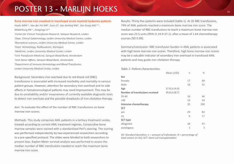
Bone marrow iron overload in transfused acute myeloid leukemia patientsHoeks MPA1,2, Van der Pol MR3, Evers D4, Van Kerkhof BW5, Van Kraaij MG1,6,7, Middelburg RA1,2, Zwaginga JJ1,8
1Center for Clinical Transfusion Research, Sanquin Research, Leiden2Dept. Clinical Epidemiology, Leiden University Medical Center, Leiden3Biomedical sciences, Leiden University Medical Center, Leiden4Dept. Hematology, Radboudumc, Nijmegen5Medicine, Leiden University Medical Center, Leiden6Unit Transfusion Medicine, Sanquin Blood Bank, Amsterdam7Unit Donor Affairs, Sanquin Blood Bank, Amsterdam8Department of Immuno-hematology and Blood Transfusion, Leiden University Medical Center, Leiden
Background: Secondary iron overload due to red blood cell (RBC) transfusions is associated with increased morbidity and mortality in various patient groups. However, attention for secondary iron overload and its side effects in hematooncological patients may need improvement. This may be due to unreliability and/or invasiveness of currently available diagnostic tests to detect iron overload and the possible drawbacks of iron chelation therapy.
Aim: To evaluate the effect of the number of RBC transfusions on bone marrow iron scores.
Methods: This study comprises AML patients in a tertiary treatment center, treated according to current AML treatment regimes. Consecutive bone marrow samples were stained with a standardized Perl’s staining. The scoring was performed independently by two experienced researchers according to a pre-specified protocol. The slides were blinded to both researchers to prevent bias. Kaplan-Meier survival analysis was performed to assess themedian number of RBC transfusions needed to reach the maximum bone marrow iron score.
Results: Thirty-five patients were included (table 1). At 35 RBC transfusions, 74% of AML patients reached a maximum bone marrow iron score. The median number of RBC transfusions to reach a maximum bone marrow iron score was 23.5 units (95% CI 19.9-27.1), after a mean of 1.64 chemotherapy courses (SD 0.99).
Summary/conclusion: RBC transfusion burden in AML patients is associated with high bone marrow iron scores. Therefore, high bone marrow iron scores may be a valuable indicator of secondary iron overload in transfused AML patients and may guide iron chelation therapy.
Table 1: Patient characteristics Mean (±SD) n %SexFemale 17 49Male 18 51Age 57.8 (±14.0)Number of transfusions received 45.8 (±18.7)20-40 16 46>40 19 54Intensive chemotherapy 35 100SCTyes 29 83no 6 17SCT typeallogeneic 28 97autologous 1 3
SD: Standard deviation; n = amount of individuals; % = percentage of total cohort (n=35); SCT: Stem cell transplantation
53POSTER 13 - MARLIJN HOEKS

Short-term prognostic value of preoperative intravenous iron in colorectal cancer patientsWilson MJ1,2, Bruns ER3, Borstlap WA3, Dekker JWT4, Jeekel J5, Zwaginga JJ6,7, Bemelman WA3, Schipperus M8,9
1Stichting TRIP, Leiden, Nationale Hemo-en Biovigilantie2Erasmus MC - Centrumlocatie, Rotterdam, Chirurgie3Academisch Medisch Centrum (AMC), Amsterdam, Chirurgie4Reinier de Graaf Gasthuis, Delft, Chirurgie5Erasmus MC - Centrumlocatie, Rotterdam, Neuroscience6Sanquin Leiden , Leiden, Klinische transfusiegeneeskunde7Leids Universitair Medisch Centrum (LUMC), Leiden, Hematologie8Stichting TRIP, Leiden, nationale hemo- en biovigilantie9HagaZiekenhuis - Locatie Leyweg (Reinier Haga Groep), Den Haag, Hematologie
Background: Both preoperative anemia and the treatment of anemia, blood transfusions and erythropoiesis-stimulating agents, are associated with increased postoperative morbidity and increased risk of tumor recurrence.
Aim: To assess the efficacy of preoperative intravenous iron infusion in colorectal cancer patients.
Methods: The prognostic value of preoperative intravenous iron was asses-sed in a retrospective cohort, including all patients who underwent surgery for colorectal cancer between 2010-2016 in a single center hospital. For comparative analyses all anemic patients (at presentation) were divided in 2 groups: usual care (UC) group (i.e. no therapy) and intravenous iron (IV) group (iron infusion <6 weeks preoperative), excluding preoperative blood transfusion and neoadjuvant chemotherapy. For logistic regression analyses, all anemic patients were included. Primary outcome was the change in hemoglobin level; secondary outcomes were the percentage of patients with postoperative complications and blood transfusions.
Results: In total, 758 colorectal cancer patients, eligible for inclusion, underwent surgery, of which 318 (41.9%) were anemic. The IV and the UC group, both excluding blood transfusion, included 52 and 153 patients with mean hemoglobin (Hb) at diagnosis of 6.3 and 6.9 mmol/L, respectively. In IV patients, Hb level was significantly increased (IV=0.65 mmol/L vs UC=0.10 mmol/L, p<0.001), most distinct in patients with advanced anemia and characteristics of absolute iron deficiency, and less peri- (p=0.3) and postoperative blood transfusions (p=0.4), and complications (p=0.03) were observed, as compared to UC patients. In multivariate logistic regression analyses administration of intravenous iron therapy did not affect postoperative blood transfusion and complication rate (OR 0.54, p=0.14 and OR=0.91, p=0.77, respectively).
Conclusion: Based on this retrospective cohort study, implementation of intravenous iron therapy in anemic colorectal cancer patients leads to higher increase of preoperative hemoglobin level, especially in patients with advanced anemia and characteristics of absolute iron deficiency, but this effect did not translate into a significant decrease in peri- and postoperative blood transfusions and complications.
54 POSTER 14 - MICHAEL WILSON
A method for the biotinylation of red blood cells for clinical research that complies with Good Manufacturing Practice regulationDe Back DZ1, Vlaar R1, Beuger B1, Daal B2, Lagerberg J2, De Korte D2, Van Kraaij MG3, Van Bruggen R1
1Dept. Blood Cell Research, Sanquin Research, and Landsteiner Laboratory, University of Amsterdam, Amsterdam, The Netherlands.2Dept. Transfusion Medicine, Sanquin Research, and Landsteiner Laboratory, University of Amsterdam, Amsterdam, The Netherlands.
Background: Biotinylated RBC can be used to independently and accurately measure RBC survival and clearance. This method has several advantages over the standard 51Cr method: 1) study subjects are not exposed to radiation; 2) small blood volumes are required to analyze results, and 3) multiple RBC populations can be measured simultaneously in the same individual. However, so far only experimental non-validated biotin-labeled RBC products have been transfused.
Aim: The goal of this study was to produce a standardized biotin-labeled RBC product in a fast, simple and sterile manner that can be used for clinical research and for the evaluation of new blood products.
Methods: Red Cell Concentrate (RCC) fractions were labeled with two different concentrations of biotin in a closed system (according to GMP), to ensure sterility of the labeled end product. Using a flow cytometric analysis, the reproducibility and robustness of the biotin labeling protocol was assessed, as well as the stability of the labeled (un-)irradiated end product. Additionally, RBC parameters such as phosphatidylserine exposure (PS), Na, K, free hemoglobin, ATP, pH and morphology were determined prior to and after biotin labeling to rule out effects of the labeling procedure on biological activity.
Results: Our data show that RCC can be labeled according to GMP with two different biotin concentrations in a standardized (GMP) manner, without affecting the biological activity.
Summary/conclusion: An easy, rapid (<2 hrs), GMP qualified and robust method was developed to generate biotin-labeled RBC for clinical research.
55POSTER 15 - DJUNA DE BACK

Does hemoglobin level influence donor return?Py JY1, Barnoux M1
1Etablissement Français du Sang, site d’Orléans
Background: Biotinylated RBC can be used to independently and accurately Background: Blood donations may worse donor pre-anaemic state, with clinical signs like fatigue.
Aim: To see if such possible signs, presumably linked to donor low haemoglobin level, may impair his return.
Methods: We studied a selection of year 2015 French whole blood donors, born in years 1955, 1965, 1975, 1985, and 1995. Donations without any return were included as “no return”. Other donations were only included if the next event was a spontaneous application. Delay between index donation and the donor return was registered, and considered if less than one year.
Results: 126.442 female and 128.887 male donations were included, with respectively a one year return of 64.7% and 71.8% and a median delay of 163 and 141 days. There is no delay rise with donations near the low hemoglobin thresholds, nor any deterioration of the return rate. By contrast, data rather point to deterioration in case of high level. Donor age does not change these results.
Summary/conclusion: Even if whole blood donation may be immediately less tolerated in pre-anemic donors, it does not affect their return after several weeks.
56 POSTER 16 - JEAN-YVES PY

Associations between SNPs and erythrocyte traits including hemoglobin in humans: a systematic literature review and Donor InSight-III data collectionTimmer T1, Van den Hurk K, Van der Schoot E2,3, Veldhuisen B2,4, Daams J5, De Kort W1,6
1Dept Donor Studies, Sanquin Research, Amsterdam, The Netherlands.2Dept. Experimental Immunohematology, Sanquin Research, Amsterdam, The Netherlands.3Landsteiner Laboratory, Academic Medical Center, Amsterdam, the Netherlands.4Dept. Diagnostic Immunohematology, Sanquin, Amsterdam, The Netherlands5Medical Library, Academic Medical Center, Amsterdam, The Netherlands6Dept Public Health, Academic Medical Center, Amsterdam, The Netherlands.
Background: Blood donation results in a loss of erythrocytes and therefore new erythrocytes and hemoglobin (Hb) need to be produced. Several genetic loci have been shown to be associated with Hb and other erythrocyte parameters.
Aim: (1) Identify which single nucleotide polymorphisms (SNPs) show associations with Hb/erythrocyte traits in humans, and (2) investigate associations of SNPs with Hb trajectories in blood donors.
Methods: (1) A systematic review on SNPs associated with Hb/erythrocyte traits is ongoing. (2) ,863 donors (with declining and stable Hb trajectories based on latent-class growth analyses and a randomly selected group) have participated in Donor InSight-III (DIS-III). Data-collection consisted of blood sampling for a full blood count, SNP array (UK Biobank Axiom® Array, Affymetrix, CA, USA) and analysis of iron parameters, and questionnaires regarding health, lifestyle and menstruation.Results: 3,597 titles/abstracts were screened of which 137 full texts are currently being screened on relevance (preliminary results on poster). DIS-III data are currently being prepared for data analyses.
Summary/conclusion: We expect to find known and new associations between SNPs and Hb (trajectories)/erythrocyte traits.
57POSTER 17 - TIFFANY TIMMER

Distribution of ferritin levels of Dutch donors – are ferritin levelsproportional to the number of whole blood donations?Prinsze FJ1, De Groot R1,2, Timmer TC1,3, De Kort WLAM1,3, Van den Hurk K1
1Dept. Donor Studies, Sanquin Research, Amsterdam, The Netherlands2Dept. Epidemiology and Biostatistics, VU University Medical Center, Amsterdam, The Netherlands3Dept. Public Health, Academic Medical Center, Amsterdam, The Netherlands
Background: Donation is associated with a loss of iron. To prevent iron deficiency a minimum haemoglobin level is set as eligibility criterion and is measured before each donation. However, ferritin might be a more suitable parameter as it is an indicator of iron stores.
Aim: To map the distribution of ferritin levels in categories of number of donations in the Donor InSight-III population and compare these with Hb measurements in order to develop a better donor screening.
Methods: Donor InSight (DIS) is a cohort study among the Dutch donor population (three questionnaire rounds). In the DIS-III population Hb and ferritin were measured (2015-2016). Data on the number of previous donations were retrieved from EProgesa, the blood bank information system. Per category of donation history mean±SD ferritin and Hb levels were calculated separately for men, premenopausal (≤44) and postmenopausal women (≥45).
Results: A total of 2845 participants were included in these analyses, all had made at least 1 donation. Mean Hb levels were similar for donors with varying donation histories. Mean ferritin levels were approximately 50% lower for donors with more than 50 donations, compared to those with 2-10 donations.
Discussion: Hb levels are relatively stable, ferritin levels drop gradually with the number of donations. However, a stabilization was found at extreme loyal donors (>50 donations), this might be due to donor selection effects. Further research is necessary to disentangle donation history from age effects. Longitudinal data collection from new donors throughout their donor career should clarify patterns of ferritin trajectories.
58 POSTER 18 - FEMMEKE PRINSZE
Risk dependent intervention on the prevalence of low hemoglobin deferralHuis in ’t Veld E1, Van den Hurk K1, De Kort WLAM1,2, Baart M1
1Dept. Donor Studies, Sanquin Research2Dept. Social Medicine, Academic Medical Center, University of Amsterdam
Background: Donors with a low hemoglobin (Hb) level are deferred for donation. Although meant to protect donors, these deferrals are demoralizing and increase the risk for donor lapse. Based on previously developed sex specific prediction models for Hb deferral risk, an intervention strategy to decrease the prevalence of donor deferrals was tested.
Aim: The aim of the current study was to conduct an intervention trial for donors with a high risk of Hb deferral, to investigate the effect of prolongation of the donation interval and/or dietary advice on the Hb level and prevalence of Hb deferral on the subsequent visit
Methods: Donors from 26 blood collection centers throughout the Netherlands with a Hb level lower than 0.2 mmol/L above the gender specific cut-off (total N = 12073) received either no intervention, a prolongation of the donation interval of six or 12 months, dietary advice, or both. The effect of the intervention on Hb levels and deferral rates at the subsequent donation attempt were analyzed using Generalized Estimating Equations.
Results: Young women and donors with lower Hb levels at the first donation attempt were at higher risk of deferral at the second donation attempt. Prolongation of the donation interval by six months significantly reduced the risk of deferral. However, there was no additional benefit of an extra-long 12 month prolongation or dietary advice.
Summary/conclusion: Implementation of a protocol which defers donors at risk for six additional months may reduce the risk of Hb related deferrals in the future.
59POSTER 19 - LISANNE HUIS IN ‘T VELD

SanquinPlesmanlaan 125Po Box 98921006 AN AmsterdamThe Netherlands+31 20 - 5123 000www.sanquin.nl/sss