ios press epo-releasing neural precursor cells promote ... et...restorative neurology and...
TRANSCRIPT

Restorative Neurology and Neuroscience 35 (2017) 583–599DOI 10.3233/RNN-170750IOS Press
583
EPO-releasing neural precursor cellspromote axonal regeneration and recoveryof function in spinal cord traumatic injury
S. Carellia,b,1,∗, T. Giallongoa,1, Z. Gombalovaa,c, D. Merlia, A.M. Di Giulioa,b and A. Gorioa,∗aDepartment of Health Sciences, Laboratory of Pharmacology, University of Milan, via A di Rudinı, Milan, ItalybPediatric Clinical Research Center Fondazione Romeo ed Enrica Invernizzi, University of Milan, Milan, ItalycPavol Jozef Safarik University in Kosice, Faculty of Science, Institute of Biology and Ecology, Moyzesova,Kosice, Slovakia (SVK)
Abstract.Background: Spinal cord injury (SCI) is a debilitating condition characterized by a complex of neurological dysfunctionsranging from loss of sensation to partial or complete limb paralysis. Recently, we reported that intravenous administrationof neural precursors physiologically releasing erythropoietin (namely Er-NPCs) enhances functional recovery in animalsfollowing contusive spinal cord injury through the counteraction of secondary degeneration. Er-NPCs reached and accumu-lated at the lesion edges, where they survived throughout the prolonged period of observation and differentiated mostly intocholinergic neuron-like cells.Objective: The aim of this study was to investigate the potential reparative and regenerative properties of Er-NPCs in amouse experimental model of traumatic spinal cord injury.Methods and Results: We report that Er-NPCs favoured the preservation of axonal myelin and strongly promoted theregrowth across the lesion site of monoaminergic and chatecolaminergic fibers that reached the distal portions of the injuredcord. The use of an anterograde tracer transported by the regenerating axons allowed us to assess the extent of such a process.We show that axonal fluoro-ruby labelling was practically absent in saline-treated mice, while it resulted very significant inEr-NPCs transplanted animals.Conclusion: Our study shows that Er-NPCs promoted recovery of function after spinal cord injury, and that this is accompaniedby preservation of myelination and strong re-innervation of the distal cord. Thus, regenerated axons may have contributed tothe enhanced recovery of function after SCI.
Keywords: Spinal cord injury, neural stem cells, transplantation, regenerative medicine, animal behavior
1. Introduction
Spinal cord injury (SCI) is most commonly causedby high-energy trauma and represents a complexemergency issue that leads most often to a chroniccondition. The majority of patients are men of 16–30
1Carelli S. and Giallongo T. equally contributed to the work.∗Corresponding authors. Alfredo Gorio and Stephana Carelli,
Department of Health Sciences University of Milan, Labora-tory of Pharmacology, Polo H. San Paolo, via A di Rudinı8, 20 142 Milan, Italy. Tel.: +39 0250323030; Fax: +390250323033; E-mails: [email protected] (Alfredo Gorio)and [email protected] (Stephana Carelli).
years of age, thus in addition to the enormous per-sonal suffering, SCI results also in substantial coststo society (Fehlings et al., 2011; Schlayer et al.,1988). There is currently no curative therapy, the carein the acute phase used to be limited to high-dosecorticosteroid treatment that is now being advisedagainst, and surgical stabilization and decompres-sion to possibly attenuate further damage (Fehlingset al., 2011; Furlan et al., 2011). Numerous reportshave described the presence of inhibiting factorsin the lesion environment that limit neural regen-eration in adult CNS and in particular across theinjury site in SCI. A variety of inhibitory molecules
0922-6028/17/$35.00 © 2017 – IOS Press and the authors. All rights reservedThis article is published online with Open Access and distributed under the terms of the Creative Commons Attribution Non-Commercial License (CC BY-NC 4.0).

584 S. Carelli et al. / SCI repair enhanced by Er-NPCs
associated with myelin and extracellular matrix havebeen described (Fawcett, 2006; Fitch et al., 2008; He& Koprivca, 2004). The limiting environmental fac-tors may include also the insufficient local presence ofgrowth-permissive matrices and growth factors. Theneutralization of such growth inhibitory moleculeshave been extensively studied (Young, 2014) and alsoa variety of stem cells or stem cell-derived moleculesas well have shown beneficial effects to a variableextent (Martino & Pluchino, 2006; Romanko et al.,2007). We had shown that intravenous administrationof erythropoietin-releasing adult neural precursorscells, isolated from SVZ six hours after donor death(Er-NPCs; formerly called post mortem-neural pre-cursor cells; PM-NPCs; Marfia et al., 2011), improvehind limb functional recovery. Er-NPCs accumulateat the lesion site, where they differentiate mostly intocholinergic neuron cells, favouring preservation ofmyelin (Carelli et al., 2014a; Carelli et al., 2014b;Carelli et al., 2015). Acute traumatic SCI is followedby vascular changes with loss of neurons, oligoden-droglia, and astrocytes, neuroinflammation quicklyfollows with consequent invasion of the injury bya variety of inflammatory cells. These acute condi-tion is then associated to Wallerian degeneration ofascending and descending tracts with gradual for-mation of cavities in the cord, the formation ofthe glial scar reduces greatly the growth ability byaxons across the injury (Gorio et al 2002, Ahujaet al. 2017). The aim of this work was to investi-gate the ability of Er-NPCs in axonal regenerationacross injury. Here, we show, that Er-NPCs adminis-tration enhances restauration of TH and 5-HT positivefibres in the caudal cord, and increases significantlythe number of axons able to cross the injury site.This enhanced re-innervation of the caudal cord mayunderlay the gradual and continuous improvement inhind limb function that has been observed even at 90days after lesioning.
2. Materials and methods
2.1. Animal care
In this study, adult CD1 male mice 25–30 g inweight (Charles River, Calco, Italy) were used. Allof the procedures were approved by the ReviewCommittee of the University of Milan and metthe Italian Guidelines for Laboratory Animals,which conform to the European Communities Direc-tive (2010/63/UE). Animals were kept for at least
3 days before the experiments in standard conditions(22 ± 2◦C, 65% humidity, and artificial light between8:00 a.m. to 8:00 p.m.).
2.2. Er-NPCs isolation, characterization, andlabelling
Er-NPCs were obtained from 6 weeks old CD-1albino mice; their isolation, growth and characteriza-tion were performed by following methods describedin previously published papers (Marfia et al., 2011;Carelli et al., 2014a; Carelli et al., 2014b; Carelliet al., 2015) and set up in the past by Gritti andco-workers (Gritti et al., 2002). Briefly, cells wereisolated from the SVZ 6 hours after sacrifice bycervical dislocation. Brains were removed, and tis-sues containing the SVZ region were dissected,transferred to Earl’s balanced salt solution (Life Tech-nologies, Monza, Italy) containing 1 mg/ml papain(27 U/mg; Sigma-Aldrich, Milan, Italy), 0.2 mg/mlcysteine (Sigma-Aldrich), and 0.2 mg/ml EDTA(Sigma-Aldrich). Tissue was incubated for 45 minat 37◦C on a rocking platform. Tissues were thentransferred to DMEM-F12 medium (Euroclone, Pero,Milan, Italy) and mechanically dissociated with aPasteur pipette. Cells were counted and plated at3500 cells/cm2 in DMEM-F-12 (Euroclone, Pero,Milan, Italy) containing 2 ml-glutamine (Euroclone),0.6% glucose (Sigma-Aldrich), 9.6 gm/ml putrescine(Sigma-Aldrich), 6.3 ng/ml progesterone (Sigma-Aldrich), 5.2 ng/ml sodium selenite (Sigma-Aldrich),0.025 mg/ml insulin (Sigma-Aldrich), 0.1 mg/mltransferrin (Sigma-Aldrich), and 2 �g/ml heparin(sodium salt, grade II; Sigma-Aldrich), bFGF (humanrecombinant, 10 ng/mL; Life Technologies) andEGF (human recombinant, 20 ng/mL; Life Tech-nologies). Spheres formed after 5–7days wereharvested, collected by centrifugation (10 min at123 g), mechanically dissociated to a single-cell sus-pension, and re-plated in the medium indicated above(Marfia et al., 2011; Gritti et al., 2002; Bottai et al.,2008). As previously described, Er-NPCs differenti-ation was performed by plating the dissociated stemcells at the density of 40,000 cells/cm2 in presenceof adhesion molecules (Matrigel™, BD Biosciences,Buccinasco, MI, Italy) and bFGF (10 ng/ml) for 48hours. After this time, the medium was changedand cells were exposed to the same medium con-taining foetal bovine serum (1% vol/vol; Euroclone)and depleted of bFGF. This incubation lasted forthe following 5 days (Marfia et al. 2011; Carelliet al. 2014b, Carelli et al. 2015). Then, the extent of

S. Carelli et al. / SCI repair enhanced by Er-NPCs 585
differentiation was investigated by immunocyto-chemical staining (Marfia et al., 2011). To monitorthe fate of Er-NPCs after transplantation PKH26 andH33342 were used (Sigma-Aldrich). PKH26 is a nontoxic cell dye characterized by a long aliphatic tails(PKH26) that allow the dye incorporation in lipidregions of the cell membrane (Horan et al., 1990; Wal-lace et al., 2008). Er-NPCs were labelled just beforethe injection, following manufacturer’s instructionsand as described previously (Carelli et al., 2014a; Liuet al., 2014).
Er-NPCs were stained with Hoechst 33342(Sigma-Aldrich, Steinheim, Germany) at not toxicconcentration (0.5 �g/mL). Floating mechanicallydissociated NPCs were incubated with H33342 solu-tion at the final concentration of 0.5 �g/ml for 90minutes at 37◦C in an incubator. After staining, thecells were pelleted by centrifugation (500 × g for5 min) and rinsed twice with HBSS (10 ml) (Carelliet al., 2014a). For transplantation labelled cells wereresuspended in sterile physiologic solution at a con-centration of 3.3 × 10⁵ cells/50 �l (Carelli et al.,2014a; Carelli et al., 2015).
2.3. Setting of experimental groups and celladministration
Experimental animals were divided into threegroups: 1) Laminectomies mice (n = 6); 2) Lesionedmice treated with phosphate buffer (PBS, n = 15); 3)Lesioned mice transplanted with Er-NPCs (n = 15).The traumatic SCI was performed using an InfiniteHorizon (IH; Precision Systems and Instrumenta-tion, LLC, Lexington, KY, USA) device (Carelliet al., 2014a; Gorio et al., 2007) at the T8 level.Surgery on the animals was performed as previouslydescribed (Carelli et al., 2014a; Carelli et al., 2014b;Carelli et al., 2015; Gorio et al., 2007). Er-NPCs wereadministered after spinal cord lesion and intravenousadministration (i.v.) was performed by tail vein injec-tion. As previously described, the treatment includesthree different i.v. injection of 50 �l in volume, thefirst administration 30 min after injury, followed bya second injection 6 h later and a third one 18 h afterthe lesion (Carelli et al., 2014a; Carelli et al., 2014b;Carelli et al., 2015).
2.4. Behavioural tests and hind limb function
As described by Basso and co-workers (Bassoet al., 2006) functional recovery evaluations wereassessed in a blinded fashion. Neurological function
was evaluated first 24 h after injury and then twice aweek for the first 4 weeks. The methods utilized arewell known in the field of behavioural evaluation ofrecovery of function after SCI (Basso et al., 2006).For behavioural experiments we used 12 animals forall experimental groups. Allodynia-like responses inthe unaffected forepaw were assessed by means ofstandard hotplate test and cold stimulation as previ-ously reported (Carelli et al., 2014b; Carelli et al.,2015; Hofstetter et al., 2005).
2.5. Histology and immunohistochemistry
At the end of the experimental period, animals wereanesthetized and perfused as described previously[11–13]. Spinal cords were post-fixed overnight,cryoprotected with 30% sucrose (Sigma-Aldrich),quickly frozen, stored at –80◦C and sectioned bymeans of a cryostat (Leica) (Carelli et al. 2014a;Carelli et al., 2014b; Carelli et al., 2015). Cryostatsections (15 �m) were collected onto glass slidesand processed for immunohistochemistry. Sectionswere rinsed with PBS (Euroclone), treated withblocking solution (Life-Technologies) and incubatedwith primary antibodies overnight at 4◦C. Aftertreatment with primary antibodies, the sections werewashed with PBS and incubated with appropriatesecondary antibodies (Alexa Fluor® 488, MolecularProbes®, Life Technologies) for 2 hours at roomtemperature. Sections were washed in PBS, nucleiwere stained with DAPI (1�g/ml final concentration,10 minutes at room temperature; Sigma-Aldrich)and then mounted using the FluorSave Reagent(Calbiochem, Merck Chemical, Darmstadt, Ger-many) and analyzed by confocal microscopy. Incontrol determinations, primary antibodies wereomitted and replaced with equivalent concentrationsof unrelated IgG of the same subclass. The follow-ing primary antibodies were used:, �-Tubulin III(1:150; Covance). Microtubule-Associated Protein 2(MAP-2; 1:300; Chemicon), Tyrosine hydroxylase(TH; 1:500; Millipore), Choline Acetyltransferase(ChAT; 1:1000; Chemicon), 5-hydroxytryptamine(5-HT; 1:200 Millipore); GAP-43 (1:1000;Millipore).
2.6. Assessment of myelin preservation
Assessment of myelin preservation was evaluatedon cord sections of non- lesioned (healthy), lesioned+ PBS and lesioned + Er-NPCs animals placed onthe same coverslip as described previously (Pertici

586 S. Carelli et al. / SCI repair enhanced by Er-NPCs
et al., 2013; Carelli et al., 2014b; Carelli et al.,2015). This approach allows for the homogeneousevaluation of quantitative data obtained by confocalanalysis. Myelin preservation was evaluated compar-ing the levels of myelin in the ventral white matter at0.4 mm (rostral and caudal) laterally from the lesionepicenter in healthy, saline and cells treated ani-mals. The choice of the ventral white matter wasbased on the knowledge that the reticular spinalpathway descends mostly in the ipsilateral dorso-and ventrolateral funiculi and is directly involvedin the regulation of the movement of the mousefoot (Vitellaro-Zuccarello et al., 2007). The confo-cal microscope images for the laminectomies animalsand saline and cells-treated mice were obtained usingthe same intensity, pinhole, wavelength and thicknessof the acquisition. As reference, we used sectionsclose to the ones analyzed and not treated with fluo-romyelin (Pertici et al., 2013; Carelli et al., 2014b;Carelli et al., 2015). Briefly, the procedure of thestaining was carried out by incubating the cryosec-tions with fluoromyelin diluted 1:300 in PBS for20 minutes; slides were then washed three timesfor 10 min each with PBS and mounted with Fluor-Save (Merck, Darmstadt, Germany), and qualitativelyand quantitatively analyzed by confocal microscopy(Leica TSC2; Leica Microsystems, Heidelberg,Germany).
2.7. Fluoro-Ruby Protocol
Fluororuby is a fluorescent rhodamine-conjugateddextran, which has been used to study in vivo axonaltransport within the central nervous system (Lu et al.,2001). A 10% solution of Fluororuby is made by dis-solving 10 mg of dry powder in 100 �L of PBS, thatwas delivered via intraspinal injection at T6/T7 usinga 5 �L Hamilton microsyringe. Injection volumestypically ranged from 0.5–1 �L and were graduallyinjected over a 10–15 minute interval (Schofield etal., 2007). The animals were then allowed to recover.The animals were perfused 5 days after the tracerinjection with neutral buffered formaldehyde (10%formalin in 0.1 M neutral phosphate buffer). Thespinal cord was then removed, and post-fixed forat least overnight in the same fixative solution andincluded in paraffin. The cords were then sectionedby means of a microtome (Zeiss) with thickness setat 10 microns. This analysis was performed on 3 ani-mals per experimental group: (i) injected with salinesolution as a control and (ii) Er-NPCs-transplantedanimals.
2.8. Semiquantitative method of analysis for thedetermination of serotoninergic andcatecholaminergic fiber density
Data were collected from sections taken at thesame distance (2 mm) from the lesion epicenter andimmunostained in a single batch to minimize vari-ability. Images were acquired using standardizedconfocal microscopy (Hawthorne et al., 2011; Shigyoet al., 2016). Five consecutive sections (10 �m thick)were averaged, and the count was serially repeatedevery 50 �m for the length of 500 �m. Semiquanti-tative analysis of immunoreactivity was performedby using confocal microscopy (Leica SP2 confocalmicroscope with He/Kr and Ar lasers; Heidelberg,Germany) to evaluate the mean relative optical den-sity. Quantification of serotonin (5-HT) and tyrosinhydroxilase (TH) immunoreactivity was carried outin the regions reported in Figs. 4 and 5 following theprocedure described for myelin assessment.
2.9. Statistical analysis
Data were expressed as the mean ± S.D. Multiplegroups comparison were made by ANOVA followedby Bonferroni’s multiple comparisons test to assessstatistical significance versus respective control. Theanalyses were performed using Prism 3.0 software(GraphPad Software, Inc.). Statistical significancewas accepted for a P < 0.05.
3. Results
3.1. Er-NPCs elicit neuronal markers expressionin recipient spinal cord
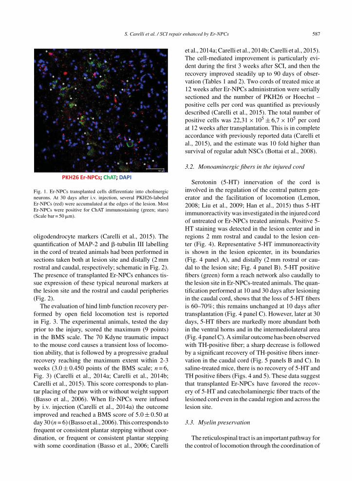
Transplanted Er-NPCs localize at the edges ofinjury site and show a well-structured morphologywith dendrite-like processes; they are mostly positivefor Choline-AcetylTransferase (ChAT) (Carelli et al.,2015 and Fig. 1). The quantification performed 30days after transplantation shows that 75 ± 5.35 per-cent of transplanted Er-NPCs (PKH26 positive cells)are positive for ChAT, with a dotted (Fig. 1) distri-bution that in most cases is perinuclear, with positivelabeling of processes in several instances (Fig. 1).The high majority of Er-NPCs localized at the bound-aries of lesion site are also positive for MAP-2 and�-tubulin III (Fig. 2). As reported in our previouswork also in this new set of experiments none ofengrafted Er-NPCs resulted positive to GFAP or
S. Carelli et al. / SCI repair enhanced by Er-NPCs 587
Fig. 1. Er-NPCs transplanted cells differentiate into cholinergicneurons. At 30 days after i.v. injection, several PKH26-labeledEr-NPCs (red) were accumulated at the edges of the lesion. MostEr-NPCs were positive for ChAT immunostaining (green; stars)(Scale bar = 50 �m).
oligodendrocyte markers (Carelli et al., 2015). Thequantification of MAP-2 and �-tubulin III labellingin the cord of treated animals had been performed insections taken both at lesion site and distally (2 mmrostral and caudal, respectively; schematic in Fig. 2).The presence of transplanted Er-NPCs enhances tis-sue expression of these typical neuronal markers atthe lesion site and the rostral and caudal peripheries(Fig. 2).
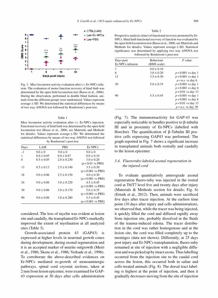
The evaluation of hind limb function recovery per-formed by open field locomotion test is reportedin Fig. 3. The experimental animals, tested the dayprior to the injury, scored the maximum (9 points)in the BMS scale. The 70 Kdyne traumatic impactto the mouse cord causes a transient loss of locomo-tion ability, that is followed by a progressive gradualrecovery reaching the maximum extent within 2-3weeks (3.0 ± 0.450 points of the BMS scale; n = 6,Fig. 3) (Carelli et al., 2014a; Carelli et al., 2014b;Carelli et al., 2015). This score corresponds to plan-tar placing of the paw with or without weight support(Basso et al., 2006). When Er-NPCs were infusedby i.v. injection (Carelli et al., 2014a) the outcomeimproved and reached a BMS score of 5.0 ± 0.50 atday 30 (n = 6) (Basso et al., 2006). This corresponds tofrequent or consistent plantar stepping without coor-dination, or frequent or consistent plantar steppingwith some coordination (Basso et al., 2006; Carelli
et al., 2014a; Carelli et al., 2014b; Carelli et al., 2015).The cell-mediated improvement is particularly evi-dent during the first 3 weeks after SCI, and then therecovery improved steadily up to 90 days of obser-vation (Tables 1 and 2). Two cords of treated mice at12 weeks after Er-NPCs administration were seriallysectioned and the number of PKH26 or Hoechst –positive cells per cord was quantified as previouslydescribed (Carelli et al., 2015). The total number ofpositive cells was 22,31 × 10⁵ ± 6,7 × 10⁵ per cordat 12 weeks after transplantation. This is in completeaccordance with previously reported data (Carelli etal., 2015), and the estimate was 10 fold higher thansurvival of regular adult NSCs (Bottai et al., 2008).
3.2. Monoaminergic fibers in the injured cord
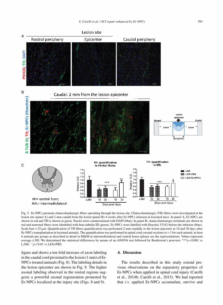
Serotonin (5-HT) innervation of the cord isinvolved in the regulation of the central pattern gen-erator and the facilitation of locomotion (Lemon,2008; Liu et al., 2009; Han et al., 2015) thus 5-HTimmunoreactivity was investigated in the injured cordof untreated or Er-NPCs treated animals. Positive 5-HT staining was detected in the lesion center and inregions 2 mm rostral and caudal to the lesion cen-ter (Fig. 4). Representative 5-HT immunoreactivityis shown in the lesion epicenter, in its boundaries(Fig. 4 panel A), and distally (2 mm rostral or cau-dal to the lesion site; Fig. 4 panel B). 5-HT positivefibers (green) form a reach network also caudally tothe lesion site in Er-NPCs-treated animals. The quan-tification performed at 10 and 30 days after lesioningin the caudal cord, shows that the loss of 5-HT fibersis 60–70%; this remains unchanged at 10 days aftertransplantation (Fig. 4 panel C). However, later at 30days, 5-HT fibers are markedly more abundant bothin the ventral horns and in the intermediolateral area(Fig. 4 panel C). A similar outcome has been observedwith TH-positive fiber; a sharp decrease is followedby a significant recovery of TH-positive fibers inner-vation in the caudal cord (Fig. 5 panels B and C). Insaline-treated mice, there is no recovery of 5-HT andTH positive fibers (Figs. 4 and 5). These data suggestthat transplanted Er-NPCs have favored the recov-ery of 5-HT and catecholaminergic fiber tracts of thelesioned cord even in the caudal region and across thelesion site.
3.3. Myelin preservation
The reticulospinal tract is an important pathway forthe control of locomotion through the coordination of

588 S. Carelli et al. / SCI repair enhanced by Er-NPCs
A
B
Fig. 2. Er-NPCs transplantation improves neural markers expression in injured spinal cord. Pictures represent confocal acquisitions ofcoronal sections taken at the edges of the lesion at 30 days after i.v. administration of PKH26 – labeled Er-NPCs (showed in red) (panelA). Sections were immunostained for � tubulin III and MAP-2 (showed in green). PKH26-labeled cells were positive for �-tubulin III andMAP-2 (white stars) (Scale bar = 75 and 100 �m) (panel A). Graphs reported in panel B show the quantification of immunoreactivity insections taken at the lesion epicenter, 2 mm rostral or caudal to the lesion epicenter (please see schematic representation). Values representaverage ± SD. We determined the statistical differences by means of ANOVA test followed by Bonferroni’s post-test. ***p < 0.001 vs LAM;$$p < 0.01 vs PBS.
rhythmic stepping movements (Ballerman & Fouad,2006). It runs in the ipsilateral dorso- and ventro-lateral funiculi of the cord, and is predominantlyconstituted by myelinated axons (Loy et al., 2002).The extent of myelination of these pathways dependsmarginally on the presence of serotonergic axonsin these bundles since they are scarcely myelinated(Loy et al., 2002; Hildebrand & Hahn, 1978). The
condition of myelin in these descending structureswas determined by evaluation of FluoroMyelin™staining by means of confocal quantitative analysisas detailed in Materials and Methods. Myelin wasquantified 30 days after lesion; sections were takenat the center of the lesion and 2 mm caudally to thelesion site (Fig. 6). Only intact myelin was assessed,and the degenerated crumbled labeled parts were not
S. Carelli et al. / SCI repair enhanced by Er-NPCs 589
Fig. 3. Mice locomotor activity evaluation after i.v. Er-NPCs infu-sion. The evaluation of motor function recovery of hind limb wasdetermined by the open field locomotion test (Basso et al., 2006).During the observation, performed in double blind fashion, ani-mals from the different groups were randomized. Values representaverage ± SD. We determined the statistical differences by meansof two way ANOVA test followed by Bonferroni’s post-test.
Table 1
Mice locomotor activity evaluation after i.v. Er-NPCs injection.Functional recovery of hind limb was determined by the open fieldlocomotion test (Basso et al., 2006; see Materials and Methodsfor details). Values represent average ± SD. We determined thestatistical differences by means of two way ANOVA test followed
by Bonferroni’s post-test
Days LAM PBS Er-NPCs
–1 9.0 ± 0 9.0 ± 0 9.0 ± 01 8.0 ± 0.05 0.0 ± 0.87 0.0 ± 0.106 8.5 ± 0.05 2.0 ± 0.230 3.0 ± 0.20
(p < 0.01 vs PBS)13 8.5 ± 0.13 2.5 ± 0.140 3.5 ± 0.30
(p < 0.001 vs PBS)18 9.0 ± 0.06 2.5 ± 0.130 4.0 ± 0.30
(p < 0.001 vs PBS)24 9.0 ± 0.06 3.0 ± 0.220 4.5 ± 0.45
(p < 0.001 vs PBS)30 9.0 ± 0.06 3.0 ± 0.170 5.0 ± 0.35
(p < 0.001 vs PBS)90 9.0 ± 0.06 3.0 ± 0.280 5.5 ± 0.45
(p < 0.001 vs PBS)
considered. The loss of myelin was evident at lesionsite and caudally, the transplanted Er-NPCs markedlyimproved the extent of myelination at all analyzedsites (Table 3).
Growth-associated protein 43 (GAP43) isexpressed at higher levels in neuronal growth conesduring development, during axonal regeneration andit is an accepted marker of neurite outgrowth (Meiriet al., 1986; Skene et al., 1986; Verkade et al., 1996).To corroborate the above-described evidences onEr-NPCs mediated re-growth of monoaminergicpathways, spinal cord cryostat sections, taken at2 mm from lesion epicenter, were examined for GAP-43 expression at 30 days after cells administration
Table 2
Prospective analysis (time) of functional recovery promoted by Er-NPCs. Hind limb functional recovery of function was evaluated bythe open field locomotion test (Basso et al., 2006; see Materials andMethods for details). Values represent average ± SD. Statisticalsignificance was determined by applying two way ANOVA test
followed by Bonferroni’s post-test
Days post Behaviour P valueEr-NPCs infusion (BMS scale)
1 0.0 ± 0.106 3.0 ± 0.20 p < 0.001 vs day 113 3.5 ± 0.30 p < 0.001 vs day 1
p = n.s. vs day 630 5.0 ± 0.35 p < 0.001 vs day 1
p < 0.001 vs day 6p < 0.01 vs day 13
90 5.5 ± 0.45 p < 0.001 vs day 1p < 0.001 vs day 6p < 0.01 vs day 13p = n.s. vs day 30
(Fig. 7). The immunoreactivity for GAP-43 wasespecially noticeable in bundles positive to �-tubulinIII and in proximity of Er-NPCs (labelled withHoechst). The quantification of �-Tubulin III pos-itive cells expressing GAP43 was performed. Thegraph reported in Fig. 7 shows a significant increasein transplanted animals both rostrally and caudallyto the lesion epicenter.
3.4. Fluororuby-labeled axonal regeneration inthe injured cord
To evaluate quantitatively anterograde axonalregeneration fluoro-ruby was injected in the rostralcord at T6/T7 level five and twenty days after injury(Materials & Methods section for details; Fig. 6)(Erturk et al., 2012). Then, animals were sacrificedfive days after tracer injection. At the earliest timepoint (10 days after injury and cells administration),we observed that, while the tracer was being injected,it quickly filled the cord and diffused rapidly awayfrom injection site, probably dissolved in the fluidsof the trauma-induced edema. The tracer distribu-tion in the cord was rather homogenous and at thelesion site, the cord was filled completely up to themeninges (data not shown). Differently, at 25 dayspost injury and Er-NPCs transplantation, fluoro-rubyremained at site of injection with a negligible diffu-sion and was picked up by intact axons. Thus labellingoccurred from the injection site to the caudal cordacross the lesion, this occurred both in saline andcells-treated animals (Fig. 8). The dorsal tract label-ing is highest at the point of injection, and then itgradually decreases moving from the site of injection

590 S. Carelli et al. / SCI repair enhanced by Er-NPCs
A
C
B
Fig. 4. Er-NPCs promote serotoninergic fibres sprouting through the lesion site. Serotoninergic (5-HT) fibers were investigated at the lesionsite (panel A) and 2 mm away from the lesion (panel B) 4 weeks after Er-NPCs infusion in lesioned mice. In panel A, Er-NPCs are shownin red (PKH26) and 5-HT is shown in green. Nuclei were counterstained with DAPI (blue). In panel B, 5-HT staining is showed in red andneuronal fibers were identified with beta-tubulin III (green). Er-NPCs were labelled with Hoechst 33342 before the infusion (blue). Scalebars = 50 and 100 �m. Quantification was performed 2 mm caudally to the lesion epicenter at 10 and 30 days after Er-NPCs transplantationin lesioned animals (panel C). The quantification was performed in spinal cord coronal sections (n = 3 for each animal; at least 6 animal pergroup) as described in detail in M&M in intermediolateral and ventral horns (please see the representation). Values represent average ± SD.We determined the statistical differences by means of an ANOVA test followed by Bonferroni’s post-test. ***p < 0.001 vs LAM; ◦p < 0.05vs LES+PBS.
to the lesion site and it is very limited in the caudalcord of saline-treated mice (Fig. 8 panel A). Such alabelling is, however, much higher in the Er-NPCstransplanted animals (Fig. 8 panel A). The highestnumber of labelled axons is localized in the rostralregion of the lesion, but, as expected, they accumulate
at the injury boundaries where transplanted cellsare located. Several of these axons cross the lesionand their number increases again just caudal to thelesion as axons likely sprout when entering the den-ervated caudal cord. The quantification of fluoro-rubylabelled axons is reported in panel C of the same
S. Carelli et al. / SCI repair enhanced by Er-NPCs 591
A
C
B
Fig. 5. Er-NPCs promote chatecolaminergic fibres sprouting through the lesion site. Chatecolaminergic (TH) fibers were investigated at thelesion site (panel A) and 2 mm caudal from the lesion (panel B) 4 weeks after Er-NPCs infusion in lesioned mice. In panel A, Er-NPCs areshown in red and TH is shown in green. Nuclei were counterstained with DAPI (blue). In panel B, chatecolaminergic terminals are shown inred and neuronal fibers were identified with beta-tubulin III (green). Er-NPCs were labelled with Hoechst 33342 before the infusion (blue).Scale bars = 25 �m. Quantification of TH fibers quantification was performed 2 mm caudally to the lesion epicenter at 10 and 30 days afterEr-NPCs transplantation in lesioned animals. The quantification was performed in spinal cord coronal sections (n = 3 for each animal; at least6 animals per group) as described in detail in M&M in intermediolateral and ventral horns (please see the representation). Values representaverage ± SD. We determined the statistical differences by means of an ANOVA test followed by Bonferroni’s post-test. ***p < 0.001 vsLAM; ◦◦p < 0.01 vs LES+PBS.
figure and shows a ten-fold increase of axon labelingin the caudal cord proximal to the lesion (1 mm) of Er-NPCs-treated animals (Fig. 8). The labeling details inthe lesion epicenter are shown in Fig. 9. The higheraxonal labeling observed in the rostral regions sug-gests a powerful axonal regeneration promoted byEr-NPCs localized at the injury site (Figs. 8 and 9).
4. Discussion
The results described in this study extend pre-vious observations on the reparatory properties ofEr-NPCs when applied in spinal cord injury (Carelliet al., 2014b; Carelli et al., 2015). We had reportedthat i.v. applied Er-NPCs accumulate, survive and

592 S. Carelli et al. / SCI repair enhanced by Er-NPCs
Fig. 6. Myelin preservation in the injured cord of animals treated with Er-NPCs. Myelin preservation was evaluated by means ofFluoromyelin™ staining (green) performed in sections at the lesion epicenter, and 2 mm caudally to the lesion site (please see schematicrepresentation).
Table 3
Er-NPCs treatment preserves myelin in the injured cord. Myelin preservation was evaluated by means of Fluoromyelin™ staining (green)performed in sections at the lesion epicenter, and 2 mm caudally to the lesion site (please see Supplementary Figure 2). Quantificationwas performed by confocal analysis of ventral and lateral white matter sections taken as indicated in the graphs. For the quantification, weconsidered sections (n = 3) from three animals per group. Values represent average ± SD. We determined the statistical differences by means
of ANOVA test followed by Tukey’s post-test
Lesion site 2mm caudalvWM iWM vWM iWM
LAM 1.023 ± 0.051 1.043 ± 0.048 1.036 ± 0.236 1.032 ± 0.235LES+PBS 0.068 ± 0.032 0.495 ± 0.086 0.586 ± 0.041 0.505 ± 0.042
(p < 0.01 vs LAM) (p < 0.01 vs LAM) (p < 0.01 vs LAM) (p < 0.01 vs LAM;p < 0.05 vs PBS)
LES+Er-NPCs 0.885 ± 0.251 0.797 ± 0.201 0.737 ± 0.075 0.664 ± 0.068(p < 0.05 vs PBS) (p < 0.05 vs PBS) (p < 0.01 vs LAM) (p < 0.01 vs LAM)
differentiate at injury site and improve recovery offunction through the reduction of post-traumatic neu-roinflammation (Carelli et al., 2014a; Carelli et al.,2014b; Carelli et al., 2015). Er-NPCs localize at theedges of injury where the microenvironment might bepositively affected by the powerful counteraction ofreactive inflammation (Carelli et al., 2014b; Carelliet al., 2015). This is accompanied by the significantpreservation of lesion site spinal cord parenchymaand significantly attenuated myelin loss in the ven-tral and medioventral pathways. Here it is shownthat such an improvement of the injury site mayhave changed the cord local microenvironment nowcapable of sustaining axonal regeneration across theinjury as demonstrated by red ruby axonal labelling.
This is also specifically true for descending 5-HTand catecholamine containing fibers that markedlyre-innervate the caudal cord. GAP-43 expressionis often correlated with axon re-growth potential(Aigner et al., 1995; Benowitz and Routtemberg,1997; Chaisuksunt et al., 2000; Donnelly et al., 2013),and its higher expression in the caudal cord sup-ports the enhanced regeneration across the lesion inEr-NPCs transplanted mice.
We previously reported that Er-NPCs release ery-thropoietin (Marfia et al., 2011), and their intrastriatalinjection promoted recovery of function in a modelof Parkinson’s disease through erythropoietin release(Carelli et al., 2016; Carelli et al., 2017). Afteri.v. infusion Er-NPCs were able to counteract the

S. Carelli et al. / SCI repair enhanced by Er-NPCs 593
Fig. 7. GAP43 expression in Er-NPCS transplanted cord. Qualitative picture of GAP43 expression investigated 2 mm away (rostral andcaudal) from the cord lesion site of lesioned animals transplanted with Er-NPCs and PBS. GAP 43 is showed in red and neuronal fibres weredetected with beta-tubulin III staining (green). In order to perform double staining in these experiments Er-NPCs were labelled with Hoechst33342 (blue). Scale bars = 75 �m. Pictures are representative of at least three different immunostaining experiments. The fluorescenceintensity of GAP 43 is showed in the graph (below) and was performed in three animals per group. Statistical significance was determined byANOVA test followed by Bonferroni’s post-test. ***p < 0.001 vs Les+Er-NPCs; ◦◦◦p < 0.001 vs Les+PBS rostral; $$$p < 0.001 vs Les+PBScaudal.
secondary degeneration process by inhibiting theexpression of pro-inflammatory cytokines (Carelliet al., 2014b; Carelli et al., 2015). Here, we show thatthe neuroprotective action of transplanted Er-NPCsallowed the structural preservation or neo-formationof a favorable milieu, this is suggested by the higherpreservation of neuronal markers such as �-tubulinIII and MAP-2 at the lesion site. This is in agreement
with previously reported reduction of the lesion sizeby Er-NPCs administration as shown by quantitativemorphology and in vivo NMR (Carelli et al., 2015).Our lesioning paradigm does not completely severethe cord, and in addition to the lateral and ventral path-ways there are channels of survived spinal tissue, butthe post-traumatic inflammation and hypoxia rendersthese portions quite hostile for axonal regrowth. It is,

594 S. Carelli et al. / SCI repair enhanced by Er-NPCs
A
B
Fig. 8. In vivo axonal transport recovery in spinal cord of animal transplanted or not with Er-NPCs. Panel A. Qualitative image of anterogradeaxonal transport at 25 days after lesion in PM-NPC treated animals. As described in material and methods section fluororuby was injected atT6/T7 at day 20 after lesioning and animal sacrificed five days later. Schematic reconstruction of spinal cord longitudinal sections of lesioned(below) and lesioned+Er-NPCs (above) treated animals. Er-NPCs were stained with Hoechst (blue). Panel B. Quantification of fluorescence2 cm, 1 cm and 1 mm away from the lesion. Sections were taken from animals transplanted or not with Er-NPCs. Quantification wasperformed in three animals per group 25 days after lesion. We determined the statistical differences by means of an ANOVA test followed byBonferroni’s post-test. **p < 0.01, ***p < 0.001 vs saline treatment; ◦◦p < 0.01 vs 2 cm transplanted mice; ###p < 0.001 vs 1 cm transplantedmice.
thus, conceivable that the prompt effect of Er-NPCsrenders these territories amenable to sustain axonalregrowth as we report here. This is likely the biolog-ical base for the highly more trophic cord observedeven 10 months after lesioning and transplantation byNMR (Carelli et al., 2015).
Perrin and coworkers transplanted lentiviral trans-duced human embryonic neural progenitors capable
of expressing neurogenin-2 (Perrin et al., 2011) andreported that animal functional recovery correlatedwith partial restoration of serotonin fiber densitycaudal to the lesion (Perrin et al., 2011). In addi-tion, immature astrocytes derived from bone marrowstromal cells grafted into the injured spinal cordwere able to promote the outgrowth of 5HT-positivefibers since they offered a growth-permissive surface

S. Carelli et al. / SCI repair enhanced by Er-NPCs 595
Fig. 9. Detail of anterograde axonal labelling relative to Er-NPCs transplanted animals (at 30 days). The reconstruction is referring tolongitudinal sections of lesion site epicenter (T9) and 1 mm away from the lesion. Scale bars = 100 �m.
(Hofstetter et al., 2005). The positive effectiveness ofstem cells transplants in the injured CNS might bedue to the release of trophic factors by the engraftedcells (Martino & Pluchino, 2006; McTigue et al.,1998; Widenfalk et al., 2001; De Lima et al., 2012).The present study reports that TH-positive fiber den-sity in the ventral portion of the lumbosacral cordof lesioned Er-NPCs treated mice is much higherthan in saline-treated lesioned mice. Interestingly,these evidences are in accordance with what hasbeen reported previously in spinal cord lesioned ratsacutely treated with rhEPO (Cerri et al., 2012). Hereit was reported that exogenous administration ofrhEPO increased the preservation of TH – positivefibers and buttons in the lesioned cord and this cor-responded to larger descending spinal and ascendingcortical evoked potentials (Cerri et al., 2012). Plas-ticity and its adaptive changes following injury areamong the most studied problems in neuroscience,and there is much evidence that the serotonin andnoradrenaline systems are involved in the suble-sional changes following traumatic lesions to the cord(Parker, 2000; Jordan et al., 2008; Harkema et al.,2011).
An ideal treatment capable of inducing axon regen-eration in the injured central spinal cord shouldattenuate scarring (Abematsu et al., 2006), the pro-duction of growth-inhibitory factors at the lesion site(Ballerman & Fouad, 2006; Cao et al., 2002) and,at the same time, stimulate axon growth (Tran et al.,2015; Williams et al., 2015; Young, 2015). Er-NPCscould improve the recovery of function and favouraxon regeneration likely by supplying a favorable
environment and releasing EPO that has powerfulanti-inflammatory activity by reducing the expres-sion of inflammatory cytokines and counteracting theinvasion by inflammatory cells into the injury site(Gorio et al., 2002; Gorio et al., 2005). This resultsin the attenuation of secondary degeneration. Thisresults in the enhancement of spared tissue at theinjury site with particular protective effects on thereticulospinal tract leading to behavioral recovery.Transplanted Er-NPCs have a higher survival capabil-ity in such an unfavorable environment compared toregular adult NSCs. We had previously reported thati.v. infused adult neural stem cells (NSCs), isolatedfrom the subventricular zone (SVZ), are able to reachthe injury site and improve the early rate of hindlimbfunctional recovery, but they are vital for a coupleof weeks since at 3 weeks they are phagocytized bymacrophages and the process of recovery stops (Bot-tai et al., 2008). Thus, the effects of Er-NPCs might bea combination of anti-inflammatory and neuroprotec-tive actions that finally result in spinal tissue sparing(Gorio et al., 2002; Gorio et al., 2005) with the supple-mentation of a positive microenvironment that mightbe of a key importance in allowing axonal regenera-tion across the lesion. Further studies are required toelucidate the mechanisms underlying Epo–mediatedreduction of the inhibitory properties of the glialscar. In our previous work, we reported that intra-venously infused Er-NPCs reach the lesion site wherethey modify the local microenvironment by reduc-ing the production of pro-inflammatory cytokines(Carelli et al., 2015) and the infiltration of inflam-matory cells, such as macrophages and neutrophils,

596 S. Carelli et al. / SCI repair enhanced by Er-NPCs
which contribute greatly to secondary degenera-tion (Carelli et al., 2014b; Carelli et al., 2015).Moreover, the local production of growth factors suchas BDNF and NGF is increased (Carelli et al., 2015),and this might stimulate neuronal survival and axonalregrowth (Lindsay, 1998; Zhang et al., 2000; Li et al.,2015). Recently it has been reported that embryonicNSCs transplantation in aged mice is able to enhancefunctional recovery from spinal cord injury by mod-ification of cord microenvironement by inducing thelocal expression of growth factors, especially HGF(Takano et al., 2017). We also reported recently thatthe administration of Er-NPCs in a mouse experimen-tal model of Parkinson’s disease promoted both therecovery of function and restoration of TH positiveneurons in the substantia nigra (Carelli et al., 2016;Carelli et al., 2017). These curative effects were dueto the physiologic release of erythropoietin as the co-injection of the specific inhibitory antibody abrogatedthe effect of transplanted cells (Carelli et al., 2016;Carelli et al., 2017).
Many evidences show that stem cells implants bythemselves are not able to stimulate significant num-bers of axons to exit the injury site, and provide onlymodest improvements in functional outcome (Fitchet al., 2008; De Lima et al., 2012). For these rea-sons, in order to mediate significant anatomical repairand/or functional improvements, the effects of stemcell implants have been increased with various phar-macological, molecular, or biomaterial approachesthat overcome intrinsic or extrinsic inhibitors of axongrowth, such as neurotrophin supplementation, chon-droitinase ABC, polysialic acid, matrix suspension,or cyclic AMP elevation (Fawcett et al., 2006; Pearseet al., 2004; Iorgulescu et al., 2015; Takami et al.,2002; Flora et al., 2013). Differently, the presentreport provides new insights into the effects of trans-planted adult neural precursors that likely via localphysiologic release of EPO, promote neural tissuesparing and axonal regeneration leading to a sig-nificant recovery of function. We also suggest thatEr-NPCs, and cells with similar properties, mayrepresent good candidates for cellular therapy in neu-rodegenerative disorders.
5. Conclusions
This study shows that Er-NPCs promoted recoveryof function after spinal cord injury, accompanied bypreservation of myelination and strong re-innervationof the distal cord.
Abbreviations
Er-NPCs, erythropoietin-releasing neural precur-sor cells; TH, tyrosine hydroxilase, 5-HT, serotonin;�-TUBIII, �-Tubulin III; SCI, spinal cord injury.
Acknowledgments
The authors acknowledge Drs. Laura Madaschiand Raffaella Adami for outstanding technical con-tribution and assistance. Dr. Zuzanna Gombalovaparticipated to research when she was visiting stu-dent in the Laboratory of Pharmacology, Dept. ofHealth Sciences, University of Milan, supported bythe Erasmus program for PhD students.
Funding
The authors acknowledge the economic support ofthe “Neurogel-en-Marche” Foundation (France) andAUS Niguarda Onlus (Italy) to AG, and Fondazione“Romeo and Enrica Invernizzi” to AMDG.
Author contributions
Stephana Carelli: conception and design, data anal-ysis and interpretation, manuscript writing. ToniellaGiallongo: performed experiments and data analy-sis. Zuzana Gombalova and Davide Merli: performedexperiments. Anna Maria Di Giulio: data analysis andinterpretation, financial support, manuscript writing.Alfredo Gorio: conception and design, data analy-sis and interpretation, financial support, manuscriptwriting.
Conflict of interest
All the contributing authors have seen andapproved the manuscript, and any authors of thismanuscript do not have any conflict of interest todisclose.
Consent for publication
All authors provide consent for publication of thispaper.

S. Carelli et al. / SCI repair enhanced by Er-NPCs 597
References
Abematsu, M., Smith, I., & Nakashima, K. (2006). Mechanismsof neural stem cell fate determination: Extracellular cues andintracellular programs. Curr. Stem Cell Research and Ther-apy, 1, 267-277.
Ahuja, C.S., Nori, S., Tetreault, L., Wilson, J., Kwon, B., Harrop,J., Choi, D., & Fehlings, M.G. (2017). Traumatic Spinal CordInjury-Repair and Regeneration. Neurosurgery, 80(3S): S9-S22. doi: 10.1093/neuros/nyw080
Aigner, L., Arber, S., Kapfhammer, J.P., Laux, T., Schneider, C.,Botteri, F., Brenner, H.R., & Caroni, P. (1995). Overexpres-sion of the neural growth-associated protein GAP-43 inducesnerve sprouting in the adult nervous system of transgenicmice. Cell, 83, 269-278.
Ballermann, M., & Fouad K. (2006). Spontaneous locomo-tor recovery in spinal cord injured rats is accompaniedby anatomical plasticity of reticulospinal fibers. EuropeanJournal of Neuroscience, 8, 1988-1996. doi: 10.1111/j.1460-9568.2006.04726.x
Basso, D.M., Fisher, L.C., Anderson, A.J., Jakeman, L.B.,Mctigue, D.M, & Popovich, P.G. (2006). Basso MouseScale for locomotion detects differences in recovery afterspinal cord injury in five common mouse strains. Jour-nal of Neurotrauma, 23, 635-659. doi: 10.1089/neu.2006.23.635
Benowitz, L.I., & Routtenberg, A. (1997). GAP-43: An intrinsicdeterminant of neuronal development and plasticity. Trendsin Neurosciences, 20, 84-91.
Bottai, D., Madaschi, L., Di Giulio, A.M., & Gorio, A. (2008).Viability-dependent promoting action of adult neural precur-sors in spinal cord injury. Molecular Medicine, 14, 634-644.doi: 10.2119/2008-00077.Bottai
Cao, Q.L., Howard, R.M., Dennison, J.B., & Whittemore, S.R.(2002). Differentiation of engrafted neuronal-restricted pre-cursor cells is inhibited in the traumatically injured spinalcord. Experimental Neurology, 177, 349-359.
Carelli, S., Giallongo, T., Gerace, C., De Angelis, A., Basso,M.D., Di Giulio, A.M., & Gorio, A. (2014a). Neural stemcell transplantation in experimental contusive model of spinalcord injury. Journal of Visualized Experiments, 17(94). doi:10.3791/52141
Carelli, S., Giallongo, T., Latorre, E., Caremoli, F., Gerace, C.,Basso, M.D., Di Giulio, A.M., & Gorio, A. (2014b). AdultMouse Post Mortem Neural Precursors survive, differentiate,counteract cytokine production and promote functional recov-ery after transplantation in experimental traumatic spinalcord. Journal of Stem Cell Research and Transplantation, 1,1008.
Carelli, S., Giallongo, T., Marfia G., Merli, D., Ottobrini, L.,Degrassi, A., Basso, M.D., Di Giulio, A.M., & Gorio,A. (2015). Exogenous adult postmortem neural precursorsattenuate secondary degeneration and promote myelin spar-ing and functional recovery following experimental spinalcord injury. Cell Transplantation, 24(4), 703-719. doi:10.3727/096368914X685140
Carelli, S., Giallongo, T., Viaggi, C., Gombalova, Z., Latorre, E.,Mazza, M., Vaglini, F., Di Giulio, A.M., & Gorio, A. (2016).Grafted neural precursors Integrate into mouse striatum dif-ferentiate and promote recovery of function through release
of erythropoietin in MPTP-treated mice. ASN Neuro, 27, 8(5).doi: 10.1177/1759091416676147
Carelli, S., Giallongo, T., Viaggi, C., Latorre, E., Gombalova,Z., Raspa, A., Mazza, M., Vaglini, F., Di Giulio, A.M., &Gorio, A. (2017). Recovery from experimental parkinsonismby intrastriatal application of erythropoietin or EPO-releasingneural precursors. Neuropharmacology, 119, 76-90. doi:10.1016/j.neuropharm.2017.03.035
Cerri, G., Montagna, M., Madaschi, L., Merli, D., Borroni, P.,Baldissera, F., & Gorio A. (2012). Erythropoietin effect onsensorimotor recovery after contusive spinal cord njury: Anelectrophysiological study in rats. Neuroscience, 219, 290-301. doi: 10.1016/j.neuroscience.2012.05.041
Chaisuksunt, V., Zhang, Y., Anderson, P.N., Campbell, G., Vau-dano, E., Schachner, M., & Lieberman, A.R. (2000). Axonalregeneration from CNS neurons in the cerebellum and brain-stem of adult rats: Correlation with the patterns of expressionand distribution of messenger RNAs for L1, CHL1, c-jun and growth-associated protein-43. Neuroscience, 100,87-108.
De Lima, S., Koriyama, Y., Kurimoto, T., Oliveira, J.T., Yin,Y., Li, Y., Gilbert, H.Y., Fagiolini, M., Martinez, A.M., &Benowitz, L. (2012). Full-length axon regeneration in theadult mouse optic nerve and partial recovery of simple visualbehaviors. Proceedings of the National Academy of Sciencesof the United States of America (PNAS), 109, 9149-9154. doi:10.1073/pnas.1119449109
Donnelly, C.J., Park, M., Spillane, M., Yoo, S., Pacheco, A.,Gomes, C., Vuppalanchi, D., McDonald, M., Kim, H.H.,Merianda, T.T., Gallo, G., & Twiss, J.L. (2013). AxonallySynthesized �-Actin and GAP-43 Proteins Support DistinctModes of Axonal Growth. Journal of Neuroscience, 33, 3311-3322. doi: 10.1523/JNEUROSCI.1722-12.2013
Erturk, A., Mauch, C.P., Hellal, F., Forstner, F., Keck, T., Becker,K., Jahrling, N., Steffens, H., Richter, M., Hubener, M.,Kramer, E., Kirchhoff, F., Dodt, H.U., & Bradke, F. (2012).Three-dimensional imaging of the unsectioned adult spinalcord to assess axon regeneration and glial responses afterinjury. Nature Medicine, 18, 166-171. doi: 10.1038/nm.2600
Fawcett, J.W. (2006). Novel strategies for protection and repair ofthe central nervous system. Clinical Medicine, 6, 598-603.
Fehlings, M.G., Cadotte, D.W., & Fehlings, L.N. (2011). A seriesof systematic reviews on the treatment of acute spinal cordinjury: A foundation for best medical practice. Journal ofNeurotrauma, 28, 1329-1333. doi: 10.1089/neu.2011.1955
Fitch, M.T., & Silver, J. (2008). CNS injury, glial scars, andinflammation: Inhibitory extracellular matrices and regen-eration failure. Experimental Neurology, 209, 294-301. doi:10.1016/j.expneurol.2007.05.014
Flora, G., Joseph, G., Patel, S., Singh, A., Bleicher, D., Barakat,D.J., Louro, J., Fenton, S., Garg, M., Bunge, M.B., & Pearse,D.D. (2013). Combining neurotrophin-transduced Schwanncells and rolipram to promote functional recovery from suba-cute spinal cord injury. Cell Transplantation, 22, 2203-2217.doi: 10.3727/096368912X658872
Furlan, J.C., Noonan, V., Cadotte, D.W., & Fehlings, M.G.(2011). Timing of decompressive surgery of spinal cord aftertraumatic spinal cord injury: An evidence-based examinationof pre-clinical and clinical studies. Journal of Neurotrauma,28, 1371-1399. doi: 10.1089/neu.2009.1147

598 S. Carelli et al. / SCI repair enhanced by Er-NPCs
Gorio, A., Gokmen, N., Erbayraktar, S., Yilmaz, O., Madaschi,L., Cichetti, C., Di Giulio, A.M., Vardar, E., Cerami, A.,& Brines, M. (2002). Recombinant human erythropoietincounteracts secondary injury and markedly enhances neu-rological recovery from experimental spinal cord trauma.Proceedings of the National Academy of Sciences of theUnited States of America (PNAS), 99, 9450-9455. doi:10.1073/pnas.142287899
Gorio, A., Madaschi, L., Di Stefano, B., Carelli, S., Di Giulio,A.M., De Biasi, S., Coleman, T., Cerami, A., & BrinesM. (2005). Methylprednisolone neutralizes the beneficialeffects of erythropoietin in experimental spinal cord injury.Proceedings of the National Academy of Sciences of theUnited States of America (PNAS), 102, 16379-16384. doi:10.1073/pnas.0508479102
Gorio, A., Madaschi, L., Zadra, G., Marfia, G., Cavalieri, B.,Bertini, R., & Di Giulio, A.M. (2007). Reparixin, an inhibitorof CXCR2 function, attenuates inflammatory responses andpromotes recovery of function after traumatic lesion tothe spinal cord. Journal of Pharmacology and Experi-mental Therapeutics, 322, 973-981. doi: 10.1124/jpet.107.123679
Gritti, A., Bonfanti, L., Doetsch, F., Caille, I., Alvarez-Buylla, A.,Lim, D.A., Galli, R., Verdugo, J.M., Herrera, D.G., & Vescovi,A.L. (2002). Multipotent neural stem cells reside into the ros-tral extension and olfactory bulb of adult rodents. Journal ofNeuroscience, 22, 437-445.
Han, Q., Cao, C., Ding, Y., So, K.F., Wu, W., Qu, Y., &Zhou, L. (2015). Plasticity of motor network and func-tion in the absence of corticospinal projection. ExperimentalNeurology, 267, 194-208. doi: 10.1016/j.expneurol.2015.03.008
Harkema, S., Gerasimenko, Y., Hodes, J., Burdick, J., Angeli, C.,Chen, Y., Ferreira, C., Willhite, A., Rejc, E., Grossman, R.G.,& Edgerton, V.R. (2011). Effect of epidural stimulation ofthe lumbosacral spinal cord on voluntary movement, stand-ing, and assisted stepping after motor complete paraplegia:A case study. Lancet, 377, 1938-1947. doi: 10.1016/S0140-6736(11)60547-3
Hawthorne, A.L., Hu, H., Kundu, B., Steinmetz, M.P., Wylie,C.J., Deneris, E.S., & Silver J. (2011). The unusual responseof serotonergic neurons after CNS Injury: Lack of axonaldieback and enhanced sprouting within the inhibitory environ-ment of the glial scar. Journal Neuroscience, 31, 5605-5616.doi: 10.1523/JNEUROSCI.6663-10.2011
He, Z., & Koprivica, V. (2004). The Nogo signaling pathway forregeneration block. Annual Review of Neuroscience, 27, 341-368. doi: 10.1146/annurev.neuro.27.070203.144340
Hildebrand, C., & Hahn, R. (1978). Relation between myelinsheath thickness and axon size in spinal cord white mat-ter of some vertebrate species. Journal of the NeurologicalSciences, 38, 421-434.
Hofstetter, C.P., Holmstrom, N.A., Lilja, J.A., Schweinhardt, P.,Hao, J., Spenger, C., Wiesenfeld-Hallin, Z., Kurpad, S.N.,Frisen, J., & Olson, L. (2005). Allodynia limits the usefulnessof intraspinal neural stem cell grafts; directed differentiationimproves outcome. Nature Neuroscience, 8, 346-353. doi:10.1038/nn1405
Horan, P.K., Melnicoff, M.J., Jensen, B.D., & Slezak, S.E. (1990).Fluorescent cell labeling for in vivo and in vitro cell tracking.Methods in Cell Biology, 33, 469-490.
Iorgulescu, J.B., Patel, S.P., Louro, J., Andrade, C.M., Sanchez,A.R., Pearse D.D. (2015). Acute Putrescine Supplemen-tation with Schwann Cell Implantation Improves Sensoryand Serotonergic Axon Growth and Functional Recovery inSpinal Cord Injured Rats. Neural Plasticity, 186385. doi:10.1155/2015/186385
Jordan, L.M., Liu, J., Hedlund, P.B., Akay, T., Pearson, K.G.(2008). Descending command systems for the initiationof locomotion in mammals. Brain Research Reviews, 57,183-191. doi: 10.1016/j.brainresrev.2007.07.019
Lemon, R.N. (2008). Descending pathways in motor con-trol. Annual Review of Neuroscience, 31, 195-218. doi:10.1146/annurev.neuro.31.060407.125547
Li, S., Wang, X., Gu, Y., Chen, C., Wang, Y., Liu, J., Hu, W., Yu, B.,Wang, Y., Ding, F., Liu, Y., & Gu, X. (2015). Let-7 microR-NAs Regulate Peripheral Nerve Regeneration by TargetingNerve Growth Factor. Molecular Therapy, 23, 790-798. doi:10.1038/mt.2014.220
Lindsay, R.M. (1998). Nerve growth factors (NGF, BDNF)enhance axonal regeneration but are not required for sur-vival of adult sensory neurons. Journal of Neuroscience, 8,2394-2405.
Liu, J., Akay, T., Hedlund, P.B., Pearson, K.G., & Jordan, L.M.(2009). Spinal 5-HT7 receptors are critical for alternatingactivity during locomotion: In vitro neonatal and in vivo adultstudies using 5-HT7 receptor knockout mice. Journal of Neu-rophysiology, 102, 337-348. doi: 10.1152/jn.91239.2008
Liu, S.J., Zou, Y., Belegu, V., Lv, L.Y., Lin, N., Wang, T.Y.,McDonald, J.W., Zhou, X., Xia, Q.J., & Wang, T.H. (2014).Co-grafting of neural stem cells with olfactory en sheathingcells promotes neuronal restoration in traumatic brain injurywith an anti-inflammatory mechanism. Journal of Neuroin-flammation, 11, 66-75. doi: 10.1186/1742-2094-11-66
Loy, D.N., Talbott, J.F., Onifer, S.M., Mills, M.D., Burke, D.A.,Dennison, J.B., Fajardo, L.C., Magnuson, D.S., & Whit-temore, S.R. (2002). Both dorsal and ventral spinal cordpathways contribute to overground locomotion in the adultrat. Experimental of Neurology, 177, 575-580.
Lu, J., Ashwell, K.W., Hayek, R., & Waite, P. (2001). Fluororubyas a marker for detection of acute axonal injury in rat spinalcord. Brain Research, 915, 118-123.
Marfia, G., Madaschi, L., Marra, F., Menarini, M., Bottai, D.,Formenti, A., Bellardita, C., Di Giulio, A.M., Carelli, S.,& Gorio, A. (2011). Adult neural precursors isolated frompost mortem brain yield mostly neurons: An erythropoietin-dependent process. Neurobiology of Disease, 43, 86-98. doi:10.1016/j.nbd.2011.02.004
Martino, G., & Pluchino, S. (2006). The therapeutic poten-tial of neural stem cells. Nature Reviews Neuroscience, 7,395-406.
McTigue, D.M., Horner, P.J., Stokes, B.T., & Gage, F.H. (1998).Neurotrophin-3 and brain-derived neurotrophic factor induceoligodendrocyte proliferation and myelination of regenerat-ing axons in the contused adult rat spinal cord. Journal ofNeuroscience, 18, 5354-5365.
Meiri, K.F., Pfenninger, K.H., & Willard, M.B. (1986). Growth-associated protein, GAP-43, a polypeptide that is inducedwhen neurons extend axons, is a component of growth conesand corresponds to pp46, a major polypeptide of a subcel-lular fraction enriched in growth cones. Proceedings of the

S. Carelli et al. / SCI repair enhanced by Er-NPCs 599
National Academy of Sciences of the United States of America(PNAS), 83, 3537-3541.
Parker, D. (2000). Spinal-Cord plasticity: Independent and inter-active effects of neuromodulator and activity-dependentplasticity. Molecular Neurobiology, 22, 55-80.
Pearse, D.D., Pereira, F.C., Marcillo, A.E., Bates, M.L., Berro-cal, Y.A., Filbin, M.T., & Bunge, M.B. (2004). cAMP andSchwann cells promote axonal growth and functional recov-ery after spinal cord injury. Nature Medicine, 10, 610-616.
Perrin, F.E., Boniface, G., Serguera, C., Lonjon, N., Serre,A., Prieto, M., Mallet, J., & Privat, A. (2010). Graftedhuman embryonic progenitors expressing neurogenin-2 stim-ulate axonal sprouting and improve motor recovery aftersevere spinal cord injury. PLoS One, 5(12), e15914. doi:10.1371/journal.pone.0015914
Pertici, V., Amendola, J., Laurin, J., Gigmes, D., Madaschi, L.,Carelli, S., Marqueste, T., Gorio, A., & Decherchi, P. (2013).The use of poly(N-[2-hydroxypropyl]-methacrylamide)hydrogel to repair a T10 spinal cord hemisection in rat: Abehavioural, electrophysiological and anatomical examina-tion. ASN Neuro, 5(2), 149-166. doi: 10.1042/AN20120082
Romanko, M.J., Zhu, C., Bahr, B.A., Blomgren, K., & Levison,S.W. (2007). Death effector activation in the subventricularzone subsequent to perinatal hypoxia/ischemia. Journal ofNeurochemistry, 103, 1121-1131.
Schlayer, H.J., Laaff, H., Peters, T., Woort-Menker, M., Estler,H.C., Karck, U., Schaefer, H.E., & Decker, K. (1988).Involvement of tumor necrosis factor in endotoxin-triggeredneutrophil adherence to sinusoidal endothelial cells of mouseliver and its modulation in acute phase. Journal of Hepatol-ogy, 7, 239-249.
Schofield, B.R., Schofield, R.M., Sorensen, K.A., & Motts, S.D.(2007). On the use of retrograde tracers for identification ofaxon collaterals with multiple fluorescent retrograde tracers.Neuroscience, 146, 773-783.
Shigyo, M., & Tohda, C. (2016). Extracellular vimentin is anovel axonal growth facilitator for functional recovery inspinal cord-injured mice. Scientific Reports, 6, 28293. doi:10.1038/srep28293
Skene, J.H., Jacobson, R.D., Snipes, G.J., McGuire, C.B., Nor-den, J.J., & Freeman, J.A. (1986). A protein induced duringnerve growth (GAP-43) is a major component of growth-conemembranes. Science, 233, 783-786.
Takami, T., Oudega, M., Bates, M.L., Wood, P.M., Kleitman, N., &Bunge, M.B. (2002). Schwann cell but not olfactory ensheath-ing glia transplants improve hindlimb locomotor performancein the moderately contused adult rat thoracic spinal cord.Journal of Neuroscience, 22, 6670-6681.
Takano, M., Kawabata, S., Shibata, S., Yasuda, A., Nori, S., Tsuji,O., Nagoshi, N., Iwanami, A., Ebise, H., Horiuchi, K., Okano,H., Nakamura, M. (2017). Enhanced Functional Recoveryfrom Spinal Cord Injury in Aged Mice after Stem Cell Trans-plantation through HGF Induction. Stem Cell Reports, 8(3),509-518. doi: 10.1016/j.stemcr.2017.01.013
Tran, A.P., & Silver, J. (2015). Neuroscience. Systemically treatingspinal cord injury. Science, 348, 285-286. doi: 10.1126/sci-ence.aab1615
Verkade, P., Verkleij, A.J., Annaert, W.G., Gispen, W.H., &Oestreicher, A.B. (1996). Ultrastructural localization ofB-50/growth-associated protein-43 to anterogradely trans-ported synaptophysin-positive and calcitonin gene-relatedpeptide-negative vesicles in the regenerating rat sciatic nerve.Neuroscience, 71, 489-505.
Vitellaro-Zuccarello, L., Mazzetti, S., Madaschi, L., Bosisio, P.,Gorio, A., & De Biasi, S. (2007). Erythropoietin-mediatedpreservation of the white matter in rat spinal cord injury.Neuroscience, 144, 865-877.
Wallace, P.K., Tario, J.D. Jr, Fisher, J.L., Wallace, S.S., Ernstoff,M.S., & Muirhead, K.A. (2008). Tracking Antigen-DrivenResponses by Flow Cytometry: Monitoring Prolifera-tion by Dye Dilution. Cytometry, 73A, 1019-1034. doi:10.1002/cyto.a.20619
Widenfalk, J., Lundstromer, K., Jubran, M., Brene, S., & Olson, L.(2001). Neurotrophic factors and receptors in the immatureand adult spinal cord after mechanical injury or kainic acid.Journal of Neuroscience, 21, 3457-3475.
Williams, R.R., Henao, M., Pearse, D.D., & Bunge, M.B. (2015).Permissive Schwann cell graft/spinal cord interfaces foraxon regeneration. Cell Transplantation, 24, 115-113. doi:10.3727/096368913X674657
Young, W. (2014). Spinal Cord regeneration. Cell Transplantation,23, 573-611. doi: 10.3727/096368914X678427
Zhang, J.Y., Luo, X.G., Xian, G.J., Liu, Z.H., & Zhou, X.F. (2000).Endogenous BDNF is required for myelination and regener-ation of injured sciatic nerve in rodents. European Journal ofNeuroscience, 12, 4171-4180.