iol power calculations… - esoirs · •optical biometers : iolmaster, lenstar iolmaster...
TRANSCRIPT

6/22/2015
1
IOL Power Calculations…
Achieving accurate results in LACS era
Hany Helaly, MD
Lecturer of Ophthalmology,
Alexandria university, Egypt.
• IOL power calculations have become a
focal point of cataract surgery.
• In the UK (2006): "benchmark" standard
for refractive outcomes for normal eyes
after cataract surgery should be within
±0.50D for 55% of cases and within
±1.00D for 85% of cases
6/22/2015
2
• Today, it is possible to be within ±0.50 D
for >70% of cases and ±1.00 D for >90% of
cases.
Sources of error in IOL power calculation
• Error from instrumentation and technique
of measurement:
• Axial length measurement
• Keratometric error
• Errors from wrong choice of calculation
formulas.
• Errors from implant power labeling.
• Errors resulting from operative technique
and post-operative changes.
6/22/2015
3
Methods of Measuring Keratometric Power
• Manual keratometer
• Automated keratometer (AK)
• Corneal Topography
• Pentacam (Oculus, Germany)
• Optical biometers : IOLMaster, Lenstar
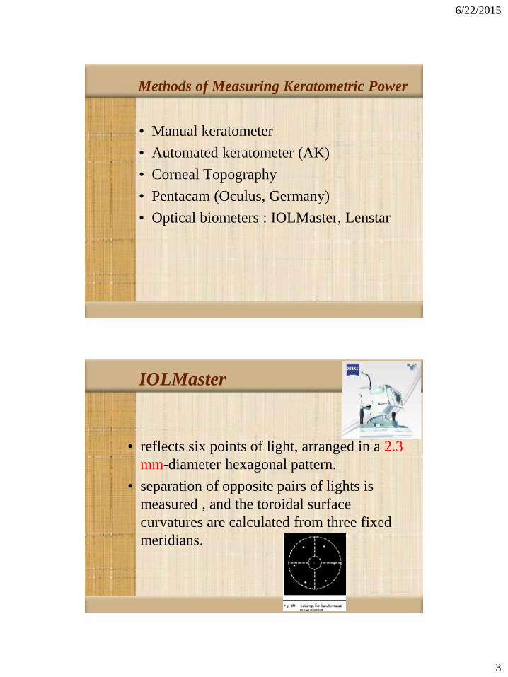
IOLMaster
• reflects six points of light, arranged in a 2.3
mm-diameter hexagonal pattern.
• separation of opposite pairs of lights is
measured , and the toroidal surface
curvatures are calculated from three fixed
meridians.

6/22/2015
4
LENSTAR LS 900
• Based on optical low coherence
reflectometry (OLCR) using a wavelength
of 820 μm.
• To calculate K readings, it evaluates 32
points at 2.3 and 1.65 mm optical zones.
A-scan Biometry (Applanation).
• Variable corneal compression.
• The applanation technique yields a shorter
axial length than immersion, as a result it
would require a lower lens constant.
• a: Initial spike (probe tip and cornea)
• b: Anterior lens capsule
• c: Posterior lens capsule
• d: Retina
• e: Sclera
• f: Orbital fat

6/22/2015
5
A-scan Biometry (Immersion).
• Better reproducibility than the applanation
method.
• Coupling fluid prevents compression.
• Making the change from the
applanation to immersion is
well worth the small learning
curve.
A-scan Biometry (Immersion).
• a: Probe tip. (moved away from cornea)
• b: Cornea. (Double-peaked)
• c: Anterior lens capsule.
• d: Posterior lens capsule.
• e: Retina. (sharp 90 degree take-off from
the baseline)
• f: Sclera.
• g: Orbital fat.
6/22/2015
6
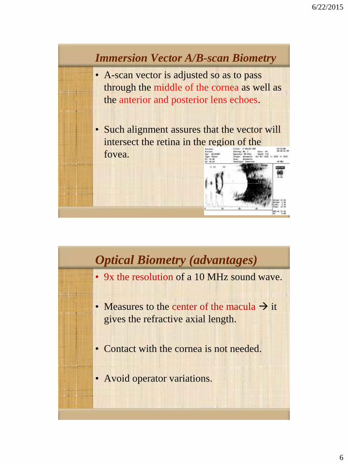
Immersion Vector A/B-scan Biometry
• A-scan vector is adjusted so as to pass
through the middle of the cornea as well as
the anterior and posterior lens echoes.
• Such alignment assures that the vector will
intersect the retina in the region of the
fovea.
Optical Biometry (advantages)
• 9x the resolution of a 10 MHz sound wave.
• Measures to the center of the macula it
gives the refractive axial length.
• Contact with the cornea is not needed.
• Avoid operator variations.

6/22/2015
7
LENSTAR LS 900
Pachymetry ACD
Lens Thickness Axial Length
Keratometry White to White
Pupillometry Retinal Thickness
Eccentricity of the Visual Axis
Comparing the accuracy of OLCR vs. PCI
• Study published in DJO 2013.
• Authors : Prof. Dr. Ahmed A. El-Massry,
Dr. Hany A. Helaly.
• A prospective study that included 50 eyes of
50 patients scheduled for cataract surgery.

6/22/2015
8
Comparing the accuracy of OLCR vs. PCI
Lenstar LS 900 IOLMasterMean + Standard deviation
(Range)
Mean + Standard deviation
(Range)
Axial Length (mm)
different recaliberation alogrithm23.93 + 1.74
(21.7 to 29.2)
23.92 + 1.73
(21.69 to 29.0)
Average K readings (D)
32 points vs 6 points
43.74 + 1.05
(41.42 to 46.20)
43.83 + 0.96
(42.27 to 46.34)
Anterior Chamber Depth (mm)
optical biometry vs. image analysis 3.17 + 0.24
(2.80 to 3.90)
3.11 + 0.24
(2.70 to 3.80)
Comparing the accuracy of OLCR vs. PCI
Lenstar LS 900 IOLMaster
Mean arithmetic error (D) + SD - 0.017 + 0.56 - 0.0048 + 0.60
Mean absolute error (D) + SD 0.428 + 0.36 0.472 + 0.37
Range (D) (- 1.40 to + 1.00) (- 1.45 to 0.98)
Prediction error
Within + 0.5 D 68 % 70 %
Within + 1 D 94 % 90 %
Within + 2 D 100 % 100 %

6/22/2015
9
IOLMaster 700 (next generation)
• The first Swept Source OCT-based
biometer (the entire eye is scanned three
dimensionally).
IOLMaster 700 (next generation)
• Provides an image-based measurement,
allowing one to view the complete
longitudinal section of the eye—from the
cornea to the retina.
• It can also be used to identify irregular eye
geometries, such as lens tilt.

6/22/2015
10
IOLMaster 700 (next generation)
• Preoperative crystalline lens position and
tilt (top) and postoperative IOL position
and tilt in horizontal (0°) B-Scan image
(bottom).
IOLMaster 700 (next generation)
• Imaging of the fovea alert the user to
insufficient fixation during measurements.
Poor (left) vs. correct (right) fixation

6/22/2015
11
IOLMaster 700 (next generation)
• It has a built-in toric IOL calculator
(Haigis-T).
• It acquires a reference image of scleral &
conjunctival vessels CALLISTO eye
computer in the OR connected to the
LUMERA microscope During surgery,
the image allows intraoperative matching
with the live eye image.
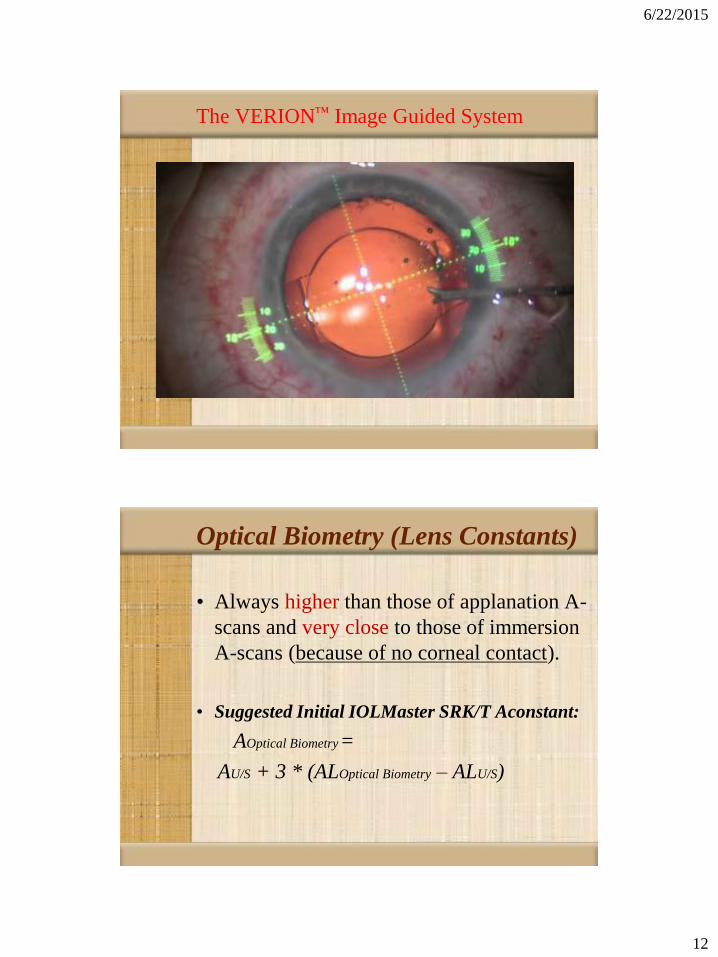
The VERION™ Image Guided System
6/22/2015
12
The VERION™ Image Guided System
Optical Biometry (Lens Constants)
• Always higher than those of applanation A-
scans and very close to those of immersion
A-scans (because of no corneal contact).
• Suggested Initial IOLMaster SRK/T Aconstant:
AOptical Biometry=
AU/S + 3 * (ALOptical Biometry – ALU/S)
6/22/2015
13
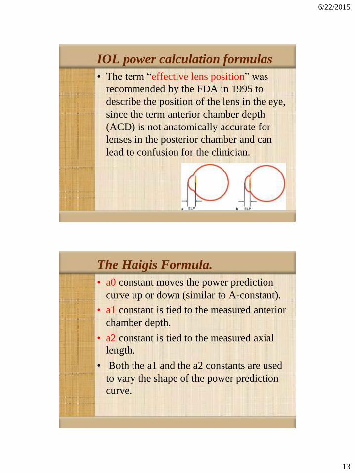
IOL power calculation formulas
• The term “effective lens position” was
recommended by the FDA in 1995 to
describe the position of the lens in the eye,
since the term anterior chamber depth
(ACD) is not anatomically accurate for
lenses in the posterior chamber and can
lead to confusion for the clinician.
The Haigis Formula.
• a0 constant moves the power prediction
curve up or down (similar to A-constant).
• a1 constant is tied to the measured anterior
chamber depth.
• a2 constant is tied to the measured axial
length.
• Both the a1 and the a2 constants are used
to vary the shape of the power prediction
curve.
6/22/2015
14
Choice of IOL power calculation formulas
• For axial lengths from 22.50 mm to 26.00
mm, and central corneal powers ranging
from 41.00 D to 46.00 D, almost any
modern IOL power calculation formula will
give good outcomes.
• Short eyes : Hoffer Q, Haigis.
• Long eyes: Holladay, SRK/T
• 4th generation formulas: Holladay 2 &
Haigis.
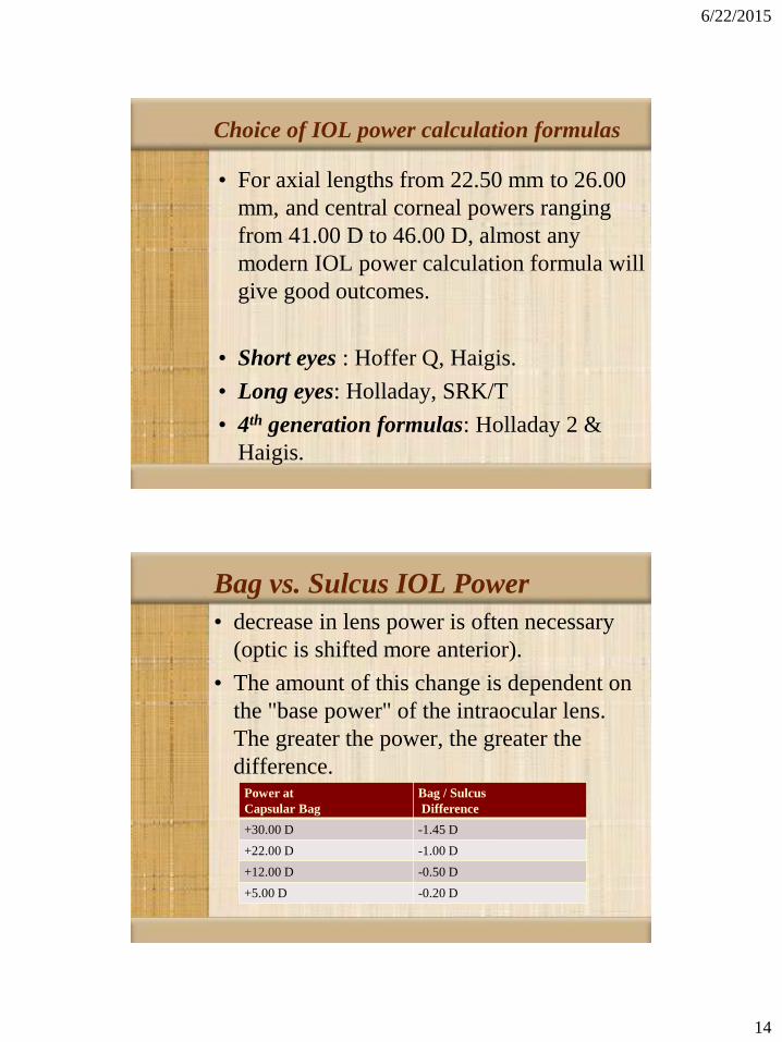
Bag vs. Sulcus IOL Power
• decrease in lens power is often necessary
(optic is shifted more anterior).
• The amount of this change is dependent on
the "base power" of the intraocular lens.
The greater the power, the greater the
difference.Power at
Capsular Bag
Bag / Sulcus
Difference
+30.00 D -1.45 D
+22.00 D -1.00 D
+12.00 D -0.50 D
+5.00 D -0.20 D

6/22/2015
15
Corneal Transplantation
• There is no method that can be used to
accurately carry out IOL power calculations
prior to corneal transplantation combined
with cataract removal.
• This is because it is impossible to know
the central power of the donor graft
prior to surgery.
Corneal Transplantation
• Either use a "best guess" of post-operative
corneal power (such as 44.0 D) often
lead to an unpleasant post-operative
refractive surprise.
• Or carry out corneal transplantation with
cataract removal, but without intraocular
lens implantation. The lens implantation
would then be carried out at a later time, as
a secondary procedure.
6/22/2015
16
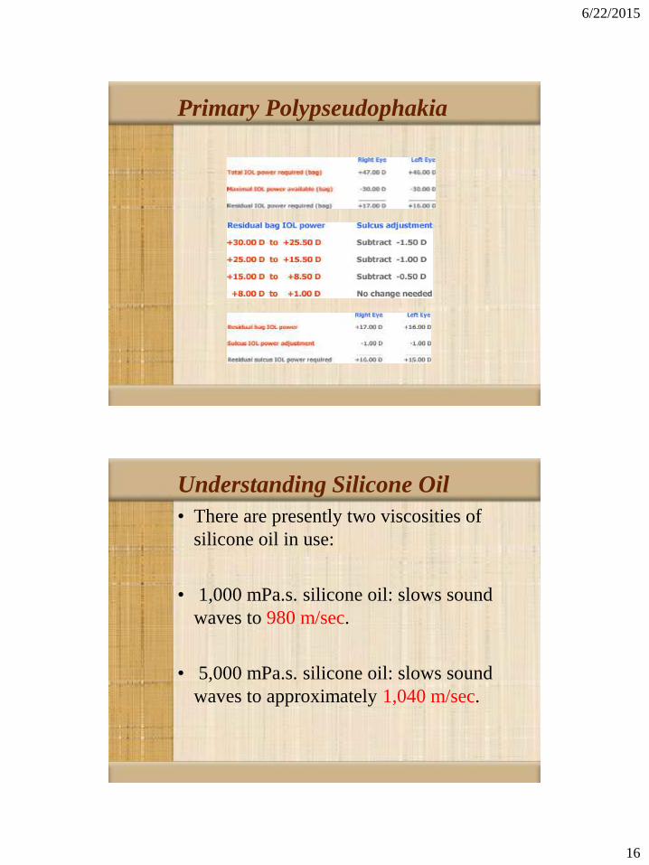
Primary Polypseudophakia
Understanding Silicone Oil
• There are presently two viscosities of
silicone oil in use:
• 1,000 mPa.s. silicone oil: slows sound
waves to 980 m/sec.
• 5,000 mPa.s. silicone oil: slows sound
waves to approximately 1,040 m/sec.

6/22/2015
17
Understanding Silicone Oil
• Use optical biometry.
• Adjust U/S speed in the machine.
• Multiply result X 980(silicone speed) /
1532.
• Use 2ry IOL implantation.
Understanding Silicone Oil
• If the silicone oil is to remain in the eye for
an extended period of time after cataract
surgery
For an eye of average dimensions, the
additional power needed for a convex-
plano PMMA intraocular lens is typically
between +3.0 D to +3.5 D.
6/22/2015
18
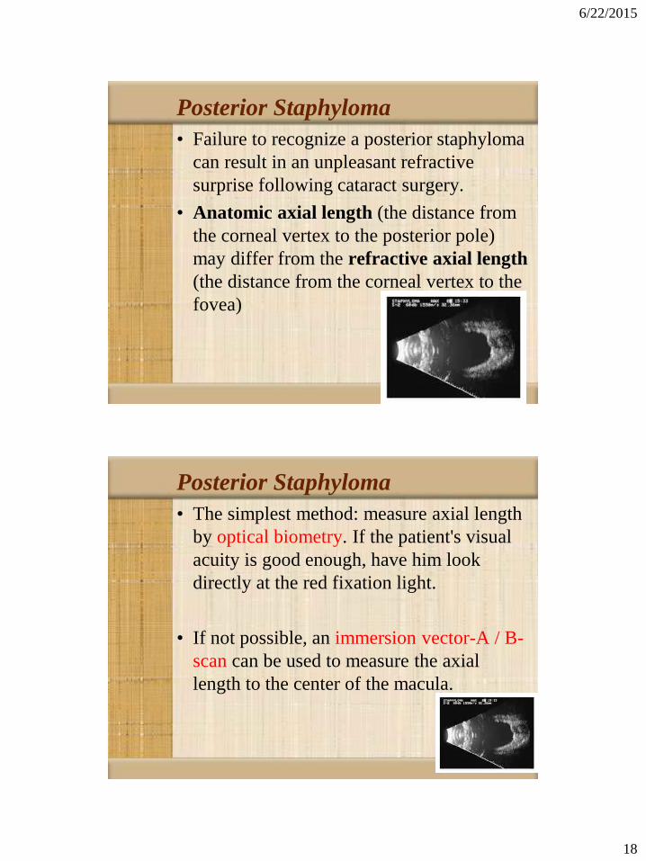
Posterior Staphyloma
• Failure to recognize a posterior staphyloma
can result in an unpleasant refractive
surprise following cataract surgery.
• Anatomic axial length (the distance from
the corneal vertex to the posterior pole)
may differ from the refractive axial length
(the distance from the corneal vertex to the
fovea)
Posterior Staphyloma
• The simplest method: measure axial length
by optical biometry. If the patient's visual
acuity is good enough, have him look
directly at the red fixation light.
• If not possible, an immersion vector-A / B-
scan can be used to measure the axial
length to the center of the macula.

6/22/2015
19
IOL power calculation following
Keratorefractive surgery
• Measured average k-reading + standard
IOL power calculation formulas
overestimation ( postop. hyperopia )
IOL power calculation following
Keratorefractive surgery
• After myopic LASIK : postop. hyperopia
• After hyperopic LASIK : postop. myopia
• Error 1 D in IOL power 0.71 D error at
the spectacle plane
Feiz V, Mannis MJ, Garcia-Ferrer F, et al. Intraocular lens power calculation after laser in situ
keratomileusis for myopia and hyperopia: a standardized approach. Cornea 2001;20:792-7.
6/22/2015
20
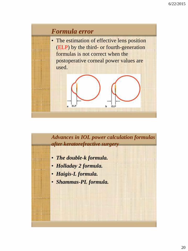
Formula error
• The estimation of effective lens position
(ELP) by the third- or fourth-generation
formulas is not correct when the
postoperative corneal power values are
used.
Advances in IOL power calculation formulas
after keratorefractive surgery
• The double-k formula.
• Holladay 2 formula.
• Haigis-L formula.
• Shammas-PL formula.

6/22/2015
21
The double-k formula
• For 3rd generation formulas:
• SRK/T.
• Holladay 1.
• Hoffer Q.
• This method uses 2 k-values:
• pre refractive for calc of ELP
• post refractive for the vergence formula that
finally gives the IOL power
Holladay 2 formula
• Uses another innovative approach, which is
to use measurements of
• corneal power.
• corneal diameter.
• anterior chamber depth (ACD).
• lens thickness.
• refractive error.
• axial length.
to further refine the ELP calculation.
6/22/2015
22
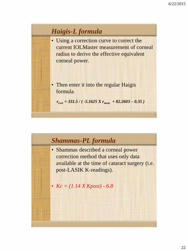
Haigis-L formula
• Using a correction curve to correct the
current IOLMaster measurement of corneal
radius to derive the effective equivalent
corneal power.
• Then enter it into the regular Haigis
formula.
rcorr = 331.5 / ( -5.1625 X rmeas + 82.2603 – 0.35 )
Shammas-PL formula
• Shammas described a corneal power
correction method that uses only data
available at the time of cataract surgery (i.e.
post-LASIK K-readings).
• Kc = (1.14 X Kpost) - 6.8

6/22/2015
23
Shammas-PL formula
• This corrected K-value is used in the
Shammas post-LASIK (Shammas-PL)
formula
• In which the ELP does not vary with the
corneal curvature that has been altered by
the LASIK procedure.
radial keratotomy
• Any map that provides average anterior
corneal power over central 2–3 mm
gives an accurate estimation of corneal
refractive power.
• Still needs to compensate for potential
errors in ELP by using
• Holladay 2 formula
• double-K approach with 3rd generation
formulas.

6/22/2015
24
Take Home Message
• Immersion A scan is still the gold standard
for biometry and worth the effort to shift to.
• Optical biometry is a new simple method to
achieve reproducible accurate results
comparable to the gold standard.
• Personal optimization of lens constants is
mandatory for more accurate postoperative
refractive outcome.
THANK YOU